xxx meeting of the italian society ... - … meeting of the italian society for the study of...
TRANSCRIPT

XXX MEETING OF THE ITALIAN SOCIETY FOR THE STUDY OF
CONNECTIVE TISSUES (SISC)
ABSTRACT BOOK
University of Palermo, Palazzo Steri, Sala Magna, Palermo, Italy
27 – 29 October 2010
With scientific patrocinium

- XXX Meeting of the Italian Society for the Study of Connective Tissues (SISC)- University of Palermo, 27-29 October 2010 - 2

- XXX Meeting of the Italian Society for the Study of Connective Tissues (SISC)- University of Palermo, 27-29 October 2010 - 3
CONTENTS
ORGANIZING COMMITTEE PAG. 5
OPENING ADDRESS PAG. 7
PROGRAMME PAG. 9
INVITED SPEAKERS’ CURRICULA PAG. 15
PLENARY LECTURES PAG. 23
ORAL COMMUNICATIONS PAG. 37
SELECTED POSTER PRESENTATIONS PAG. 55
POSTERS PAG. 65
SOCIAL EVENTS PAG. 75
AUTHORS’ INDEX PAG. 83

- XXX Meeting of the Italian Society for the Study of Connective Tissues (SISC)- University of Palermo, 27-29 October 2010 - 4
- XXX Meeting of the Italian Society for the Study of Connective Tissues (SISC)- University of Palermo, 27-29 October 2010 - 5
.
XXX MEETING
of the Italian Society for the Study of Connective Tissues (SISC)
October 27-29, 2010 – University of Palermo
Palazzo Steri, Sala Magna, Palermo, Italy
www.siscpalermo2010.it
www-3.unipv.it/sisc/
Scientific Advisory Board Prof. Guido Filosto, Palermo, Honorary President Prof.ssa Ida Pucci-Minafra, Palermo, President Prof. Alberto Calatroni, Messina Prof. Ranieri Cancedda, Genova Prof.ssa Alida Ferlazzo, Messina Prof.ssa Vittoria Ottani, Bologna Prof. Alberto Passi, Varese Prof. Daniela Quaglino, Modena Prof. Antonio Rossi, Pavia Prof. Michele Spina, Padova
ORGANIZING COMMITTEE
Local Scientific Committee
Prof.ssa Ida Pucci-Minafra Prof. Salvatore Minafra Dott.ssa Patrizia Cancemi Dott.ssa Nadia Ninfa Albanese Dott. Gianluca Di Cara Dott.ssa Maria Rita Marabeti Dott.ssa Francesca Costantini Dott.ssa Rosa Musso
Administrative and technical support Studio Zito Dr.ssa Maria Cinà Sg. Marco Andrea Cottone Sg. Anna Maria Buscemi
www.unipa.it
www.cobsunipa.it
www.lamaddalenanet.it

- XXX Meeting of the Italian Society for the Study of Connective Tissues (SISC)- University of Palermo, 27-29 October 2010 - 6
- XXX Meeting of the Italian Society for the Study of Connective Tissues (SISC)- University of Palermo, 27-29 October 2010 - 7
A Welcome from the President
It is a great pleasure and honour for me to open this XXX Meeting of the Italian Society for the Study of Connective Tissues held in this historical residence of Palazzo Steri. It is a particular pleasure to welcome our Invited Speakers and Chairpersons, the Academic Authorities and Colleagues of the University of Palermo and all participants and members of the Society. This is a special occasion for me, for several reasons. The first because this is my second and last appointment as President of this important Society, where outstanding Scientists have made major contributions to the Society before myself. The second is because it also coincides with my retirement from the academy (but I hope not from research!). My retirement means that I have had the privilege to work through five decades of Biological and Medical Science; from discovery of the double helix to the global revolution of the genomic
and proteomic era. During this time many other science milestones have been achieved, especially by contributions from researchers of my generation. We have observed biology going from descriptive morphology to molecular and virtual biology, with advancements and discoveries in the fields of cell biology, reproduction, gene expression, cancer and development, to mention some. In parallel, the advancement in technologies of imaging, such as photosensitive fluorescent probes, super-resolution microscopy imaging, electron microscope 3D reconstructions, as well as the field of spectrometry applied to the proteomics and clinical diagnostics have changed the way of looking at and studying tissues and cells.
Going back “in my time” connective tissue was described as a mere supporting scaffold for cells and tissues sitting on it, and researches emphasizing its possible relevance in the dynamics of bio-pathological processes were in some way considered irrelevant or bizarre. Indeed, the recent scientific and technical development have highlighted new ideas and interests to the study of this fascinating “ensemble” of tissues. “Loose” connective tissues form beautifully intricate and complex mesh-works of specialized molecules which permeate all tissues and organs, capturing and releasing information from and to neighbouring systems. For this reason, the study of connective tissue is multifactorial, covering different fields of science. There is no human pathology which does not involve connective tissues, either directly, such as many genetic disorders, or indirectly such as inflammation and cancer.
In this meeting we attempt to bring together different aspects of the biology and pathology of the connective tissues, which will be presented by distinguished Scientists in the field, but also by promising young researchers. To them I want to manifest all my solidarity for the difficult time that they are living because of the uncertainty inherent the future of the research and academy in Italy, but also in other countries of the western world.
But let me manifest my deepest gratitude and welcome to ours invited foreign Scientists, who come so far away from Sicily to attend this program and giving highly qualified contributions and prestige to our meeting. I am sure that this event will be an enriching experience for all of us, not only for generating new good ideas and suggestions, but also for the enhancement of existing, and the beginning of future scientific and personal friendship among participants.
Finally, I wish to express my thanks and appreciation to the Institutions who contributed to the meeting organization, and, particularly, to all the components of my team, who made possible the actuation of this event.
Ida Pucci Minafra

- XXX Meeting of the Italian Society for the Study of Connective Tissues (SISC)- University of Palermo, 27-29 October 2010 - 8

- XXX Meeting of the Italian Society for the Study of Connective Tissues (SISC)- University of Palermo, 27-29 October 2010 - 9
PROGRAMME

- XXX Meeting of the Italian Society for the Study of Connective Tissues (SISC)- University of Palermo, 27-29 October 2010 - 10
- XXX Meeting of the Italian Society for the Study of Connective Tissues (SISC)- University of Palermo, 27-29 October 2010 - 11
Wednesday, October 27 14.00-15.00 Registration 15.00-16.00 Opening Cerimony
Greetings from the Academic Authorities Opening address: Ida Pucci-Minafra (SISC President; Palermo, IT) Historical Perspective: Ivonne Pasquali-Ronchetti (Cofounder SISC; Modena, IT)
16.00-17.30 Inaugural plenary lectures
Chairs: Ida Pucci-Minafra (Palermo, IT), David Woolley (Manchester, UK)
Paola Defilippi (University of Torino, IT): The integrin adaptor proteins p140Cap and p130Cas as molecular hubs in cell migration and invasion.
Hideaki Nagase (Imperial College London, UK): Fibrillar collagen degradation
and cell migration.
Welcome Cocktail at Grand Hotel Villa Igiea Thursday, October 28 09.00-10.30 Plenary lectures
Chairs: Gillian Murphy (London, UK), M. Letizia Vittorelli (Palermo, IT)
Lance A. Liotta (George Mason University Manassas, USA): When does cancer metastasis begin? How the tissue microenvironment spawns invasive breast carcinoma cells.
Alberto Passi (University of Insubria, Varese, IT): Hyaluronan: simple polymer with complex biological activities.
10.30-11.00 Coffee break 11.00-12.20 Oral communications: Matrix Molecules
Chairs: Alessandro Ruggeri (Bologna, IT), Antonio Rossi (Pavia, IT)
John E. Scott (Manchester, UK): Quantitative proteoglycan/collagen compositions of extracellular matrices (ECMS, E.G. cornea and tendon) are predictable, based on simple ultrastructural geometry.
Marilisa Quaranta (University of Bologna, IT): Crimping pattern in positional and
energy storing tendons: Morphological and functional correlations. Marco Franchi (University of Bologna, IT): Role of decorin and fibrillar crimps in
elastic recoil of tendon. Mario Raspanti (University of Insubria, Varese, IT): Fibrillogenesis reloaded.
Evidence of two different mechanisms of collagen fibril formation. 12.20-12.40 Selected poster presentations

- XXX Meeting of the Italian Society for the Study of Connective Tissues (SISC)- University of Palermo, 27-29 October 2010 - 12
Simone D. Scilabra (Imperial College London, UK): LRP-dependent and -
independent endocytic pathways of TIMP-3. Giancarlo Nastasi (University of Messina, IT): Is versican a primary regulator of
neural crest cells migration in the early Xenopus embryo? 13.00-15.00 Lunch and poster session 15.00-17.00 Oral communications: Connective tissue diseases and treatment
Chairs: Cesare Balduini (Pavia, IT), Alberto Calatroni (Messina, IT)
Antonella Forlino (University of Pavia, IT): Characterization of normal and mutant forms of human recombinant prolidase.
Angela D’Ascola (University of Messina, IT): Treatment with high molecular
weight hyaluronan reduces inflammation in experimental arthritis by affecting toll like receptors.
Daniela Quaglino (University of Modena, IT): In vitro antioxidant treatments
revert parameters of oxidative stress and decrease TNAP expression in PXE fibroblasts.
Antonio Rossi (University of Pavia, IT): CANT1 mutations in desbuquois
dysplasia cause a defect in proteoglycan synthesis.
Antonio J. Lepedda (University of Sassari, IT): Apolipoprotein profiles in atherosclerotic disease.
Cristian Gruppi (University of Pavia, IT): Do spreading and proplatelet formation
by human megakaryocytes depend on type, interactions and structure of extracellular matrix proteins?
17.00-17.20 Selected poster presentations
Maria Rita Marabeti (University of Palermo, IT): Circulating and tissue forms of MMP2 and MMP9 in breast cancer progression. Tiziana Apuzzo (Bellinzona, CH): CXCR4 associated proteins.
17.30 SISC General Assembly
19.00 Organ Recital at Basilica di San Francesco di Assisi 20.30 Social Dinner in the Old Palermo

- XXX Meeting of the Italian Society for the Study of Connective Tissues (SISC)- University of Palermo, 27-29 October 2010 - 13
Friday, October 29 09.00-09.45 Plenary lecture
Chairs: Paola De Filippi (Torino, IT), Daniela Quaglino (University of Modena, IT) Zena Werb (University of California, San Francisco, USA): Of mice and women: new insights into breast development and breast cancer.
09.45-10.25 Oral communications: Microenvironment and cancer Antonella Naldini (University of Siena, IT): Non-hypoxic induction of HIF-1 by
interleukin-1 promotes cell migration in MDAMB231 breast cancer cells. Ida Pucci Minafra (University of Palermo, IT): Permissive and restrictive
influences from breast cancer stroma. 10.25-10.55 Selected poster presentations
Patrizia Cancemi (University of Palermo, IT): S100s protein expression in a large sample-set of breast cancer tissues. Gemma Palazzolo (University of Palermo, IT): Proteomic analysis of extracellular vesicles shed in vitro by MDAMB231 breast carcinoma cells. Riccardo Alessandro (University of Palermo, IT): Role of exosomes released by chronic myleogenous leukemia cells in the modulation of tumor microenviroment.
10.55-11.25 Coffee break
11.25-12.25 Oral communications: Biomaterials and Imaging Chairs: Vittoria Ottani (Bologna, IT), Mario Raspanti (Varese, IT) Alessandro Gandaglia (University of Padova, IT): Time-course evaluation of
tissue guided regenerated valved conduits: long term results of porcine decellularized �-Gal negative roots.
Valerio Brucato (University of Palermo, IT): Pre-vascularized PLLA scaffolds: a
new approach to develop deep tissue regeneration. Paolo Minafra (University of Pavia, IT): Value of ultrasound elastosonography
applied to the musculoskeletal apparatus. 12.25-12.35 Selected poster presentation
Filippo Naso (University of Padova, IT): Alpha-Gal removal tool for the creation of biocompatible heart valve tissue.
12.35 Closing remarks
John E. Scott (Manchester, UK).

- XXX Meeting of the Italian Society for the Study of Connective Tissues (SISC)- University of Palermo, 27-29 October 2010 - 14

- XXX Meeting of the Italian Society for the Study of Connective Tissues (SISC)- University of Palermo, 27-29 October 2010 - 15
INVITED SPEAKERS’ CURRICULA

- XXX Meeting of the Italian Society for the Study of Connective Tissues (SISC)- University of Palermo, 27-29 October 2010 - 16

- XXX Meeting of the Italian Society for the Study of Connective Tissues (SISC)- University of Palermo, 27-29 October 2010 - 17
Paola Defilippi
Full Professor of Cell Biology, University of Torino. Place of work: Molecular and Biotechnology Center, Dept. of Genetics, Biology and Biochemistry, University of Torino (Italy). The research in Dr Defilippi group is focused on the identification of signaling platforms that controls proliferation and migration of normal and transformed cells in response to cell-matrix adhesion and growth factor stimulation. Originally they
identified the molecular basis of integrins, growth factor and cytokine receptors (EGF and IL-3 receptors) cross-talk, to investigate interactions and new mechanisms of co-operation between these distinct classes of receptors. They have recently characterized two adaptor proteins, p130Cas and p140Cap, with opposite, but essential roles in breast cancer cell tumorigenesis. Both proteins are molecular hubs that upon cell-matrix, cell-cell and growth factor stimulation are involved in the control of cell proliferation, migration and invasion of transformed cells. They are currently investigating at molecular level, their mechanistical functions with in vitro models and in vivo transgenic and KO animals.
Paola Defilippi got a degree in Biology at the University of Torino and a Ph.D. in Biochemistry at the Universitè Libre de Bruxelles (Belgium). She is a member of the Italian ABCD Society (Associazione Biologia Cellulare e del Differenziamento). Her research has generated two patents and sixty peer-reviewed publications.
- XXX Meeting of the Italian Society for the Study of Connective Tissues (SISC)- University of Palermo, 27-29 October 2010 - 18
Lance A. Liotta
Professor of life sciences at George Mason University and co-director of the university's Center for Applied Proteomics and Molecular Medicine. He also serves as medical director of the George Mason University / Inova Health System Clinical Proteomics Laboratory in the GMU/Inova Health System Translational Research Centers. Before joining the George Mason faculty in May 2005, Dr. Liotta was chief of the Laboratory of Pathology at
the National Cancer Institute's (NCI) Center for Cancer Research, and deputy director for Intramural Research at the National Institutes of Health. One of the first scientists to investigate the process of tumor invasion and metastasis at the molecular level, he has invented technologies in the fields of diagnostics, immunoassays, micro dissection, and proteomics, which have been used to make broad discoveries in genomics, functional genetics, and tissue proteomics. In partnership with Dr. Emanuel F. Petricoin III, formerly of the U.S. Food and Drug Administration (FDA), Dr. Liotta founded the NCI/FDA Clinical Proteomics Program, the first joint initiative between NCI and FDA to develop technologies for the discovery of proteins and the profiling of signal pathways in human tissues. These innovative proteomic technologies are applied to patient tissue biopsies and blood samples collected before, during, and after experimental therapies in clinical research trials. At George Mason, Drs. Liotta and Petricoin are exploring their recent discovery of an archive of protein fragments in the blood that are potential biomarker candidates for breast, ovarian and lung cancers. Their immediate goals are to validate these potential biomarkers in clinical trials to determine their feasibility in the diagnosis of cancer prior to metastasis, and to develop patient-tailored medical treatment strategies. Dr. Liotta earned his medical degree from Case Western Reserve Medical School and is licensed to practice medicine in the state of Maryland. He also holds a doctoral degree in biomedical engineering from Case Western Reserve University. His research contributions have generated 80 issued patents and more than 550 peer-reviewed publications.

- XXX Meeting of the Italian Society for the Study of Connective Tissues (SISC)- University of Palermo, 27-29 October 2010 - 19
Hideaki Nagase
Professor and Head of Matrix Biology Department at the Kennedy Institute of Rheumatology Division, Imperial College London, London, UK., 1999-present. He is investigating the structure and function of matrix metalloproteinases and tissue inhibitors of metalloproteinases (TIMPs) and their roles in cartilage matrix destruction during the progression of arthritis. He holds a B.Sc. in Pharmacy from Tokyo
College of Pharmacy, a M.Sc. degree in Physiological Chemistry from Science University of Tokyo in Japan and a Ph.D. degree in Biochemistry from the University of Miami, USA. He received his postdoctoral training at Strangeways Research Laboratory, Cambridge, UK and Dartmouth Medical School, USA. He was elected as an Honorary Fellow of The Royal College of Physicians in 2004. He elucidated the stepwise mechanism of activation of matrix metalloproteinases and the inhibition mechanism of TIMPs, the latter in collaboration with Professor Wolfram Bode and Professor Keith Brew.
Dr Nagase is a member of several Scientific Societies, among which the American Society for Biochemistry and Molecular Biology, International Society for Matrix Biology, British Society of Matrix Biology, British Biochemical Society. He was a member of the Editorial board of the FASEB Journal, the International Journal of Biochemistry and Cell Biology, the Biochemical Journal, Cell Research, the Biochemical Journal, and currently of the Journal of Biological Chemistry. He has co-authored 192 papers, 31 book chapters and one book.

- XXX Meeting of the Italian Society for the Study of Connective Tissues (SISC)- University of Palermo, 27-29 October 2010 - 20
Alberto Passi
Full professor in biochemistry at the Medical School, University of Insubria (Varese – Italy), dean of the Faculty of Exercise and Sport Sciences of University of Insubria, director of biochemistry laboratories at Department of Biomedical Experimental and Clinical Sciences (DSBSC) of University of Insubria. Since obtaining his Ph.D., he has focused his scientific interest in extracellular matrix metabolism. Cell cultures, lung
and vascular tissues were initially approached studying proteoglycan metabolism including the effect of free radicals and proteases on extracellular matrix macromolecules. After a sabbatical period in Hascall’s lab in Cleveland (OH – USA), the Passi work moved towards hyaluronan. In particular, studies were done on hyaluronan metabolism and its control. The role of UDP sugars precursors in glycosaminoglycan synthesis and the covalent modification of enzymes involved in glycosaminoglycan metabolism are currently the main interest in Passi’s lab. Passi is member of Italian Biochemistry and Molecular Biology Society, Italian Connective Tissue Society, International Society of Matrix Biology, American Society for Matrix Biology and American Biochemical and Molecular Biology Society.
He is in the editorial board of several journals of biochemistry and presented as invited speaker more than 50 talks during international meetings and seminars, is co-author of 64 peer reviewed papers in international journals.
- XXX Meeting of the Italian Society for the Study of Connective Tissues (SISC)- University of Palermo, 27-29 October 2010 - 21
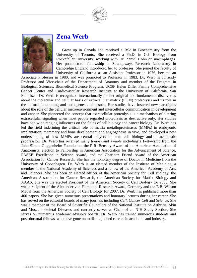
Zena Werb
Grew up in Canada and received a BSc in Biochemistry from the University of Toronto. She received a Ph.D. in Cell Biology from Rockefeller University, working with Dr. Zanvil Cohn on macrophages. Her postdoctoral fellowship at Strangeways Research Laboratory in Cambridge England introduced her to proteases. She joined the faculty of University of California as an Assistant Professor in 1976, became an
Associate Professor in 1980, and was promoted to Professor in 1983. Dr. Werb is currently Professor and Vice-chair of the Department of Anatomy and member of the Program in Biological Sciences, Biomedical Science Program, UCSF Helen Diller Family Comprehensive Cancer Center and Cardiovascular Research Institute at the University of California, San Francisco. Dr. Werb is recognized internationally for her original and fundamental discoveries about the molecular and cellular basis of extracellular matrix (ECM) proteolysis and its role in the normal functioning and pathogenesis of tissues. Her studies have fostered new paradigms about the role of the cellular microenvironment and intercellular communication in development and cancer. She pioneered the concept that extracellular proteolysis is a mechanism of altering extracellular signaling when most people regarded proteolysis as destructive only. Her studies have had wide ranging influences on the fields of cell biology and cancer biology. Dr. Werb has led the field indefining the critical role of matrix metalloproteinases (MMPs) in embryonic implantation, mammary and bone development and angiogenesis in vivo, and developed a new understanding of how MMPs are central players in stem cell biology and in neoplastic progression. Dr. Werb has received many honors and awards including a Fellowship from the John Simon Guggenheim Foundation, the R.R. Bensley Award of the American Association of Anatomists, election to Fellowship in American Association for the Advancement of Science, FASEB Excellence in Science Award, and the Charlotte Friend Award of the American Association for Cancer Research. She has the honorary degree of Doctor in Medicine from the University of Copenhagen. Dr. Werb is an elected member of the Institute of Medicine, a member of the National Academy of Sciences and a fellow of the American Academy of Arts and Sciences. She has been an elected officer of the American Society for Cell Biology, the American Association for Cancer Research, the American Society for Matrix Biology and AAAS. She was the elected President of the American Society of Cell Biology for 2005. She was a recipient of the Alexander von Humboldt Research Award, Germany and the E.B. Wilson Medal from the American Society of Cell Biology for 2007. Dr. Werb has published more than 400 papers. She has given numerous presentations and honorary lectures during her career. She has served on the editorial boards of many journals including Cell, Cancer Cell and Science. She was a member of the Board of Scientific Councilors of the National Institute on Arthritis, Skin and Musculo-skeletal Diseases and currently serves as Chair of an NIH Study Section. She serves on numerous academic advisory boards. Dr. Werb has trained numerous students and post-doctoral fellows, who have gone on to distinguished careers in academia and industry.

- XXX Meeting of the Italian Society for the Study of Connective Tissues (SISC)- University of Palermo, 27-29 October 2010 - 22
- XXX Meeting of the Italian Society for the Study of Connective Tissues (SISC)- University of Palermo, 27-29 October 2010 - 23
PLENARY LECTURES

- XXX Meeting of the Italian Society for the Study of Connective Tissues (SISC)- University of Palermo, 27-29 October 2010 - 24

- XXX Meeting of the Italian Society for the Study of Connective Tissues (SISC)- University of Palermo, 27-29 October 2010 - 25
THE INTEGRIN ADAPTOR PROTEINS p140CAP AND p130CAS AS MOLECULAR HUBS IN CELL MIGRATION AND INVASION
DI STEFANO P., CAMACHO LEAL M.P., TORNILLO G., BISARO B., DAMIANO L., MORELLO V., REPETTO D., PINCINI A.,
ARAMU S., SHARMA N., TURCO E., CABODI S., DEFILIPPI P. Molecular Biotechnology Center, University of Torino, Torino, Italy.
Corresponding Author: [email protected] Integrins are cell surface heterodimeric receptors for the extracellular matrix (ECM), formed by the non
covalent association of alpha and beta subunits. The mammalian genome comprises 8 beta subunit and 18 alpha subunit genes, that assemble into 24 different functional integrins [1]. Integrins are transmembrane proteins characterised by a large extracellular domains that forms elongated stalks and a globular ligand-binding head region, through which they bind to the components of the extracellular matrix, and a short cytoplasmic tails connected to the actin cytoskeleton [1]. Integrins specifically localize to focal adhesions, which are sites of close apposition with the extracellular matrix (ECM) where actin filament are anchored to the plasma membrane. At these sites, integrins co-distribute with cytoskeletal proteins such as talin, kindlin, vinculin and alpha actinin, which serve as multi-functional scaffolding proteins, leading to protein-protein interactions by the recruitment of kinases, phophatases and their substrates together, thus regulating the dynamic of integrin-cytoskeleton connection [2]. Integrins are enzymatically inactive receptors, which link to cellular components to elicit signal transduction. Integrin ability to transduce signals inside the cells upon ECM binding, the so called “outside-in signalling” is required for polymerization of actin cytoskeleton during cell adhesion and for control of cell migration, proliferation, survival and differentiation [2].
Tyrosine phosphorylation of proteins represents a primary response to integrin stimulation and a preferential way to transduce signals throughout the cell. Among the kinases that are activated upon integrin activation, Src Family Kinases (SFKs) and Focal adhesion kinase (Fak) are crucial regulators of downstream signalling in normal and pathological conditions [3]. Increasing evidence highlights the importance of adaptor proteins in transducing integrin signalling by their ability to create molecular complexes regulating a variety of cellular functions such as survival, proliferation, migration and invasion. In this review, we will discuss the contribution of p130Cas and p140Cap adaptor proteins to cell migration and invasion in response to cell matrix adhesion and growth factor receptors signalling.
The p130Cas protein is characterized by the presence of multiple conserved sequence motifs and extensive post-translational modification, mainly consisting of tyrosine and serine phosphorylation. These structural features allow the assembly of specific multi-protein complexes, that result in the regulation of cell adhesion, migration and survival. In particular, p130Cas consists of an N-terminal Src homology 3 (SH3) domain, a substrate domain, a serine rich region, and a C-terminal domain. The SH3 domain interacts with polyproline-rich sequences present in several proteins including Fak, PYK2/RAFTK, FRNK, PTP-PEST, PTP1B, C3G and CIZ [4,5]. The substrate domain, characterized by 15 YxxP motifs, upon Src family kinases phosphorylation, exposes additional binding sites for SH2 containing proteins such as Crk adaptors [6], while the serine rich region represents a docking site for other partners such as 14-3-3 and GRB2. Lastly, the C-terminus contains a polyproline-rich region responsible for the binding of the Src family kinase, PI3K, BCAR3/AND-34, CHAT-H and ubiquitin ligases such as AIP4, APC/C and CDH1, as well as a binding site for the adaptor protein p140Cap [7]. p130Cas represents a nodal signalling platform on which integrin and growth factor receptor signalling convey. Integrins, receptor tyrosine kinases (RTKs) and oestrogen receptor (ER) are major upstream regulators of p130Cas, mainly through the activation of SRC and FAK kinases, leading to p130CAS tyrosine phosphorylation on the C-terminal binding site YDYVHL [4]. Moreover, physical stretching of p130Cas induces a conformational change that enables Src-family kinase-dependent p130Cas tyrosine phosphorylation. These findings point out a function for p130Cas as a sensor that integrates mechanical forces coming from the extracellular environment into intracellular signals leading to actin cytoskeleton reorganization [8].
During migration, lamellipodia and filopodia extend from the cell leading edge and create new dynamic adhesions, which form and rapidly disassemble at the base of protrusions [9,10]. The role of p130Cas in cell migration was initially inferred by studies performed on mouse embryo fibroblasts derived from p130Cas KO. p130Cas null MEFS show defects in stress fibre formation and cell spreading, impaired actin bundling and cell migration, that were restored by full-length p130Cas expression [11]. p130Cas protein regulates cell migration and invasion mainly through two signalling mechanisms: the tyrosine phosphorylation of p130Cas by FAK and SRC-family kinases and the association with Zyxin family proteins.
The tyrosine phosphorylation of the substrate domain of p130Cas provides binding sites for Crk proteins that in turn associates with DOCK180, a guanine nucleotide exchange factors that stwiches the small GTPase Rac1 from a GDP-bound inactive to a GTP-bound active state at lamellipodia and filopodia adhesion sites [12,13]. This drives localized Rac activation, membrane ruffling and actin cytoskeleton remodelling, focal adhesion turnover, pseudopodia formation and extension. In addition, ARP2/3 and PAK kinase activation enhance cell migration [14].

- XXX Meeting of the Italian Society for the Study of Connective Tissues (SISC)- University of Palermo, 27-29 October 2010 - 26
It has been reported that expression of Bmx/Etk, a member of the Tec/Btk family of nonreceptor kinases, enhances tyrosine phosphorylation of p130Cas and the formation of p130Cas/Crk complex. Moreover, the coexpression of Bmx with p130Cas results in an enhanced membrane ruffling and haptotactic cell migration. The expression of a mutant form of p130Cas, unable to interact with Crk, inhibits Bmx-induced membrane ruffling and cell migration, indicating that p130Cas and the adaptor protein Crk complex formation is instrumental for connecting several stimuli to the regulation of actin cytoskeleton and cell motility [15].
Additional molecules that play important roles in modulating tyrosine phosphorylation of p130Cas leading to cell migration are the zyxin/Ajuba family of LIM proteins. These proteins bind to actin cytoskeleton and are implicated in cell motility. Ajuba allows p130Cas localization to nascent adhesive sites in migrating cells thereby leading to the activation of the small GTPase Rac, whereas Zyxin interacts with the SH3 domain of p130Cas and with a nucleocytoplasmic transcription factor, CIZ/NMP4/ZNF384 [16]. Recent data also show that p130Cas activates several GTPases other than Rac. The association between p130Cas and AND-34, an NSP family member, which acts as a GTP exchange factor for Ral, Rap1 and R-Ras enhances Src activation and cell migration, likely through a Rap1-dependent mechanism [17].
Cell invasion is a complex process that can be initiated by alterations in integrin surface expression, by the release or activation of proteases that degrade the extracellular matrix, and by changes in gene expression during cell transformation [18]. p130Cas tyrosine phosphorylation upon integrin or growth factor receptor activation has also been linked to cell invasion and it has been reported that the SH3 domain of p130Cas is also required for this process. Indeed, Fak-null cells are not invasive when transformed by v-Src, but they acquire invasive properties upon over-expression of p130Cas SH3 domain, indicating that this domain is required for rescue of v-Src cell invasion. In this context, the formation of a Src/p130Cas/Crk/DOCK180 complex increases Rac1 and JNK activities and MMP-9 expression, leading to an invasive cell phenotype [19]. Another effector that associates to p130Cas SH3 domain and mediates invasion is CIZ (Cas-Interacting Zinc finger protein). This nucleocytoplasmic shuttling protein, by binding to a consensus-like sequence in the matrix metallo proteinase (MMPs) promoter, up-regulates the transcription of MMPs [20], thus acting as a downstream element connecting p130Cas to the invasive program. Negative regulators of cell invasion have also been shown to impact on p130Cas. One of this is p140Cap, recently described as a p130Cas-associated protein [7], which when over-expressed in breast carcinoma cells inhibits cell invasion, by down-regulating p130Cas tyrosine phosphorylation, through inhibition of Src kinase activity [21]. In addition, the KAI1/CD82 protein, a member of the tetraspanin superfamily, has been recently rediscovered as a cancer metastasis suppressor and suppresses prostate carcinoma cell migration by decreasing p130Cas protein levels and its coupling with Crk [22].
In breast carcinoma cells, the ErbB2 oncogene potentiate the migration and invasion machinery facilitating p130Cas/Crk coupling and ERK activation [23]. Recently, in p130Cas -/- MEFs transformed by ErbB2, the absence of p130Cas resulted in the inability to migrate and invade, that was rescued by re-expression of FL p130Cas [24]. Moreover, p130Cas silencing in ErbB2-dependent breast carcinoma impairs in vitro migration and invasion as well as in vivo lung metastasis formation. Consistently, p130Cas over-expression in MCF-10A cells transformed by a chimeric ErbB2 receptor, grown in 3D, confers invasive capacity to the acinar structures with degradation of the basement membrane [25]. The 3D invasion of p130Cas over-expressing/ErbB2 transformed epithelial cells is dependent on activation of both Erk1/2 MAPK and PI3K Akt signalling pathways with subsequent hyperactivation of mTOR/p70S6K and Rac1, as two distinct downstream effectors (Tornillo et al., unpublished data). The human p140Cap (Cas-associated protein) is coded by the Srcin1 gene, previously known as SNIP, P140 or p140Cap. The Srcin1 gene is conserved in human, mouse, rat, dog, cow, and zebrafish and is localized on Chromosome 17 q21.1 in humans.
The p140Cap protein was originally identified as SNIP, SNAP-25b-Interacting Protein, in rat brain and in this system p140Cap/SNIP is involved in the regulation of neurosecretion, likely by anchoring SNAP-25 to the membrane cytoskeleton [25]. p140Cap is a multi-site docking protein, composed by a putative N-terminal myrystylation site, a tyrosine-rich domain, two proline-rich regions, a coil-coiled domain, two charged amino acid rich regions and a putative actin-binding site [7,25]. p140Cap is mainly expressed in brain, testis and epithelial-rich tissues such as mammary gland, lung, colon and kidney [7,25,26]. The presence of many conserved sequence motifs that can undergo extensive post-translational modification, such as tyrosine and serine phosphorylation, suggests that p140Cap can elicit protein–protein interactions, leading to the formation of multi-protein complexes. Indeed p140Cap is tyrosine phosphorylated in epithelial cells upon integrin-mediated adhesion and EGF receptor activation [7] and it has been shown to associate to many proteins. In normal epithelial cells, p140Cap and p130Cas are found to indirectly interact and their association is occurring through the last 217 amino acids of the p140Cap C-terminal region and the amino acids 544-678 of p130Cas. The same C-terminal region of p140Cap has been described to bind directly to the SH3 domain of the Src kinase in both breast tumour MCF7 cells and in primary hippocampal neurons [21,27]. Moreover in MCF7 cells p140Cap has been shown by Far Western Blotting to bind directly the kinase C-terminal Src kinase (Csk), a potent negative regulator of Src. The domain of Csk involved in the interaction with p140Cap remains to be elucidated. The physiological significance of p140Cap interaction with Src and Csk relates to p140Cap ability to regulate Src activation and downstream signaling [21].

- XXX Meeting of the Italian Society for the Study of Connective Tissues (SISC)- University of Palermo, 27-29 October 2010 - 27
By two hybrid screening in human brain, the C-terminal motif of p140Cap has also been found to associate with Vinexin SH3 domains. Vinexin belongs to a family composed of vinexin, c-Cbl associated protein/ponsin, and Arg-binding protein 2. This association has been confirmed by immunoprecipitation in epithelial cells and rat brain. In non-neuronal cells, Vinexin is localized at focal adhesions and shown to be involved in growth factor- and integrin-mediated signal transduction, actin cytoskeletal organization, cell spreading, motility, and growth [28]. The Vinexin-p140Cap interaction might affect subcellular localization of these proteins and be involved in the formation and/or maintenance of synapses [26].
In the brain, p140Cap directly associates with all members of the microtubule plus-end tracking protein EB family, through a short 92 amino acid C-terminal region (aa 1124–1216), likely via a positively charged S/P-rich region [29]. Finally, p140Cap has also been shown to pull-down with Cortactin, a Src kinase substrate and F-actin binding protein, capable of promoting actin stability and nucleation in non-neuronal cells [30] and dendritic spines of cultured hippocampal neurons [31]. In hippocampal neurons, EB3 and p140Cap depletion causes defective spine morphology, indicating that the two proteins are involved in stabilization of mature mushroom-like spines. The interactions of p140Cap with EB3-positive MT plus-end and Cortactin may therefore represent a link between the local signalling of MTs and the actin cytoskeleton within the dendritic spines [27].
Overall, the p140Cap binding partners are mainly implicate in membrane fusion and actin cytoskeleton remodelling. p140Cap association to p130Cas, Src, Cortactin and the presence in the p140Cap sequence of a putative actin-binding domain homologous to the yeast protein AIP3 [32] suggest that p140Cap could be an actin-binding protein.
One of the major function so far described for the p140Cap is to inhibit the activation of Src kinase. The mechanism through which p140Cap acts on Src activation has been recently defined. In breast cancer cells, upon cell-matrix adhesion or EGF stimulation, p140Cap activates Csk kinase that by phosphorylating the negative regulatory tyrosine on the C-terminal domain of c-Src induces a conformational change in the c-Src structure that prevents its activity. Consistently, silencing of p140Cap increases Src activation, leading to a fine tuning of integrin and growth factor receptor signalling [21,33]. As a consequence, in cells expressing high levels of p140Cap, upon integrin-mediated adhesion, the association between Src and Fak is impaired. p140Cap over-expression does not modify phosphorylation of Fak on autocatalytic tyrosine 397, but rather inhibits Src-dependent phosphorylation of Fak tyrosine 925. Moreover, p140Cap elevated expression also impairs integrin-dependent p130Cas phosphorylation, as already described above, leading to inhibition of Rac1 activity and cell migration [21]. As expected for the major role of Src in actin cytoskeleton dynamics and cell migration, high levels of p140Cap impair spreading and extension of lamellipodia and filopodia on ECM proteins of breast cancer cells. In addition, p140Cap over-expression also inhibits migration on fibronectin-coated transwells and invasion in Matrigel. Consistently, p140Cap silencing induces an increase in cell spreading in the early phases of cell adhesion, a fibroblastic-like shape and increased motility and invasion. Cells expressing a truncated form of p140Cap, lacking the Src-binding domain, restores integrin-dependent Src and Rac activation and are capable of migrating and invading properly [21].
In addition, p140Cap specifically interferes with invasive and migratory properties of cancer cells blocking E-cadherin/EGFR cross-talk in both breast and colon cancer cells. Indeed, it has been demonstrated that p140Cap plays a crucial role in the regulation of cell-cell adhesion. In particular, in MCF7 breast cancer cells, p140Cap controls cell-cell contact dynamics by increasing the amount of immobilized E-cadherin at the cell surface, thereby regulating the strength of cell-cell adhesion. This mechanism depends on Src kinase activity [33] and on the inhibition of EGFR downstream signalling. Indeed EGFR activation and its downstream signalling are profoundly impaired by p140Cap over-expression and enhanced by its silencing. p140Cap functionally interacts with E-
cadherin and EGFR at the cell membrane, thus behaving in breast cancer cells as a new player in E-cadherin-dependent down-regulation of EGFR signalling and in the control of cell invasion [33]. p130Cas and p140Cap signalling involved in migration and invasion. Upon extracellular matrix binding or growth factors stimulation, integrins and Receptor Protein Tyrosine Kinases (RPTK) represent the major upstream regulators of p130Cas and p140Cap, mainly through the regulation of Src kinase activity. Once tyrosine phosphorylated by Src, p130Cas recruits proteins that activate downstream pathways, resulting in actin cytoskeleton re-organization, increased cell motility and migration. p130Cas by acting on metalloproteinases (MMPs) promoter is also required for the invasive program.

- XXX Meeting of the Italian Society for the Study of Connective Tissues (SISC)- University of Palermo, 27-29 October 2010 - 28
Upon cell matrix adhesion or mitogen stimulus, p140Cap inhibits Src kinase activity and p130Cas tyrosine phosphorylation and p130Cas/Crk complex formation. As a consequence, the effect of p130Cas on actin cytoskeleton re-organization is impaired and cell migration and invasion are inhibited. Moreover, by inactivating Src, p140Cap also regulates the epidermal growth factor receptor (EGFR) pathway through E-cadherin-dependent inactivation of EGFR signalling. p140Cap by interacting with E-cadherin and EGFR at the cell membrane, immobilizes E-Cadherin at the cell membrane thus preventing cell migration and invasion. References: 1. Hynes R.O. (2004). Matrix Biol., 23: 333-340. 2. Cabodi S., Di Stefano P., Leal Mdel P., Tinnirello A., et al. (2010). Adv. Exp. Med. Biol., 674: 43-54. 3. Desgrosellier J.S., Cheresh D.A. (2010). Nat. Rev. Cancer, 10: 9-22. 4. Defilippi P., Di Stefano P., Cabodi S. (2006). Trends Cell. Biol., 16: 257-263. 5. Tikhmyanova N., Little J.L., Golemis E.A. (2010). Cell. Mol. Life Sci., 67: 1025-48. 6. Sakai R., Iwamatsu A., Hirano N., Ogawa S., Tanaka T., et al. (1994). J. Biol. Chem., 269: 32740-32746. 7. Di Stefano P., Cabodi S., Boeri Erba E., Margaria V., Bergatto E., et al. (2004). Mol. Biol. Cell., 15: 787-800. 8. Sawada Y., Tamada M., Dubin-Thaler B.J., Cherniavskaya O., Sakai R., et al. (2006). Cell., 127: 1015-1026. 9. Ridley A.J., Schwartz M.A., Burridge K., Firtel R.A., Ginsberg M.H., et al. (2003). Science, 302: 1704-1709. 10. Mitra S.K., Hanson D.A., Schlaepfer D.D. (2005). Nat. Rev. Mol. Cell. Biol., 6: 56-68. 11. Honda H., Oda H., Nakamoto T., Honda Z., Sakai R., et al. (1998). Nat. Genet., 19: 361-365. 12. Gustavsson A., Yuan M., Fallman M. (2004). J. Biol. Chem., 279: 22893-22901. 13. Webb D.J., Donais K., Whitmore L.A., Thomas S.M., Turner C.E., et al. (2004). Nat. Cell. Biol., 6: 154-161. 14. Heasman S.J., Ridley A.J. (2008). Nat. Rev. Mol. Cell. Biol., 9: 690-701. 15. Abassi Y.A., Rehn M., Ekman N., Alitalo K., Vuori K. (2003). J. Biol. Chem., 278: 35636-35643. 16. Janssen H., Marynen P. (2006). Exp. Cell. Res., 312: 1194-1204. 17. Riggins R.B., Quilliam L.A., Bouton A.H. (2003). J. Biol. Chem., 278: 28264-28273. 18. Hood J.D., Cheresh D.A. (2002). Nat. Rev. Cancer, 2: 91-100. 19. Hsia D.A., Mitra S.K., Hauck C.R., Streblow D.N., Nelson J.A., et al. (2003). J. Cell. Biol., 160: 753-767. 20. Nakamoto T., Yamagata T., Sakai R., Ogawa S., Honda H., et al. (2000). Mol. Cell. Biol., 20: 1649-1658. 21. Di Stefano P., Damiano L., Cabodi S., Aramu S., Tordella L., et al. (2007). Embo J., 26: 2843-2855. 22. Zhang X.A., He B., Zhou B., Liu L. (2003). J. Biol. Chem., 278: 27319-27328. 23. Spencer K.S., Graus-Porta D., Leng J., Hynes N.E., Klemke R.L. (2000). J. Cell. Biol., 148: 385-397. 24. Cabodi S., Tinnirello A., Bisaro B., Tornillo G., Camacho-Leal M.D., et al. (2010). FASEB J., 24 (10): 3796-3808. 25. Chin L.S., Nugent R.D., Raynor M.C., Vavalle J.P., Li L. (2000). J. Biol. Chem., 275: 1191-1200. 26. Ito H., Atsuzawa K., Sudo K., Di Stefano P., Iwamoto I., et al. (2008). J. Neurochem., 107: 61-72. 27. Jaworski J., Kapitein L.C., Gouveia S.M., Dortland B.R., Wulf P.S., et al. (2009). Neuron, 61: 85-100. 28. Kioka N., Ueda K., Amachi T. (2002). Cell. Struct. Funct., 27: 1-7. 29. Akhmanova A., Hoogenraad C.C. (2005). Curr. Opin. Cell. Biol., 17: 47-54. 30. Wu H., Parsons J.T. (1993). J. Cell. Biol., 120: 1417-1426. 31. Hering H., Sheng M. (2003). J. Neurosci. 23: 11759-11769. 32. Amberg D.C., Zahner J.E., Mulholland J.W., Pringle J.R., Botstein D. (1997). Mol. Biol. Cell., 8: 729-753. 33. Damiano L., Di Stefano P., Camacho Leal M.P., Barba M., Mainiero F., et al. (2010). Oncogene, 29: 3677-90.

- XXX Meeting of the Italian Society for the Study of Connective Tissues (SISC)- University of Palermo, 27-29 October 2010 - 29
FIBRILLAR COLLAGEN DEGRADATION AND CELL MIGRATION
VISSE R., MANKA S., ITOH Y., NAGASE H. Department of Matrix Biology, The Kennedy Institute of Rheumatology Division, Faculty of Medicine, Imperial College London, UK.
Corresponding Author: [email protected]
1. Introduction Collagens are major structural proteins of connective tissues such as skin, tendon, bone, cartilage, blood vessels
and basement membranes. Interstitial collagens I, II and III are the most abundant. In the tissue they form insoluble fibrils and provide the scaffolding of the tissue and organs. They also guide cells to migrate, proliferate and differentiate. The degradation of these macromolecules is therefore essential in biological processes such as embryogenesis, organ morphogenesis, tissue remodelling, angiogenesis and wound healing. Aberrant degradation of collagens is associated with diseases such as arthritis, cancer, atherosclerosis, aneurysm, and fibrosis. The interstitial collagens consist of three � chains of approximately 1000 residues with repeating Gly-X-Y tripeptides, where X and Y are often proline and hydroxyproline, respectively. Due to high imino acid content and glycine repeated every third residue, the � chain adopts a left-handed poly-Pro II-like helix and three chains intertwine to form a right-handed superhelix. Such triple helical structures make interstitial collagens resistant to most proteolytic proteinases. In vertebrates, the key enzymes responsible for interstitial collagen degradation are collagenases which belong to the MMP family. They are produced by a number of cell types and there are two types: secreted collagenases (MMPs -1, 2, -8 and -13) and a plasma membrane-anchored collagenase, MMP-14. All cleave interstitial collagens at the specific site ¾ away from the N-terminus and generate characteristic ¾ and ¼ fragments. I will discuss how those MMPs cleave triple helical collagens and their implication in physiology and pathology.
2. Collagenses unwind triple helical collagens
Secreted collagenases (MMPs -1, -8 and -13) consist of a propeptide, catalytic metalloproteinase domain, linker region (or “hinge”) and a hemopexin (Hpx) domain. For these enzymes to express collagenolytic activity they must have their Hpx domain. MMP-2 has three fibronectin type II motifs that are inserted in the catalytic domain; otherwise its domain arrangement is similar to other MMPs. MMP-14 is anchored on the cell surface with a C-terminal transmembrane domain and a short cytoplasmic domain. When the 3D structure of full-length MMP-1(collagenase 1) was reported [1], it became apparent that the active site of the enzyme is only 5 Å wide, too narrow
to accommodate a 15 Å wide triple helical collagen. We have previously demonstrated that collagenases locally
unwind the triple helical collagen before they hydrolyse the peptide bond using the catalytically inactive MMP-1 mutant whose Glu200 was mutated to Ala [MMP-1(E200A)] [2]. In the presence of this mutant, enzymes that do not have collagenolytic activity, such as the catalytic domain of MMP-1 (MMP-1Cat), MMP-3 and leukocyte elastase, could digest collagen into typical ¾ and ¼ fragments at 25 ºC (Fig. 1). This collagenase-induced collagen unwinding model was recently challenged by Stultz and colleagues [3]. Based on their theoretical molecular simulation, they proposed that the
collagen triple helix is less stable around the collagenase-cleavage site than the rest of the molecule and it can be partially unfolded. They considered that such a “vulnerable” state is
sufficient for collagenase to hydrolyse triple helical collagen. However, this model does not explain the fact that non-collagenolytic enzymes can cleave in the presence of MMP-1(E200A). We have also tested whether the catalytic domain of MMP-1 alone can cleave triple helical collagen I at a higher concentration and found that MMP-1Cat can cleave collagen very slowly into ¾ and ¼ fragments at 25 °C, and this activity was 1/60,000 of that of full-length MMP-1. Nevertheless, this suggests that a vulnerable state of collagen is present, but it alone is not sufficient for effective catalysis of triple helical collagen. More importantly, when the collagenolytic activity of MMP-1Cat was measured in the presence of MMP-1(E200A) (i.e. the “cutter” activity or the ability to cut unwound collagen; see Figure 1), it was 10-fold greater than that of MMP-1Cat alone. The collagenolytic activities of the catalytic domain of MMP-1 mutants showed only small variability (60-120 % of MMP-1Cat), but their ability to cleave collagen I increased variably in the presence of MMP-1(E200A) ranging from 4.5-fold to 58-fold compared to those of Cat domain only. These results suggest that considerable structural changes are induced in collagen I by interacting with MMP-1(E200A). If MMP-1(E200A) were to simply stabilise the vulnerable state of collagen without causing structural changes in triple helical collagen, the fold of increase in the collagenolytic activity among these mutants would have been similar.
In developing methods to measure collagen unwinding and cutting activity of collagenase, we found that collagen unwinding activity is one of rate-limiting steps in collagenolysis. In collaboration with Sergey Leikin at
Fig. 1. Collagen unwinding assay. Full-length MMP-1(E200A) has a catalytic site mutation. It can bind and locally unwind triple helical collagen but not cleave it.

- XXX Meeting of the Italian Society for the Study of Connective Tissues (SISC)- University of Palermo, 27-29 October 2010 - 30
NIH we found that �1(I)3 homotrimers of type I collagen, which are produced in embryonic cells and cancer cells is resistant to collagenases and this is due to less efficient unwinding of homotrimers by MMP-1 [4]. We also found that some collagenase mutants showed 3-4-fold higher unwinding activity and several-fold higher cutting activity than those of MMP-1. Thus, one would expect them to express much greater collagenolytic activity than that of MMP-1, but they exhibited only 10-25 % activity, suggesting that the unwound �-chains are not presented correctly to their active sites, resulting in poor overall collagenase activity. This means that the triple helicase activity of collagenases requires multiple steps and impairment of any of these steps will greatly affect the overall collagenolytic activity and their specificity. Those steps are: collagen recognition; unwinding of collagen; correct presentation of unwound �-chains to the active site; and catalysis of peptide bonds.
3. Insights into the cooperative binding of the catalytic (Cat) domain to collagen
Full-length MMP-1(E200A) exhibited saturable binding with an apparent Kd of ~ 0.6µM at 25 ºC. The Hpx bound very weakly to collagen I, and was not saturable. The Cat did not bind to collagen I. The binding of MMP-1(E200A) to collagen is therefore due to the cooperativity of the two domains. More importantly, the binding affinity of MMP-1(E200A) to collagen I was temperature dependent. Maximal binding occurred at 30 ºC and was slightly reduced at 37 ºC. At 40 ºC, however, the binding affinity was drastically reduced to below that of 4 ºC. We therefore hypothesise that structural changes in collagen enhance collagenase binding and help collagenase to induce further structural changes in collagen (a temperature-dependent cross-talk between collagen and collagenase). This temperature-dependent increase of MMP-1(E200A) binding to collagen was reduced by the active site-directed synthetic inhibitor GM6001 to the level of binding at 10 °C. This suggests that the binding of MMP-1(E200A) to collagen above 20°C involves the active site of the Cat domain which then participates in collagen unwinding. GM6001 inhibits the MMP-1(E200A)-induced collagen unwinding by blocking the substrate binding site.
4. A unique triple helicase activity of plasma membrane-anchored MMP-14 (MT1-MMP) and cell migration
MMP-14 is the first characterized member of the membrane-type MMPs and was discovered as a proMMP-2 activator [5]. It also digests collagen I [6]. It is now well established that MMP-14 is a key pericellular collagenase and it plays an important role in many biological and pathological events, which include cell migration, matrix invasion, cell proliferation, angiogenesis and cancer cell metastasis [7]. We found that a key factor for MMP-14 to exhibit collagenolytic activity is dimerization of the enzyme on the cell surface mediated by the interaction of two neighbouring Hpx domains of the enzyme [8]. However, it is not known why the dimerisation of the enzyme is required for collagenolytic activity, since the enzyme is active on gelatin even if dimerisation is disrupted. Does MMP-14 form oligomers or cluster together at the site of collagen cleavage? It may be speculated that freedom of movement of the enzyme is crucial and/or that MMP-14 may interact with other cell surface molecules which help present MMP-14 to fibrillar collagens on the cell surface. Interestingly, when MMP-13 was expressed in COS-7 cells as a transmembrane enzyme, it failed to cleave solid-phase collagen, but when fused with the Hpx of MMP-14, it regained collagenolytic activity. Hence, a similar restriction of collagenolytic capacity may apply to MMP-13 when it is experimentally anchored to the cell membrane. 5. Degradation of collagen fibrils
In the tissue, interstitial collagens form insoluble fibrils with a characteristic axial D periodicity of 67 nm. Reconstituted acid-soluble collagen I fibrils are more resistant to collagenolysis by MMP-1, compared with monomeric collagen I in solution due to accessibility. Rat-tail tendon collagen is even more resistant to MMP-1 and MMP-13. The resistance of collagen fibrils is also explained by the three-dimensional structure obtained by fiber diffraction of an intact collagen I fiber from rat tail tendon solved by Orgel et al. [9]. The structure shows that the topology of the collagen molecule is such that neighbouring molecules are arranged to form a supertwisted, discontinuous, right-handed microfibril that interdigitates with neighbouring microfibrils. This structure indicates that the collagenase cleavage site in the collagen molecule is largely blocked by the C-telopeptide of the neighbouring collagen molecules [10]. This suggests that proteinases that have telopeptidase activity such as MMP-2, MMP-3, MMP-13, cathepsin B, cathepsin L, neutrophil elastase and cathepsin G may be important components for fibrillar collagen hydrolysis. Such action would also remove cross linking sites from the fibrils [11]. Alternatively thinner fibrils or mechanically damaged collagen fibrils may expose the collagenase cleavage sites and initiate collagenolysis.
Saffarian et al. [12] reported that active MMP-1 moves in one direction on reconstituted collagen fibrils, like a molecular ratchet which is driven by proteolysis. This movement may be explained from the Orgel model of collagen fibrils: Upon removal of the C-telopetide or damaging of a part of fibrils, collagenase can cleave and and remove the C-terminal ¼ fragment, including the C-telopeptide of the collagen molecule, which then reveals the C-terminally adjacent collagenase cleavage site. Subsequent cleavage of this site will expose another cleavage site on the C-terminal of the previous cleavage site. Such directional collagenolytic action also exposes integrin binding sites that are buried interior of collagen fibrils based on the Orgel model. The removal of the C-terminal ¼ fragment therefore promotes the cell attachment to extracellular matrices and enhances directional cell movement within the
- XXX Meeting of the Italian Society for the Study of Connective Tissues (SISC)- University of Palermo, 27-29 October 2010 - 31
tissue. The expression of MMP-14 on the cell surface or attachment of soluble MMP-1 to the cell surface �2�1 integrin [13] or Emmprin (CD147/Basigin) [14] participates in pericellular cleavage of collagen fibrils. Cells such as cancer cells, inflammatory cells and keratinocytes may use such mechanisms and move along in a certain direction depending on the orientation of collagen fibrils. This work is supported by grants from Wellcome Trust, Arthritis Research UK, Cancer Research UK. References: 1. Li J., Brick P., O'Hare M.C., Skarzynski T., Lloyd L.F., et al. (1995). Structure, 3: 541-549. 2. Chung L., Dinakarpandian D., Yoshida N., Lauer-Fields J.L., Fields G.B., Visse R., Nagase H. (2004). EMBO Journal, 23: 3020-3030. 3. Nerenberg P.S., Salsas-Escat R., Stultz C.M. (2008). Proteins, 70, 1154-1161. 4. Han S., Makareeva E., Kuznetsova N.V., DeRidder A.M., Sutter M.B., et al. (2010). J. Biol. Chem., 285: 22276-22281. 5. Sato H., Takino T., Okada Y., Cao J., Shinagawa A., Yamamoto E., Seiki M. (1994). Nature, 370: 61-65. 6. Ohuchi E., Imai K., Fujii Y., Sato H., Seiki M., Okada Y. (1997). J. Biol. Chem., 272: 2446-2451. 7. Itoh Y., Seiki M. (2006). J. Cell. Physiol., 206: 1-8. 8. Itoh Y., Ito N., Nagase H., Evans R.D., Bird S.A., Seiki M. (2006). Mol. Biol. Cell., 17: 5390-5399. 9. Orgel J.P., Irving T.C., Miller A., Wess T.J. (2006). Proc. Natl. Acad. Sci. U S A, 103: 9001-9005. 10. Perumal S., Antipova O., Orgel J.P. (2008). Proc. Natl. Acad. Sci. U S A, 105: 2824-2829. 11. Wu J.J., Lark M.W., Chun L.E., Eyre D.R. (1991). J. Biol. Chem., 266: 5625-5628. 12. Saffarian S., Collier I.E., Marmer B.L., Elson E.L., Goldberg G. (2004). Science, 306: 108-111. 13. Stricker T.P., Dumin J.A., Dickeson S.K., Chung L., Nagase H., Parks W.C., Santoro S.A. (2001). J. Biol. Chem., 276: 29375-29381. 14. Guo H.M., Li R.S., Zucker S., Toole B.P. (2000). Cancer Research, 60: 888-891.

- XXX Meeting of the Italian Society for the Study of Connective Tissues (SISC)- University of Palermo, 27-29 October 2010 - 32
WHEN DOES CANCER METASTASIS BEGIN? HOW THE TISSUE MICROENVIRONMENT SPAWNS INVASIVE BREAST CARCINOMA CELLS
ESPINA V.1, MARIANI B.4, GALLAGHER R.I.1, EDMISTON K.H.2, LIOTTA L.A.1
1George Mason University, Center for Applied Proteomics and Molecular Medicine, 10900 University Blvd., Manassas, VA 20110; 2Inova Fairfax Hospital, Cancer Research Center, 3300 Gallows Road, Falls Church, VA 22042; 3Inova Fairfax Hospital, Department of Pathology, 3300 Gallows Road, Falls Church, VA 22042;
4Advanced Genomic Technology, Center of Genetics & IVF Institute, 3015 Williams Dr. Fairfax, VA 22031. Corresponding Author: [email protected]
While it is accepted that a majority of invasive breast cancer progresses from a ductal carcinoma in situ (DCIS)
precursor stage, very little is known about the factors that promote survival of DCIS neoplastic cells within the duct and induce the emergence of invasive carcinoma.
Invasive carcinoma progenitors pre-exist in human DCIS: We examined the hypothesis that fresh human
DCIS lesions contain pre-existing carcinoma precursor cells. We characterized these cells by full genome molecular cytogenetics (300,000 SNPs, Illumina HumanCytoSNP profile), and signal pathway profiling [Reverse Phase Protein Microarray (RPMA) 59 endpoints], and demonstrated that autophagy is a candidate therapeutic target for DCIS. Our model system for ex vivo organoid culture of pure fresh human DCIS lesions, without enzymatic treatment or sorting, induced the emergence of neoplastic epithelial cells exhibiting the following characteristics [1]: a) spontaneous generation of hundreds of spheroids and duct-like 3-D structures in culture within 2-4 weeks, b) tumorigenicity in NOD SCID mice, c) cytogenetically abnormal (copy number loss or gain in chromosomes including 1, 5, 6, 8, 13, 17) compared to the normal karyotype of the non-neoplastic cells in the source patient’s breast tissue, d) in vitro migration and invasion of autologous breast stroma, and e) up-regulation of signal pathways linked to, and components of, cellular autophagy. Multiple autophagy markers were present in the patient’s original DCIS lesion and the mouse xenograft. Treatment with a lysosomotropic inhibitor (chloroquine phosphate) of autophagy completely suppressed the generation of DCIS spheroids/3-D structures, suppressed ex vivo invasion of autologous stroma, induced apoptosis, suppressed autophagy associated proteins including Atg5, AKT/PI3 Kinase and mTOR, and eliminated cytogenetically abnormal cells from the organ culture. Conclusions: These results support the hypothesis that cytogenetically abnormal invasive cells pre-exist in human DCIS, but are held in check in the duct microenvironment. DCIS malignant precursor cells may require cellular autophagy for survival.
DCIS neoadjuvant therapy: Targeting the autophagy pathway in malignant precursor cells. Based on
these findings, we are testing the safety and effectiveness of chloroquine phosphate (Aralen), alone or in combination with tamoxifen, administered for a 3 month period to patients with any grade DCIS. This unique DCIS neoadjuvant therapy study design provides immediate molecular and biologic feedback about the in vivo effectiveness of a therapy targeting intraductal neoplastic cells within breast premalignant lesions. Patients with high grade ER+ DCIS receive tamoxifen plus Aralen. ER- patients receive Aralen alone. Patients with low grade ER+ DCIS receive tamoxifen only. All patients receive standard of care surgical therapy post drug treatment. MRI is performed before and after treatment. “Effectiveness” is measured at the molecular level: The activated (post translationally modified) state of 59 proteins associated with autophagy, hypoxia, apoptosis, angiogenesis, invasion, and cell cycle pathways are measured, by Reverse Phase Protein Microarray, before and after therapy within the microdissected DCIS biopsy. In parallel, DCIS living organoids are harvested, cultured, and scored for a) invasive potential in autologous human breast stroma ex vivo, b) DCIS progenitor cell yield and growth, and c) growth in NOD SCID mouse xenotransplantation. Full genome molecular cytogenetics is conducted (300,000 SNPs, Illumina HumanCytoSNP profile). Current enrollment: trial opened in Aug. 2010. The planned accrual is 30 patients in each treatment arm. This trial, if successful, provides neoadjuvant therapy of ER negative DCIS and can support a future chemoprevention strategy designed to suppress occult or overt breast premalignant lesions. Supported by DOD BCRP funding. References: 1. Espina V., Mariani B.D., Gallagher R.I., Tran K., Banks S., et al. (2010). PLoS One, 5 (4): e10240.

- XXX Meeting of the Italian Society for the Study of Connective Tissues (SISC)- University of Palermo, 27-29 October 2010 - 33
HYALURONAN: SIMPLE POLYMER WITH COMPLEX BIOLOGICAL ACTIVITIES
PASSI A., VIGETTI D., VIOLA M., KAROUSOU E., DELEONIBUS S., DE LUCA G. Department of Biomedical Experimental and Clinical Sciences (DSBSC), University of Insubria, Varese, Italy.
Corresponding Author: [email protected]
Regulation of hyaluronan (HA) synthesis shows several aspects of remarkable interest. In mammals, HA is synthesized by three homologues HA synthases on the cell membrane, identified as HAS1, HAS2 and HAS3, which polymerize HA chain using UDP-glucuronic acid and UDP-N-Acetyl glucosamine as precursors. The availability of sugars precursors plays a role in the HA synthesis and depends on the different pathways involved in UDP sugars synthesis. Moreover, the protein sorting process of HASes shows an intriguing mechanisms. In order to shed light over it, we developed a non-radioactive assay for HASes activity in eukaryotic cells, addressing in the same time the question about HASes activity during protein intracellular trafficking. Active proteins from three fractions: plasma membrane, microsomal cytosol [containing membrane proteins mainly from endoplasmic reticulum (ER) and Golgi] and nuclei were obtained [1-3]. After the incubation with UDP-precursors, we quantify new synthesised HA by electrophoretic approach (FACE) and HPLC. This new method is able to measure HASes activity in plasma membrane fraction as well as in cytosolic membranes. This new technique was used to evaluate the effect of 4-methylumbelliferone, phorbol 12-myristate 13 acetate (PMA), IL-1β, PDGF-BB and tunicamycin on human mammalian cells and we found that the HASes activity can be modulated by post-translational modification such as phosphorylation and glycosylation. Interestingly, we were able to detect a significant increase in HASes activity in plasma membrane and cytosolic fractions after tunicamycin treatment. Such a compound is known to induce ER stress by inhibiting N-glycosilation and to promote particular HA structures called “HA cables” that have been observed to interact directly with intracellular vesicular structures [1,2]. Even the “aging” in vitro was able to modify the amount of HA produced by the cells modifying therefore their behaviour [4]. The effect of tunicamycin on the HASes activity was reproduced by treatment of the cytosolic membrane by PNGase, which removes N-glycosilation. Moreover, the use of phosphorilase modulated the activity of the enzymes in the membrane preparation showing that phosphorilaton is critical for enzyme activity. Using the AMPK kinase and mutated protein we were able to develop a model for this covalent modification. These results indicate that a covalent modifications of HASes may occur in the cells and that this phenomenon may affect the production of HA during intracellular processing of the enzymes. References: 1. Vigetti D., et al. (2009). Glycobiology, 19: 537-46. 2. Vigetti D., et al. (2009). J. Biol. Chem., 30 (284): 30684-94. 3. Vigetti D., et al. (2010). J. Biol. Chem., 285 (32): 24639-45. 4. Vigetti D., et al. (2008). J. Biol. Chem., 283: 4448-58.

- XXX Meeting of the Italian Society for the Study of Connective Tissues (SISC)- University of Palermo, 27-29 October 2010 - 34
OF MICE AND WOMEN: NEW INSIGHTS INTO BREAST DEVELOPMENT AND BREAST CANCER
WERB Z.
Department of Anatomy, University of California, San Francisco CA 94143-0452, USA. Corresponding Author: [email protected]
In 1863, Virchow hypothesized that the cancer originated at sites of chronic tissue injury and that the ensuing
inflammation they cause enhances cell proliferation. While it is now clear that proliferation of cells alone does not cause cancer, sustained cell proliferation in an environment rich in inflammatory cells, growth factors, activated stroma and DNA damage promoting agents, certainly potentiates and/or promotes neoplastic risk. In fact, in some cases, the trigger for neoplastic progression may even come from signals within the stromal microenvironment. The stromal microenvironment consists of several cell types (fibroblasts, macrophages, vascular components, and inflammatory cells of the innate and acquired immune response), as well as the extracellular matrix that they elaborate and all the molecules that are concentrated and immobilized on it [2,3]. All of these components communicate with each other and with the neoplastic cells to contribute to the aberrant tumor organ. Both extrinsic and intrinsic mechanisms regulate the development of the aberrant tumor organ and regulate its progression [2,4]. Applying lessons learned by studying normal mammary gland development [9,12], we have used genetic and in vivo imaging techniques to study epithelial cancer cells and stromal cells present in the tumor microenvironment during tumor progression. Transcriptional Regulation of Mammary Tumor Progression
Transcriptional regulation of epithelial differentiation, cell-cell interaction and motility is a key intrinsic event in tumor progression. We have investigated GATA3, a master regulator for luminal differentiation [6], which is lost upon malignant conversion, and found that it allows the malignant tumor cells to disseminate throughout the body [7]. GATA3 expression is progressively lost during luminal breast cancer progression as cancer cells acquire a stem cell-like phenotype. GATA3 levels in human breast cancer cell lines inversely correlate with their metastatic capability. Metastatic cell lines such as the MDA-MB-231 cells have low GATA3 levels, whereas non-metastatic cell lines such as the MCF7 cells have high GATA3 levels [7]. Furthermore, in several mouse models, including the MMTV-PyMT (polyoma middle T antigen) and MMTV-Neu models which develop luminal breast cancer, loss of GATA3 correlates with loss of differentiation genes, the transition from adenoma to early carcinoma and the onset of tumor dissemination and metastasis. Importantly, expression of GATA3 in GATA3-negative, undifferentiated breast carcinoma cells is sufficient to induce tumor differentiation and inhibits tumor dissemination in a mouse model. In addition to regulating cell differentiation, adhesion and proliferation, GATA3 may influence tumor progression and metastasis in an indirect mechanism by affecting the microenvironment. These findings demonstrate the exquisite ability of a differentiation factor to affect malignant properties, and raise the possibility that GATA3 or its downstream genes could be used in treating luminal breast cancer. Imaging the Dynamic Tumor Microenvironment
The progressing tumor cells develop in an extrinsic microenvironment rich in inflammatory cells, growth factors and activated stroma that promotes neoplastic risk. All of these components communicate with each other to contribute to the aberrant tumor organ. We have visualized the behavior of the cancer cells and leukocytes in living, anaesthetized mice using spinning disk confocal microscopy [3,8]. The inflammatory myeloid cells in the microenvironment increase dramatically upon the malignant conversion of the tumor cells. The macrophages and myeloid cells are present in multiple distinct behavioral subpopulations. Immunosuppressive M2 macrophages are relatively immotile, while immature myeloid cells observed at the tumor-stroma interface are very motile.
Breast carcinomas are infiltrated with different types of myeloid cells, including monocytes, neutrophils and different types of macrophages [2,3]. The myeloid cells can promote tumor progression by stimulating cancer cell proliferation, tumor angiogenesis and metastasis, or by suppressing the immune response. The histology and progression of mammary carcinomas developing in these mice resemble that of human luminal breast cancer. The MMTV-PyMT mice were cross bred with mice expressing cyan fluorescent protein in all cells, including cancer cells, and with mice expressing green fluorescent protein in the myeloid cell population. This labeling enabled us to use spinning disk confocal microscopy to follow individual cells in tumors of live mice for up to 40 hours [3,8].
The inflammatory cells modulate the tumor ecology, altering vascular permeability and responses to chemotherapy. The dynamic interplay of tumor cells and host cells responding to the tumor contribute to tumor evolution and evasion of therapeutic responses. In particular, the inflammatory cells contribute matrix metalloproteinases that regulate the tumor microenvironment and regulate inflammation and angiogenesis [1,5,10]. One important function of the proteinases is the regulation of vascular permeability through activation of TGF-� [11]. Blocking TGF- � or the enzymes increases vascular permeability and may facilitate access of therapeutics to tumors. We are now observing how cells in tumors are affected by cytotoxic chemotherapeutic treatment.

- XXX Meeting of the Italian Society for the Study of Connective Tissues (SISC)- University of Palermo, 27-29 October 2010 - 35
Supported by funds from the NIH. References: 1. Egeblad M., Nakasone N., Werb Z. (2010). Dev. Cell. In press (June). 2. Egeblad M., Ewald A.J., Askautrud H.A., Truitt M.L., Welm B.E., et al.(2008). Dis. Model. Mech., 1: 155-167. 3. Ewald A.J., Brenot A., Duong M., Chan B.S., Werb Z. (2008). Dev. Cell., 14: 570-581. 4. Lu P., Werb Z. (2008). Science, 322: 1506-1509. 5. Welm B.E., Dijkgraaf G.J., Bledau A.S., Welm A.L., Werb Z. (2008). Cell. Stem. Cell., 2: 90-102. 6. Kouros-Mehr H., Slorach E.M., Sternlicht M.D., Werb Z. (2006). Cell., 127: 1041-1055. 7. Kouros-Mehr H., Bechis S.K., Slorach E.M., Littlepage L.E., Egeblad M., et al. (2008). Cancer Cell, 13: 141-152. 8. Lohela M., Werb Z. (2010). Curr. Opin. Genet. Dev., 20: 72-78. 9. Du R., Lu K., Petritsch C., Liu P., Ganss R., Passague E., Song H., et al. (2008). Cancer Cell, 13: 206-220. 10. Kessenbrock K., Plaks V., Werb Z. (2010). Cell, 141: 52-67. 11. Melani C., Sangaletti S., Barazzetta F.M., Werb Z., Colombo M. P. (2007). Cancer Res., 67: 11438-11446. 12. Sounni N.E., Dehne K., van Kempen L., Egeblad M., Affara N.I., et al. (2010). Dis. Model. Mech., 3: 317-332.

- XXX Meeting of the Italian Society for the Study of Connective Tissues (SISC)- University of Palermo, 27-29 October 2010 - 36