xrcc1 mutation is associated with parp1 …sro.sussex.ac.uk/66490/1/merged file.pdf · xrcc1...
TRANSCRIPT

XRCC1 mutation is associated with PARP1 hyperactivation
and cerebellar ataxia
Article (Accepted Version)
http://sro.sussex.ac.uk
Hoch, Nicolas C, Hanzlikova, Hana, Rulten, Stuart L, Tétreault, Martine, Komulainen, Emilia, Ju, Limei, Hornyak, Peter, Zeng, Zhihong, Gittens, William, Rey, Stephanie A, Staras, Kevin, Mancini, Grazia M S, McKinnon, Peter J, Wang, Zhao-Qi, Wagner, Justin et al. (2016) XRCC1 mutation is associated with PARP1 hyperactivation and cerebellar ataxia. Nature, 541 (7635). pp. 87-91. ISSN 0028-0836
This version is available from Sussex Research Online: http://sro.sussex.ac.uk/66490/
This document is made available in accordance with publisher policies and may differ from the published version or from the version of record. If you wish to cite this item you are advised to consult the publisher’s version. Please see the URL above for details on accessing the published version.
Copyright and reuse: Sussex Research Online is a digital repository of the research output of the University.
Copyright and all moral rights to the version of the paper presented here belong to the individual author(s) and/or other copyright owners. To the extent reasonable and practicable, the material made available in SRO has been checked for eligibility before being made available.
Copies of full text items generally can be reproduced, displayed or performed and given to third parties in any format or medium for personal research or study, educational, or not-for-profit purposes without prior permission or charge, provided that the authors, title and full bibliographic details are credited, a hyperlink and/or URL is given for the original metadata page and the content is not changed in any way.

XRCC1 Mutation is Associated with PARP1 Hyperactivation and Cerebellar Ataxia
Nicolas Hoch1,2*, Hana Hanzlikova1*, Stuart L. Rulten1, Martine Tétreault3, Emilia Koumulainen1, Limei Ju1, Peter Hornyak1, Zhihong Zeng1, William Gittens1, Stephanie Rey4, Kevin Staras4, Grazia M.S. Mancini5, Peter J. McKinnon6, Zhao-Qi Wang7, Justin Wagner8, Care4Rare Canada Consortium, Grace Yoon9** and Keith W. Caldecott1**
*these authors contributed equally **Corresponding authors.
1. Genome Damage and Stability Centre, School of Life Sciences, University of Sussex, Falmer, Brighton, UK
2. CAPES Foundation, Ministry of Education of Brazil, Brasilia/DF 70040-020, Brazil 3. Department of Human Genetics, McGill University and Genome Québec Innovation
Centre, Montréal, Québec, H3A 0G4, Canada, 4. Neuroscience, School of Life Sciences, University of Sussex, Falmer, Brighton, UK 5. Department of Clinical Genetics, Erasmus MC, P.O. Box 2040, 3000 CA, Rotterdam, the
Netherlands 6. St Jude Children’s Research Hospital, Memphis, USA 7. Leibniz Institute for Age Research, Fritz Lipmann Institute, 1107745 Jena, Germany 8. The Children's Hospital of Eastern Ontario Research Institute, Ottawa, K1L 8H1, Canada, 9. Division of Clinical and Metabolic Genetics, and Division of Neurology, The Hospital for
Sick Children, University of Toronto, Toronto, M5G 1X8, Canada,
Correspondence to: Keith W. Caldecott ([email protected]) and Grace Yoon ([email protected])

XRCC1 is a molecular scaffold protein that assembles multi-protein complexes involved in DNA single-strand break repair1,2. Here, we show that biallelic mutations in human XRCC1 are associated with ocular motor apraxia, axonal neuropathy, and progressive cerebellar ataxia. XRCC1-mutant patient cells exhibit not only reduced rates of single-strand break repair but also elevated levels of protein ADP-ribosylation; a phenotype recapitulated in a related syndrome caused by mutations in the XRCC1 partner protein PNKP and implicating hyperactivation of poly (ADP-ribose) polymerase/s as a cause of cerebellar ataxia. Indeed, remarkably, genetic deletion of Parp1 rescued normal cerebellar ADP-ribose levels and reduced the loss of cerebellar neurons and ataxia in Xrcc1-defective mice, identifying a molecular mechanism by which endogenous single-strand breaks trigger neuropathology. Collectively, these data establish the importance of XRCC1 protein complexes for normal neurological function and identify PARP1 as a therapeutic target in DNA strand break repair-defective disease. A forty-seven year old woman of East Indian descent and non-
consanguineous parents was diagnosed at age forty-one with cerebellar atrophy,
gait and limb ataxia, ocular motor apraxia, and peripheral neuropathy (Fig. 1a, b).
Prenatal and early developmental history was completely normal and difficulties
with balance and gait were first noticed at twenty-eight years, but this was not
fully investigated until age forty. Magnetic resonance imaging (MRI) and
cerebellar examination revealed progressive cerebellar atrophy (Fig. 1c) and
multiple ataxic abnormalities including dysmetria, dysdiadochokinesis, and
dysarthria, and nerve conduction studies revealed chronic length-dependent
sensory-motor predominantly axonal peripheral neuropathy (Fig. 1b,
Supplementary Information). After ruling out more than ten known spinocerebellar
ataxias by genetic and metabolic screening (Supplementary Information) exome
sequencing of the proband identified compound heterozygous mutations in
XRCC1 (NM_006297). The mutations were confirmed by Sanger sequencing as
c.1293G>C (p.K431N) and c.1393C>T (p.Q465*) and were present in trans, with
the unaffected sibling of the proband heterozygous for c.1293G>C (Fig. 1d).
c.1393C>T has not previously been described in the population, whereas

c.1293G>C was previously detected in heterozygous state in four individuals of
South Asian descent (ExAC Consortium, Cambridge, MA). After ruling out other
rare gene variants on the basis of their presence in the unaffected sibling,
presence in homozygous state in unaffected in-house controls and/or ExAC, and
lack of functional and/or disease relevance no other candidate causative
mutations remained.
The c.1393C>T mutation is located within exon 12 and creates a
premature stop codon at amino acid 465, most likely triggering nonsense
mediated mRNA decay. The c.1293G>C mutation is located at the end of exon
11 and is also part of the donor splice site for intron 11, most likely affecting
splicing and inducing premature stop codons/nonsense-mediated decay and/or
encoding XRCC1 with the missense mutation, K431N. Consistent with this,
reduced total levels of XRCC1 mRNA were observed in patient cells, as well as
aberrant splicing of XRCC1 transcripts if nonsense-mediated decay was inhibited
with cycloheximide (Extended Data Fig. 1).
To establish the pathogenic impact of the biallelic XRCC1 mutations we
examined patient fibroblasts and lymphoblastoid cells (LCLs) for levels of XRCC1
protein by indirect immunofluorescence and Western blotting. The level of
XRCC1 in the patient primary fibroblasts was greatly reduced when compared to
wild type primary human fibroblasts (1BR) by indirect immunofluorescence and
was not measurably higher than in human RPE-1 cells in which XRCC1 was
deleted by CRISPR-Cas9 (Fig. 2a). However, Western blotting suggested that
patient fibroblasts and LCLs both retained a small amount (~5%) of residual
XRCC1 (Fig. 2b, Extended Data Fig. 2a). Indeed, this was confirmed using
XRCC1 siRNA, which reduced the anti-XRCC1 signal on Western blots of patient
fibroblasts even further (Fig. 2b, bottom). Levels of DNA ligase III (Lig3) were
also greatly reduced (by >80%) in patient cells, consistent with the established
impact of XRCC1 on the cellular stability of this partner protein (Fig. 2b, top,
Extended Data Fig. 2b)3-5. Since germ-line deletion of Xrcc1 in mouse is
embryonic lethal6 we suggest that the small amount of XRCC1 remaining in the
patient was important for embryonic viability. Consistent with this idea, embryonic
viability in mice is supported by a little as ~10% of normal Xrcc1 levels7.

To determine whether the residual XRCC1 in patient cells can engage in
single-strand break repair (SSBR) we quantified the extent to which it bound
oxidised chromatin. XRCC1 was primarily detected in nucleoli in undamaged wild-
type RPE-1 cells and normal primary 1BR fibroblasts, following the extraction of
soluble proteins with detergent, but was rapidly recruited into global nuclear
chromatin following treatment with H2O2 (Fig. 2c, d); a physiological source of
oxidative single-strand breaks (SSBs)8. In contrast, little or no XRCC1 recruitment
into chromatin was detected in XRCC1-patient fibroblasts by high-resolution or
high-content imaging (Fig. 2c, d, Extended Data Fig. 3a). Similar results were
observed following treatment with camptothecin (CPT), a topoisomerase poison
that induces SSBs triggered by abortive topoisomerase I activity (Extended Data
Fig. 3b).
Importantly, the defect in XRCC1 recruitment in patient fibroblasts was
accompanied by a delay in the kinetics of DNA single-strand break repair (SSBR)
following H2O2 treatment (Fig. 3a). This phenotype was recapitulated in XRCC1-/-
RPE-1 cells and is consistent with the established molecular role of XRCC12,9. In
contrast, we failed to detect a major difference in double-strand break repair in
XRCC1-patient fibroblasts, as measured by H2AX immunostaining following
ionising radiation (Fig. 3b, Extended Data Fig. 4). In agreement with the defect in
SSBR, XRCC1-patient LCLs exhibited a four-fold increase in sister chromatid
exchange; a hyper-recombination phenotype resulting from elevated homologous
recombination triggered by unrepaired SSBs in S/G2 phase of the cell cycle (Fig.
3c)10,11.
XRCC1 is a scaffold protein that assembles SSBR multi-protein complexes
and, importantly, components of these complexes are mutated in the cerebellar
ataxias spinocerebellar ataxia with axonal neuropathy-1 (SCAN1; mutated in
TDP1)12,13, ataxia oculomotor apraxia-1 (AOA1; mutated in Aprataxin)14, and
ataxia oculomotor apraxia-4 (AOA4; mutated in PNKP)15,16. Indeed, it is striking
that the pathology of the XRCC1 patient combines features of each of these
diseases, consistent with the role played by XRCC1 in coordinating their activity.
The discovery that XRCC1 is itself mutated in cerebellar ataxia is thus significant
because it demonstrates the importance of these complexes in preventing
neurodegeneration in humans.

To investigate the mechanism(s) by which unrepaired SSBs trigger
neuropathology we considered the possibility that persistent unrepaired SSBs
might result in prolonged activity of the SSB sensor protein, PARP1. This
hypothesis was prompted by the observation that excessive synthesis of poly
(ADP-ribose) and/or excessive depletion of NAD+ by PARP1 is neurotoxic and
associated with ischemia reperfusion injury17,18. Consistent with this idea, whilst
ADP-ribose was rapidly detected in both wild type and patient fibroblasts following
H2O2 treatment it persisted at a higher level in the latter cells during subsequent
incubation in drug-free medium (Fig. 4a, left). This was also evident in XRCC1-/-
human RPE-1 cells, confirming that this phenotype was induced by loss of
XRCC1. Elevated ADP-ribose levels were also detected in XRCC1 patient
fibroblasts and XRCC1-/- RPE-1 cells following treatment with camptothecin (CPT)
(Fig. 4a, right). Indeed, the difference in ADP-ribose levels between wild type and
XRCC1-mutant cells was even greater following CPT than following H2O2
treatment. The type of SSB induced by CPT has been linked previously with
SSBR-defective neurodegenerative disease and is a possible source of
pathogenic SSBs in SSBR-defective individuals13,19. Consistent with this idea,
CPT-induced ADP-ribose levels were also elevated in fibroblasts from a patient
with ataxia oculomotor apraxia-4 (AOA4); the cerebellar ataxia resulting from
mutation of the XRCC1 protein partner, PNKP (Fig. 4b, Extended Data Fig. 5).
Importantly, the elevated ADP-ribose observed in CPT-treated XRCC1-/- RPE-1
cells, XRCC1- patient cells, and PNKP-patient cells was entirely dependent on
PARP1 activity (Fig. 4c, Extended Data Figs 6, 7). Moreover, this phenotype was
rescued by the introduction of wild-type recombinant XRCC1 into XRCC1 patient
fibroblasts by electroporation (Fig. 4d, Extended Data Fig. 8).
Next, to examine directly whether hyperactive PARP1 triggers cerebellar
ataxia in the absence of efficient SSBR we employed a mouse model in which
Xrcc1 was conditionally deleted in brain (Xrcc1Nes-Cre)4. Xrcc1Nes-Cre mice exhibit
pronounced cerebellar histopathology including increased apoptosis of cerebellar
granule neurons4, greatly reduced numbers of cerebellar interneurons4, and
decreased electrophysiological spike activity in Purkinje cells (Extended Data Fig.
9). Moreover, consistent with the pathology of the XRCC1-mutant patient and
other SSBR-defective patients, Xrcc1Nes-Cre mice exhibit cerebellar ataxia4.
Strikingly, we detected elevated levels of ADP-ribose in the cerebellum of

Xrcc1Nes-Cre mice suggesting that the loss of Xrcc1 can trigger Parp1 hyper-
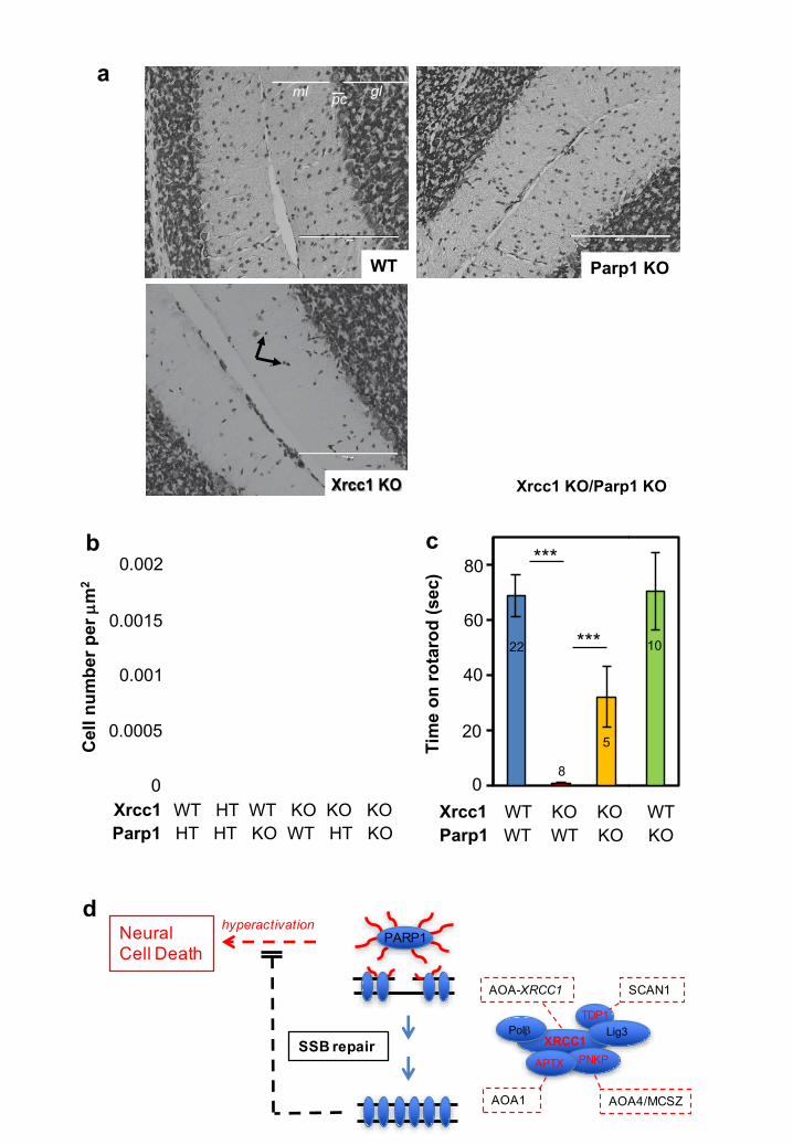
activation in brain even at endogenous levels of SSBs (Fig. 4e). Indeed, the
deletion of Parp1 ablated both the elevated level of ADP-ribose and the
characteristic loss of cerebellar interneurons in Xrcc1Nes-Cre mice, thereby
increasing neuronal density in the molecular layer ~4-fold to wild type levels
(Fig. 5a, b). This did not reflect an impact of Parp1 deletion on the rate of SSBR
because the latter was similarly slow in XRCC1-/- and XRCC1-/-/PARP1-/- RPE-1
cells (Extended Data Fig. 10). Rather, these data demonstrate that in the
absence of Xrcc1-dependent SSB repair Parp1 is hyperactivated, resulting in the
loss and/or dysfunction of cerebellar neurones. Finally, to examine whether Parp1
deletion also rescued the cerebellar ataxia observed in Xrcc1Nes-Cre mice we
compared Xrcc1Nes-Cre and Xrcc1Nes-Cre/Parp1-/- mice for their performance on an
accelerating rotarod. Indeed, remarkably, whereas Xrcc1Nes-Cre mice were
profoundly ataxic and unable to remain on the rotarod for more than a few
seconds the additional deletion of Parp1 improved the rotarod performance of
Xrcc1Nes-Cre mice >30-fold, increasing their mean retention time to ~30 sec
(Fig. 5c).
Collectively, these data identify elevated ADP-ribose levels as a biomarker
of PARP1 hyperactivity and as a cause of cerebellar ataxia induced by unrepaired
SSBs (Fig. 5d). This scenario might also extend to other more common
neurodegenerative diseases because elevated levels of oxidative stress and DNA
strand breakage are also implicated in diseases such as Alzheimer’s disease,
Huntington’s disease, and Parkinson’s disease20-22. Finally, these data identify
PARP1 as a possible drug-target for the treatment of cerebellar ataxias
associated with unrepaired SSBs. Inhibition of PARP1 with currently available
chemical inhibitors may not be useful in this context, however, because these
inhibitors “trap” PARP1 on DNA23 and do not mimic PARP1 genetic deletion
(Extended Data Fig. 10). However, the development of selective inhibitors of
PARP1 that prevent DNA binding by this enzyme may have significant
therapeutic potential.
Figure Legends

Figure 1. Biallelic XRCC1 mutations are associated with cerebellar ataxia, ocular motor apraxia and axonal neuropathy. a, Family pedigree of the
affected proband (III.1; black circle) and her unaffected sister (III.2; circle with
black dot). b, Summary of proband clinical features. The full clinical history and
electrophysiological analysis is presented in Supplementary Information. c, MRI
of the proband at age 40 and 47. Sagittal T1 weighted images (middle top)
demonstrating moderately severe vermian atrophy and Axial FLAIR images (right
top) demonstrating atrophy of the cortex of the vermis and cerebellar
hemispheres at age 40. Note that imaging at age 47 reveals progression of the
vermian atrophy and atrophy of the vermian cortex and cerebellar hemispheres
(bottom). The cerebellum is circled in white and insets (left) are magnifications of
the sagittal images to highlight the cerebellar atrophy. d, Confirmation by Sanger
sequencing of the mutations c.1293G>C (p.K431N) and c.1393C>T (p.Q465*) in
the proband (III.1) and c.1293G>C (p.K431N) in the unaffected sibling (III.2).
Figure 2. XRCC1 patient mutations greatly reduce XRCC1 protein levels and XRCC1 recruitment into damaged chromatin. a, 1BR wild type human
fibroblasts (“WT Fib.”) and wild type RPE-1 cells (“WT RPE-1”) were compared
with XRCC1 patient fibroblasts (“Patient Fib.”) and XRCC1-/- RPE-1 cells for
levels of XRCC1 protein by indirect immunofluorescence. Cells were
counterstained with DAPI and images captured on a Zeiss microscope. b, Top,
wild type RPE-1 cells, wild type 1BR fibroblasts, and wild type lymphoblastoid
cells (“LCLs”) were compared with XRCC1-/- RPE-1 cells, XRCC1 patient
fibroblasts, and with LCLs from the XRCC1 patient or the unaffected sibling for
levels of XRCC1, Lig3, and Tubulin by Western blotting. Bottom, wild type or
patient fibroblasts were transfected with non-targeting or XRCC1 siRNA and
XRCC1 and Tubulin protein levels analysed by Western blotting. c, XRCC1
recruitment into chromatin was compared in the indicated cell lines before and 10
min after treatment with 1 mM H2O2. Cells were pre-extracted with detergent prior
to fixation and immunostaining, and images were captured as above. d,
Quantitation of XRCC1 in chromatin (excluding the nucleoli) from >1000 cells per
sample using Olympus ScanR acquisition and analysis software. Data are the
mean (+/-1SD) of three independent experiments. Statistical significance (two-

tailed t-test) was assessed where indicated (**p<0.01; “ns”, not significant).
Representative ScanR images are shown in Extended Data Fig. 3a. Scale bars
are 10 µm.
Figure 3. Defective DNA single-strand break repair in XRCC1-mutant patient cells. a, DNA strand breakage was quantified by alkaline comet assays in wild
type RPE-1 cells, XRCC1-/- RPE-1 cells, normal human fibroblasts (1BR), and
XRCC1-mutated patient fibroblasts before and immediately after treatment with
H2O2 on ice and after the indicated repair periods in drug-free medium. Data are
the average comet tail moment (an arbitrary unit-measure of DNA strand breaks)
of 100 cells per sample and are the mean (+/-SEM) of three independent
experiments. Statistically significant differences (two-way ANOVA) are indicated
(*p<0.05; **p<0.01). b, H2AX foci were quantified as a measure of DSBs in the
indicated cell lines before and at the indicated times after ionising radiation
(2 Gy). Data are the average number of H2AX foci per cell from ~1000
cells/sample and are the mean (+/-1SD) of three independent experiments. Data
were quantified by ScanR high content imaging. Statistically significant
differences (two-way ANOVA) are indicated (**p<0.01; ns, not significant).
Representative images are presented in Extended Data Fig. 4. c, Representative
images (left) and quantification (right) of sister chromatid exchanges in LCLs from
an unrelated normal control (“WT”), the unaffected sibling, and the XRCC1-
mutated patient. A total of 36 metaphases per genotype were scored and the
mean number of SCEs (+/-1SEM) per chromosome was plotted for each
metaphase. Statistically significant differences (two-tailed t-test) are indicated
(**p<0.01; ns, not significant). Scale bars are 10 µm.
Figure 4. Elevated ADP-ribosylation in XRCC1-mutated patient cells and cerebellum. a, Levels of ADP-ribosylation were measured in 1BR wild type
human fibroblasts (“WT Fib.”), wild type RPE-1 cells (“WT RPE-1”), XRCC1
patient fibroblasts (“Patient Fib.”), and XRCC1-/- RPE-1 cells by indirect
immunofluorescence using Anti-pan-ADP-ribose binding reagent (Millipore). Cells
were treated with 150 µM H2O2 for 10 min and then incubated in drug-free
medium for 60 min to allow DNA repair or were incubated with 30 µM CPT for

45 min. b, Left, ADP-ribosylation was detected in 1BR wild type and PNKP
patient fibroblasts after CPT treatment by indirect immunofluorescence as above.
Middle, Western blot showing PNKP, XRCC1, and PARP1 levels in the two cell
lines. Right, Quantitative comparison of ADP-ribose levels in 1BR wild type,
XRCC1-mutant patient, and PNKP patient fibroblasts before and after CPT
treatment, measured by ScanR imaging and plotted relative to the ADP-ribose
level in untreated 1BR cells. Data are from >1000 cells/sample and are the mean
(+/-1SD) of three independent experiments. Statistically significant differences
(two-tailed t-test) are indicated (*p<0.05). c, ADP-ribosylation was detected in
wild type (“WT”), XRCC1-/-, PARP1-/-, and XRCC1-/-/PARP1-/- RPE-1 cells after
CPT treatment by indirect immunofluorescence as above. d, Levels of ADP-
ribosylation were quantified before and after CPT treatment by high content
imaging as above in wild type 1BR fibroblasts, XRCC1 patient fibroblasts (“X1
pat.”), and XRCC1 patient fibroblasts transfected by electroporation in the
presence of 1 µg or 2 µg, as indicated, of control BSA or purified recombinant
human XRCC1-His protein. Statistically significant differences (two-tailed t-test) to
CPT treated 1BR cells are indicated (*p<0.05). Representative ScanR images are
presented in Extended Data Figure 8. e, Levels of ADP-ribosylation were
measured in cerebellar sections from wild type (WT), Xrcc1Nes-Cre (Xrcc1 KO),
Parp1-/- (Parp1 KO), and Xrcc1Nes-Cre/Parp1-/- (Xrcc1 KO/Parp1 KO) mice by
immunohistochemistry using rabbit anti-poly (ADP-ribose) primary antibody
(Trevigen). Scale bars in IF images are 10 µm and in the histology section is
1 mm.
Figure 5. Parp1 deletion restores normal interneuron density and reduces cerebellar ataxia in XRCC1Nes-Cre mice. a, Neurons in cerebellar sections from
wild type (WT), Xrcc1Nes-Cre (Xrcc1 KO), Parp1-/- (Parp1 KO), and
Xrcc1Nes-Cre/Parp1-/- (Xrcc1 KO/Parp1 KO) mice were detected by Nissl staining.
The position of the granule layer (“gl”), molecular layer (“ml”), and Purkinje cells
(“pc”) is indicated. Note the reduced number of interneurons (black arrows) in the
molecular layer of Xrcc1Nes-Cre mice. Representative images are shown and scale
bars are 200 µm. b, Quantitation of interneuron density in the molecular layer in
mouse cerebellum of the indicated genotype. Data are the mean (+/-SEM) Nissl-
positive cells per µm2 in the molecular layer of sections from 5-10 mice per

genotype. T-test comparisons are indicated (**p<0.01, ***p<0.001). c, Motor
coordination/cerebellar ataxia was measured in mice of the indicated genotype on
an accelerating rotarod. The number of mice analysed for each genotype are
indicated in/above the bars. Statistically significant t-test comparisons are
indicated (**p<0.01, ***p<0.001). d, Model for PARP1 hyperactivation and neural
death triggered by unrepaired SSBs. PARP1 binding and activation at SSBs
results in ribosylation of itself and histones with poly (ADP-ribose) (red wavy
lines). Poly (ADP-ribose) binding by XRCC1 assembles protein complexes
containing the enzymes required for repair of the SSBs. However, if SSB repair is
inefficient, PARP1 activity persists resulting in excessive poly (ADP-ribose) and
concomitant depletion of NAD+; both of which are implicated in cytotoxicity.
Protein partners associated previously with cerebellar ataxia in humans are
highlighted in red and their associated cerebellar ataxias are boxed in red.
SCAN1; spinocerebellar ataxia with axonal neuropathy-1 (mutated in TDP1).
AOA1; ataxia oculomotor apraxia-1 (mutated in APTX). AOA4; ataxia oculomotor
apraxia-4 (mutated in PNKP). We suggest that the patient with XRCC1 mutations
will be the founder member of a new Syndrome; ataxia oculomotor apraxia-
XRCC1 (AOA-XRCC1), as suggested in new guidelines for naming movement
disorders24.
Methods
Whole-exome sequencing. Whole-exome library preparation, exon capture and
sequencing were performed at the Genome Québec Innovation Center (Montréal,
QC, Canada) as previously described25. Genomic DNA was captured using the
SureSelect Human 50Mb All Exon kit v5 (Agilent Technologies, Santa Clara, CA,
USA). Sequencing was performed on an Illumina HiSeq2000 (Illumina, San
Diego, CA, USA) with paired-end 100-bp reads. A mean coverage of 137x was
obtained and 97% of the bases were covered at more than 10x. Read alignment,
variant calling, and annotation were done with a pipeline based on BWA,
SAMtools, Annovar, and custom annotation scripts. All sequences were aligned
to Human genome Hg19. We excluded variants with minor allele frequency
greater than 5% in either the 1000 genomes project
(http://browser.1000genomes.org/index.html) or the 6500 NHLBI EVS

(http://evs.gs.washington.edu/EVS), and seen in more than 30 samples from our
in-house database (containing approximately 2000 samples). The WES data was
further filtered to keep protein-damaging variants (nonsense, missense,
frameshift, indel, and splice variants).
Antibodies and chemicals. The antibodies employed in this study were anti-
XRCC1 rabbit polyclonal (Millipore; ABC738), anti-Lig3 (TL25) rabbit
polyclonal26, anti-PNKP (SK3195) rabbit polyclonal27, rabbit Fc-fused Anti-pan-
ADP-ribose binding reagent (Millipore; MABE1016), anti-poly (ADP-ribose) rabbit
polyclonal (Trevigen; 4336), anti-α-tubulin rat polyclonal (Abcam; ab6160), anti-
BrdU rat monoclonal, crossreacting with CldU, (BioRad; OBT0030G), anti-
nucleophosmin (B23) mouse monoclonal (Invitrogen; 325200), anti-PARP1
mouse monoclonal (Serotec; MCA1522G) and anti-H2AX mouse monoclonal
(Millipore; 05-636). The secondary antibodies employed for Western blotting were
HRP-conjugated goat anti-rabbit (Bio-Rad; 170-6515), goat anti-mouse (Bio-Rad;
170-6516) and rabbit anti-rat (Abcam; ab6734) and for indirect
immunofluorescence were goat anti-mouse or anti-rabbit Alexa 488 (Invitrogen;
A11001 and A31628), goat anti-rabbit Alexa 568 (Invitrogen; A11036), donkey
anti-mouse Alexa 647 (Invitrogen; A39571) and goat anti-rat Alexa 568
(Invitrogen; A11077). Camptothecin (CPT) was purchased from Sigma and
Hydrogen Peroxide (H2O2) was obtained from Fischer Scientific. Veliparib (ABT-
888) was purchased from Selleckchem and KU0058948 hydrochloride from Axon.
Cell lines. All cell lines were tested for absence of mycoplasma. Wild type human
hTERT RPE-1 cells (ATCC; CRL4000)(denoted “RPE-1” for simplicity) and their
XRCC1-/- derivative (XRCC1-/- RPE-1 cells) were cultured in Dulbecco’s Modified
Eagle’s Medium (DMEM/F12; Sigma) supplemented with 10% fetal calf serum
and 0.01 mg/ml hygromycin B in a humidified atmosphere of 5% CO2 at 37°C.
Wild type control cells were 1BR3 (denoted 1BR in the text for simplicity) primary
human fibroblasts and the lymphoblastoid cell line (LCL) 11-27 isolated from an
normal unaffected control. XRCC1 patient primary fibroblasts (identifier number;
5596502b) were generated from a patient skin biopsy and the LCL cell lines
HEP15-00082 and HEP15-00083 were obtained from fresh blood from,

respectively, the unaffected sibling and affected patient by EBV transformation.
Appropriate patient consent was provided for preparation of primary fibroblasts.
Primary human fibroblasts from a PNKP-mutated patient with cerebellar ataxia
have been described previously28. Primary human fibroblasts were grown in
Minimum Essential Media (MEM; Gibco) containing 15% fetal calf serum, 2 mM
glutamine, and the antibiotics penicillin (100 units/ml) and streptomycin (100
μg/ml) at low oxygen (5%) at 37oC. LCLs were cultured in RPMI medium (Gibco)
containing 10% FBS, 2 mM glutamine and penicillin/streptomycin, in a humidified
atmosphere of 5% CO2 at 37°C.
Generation of gene edited RPE-1 cells. Guide sequences were identified using
either E-CRISP (http://www.e-crisp.org/E-CRISP/) or CRISPRdirect
(http://crispr.dbcls.jp). For XRCC1 gene editing we chose the 23-mer CRISPR
complementary guide RNA sequences 5’-CCGCCUCCGCCAUGUCGUGUCCU-
3’ & 5’-AGGGACACGACAUGGCGGAGGCGG-3’ (PAM underlined) spanning
XRCC1 ORF nucleotides 12-34, and employed the 58-
mer synthetic oligonucleotides
(XCr2F; 5’-TTTCTTGGCTTTATATATCTTGTGGAAAGGACGAAACACCGACAC
GACATGGCGGAGG &
XCr2R; 5’-GACTAGCCTTATTTTAACTTGCTATTTCTAGCTCTAAAACCCTCCG
CCATGTCGTGTC) (Eurofins) encoding 18 bp Tru-guide29 versions of the guide
(underlined) minus the PAM. For PARP1 gene editing we chose the 20-mer ‘Tru-
guide’ sequences 5’-GCACCCUGACGUUGAGGUGG-3’ and
5’-CCACCUCAACGUCAGGGUGC-3’ (PAM underlined) spanning nucleotides
195-214 of the human PARP1 ORF, and employed the 57-mer synthetic
oligonucleotides PARP1-2F:
5’-TTTCTTGGCTTTATATATCTTGTGGAAAGGACGAAACACCGCACCCTGACG
TTGAGG-3’ & PARP1-2R:
5’-GACTAGCCTTATTTTAACTTGCTATTTCTAGCTCTAAAACCCTCAACGTCA
GGGTGC-3’ encoding the 17 bp Tru-guide versions of the guide (underlined)
minus the PAM.
The relevant oligonucleotide guide pairs were annealed and extended into
a 98-mer oligonucleotide duplexes using Phusion polymerase (NEB) and then
subcloned into the guide RNA vector (Addgene; #41824)30 using Gibson

Assembly (NEB). hTERT RPE-1 cells were co-transfected with the relevant guide
construct/s separately (XRCC1-/-, PARP1-/-) or together (XRCC1-/-/PARP1-/-) and
with the Cas9 expression construct Addgene #4181530 using a NEON
Transfection System (Invitrogen). 24 h later, the transfected cells were selected in
medium containing 0.5 mg/ml G418 for 5 days and subcloned into 96-well plates.
Once at sufficient cell density the subclones were analyzed for expression of the
relevant protein/s by indirect immunofluorescence. Absence of the relevant
protein/s in selected clones was then confirmed by Western blotting. For XRCC1-/-
, PARP1-/-, and XRCC1-/-/PARP1-/- RPE-1 cells we selected clones #3, #G7, and
#D1 for further work, respectively.
Complementation of XRCC1 patient cells with recombinant XRCC1 protein. Recombinant human XRCC1 harbouring a C-terminal decahisitidine tag (denoted
XRCC1-His) was expressed in E.coli from pET16b-XH and purified by metal-
chelate affinity chromatography and gel filtration3. 1 or 2 µg of control bovine
serum albumin (BSA) or purified XRCC1-His was electroporated into 1x10⁵
XRCC1 patient fibroblasts using a NEON Transfection System (Invitrogen)
according to the manufacturer’s protocol. 18 hours later the cells were treated
with CPT for 45 min, fixed, and immunostained for levels of XRCC1 and ADP-
ribose as indicated.
RNA extraction, cDNA synthesis and qPCR analysis. LCLs were treated with
either 100 µg/ml Cycloheximide (Sigma) or vehicle alone for 4 h and total RNA
extracted with RNAeasy Kit (Qiagen) essentially as described by the
manufacturer but with an additional 15 min DNAse I (Promega) digest of the
samples on the column. 1 µg total RNA was annealed to oligodT(15) primer and
reverse transcribed using M-MuLV RT (NEB) for 2 h at 42°C. After RNAse A
digest, the cDNA was purified using PCR purification kit (Qiagen) and 1/40 of the
eluate used per reaction. Three replicate qPCR reactions using ABsolute qPCR
SYBR Low ROX (Thermo) were performed per experiment in a MX3005P
(Agilent) thermocycler and analysed using MxPro software (Agilent). The fold
change was calculated from Ct values relative to actin and Ct values relative
to WT untreated for three independent experiments. Primers were:

XRCC1 exon10 forward: CAACACCCCCAAGTACAGC
XRCC1 exon 10 reverse: AGTCCAGCACCCACTCCTTAC
XRCC1 exon11 forward: TCCAGCAGTGAGGAGGATG
XRCC1 intron11 reverse: AGGCAAGAGTGGGAAGTTTG
XRCC1 exon 12 reverse: AGTGGGCTTGGTTTTGGTC
Actin forward: CTCGTCATACTCCTGCTTGC
Actin reverse: GAAGTGTGACGTGGACATCC
XRCC1 cDNA cloning. cDNA prepared as above from patient cells treated with
cycloheximide was used for Phusion polymerase (NEB) amplification of full length
XRCC1 transcripts using primers ATGCCGGAGATCCGCCTCCG and
GGCTTGCGGCACCACCCCAT. PCR products were purified using Gel extraction
kit (Qiagen), cloned using TOPO cloning kit (Thermo Fischer Scientific) and
plasmids originating from single colonies purified using Miniprep kit (Qiagen) and
sequenced by Sanger sequencing (Beckman Coulter).
siRNA. Wild type and patient primary fibroblasts were reverse-transfected with
Lipofectamine RNA iMAX (Life Technologies) as indicated by the manufacturer,
using non-targeting siRNA (ON-TARGETplus, Dharmacon) and siXRCC1
SMARTpool (Dharmacon).
Western blotting. PBS-washed cells were lysed in Laemmli buffer, heated for 10
min at 95°C, and sonicated for 30 sec using Bioruptor® Pico (Diagenode). Protein
concentrations were determined using the BCA assay (Pierce). Samples were
subjected to SDS PAGE, proteins transferred onto nitrocellulose membrane and
detected by immunoblotting using the relevant primary and horseradish
peroxidase-conjugated secondary antibodies. Peroxidase activity was detected
by ECL reagent (GE Healthcare) and Amersham Hyperfilm ECL (GE Healthcare).
Non-saturated film exposures were digitalized using an EPSONperfection 2400
photo scanner and quantified using ImageJ software.
Immunofluorescence and microscopy. Cells cultured on glass coverslips were
fixed in 4% paraformaldehyde for 10 min at room temperature, permeabilized in
methanol/acetone solution (1:1), blocked in 10% foetal calf serum and incubated

with primary antibodies for 60 min at room temperature. Following rising in PBS,
coverslips were incubated with secondary antibodies at room temperature for
60 min. Finally, after washing in PBS, nuclei were counterstained with DAPI
(Sigma) and coverslips mounted using anti−fading mounting reagent
(Vectashield, Vector Laboratories). To measure chromatin retention of proteins,
cells were pre-extracted in cold 0.2% Triton X-100 for 2 min on ice prior to fixation
as above. High-resolution microscopy of fixed samples was carried out on a Zeiss
AxioObserver.Z1 microscope, equipped with oil immersion objectives (Plan-
Apochromat 63x/1.4 and 100x/1.4), Hamamatsu ORCA-Flash4.0 LT camera and
ZEN 2 core imaging software. Automated wide-field microscopy was performed
on an Olympus ScanR system (motorized IX83 microscope) with ScanR Image
Acquisition and Analysis Software, 20x/0.45 (LUCPLFLN 20x PH) and 40x/0.6
(LUCPLFLN 40x PH) dry objectives and Hamamatsu ORCA-R2 digital CCD
camera C10600.
ADP-ribose immunohistochemistry. Mice (P17-P23) were anaesthetized using
0.2 mg/g Euthanal (Vetoquinol UK Ltd) and perfused transcardially with PBS
followed by 4% paraformaldehyde. The brains were postfixed in 4%
paraformaldehyde for 48 h and stored in 25% sucrose/PBS until moulding and
freezing. 7 μm sagittal sections were obtain using a cryostat (Leica CM1850).
Immunohistochemistry was conducted essentially as described4. Briefly, slides
were washed in PBS and heated until boiling in antigen retrieval buffer (Nacalai
Tesque, Histo VT One). Endogenous peroxidase was blocked by incubating
slides in 0.6% H2O2 in methanol. After incubating in blocking solution (5% goat
serum, 1% BSA, 0.4% Triton-X-100 in PBS) the indicated primary antibodies
were applied on overnight. The appropriate Biotin-SP-conjugated AffiniPure Goat
antibody (JacksonImmuno Research, 1:500) was incubated on the slides for 1 hr
followed by ABC reagent (Vectstain Elite ABC kit, Vector Laboratories) according
to manufacturer’s instructions. The chromogen was developed using VIP reagent
(Vector VIP peroxide substrate kit, Vector laboratories, Inc.; SK-4600). Images
were obtained using a Nikon Eclipse E400 mounted with Electronic Digital
Eyepiece Camera CMOS (C-mount UK).

Sister chromatid exchanges. Lymphoblastoid cell lines were incubated in
medium containing 8 µM chlorodeoxyuridine, 32 µM thymidine, 10 µM
fluorodeoxyuridine and 200 µM cytidine (such that 20% of incorporated thymidine
is replaced with chlorodeoxyuridine)11,31 for 20 h, washed twice in media and then
allowed to grow for another 20 h in medium containing 10 µM thymidine. Cells
were treated with 100 ng/ml colcemid for 1 h, swollen in 75 mM KCl for 5 min at
37°C and fixed in Carnoy’s fixative before preparation of metaphase spreads.
Slides were allowed to air dry, rehydrated in PBS, incubated in 2 M HCl for
30 min, washed twice in 100 mM borate buffer pH 8.5 for 10 min and blocked in
10% foetal calf serum in PBS for 30 min. Further immunofluorescence and
imaging was performed as above.
SSBR & DSBR assays. Alkaline comet assays were performed essentially as
described32 and inducing DNA breaks with 50 µM H2O2 (RPE-1 cells) or 25 µM
H2O2 (primary fibroblasts) for 10 min on ice. For DSB repair assays, cells were
irradiated with 2 Gy using Gammacell 1000 machine, and H2AX quantified at the
indicated times, afterwards.
Mouse maintenance and analysis. Animals were maintained and used under
the auspices of UK Home Office project licence number 70/8300. The generation
of Parp1-/- and Xrcc1Nes-Cre mice have been reported previously4,33. Intercrosses
between Parp1-/- and Xrcc1+/loxp mice were maintained in a mixed C57/Bl6 x S129
strain and housed on a 12 h light/dark cycle with lights on at 7 am. Temperature
and humidity were maintained at 21oC (+/-2oC) and 50% (+/-10%), respectively.
All experiments were carried out under the UK Animal (Experimental Procedures)
Act, 1986. Genomic DNA was extracted from biopsied tail using the REDExtract-
N-Amp Tissue PCR Kit in accordance to the manufacturer’s instructions
(Sigma). For Parp1, the following primers were used: PARP1 F1 (5’-GTT GTG
AAC GAC CTT CTG GG-3’), PARP1 R1 (5’-CCT TCC AGA AGC AGG AGA AG-
3’) and PARP R2 (5’-GCT TCA GTG ACA ACG TCG AG-3’). PCR products were
generated by an initial denaturation step at 94C for 2 min followed by 35 cycles
of 94C for 30 s, 54C for 40 s and 72C for 180 s. Xrcc1 was amplified using the
forward PC1 (5’-TAT GCT TGC TGT ACA GGG ATT GGG-3’) and reverse PC2

(5’-TGG ACC ATG AAA AAG CTG TGT GC-3’) primers. A 400 bp cre PCR
product was generated using the forward Cre-3 (5’-CTG CCA CGA CCA AGT
GAC AGC-3’) and reverse Cre-4 (5’-ACC TGC GGT GCT AAC CAG CG-3’)
primers. Both Xrcc1 and Cre PCR products were amplified using the following
PCR conditions: initial denaturation for 5 min at 94C followed by 35 cycles of
94C for 30 s, 59C for 45 s and 72C for 45 s.
Nissl staining of paraffin-embedded sections. Brains were removed at the
indicated times and placed in 10% neutral buffered formalin (3.7% formaldehyde,
3.5 g/l NaH2PO4, 6.5 g/l Na2HPO4) for 24 h and then transferred to PBS. Paraffin
embedding, sagittal sectioning and Nissl staining were carried out by Propath UK
Ltd (Hereford, UK) and UCL IQPath (Institute of Neurology, London, UK). Briefly,
deparaffinised sectioned were stained in 0.1% cresyl violet solution (0.1% cresyl
violet, 0.3% glacial acetic acid) for 3-10 minutes, rinsed in water, then 95%
ethanol for 30 sto 5 min, 100% ethanol for 2 x 5 min and xylene for 2 x 5 min
before mounting. Nissl-positive interneurons were counted within three randomly-
selected 16875 µm2 regions of each molecular layer, and the number of cells per
µm2 was calculated.
Rotarod analysis. To evaluate motor coordination/cerebellar ataxia, an
accelerating Panlab Rotarod (Harvard Apparatus Ltd., UK) was used. The
apparatus was composed of a rod with a diameter of 30 mm divided into five
opaque methacrylate arnite barriers, separating the rod into five 50 mm sections.
Mice were trained for three successive attempts on the day prior to assessment.
For assessment, mice were positioned on the rod facing away from the
experimenter whilst the rod was stationary. Once activated, the rod accelerated at
0 to 40 rpm at an acceleration rate of 5 min. Mice were rested for 15 min
between each of the three trials and then positioned back on the rod for the next
assessment. The time spent on the rod was recorded for each mouse and the
mean time was calculated from the three independent trials.
Purkinje cell recordings. Vermis parasagittal slices (200 μm) were taken from
P13-P17 mice using a vibroslicer (VT1200S, Leica Microsystems, Germany) in

ice-cold artificial cerebrospinal fluid (ACSF) containing (in mM): 125 NaCl, 2.5
KCl, 25 glucose, 1.25 NaH2PO4, 26 NaHCO3, 1 MgCl2, 2 CaCl2 (bubbled with
95% O2 and 5% CO2, pH 7.3). Slices were maintained in ACSF at 34-35°C for 45
min and then at 23-25°C for electrophysiology. Voltage-clamp recordings of
Purkinje cells were performed in cell-attached mode using a Multiclamp 700A
amplifier (Molecular Devices) with pipettes (5-7 MΩ) containing 150 mM NaCl.
Signals were filtered at 10 kHz and digitized at 50 kHz and analysis of spike
activity performed offline using Clampfit (Molecular Devices).
Data Availability. The datasets generated during and/or analysed during the
current study are available from the corresponding author on reasonable request.
Data analysis and statistics. The number of experimental repeats and statistical
tests (conducted in Excel or GraphPad) are indicated in the relevant figure
legends. The means of population averages from multiple independent
experiments (+/-SD) are indicated. Where appropriate, power calculations were
conducted using online statistical resources. Samples were not blinded but the
collection and analysis of microscopic data was automated and free of user bias.
No animals/samples were omitted from data points/data analyses.
Acknowledgments. We would like to thank the patient and her family for their
contribution to this study. This work was selected for study by the Care4Rare
Canada (Enhanced Care for Rare Genetic Diseases in Canada) Consortium
Gene Discovery Steering Committee: Kym Boycott (lead; University of Ottawa),
Alex MacKenzie (co-lead; University of Ottawa), Jacek Majewski (McGill
University), Michael Brudno (University of Toronto), Dennis Bulman (University of
Ottawa), and David Dyment (University of Ottawa). We especially thank David
Dyment (University of Ottawa) for his advice and discussion. We thank Saskia
vanderVelde-Visser and Janneke Schuurs-Hoeijmakers at the Radboud Nijmegen
University Medical Centre for EBV transformation of the patient and sibling LCLs.
Funding. This work was funded by a MRC Programme Grant (MR/J006750/1) to
KWC, a “Science without Borders” postdoctoral fellowship (CAPES Foundation,
Ministry of Education, Brazil, BEX9769-13-7) to NH, and funding to GY from

Genome Canada, the Canadian Institutes of Health Research, the Ontario
Genomics Institute, Ontario Research Fund, Genome Quebec, Children’s
Hospital of Eastern Ontario Foundation and the Hospital for Sick Children. PMcK
acknowledges the NIH (NS-37956, CA-21765), the CCSG (P30 CA21765) and
the American Lebanese and Syrian Associated Charities of St. Jude Children's
Research Hospital for support.
1. Caldecott, K. W. Single-strand break repair and genetic disease. Nat. Rev. Genet. 9, 619–631 (2008).
2. Caldecott, K. W. XRCC1 and DNA strand break repair. DNA Repair (Amst)2, 955–969 (2003).
3. Caldecott, K. W., Tucker, J. D., Stanker, L. H. & Thompson, L. H. Characterization of the XRCC1-DNA ligase III complex in vitro and its absence from mutant hamster cells. Nucleic Acids Res 23, 4836–4843 (1995).
4. Lee, Y. et al. The genesis of cerebellar interneurons and the prevention of neural DNA damage require XRCC1. Nat. Neurosci. 12, 973–980 (2009).
5. Caldecott, K. W., McKeown, C. K., Tucker, J. D., Ljungquist, S. & Thompson, L. H. An interaction between the mammalian DNA repair protein XRCC1 and DNA ligase III. Mol Cell Biol 14, 68–76 (1994).
6. Tebbs, R. S. et al. Requirement for the Xrcc1 DNA base excision repair gene during early mouse development. Dev. Biol. 208, 513–529 (1999).
7. Tebbs, R. S., Thompson, L. H. & Cleaver, J. E. Rescue of Xrcc1 knockout mouse embryo lethality by transgene-complementation. DNA Repair (Amst)2, 1405–1417 (2003).
8. Bradley, M. O. & Kohn, K. W. X-ray induced DNA double strand break production and repair in mammalian cells as measured by neutral filter elution. Nucleic Acids Res 7, 793–804 (1979).
9. Thompson, L. H. & West, M. G. XRCC1 keeps DNA from getting stranded. Mutat Res 459, 1–18 (2000).
10. Thompson, L. H. et al. A CHO-cell strain having hypersensitivity to mutagens, a defect in DNA strand-break repair, and an extraordinary baseline frequency of sister-chromatid exchange. Mutat Res 95, 427–440 (1982).
11. Dillehay, L. E., Thompson, L. H., Minkler, J. L. & Carrano, A. V. The relationship between sister-chromatid exchange and perturbations in DNA replication in mutant EM9 and normal CHO cells. Mutat Res 109, 283–296 (1983).
12. Takashima, H. et al. Mutation of TDP1, encoding a topoisomerase I-dependent DNA damage repair enzyme, in spinocerebellar ataxia with axonal neuropathy. Nat Genet 32, 267–272 (2002).
13. El-Khamisy, S. F. et al. Defective DNA single-strand break repair in spinocerebellar ataxia with axonal neuropathy-1. Nature 434, 108–113 (2005).
14. Moreira, M. C. et al. The gene mutated in ataxia-ocular apraxia 1 encodes the new HIT/Zn-finger protein aprataxin. Nat Genet 29, 189–193 (2001).
15. Bras, J. et al. Mutations in PNKP cause recessive ataxia with oculomotor

apraxia type 4. Am J Hum Genet 96, 474–479 (2015). 16. Shen, J. et al. Mutations in PNKP cause microcephaly, seizures and
defects in DNA repair. Nat Genet 42, 245–249 (2010). 17. Eliasson, M. J. et al. Poly(ADP-ribose) polymerase gene disruption renders
mice resistant to cerebral ischemia. Nat. Med. 3, 1089–1095 (1997). 18. Chiarugi, A. Poly(ADP-ribose) polymerase: killer or conspirator? The
‘suicide hypothesis’ revisited. Trends Pharmacol. Sci. 23, 122–129 (2002). 19. Katyal, S. et al. Aberrant topoisomerase-1 DNA lesions are pathogenic in
neurodegenerative genome instability syndromes. Nat. Neurosci. (2014). doi:10.1038/nn.3715
20. Hou, Y., Song, H., Croteau, D. L., Akbari, M. & Bohr, V. A. Genome instability in Alzheimer disease. Mech. Ageing Dev. (2016). doi:10.1016/j.mad.2016.04.005
21. Shiwaku, H. & Okazawa, H. Impaired DNA damage repair as a common feature of neurodegenerative diseases and psychiatric disorders. Curr. Mol. Med. 15, 119–128 (2015).
22. Narciso, L. et al. The Response to Oxidative DNA Damage in Neurons: Mechanisms and Disease. Neural Plast. 2016, 3619274 (2016).
23. Murai, J. et al. Trapping of PARP1 and PARP2 by Clinical PARP Inhibitors. Cancer Res 72, 5588–5599 (2012).
24. Marras, C. et al. Nomenclature of genetic movement disorders: Recommendations of the international Parkinson and movement disorder society task force. Mov. Disord. 31, 436–457 (2016).
25. Tetreault, M. et al. Whole-exome sequencing identifies novel ECHS1 mutations in Leigh syndrome. Hum. Genet. 134, 981–991 (2015).
26. Cappelli, E. et al. Involvement of XRCC1 and DNA ligase III gene products in DNA base excision repair. J Biol Chem 272, 23970–23975 (1997).
27. Breslin, C. & Caldecott, K. W. DNA 3'-phosphatase activity is critical for rapid global rates of single-strand break repair following oxidative stress. Mol Cell Biol 29, 4653–4662 (2009).
28. Poulton, C. et al. Progressive cerebellar atrophy and polyneuropathy: expanding the spectrum of PNKP mutations. Neurogenetics (2012). doi:10.1007/s10048-012-0351-8
29. Fu, Y., Sander, J. D., Reyon, D., Cascio, V. M. & Joung, J. K. Improving CRISPR-Cas nuclease specificity using truncated guide RNAs. Nat. Biotechnol. 32, 279–284 (2014).
30. Mali, P. et al. RNA-guided human genome engineering via Cas9. Science339, 823–826 (2013).
31. Pinkel, D., Thompson, L. H., Gray, J. W. & Vanderlaan, M. Measurement of sister chromatid exchanges at very low bromodeoxyuridine substitution levels using a monoclonal antibody in Chinese hamster ovary cells. Cancer Res 45, 5795–5798 (1985).
32. Breslin, C. et al. Measurement of chromosomal DNA single-strand breaks and replication fork progression rates. Meth. Enzymol. 409, 410–425 (2006).
33. Wang, Z. Q. et al. Mice lacking ADPRT and poly(ADP-ribosyl)ation develop normally but are susceptible to skin disease. Genes Dev 9, 509–520 (1995).

ba
c d
40yr
47yr
c.1293G>C(p.K431N)
c.1393C>T(p.Q465*)
XRCC1 (NM_006297)
III.1
A A G/C G T
III.1
A A G/C G T
III.2 III.2
T C C/T A G
T C C A G
Proband
Sibling
Age/sex 47/F
Onset(yr) 28
PresentingSymptom Gaitataxia
Intelligence Normal
Muscleweakness Mild(DistalMuscles)
Dystonia No
SensoryDisturbance:
TouchandPain Normal
Vibration Decreased
PositionSense Decreased
Ataxia:
Gait Yes(Severe)
Dysmetria Yes
Dysarthria Yes
Dysdiadochokinesia Yes
DeepTendonReflex Absent
OculomotorApraxia Yes
MRI CerebellarAtrophy
MotorNerveConductionVelocity:
upperlimbs Mildreduction/Normal
lowerlimbs Mildreduction/Normal
SensoryNerveConductionVelocity:
upperlimbs Absent/notrecordable
lowerlimbs Absent/notrecordable

-H2 O
2+
H2 O
2
DAPI/B23
XRCC1
WT RPE-1
WT Fib. (1BR) Patient Fib.
-H2 O
2+
H2 O
2
WT RPE-1 XRCC1-/- RPE-1
WT RPE-1 XRCC1-/- RPE-1
WT Fib. (1BR) Patient Fib. a
Lig3aa
Tubulin
RPE-1 Fib. LCLs
Patient
XRCC1
Tubulin
b
WT
RPE-1
XR
CC
1 f
luo
rescen
ce (
rela
tive levels
)
- +
DAPIXRCC1
DAPIXRCC1
DAPIXRCC1
DAPIXRCC1
c
d
100
75
50
XRCC1
WT(1BR)
siRNA
100
50
0
1
2
3
4
5
6
7
H2O2
X1-/-
RPE-1
WT
Fib.
(1BR)
X1 Pat.
Fib.
- + - + - +
ns ns
**

SC
Es/c
hro
mo
so
me
c Sibling LCLs Patient LCLs
WTW T s ib lin g p a tie n t
0 .0
0 .5
1 .0
1 .5
2 .02
1
0
Sibling Patient
ns ****
Metaphases
a b
0 min 30 min 4 h
7 h 12 h 24 h
0
5
10
15
20
Mean T
ail
Mom
ent
0
5
10
15
20
Mean T
ail
Mom
ent
-H2O2
Time (min) after H2O2
0 7.5 15 30-H2O20 15 30 60
Time (min) after H2O2
WT (1BR)XRCC1 patient *
WT RPE-1XRCC1-/- RPE-1 **
Mean g
H2A
X foci/cell
20
25
15
10
5
0WT
RPE-1
XRCC1-/-
RPE-1
WT
(1BR)
Patient
** ns

b
XR
CC
1
PA
RP
1
PN
KP
c+
CP
T
WT XRCC1-/-PARP1-/-XRCC1-/-
PARP1-/- DA
PI
AD
P-rib
ose
a+
CP
T
WT RPE-1XRCC1-/-
RPE-1
WT Fib.
(1BR)
XRCC1
Patient Fib.
DA
PI
AD
P-rib
ose
+H
2 O2
(+ re
pa
ir)
DA
PI
AD
P-rib
ose
+C
PT
WT (1BR)PNKP Fib. DA
PI
AD
P-rib
ose
0 2 4 6 8
10
-C
PT
+ C
PT
*
**
ADP-ribose (relative levels)
1B
R
XR
CC
1 p
at.
PN
KP
pat.
d
0 1 2 3 4 5
1BR -CPT
XD1 -CPT
1BR
XD1
1 ug BSA
2 ug BSA
1 ug XRCC1
2 ug XRCC1
CP
T-
-+
+ +
+ +
+
ADP-ribose (relative levels)
**
*nsns
WT
Parp
1 K
O
Xrc
c1 K
OX
rcc1 K
O/P
arp
1 K
O
ADP-ribose
e
a
Xrcc1
Parp1
WT KO KO WT
***
***
cb
Xrcc1 KO Xrcc1 KO/Parp1 KO
Parp1 KO WT
ml glpc
WT WT KO KO
20
40
60
80
Tim
e o
n r
ota
rod
(sec)
0.001
0.002
0 0
Cell
nu
mb
er
per µµ
m2
0.0005
0.0015
HT HT KO WT HT KO
WT HT WT KO KO KO
Parp1
Xrcc1
d
22
8
5
10
SSB repair
hyperactivationPARP1Neural
Cell Death
XRCC1Polb
TDP1
PNKPAPTX
Lig3
AOA1 AOA4/MCSZ
SCAN1AOA-XRCC1