world journal of - bsdwebstorage.blob.core.windows.net · kung-chia young, pingtung ... chun-fu...
TRANSCRIPT

Published by Baishideng Publishing Group Inc
World Journal of VirologyWorld J Virol 2015 November 12; 4(4): 313-376
ISSN 2220-3249 (online)

World Journal of VirologyW J V
EDITOR-IN-CHIEFLing Lu, Kansas
GUEST EDITORIAL BOARD MEMBERSChi-Ho Chan, TaichungShih-Cheng Chang, TaoyuanHsin-Wei Chen, Miaoli CountyShun-Hua Chen, TainanSteve S Chen, TaipeiWei-June Chen, TaoYuanJiann Ruey Hong, TainanReuben Jih-Ru Hwu, HsinchuCheng-Wen Lin, TaichungNa-Sheng Lin, TaipeiTzou-Yien Lin, TaoyuanHsin-Fu Liu, New TaipeiHung-Jen Liu, TaichungSzecheng J Lo, Tao YuanMenghsiao Meng, TaichungWen-Ling Shih, PingtungRobert YL Wang, TaoYuanChang-Jer Wu, KeelungChi-Chiang Yang, TaichungKung-Chia Young, Pingtung
MEMBERS OF THE EDITORIAL BOARD
Argentina
Angela Gentile, Buenos AiresPablo Daniel Ghiringhelli, BernalGiselle Paula Martín Ocampos, La PlataJorge Victorio Pavan, Córdoba
Laura Elena Valinotto, Buenos Aires
Australia
Shisan Bao, SydneyJiezhong Chen, WollongongRussell J Diefenbach, WestmeadIan Maxwell Mackay, BrisbaneDavid Peter Wilson, SydneyKong-Nan Zhao, Herston
Austria
Adly MM Abd-Alla, ViennaSabine Brandt, ViennaThomas Lion, Vienna
Barbados
Alok Kumar, Bridgetown
Belgium
Jan P Clement, LeuvenJelle Matthijnssens, Leuven
Brazil
Luciano K de Souza Luna, Ribeirão PretoLuciane Pinto Gaspar, CuritibaThiago Moreno Le Souza, Rio De JaneiroJosé P G Leite, Rio de Janeiro
Sonia Mara Raboni, CuritibaLivia Melo Villar, Rio De Janeiro
BulgariaIrena Petkova Kostova, Sofia
CameroonRichard Njouom, Yaounde
CanadaEarl Garnet Brown, OttawaIvan Brukner, MontrealMax Alexander Chernesky, HamiltonAlain Houde, QuebePeter J Krell, GuelphJean F Laliberté, VancouverHonglin Luo, VancouverXianzhou Nie, FrederictonJean-Pierre Routy, MontrealAiming Wang, OntarioDecheng Yang, Vancouver
ChileMarcelo López-Lastra, Santiago
ChinaKun-Long Ben, KunmingGuang-Wen Cao, Shanghai
I
Editorial Board2011-2015
The World Journal of Virology Editorial Board consists of 341 members, representing a team of worldwide experts in virology. They are from 58 countries, including Argentina (5), Australia (6), Austria (3), Barbados (1), Belgium (2), Brazil (6), Bulgaria (1), Cameroon (1), Canada (11), Chile (1), China (52), Croatia (2), Cuba (1), Czech Republic (1), Denmark (1), Egypt (2), Ethiopia (1), Finland (4), France (11), Germany (12), Ghana (1), Greece (2), Hungary (1), India (11), Indonesia (1), Iran (1), Ireland (3), Israel (5), Italy (24), Japan (13), Kazakhstan (1), Kenya (1), Kosovo (1), Mexico (2), Netherlands (6), New Zealand (1), Nigeria (1), Pakistan (2), Palestine (1), Poland (1), Portugal (1), Romania (1), Russia (2), Saudi Arabia (1), Senegal (1), Singapore (2), Slovakia (1), Slovenia (2), South Africa (3), South Korea (4), Spain (14), Sweden (3), Thailand (8), Tunisia (1), Turkey (4), United Kingdom (7), United States (82), and Uruguay (1).
August 12, 2013WJV|www.wjgnet.com

Paul Kay Sheung Chan, Hong KongYuan-Ding Chen, KunmingAn-Chun Cheng, Ya’anShang-Jin Cui, HarbinXiao-Ping Dong, BeijingZai-Feng Fan, BeijingJean-Michel Garcia, Hong KongXiu-Guo Hua, ShanghaiWen-Lin Huang, GuangdongMargaret Ip, Hong KongDao-Hong Jiang, WuhanJian-Qi Lian, Xi’anXin-Yong Liu, JinanXiao-Yang Mo, ChangshaBeatrice Nal, Hong KongCheng-Feng Qin, BeijingHua-Ji Qiu, HarbinXiao-Feng Ren, HarbinHuai-Chang Sun, YangzhouJian-Wei Wang, BeijingNing Wang, BeijingYou-Chun Wang, BeijingMary Miu Yee Waye, Hong KongPatrick CY Woo, Hong KongJian-Qing Wu, NanjingRui Wu, LuoyangYu-Zhang Wu, ChongqingChuang-Xi Zhang, HangzhouGuo-Zhong Zhang, BeijingChun-Fu Zheng, Wuhan
Croatia
Snjezana Zidovec Lepej, ZagrebPero Lučin, Rijeka
Cuba
Maria G Guzman, La Habana
Czech Republic
Daniel Ruzek, Ceske Budejovice
Denmark
Håvard Jenssen, Roskilde
Egypt
Samia Ahmed Kamal, CairoAbdel-Rahman Zekri, Cairo
Ethiopia
Woldaregay Erku Abegaz, Addis Ababa
Finland
Jussi Hepojoki, HelsinkiAnne Jääskeläinen, HelsinkiIrmeli Lautenschlager, Helsinki
Antti Vaheri, Helsinki
France
Laurent Belec, ParisChristian A Devaux, MontpellierJean Dubuisson, LilleWattel Eric, LyonDuverlie Gilles, AmiensGilles Gosselin, MontpellierBedouelle Hugues, ParisEric J Kremer, MontpellierDenis Rasschaert, ToursFarzin Roohvand, Tehran and ParisChristian Trépo, Lyon
Germany
Gualtiero Alvisi, HeidelbergClaus Thomas Bock, BerlinAndreas Dotzauer, BremenIngo Drexler, DüsseldorfChristoph Eisenbach, HeidelbergThomas Iftner, GöttingenFlorian Lang, TuBingenMichael Nevels, RegensburgStefan Pöhlmann, GöttingenAndreas MH Sauerbrei, JenaJonas Schmidt-Chanasit, HamburgFrank Tacke, Aachen
Ghana
Kwamena W Sagoe, Accra
Greece
Apostolos I Beloukas, AthensGeorge V Papatheodoridis, Athens
Hungary
Krisztián Bányai, Budapest
India
Akhil C Banerjea, New DelhiJayta Bhattacharyaan, PuneRunu Chakravarty, KolkataSibnarayan Datta, TezpurJitendra Kumar, PunjabSunil Kumar Mukherjee, New DelhiRamesh S Paranjape, PuneSharma Pradeep, KamalHK Pradhan, New DelhiShamala D Sekaran, New DelhiRasappa Viswanathan, Coimbatore
Indonesia
Andi Utama, Tangerang
Iran
Seyed M Ghiasi, Tehran
Ireland
Carlo Bidoia, DublinLiam J Fanning, CorkWeifeng Shi, Dublin
Israel
Irit Davidson, Bet DaganYedidya Gafni, Bet DaganMurad Ghanim, Bet DaganMurad Ghanim, RehovotRaz Jelinek, Beer Sheva
Italy
Alberto Alberti, SassariGualtiero Alvisi, PaduaGiorgio Barbarini, VogheraMassimiliano Berretta, AvianoFranco Maria Buonaguro, NaplesMaria R Capobianchi, ProcidaArnaldo Caruso, BresciaDaniel Oscar Cicero, Buenos AiresMarco Ciotti, RomeCristina Costa, TurinPiergiuseppe De Berardinis, NaplesFederico De Marco, RomeMassimo EA De Paschale, LegnanoMaurizia Debiaggi, PaviaPaolo Fabris, VicenzaDaniele Focosi, PisaSimone Giannecchini, FlorenceRoberto Manfredi, BolognaVito Martella, BariNicola Principi, MilanGiuseppe Portella, Aichi PrefectureGiovanni Rezza, RomeDiego Ripamonti, BergamoTeresa Antonia Santantonio, Foggia
Japan
Masashi Emoto, MaebashiBin Gotoh, OtsuKazuyoshi Ikuta, SuitaHiroki Isomura, NagoyaHideya Kawasaki, SuitaEiichi N Kodama, SendaiHiromitsu Moriyama, TokyoKenji Okuda, Aichi PrefectureIkuo Shoji, Aichi PrefectureNobuhiro Suzuki, KurashikiTakashi Suzuki, KurashikiAkifumi Takaori-Kondo, KyotoTetsuya Toyoda, Toyohashi
Kazakhstan
Vladimir E Berezin, Almaty
II August 12, 2013WJV|www.wjgnet.com

III August 12, 2013WJV|www.wjgnet.com
Kenya
George Gachara Maina, Nairobi
Kosovo
Lul Raka, Nairobi
Mexico
Juan Ernesto Ludert, Mexico CityJulio Reyes-Leyva, Metepec
Netherlands
KS Meriaha Benschop, AmsterdamBen Berkhout, AmsterdamByron EE Martina, RotterdamWillem JG Melchers, NijmegenMonique Nijhuis, UtrechtJohn W Rossen, Tilburg
New Zealand
Olga S Garkavenko, Auckland
Nigeria
Olajide Adewale Owolodun, Jos
Pakistan
Muhammad Masroor Alam, IslamabadMuhammad Imran Qadir, Faisalabad
Palestine
Ahamd Y Amro, Jerusalem
Poland
Brygida Knysz, Wroclaw
Portugal
Celso Cunha, Lisbon
Romania
Anda Baicus, Bucharest
Russia
Anton Buzdin, MoscowElena Vasil’evna Gavrilova, Novosibirsk
Saudi ArabiaAhmed Sayed Abdel-Moneim, Al-Taif
SenegalAssan Jaye, Banjul
SingaporeSophie Bellanger, SingaporeDing Xiang Liu, Singapore
SlovakiaGabriela Bukovska, Bratislava
SloveniaUros Krapez, LjubljanaAndrej Steyer, Ljubljana
South AfricaHuub C Gelderblom, DurbanDirk Stephan, StellenboschJanusz Tadeusz Paweska, Stellenbosch
South KoreaSang Hoon Ahn, SeoulTae-Jin Choi, BusanJunsoo Park, WonjuSang heui Seo, Daejeon
SpainAlfredo Berzal-Herranz, GranadaRafael Blasco, MadridLuis Enjuanes, MadridJuan Martínez Hernández, MadridJaime Gómez Laguna, CórdobaCecilio Lopez-Galindez, MadridF Xavier López-Labrador, ValenciaJosé A Melero, MadridLuis Menéndez-Arias, MadridAndrés Moya, ValenciaDavid Roiz Pereda, GranadaPilar Perez-Romero, SevillaJuan-Carlos Saiz, MadridNatalia Soriano-Sarabia, Madrid
SwedenGöran P L Bucht, UmeåAli Mirazimi, StockholmBo F Oberg, Huddinge
ThailandPrasert Auewarakul, Bangkok
Parin Chaivisuthangkura, BangkokWasin Charerntantanakul, Chiang MaiWansika Kiatpathomchai, BangkokSasisopin Kiertiburanakul, BangkokWinyou Mitarnun, Chiang MaiYong Poovorawan, BangkokViroj Wiwanitkit, Bangkok
Tunisia
Olfa Bahri, Tunis
Turkey
Ömer Coşkun, AnkaraIftihar Koksal, TrabzonAykut Ozdarendeli, KayseriAyca Arzu Sayiner, Izmir
United Kingdom
Shiu-Wan Chan, ManchesterMaurizio Chiriva-Internati, NottinghamIain M Morgan, GlasgowMark Richard Nelson, LondonAdrian William Philbey, GlasgowJames P Stewart, LiverpoolGavin W G Wilkinson, Cardiff
United States
Nafees Ahmad, TucsonAshok Aiyar, Los AngelesJudith M Ball, TexasIgor M Belyakov, GaithersburgLbachir BenMohamed, IrvinePreeti Bharaj, OrlandoJay C Brown, VirginiaVictor Ephraim Buckwold, WalkersvilleAlexander Bukreyev, GalvestonJoseph John Carter, SeattleMaria Graciela Castro, Los AngelesYanPing Chen, BeltsvilleXiaojiang S Chen, Los AngelesPawel S Ciborowski, OmahaHarel Dahari, ChicagoDavid A Davis, OmahaDon J Diamond, DuarteVincent N Fondong, DoverPhillip A Furman, PrincetonShou-Jiang Gao, San AntonioKaplan Gerardo, BethesdaDavid Richard Gretch, SeattleHailong Guo, RochesterHaitao Guo, DoylestownYoung Shin Hahn, CharlottesvilleAmnon Hizi, BethesdaKuan-The Jeang, BethesdaWei Jiang, CharlestonXia Jin, RochesterClinton Jimmie Jones, LincolnRobert Jordan, OregonAdriana Elisa Kajon, AlbuquerqueKrishna MV Ketha, BethesdaPaul R Kinchington, PittsburghPrasad S Koka, San Diego

IV August 12, 2013WJV|www.wjgnet.com
Sachin Kumar, College ParkMajid Laassri, RockvilleFeng Li, BrookingsJin Ling, corvallisLing Lu, Kansas CityYuanan Lu, HonoluluPaolo Lusso, BethesdaBarry Joseph Margulies, TowsonMichael Raymond McConnell, San DiegoUlrich Karl Melcher, StillwaterGeorge Miller, StillwaterMansour Mohamadzadeh, ChicagoThomas P Monath, Menlo ParkJonathan Patrick Moorman, Johnson CityEgbert Mundt, StillwaterKaruppiah Muthumani, PhiladelphiaEleftherios Mylonakis, Boston
Hiroyuki Nakai, PittsburghDebiprosad Nayak, Los AngelesAnthony V Nicola, RichmondShunbin Ning, MiamiPhillipe N Nyambi, New YorkKrishan K Pandey, Saint LouisVirendra N Pandey, Saint LouisEric Murnane Poeschla, RochesterAndrew Patrick Rice, HoustonJacques Robert, RochesterRachel Lee Roper, GreenvilleDeepak Shukla, ChicagoAndrey Sorokin, MilwaukeeQiyi Tang, PonceYajarayma J Tang Feldman, DavisIkuo Tsunoda, ShreveportSharof M Tugizov, San Francisco
Xiu-Feng Wan, Mississippi StateJane Huiru Wang, Willowbrook,Xiuqing Wang, BrookingsXinzhen Yang, BostonZhiping Ye, BethesdaDongwan Yoo, UrbanaKyoungjin J Yoon, AmesLijuan Yuan, BlacksburgYan Yuan, BostonHong Zhang, RockvilleLuwen Zhang, LincolnZhi-Ming Zheng, Bethesda
Uruguay
Matías Victoria, Salto

Contents
IWJV|www.wjgnet.com November 12, 2015|Volume 4|Issue 4|
Quarterly Volume 4 Number 4 November 12, 2015
REVIEW313 Hepatitisdeltavirus:Afascinatingandneglectedpathogen
Cunha C, Tavanez JP, Gudima S
323 UpdateonhepatitisBandCvirusdiagnosis
Villar LM, Cruz HM, Barbosa JR, Bezerra CS, Portilho MM, Scalioni LP
343 HepatitisEvirusinfection:Epidemiologyandtreatmentimplications
Lee GY, Poovorawan K, Intharasongkroh D, Sa-nguanmoo P, Vongpunsawad S, Chirathaworn C, Poovorawan Y
MINIREVIEWS356 Humanimmunodeficiencyvirus/acquiredimmunedeficiencysyndrome:Usingdrugfrommathematical
perceptive
Chatterjee AN, Saha S, Roy PK
ORIGINAL ARTICLE Basic Study
365 RapidgenotypingofhumanrotavirususingSYBRgreenreal-timereversetranscription-polymerasechain
reactionwithmeltingcurveanalysis
Tong Y, Lee BE, Pang XL
Retrospective Study
372 Prevalenceofadenovirusandrotavirusinfectioninimmunocompromisedpatientswithacutegastroenteritis
inPortugal
Ribeiro J, Ferreira D, Arrabalde C, Almeida S, Baldaque I, Sousa H
W J World Journal of VirologyV
ContentsWorld Journal of Virology
Volume 4 Number 4 November 12, 2015
EDITORS FOR THIS ISSUE
Responsible Assistant Editor: Xiang Li Responsible Science Editor: Fang-Fang Ji Responsible Electronic Editor: Su-Qing Liu Proofing Editorial Office Director: Xiu-Xia SongProofing Editor-in-Chief: Lian-Sheng Ma
World Journal of VirologyRoom 903, Building D, Ocean International Center, No. 62 Dongsihuan Zhonglu, Chaoyang District, Beijing 100025, ChinaTelephone: +86-10-85381891Fax: +86-10-85381893E-mail: [email protected] Desk: http://www.wjgnet.com/esps/helpdesk.aspxhttp://www.wjgnet.com
PUBLISHERBaishideng Publishing Group Inc8226 Regency Drive, Pleasanton, CA 94588, USATelephone: +1-925-223-8242Fax: +1-925-223-8243E-mail: [email protected] Desk: http://www.wjgnet.com/esps/helpdesk.aspxhttp://www.wjgnet.com
PUBLICATIONDATENovember 12, 2015
COPYRIGHT© 2015 Baishideng Publishing Group Inc. Articles published by this Open-Access journal are distributed under the terms of the Creative Commons Attribution Non-commercial License, which permits use, distribution, and reproduction in any medium, provided the original work is properly cited, the use is non-commercial and is otherwise in compliance with the license.
SPECIALSTATEMENTAll articles published in journals owned by the Baishideng Publishing Group (BPG) represent the views and opin-ions of their authors, and not the views, opinions or policies of the BPG, except where otherwise explicitly indicated.
INSTRUCTIONSTOAUTHORSFull instructions are available online at http://www.wjg-net.com/2220-3249/g_info_20100722180909.htm.
ONLINESUBMISSIONhttp://www.wjgnet.com/esps/
IIWJV|www.wjgnet.com
ABOUT COVER
AIM AND SCOPE
INDExING/ABSTRACTING
FLYLEAF
November 12, 2015|Volume 4|Issue 4|
NAMEOFJOURNALWorld Journal of Virology
ISSNISSN 2220-3249 (online)
LAUNCHDATEFebruary 12, 2012
FREQUENCYQuarterly
EDITOR-IN-CHIEFLing Lu, MD, PhD, Department of Pathology and Laboratory Medicine, University of Kansas Medical Center, Kansas City, 3901 Rainbow Blvd, WHE 3020, KS 66160, United States
EDITORIALOFFICEJin-Lei Wang, DirectorXiu-Xia Song, Vice Director
EditorialBoardMemberofWorld JournalofVirology ,YongPoovorawan,Professor,Center of Excellence inClinicalVirology, Faculty ofMedicineChualongkornUniversityandHospital,Bangkok10330,Thailand
World Journal of Virology (World J Virol , WJV, online ISSN 2220-3249, DOI: 10.5501) is a peer-reviewed open access academic journal that aims to guide clinical practice and improve diagnostic and therapeutic skills of clinicians.
WJV covers topics concerning arboviral infections, bronchiolitis, central nervous system viral diseases, DNA virus infections, encephalitis, eye infections, fatigue syndrome, hepatitis, meningitis, opportunistic infections, pneumonia, RNA virus infections, sexually transmitted diseases, skin diseases, slow virus diseases, tumor virus infections, viremia, zoonoses, and virology-related traditional medicine, and integrated Chinese and Western medicine. Priority publication will be given to articles concerning diagnosis and treatment of viral diseases. The following aspects are covered: Clinical diagnosis, laboratory diagnosis, differential diagnosis, imaging tests, pathological diagnosis, molecular biological diagnosis, immunological diagnosis, genetic diagnosis, functional diagnostics, and physical diagnosis; and comprehensive therapy, drug therapy, surgical therapy, interventional treatment, minimally invasive therapy, and robot-assisted therapy.
We encourage authors to submit their manuscripts to WJV. We will give priority to manuscripts that are supported by major national and international foundations and those that are of great basic and clinical significance.
World Journal of Virology is now indexed in PubMed Central, PubMed, and Digital Object
Identifier.
I-IV EditorialBoard

Celso Cunha, João Paulo Tavanez, Severin Gudima
Celso Cunha, João Paulo Tavanez, Global Health and Tropical Medicine, Medical Microbiology Unit, Institute of Hygiene and Tropical Medicine, Universidade Nova de Lisboa, 1349-008 Lisboa, Portugal
Severin Gudima, Department of Microbiology, Molecular Genetics and Immunology, University of Kansas Medical Center, Kansas City, KS 67874, United States
Author contributions: Cunha C coordinated and designed the study, wrote and edited the manuscript; Tavanez JP wrote the manuscript; Gudima S designed the study, wrote and edited the manuscript.
Supported by NIH to Dr. Gudima, Nos. R01CA166213, R21AI097647, and R21AI099696.
Conflict-of-interest statement: The authors declare to have no conflicts of interests.
Open-Access: This article is an open-access article which was selected by an in-house editor and fully peer-reviewed by external reviewers. It is distributed in accordance with the Creative Commons Attribution Non Commercial (CC BY-NC 4.0) license, which permits others to distribute, remix, adapt, build upon this work non-commercially, and license their derivative works on different terms, provided the original work is properly cited and the use is non-commercial. See: http://creativecommons.org/licenses/by-nc/4.0/
Correspondence to: Celso Cunha, Associate Professor, Global Health and Tropical Medicine, Medical Microbiology Unit, Institute of Hygiene and Tropical Medicine, Universidade Nova de Lisboa, Rua da Junqueira, 100, 1349-008 Lisboa, Portugal. [email protected]: +351-21-3652620Fax: +351-21-3632105
Received: July 4, 2015Peer-review started: July 10, 2015First decision: July 31, 2015Revised: October 14, 2015Accepted: October 23, 2015Article in press: October 27, 2015Published online: November 12, 2015
AbstractHepatitis delta virus (HDV) is the etiologic agent of the most severe form of virus hepatitis in humans. Sharing some structural and functional properties with plant viroids, the HDV RNA contains a single open reading frame coding for the only virus protein, the Delta antigen. A number of unique features, including ribozyme activity, RNA editing, rolling-circle RNA replication, and redirection for a RNA template of host DNA-dependent RNA polymerase Ⅱ, make this small pathogen an excellent model to study virus-cell interactions and RNA biology. Treatment options for chronic hepatitis Delta are scarce and ineffective. The disease burden is perhaps largely underestimated making the search for new, specific drugs, targets, and treatment strategies an important public health challenge. In this review we address the main features of virus structure, replication, and interaction with the host. Virus pathogenicity and current treatment options are discussed in the light of recent developments.
Key words: Hepatitis delta virus; Hepatitis B virus; RNA replication; Pathogenesis; Treatment
© The Author(s) 2015. Published by Baishideng Publishing Group Inc. All rights reserved.
Core tip: Hepatitis delta virus (HDV) is the etiologic agent of probably the most severe form of virus hepatitis. HDV replication and spread depends on the presence of hepatitis B virus which provides the enve-lope proteins coded exclusively by its own genome. About 20 million people are currently chronically infected with HDV and no specific therapy is still available. Here, we review the current knowledge on HDV biology, epidemiology, pathogenesis, and treatment. Future trends and perspectives are discussed in the light of recent developments on HDV biology and its interaction with the host.
Cunha C, Tavanez JP, Gudima S. Hepatitis delta virus: A fascinating
REVIEW
313 November 12, 2015|Volume 4|Issue 4|WJV|www.wjgnet.com
Hepatitis delta virus: A fascinating and neglected pathogen
Submit a Manuscript: http://www.wjgnet.com/esps/Help Desk: http://www.wjgnet.com/esps/helpdesk.aspxDOI: 10.5501/wjv.v4.i4.313
World J Virol 2015 November 12; 4(4): 313-322ISSN 2220-3249 (online)
© 2015 Baishideng Publishing Group Inc. All rights reserved.
World Journal of VirologyW J V

and neglected pathogen. World J Virol 2015; 4(4): 313-322 Available from: URL: http://www.wjgnet.com/2220-3249/full/v4/i4/313.htm DOI: http://dx.doi.org/10.5501/wjv.v4.i4.313
INTRODUCTIONOver 35 years have passed since Rizzetto et al[1] reported the discovery of what has been called Delta antigen in a patient with diagnosis of severe hepatitis B infection. Subsequent research on the nature of this antigen led to the identification, in 1980, of a new hepatotropic virus, hepatitis delta virus (HDV)[2,3]. This new infectious agent was later found to be a sub-viral agent dependent on the presence, in infected cells, of hepatitis B virus (HBV) to accomplish the replication cycle[3,4]. In nature, both viruses, HBV and HDV, share the same envelope proteins coded exclusively by the HBV genome[5,6].
Today, the World Health Organization estimates that about 400 million people are chronically infected with HBV worldwide[7,8], of which approximately 20 million are co-infected with HDV[9,10]. The Amazon basin and some central African and east European countries are among the regions with higher prevalence. However, there is still a considerable lack of information concerning a significant number of countries mostly situated in Africa, Asia, and Latin America (Figure 1). The geographic distribution of the so far identified eight HDV clades is also far from being uniform. Clade 1 may be found worldwide, in contrast with clade 3 which seems to be confined to the Amazon region (Figure 1). The most frequent outcome of the acute co-infection with HDV is virus clearance and patient’s recovery. However, in up to 5% of the infected individuals a chronic form of HDV infection will develop[11]. In the case of super-infection, when a chronic HBV carrier gets super-infected with HDV, the outcome is distinct. About 70%-90% of super-infected individuals will become chronic carriers for both viruses, HBV and HDV[12].
As compared to the individuals that are chronic carriers of HBV alone, HDV additionally increases the risk of hepatocellular carcinoma (HCC) and mortality threefold and twofold, respectively, in HDV/HBV carriers[13,14]. Currently, in clinical practice, there are no drugs used that directly and specifically target HDV. None of the currently approved anti-HBV drugs efficiently blocks HDV infection[7,9,14-17].
All of the above, given additional HDV-inflicted liver pathogenesis, and inability to efficiently circumvent HDV infection by anti-HBV drugs, makes HDV a very serious pathogen, and it does call for additional attention to HDV and development of specific anti-HDV interventions.
HDV is mostly endemic in low income countries in which the budget for new, potentially expensive drugs is, of course, not the first priority. Accordingly, development of new treatment options based on specific drugs has not only proved to be difficult (the virus apparently does not code for any specific enzymatic activity that could be
targeted) but may also represent an uninteresting option for pharmaceutical companies, speaking from a strictly financial point of view.
Nevertheless, this small human pathogen bears a set of features that make it a formidable model to study fundamental aspects of host-pathogen interactions and RNA biology including mechanisms of transcription, replication, and genome evolution[18,19]. The small size and structure of the genome bearing only one open reading frame (ORF), which is edited by host enzymes, its ribozyme activity and still largely undeciphered mechanism of RNA-directed RNA replication, are prominent examples of the uniqueness of this human pathogen[19].
In this review, we will address the specific features of HDV structure and replication, its interaction with host cells and HBV. Future perspectives of research based on recent important developments will be discussed.
The virus and its replicationThe virus: HDV is an enveloped spherical subviral agent about 36 nm in diameter[19]. The virus particle contains a ribonucleoprotein (RNP) core consisting of one copy of the RNA genome and approximately 200 copies of the only virus encoded protein, the Delta antigen (HDAg)[20]. The HDV envelope contains hepatitis B virus surface antigens (HBsAg), provided solely by HBV. In accordance, the two viruses share virtually indistinguishable envelopes[6].
The virus genome is a circular single-stranded RNA molecule of around 1.7 kb and negative polarity[21,22]. A significant degree of internal base-pairing (about 70% of all nucleotides) is an important feature, with potential not yet unveiled functional implications, observed in this molecule[23,24]. This structure is similar to that described for plant viroids, albeit the latters have a smaller size and do not code for any protein (Table 1). On the contrary, the HDV genome displays one ORF which codes for the only viral protein, the Delta antigen[25-27]. This protein can be found in virions under two distinct forms: Small (S-HDAg, 195 aa) and large (L-HDAg; 213 or 214 aa, depending on the genotype). L-HDAg is synthesized mainly later in the replication cycle[28,29] as a consequence of an editing mechanism that takes place in the so-called anti-genome, an exact copy of the genome that arises as a replicative intermediate during RNA replication. The editing reaction is catalyzed by cellular adenosine deaminase 1 which converts an amber stop codon into a tryptophan codon (UGG) allowing a 57 nucleotide and consequently 19 aa extension of the ORF[30,31].
Both L-HDAg and S-HDAg share the same functional domains with the exception of the L-HDAg-specific C-terminal extension, which bears an isoprenylation signal present in cysteine residue 211[32]. Farnesylation of this residue is reported to be crucial albeit not sufficient for interaction with HBsAg and subsequent virion packaging and release from the cells[33,34]. The common functional motifs are a nuclear localization signal (NLS; aa 66-75), a coiled-coil domain (aa 12-60), and a
314 November 12, 2015|Volume 4|Issue 4|WJV|www.wjgnet.com
Cunha C et al . Hepatitis delta virus biology and pathogenicity

bipartite arginine-rich RNA binding domain (aa 97-107 and 136-146; ARM1 and ARM2, respectively)[35-37]. More recently, however, it was shown that mutation in the core arginines of both ARM1 and ARM2 did not impair the RNA-binding ability of a C-terminal HDAg-160 truncated form of HDAg[38]. The authors suggested that HDAg establishes numerous contacts with HDV RNA to assemble ribonucleoprotein complexes.
Delta antigens: Several properties have been assigned to S-HDAg but none related to any known enzymatic activity. Among the reported putative and observed functions are the promotion of nuclear import of HDV RNPs[39], regulation of HDV RNA editing[40], facilitation of ribozyme cleavage (chaperone)[41,42], and facilitation of accumulation of processed RNA transcripts[43,44]. Both Delta antigens are post-translationally modified by host enzymes. Several post-translational modifications (PTM) have been described in HDAg and these include phosphorylation, methylation, acetylation, and sumoy-lation[45-48]. Phosphorylation occurs at multiple sites and can be mediated by different host kinases, dsRNA-activated protein kinase R, protein kinase C, and ERK1/2[49-51]. All these modifications may have distinct functional significance but it seems consensual that they are all involved in promoting virus RNA replication[52].
Methylation of Arg 13 on S-HDAg by arginine methyltransferase Ⅰ was reported and proposed to be important to enhance both genomic RNA and mRNA
synthesis[46]. Additionally, cellular p300 acetyltransferase was found to acetylate Lys72 on the NLS of S-HDAg[53]. Although speculative, this modification may have impact on the efficiency of nuclear import.
Finally, sumoylation was the most recent PTM to be reported on S-HDAg. It occurs at multiple lysine residues and is catalyzed by host small ubiquitin-related modifier isoform 1. Sumoylation was proposed to be important to promote genomic RNA and mRNA synthesis[48].
Undoubtedly, these observations represent only a tiny part of the whole picture drawn by HDAgs inside the cell. In fact, Delta antigens can also be found as peptides of different smaller sizes in the nucleus of HDV replicating cells[54]. Do these additional smaller forms correspond to distinct functional features? The answer is still far from being clear as no evidence supporting this point of view have been reported. In addition, it has been shown that S-HDAg can form multimers in HDV replicating cells[20,55,56]. These multimers may play an important role in virus replication by facilitating the accumulation of virus RNAs. Moreover, it is known that HDAgs are basic proteins with an estimated overall + 12 charge[57]. Thus, it is not surprising that, at least in vitro, the protein can bind nonspecifically to several types of nucleic acids including dsDNA and several distinct RNAs[58].
Furthermore, S-HDAg may also be involved in sequestering and manipulating host cell components to facilitate HDV replication. In this context, it is not surprising that the search for S-HDAg interacting proteins unveiled a considerable number of potential partners. First Cao et al[59] used an immunoprecipitation followed by mass spectrometry approach being able to identify more than 100 host proteins in the assay. Later, Gowans et al[60] performed a yeast two-hybrid screen using a human liver cDNA library and identified 30 host candidate proteins capable of specifically interacting with S-HDAg. Making use of RNA silencing strategies some of these candidate interactions were found to be of potential functional significance. However, the above mentioned strong positive charge of HDAgs compels one to be careful when analyzing the specificity and role of these interactions in the HDV replication cycle.
315 November 12, 2015|Volume 4|Issue 4|WJV|www.wjgnet.com
Table 1 Similarities and differences between hepatitis delta virus and plant viroids
HDV (1700 nt) Pospiviroidae (200-400 nt) Avsunviroidae (200-400 nt)
Circular ssRNA Circular ssRNA Circular ssRNA Extensive intramolecular base pairing Extensive intramolecular base pairing Extensive intramolecular base pairing A DNA-directed RNA polymerase makes both plus and minus strands
A DNA-directed RNA polymerase makes both plus and minus strands
A DNA-directed RNA polymerase makes both plus and minus strands
Encodes for protein No proteins encoded No proteins encodedVirion maturation depends on a helper virus Replication does not depend on the presence of a
helper virus Replication does not depend on the presence of a
helper virus Symmetric rolling circle RNA replication Asymmetric rolling circle RNA replication Symmetric rolling circle RNA replicationReplicates in the nucleus Replicates in the nucleus Replicates in chloroplastsRibozyme activity No ribozyme activity Ribozyme activity
HDV: Hepatitis delta virus.
1, 2
1
1
1 1
1, 2
1, 3
4, 5, 6,7, 8
1, 2
1, 2, 4
1, 2, 4
HighIntermediateLowVery lowNon available
Figure 1 Prevalence and geographical distribution of eight hepatitis delta virus clades in the world.
Cunha C et al . Hepatitis delta virus biology and pathogenicity

316 November 12, 2015|Volume 4|Issue 4|WJV|www.wjgnet.com
contrast, other groups, using different types of trans-cription inhibitors, actinomycin D, 5,6-dichloro-1-β-D-ribofuranosylbenzimidazole, α-amanitin, provided data suggesting the involvement of solely RNA pol Ⅱ[77].Furthermore, the presence of virus RNA in the nucleo-lus could not be observed in the absence of Delta antigen suggesting that this presence lacks functional relevance[62,63]. In recent years, the use of immunopre-cipitation and proteomic approaches, among others, led to the identification of several pol Ⅰ, pol Ⅱ, and pol Ⅲ subunits as binding partners for HDV RNA[59]. These results need to be interpreted with care since the observed binding to HDV RNA could be a result of indirect interaction through other non-identified partners. However, independently of the host polymerase(s) involved in replication of virus RNAs a striking question is still hanging in the air: How does the virus redirect a host DNA-dependent RNA polymerase to use an RNA template? Here, the eventual participation of the S-HDAg, which as mentioned before displays a net positive charge and intrinsic disorder, may play a crucial role allowing the virus to overcome obstacles posed by the host environment for its replication.
The search for promoter sequences in virus RNA has also been followed by a few groups with inconclusive results. Yet, there is evidence from in vivo models supporting that mRNA synthesis initiates at nt 1630[78,79]. It may additionally be possible that multiple binding sequences for host RNA polymerases are present both in the virus genome and antigenome. This “nonspecific” binding could be a consequence of the RNA secondary structure bearing an extensive base-pairing with a number of predicted internal loops. Additionally, S-HDAg could also play an important role since it can bind nonspecifically to several nucleic acids, from dsDNA to ssRNA. It could be possible that S-HDAg plays a role as mediator between the virus RNA and a host RNA polymerase promoting its binding to several sequences in the genome and antigenome. Alternatively, S-HDAg could simply act as a chaperone, stabilizing RNA mole-cules and making them available for transcription. Assembly of HDV virions takes place in the cytoplasm. In this cellular compartment HBV-derived HBsAgs interact with HDV RNPs that are exported from the nucleus[80,81]. This interaction was shown to be mediated by L-HDAgs[82,83]. Tavanez et al[81] used heterokaryon assays to show that HDV RNPs shuttle between the nucleus and the cytoplasm. The authors claimed that nuclear import is mediated by an NLS located in Delta antigens (aa 66-75) and provided evidence that export to the cytoplasm is mediated by a cis-acting sequence in virus RNA[35]. However, Lee et al[84] (2001) have shown a year before that aa 198-210 in L-HDAg were able to promote the export of a reporter protein. More recently, Freitas and Cunha used a well-established CAT reporter system to investigate a possible presence of nuclear export elements (NEEs) in HDV RNAs[85]. The authors showed that NEEs may be present in both genomic and antigenomic molecules and that nuclear export is, at
S-HDAg is predicted to be an intrinsically disordered protein, a property already assigned to several other virus and cellular proteins[61]. This feature may be responsible for the lack of success in all, to our know-ledge at least in three different laboratories, attempts to crystalize and solve the 3D structure of the Delta antigen. These properties of the Delta antigen make the study of HDV biology much more complex than perhaps initially believed. However, as we shall discuss below, they are not the only most important ones.
HDV replication: HDV replication takes place in the nucleus of infected cells[60,62,63]. The study of the HDV replication has long been difficult due to the lack of an appropriate cell culture system capable of supporting all steps of the virus life cycle, from attachment to release from the cells. Primary human hepatocytes have been long the only cells known to support the complete life cycle of HDV[64]. These are expensive and not easy to cultivate. Thus, other approaches needed to be developed and a number of alternatives arose with time. Among them are the Hepa RG cell line and the stably transfected HEK-293 cells expressing S-HDAg under the control of a tetracycline inducible promoter[65,66]. Although not representing ideal models, they became important tools for HDV research. The recent identification of the sodium-taurocholate co-transporting polypeptide (NTCP, encoded by SLC10A1) as the bona fide receptor for HBV and HDV culminated a long run that included a number of tested hypothesis and putative isolations[67,68]. It represented an important breakthrough since it allowed engineering cell lines overexpressing it and consequently also supporting the initial steps of virus attachment and entry. So far, these human NTCP-expressing cell lines include human HepG2 and Huh7 as well as mouse Hepa1-6, AML-12, and primary mouse hepatocytes[69].
After the uncoating of virus particles, HDV RNPs are transported to the nucleus, where RNA replication takes place[70]. The existing data indicates replication of the virus genome involves a double rolling-circle mechanism with formation of multimeric anti-genomic and genomic molecules[71]. These RNA multimers are cleaved at precise monomeric intervals by a rybozime activity present in both genomic and antigenomic molecules[72,73]. The presence of ribozymes in HDV RNAs is a feature shared with the viroid family of Avsunviroidae[74] (Table 1).
Although it is well established that the presence of S-HDAg stimulates virus RNA accumulation, the precise role of this virus antigen in the mechanism of HDV RNA replication remains elusive. Controversy on which host polymerase or polymerases are involved in synthesis of genomes and antigenomes lasted, for a long time. Some groups claimed that both RNA pol Ⅰ and pol Ⅱ are involved in genome and antigenome synthesis, respectively[75]. Mainly, these evidences were obtained in in vitro assays using different inhibitory concentrations of α-amanitin and on reports showing the presence of virus RNA in the nucleolus[75,76]. By
Cunha C et al . Hepatitis delta virus biology and pathogenicity

317 November 12, 2015|Volume 4|Issue 4|WJV|www.wjgnet.com
least in part, sensitive to leptomycin B, an inhibitor of the host CRM1-mediated export pathway. Whether a NES present in L-HDAg or a NEE in virus RNA are responsible for promoting HDV RNP export may be considered still controversial. Consequently, further research is mandatory to unequivocally answer this question.
Clinical manifestations and therapy: It is widely and for a longtime known that HDV infection is associated with a broad range of clinical manifestations, from asymptomatic to fulminant hepatitis. In the latter cases, mortality often reaches 80% of the affected individuals[86,87].
Concomitant infection of HBV and HDV usually displays more severe symptoms when compared with a single HBV infection. Nevertheless, the most frequent outcome is virus clearance, a situation reported in about 95% of the cases[88]. In contrast, HDV super-infection of chronic HBV patients results in progression to chronicity in up to 80% of patients. Moreover, about 60%-70% of these patients will develop cirrhosis[89]. These patients usually progress more rapidly to cirrhosis, show in-creased liver decompensation, and eventually death when compared with those chronically infected with HBV alone[90,91].
The factors influencing the distinct clinical course in coinfected and superinfected patients are still poorly understood. In both cases the organism produces a strong anti-HDAg antibody response which is, unfor-tunately, unable to modulate the course of infection[92-94]. The majority of superinfected patients progresses to chronic disease independent of the presence of high titers of anti-HDV antibodies. Despite the limited number of studies there are evidences supporting a role of cytotoxic T cells in HDV infection including the destruction of infected hepatocytes[95]. In any case, immunology of HDV infection is perhaps one of the most poorly understood aspects of the disease.
From the histologic point of view there are no detectable differences between anomalies observed in the liver of HDV-infected patients and patients with other acute or chronic virus liver disease[96,97]. These anomalies mostly consist of hepatocellular necrosis and inflammation and may represent, at least in part, a consequence of the immune response of the host. Proteomic and systems biology approaches have more recently been used to investigate changes in protein expression patterns and metabolic pathways altered during HDV replication. Although the model systems used can hardly be considered ideal, the obtained results provided consistent evidence that HDV replication results in significant alterations in pyruvate and glycolysis metabolism[98-100]. Of note, these studies have shown that cancer was the most likely disease associated with HDV replication and provided evidence that the G2/M cell cycle checkpoint is altered as a consequence of the presence of the virus[100]. Definitely, these observations, of which a significant number of arise from proteomic
experiments and analysis, need to be interpreted and handled with care. In any case, it seems uncontroversial that further research on liver biopsies of infected patients may possibly help confirming these findings.
There is no efficient therapy for chronic HBV/HDV infection. Pegylated interferon-α (PEG-IFN-α) is perhaps the most popular therapy and the one that has shown some antiviral activity against HDV[15,101]. However, the efficacy is limited - a temporary reduction in virus titers is usually observed in 15%-40% patients - and the need for prolonged administration often results in severe adverse effects[101,102]. These effects include fatigue, weight loss, and psychiatric disturbances. Ribavirine, lamivudine and other nucleotide analogues have also been tested but have shown a very limited, if any, efficacy[103-106]. The Hep-Net International hepatitis D intervention trial included 77 patients from Germany, Greece, and Turkey. In this study a PEG-IFN-α2a therapy was compared with adefovir and a combination of PEG-IFN-α2a and adefovir[107]. Adefovir showed a very limited efficacy and the combination therapy based on PEG-IFN-α2a and adefovir was only superior in reducing HBsAg levels but not in HDV RNA[17]. In any case, HDV RNA relapses were often observed in a long-term follow-up (median time 4.5 years). The nucleoside analog entecavir, which showed antiviral efficacy in the woodchuck model of hepatitis B, was assayed in thirteen chronic hepatitis D patients for one year also proving to be ineffective[17]. It thus seems evident that current anti-HBV drugs are unable to efficiently circumvent HDV infection.
Today, it is usually recommended to treat chronic hepatitis D with PEG-IFN-α for at least one year if the patient tolerates the eventual adverse effects. However, in patients with advanced liver disease, liver transplantation may represent the only available option[108]. It is thus clear that current therapeutic options are unsatisfactory and there is an urgent need for more effective and specific anti-HDV drugs that will directly target HDV. Prenylation inhibitors may become an interesting and effective option and have been shown to be safe when used to treat neoplasias[109,110]. As discussed before, prenylation of L-HDAg is essential for interaction with HBsAg and virion assembly, and thus may be regarded as a potential target for therapeutic intervention.
Most recently, and as a consequence of the iden-tification of NCTP as the host cell HDV receptor, inhi-bitors of viral entry have been tested and proposed as potential anti-viral drugs. Namely, Myrcludex B, a synthetic N-acylated preS1 lipopeptide and cyclosporine A were shown to inhibit virus entry by interfering with the receptor functions of NCTP, however, currently there is no data available regarding the performance of this drug in actual HDV-infected individuals[111,112].
However, it is clear that a higher investment in research of fundamental aspects of HDV biology as well as of anti-HDV specific compounds is crucial in order to improve the quality of life and life expectancy of chronic HBV/HDV carriers.
Cunha C et al . Hepatitis delta virus biology and pathogenicity
318 November 12, 2015|Volume 4|Issue 4|WJV|www.wjgnet.com
Recent trends in HDV researchIn the past few years a number of interesting develop-ments have occurred in the field of HDV research and its interaction with HBV.
Using super-infection with WHV-enveloped HDV of the woodchucks that were chronic carriers of WHV and already developed HCCs, it was found that HDV was able to infect fractions of the cells of WHV-induced HCCs. These results suggest that at least a certain percentage of HCC cells in vivo express functional WHV receptors and support the attachment, entry, trafficking, and complete replication cycle of HDV[113]. The data also opens new avenues of research that will further address the mechanisms of the relationship between established HCCs and ongoing virus infection.
A second study compared several types of HDV that differed only by the envelope proteins of HBV that coated the virions[114]. Twenty five different types of HBV envelope proteins that belonged to twenty five different HBV variants of nine genotypes A-I were analyzed. It was found that all nine HBV genotypes tested were able to support the production of infectious HDV virions that contained HDV genome of genotype I. Significant differences in infectivity were found for the envelope proteins of different HBV variants. The data generated strongly suggest that HBV envelope proteins facilitate not only attachment and entry, but also at least one additional immediate post-entry step of the HDV life cycle. In addition, testing of infectivity suggested that it cannot be concluded that the envelope proteins of HBV produced during chronic stage of HBV infection are mainly responsible for assembly of the virions with diminished infectivity. The study also suggested that correctly regulated disassembly of HDV RNP from the HBV envelope proteins after entry is critical for the overall infectivity of HDV particles[114].
Finally, a third recent study demonstrated that infectious HDV virions can be assembled by the envelope proteins derived from the naturally integrated HBV DNA in the absence of ongoing HBV replication[115]. These findings suggest that HDV can possibly persist in vivo in the absence of HBV replication (or when HBV replication is suppressed by a drug), when functional HBV envelope proteins are supplied from integrated HBV DNA. Such a mechanism of HDV persistence was not explored previously. The results obtained explain, at least in part, inability of anti-HBV drugs to efficiently block HDV infection in vivo. Additionally, they also suggest that HDV can be actually a more independent and more significant pathogen than it is currently assumed[116].
Origin of the virusAs discussed earlier, HDV bears a number of chara-cteristics similar to those found in plant viroids (Table 1).
These similarities may allow speculation on a possible HDV origin from the plant world. According to this hypothesis, HDV could have evolved to encode the Delta antigen thus providing an explanation for its larger genome when compared with viroids[117]. However, a
deeper analysis of this homology was evaluated as non-significant and this hypothesis seems to be, at least for the time being, ruled out.
One of the key features of HDV genomic and anti-genomic RNA molecules is their ribozyme activity. Ribozymes are considered to be characteristic of viroids. However, the two HDV ribozymes are not only struc-turally different from those of Avsunviroidae but also display similarities to several HDV-like ribozymes found in eukaryotes[74,116]. This finding rather supports the hypothesis of a human transcriptome origin of HDV.
We can thus conclude that the plant or animal origins of HDV are still questionable and highly speculative. But this is one of the many fascinating questions that still remain to be unveiled for this awkward and awesome virus.
CONCLUSIONAlmost 40 years after its discovery, HDV remains a challenge for clinicians and researchers. It is discon-certing simplicity, with a small RNA genome and a single protein, the Delta antigen, make it an excellent model not only for virologists but also for those interested in RNA and cell biology. The virus bears a number of unique features including a RNA-directed RNA replication mechanism of the genome catalyzed by host RNA poly-merase Ⅱ. Enzymatic activities were not identified in Delta antigens thus making difficult the identification of potential targets for specific and effective therapies. Development of such therapies is crucial to reduce the number of chronic patients progressing to cirrhosis and hepatocellular carcinoma. The burden of disease caused by HDV is most probably underestimated since there is a considerable lack of epidemiologic data from several countries where HBV is highly prevalent.
In conclusion, despite considerable progress made in HDV research a significant number of questions remain to be answered concerning fundamental aspects of its biology, pathogenesis, and interaction with the host. The next few years will hopefully bring to light new answers but also new exciting questions, helping understand this fascinating pathogen, and contributing to reducing morbidity and mortality among infected individuals.
ACKNOWLEDGMENTSJoão Paulo Tavanez is a recipient of a Fundação para a Ciência e Tecnologia post-doctoral fellowship.
REFERENCES1 Rizzetto M, Canese MG, Aricò S, Crivelli O, Trepo C, Bonino F,
Verme G. Immunofluorescence detection of new antigen-antibody system (delta/anti-delta) associated to hepatitis B virus in liver and in serum of HBsAg carriers. Gut 1977; 18: 997-1003 [PMID: 75123 DOI: 10.1136/gut.18.12.997]
2 Rizzetto M, Canese MG, Gerin JL, London WT, Sly DL, Purcell RH. Transmission of the hepatitis B virus-associated delta antigen to chimpanzees. J Infect Dis 1980; 141: 590-602 [PMID: 6989929 DOI: 10.1093/infdis/141.5.590]
Cunha C et al . Hepatitis delta virus biology and pathogenicity

319 November 12, 2015|Volume 4|Issue 4|WJV|www.wjgnet.com
3 Rizzetto M, Hoyer B, Canese MG, Shih JW, Purcell RH, Gerin JL. delta Agent: association of delta antigen with hepatitis B surface antigen and RNA in serum of delta-infected chimpanzees. Proc Natl Acad Sci USA 1980; 77: 6124-6128 [PMID: 6934539 DOI: 10.1073/pnas.77.10.6124]
4 Ponzetto A, Negro F, Popper H, Bonino F, Engle R, Rizzetto M, Purcell RH, Gerin JL. Serial passage of hepatitis delta virus in chronic hepatitis B virus carrier chimpanzees. Hepatology 1988; 8: 1655-1661 [PMID: 3192181 DOI: 10.1002/hep.1840080631]
5 Smedile A, Rizzetto M, Gerin JL. Advances in hepatitis D virus biology and disease. Prog Liver Dis 1994; 12: 157-175 [PMID: 7746872]
6 Sureau C. The role of the HBV envelope proteins in the HDV replication cycle. Curr Top Microbiol Immunol 2006; 307: 113-131 [PMID: 16903223 DOI: 10.1007/3-540-29802-9_6]
7 Gish RG. Current treatment and future directions in the mana-gement of chronic hepatitis B viral infection. Clin Liver Dis 2005; 9: 541-565, v [PMID: 16207563 DOI: 10.1016/j.cld.2005.08.005]
8 Akbar F, Yoshida O, Abe M, Hiasa Y, Onji M. Engineering immune therapy against hepatitis B virus. Hepatol Res 2007; 37 Suppl 3: S351-S356 [PMID: 17931186 DOI: 10.1111/j.1872-034X.2007.00251.x]
9 Heidrich B, Manns MP, Wedemeyer H. Treatment options for hepatitis delta virus infection. Curr Infect Dis Rep 2013; 15: 31-38 [PMID: 23242761 DOI: 10.1007/s11908-012-0307-z]
10 Reinheimer C, Doerr HW, Berger A. Hepatitis delta: on soft paws across Germany. Infection 2012; 40: 621-625 [PMID: 22753115 DOI: 10.1007/s15010-012-0287-9]
11 Hadziyannis SJ. Review: hepatitis delta. J Gastroenterol Hepatol 1997; 12: 289-298 [PMID: 9195369]
12 Lau DT, Kleiner DE, Park Y, Di Bisceglie AM, Hoofnagle JH. Resolution of chronic delta hepatitis after 12 years of interferon alfa therapy. Gastroenterology 1999; 117: 1229-1233 [PMID: 10535887]
13 Rizzetto M, Verme G, Recchia S, Bonino F, Farci P, Aricò S, Calzia R, Picciotto A, Colombo M, Popper H. Chronic hepatitis in carriers of hepatitis B surface antigen, with intrahepatic expression of the delta antigen. An active and progressive disease unresponsive to immunosuppressive treatment. Ann Intern Med 1983; 98: 437-441 [PMID: 6340574 DOI: 10.7326/0003-4819-98-4-437]
14 Fattovich G, Giustina G, Christensen E, Pantalena M, Zagni I, Realdi G, Schalm SW. Influence of hepatitis delta virus infection on morbidity and mortality in compensated cirrhosis type B. The European Concerted Action on Viral Hepatitis (Eurohep). Gut 2000; 46: 420-426 [PMID: 10673308 DOI: 10.1136/gut.46.3.420]
15 Yurdaydin C. Treatment of chronic delta hepatitis. Semin Liver Dis 2012; 32: 237-244 [PMID: 22932972 DOI: 10.1055/s-0032-1323629]
16 Heidrich B, Yurdaydın C, Kabaçam G, Ratsch BA, Zachou K, Bremer B, Dalekos GN, Erhardt A, Tabak F, Yalcin K, Gürel S, Zeuzem S, Cornberg M, Bock CT, Manns MP, Wedemeyer H. Late HDV RNA relapse after peginterferon alpha-based therapy of chronic hepatitis delta. Hepatology 2014; 60: 87-97 [PMID: 24585488 DOI: 10.1002/hep.27102]
17 Kabaçam G, Onder FO, Yakut M, Seven G, Karatayli SC, Karatayli E, Savas B, Idilman R, Bozdayi AM, Yurdaydin C. Entecavir treatment of chronic hepatitis D. Clin Infect Dis 2012; 55: 645-650 [PMID: 22573857 DOI: 10.1093/cid/cis459]
18 Taylor JM. Hepatitis delta virus. Virology 2006; 344: 71-76 [PMID: 16364738 DOI: 10.1016/j.virol.2005.09.033]
19 Alves C, Branco C, Cunha C. Hepatitis delta virus: a peculiar virus. Adv Virol 2013; 2013: 560105 [PMID: 24198831]
20 Gudima S, Chang J, Moraleda G, Azvolinsky A, Taylor J. Parameters of human hepatitis delta virus genome replication: the quantity, quality, and intracellular distribution of viral proteins and RNA. J Virol 2002; 76: 3709-3719 [PMID: 11907210 DOI: 10.1128/JVI.76.8.3709-3719.2002]
21 Saldanha JA, Thomas HC, Monjardino JP. Cloning and sequencing of RNA of hepatitis delta virus isolated from human serum. J Gen Virol 1990; 71 (Pt 7): 1603-1606 [PMID: 2374010]
22 Makino S, Chang MF, Shieh CK, Kamahora T, Vannier DM,
Govindarajan S, Lai MM. Molecular cloning and sequencing of a human hepatitis delta (delta) virus RNA. Nature 1987; 329: 343-346 [PMID: 3627276 DOI: 10.1038/329343a0]
23 Kos A, Dijkema R, Arnberg AC, van der Meide PH, Schellekens H. The hepatitis delta (delta) virus possesses a circular RNA. Nature 1986; 323: 558-560 [PMID: 2429192 DOI: 10.1038/323558a0]
24 Chen PJ, Kalpana G, Goldberg J, Mason W, Werner B, Gerin J, Taylor J. Structure and replication of the genome of the hepatitis delta virus. Proc Natl Acad Sci USA 1986; 83: 8774-8778 [PMID: 2430299]
25 Bergmann KF, Gerin JL. Antigens of hepatitis delta virus in the liver and serum of humans and animals. J Infect Dis 1986; 154: 702-706 [PMID: 3745977 DOI: 10.1093/infdis/154.4.702]
26 Bonino F, Heermann KH, Rizzetto M, Gerlich WH. Hepatitis delta virus: protein composition of delta antigen and its hepatitis B virus-derived envelope. J Virol 1986; 58: 945-950 [PMID: 3701932]
27 Weiner AJ, Choo QL, Wang KS, Govindarajan S, Redeker AG, Gerin JL, Houghton M. A single antigenomic open reading frame of the hepatitis delta virus encodes the epitope(s) of both hepatitis delta antigen polypeptides p24 delta and p27 delta. J Virol 1988; 62: 594-599 [PMID: 2447291]
28 Luo GX, Chao M, Hsieh SY, Sureau C, Nishikura K, Taylor J. A specific base transition occurs on replicating hepatitis delta virus RNA. J Virol 1990; 64: 1021-1027 [PMID: 2304136]
29 Chao M, Hsieh SY, Taylor J. Role of two forms of hepatitis delta virus antigen: evidence for a mechanism of self-limiting genome replication. J Virol 1990; 64: 5066-5069 [PMID: 2398535]
30 Polson AG, Bass BL, Casey JL. RNA editing of hepatitis delta virus antigenome by dsRNA-adenosine deaminase. Nature 1996; 380: 454-456 [PMID: 8602246 DOI: 10.1038/380454a0]
31 Wong SK, Lazinski DW. Replicating hepatitis delta virus RNA is edited in the nucleus by the small form of ADAR1. Proc Natl Acad Sci USA 2002; 99: 15118-15123 [PMID: 12399548 DOI: 10.1073/pnas.232416799]
32 Glenn JS, Watson JA, Havel CM, White JM. Identification of a prenylation site in delta virus large antigen. Science 1992; 256: 1331-1333 [PMID: 1598578 DOI: 10.1126/science.1598578]
33 Lee CZ, Chen PJ, Lai MM, Chen DS. Isoprenylation of large hepatitis delta antigen is necessary but not sufficient for hepatitis delta virus assembly. Virology 1994; 199: 169-175 [PMID: 8116240 DOI: 10.1006/viro.1994.1109]
34 Otto JC, Casey PJ. The hepatitis delta virus large antigen is farnesylated both in vitro and in animal cells. J Biol Chem 1996; 271: 4569-4572 [PMID: 8617711 DOI: 10.1074/jbc.271.9.4569]
35 Alves C, Freitas N, Cunha C. Characterization of the nuclear localization signal of the hepatitis delta virus antigen. Virology 2008; 370: 12-21 [PMID: 17897693 DOI: 10.1016/j.virol.2007.07.034]
36 Lee CZ, Lin JH, Chao M, McKnight K, Lai MM. RNA-binding activity of hepatitis delta antigen involves two arginine-rich motifs and is required for hepatitis delta virus RNA replication. J Virol 1993; 67: 2221-2227 [PMID: 8445729]
37 Zuccola HJ, Rozzelle JE, Lemon SM, Erickson BW, Hogle JM. Structural basis of the oligomerization of hepatitis delta antigen. Structure 1998; 6: 821-830 [PMID: 9687364]
38 Daigh LH, Griffin BL, Soroush A, Mamedov MR, Casey JL. Arginine-rich motifs are not required for hepatitis delta virus RNA binding activity of the hepatitis delta antigen. J Virol 2013; 87: 8665-8674 [PMID: 23740973 DOI: 10.1128/JVI.00929-13]
39 Xia YP, Yeh CT, Ou JH, Lai MM. Characterization of nuclear targeting signal of hepatitis delta antigen: nuclear transport as a protein complex. J Virol 1992; 66: 914-921 [PMID: 1731113]
40 Cheng Q, Jayan GC, Casey JL. Differential inhibition of RNA editing in hepatitis delta virus genotype III by the short and long forms of hepatitis delta antigen. J Virol 2003; 77: 7786-7795 [PMID: 12829818 DOI: 10.1128/JVI.77.14.7786-7795.2003]
41 Huang ZS, Wu HN. Identification and characterization of the RNA chaperone activity of hepatitis delta antigen peptides. J Biol Chem 1998; 273: 26455-26461 [PMID: 9756880 DOI: 10.1074/jbc.273.41.26455]
42 Wang CC, Chang TC, Lin CW, Tsui HL, Chu PB, Chen BS,
Cunha C et al . Hepatitis delta virus biology and pathogenicity

320 November 12, 2015|Volume 4|Issue 4|WJV|www.wjgnet.com
Huang ZS, Wu HN. Nucleic acid binding properties of the nucleic acid chaperone domain of hepatitis delta antigen. Nucleic Acids Res 2003; 31: 6481-6492 [PMID: 14602906 DOI: 10.1093/nar/gkg857]
43 Lazinski DW, Taylor JM. Relating structure to function in the hepatitis delta virus antigen. J Virol 1993; 67: 2672-2680 [PMID: 8474167]
44 Wu TT, Netter HJ, Lazinski DW, Taylor JM. Effects of nucleotide changes on the ability of hepatitis delta virus to transcribe, process, and accumulate unit-length, circular RNA. J Virol 1997; 71: 5408-5414 [PMID: 9188612]
45 Hong SY, Chen PJ. Phosphorylation of serine 177 of the small hepatitis delta antigen regulates viral antigenomic RNA replication by interacting with the processive RNA polymerase II. J Virol 2010; 84: 1430-1438 [PMID: 19923176 DOI: 10.1128/JVI.02083-09]
46 Li YJ, Stallcup MR, Lai MM. Hepatitis delta virus antigen is methylated at arginine residues, and methylation regulates subcellular localization and RNA replication. J Virol 2004; 78: 13325-13334 [PMID: 15542683 DOI: 10.1128/JVI.78.23.13325-13334.2004]
47 Mu JJ, Tsay YG, Juan LJ, Fu TF, Huang WH, Chen DS, Chen PJ. The small delta antigen of hepatitis delta virus is an acetylated protein and acetylation of lysine 72 may influence its cellular localization and viral RNA synthesis. Virology 2004; 319: 60-70 [PMID: 14967488 DOI: 10.1016/j.virol.2003.10.024]
48 Tseng CH, Cheng TS, Shu CY, Jeng KS, Lai MM. Modification of small hepatitis delta virus antigen by SUMO protein. J Virol 2010; 84: 918-927 [PMID: 19889771 DOI: 10.1128/JVI.01034-09]
49 Chen CW, Tsay YG, Wu HL, Lee CH, Chen DS, Chen PJ. The double-stranded RNA-activated kinase, PKR, can phosphorylate hepatitis D virus small delta antigen at functional serine and threonine residues. J Biol Chem 2002; 277: 33058-33067 [PMID: 12060652 DOI: 10.1074/jbc.M200613200]
50 Yeh TS, Lo SJ, Chen PJ, Lee YH. Casein kinase II and protein kinase C modulate hepatitis delta virus RNA replication but not empty viral particle assembly. J Virol 1996; 70: 6190-6198 [PMID: 8709245]
51 Chen YS, Huang WH, Hong SY, Tsay YG, Chen PJ. ERK1/2-mediated phosphorylation of small hepatitis delta antigen at serine 177 enhances hepatitis delta virus antigenomic RNA replication. J Virol 2008; 82: 9345-9358 [PMID: 18632853 DOI: 10.1128/JVI.00656-08]
52 Huang WH, Chen CW, Wu HL, Chen PJ. Post-translational modification of delta antigen of hepatitis D virus. Curr Top Microbiol Immunol 2006; 307: 91-112 [PMID: 16903222]
53 Huang WH, Mai RT, Lee YH. Transcription factor YY1 and its associated acetyltransferases CBP and p300 interact with hepatitis delta antigens and modulate hepatitis delta virus RNA replication. J Virol 2008; 82: 7313-7324 [PMID: 18480431 DOI: 10.1128/JVI.02581-07]
54 Wang JG, Lemon SM. Hepatitis delta virus antigen forms dimers and multimeric complexes in vivo. J Virol 1993; 67: 446-454 [PMID: 7677957]
55 Cornillez-Ty CT, Lazinski DW. Determination of the multime-rization state of the hepatitis delta virus antigens in vivo. J Virol 2003; 77: 10314-10326 [PMID: 12970416 DOI: 10.1128/JVI.77.19.10314-10326.2003]
56 Lin BC, Defenbaugh DA, Casey JL. Multimerization of hepatitis delta antigen is a critical determinant of RNA binding specificity. J Virol 2010; 84: 1406-1413 [PMID: 19923178 DOI: 10.1128/JVI.01723-09]
57 Kuo MY, Goldberg J, Coates L, Mason W, Gerin J, Taylor J. Molecular cloning of hepatitis delta virus RNA from an infected woodchuck liver: sequence, structure, and applications. J Virol 1988; 62: 1855-1861 [PMID: 3367426]
58 Alves C, Cheng H, Roder H, Taylor J. Intrinsic disorder and oligomerization of the hepatitis delta virus antigen. Virology 2010; 407: 333-340 [PMID: 20855099 DOI: 10.1016/j.virol.2010.08.019]
59 Cao D, Haussecker D, Huang Y, Kay MA. Combined proteomic-RNAi screen for host factors involved in human hepatitis delta
virus replication. RNA 2009; 15: 1971-1979 [PMID: 19776158 DOI: 10.1261/rna.1782209]
60 Gowans EJ, Baroudy BM, Negro F, Ponzetto A, Purcell RH, Gerin JL. Evidence for replication of hepatitis delta virus RNA in hepatocyte nuclei after in vivo infection. Virology 1988; 167: 274-278 [PMID: 3188398]
61 Casaca A, Fardilha M, da Cruz e Silva E, Cunha C. The hetero-geneous ribonuclear protein C interacts with the hepatitis delta virus small antigen. Virol J 2011; 8: 358 [PMID: 21774814 DOI: 10.1186/1743-422X-8-358]
62 Cunha C, Monjardino J, Cheng D, Krause S, Carmo-Fonseca M. Localization of hepatitis delta virus RNA in the nucleus of human cells. RNA 1998; 4: 680-693 [PMID: 9622127]
63 Han Z, Alves C, Gudima S, Taylor J. Intracellular localization of hepatitis delta virus proteins in the presence and absence of viral RNA accumulation. J Virol 2009; 83: 6457-6463 [PMID: 19369324 DOI: 10.1128/JVI.00008-09]
64 Sureau C, Jacob JR, Eichberg JW, Lanford RE. Tissue culture system for infection with human hepatitis delta virus. J Virol 1991; 65: 3443-3450 [PMID: 2041075]
65 Sureau C. The use of hepatocytes to investigate HDV infection: the HDV/HepaRG model. Methods Mol Biol 2010; 640: 463-473 [PMID: 20645068 DOI: 10.1007/978-1-60761-688-7_25]
66 Chang J, Gudima SO, Tarn C, Nie X, Taylor JM. Development of a novel system to study hepatitis delta virus genome replication. J Virol 2005; 79: 8182-8188 [PMID: 15956563 DOI: 10.1128/JVI.79.13.8182-8188.2005]
67 Yan H, Zhong G, Xu G, He W, Jing Z, Gao Z, Huang Y, Qi Y, Peng B, Wang H, Fu L, Song M, Chen P, Gao W, Ren B, Sun Y, Cai T, Feng X, Sui J, Li W. Sodium taurocholate cotransporting polypeptide is a functional receptor for human hepatitis B and D virus. Elife 2012; 1: e00049 [PMID: 23150796 DOI: 10.7554/eLife.00049]
68 Ni Y, Lempp FA, Mehrle S, Nkongolo S, Kaufman C, Fälth M, Stindt J, Königer C, Nassal M, Kubitz R, Sültmann H, Urban S. Hepatitis B and D viruses exploit sodium taurocholate co-transporting polypeptide for species-specific entry into hepatocytes. Gastroenterology 2014; 146: 1070-1083 [PMID: 24361467 DOI: 10.1053/j.gastro.2013.12.024]
69 Li H, Zhuang Q, Wang Y, Zhang T, Zhao J, Zhang Y, Zhang J, Lin Y, Yuan Q, Xia N, Han J. HBV life cycle is restricted in mouse hepatocytes expressing human NTCP. Cell Mol Immunol 2014; 11: 175-183 [PMID: 24509445 DOI: 10.1038/cmi.2013.66]
70 Chou HC, Hsieh TY, Sheu GT, Lai MM. Hepatitis delta antigen mediates the nuclear import of hepatitis delta virus RNA. J Virol 1998; 72: 3684-3690 [PMID: 9557649]
71 Modahl LE, Lai MM. Transcription of hepatitis delta antigen mRNA continues throughout hepatitis delta virus (HDV) repli-cation: a new model of HDV RNA transcription and replication. J Virol 1998; 72: 5449-5456 [PMID: 9621000]
72 Wadkins TS, Been MD. Core-associated non-duplex sequences distinguishing the genomic and antigenomic self-cleaving RNAs of hepatitis delta virus. Nucleic Acids Res 1997; 25: 4085-4092 [PMID: 9321662 DOI: 10.1093/nar/25.20.4085]
73 Wang KS, Choo QL, Weiner AJ, Ou JH, Najarian RC, Thayer RM, Mullenbach GT, Denniston KJ, Gerin JL, Houghton M. Structure, sequence and expression of the hepatitis delta (delta) viral genome. Nature 1986; 323: 508-514 [PMID: 3762705 DOI: 10.1038/323508a0]
74 Flores R, Grubb D, Elleuch A, Nohales MÁ, Delgado S, Gago S. Rolling-circle replication of viroids, viroid-like satellite RNAs and hepatitis delta virus: variations on a theme. RNA Biol 2011; 8: 200-206 [PMID: 21358283 DOI: 10.4161/rna.8.2.14238]
75 Macnaughton TB, Shi ST, Modahl LE, Lai MM. Rolling circle replication of hepatitis delta virus RNA is carried out by two different cellular RNA polymerases. J Virol 2002; 76: 3920-3927 [PMID: 11907231 DOI: 10.1128/JVI.76.8.3920-3927.2002]
76 Li YJ, Macnaughton T, Gao L, Lai MM. RNA-templated replication of hepatitis delta virus: genomic and antigenomic RNAs associate with different nuclear bodies. J Virol 2006; 80: 6478-6486 [PMID: 16775335 DOI: 10.1128/JVI.02650-05]
Cunha C et al . Hepatitis delta virus biology and pathogenicity

321 November 12, 2015|Volume 4|Issue 4|WJV|www.wjgnet.com
77 Chang J, Nie X, Gudima S, Taylor J. Action of inhibitors on accumulation of processed hepatitis delta virus RNAs. J Virol 2006; 80: 3205-3214 [PMID: 16537588 DOI: 10.1128/JVI.80.7.3205-3214.2006]
78 Gudima S, Dingle K, Wu TT, Moraleda G, Taylor J. Charac-terization of the 5’ ends for polyadenylated RNAs synthesized during the replication of hepatitis delta virus. J Virol 1999; 73: 6533-6539 [PMID: 10400749]
79 Gudima S, Wu SY, Chiang CM, Moraleda G, Taylor J. Origin of hepatitis delta virus mRNA. J Virol 2000; 74: 7204-7210 [PMID: 10906174 DOI: 10.1128/JVI.74.16.7204-7210.2000]
80 Jenna S, Sureau C. Effect of mutations in the small envelope protein of hepatitis B virus on assembly and secretion of hepatitis delta virus. Virology 1998; 251: 176-186 [PMID: 9813213 DOI: 10.1006/viro.1998.9391]
81 Tavanez JP, Cunha C, Silva MC, David E, Monjardino J, Carmo-Fonseca M. Hepatitis delta virus ribonucleoproteins shuttle between the nucleus and the cytoplasm. RNA 2002; 8: 637-646 [PMID: 12022230]
82 Chang FL, Chen PJ, Tu SJ, Wang CJ, Chen DS. The large form of hepatitis delta antigen is crucial for assembly of hepatitis delta virus. Proc Natl Acad Sci USA 1991; 88: 8490-8494 [PMID: 1924308]
83 Ryu WS, Bayer M, Taylor J. Assembly of hepatitis delta virus particles. J Virol 1992; 66: 2310-2315 [PMID: 1548764]
84 Lee CH, Chang SC, Wu CH, Chang MF. A novel chromosome region maintenance 1-independent nuclear export signal of the large form of hepatitis delta antigen that is required for the viral assembly. J Biol Chem 2001; 276: 8142-8148 [PMID: 11076934 DOI: 10.1074/jbc.M004477200]
85 Freitas N, Cunha C. Searching for nuclear export elements in hepatitis D virus RNA. World J Virol 2013; 2: 123-135 [PMID: 24255883 DOI: 10.5501/wjv.v2.i3.123]
86 Farci P, Niro GA. Clinical features of hepatitis D. Semin Liver Dis 2012; 32: 228-236 [PMID: 22932971 DOI: 10.1055/s-0032-1323628]
87 Lee WM. Acute liver failure. N Engl J Med 1993; 329: 1862-1872 [PMID: 8305063 DOI: 10.1056/NEJM199312163292508]
88 Caredda F, Rossi E, d’Arminio Monforte A, Zampini L, Re T, Meroni B, Moroni M. Hepatitis B virus-associated coinfection and superinfection with delta agent: indistinguishable disease with different outcome. J Infect Dis 1985; 151: 925-928 [PMID: 3989325 DOI: 10.1093/infdis/151.5.925]
89 Fattovich G, Boscaro S, Noventa F, Pornaro E, Stenico D, Alberti A, Ruol A, Realdi G. Influence of hepatitis delta virus infection on progression to cirrhosis in chronic hepatitis type B. J Infect Dis 1987; 155: 931-935 [PMID: 3559292 DOI: 10.1093/infdis/155.5.931]
90 Smedile A, Farci P, Verme G, Caredda F, Cargnel A, Caporaso N, Dentico P, Trepo C, Opolon P, Gimson A, Vergani D, Williams R, Rizzetto M. Influence of delta infection on severity of hepatitis B. Lancet 1982; 2: 945-947 [PMID: 6127458 DOI: 10.1016/S0140-6736(82)90156-8]
91 Buti M, Homs M, Rodriguez-Frias F, Funalleras G, Jardí R, Sauleda S, Tabernero D, Schaper M, Esteban R. Clinical outcome of acute and chronic hepatitis delta over time: a long-term follow-up study. J Viral Hepat 2011; 18: 434-442 [PMID: 20546496 DOI: 10.1111/j.1365-2893.2010.01324.x]
92 Purcell RH, Rizzetto M, Gerin JL. Hepatitis delta virus infection of the liver. Semin Liver Dis 1984; 4: 340-346 [PMID: 6395342 DOI: 10.1055/s-2008-1040663]
93 DeCock KM, Govindarajan S, Redeker AG. Serological response to hepatitis delta virus in hepatitis D. Lancet 1987; 1: 1438 [PMID: 2884532]
94 Fiedler M, Roggendorf M. Immunology of HDV infection. Curr Top Microbiol Immunol 2006; 307: 187-209 [PMID: 16903227]
95 Huang YH, Tao MH, Hu CP, Syu WJ, Wu JC. Identification of novel HLA-A*0201-restricted CD8+ T-cell epitopes on hepatitis delta virus. J Gen Virol 2004; 85: 3089-3098 [PMID: 15448372]
96 Colombari R, Dhillon AP, Piazzola E, Tomezzoli AA, Angelini GP, Capra F, Tomba A, Scheuer PJ. Chronic hepatitis in multiple
virus infection: histopathological evaluation. Histopathology 1993; 22: 319-325 [PMID: 8514275]
97 Mathurin P, Thibault V, Kadidja K, Ganne-Carrié N, Moussalli J, El Younsi M, Di Martino V, Lunel F, Charlotte F, Vidaud M, Opolon P, Poynard T. Replication status and histological features of patients with triple (B, C, D) and dual (B, C) hepatic infections. J Viral Hepat 2000; 7: 15-22 [PMID: 10718938 DOI: 10.1046/j.1365-2893.2000.00195.x]
98 Mota S, Mendes M, Penque D, Coelho AV, Cunha C. Changes in the proteome of Huh7 cells induced by transient expression of hepatitis D virus RNA and antigens. J Proteomics 2008; 71: 71-79 [PMID: 18541475 DOI: 10.1016/j.jprot.2007.12.002]
99 Mota S, Mendes M, Freitas N, Penque D, Coelho AV, Cunha C. Proteome analysis of a human liver carcinoma cell line stably expressing hepatitis delta virus ribonucleoproteins. J Proteomics 2009; 72: 616-627 [PMID: 19136081 DOI: 10.1016/j.jprot.2008.12.003]
100 Mendes M, Pérez-Hernandez D, Vázquez J, Coelho AV, Cunha C. Proteomic changes in HEK-293 cells induced by hepatitis delta virus replication. J Proteomics 2013; 89: 24-38 [PMID: 23770296 DOI: 10.1016/j.jprot.2013.06.002]
101 Goyal A, Murray JM. Effect of interferon-alpha therapy on hepatitis D virus. Hepatology 2015; 61: 2117-2118 [PMID: 25363368 DOI: 10.1002/hep.27595]
102 Heller T, Rotman Y, Koh C, Clark S, Haynes-Williams V, Chang R, McBurney R, Schmid P, Albrecht J, Kleiner DE, Ghany MG, Liang TJ, Hoofnagle JH. Long-term therapy of chronic delta hepatitis with peginterferon alfa. Aliment Pharmacol Ther 2014; 40: 93-104 [PMID: 24815494 DOI: 10.1111/apt.12788]
103 Triantos C, Kalafateli M, Nikolopoulou V, Burroughs A. Meta-analysis: antiviral treatment for hepatitis D. Aliment Pharmacol Ther 2012; 35: 663-673 [PMID: 22273482 DOI: 10.1111/j.1365-2036.2012.04993.x]
104 Gunsar F, Akarca US, Ersoz G, Kobak AC, Karasu Z, Yuce G, Ilter T, Batur Y. Two-year interferon therapy with or without ribavirin in chronic delta hepatitis. Antivir Ther 2005; 10: 721-726 [PMID: 16218171]
105 Yurdaydin C, Bozkaya H, Onder FO, Sentürk H, Karaaslan H, Akdoğan M, Cetinkaya H, Erden E, Erkan-Esin O, Yalçin K, Bozdayi AM, Schinazi RF, Gerin JL, Uzunalimoğlu O, Ozden A. Treatment of chronic delta hepatitis with lamivudine vs lamivudine + interferon vs interferon. J Viral Hepat 2008; 15: 314-321 [PMID: 18307594 DOI: 10.1111/j.1365-2893.2007.00936.x]
106 Wedemeyer H, Yurdaydìn C, Dalekos GN, Erhardt A, Çakaloğlu Y, Değertekin H, Gürel S, Zeuzem S, Zachou K, Bozkaya H, Koch A, Bock T, Dienes HP, Manns MP. Peginterferon plus adefovir versus either drug alone for hepatitis delta. N Engl J Med 2011; 364: 322-331 [PMID: 21268724 DOI: 10.1056/NEJMoa0912696]
107 Nattermann J, Nitschmann S. [Therapy of hepatitis delta. The Hep-Net International Delta Hepatitis Intervention Trial]. Internist (Berl) 2011; 52: 1365-1366 [PMID: 22002764 DOI: 10.1007/s00108-011-2943-z]
108 Roche B, Samuel D. Liver transplantation in delta virus infection. Semin Liver Dis 2012; 32: 245-255 [PMID: 22932973 DOI: 10.1055/s-0032-1323630]
109 Bordier BB, Ohkanda J, Liu P, Lee SY, Salazar FH, Marion PL, Ohashi K, Meuse L, Kay MA, Casey JL, Sebti SM, Hamilton AD, Glenn JS. In vivo antiviral efficacy of prenylation inhibitors against hepatitis delta virus. J Clin Invest 2003; 112: 407-414 [PMID: 12897208 DOI: 10.1172/JCI200317704]
110 Koh C, Yurdaydin C, Cooper S, Cory D, Dahari H, Haynes-Williams V, Winters MA, Bys M, Choong IC, R Idilman R, Keskin O, Canini L, Pinto P, Wolff EF, Bishop R, Kleiner DE, Hoofnagle JH, JGlenn J, Heller T. Prenylation inhibition with lonafarnib decreases hepatitis D levels in humans. Hepatology 2014; 60: 1092A
111 Volz T, Giersch K, Allweiss L, Bhadra OD, Petersen J, Lohse AW, Lütgehetmann M, Urban S, Dandri M. Myrcludex-B inhibits establishment of HDV super-infection in HBV infected mice and reduces HDV viremia in stably HBV/HDV co-infected mice. J
Cunha C et al . Hepatitis delta virus biology and pathogenicity

322 November 12, 2015|Volume 4|Issue 4|WJV|www.wjgnet.com
Hepatol 2015; 62, S514112 Nkongolo S, Ni Y, Lempp FA, Kaufman C, Lindner T, Esser-Nobis
K, Lohmann V, Mier W, Mehrle S, Urban S. Cyclosporin A inhibits hepatitis B and hepatitis D virus entry by cyclophilin-independent interference with the NTCP receptor. J Hepatol 2014; 60: 723-731 [PMID: 24295872 DOI: 10.1016/j.jhep.2013.11.022]
113 Freitas N, Salisse J, Cunha C, Toshkov I, Menne S, Gudima SO. Hepatitis delta virus infects the cells of hepadnavirus-induced hepatocellular carcinoma in woodchucks. Hepatology 2012; 56: 76-85 [PMID: 22334419 DOI: 10.1002/hep.25663]
114 Freitas N, Abe K, Cunha C, Menne S, Gudima SO. Support of the infectivity of hepatitis delta virus particles by the envelope
proteins of different genotypes of hepatitis B virus. J Virol 2014; 88: 6255-6267 [PMID: 24648462 DOI: 10.1128/JVI.00346-14]
115 Freitas N, Cunha C, Menne S, Gudima SO. Envelope proteins derived from naturally integrated hepatitis B virus DNA support assembly and release of infectious hepatitis delta virus particles. J Virol 2014; 88: 5742-5754 [PMID: 24623409 DOI: 10.1128/JVI.00430-14]
116 Flores R, Ruiz-Ruiz S, Serra P. Viroids and hepatitis delta virus. Semin Liver Dis 2012; 32: 201-210 [PMID: 22932968 DOI: 10.1055/s-0032-1323624]
117 Taylor J, Pelchat M. Origin of hepatitis delta virus. Future Microbiol 2010; 5: 393-402 [PMID: 20210550 DOI: 10.2217/fmb.10.15]
P- Reviewer: Marzuillo P, Mishra PK, Russell RS S- Editor: Qiu S L- Editor: A E- Editor: Liu SQ
Cunha C et al . Hepatitis delta virus biology and pathogenicity

Livia Melo Villar, Helena Medina Cruz, Jakeline Ribeiro Barbosa, Cristianne Sousa Bezerra, Moyra Machado Portilho, Letícia de Paula Scalioni
Livia Melo Villar, Helena Medina Cruz, Jakeline Ribeiro Barbosa, Cristianne Sousa Bezerra, Moyra Machado Portilho, Letícia de Paula Scalioni, Viral Hepatitis Laboratory, Oswaldo Cruz Institute, Oswaldo Cruz Foundation, Rio de Janeiro 21041-210, Brazil
Author contributions: Villar LM designed the outline and coordinated the writing of the paper; Cruz HM, Barbosa JR, Bezerra CS, Portilho MM and de Paula Scalioni L performed data acquisition and writing; Cruz HM, Barbosa JR, Portilho MM and de Paula Scalioni L prepared the tables.
Conflict-of-interest statement: There is no conflict of interest associated with any of the senior author or other coauthors contributed their efforts in this manuscript.
Open-Access: This article is an open-access article which was selected by an in-house editor and fully peer-reviewed by external reviewers. It is distributed in accordance with the Creative Commons Attribution Non Commercial (CC BY-NC 4.0) license, which permits others to distribute, remix, adapt, build upon this work non-commercially, and license their derivative works on different terms, provided the original work is properly cited and the use is non-commercial. See: http://creativecommons.org/licenses/by-nc/4.0/
Correspondence to: Livia Melo Villar, PhD, Technologist, Viral Hepatitis Laboratory, Oswaldo Cruz Institute, Oswaldo Cruz Foundation, Av. Brasil, 4365, Rio de Janeiro 21041-210, Brazil. [email protected] Telephone: +55-21-25621918 Fax: +55-21-22706397
Received: June 3, 2015Peer-review started: June 4, 2015First decision: August 8, 2015Revised: September 25, 2015Accepted: October 23, 2015Article in press: October 27, 2015Published online: November 12, 2015
AbstractViral hepatitis B and C virus (HBV and HCV) are res-ponsible for the most of chronic liver disease worldwide
and are transmitted by parenteral route, sexual and vertical transmission. One important measure to reduce the burden of these infections is the diagnosis of acute and chronic cases of HBV and HCV. In order to provide an effective diagnosis and monitoring of antiviral treatment, it is important to choose sensitive, rapid, inexpensive, and robust analytical methods. Primary diagnosis of HBV and HCV infection is made by using serological tests for detecting antigens and antibodies against these viruses. In order to confirm primary diagnosis, to quantify viral load, to determine genotypes and resistance mutants for antiviral treatment, quali-tative and quantitative molecular tests are used. In this manuscript, we review the current serological and molecular methods for the diagnosis of hepatitis B and C.
Key words: Diagnostic methods; Genotypes; Hepatitis B virus; Molecular diagnostic techniques; Serological tests; Hepatitis C virus
© The Author(s) 2015. Published by Baishideng Publishing Group Inc. All rights reserved.
Core tip: Reliable methods for diagnosing hepatitis B virus (HBV) and hepatitis C virus (HCV) infection are essential to reduce the burden of these infections. Serological and molecular assays are used to identify acute and chronic cases of infection. In this article, the current knowledge about HBV and HCV diagnosis is updated and emphased the characteristics of each techniques to be useful to most laboratory personnel.
Villar LM, Cruz HM, Barbosa JR, Bezerra CS, Portilho MM, Scalioni LP. Update on hepatitis B and C virus diagnosis. World J Virol 2015; 4(4): 323-342 Available from: URL: http://www.wjgnet.com/2220-3249/full/v4/i4/323.htm DOI: http://dx.doi.org/10.5501/wjv.v4.i4.323
INTRODUCTIONHepatitis B virus (HBV) infection is caused by a virus that
REVIEW
323 November 12, 2015|Volume 4|Issue 4|WJV|www.wjgnet.com
Update on hepatitis B and C virus diagnosis
Submit a Manuscript: http://www.wjgnet.com/esps/Help Desk: http://www.wjgnet.com/esps/helpdesk.aspxDOI: 10.5501/wjv.v4.i4.323
World J Virol 2015 November 12; 4(4): 323-342ISSN 2220-3249 (online)
© 2015 Baishideng Publishing Group Inc. All rights reserved.
World Journal of VirologyW J V

shows a diameter of 42 nm and comprises an icosahedral capsid surrounded by a lipid envelope containing hepatitis B surface antigen (HBsAg)[1]. Viral capsid is formed by core protein and carries viral genome and polymerase[2]. HBV genome is composed by a circle DNA partially double stranded and has four open reading frames regions overlaped (ORFs): PreC/C that encodes for hepatitis B e Antigen (HBeAg) and core protein (HBcAg); P for polymerase (reverse transcriptase), S for surface proteins [three structures of HBsAg, small (S), middle (M) and large (L)], and X for a transcriptional transactivator protein[3,4]. Despite HBV has a DNA genome, it shows high mutation rates, similar to those observed in RNA viruses and retroviruses[5] what could be due to viral polymerase mistakes associated with the additional step of reverse transcription, necessary for genetic material replication[6]. HBV has been classified in 10 genotypes (A to J) with between approximately 4 and 8% intergroup nucleotide divergence across the complete genome and genotypes A-D, F, H, and I are classified further into subgenotypes[4,7].
In 1989, hepatitis C virus (HCV) was described after extensive research conducted to find the cause of post-transfusion hepatitis[8]. In 1991, Choo et al[9] were able to identify complete HCV genomic sequence and thereafter, it was possible to conceive hybridization probes and primers to amplify viral genome by polymerase chain reaction (PCR)[10]. HCV particle is constituted by a spherical envelope which involves an icosahedral capsid. It possess a RNA genome with 9.5 kb and an ORF responsible for encoding viral polyprotein, constituted by structural (core, glycoproteins E1 and E2 and protein P7) and nonstructural (NS2, NS3, NS4a, NS4b, NS5a, NS5b-RNA polymerase) proteins. HCV presents high genome variability due to low proofreading of viral RNA polymerase. This variability allows virus classification into 7 genotypes (1 to 7), based on their genomic features[11].
Worldwide HBV and HCV infection are the principal etiological agents of chronic liver disease[12-14]. Both viruses are transmitted by parenteral route, sexual and vertical transmission is more common for HBV infection compared to HCV[15,16]. Approximately 240 million people are HBV chronic patients while 150 million individuals present HCV infection in the world[13,14].
HBV endemicity is classified as high, intermediate or low according HBsAg prevalence[12,17]. South East Asia, sub-Saharan Africa, China, Indonesia and Nigeria are considered high endemicity regions since HBV chronic infection can be present in more than 8% of the population[17]. Developed countries in Western Europe and North America are classified as low endemicity areas since chronic infection rates range from 0.5% to 2.0% of population. South America and Central countries, Eastern and Southern Europe and South West Asia are considered intermediate areas since HBV prevalence rates between 2% and 7% of population[12,17].
HCV global prevalence is 1.6% corresponding to 115 millions of infected individuals and viremia prevalence is
1.1% corresponding to 80 million of cases[18]. Although HCV infection presents global distribution, different prevalences are observed according geographic area[18,19]. High anti-HCV prevalence is observed in Central Asia (5.4%), Eastern Europe (3.3%), the Midwest of North Africa region (3.1%) and Central and Western Sub-Saharan Africa (4.2% and 5.3%, respectively). Inter-mediate prevalence rates are found in southern sub-Saharan Africa (1.3%), Central Europe (1.3%), Australia (1.4%), Latin America (1%-1.25%). Low prevalence are observed in Oceania (0.1%), Caribbean (0.8%) and Western Europe (0.9%)[18]. Highest rates of chronic infection can be found in Egypt (22%), Pakistan (4.8%) and China (3.2%) where the main infection mode is the reuse of contaminated needles[13,14,20].
One important measure to diminish the burden of HBV and HCV infection is the diagnosis of acute and chronic cases. In this article, we describe the current serological and molecular methods for HBV and HCV diagnosis.
SEROLOGICAL ASSAYS FOR HBV DIAGNOSISTakahashi et al[1] described the Australia antigen in 1965 and after this was named HBsAg, while the Dane particle (complete hepatitis B virion) was subsequently identified in 1970. After these discoveries, the identification of HBV antigens and antibodies allowed: (1) clarify the natural history of the disease; (2) evaluate the clinical phases of infection; (3) monitor antiviral treatment; (4) identify infected individuals; and (5) monitor the efficacy of immunization[21-23].
HBsAg is the first serological marker to appear indicating active infection. Chronic infection is charac-terized by the persistence of this marker for more than 6 mo[24]. Recently, HBsAg quantification has become important to monitor polyethylene glycol interferon treatment[25] since this assay could be used along to HBV DNA test to define the clinical phase of HBV infection and to evaluate the therapy[26]. To date, two chemiluminescent microparticle immunoassay (CMIA) (Architect HBsAg QT and Elecsys HBsAg Ⅱ, Elecsys) could be used for HBsAg quantification showing good agreement[27].
Anti-HBs is a neutralizing antibody, and its presence indicates immunity to HBV infection[28,29]. The simul-taneous presence of anti-HBs and HBsAg has been documented in HBsAg positive patients[30,31], probably due to the incapacibility of antibodies to neutralize the circulating virions. In this situation, these people are classified as carriers of HBV infection.
HBeAg marker indicates viral replication and risk of transmission of infection, and seroconversion of HBeAg to anti-HBe is associated with remission of liver disease[32]. However, some anti-HBe reactive subjects continue to have active viral replication and hepatic disease caused by mutations in the pre-core and core
324 November 12, 2015|Volume 4|Issue 4|WJV|www.wjgnet.com
Villar LM et al . HBV and HCV methods

region in the HBV genome, which reduces the production of HBeAg[24].
The HBcAg is intracellular and for this reason is not detected in the serum of infected individuals. Antibodies against core protein (anti-HBc) appear shortly after HBsAg in acute infection and persist after acute phase indicating previous exposure. The IgM anti-HBc antibody is the first detected during acute infection, approximately 1 mo after the onset of HBsAg and disappears after 6 mo of infection. Anti-HBc IgG remains detectable in patients cured of hepatitis B and among chronic cases of HBV infection[28,33].
Isolated Anti-HBc can be found in three situations: (1) during the window period of acute phase when the anti-HBc is predominantly IgM; (2) years after acute infection had finished and anti-HBs has diminished to undetectable levels; and (3) when the titer of HBsAg has decreased thereunder the detection level after many years of chronic HBV infection. In order to evaluate the presence of isolated anti-HBc, serum samples should be retested for anti-HBc, HBsAg, anti-HBe, and anti-HBs. If the sample still had an isolated anti-HBc positive result, this sample should be tested for IgM anti-HBc in order to eliminate the possibility of recent HBV infection. HBV DNA testing should be done in chronic liver disease patients to eliminate low-level chronic HBV infection[34,35]. The clinical significance of serum markers detected during the course of infection with HBV is disclosed in Table 1.
Specific serologic assays to detect HBV markers were developed around the 70s[36]. Various serological techniques could be employed, such as radioimmun-
oassay (RIA), enzyme immunoassay (EIA), Electroche-miluminescence immunoassay (ECLIA) and chemilumin-escence immunoassay (CLIA), micro-particle enzyme immunoassay (MEIA), CMIA[36,37].
RIA was the first technique used for HBV diagnosis where one of reactants is conjugated to radioiso-topes[38,39]. This method present good sensitivity but high cost and risk to operator. In 1971, Engvall et al[40] described a technique named EIA that is similar to RIA where enzymes are attached to one of the reactants in an immunoassay to allow detection through the development of color after the addition of a suitable substrate/chromogen. The colored product is monitored visually or by spectrophotometer where the amount of the substance measured is related to color intensity. The advantages of this technique include highly reproducible results, automation, and low cost[41]. An evaluation of 70 HBsAg kits showed that 17 HBsAg EIA kits present high analytical sensitivity 4 IU⁄mL, but reduced sensitivity for HBsAg was observed in samples containing genotypes/subtypes D/ayw3, E/ayw4, F/adw4 and S gene mutants[42].
MEIA is a method that uses very small microparticles in liquid suspension as solid-phase support. Particles are coated with related molecules specific for the measured material. MEIA is executed in less time than other immunological methods due to the presence of active surface area of microparticles what rises the kinetics study and decreases the incubation time. ECLIA uses molecules as conjugated, generate chemiluminescence like, luminol derivatives, nifropenil oxalate derivates or rutenium tri-bipridyl with tripropylamine. The electro-chemiluminescence occurs when is applied an electric current in an electrode platinum, creating an electric field that making all materials respond. This reaction hydrolyzes the chemiluminescent substrate, producing an unstable product which after stabilization generates emission of light photons (amplified) what is measured by a photomultiplier[43].
Kim et al[44] compared a radioimmunoassay, an ECLIA (Modular E170 analyzer, Roche Diagnostics, Mannheim, Germany) and CMIA (Architect i2000 analyzer, Abbott Diagnostics, Abbott Park, IL, United States) for HBV markers detection showing concordance rates among the three analyzers of 100%, 91.6%, 94.6%, and 82.2% for HBsAg, anti-HBs, HBeAg, and anti-HBe, respectively. High difference results among three immunoassays analyzers (Abbott AxSYM, Roche Modular Analytics E170, and Abbott Architect i2000) for HBV markers detection was observed in samples with low level of serum HBV markers[45]. Xu et al[46] also showed that most weak positive results, determined by ECLIA, were negative determined by ELISA.
Huzly et al[47] compared the performance of six different automated immunoassays (one MEIA and five chemiluminescence assays) and three EIAs for anti-HBs quantification. The assay specificity ranged between 96.8% and 100% and sensitivity ranged between 93.5% and 100%. There was no difference between anti-HBc-
325 November 12, 2015|Volume 4|Issue 4|WJV|www.wjgnet.com
Table 1 Clinical significance of hepatitis B virus serological markers
Marker Clinical significance
HBsAg First marker to appear in course of infectionAppears one to 3 wk before the onset of symptoms
The permanence of this marker for more than 24 wk indicates chronicity
Anti-HBc IgM Marker of recent infection markerAppears with the onset of symptoms and persists up to
32 wk after infectionAnti-HBc IgG This marker did not indicate immunity and it is not
elicited by vaccinationThis presence indicates prior contact with the virus
HBeAg It appears before the onset of symptoms and indicates viral replication independent of disease phase (acute or
chronic)This presence indicates high infectivity
Anti-HBe It appears after the disappearance of HBeAgTheir presence suggests reduction or absence of viral
replication, except when infection is due to HBV strains with pre-core mutant (not producing the protein “e”)
Anti-HBs It appears one to 3 mo after HBV vaccination or after recovery of HBV acute infection and indicates immunity
to HBV infection
HBV: Hepatitis B virus; HBeAg: Hepatitis Be Antigen; HBsAg: Hepatitis B surface antigen; Anti-HBe: Antibodies against HBeAg; Anti-HBs: Antibodies against HBsAg.
Villar LM et al . HBV and HCV methods

326 November 12, 2015|Volume 4|Issue 4|WJV|www.wjgnet.com
ng/mL of HBsAg[62,63]. For HBV antigen or antibodies detection, it is
necessary blood sample collection by venipuncture in order to obtain sera or plasma samples. However, venipuncture is difficult in some individuals like drug users, haemodialysis, obese and elder individuals. In addition, the transport of these samples from remote areas to laboratories could be difficult. This situation has led to development of methods for HBV diagnosis in alternative fluids, like saliva or dried blood spot (DBS) samples[64,65].
EIA for HBsAg detection among saliva samples present sensitivities of 74.29% to 95.24% and speci-ficities of 89.88% to 100%[66-70]. Assays for anti-HBs, total anti-HBc in saliva samples showed low sensitivity (< 15%) while assay for detecting IgM anti-HBc assay demonstrated sensitivity and specificity of 100%[71].
Using DBS samples along to immunoassays, sensi-tivities varies from 78.6% to 98% for HBsAg detection, 90.5% to 97.1% for anti-HBc detection and 74.2% to 97.5% for anti-HBs detection. In all of these studies specificities varies from 88.6% to 100%[72-76]. HBV markers could be detected in DBS samples using EIA until 60 d of storage in room temperature[74]. HBV prevalence has been evaluated using DBS in specific groups at risk for infection, such as drug users, and endemic areas[77-79].
Serological diagnostic tests for HBV have improved in sensitivity, time, need for trained personnel and cost over time. In laboratories with low infrastructure or in field situations, rapid tests could be useful since they can be storage at room temperature, results are available in few minutes and they do not need trained technicians. While in laboratories presenting good infrastructure and with high-demand of results, automated ELISA ECLIAs, MEIAs, CLIAs and CMIAs can be used due to its high sensitivity although trained technicians, sophisticated equipment, and continuous supply of electricity are necessary. The main characteristics, advantages, disad-vantages and specific applications of HBV serological assays are disclosed in Table 2.
MOLECULAR METHODS FOR HBV DIAGNOSIS HBV DNA is detectable at the beginning of infection (1 mo after exposure to HBV) and increases to get a peak approximately 3 mo after the exposure to HBV reaching usually more than 108 copies/mL and then progressively decreases in chronic infection or disappears at the resolution of infection. In many chronic patients, when HBeAg seroconversion occurs, low levels of viral load persists (< 104 copies/mL)[80].
HBV DNA qualitative and quantitative methods are useful to: (1) diagnose HBV replication in chronic infections; (2) evaluate the prognosis of the disease and follow the risk of progression to cirrhosis and hepatocellular carcinoma; (3) define the beginning of
positive and -negative individuals and, hemolysis or lipemia did not seem to influence the measurement. However, classical EIAs tend to detect lower anti-HBs levels than the automated systems.
EIA is the most widely technique used for detection of HBV serological markers. Although EIAs are sensitive and specific, they are time consuming, need for sophisticated equipment and trained technicians, continuous supply of electricity, and long turnaround time hampering the execution of these assays in field settings and during the household surveys[48]. Due to this situation, rapid point-of-care tests (RPOCTs) were developed for HBV diagnosis. These assays use particle agglutination, immunochromatography, immunodot or immunofiltra-tion. The device contains a solid support (cellulose or nylon membranes, latex microparticles or plastic cards) where viral antigens or antibodies are fixed and results can be read in up to 10 min[49]. Rapid assays offer advantages of simplicity, low need for instrumentation, minimum training to execute the assay and performance at room temperature.
Since 1990s, several RPOCTs for HBsAg detection have been developed. The sensitivity of these assays varies from 43.5% to 100% while specificity varies from 95.8% to 100%[50-54]. A recent systematic review showed that the performance of rapid tests for HBsAg detection was higher in developed countries compared to developing countries what could be due to minimal heterogeneity observed in first than in later. RPOCTs presented analytical sensitivity of 4 IU/mL, but the performance of these assays is poor in seroconversion panels and among individuals infected by several HBV mutants. Thus these tests are not indicated in situations with low concentrations of HBsAg such as in healthy blood donors, general populations, patients recovering from acute HBV infection and those on antiviral therapy[49]. RPOCTs for detection of anti-HBc, anti-HBs, HBeAg, anti-HBe markers have demonstrated sensitivities of 85.5%, 64.2%, 80% and 82.8% and specificities higher than 95% for those assays[55,56].
Actually, the evolution of development of nanoscience and nanotechnology has increased the development of immunosensors for HBV diagnosis. Immunosensors are solid-state affinity ligand-based biosensing apparatus that combine immunochemical reactions to proper transducers. Generally, an immunosensor comprises of a sensing element and a transducer. The sensing element is composed by means of the immobilization of antigens or antibodies, and the binding event is transformed into a measurable signal by the transducer[57]. The goal of immunosensor is to produce a signal proportional to the concentration of analyte[58-60]. Wang and collaborators[61] developed a gold nanorod based localized surface plasmon resonance biosensor that quantify HBsAg until 0.01 IU/mL. This limit of detection is about 40 times lower than the limit of detection of the EIA method. Other immunosensors for HBsAg detection uses magnetic nanoparticle and three dimensional carbon nanotube-conducting polymer network detecting 0.001 to 0.015
Villar LM et al . HBV and HCV methods

327 November 12, 2015|Volume 4|Issue 4|WJV|www.wjgnet.com
antiviral treatment; and (4) monitor antiviral treatment and to identify resistance to nucleoside/nucleotide analogues drugs[81,82].
Molecular assays with high-sensitivity are clearly important for the diagnosis of chronic hepatitis B without HBeAg detection in serum (core promoter, precore stop mutation), and occult HBV, where viral loads can be quite low. The principle of methods to detect and quantify HBV DNA is the signal amplification such as branched DNA technology (bDNA) and hybrid capture or target amplification such as PCR[83-85].
In therapeutic monitoring of HBV, a more sensitive assay with a lower limit of detection of 10 IU/mL is recommended for early detection of viral reactivation. In addition, the assay employed should equally quantify all HBV genotypes[86]. A commercial PCR method to quantify HBV DNA is COBAS Amplicor HBV Monitor test (Roche Molecular Systems, Pleasanton, California, United States) that measure HBV DNA in serum samples, being considered reproducible, with a high degree of accuracy and precision in both intra-assay and inter-assay com-
parison[87]. This assay presented a limit of detection of 200 HBV DNA copies/mL and has the disadvantage of requiring dilution of samples with high viral load, which is not a practical solution for all laboratories[88].
Methods of signal amplification, like bDNA, allow the direct quantification of HBV DNA in human serum or plasma. In this method, sample DNA is caught by a set of capture nucleotide probes fixed on a microtitulation well. Then, another set of target probes hybridize with both the target DNA and capture probes, and this complex is hybridized with multiple copies of alkaline phosphatase. After incubation with a chemiluminescent substrate, the luminometer measures the results as relative light units and the amount of HBV DNA in each sample is determined by comparison to a standard curve. VERSANT HBV 3.0 Assay (Siemens Healthcare, United States) is a commercial method that uses bDNA technology and presented a detection limit of 2000 HBV DNA IU/mL[83,89] (Table 3).
Cobas Amplicor HBV Monitor assay (PCR technology) is more sensitive than Quantiplex HBV DNA; Bayer
Table 2 Advantages and disadvantages characteristics of hepatitis B virus serological assays
Technique Advantages Disadvantage Commercial assays Ref.
RIA High sensitivity High cost IRMA kit (North Institute of Biological Technology, Beijing, China)
[38,39,44]Risk to operator
EIA Automation Time consuming, need sophisticated equipment and trained technicians, continuous supply of electricity, not
suitable for field settings
ETI-AB-AUK-3 (DiaSorin) [41,42,47,48]High reproducible results Enzygnost anti-HBs (Dade Behring)
Low cost Monolisa anti-HBs (Bio-Rad Laboratories)
MEIA High sensitivity Sophisticated equipment, trained technicians, continuous supply of
electricity
Abbott Axsym AUSAB assay [47]Faster than other
immunological methodsECLIA High sensitivity High cost, sophisticated equipment,
trained technicians, continuous supply of electricity
Modular E170 analyzer, Roche Diagnostics [43,44,46]Results available in few
minutes Automation
CLIA High sensitivity and specificity Automation
High cost, sophisticated equipment, trained technicians, continuous supply
of electricity
Advia Centaur anti-HBs [47]Vitros anti-HBs on the Vitros ECI Immunodiagnostic
system (Ortho Clinical Diagnostics)Roche Elecsys anti-HBs on the Modular System
(Roche Diagnostics)Liaison anti-HBs on the Liaison system (DiaSorin) Abbott Architect anti-HBs assay on the Architect
i2000 system (Abbott)CMIA High sensitivity and specificity
Automation High cost, sophisticated equipment,
trained technicians, continuous supply of electricity
Architect i2000 analyzer (Abbott Diagnostics) [44]
Rapid point-of-care tests
Simplicity, do not need sophisticated equipaments,
minimum training to execute the assay, storage and performance at room
temperature, results can be read in up to 10 min
Poor performance in seroconversion panels and among individuals infected
by several HBV mutants
Determine™ HBsAg (Abbott Laboratories) [49-56]Virucheck® HBsAg (Orchid Biomedical Systems)
Cypress HBsAg (Cypress Diagnostics)Hexagon® HBsAg (Human)
Cortez Rapidtest (Cortez Diagnostics)VIKIA HBsAg (Biomériuex)Quick Profile™ (Lumiquick)
DRW-HBsAg v2.0 (Diagnostics for the Real WorldTM)
AMRAD ICTBinax
Advanced quality™ one step test (Intec Products)
HBV: Hepatitis B virus; RIA: Radioimmunoassay; EIA: Enzyme immunoassay; MEIA: Micro-particle enzyme immunoassay; ECLIA: Electro-chemiluminescence immunoassay; CLIA: Chemiluminescence immunoassay; CMIA: Chemiluminescent microparticle immunoassay; HBsAg: Epatitis B surface antigen; Anti-HBs: Antibodies against HBsAg.
Villar LM et al . HBV and HCV methods

328 November 12, 2015|Volume 4|Issue 4|WJV|www.wjgnet.com
Diagnostics (bDNA technology) to detect HBV DNA among HBeAg negative samples and useful for moni-toring the viral load during treatment in chronic HBV infection[90]. On the other hand, Cobas Amplicor HBV Monitor presented poor agreement to Digene Hybrid Capture 2 (HC2; Digene Corporation, Gaithersburg, Md.)[88].
Currently, real time PCR has become the standard technique of choice to detect and quantify HBV DNA in clinical practice due to its capacity of detecting low viral loads (10-15 IU/mL) and having a broad dynamic range (upper range of quantification of 7-8 Log10 IU/mL) as shown in Table 2. Moreover, they do not carry over contamination and can be fully automate[91].
At this time, several real time PCR assays are com-mercially available, such as, Da-an real-time HBV DNA assay (Da-an Gene Co. Ltd, Sun Yat-Sen University, Guangdong, China), COBAS TaqMan HBV test (Roche Molecular Diagnostics, Pleasonton, CA, United States), Abbott RealTime HBV assay (Abbott Molecular, Des Plaines, IL, United States), Artus RealART HBV LC PCR kit (QIAGEN, Hamburg, Germany), AdvanSure HBV real-time PCR assay (LG Life Sciences, Ltd., Seoul, South Korea). Cobas Taqman HBV (Roche) can be used along to automated sample preparation, Cobas AmpliPrep system (CAP-CTM, Roche Molecular System, Pleasanton, CA)[92] and the Abbott HBV (Abbott Diagnostic, Chicago, IL) uses m2000rt amplification platform along to m2000sp device for sample preparation[93]. Characteris-tics of quantitative methods for HBV DNA are described on Table 3.
Kim et al[94] showed good correlations among Abbott RealTime HBV Quantification Kit, the COBAS
TaqMan HBV Test, and the VERSANT bDNA 3.0 assay, AdvanSure HBV real-time PCR assay detecting HBV genotypes A-F and without cross reactivity with high HCV RNA levels or high protein concentrations. Qiu et al[95] demonstrated good correlation among Abbott and Da-an assay for HBV DNA quantification. Morris et al[93] observed good correlation among Cobas Taqman HBV Test and Abbott RealTime HBV for HBV DNA quantification, although Abbott assay presented wide dynamic range without additional dilution or repeating of HBV high titers. In house real time PCR molecular beacon based was builder up by Paraskevis et al[96] showing good correlation to COBAS TaqMan HBV test.
HBV DNA detection and quantification are made using serum samples, but alternative fluids, like saliva, DBS samples have been studied as potential fluids for HBV molecular diagnosis[64,75,97-100]. Kidd-Ljunggren et al[98] demonstrated that concentration of HBV DNA in saliva is 1000-fold lower than in serum, while Heiberg et al[99] could quantify HBV DNA using Cobas TaqMan HBV Test (Roche Diagnostics) in saliva samples. Mohamed et al[75] demonstrated sensitivity of 98% and specificity of 100% for the detection of HBV DNA using DBS and performed HBV genotyping and mutation detection among those samples with total concordance between the 10 paired DBS and plasma samples. Jardi et al[101] also revealed no decrease in HBV DNA levels or integrity among DBS storage for 7 d at room temperature and 21 d at -20 ℃.
Besides that, the detection of HBV DNA in plasma or serum is also important to determine the occult hepatitis B defined by detectable HBV DNA in peripheral blood or liver in the absence of HBsAg. Tests for occult
Table 3 Performance characteristics of commercial methods for hepatitis B virus and hepatitis C virus detection and quantification
Assay (manufacture) Method Measurable range (IU/mL)
Limit of detection IU/mL (using WHO HBV standard)
Conversion factor (IU/mL to copies/mL)
HBV Cobas ampliPrep/Cobas TaqMan HBV test v2.0 (Roche Diagnostics, California, United States)
Semi-automated qPCR 20-1.7 × 10⁷ 20 5.82
Cobas TaqMan HBV test (for use with high pure system)
Semi-automated real time PCR
29-1.1 × 10⁷ 6 5.82
Abbott real time HBV (Abbott Diagnostic, Chicago, United States)
Automated real time PCR
10-1 × 10⁹ 10 3.41
Versant HBV 3.0 assay (Siemens Healthcare, United States)
Branched DNA 2000-1 × 10⁸ 2000 5.6
HCV qualitative Cobas Amplicor HCV v2.0 (Roche) Semiautomated RT-PCR 50 50 - Versant HCV RNA qualitative assay (TMA) Transcription-mediated
amplification < 9.6 5-9.6 -
Quantitative Versant HCV RNA 3.0 (Siemens) Branched DNA 7.7 × 10⁶ 615 5.2 Cobas Amplicor Monitor HCV v2.0 (Roche) Semiautomated RT-PCR 5 × 10⁵ 600 2.7 Real time HCV (Abbott) Real time PCR 1 × 10⁷ 10 3.8 Cobas AmpliPrep/Cobas TaqMan (Roche) Automated real time
PCR 6.9 × 10⁷ 43 3
High Pure/Cobas TaqMan (Roche) Semiautomated real time PCR
3.9 × 10⁸ 25 3
WHO: World Health Organization; HBV: Hepatitis B virus; HCV: Hepatitis C virus; PCR: Polymerase chain reaction; RT-PCR: Reverse transcriptase-PCR; qPCR: Quantitative PCR.
Villar LM et al . HBV and HCV methods

329 November 12, 2015|Volume 4|Issue 4|WJV|www.wjgnet.com
HBV detection are recommended in the following situations: (1) in cryptogenic liver disease, particularly when individual presented anti-HBc in serum; (2) prior to immunosuppression, due to the potential for hepatitis flares; and (3) in solid organ transplant donors whose only anti-HBc is detected in serum, due to the potential for transmission[83].
Evidences are increasingly suggesting that the HBV genotyping is important for designing appropriate antiviral treatment and determining HBV disease pro-gression. The disease progress faster in individuals infected by HBV genotype A than genotype D, subjects infected with genotype C progressed to end stage liver disease earlier than those infected by genotype B and higher mortality rates are observed in individuals infected with genotype F than those infected with genotype A or D[102].
HBV genotyping can be determined using several methods: Reverse hybridization, restriction fragment-length polymorphism (RFLP), genotype-specific PCR assays, sequence analysis, microarray (DNAchip), real time PCR and fluorescence polarization assay[102,103]. Among them, PCR-RFLP is widely used to genotype HBV since it is inexpensive and simple. Nevertheless, this technique is poor accurate to identify some genoty-pes[104]. A commercial method to genotype HBV is the INNO-LIPA® HBV Genotyping (Fujirebio Europe, Tokyo, Japan) based on reverse hybridization that shows high sensitivity and a detection limit of 700 copies of HBV[105,106], but is relatively cost. INNO-LIPA can be completely automated if using the systems Auto-LiPA48 and AutoBlot 3000H and the LiRAS® for LiPA HBV to reading and interpretation of strips. INNO-LIPA® HBVGenotyping and direct sequencing presented the best results when genotyping methods of HBV were com-pared[107].
To identify amino acid substitutions associated to antiviral resistance to treatment, direct sequence analysis and reverse hybridization methods are used. nowadays, early detection of HBV substitutions conferring resistance to nucleoside/nucleotide analogues could be useful to modify therapy in order to avoid HBV reactivation and hepatitis flare[108]. For this purpose, commercial assays are available, such as the Trugene HBV Genotyping Kit (Siemens Medical Solutions Diagnostics), which is based on direct sequence analysis of a portion of the reverse transcriptase domain of the HBV polymerase gene[109] and can also determine HBV genotype satisfactory[110].
However, despite commercial methods can be used for mutational analysis in HBV DNA sequence, direct DNA sequencing is still the standard method cause yields accurate genotype assignments and also the method of choice for patients infected with recombinant genotypes and drug resistance mutations[110,111].
In the past years, the diversity of HBV quasispecies and minority drug resistance mutations were estimated through cloning of individual amplicons followed by Sanger sequencing. However, the “next generation” ultradeep-sequencing allows direct sequencing of the
mixed population sample and relative quantification of individual mutations with extremely high coverage over a relatively short time frame[111-114]. The platforms of ultradeep sequencing include: 454 Sequencing (Roche Diagnostics), Illumina Sequencing (Illumina/Solexa) and Pyromark Sequencing (QIAGEN), SOLiD (Applied Biosystems/Life Technologies), Ion torrent (Life Technologies)[115]. All of them present the principle of sequencing by synthesis, that involves sequencing of a single strand of DNA through synthesis of the complementary strand, one base at a time, and the detection of the individual nucleotide incorporated at each step. Fluorescence or light is only emitted when the nucleotide solution complements the first unpaired base in the template DNA strand[116]. Because these signals are obtained by synthesizing new copies of DNA template, the results can be used for extremely reliable investigation of viral mutations[114].
For the past twenty years fluorescence based quantitative PCR (qPCR) chemistries have revolutionized nucleic acid diagnostics and become the gold standard for viral load quantification. These methods are high sensitivity and could be used to evaluate the prognosis and risk of progression, to define the beginning of antiviral treatment and to monitor antiviral treatment but the major disadvantages is the high cost for commercial assays. Methods to determine HBV genotypes and mutations, like nucleotide sequencing have been develop-ed and recently the arouse of third generation sequencing methods promises to reduce the lengthy manual handling times associated with current ultradeep-sequencing approaches, decreasing the generation of raw data, but increasing both throughput and read length in every facet of medical research.
The main characteristics, advantages, disadvantages and specific applications of HBV molecular assays are disclosed in Table 4.
SEROLOGIC TESTS FOR HCV The hepatitis C diagnosis is performed through the detection of antibodies anti-HCV in blood samples where a non-reactive test result indicates the absence of HCV infection. A positive result for anti-HCV detection or a suspected case of HCV exposure should be followed by HCV RNA test. Serological diagnosis for HCV infection are based on the detection of direct antibodies against viral antigens (non-structural and structural) in human serum or plasma. As the same as cited for HBV serologi-cal diagnosis and immunoassays (like EIA or ECLIA) are generally used for anti-HCV detection. EIA has the advantages of high sensitivity, fast processing, high reliability, ease of automation and relatively low cost[57,117]. This method has undergone some changes over time, seeking to improve their diagnostic capability and therefore increased sensitivity and specificity of the assay. Until now, three generations of EIA for detecting anti-HCV using recombinant proteins or synthetic pe-ptides have been developed[57,117,118].
Villar LM et al . HBV and HCV methods

330 November 12, 2015|Volume 4|Issue 4|WJV|www.wjgnet.com
Table 4 Main characteristics of hepatitis B virus and hepatitis C molecular techniques
HBV and HCV molecular diagnosis
Application Method Advantages Disadvantages Ref.
HBV DNA qualitative methods
Diagnose infection PCR Low cost; high sensitivity It only determines the presence or absence of HBV
DNA
[81,82,85]HBV occult cases identification
Screening on blood donorsHBV DNA quantitative methods
Evaluate the prognosis and risk of progression
bDNA Wide dynamic range Low sensitivity to detect low HBV DNA levels
[81,82]
Define the beginning of antiviral treatment
Monitor antiviral treatmentHybrid capture
More sensitive than bDNA; less labor-intensive
Low sensitivity to detect low HBV DNA levels; individual
probes are required
[83]
Real time PCR Capacity of detecting low viral loads; broad dynamic range; do not carry
over contamination; can be fully automated
High cost [85,91]
HBV DNA genotyping methods
Determination of HBV genotype
RFLP Easily done; low cost; simple, rapid and suitable for large number of
samples
Low sensitivity for typing samples with low HBV
DNA levels; poor accurate to determine some genotypes
[85,104]
Genotype specific PCR
assays
Automated systems; high sensitivity; easy to perform; suitable for detecting
mixed genotype infections
High cost [107]
Sequence analysis
Identification of patients infected with recombinant genotypes
Technically demanded; time consuming
[107]
HBV DNA aminoacid substitution identification
Identify antiviral resistance to treatment
Direct DNA sequencing
Accurate Technically demanded; time consuming; necessity of
cloning for identification of mixed population
[107,110,111]
Commercial methods
Sequencing of mixed population, relative quantification of individual
mutations with extremely high coverage
Differences between the statistical and biological/clinical relevance of HBV
mutation maximal sequence read length and PCR
amplification bias
[114]
HCV RNA qualitative methods
To confirm chronic hepatitis C in patients with positive HCV
antibodies
RT-PCR High sensitivity It only determines the presence or absence of HCV
RNA
[121,168]
To identify virological response during, at the end or
after antiviral therapy
Equal sensitivity for all genotypes
To screen blood donations for evidence of infection with
HCVTranscription-
mediated amplification
High sensitivity;amplifies viral RNA;
more sensitivity for detection of genotype 1
It only determines the presence or absence of HCV
RNA
[121,168]
HCV RNA quantitative methods
To guide treatment decisions;To evaluate the prognosis;
To monitor the antiviral efficacy of treatment
bDNA Wide range of detection of HCV independent of HCV genotype (615
IU/mL to 8 million IU/mL)
Low sensitivity to detect samples presenting low
HCV RNA levels
[172]
qRT-PCR Capacity of detecting low viral loads; broad dynamic range; not carry over contamination; can be fully
automated
High cost [170-173]
HCV RNA genotyping methods
HCV genotyping is mandatory for double antiviral treatment
(interferon and ribavirin), since patients infected with genotypes 1 or 4 are treated
for longer times than patients infected by genotypes 2 and 3
RFLP Easily done; low cost; simple, rapid and suitable for large number of
samples
Low sensitivity for typing samples with low HCV
RNA levels; Poor accurate to determine some genotypes
[186,187]
Probes Easily done; low cost; useful to detect HCV genotypes and subtypes based on region 5’UTR and core and has a
low limit of detection
Identify only subtypes 1a and 1b; discrepant results
among subtypes when compared to sequence
analysis of NS5B region
[180-184]
Villar LM et al . HBV and HCV methods

331 November 12, 2015|Volume 4|Issue 4|WJV|www.wjgnet.com
The first generation HCV EIA, which is no longer used, was created using recombinant protein derived from the NS4 region (C100-3 the polypeptide), with a sensitivity of 70%-80% and a poor specificity. The C-100 antibodies are developed approximately 16 wk after HCV infection[57,119-122].
The second generation HCV EIA have included recombinant/synthetic antigens from non-structural NS3 and NS4 (c33c and C100-3) and core (c22-3) regions improving sensitivity to about 95% and diminishing the number of false-positive results. Anti-HCV can be detected nearly 10 wk after HCV infection[57,119,121,122].
Third generation HCV EIA was developed using recombinant antigens from the core region, NS3, NS4 and NS5 regions of the viral genome. These assays allowed anti-HCV detection nearly 4 to 6 wk after infection with specificity and sensitivity greater than 99%[57,121,123-125].
Up to now, there is no reliable and simple diagnostic marker currently available to detect early HCV infection. The avidity of an antibody may be an early and reliable marker of recent viral infection, since antibodies of low avidity are usually indicative of recent infection. Commercial immunoassays for anti-HCV antibody detection have been optimized to evaluate avidity in serum and DBS samples and “in-house” anti-HCV IgG avidity assay has been developed using seroconversion panels and serum samples from chronically infected individuals[126-129].
Serum samples are necessary to investigate the presence of anti-HCV using EIA, but some studies have been conducted using alternative fluids like saliva and DBS. Anti-HCV assays using saliva and DBS samples showed sensitivity and specificity higher than 90% and DBS samples can be stored for a period of 117 d, at room temperature. Then, these samples could be useful tool to increase the access of diagnosis in remote areas or individuals with poor venous access[130-136].
Anti-HCV detection is not useful to identify current or past infection, since this marker will be in serum for life after HCV exposure[125,137]. In this situation, HCV
RNA detection is generally recommended, but the cost and availability of this assay difficult the access of this method. Assays for detection of HCV core antigen (HCV Ag) or simultaneous detection of HCV Ag and anti-HCV antibodies (HCV AgAb) were developed. HCV Ag levels seems to correlate well with HCV RNA levels indicating its potential use as an inexpensive technique for diagnosing HCV acute cases[138-141]. Brandão et al[133] optimized commercial methods for HCV Ag/Ab in DBS samples showing specificity of 99.71% for Monolisa™HCV AgAb ULTRA and 95.95% for Murex HCV AgAb and sensitivity of 97.5% for both assays. Larrat et al[142] tested Monolisa™ HCV AgAb ULTRA along to oral mucosal transudate demonstrated sensitivity of 71.7% and specificity of 94.3%. These methods were employed among haemodialysis and homosexual individuals showing good performance to early detection of hepatitis C virus infection during window period of HCV infection[143,144].
Other immunoassays for anti-HCV detection include ECLIA, CMIA, CLIA, MEIA. In these assays, HCV antigens are presented on distinct solid phases (microwell, magnetic and paramagnetic particles) and anti-HCV at clinical sample is then identified using a conjugate antibody (anti-human IgG labeled with acridinium or horseradish peroxidase) that catalyzes the oxidation of a luminol, generating light. The system measures the light signal that is normalized in relation to cut-off value [signal/cut-off (S/CO)], or given as relative lights units[145,146]. Most of these assays used antigens from core, NS3, NS4 and NS5 of HCV. Sample volume varies from 10 to 40 microliters and reaction is executed from 18 to 58 min[147].
To confirm positive or indeterminate results by immunoassays, recombinant immunoblot assays (RIBA) can be used as complementary test, especially in low prevalence settings. This test is highly specific due to the presence of recombinant proteins and synthetic peptides of envelope, NS3, NS4, NS5 regions of HCV on a membrane strip. HCV antibodies present in clinical samples should react to these proteins leading to the
qPCR Can be fully automated avoiding contamination; determines the viral
genotype and subtypes 1a, 1b, 2a, 2b, 3, 4, 5 and 6
High cost [186,187]
Direct sequencing
Gold standard; identification of patients infected with recombinant
genotypes
Technically demanded; time consuming
[120,182]
HCV RNA aminoacid substitution identification
Identify antiviral resistance to treatment
Direct Sequencing
Identification of antiviral resistance in majority population
Technically demanded; time consuming; necessity of
cloning for identification of quasispecies
[188,189,193]
Deep Sequencing
Identification on resistant variants predominate in the HCV population;
powerful tool for obtaining more profound insight into the dynamics of
variants in the HCV quasispecies
Need for in-depth knowledge to analyze the
results
[112,113,195,196]
PCR: Polymerase chain reaction; HCV: Hepatitis C virus; HBV: Hepatitis B virus; RFLP: Restriction fragment-length polymorphism; RT-PCR: Reverse transcriptase-PCR; qPCR: Quantitative PCR; RFLP: Restriction fragment-length polymorphism; bDNA: Branched DNA technology.
Villar LM et al . HBV and HCV methods

332 November 12, 2015|Volume 4|Issue 4|WJV|www.wjgnet.com
appearance of cored dots at specific antigens in the strips. Interpretation may be visual or automated and a positive result is considered when two or more bands are visualized on the nitrocellulose strip, representing a specific antigen-antibody reaction. Indeterminate result is showed when only one band is visualized and negative results are obtained when no bands are observed. The major disadvantage of RIBA is the occurrence of indeterminate results, especially in those specimens with grey-zone results in the screening assays. Currently, this assay has been substituted as a confirmatory test by widely-used molecular techniques, which can additionally distinguish between active and resolved infections[121,147,148].
Nowadays there have been emergent needs for developing highly accurate, rapid and cheap analytical tools. To achieve this goal, many attempts have been focused on the development of rapid point-of-care testing (POCT) such as lateral flow tests. These techniques are based on immunochromatographic assay, lateral flow tests (LFT) or test strips. The advantages of POCT are the time for execution, simplicity and cost-effective[149]. The principle of LFT is similar to ELISA, and the base substrate is nitrocellulose membrane in which the solid phase is immobilized capture binding protein, usually an antibody or antigen. Labels such as latex, colloidal gold, carbon, and recently up-converting phosphorus technology have been employed in LFT development[150,151]. POCT is useful for HCV detection in field situations, particularly among hard-to-reach, high-risk populations, such as drug users or individuals living in remote areas[152-154]. A good performance of POCTs compared to EIA or PCR results were observed with sensitivities and specificities above 90%[152-156]. A recent metanalysis compared seven POCT and OraQuick had the highest test sensitivity and specificity and showed better performance than a third generation enzyme immunoassay in seroconversion panels[156]. This assay could be used along to serum, whole blood and saliva samples what could increase the access of diagnosis in emergency situations[151,154].
Biosensor technology has emerged as alternative technique with low detection limit, higher selectivity and sensitivity, and faster responses for anti-HCV detec-tion. Biosensor employs specific biochemical reactions mediated by isolated enzymes, immunosystems, tissues, organelles, or whole cells to detect chemical compounds, usually by means of electrical, thermal, or optical signals. The surface of bioreceptor contains a biological recognition element that interacts to analyte; then the transducer converts the recognition event into a measurable signal; finally, the output from the transducer is amplified, processed and displayed. The concentration of the analyte is proportional to the intensity of signal[57]. The main advantages of biosensors are offering a quantitative test for detection in cases with about 100 copies of hepatitis virus, in addition to automation, multiplexing analysis and throughput. Some biosensors available for anti-HCV detection include
surface plasmon resonance, piezoelectric biosensors, microcantilever based biosensors, electrochemical bio-sensor and apta-sensor[121,149].
EIA is widely used for anti-HCV detection, and has the advantage of high sensitivity, fast processing, high reliability, ease of automation and relatively low cost[57,117], but this technique did not identify if the infection is current or past, and therefore requires confir-mation of the results by a more specific method, such as HCV RNA[125,137]. Other immunoassays for anti-HCV detection as MEIA, CMIA, ECLIA and CLIA have the advantage of being performed in less time, with less incubation period and easily automatable. While RIBA can be used as a complementary test to confirm positive or indeterminate by immunoassays. The advantage is that this test is highly specific, due to the presence of recombinant proteins and synthetic peptide regions of the envelope, NS3, NS4, NS5 of HCV. However, its drawback is the occurrence of indeterminate results. Therefore, this assay has been replaced by molecular testing to confirm results, so distinguishing active infection cured[48,121,147].
Every day more, is continuing need to develop highly accurate, rapid and inexpensive analytical tools. Therefore, the immunochromatographic techniques, such as rapid diagnostic tests have been developed and widely used. Present as the short runtime advantages, simplicity and low cost[149] as well as being useful for detecting HCV in difficult access populations, high risk, such as injecting drug users or people living in remote areas[152-154]. The biosensor technology has emerged as an alternative technique with low anti-HCV detection limit, higher sensitivity and selectivity, and faster for the detection of anti-HCV responses. The main advantages of biosensors are automation, multiplexing analysis and fast processing[121,149], but these assays are not wide-spread and present high costs. Information regarding different aspects of HCV assays is available on Table 5.
MOLECULAR TECHNIQUES FOR DIAGNOSING HCV INFECTIONIn hepatitis C infection, the molecular virological techni-ques play a key role both in diagnosis and in monitoring of HCV antiviral treatment. Due to the difficulty to grow HCV in cellular culture, molecular techniques were used to identify this virus, making HCV one of the first pathogens to be identified solely by molecular diagnostics[8].
The hepatitis C viral genome can be detected in blood, by nucleic acid testing (NAT). The presence of HCV RNA is a marker for HCV viremia and is detected only in persons who are currently infected. Patients with anti-HCV detected should be evaluated for the presence of HCV RNA in their blood[157]. In this regard, the detection and quantification is useful in clinical practice to: (1) diagnose chronic HCV infection; (2) guide treatment decisions (identify patients who need antiviral therapy and offer them the most adapted genotype treatment);
Villar LM et al . HBV and HCV methods
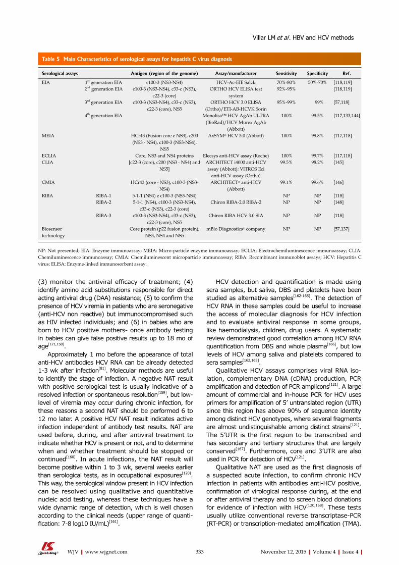
333 November 12, 2015|Volume 4|Issue 4|WJV|www.wjgnet.com
(3) monitor the antiviral efficacy of treatment; (4) identify amino acid substitutions responsible for direct acting antiviral drug (DAA) resistance; (5) to confirm the presence of HCV viremia in patients who are seronegative (anti-HCV non reactive) but immunocompromised such as HIV infected individuals; and (6) in babies who are born to HCV positive mothers- once antibody testing in babies can give false positive results up to 18 mo of age[121,158].
Approximately 1 mo before the appearance of total anti-HCV antibodies HCV RNA can be already detected 1-3 wk after infection[81]. Molecular methods are useful to identify the stage of infection. A negative NAT result with positive serological test is usually indicative of a resolved infection or spontaneous resolution[159], but low-level of viremia may occur during chronic infection, for these reasons a second NAT should be performed 6 to 12 mo later. A positive HCV NAT result indicates active infection independent of antibody test results. NAT are used before, during, and after antiviral treatment to indicate whether HCV is present or not, and to determine when and whether treatment should be stopped or continued[160]. In acute infections, the NAT result will become positive within 1 to 3 wk, several weeks earlier than serological tests, as in occupational exposures[120]. This way, the serological window present in HCV infection can be resolved using qualitative and quantitative nucleic acid testing, whereas these techniques have a wide dynamic range of detection, which is well chosen according to the clinical needs (upper range of quanti-fication: 7-8 log10 IU/mL)[161].
HCV detection and quantification is made using sera samples, but saliva, DBS and platelets have been studied as alternative samples[162-165]. The detection of HCV RNA in these samples could be useful to increase the access of molecular diagnosis for HCV infection and to evaluate antiviral response in some groups, like haemodialysis, children, drug users. A systematic review demonstrated good correlation among HCV RNA quantification from DBS and whole plasma[166], but low levels of HCV among saliva and platelets compared to sera samples[162,163].
Qualitative HCV assays comprises viral RNA iso-lation, complementary DNA (cDNA) production, PCR amplification and detection of PCR amplicons[121]. A large amount of commercial and in-house PCR for HCV uses primers for amplification of 5’ untranslated region (UTR) since this region has above 90% of sequence identity among distinct HCV genotypes, where several fragments are almost undistinguishable among distinct strains[121]. The 5’UTR is the first region to be transcribed and has secondary and tertiary structures that are largely conserved[167]. Furthermore, core and 3’UTR are also used in PCR for detection of HCV[121].
Qualitative NAT are used as the first diagnosis of a suspected acute infection, to confirm chronic HCV infection in patients with antibodies anti-HCV positive, confirmation of virological response during, at the end or after antiviral therapy and to screen blood donations for evidence of infection with HCV[120,168]. These tests usually utilize conventional reverse transcriptase-PCR (RT-PCR) or transcription-mediated amplification (TMA).
Table 5 Main Characteristics of serological assays for hepatitis C virus diagnosis
Serological assays Antigen (region of the genome) Assay/manufacturer Sensitivity Specificity Ref.
EIA 1st generation EIA c100-3 (NS3-NS4) HCV-Ac-EIE Salck 70%-80% 50%-70% [118,119]2nd generation EIA c100-3 (NS3-NS4), c33-c (NS3),
c22-3 (core)ORTHO HCV ELISA test
system92%-95% [118,119]
3rd generation EIA c100-3 (NS3-NS4), c33-c (NS3), c22-3 (core), NS5
ORTHO HCV 3.0 ELISA (Ortho)/ETI-AB-HCVK Sorin
95%-99% 99% [57,118]
4th generation EIA Monolisa™ HCV AgAb ULTRA (BioRad)/HCV Murex AgAb
(Abbott)
100% 99.5% [117,133,144]
MEIA HCr43 (Fusion core e NS3), c200 (NS3 - NS4), c100-3 (NS3-NS4),
NS5
AxSYM® HCV 3.0 (Abbott) 100% 99.8% [117,118]
ECLIA Core, NS3 and NS4 proteins Elecsys anti-HCV assay (Roche) 100% 99.7% [117,118]CLIA [c22-3 (core), c200 (NS3 - NS4) and
NS5]ARCHITECT i4000 anti-HCV assay (Abbott); VITROS Eci
anti-HCV assay (Ortho)
99.5% 98.2% [145]
CMIA HCr43 (core - NS3), c100-3 (NS3-NS4)
ARCHITECT® anti-HCV (Abbott)
99.1% 99.6% [146]
RIBA RIBA-1 5-1-1 (NS4) e c100-3 (NS3-NS4) NP NP [118]RIBA-2 5-1-1 (NS4), c100-3 (NS3-NS4),
c33-c (NS3), c22-3 (core)Chiron RIBA-2.0 RIBA-2 NP NP [148]
RIBA-3 c100-3 (NS3-NS4), c33-c (NS3), c22-3 (core), NS5
Chiron RIBA HCV 3.0 SIA NP NP [118]
Biosensor technology
Core protein (p22 fusion protein), NS3, NS4 and NS5
mBio Diagnostics® company NP NP [57,137]
NP: Not presented; EIA: Enzyme immunoassay; MEIA: Micro-particle enzyme immunoassay; ECLIA: Electrochemiluminescence immunoassay; CLIA: Chemiluminescence immunoassay; CMIA: Chemiluminescent microparticle immunoassay; RIBA: Recombinant immunoblot assays; HCV: Hepatitis C virus; ELISA: Enzyme-linked immunosorbent assay.
Villar LM et al . HBV and HCV methods

In RT-PCR-based assays, HCV RNA is the source for production of a single-stranded complementary cDNA by reverse transcriptase. DNA polymerase amplifies cDNA into multiple double-stranded DNA copies. In TMA assay, viral RNA is isolated from clinical sample and two enzymes (T7 RNA polymerase and reverse transcriptase) amplify this RNA. Hybridization protection assay detects these amplicons by probe hybridization in which only hybridized probes stay chemiluminescent and are detected in a luminometer[168].
Qualitative RT-PCR assays should detect 50 HCV RNA IU/mL or less with the same sensitivity for all genotypes. With the advent of more qPCR that has a lower limit of detection to as low as 30 copies/mL, qualitative assays were replaced especially in diagnostic laboratories[120,169]. These qualitative tests are still very common due to its higher sensitivity, but the main inconvenience is that it only verifies the absence or presence of HCV RNA[121,168].
Some qualitative commercial assays include Cobas Amplicor HCV version 2.0 (Roche Molecular Diagnostics, Pleasonton, CA, United States) and Versant HCV RNA qualitative assay (Siemens Healthcare Diagnostics, Deerfield, IL, United States). Cobas Amplicor HCV is performed according to standard RT-PCR and 50 IU/mL is the limit of detection for all HCV genotypes, while Versant HCV qualitative assay employs TMA and presents analytical sensitivity of 10 IU/mL for most genotypes and 5.3 IU/mL for genotype 1[121].
HCV RNA quantification can be accomplished by target or signal amplification, respectively quantitative RT-PCR (qRT-PCR) and branched deoxyribonucleic acid (bDNA) technology. Due to its good sensitivity (99%) and specificity (98%-99%), the classical techniques for viral genome detection and quantification are pro-gressively being replaced by quantitative PCR[120,169]. When these tests are used to monitor viral load during treatment, it is critical to use the same assay before and during therapy[120,169].
Several tests are commercially available for HCV quantification and those assays employ competitive PCR (Cobas Amplicor HCV Monitor), bDNA technique (Versant HCV RNA), real time PCR (COBAS TaqMan assay and Abbott Real Time HCV test). To quantify HCV by Cobas Amplicor HCV Monitor 2.0, the target and internal standard is amplified in a single reaction tube. The initial quantity of HCV RNA is obtained by comparing the final amounts of both templates. The dynamic range of the Amplicor™ HCV 2.0 monitor assay is 500 to approximately 500000 IU/mL with a specificity of almost 100%, independent of the HCV genotype[170,171]. The Versant HCV quantitative test (Siemens Healthcare Diagnostics) is based on signal amplification by bDNA and present a range of detection of 615 IU/mL to 8 million IU/mL independent of HCV genotype[172]. Martins et al[173] compared qualitative (in-house RT-nested PCR and COBAS AMPLICOR HCV Test v2.0 and TMA) and quantitative (COBAS AMPLICOR HCV Monitor Test v2.0 and bDNA) techniques for HCV quantification and detection, and TMA presented the highest rate (87.8%)
of HCV detection among qualitative tests being the most sensitive for HCV RNA detection over the early and late phases of HCV infection.
Real time PCR technology allows the direct moni-toring of the PCR process due to the detection and amplification of the target nucleic acid at the same time. In order to detect and amplify DNA at the same time, a probe (oligonucleotides containing a quenching molecule and a fluorescent reporter molecule) binds to target cDNA between the two PCR primers and are degraded or released by DNA polymerase during DNA synthesis. When probe is degraded, it occurs the separation of reporter and quencher molecules, which leads to emission of an increased fluorescence signal from the reporter. The quantity of RNA in the starting (first cycle) sample is proportional to the fluorescence signal. Quantification in absolute numbers is obtained by the comparison of kinetics of the target amplification with the amplification kinetics of an internal control of a defined initial concentration[168].
Cobas Taqman HCV present limit of detection and quantification of approximately 15 IU/mL among all HCV genotypes, and a linear amplification range of HCV RNA from approximately 15 to 10000000 IU/mL[174]. Abbott RealTime HCV assay has a lower detection limit of nearly 10 IU/mL, a specificity higher than 99.5% and a linear amplification range from 12 to 10000000 IU/mL independent of the hepatitis C genotype[175-177].
Currently, there are leastwise seven genotypes and more than 80 subtypes of HCV[178,179]. Methods for HCV genotyping are direct sequence analysis, real time PCR, RFLP, and reverse hybridization technology. HCV genotyping is mandatory for double antiviral treatment (interferon and ribavirin), since patients infected with genotypes 1 or 4 are treated for longer times than patients infected by genotypes 2 and 3[180]. Nevertheless, with the accessibility of new and highly effective antiviral therapies, HCV genotyping will not be important in the future.
The first commercial assays for HCV genotyping evaluated exclusively the 5’UTR, which has a high ratio of misclassification particularly on the subtype level. Nowadays commercial tests analyze coding regions, especially non-structural protein NS5B and core protein.
TRUGENE-SIEMENS HCV 5’NC Genotyping Kit (Siemens Medical Solutions Diagnostics, Tarrytown, NY, United States) is useful to detect HCV genotypes and subtypes based on region 5’UTR and has a lower limit of detection of 5000 IU/mL[181,182]. Versant HCV Genotype 2.0 Assay (LiPA) uses oligonucleotide probes specific for the 5’UTR and core regions of the six HCV genotypes and has lower limit of detection of 3700 IU/mL[183]. LIPA is efficient for HCV genotyping, but some divergent results were observed when compared to sequence analysis of the NS5B region at the subtype level (sensitivity of 95.2% for subtype 1b and 96.1% for subtype 1a)[184].
Real time PCR using TaqMan technology as Abbott Real time PCR HCV Assay (Abbott Diagnostics Europe,
334 November 12, 2015|Volume 4|Issue 4|WJV|www.wjgnet.com
Villar LM et al . HBV and HCV methods
335 November 12, 2015|Volume 4|Issue 4|WJV|www.wjgnet.com
loads with broad dynamic range. These assays are fully automated and reduced contamination. However, commercial assays present high cost compared to in house qualitative methods. For HCV genotyping, the method most useful is nucleotide sequencing, principally ultra deep sequencing that could identify resistant variants predominate in the HCV population and give information about dynamics of HCV quasispecies. How-ever, it is necessary in-depth knowledge to analyze the results.
The main characteristics, advantages, disadvantages and specific applications of HCV molecular assays are disclosed in Table 4.
CONCLUSIONIn this review, we attempted to give information regard-ing HCV and HBV serological and molecular methods available at clinical and research areas. Most of review articles regarding HBV and HCV diagnosis are relatively old[28,120,148,198] or discuss only one aspect of viral hepatitis diagnosis, for example, rapid tests for HBV[24], rapid tests for HCV[156], serological methods for HBV[49], serological methods for HCV[117], molecular methods for HBV[83] or molecular methods for HCV[120]. In the present review, both serological and molecular methods for HBV and HCV diagnosis were included and new methods such as biosensors and ultra deep sequencing were discussed giving new and updated information about this theme.
Diagnosis of HBV and HCV infection is a key tool to identify acute and chronic cases of infection in order to define preventive measures and to initiate antiviral treatment. Nowadays HBV vaccination and antiviral therapies for HBV and HCV infection have arisen drug resistant, vaccine and diagnosis escape variants that complicate diagnosis and treatment. In this situation, effective diagnosis presenting high sensitivity and speci-ficity is crucial.
Each detection method presents advantages and limitations. EIAs are the most important serological assays used for HBV and HCV detection due to its simplicity, automation and convenience. Nevertheless, they can be time-consuming and expensive and rapid assays have been developed in order to overcome these disadvantages. Molecular techniques are useful to diagnose chronic infection; to identify HBV occult cases; to evaluate the prognosis of disease; to help in treatment decisions and monitor the antiviral treat-ment efficacy; and to identify resistance mutants to antiviral treatment. Molecular methods present higher specificity and sensitivity and larger dynamic range of detection compared to other diagnostic assays like serological assays. Nevertheless, these methods are relatively expensive and require special instruments and specialized techniques. The choice of each method should be done according advantages and disadvantages and the purpose of diagnosis.
In the near future, biosensors and biochips seems to be useful technologies for serological diagnosis of
Wiesbaden, Germany) determines the viral genotype of the samples for HCV genotypes 1a, 1b, 2a, 2b, 3, 4, 5 and 6 and has limit of detection of 6053 IU/mL[185]. On the other hand, in house methods like RFLP presented a limit of detection using the Probit test ranging from 51 to 3300 IU/mL[161]. RFLP is a useful technique for HCV genotyping in different groups since presents low cost compared to commercial methods[186,187].
The direct sequencing (Sanger sequencing) is the gold standard for determining HCV genotypes and subtypes HCV. Nucleotide sequencing involves sequencing of one or more genes in the HCV genome (mainly the 5′UTR, core, E1, NS3 and NS5) and comparing these sequences to the established genotypes by computer analysis[120]. This provides the most complete information on the variations of the sequences analyzed. Furthermore, it is the most useful method for the study of viral genetic variability[182].
The sequencing technique for HCV genotyping con-sists of PCR amplification of part of the viral genome, especially these regions: The 5’NC, NS5B and core regions. These regions present diversity for the discri-mination of viral genotypes and subtypes and are sufficiently conserved for the development of reliable primers[188]. In addition to genotype determination, direct sequencing is also used for molecular epidemiological studies and to DAA resistance mutations[189-193]. In DAA treatment, the replication is intensively inhibited in drug-sensitive viral population, and the resistant variants gradually predominate in the HCV population[194]. This way, in a near future, the most appropriate treatment for HCV patients will be based on the analysis of the nucleotide or amino acid sequence[195].
Actually, deep sequencing technologies is a promising approach to characterizing viral diversity, they have the ability to generate high throughput screening that provide exceptional resolution for studying the underlying genetic diversity of complex viral populations[112,113,196]. Currently Illumina deep sequencing technology (Illu-mina Inc. San Diego, CA) and PacBio sequencing technologies (Pacific Biosciences of California, Inc.) are the newest platforms in the market. In HCV studies, deep sequencing technologies are powerful tools for obtaining more profound insight into the dynamics of variants in the HCV quasispecies of human serum. It allows sequencing the complete genome in a short time and is able to generate much more information on the viral genome sequences in internal organs[195].
Due to the rate of 10-3 mutations per nucleotide, HCV results in high-circulating quasispecies in infected patients. Recently, it was observed that approximately 15.6% of samples from Pakistan did not match any genotype[197]. Ultra deep sequencing could be useful for identification of these genotypes what is important to determine the pattern of double antiviral therapy (interferon and ribavirin) that is a standard therapy in Pakistan and other countries with few resources.
HCV quantitative methods, like qPCR are useful to monitor antiviral therapy and could detect low viral
Villar LM et al . HBV and HCV methods

336 November 12, 2015|Volume 4|Issue 4|WJV|www.wjgnet.com
HBV and HCV, principally due to real-time diagnosis and early intervention to reduce the burden of diseases. Alternative specimens and rapid assays can also be extremely useful for remote areas, low resource settings and health services with limited laboratory infrastructure. In respect to molecular HBV and HCV assays, digital PCR promises to resolve some of the deficiencies of qPCR by transforming the analog, exponential nature of PCR into a digital and linear signal. For DNA sequencing, ultra deep sequencing will be helpful for analysis of HBV and HCV mutants in order to study the dynamics of viral variants.
REFERENCES1 Takahashi T, Nakagawa S, Hashimoto T, Takahashi K, Imai M.
Large-scale isolation of Dane particles from plasma containing hepatitis B antigen and deomnstration of circular double-stranded DNA molecule extruding directly from their cores. J Immunol 1976; 117: 1392-1397 [PMID: 977955]
2 Gerlich WH, Robinson WS. Hepatitis B virus contains protein attached to the 5’ terminus of its complete DNA strand. Cell 1980; 21: 801-809 [PMID: 7438207 DOI: 10.1016/0092-8674(80)90443-2]
3 Norder H, Couroucé AM, Magnius LO. Molecular basis of hepatitis B virus serotype variations within the four major subtypes. J Gen Virol 1992; 73 (Pt 12): 3141-3145 [PMID: 1469353 DOI: 10.1099/0022-1317-73-12-3141]
4 Kramvis A. Genotypes and genetic variability of hepatitis B virus. Intervirology 2014; 57: 141-150 [PMID: 25034481 DOI: 10.1159/000360947]
5 Simmonds P. Reconstructing the origins of human hepatitis viruses. Philos Trans R Soc Lond B Biol Sci 2001; 356: 1013-1026 [PMID: 11516379 DOI: 10.1098/rstb.2001.0890]
6 Ganem D, Schneider RJ. Hepadnaviridae and their replication. In: Knipe DM, Howley PM, Chanock RM, Monath TP, Roizman B, Straus SE. Fields virology, 2001: 2703-2737
7 Lin CL, Kao JH. Hepatitis B virus genotypes and variants. Cold Spring Harb Perspect Med 2015; 5: a021436 [PMID: 25934462 DOI: 10.1101/cshperspect.a021436]
8 Choo QL, Kuo G, Weiner AJ, Overby LR, Bradley DW, Houghton M. Isolation of a cDNA clone derived from a blood-borne non-A, non-B viral hepatitis genome. Science 1989; 244: 359-362 [PMID: 2523562 DOI: 10.1126/science.2523562]
9 Choo QL, Richman KH, Han JH, Berger K, Lee C, Dong C, Gallegos C, Coit D, Medina-Selby R, Barr PJ. Genetic organization and diversity of the hepatitis C virus. Proc Natl Acad Sci USA 1991; 88: 2451-2455 [PMID: 1848704 DOI: 10.1073/pnas.88.6.2451]
10 Chiba J, Ohba H, Matsuura Y, Watanabe Y, Katayama T, Kikuchi S, Saito I, Miyamura T. Serodiagnosis of hepatitis C virus (HCV) infection with an HCV core protein molecularly expressed by a recombinant baculovirus. Proc Natl Acad Sci USA 1991; 88: 4641-4645 [PMID: 1905012 DOI: 10.1073/pnas.88.11.4641]
11 Saeed U, Waheed Y, Ashraf M. Hepatitis B and hepatitis C viruses: a review of viral genomes, viral induced host immune responses, genotypic distributions and worldwide epidemiology. Asian Pac J Trop Dis 2014; 4: 88-96 [DOI: 10.1016/S2222-1808(14)60322-4]
12 Chiba J, Ohba H, Matsuura Y, Watanabe Y, Katayama T, Kikuchi S, Saito I, Miyamura T. Serodiagnosis of hepatitis C virus (HCV) infection with an HCV core protein molecularly expressed by a recombinant baculovirus. Proc Natl Acad Sci USA 1991; 88: 4641-4645 [PMID: 1905012 DOI: 10.1073/pnas.88.11.4641]
13 World Health Organization. Hepatitis B, Fact sheet N°204. [accessed 2015 May]. Available from: URL: http//www.who.int/mediacentre/factsheets/fs204/en/
14 World Health Organization. Hepatitis C, Fact sheet N°164. [accessed 2014 May]. Available from: URL: http://www.who.int/
mediacentre/factsheets/fs164/en/15 Seeger C, Zoulim F. Hepadnaviruses. In: Fields NB. 5th ed.
Philadelphia: Lincott Williams & Wilkins, 2007: 2977-302916 Lee MH, Yang HI, Yuan Y, L’Italien G, Chen CJ. Epidemiology
and natural history of hepatitis C virus infection. World J Gastroenterol 2014; 20: 9270-9280 [PMID: 25071320 DOI: 10.3748/wjg.v20.i28.9270]
17 Ott JJ, Stevens GA, Groeger J, Wiersma ST. Global epidemiology of hepatitis B virus infection: new estimates of age-specific HBsAg seroprevalence and endemicity. Vaccine 2012; 30: 2212-2219 [PMID: 22273662 DOI: 10.1016/j.vaccine.2011.12.116]
18 Gower E, Estes C, Blach S, Razavi-Shearer K, Razavi H. Global epidemiology and genotype distribution of the hepatitis C virus infection. J Hepatol 2014; 61: S45-S57 [PMID: 25086286 DOI: 10.1016/j.jhep.2014.07.027]
19 Kretzer IF, do Livramento A, da Cunha J, Gonçalves S, Tosin I, Spada C, Treitinger A. Hepatitis C worldwide and in Brazil: silent epidemic--data on disease including incidence, transmission, prevention, and treatment. ScientificWorldJournal 2014; 2014: 827849 [PMID: 25013871 DOI: 10.1155/2014/827849]
20 Wasley A, Alter MJ. Epidemiology of hepatitis C: geographic differences and temporal trends. Semin Liver Dis 2000; 20: 1-16 [PMID: 10895428 DOI: 10.1055/s-2000-9506]
21 Hoofnagle JH. Serodiagnosis of acute viral hepatitis. Hepatology 1983; 3: 267-268 [PMID: 6299921 DOI: 10.1002/hep.1840030222]
22 Hoofnagle JH, Di Bisceglie AM. Serologic diagnosis of acute and chronic viral hepatitis. Semin Liver Dis 1991; 11: 73-83 [PMID: 1909458 DOI: 10.1055/s-2008-1040426]
23 Centers for Disease Control and Prevention. Epidemiology and Prevention of Vaccine-Preventable Diseases. Atkinson W, Hamborsky J, Wolfe S. Washington DC: Public Health Foundation, 2012: 12
24 Kao JH. Diagnosis of hepatitis B virus infection through serolo-gical and virological markers. Expert Rev Gastroenterol Hepatol 2008; 2: 553-562 [PMID: 19072403 DOI: 10.1586/17474124.2.4.553]
25 European Association For The Study Of The Liver. EASL clinical practice guidelines: Management of chronic hepatitis B virus infection. J Hepatol 2012; 57: 167-185 [PMID: 22436845 DOI: 10.1016/j.jhep.2012.02.010]
26 Martinot-Peignoux M, Lapalus M, Asselah T, Marcellin P. The role of HBsAg quantification for monitoring natural history and treatment outcome. Liver Int 2013; 33 Suppl 1: 125-132 [PMID: 23286856 DOI: 10.1111/liv.12075]
27 Maylin S, Boyd A, Delaugerre C, Zoulim F, Lavocat F, Simon F, Girard PM, Lacombe K. Comparison between Elecsys HBsAg II and architect HBsAg QT assays for quantification of hepatitis B surface antigen among patients coinfected with HIV and hepatitis B virus. Clin Vaccine Immunol 2012; 19: 242-248 [PMID: 22190396 DOI: 10.1128/CVI.05454-11]
28 Weber B. Recent developments in the diagnosis and monitoring of HBV infection and role of the genetic variability of the S gene. Expert Rev Mol Diagn 2005; 5: 75-91 [PMID: 15723594 DOI: 10.1586/14737159.5.1.75]
29 Petry A, Kupek EJ. [Effectiveness of recombinant DNA vaccines against hepatitis B in blood donors in an endemic region of South Brazil]. Rev Soc Bras Med Trop 2006; 39: 462-466 [PMID: 17160324 DOI: 10.1590/S0037-86822006000500008]
30 Tsang TK, Blei AT, O’Reilly DJ, Decker R. Clinical significance of concurrent hepatitis B surface antigen and antibody positivity. Dig Dis Sci 1986; 31: 620-624 [PMID: 3709326 DOI: 10.1007/BF01318693]
31 Dufour DR, Lott JA, Nolte FS, Gretch DR, Koff RS, Seeff LB. Diagnosis and monitoring of hepatic injury. I. Performance characteristics of laboratory tests. Clin Chem 2000; 46: 2027-2049 [PMID: 11106349]
32 Dény P, Zoulim F. Hepatitis B virus: from diagnosis to treatment. Pathol Biol (Paris) 2010; 58: 245-253 [PMID: 20580167 DOI: 10.1016/j.patbio.2010.05.002]
33 Kao JH. Hepatitis B viral genotypes: clinical relevance and molecular characteristics. J Gastroenterol Hepatol 2002; 17:
Villar LM et al . HBV and HCV methods
337 November 12, 2015|Volume 4|Issue 4|WJV|www.wjgnet.com
643-650 [PMID: 12100608 DOI: 10.1046/j.1440-1746.2002.02737.x]
34 Petersen J. Hepatitis B: Diagnostic Tests. 5th ed. Hepatology, 2014
35 Brazil Ministry of Health. 2008 Viral Hepatitis: Brazil is the careful. Health Surveillance Secretariat, Department of STD, AIDS and Viral Hepatitis, Ministry of Health, 2008 (Portuguese)
36 Diepersloot RJ, van Zantvliet-van Oostrom Y, Gleaves CA. Comparison of a chemiluminescent immunoassay with two microparticle enzyme immunoassays for detection of hepatitis B virus surface antigen. Clin Diagn Lab Immunol 2000; 7: 865-866 [PMID: 11063488 DOI: 10.1128/cdli.7.6.865-866.2000]
37 Cheng L, Guan Q, Zhang J, Sun Z. Discrepancies between two automated immunoassay systems in determining hepatitis B virus markers in serum samples with concomitant presence of antigens and antibodies. Ann Clin Lab Sci 2010; 40: 49-52 [PMID: 20124330]
38 Yalow RS, Berson SA. Assay of plasma insulin in human subjects by immunological methods. Nature 1959; 184 (Suppl 21): 1648-1649 [PMID: 13846363 DOI: 10.1038/1841648b0]
39 Annesley TM. It’s about the journey, Not the destination: The birth of radioimmunoassay. 1960. Clin Chem 2010; 56: 671-672 [PMID: 20110446 DOI: 10.1373/clinchem.2010.142950]
40 Engvall E, Perlmann P. Enzyme-linked immunosorbent assay (ELISA). Quantitative assay of immunoglobulin G. Immunochemistry 1971; 8: 871-874 [PMID: 5135623 DOI: 10.1016/0019-2791(71)90454-X]
41 Grange RD, Thompson JP, Lambert DG. Radioimmunoassay, enzyme and non-enzyme-based immunoassays. Br J Anaesth 2014; 112: 213-216 [PMID: 24431350 DOI: 10.1093/bja/aet293]
42 Scheiblauer H, El-Nageh M, Diaz S, Nick S, Zeichhardt H, Grunert HP, Prince A. Performance evaluation of 70 hepatitis B virus (HBV) surface antigen (HBsAg) assays from around the world by a geographically diverse panel with an array of HBV genotypes and HBsAg subtypes. Vox Sang 2010; 98: 403-414 [PMID: 20412171 DOI: 10.1111/j.1423-0410.2009.01272.x]
43 Forster RJ, Bertoncello P, Keyes TE. Electrogenerated chemilu-minescence. Annu Rev Anal Chem (Palo Alto Calif) 2009; 2: 359-385 [PMID: 20636067 DOI: 10.1146/annurev-anchem-060908-155305]
44 Kim H, Oh EJ, Kang MS, Kim SH, Park YJ. Comparison of the Abbott Architect i2000 assay, the Roche Modular Analytics E170 assay, and an immunoradiometric assay for serum hepatitis B virus markers. Ann Clin Lab Sci 2007; 37: 256-259 [PMID: 17709690]
45 Chen Y, Wu W, Li LJ, Lou B, Zhang J, Fan J. Comparison of the results for three automated immunoassay systems in determining serum HBV markers. Clin Chim Acta 2006; 372: 129-133 [PMID: 16713592 DOI: 10.1016/j.cca.2006.03.032]
46 Xu W, Li Y, Wang M, Gu J. Comparison of two immunoassays for determining hepatitis B virus serum markers. Clin Chem Lab Med 2012; 50: 153-157 [PMID: 21950598 DOI: 10.1515/CCLM.2011.721]
47 Huzly D, Schenk T, Jilg W, Neumann-Haefelin D. Comparison of nine commercially available assays for quantification of antibody response to hepatitis B virus surface antigen. J Clin Microbiol 2008; 46: 1298-1306 [PMID: 18256221 DOI: 10.1128/JCM.02430-07]
48 Soeung SC, Rani M, Huong V, Sarath S, Kimly C, Kohei T. Results from nationwide hepatitis B serosurvey in Cambodia using simple and rapid laboratory test: implications for National Immunization Program. Am J Trop Med Hyg 2009; 81: 252-257 [PMID: 19635879]
49 Khuroo MS, Khuroo NS, Khuroo MS. Accuracy of Rapid Point-of-Care Diagnostic Tests for Hepatitis B Surface Antigen-A Systematic Review and Meta-analysis. J Clin Exp Hepatol 2014; 4: 226-240 [PMID: 25755565 DOI: 10.1016/j.jceh.2014.07.008]
50 Randrianirina F, Carod JF, Ratsima E, Chrétien JB, Richard V, Talarmin A. Evaluation of the performance of four rapid tests for detection of hepatitis B surface antigen in Antananarivo, Madagascar. J Virol Methods 2008; 151: 294-297 [PMID: 18462816 DOI: 10.1016/j.jviromet.2008.03.019]
51 Davies J, van Oosterhout JJ, Nyirenda M, Bowden J, Moore E, Hart IJ, Zijlstra EE, Chaponda M, Faragher B, Beeching NJ, Beadsworth MB. Reliability of rapid testing for hepatitis B in a region of high HIV endemicity. Trans R Soc Trop Med Hyg 2010; 104: 162-164 [PMID: 19931107 DOI: 10.1016/j.trstmh.2009.10.010]
52 Seremba E, Ocama P, Opio CK, Kagimu M, Yuan HJ, Attar N, Thomas DL, Lee WM. Validity of the rapid strip assay test for detecting HBsAg in patients admitted to hospital in Uganda. J Med Virol 2010; 82: 1334-1340 [PMID: 20572076 DOI: 10.1002/jmv.21813]
53 Bottero J, Boyd A, Gozlan J, Lemoine M, Carrat F, Collignon A, Boo N, Dhotte P, Varsat B, Muller G, Cha O, Picard O, Nau J, Campa P, Silbermann B, Bary M, Girard PM, Lacombe K. Performance of rapid tests for detection of HBsAg and anti-HBsAb in a large cohort, France. J Hepatol 2013; 58: 473-478 [PMID: 23183527 DOI: 10.1016/j.jhep.2012.11.016]
54 Chevaliez S, Challine D, Naija H, Luu TC, Laperche S, Nadala L, Allain JP, Lee HH, Pawlotsky JM. Performance of a new rapid test for the detection of hepatitis B surface antigen in various patient populations. J Clin Virol 2014; 59: 89-93 [PMID: 24355522 DOI: 10.1016/j.jcv.2013.11.010]
55 Lau DT, Ma H, Lemon SM, Doo E, Ghany MG, Miskovsky E, Woods GL, Park Y, Hoofnagle JH. A rapid immunochro-matographic assay for hepatitis B virus screening. J Viral Hepat 2003; 10: 331-334 [PMID: 12823602]
56 El-Ghitany EM, Farghaly AG. Evaluation of commercialized rapid diagnostic testing for some Hepatitis B biomarkers in an area of intermediate endemicity. J Virol Methods 2013; 194: 190-193 [PMID: 24004823 DOI: 10.1016/j.jviromet.2013.08.026]
57 Uliana CV, Riccardi CS, Yamanaka H. Diagnostic tests for hepatitis C: recent trends in electrochemical immunosensor and genosensor analysis. World J Gastroenterol 2014; 20: 15476-15491 [PMID: 25400433 DOI: 10.3748/wjg.v20.i42.15476]
58 Malhotra BD, Turne APF. Advances in Biosensors, Elsevier Science. 5th ed. The Netherlands, 2003
59 Nakamura H, Karube I. Current research activity in biosensors. Anal Bioanal Chem 2003; 377: 446-468 [PMID: 12811457 DOI: 10.1007/s00216-003-1947-5]
60 Yao CY, Fu WL. Biosensors for hepatitis B virus detection. World J Gastroenterol 2014; 20: 12485-12492 [PMID: 25253948 DOI: 10.3748/wjg.v20.i35.12485]
61 Wang X, Li Y, Wang H, Fu Q, Peng J, Wang Y, Du J, Zhou Y, Zhan L. Gold nanorod-based localized surface plasmon resonance biosensor for sensitive detection of hepatitis B virus in buffer, blood serum and plasma. Biosens Bioelectron 2010; 26: 404-410 [PMID: 20729056 DOI: 10.1016/j.bios.2010.07.121]
62 Hu Y, Zhao Z, Wan Q. Facile preparation of carbon nanotube-conducting polymer network for sensitive electrochemical immuno-assay of Hepatitis B surface antigen in serum. Bioelectrochemistry 2011; 81: 59-64 [PMID: 21458390 DOI: 10.1016/j.bioelechem.2011.01.005]
63 Nourani S, Ghourchian H, Boutorabi SM. Magnetic nanoparticle-based immunosensor for electrochemical detection of hepatitis B surface antigen. Anal Biochem 2013; 441: 1-7 [PMID: 23831477 DOI: 10.1016/j.ab.2013.06.011]
64 Snijdewind IJ, van Kampen JJ, Fraaij PL, van der Ende ME, Osterhaus AD, Gruters RA. Current and future applications of dried blood spots in viral disease management. Antiviral Res 2012; 93: 309-321 [PMID: 22244848 DOI: 10.1016/j.antiviral.2011.12.011]
65 Mahboobi N, Porter SR, Karayiannis P, Alavian SM. Oral fluid and hepatitis A, B and C: a literature review. J Oral Pathol Med 2012; 41: 505-516 [PMID: 22188507 DOI: 10.1111/j.1600-0714.2011.01123.x]
66 Hutse V, Verhaegen E, De Cock L, Quoilin S, Vandenberghe H, Horsmans Y, Michielsen P, Van Damme P, Van Vlierberghe H, Claeys F, Vranckx R, Van Oyen H. Oral fluid as a medium for the detection of hepatitis B surface antigen. J Med Virol 2005; 77: 53-56 [PMID: 16032713 DOI: 10.1002/jmv.20413]
67 Quoilin S, Hutse V, Vandenberghe H, Claeys F, Verhaegen E, De Cock L, Van Loock F, Top G, Van Damme P, Vranckx R, Van Oyen
Villar LM et al . HBV and HCV methods

338 November 12, 2015|Volume 4|Issue 4|WJV|www.wjgnet.com
H. A population-based prevalence study of hepatitis A, B and C virus using oral fluid in Flanders, Belgium. Eur J Epidemiol 2007; 22: 195-202 [PMID: 17356926 DOI: 10.1007/s10654-007-9105-6]
68 Cruz HM, da Silva EF, Villela-Nogueira CA, Nabuco LC, do Ó KM, Lewis-Ximenez LL, Yoshida CF, Lampe E, Villar LM. Evaluation of saliva specimens as an alternative sampling method to detect hepatitis B surface antigen. J Clin Lab Anal 2011; 25: 134-141 [PMID: 21438008 DOI: 10.1002/jcla.20447]
69 Arora G, Sheikh S, Pallagatti S, Singh B, Singh VA, Singh R. Saliva as a tool in the detection of hepatitis B surface antigen in patients. Compend Contin Educ Dent 2012; 33: 174-176, 178; quiz 180, 182 [PMID: 22479783]
70 de Paula Scalioni L, Cruz HM, de Paula VS, Corrêia Oliveira J, Tourinho Dos Santos R, Motta-Castro AR, Murat PG, Villela-Nogueira CA, Lewis-Ximenez LL, Lampe E, Villar LM. Importance of collection methods and stability of oral fluid samples for hepatitis B surface antigen detection. J Clin Lab Anal 2013; 27: 186-194 [PMID: 23440736 DOI: 10.1002/jcla.21582]
71 Amado LA, Villar LM, de Paula VS, de Almeida AJ, Gaspar AM. Detection of hepatitis A, B, and C virus-specific antibodies using oral fluid for epidemiological studies. Mem Inst Oswaldo Cruz 2006; 101: 149-155 [PMID: 16830707 DOI: 10.1590/S0074-02762006000200006]
72 Forbi JC, Obagu JO, Gyar SD, Pam CR, Pennap GR, Agwale SM. Application of dried blood spot in the sero-diagnosis of hepatitis B infection (HBV) in an HBV hyper-endemic nation. Ann Afr Med 2010; 9: 44-45 [PMID: 20418650 DOI: 10.4103/1596-3519.62625]
73 Lee CE, Sri Ponnampalavanar S, Syed Omar SF, Mahadeva S, Ong LY, Kamarulzaman A. Evaluation of the dried blood spot (DBS) collection method as a tool for detection of HIV Ag/Ab, HBsAg, anti-HBs and anti-HCV in a Malaysian tertiary referral hospital. Ann Acad Med Singapore 2011; 40: 448-453 [PMID: 22206053]
74 Villar LM, de Oliveira JC, Cruz HM, Yoshida CF, Lampe E, Lewis-Ximenez LL. Assessment of dried blood spot samples as a simple method for detection of hepatitis B virus markers. J Med Virol 2011; 83: 1522-1529 [PMID: 21739441 DOI: 10.1002/jmv.22138]
75 Mohamed S, Raimondo A, Pénaranda G, Camus C, Ouzan D, Ravet S, Bourlière M, Khiri H, Dukan P, Olive D, Halfon P. Dried blood spot sampling for hepatitis B virus serology and molecular testing. PLoS One 2013; 8: e61077 [PMID: 23613788 DOI: 10.1371/journal.pone.0061077]
76 Ross RS, Stambouli O, Grüner N, Marcus U, Cai W, Zhang W, Zimmermann R, Roggendorf M. Detection of infections with hepatitis B virus, hepatitis C virus, and human immunodeficiency virus by analyses of dried blood spots--performance characteristics of the ARCHITECT system and two commercial assays for nucleic acid amplification. Virol J 2013; 10: 72 [PMID: 23497102 DOI: 10.1186/1743-422X-10-72]
77 Lukacs Z, Dietrich A, Ganschow R, Kohlschütter A, Kruithof R. Simultaneous determination of HIV antibodies, hepatitis C antibodies, and hepatitis B antigens in dried blood spots--a feasibility study using a multi-analyte immunoassay. Clin Chem Lab Med 2005; 43: 141-145 [PMID: 15843206 DOI: 10.1515/CCLM.2005.023]
78 Vallejo F, Toro C, de la Fuente L, Brugal MT, Soriano V, Silva TC, Bravo MJ, Ballesta R, Barrio G. Prevalence of and risk factors for hepatitis B virus infection among street-recruited young injection and non-injection heroin users in Barcelona, Madrid and Seville. Eur Addict Res 2008; 14: 116-124 [PMID: 18552487 DOI: 10.1159/000130415]
79 Komas NP, Vickos U, Hübschen JM, Béré A, Manirakiza A, Muller CP, Le Faou A. Cross-sectional study of hepatitis B virus infection in rural communities, Central African Republic. BMC Infect Dis 2013; 13: 286 [PMID: 23800310 DOI: 10.1186/1471-2334-13-286]
80 Whalley SA, Murray JM, Brown D, Webster GJ, Emery VC, Dusheiko GM, Perelson AS. Kinetics of acute hepatitis B virus infection in humans. J Exp Med 2001; 193: 847-854 [PMID: 11283157 DOI: 10.1084/jem.193.7.847]
81 Chevaliez S, Pawlotsky JM. Diagnosis and management of chronic viral hepatitis: antigens, antibodies and viral genomes. Best Pract Res Clin Gastroenterol 2008; 22: 1031-1048 [PMID: 19187865 DOI: 10.1016/j.bpg.2008.11.004]
82 Mangia A, Antonucci F, Brunetto M, Capobianchi M, Fagiuoli S, Guido M, Farci P, Lampertico P, Marzano A, Niro G, Pisani G, Prati D, Puoti M, Raimondo G, Santantonio T, Smedile A, Lauria F. The use of molecular assays in the management of viral hepatitis. Dig Liver Dis 2008; 40: 395-404 [PMID: 18321798 DOI: 10.1016/j.dld.2007.12.016]
83 Valsamakis A. Molecular testing in the diagnosis and management of chronic hepatitis B. Clin Microbiol Rev 2007; 20: 426-439, table of contents [PMID: 17630333 DOI: 10.1128/CMR.00009-07]
84 Caliendo AM, Valsamakis A, Bremer JW, Ferreira-Gonzalez A, Granger S, Sabatini L, Tsongalis GJ, Wang YF, Yen-Lieberman B, Young S, Lurain NS. Multilaboratory evaluation of real-time PCR tests for hepatitis B virus DNA quantification. J Clin Microbiol 2011; 49: 2854-2858 [PMID: 21697326 DOI: 10.1128/JCM.00471-11]
85 Datta S, Chatterjee S, Veer V. Recent advances in molecular diagnostics of hepatitis B virus. World J Gastroenterol 2014; 20: 14615-14625 [PMID: 25356025 DOI: 10.3748/wjg.v20.i40.14615]
86 Pawlotsky JM. [Virologic techniques for the diagnosis and mo-nitoring of hepatitis B]. Gastroenterol Clin Biol 2008; 32: S56-S63 [PMID: 18662611 DOI: 10.1016/S0399-8320(08)73266-4]
87 Kania D, Ottomani L, Meda N, Peries M, Dujols P, Bolloré K, Rénier W, Viljoen J, Ducos J, Van de Perre P, Tuaillon E. Performance of two real-time PCR assays for hepatitis B virus DNA detection and quantitation. J Virol Methods 2014; 201: 24-30 [PMID: 24560781 DOI: 10.1016/j.jviromet.2014.01.015]
88 Konnick EQ, Erali M, Ashwood ER, Hillyard DR. Evaluation of the COBAS amplicor HBV monitor assay and comparison with the ultrasensitive HBV hybrid capture 2 assay for quantification of hepatitis B virus DNA. J Clin Microbiol 2005; 43: 596-603 [PMID: 15695651 DOI: 10.1128/JCM.43.2.596-603.2005]
89 Yao JD, Beld MG, Oon LL, Sherlock CH, Germer J, Menting S, Se Thoe SY, Merrick L, Ziermann R, Surtihadi J, Hnatyszyn HJ. Multicenter evaluation of the VERSANT hepatitis B virus DNA 3.0 assay. J Clin Microbiol 2004; 42: 800-806 [PMID: 14766856 DOI: 10.1128/jcm.42.2.800-806.2004]
90 Dai CY, Yu ML, Chen SC, Lin ZY, Hsieh MY, Wang LY, Tsai JF, Chuang WL, Chang WY. Clinical evaluation of the COBAS Amplicor HBV monitor test for measuring serum HBV DNA and comparison with the Quantiplex branched DNA signal amplification assay in Taiwan. J Clin Pathol 2004; 57: 141-145 [PMID: 14747437 DOI: 10.1136/jcp.2003.10835]
91 Bustin SA, Benes V, Nolan T, Pfaffl MW. Quantitative real-time RT-PCR--a perspective. J Mol Endocrinol 2005; 34: 597-601 [PMID: 15956331 DOI: 10.1677/jme.1.01755]
92 Berger A, Gohl P, Stürmer M, Rabenau HF, Nauck M, Doerr HW. Detection and quantitation of HBV DNA in miniaturized samples: multi centre study to evaluate the performance of the COBAS ® AmpliPrep/COBAS ® TaqMan ® hepatitis B virus (HBV) test v2.0 by the use of plasma or serum specimens. J Virol Methods 2010; 169: 404-408 [PMID: 20728470 DOI: 10.1016/j.jviromet.2010.07.025]
93 Morris CJ, Hill M, de Medina M, Herman C, Cloherty GA, Martin P. Comparison of detection and quantification of HBV DNA in chronic HBeAg negative and positive patients by Abbott RealTime HBV and Roche Cobas TaqMan HBV assays. J Virol Methods 2013; 193: 391-393 [PMID: 23835030 DOI: 10.1016/j.jviromet.2013.06.036]
94 Kim H, Shin S, Oh EJ, Kahng J, Kim Y, Lee HK, Kwon HJ. Comparison of the AdvanSure HBV real-time PCR test with three other HBV DNA quantification assays. Ann Clin Lab Sci 2013; 43: 230-237 [PMID: 23694800]
95 Qiu N, Li R, Yu JG, Yang W, Zhang W, An Y, Li T, Liu XE, Zhuang H. Comparison of Abbott and Da-an real-time PCR for quantitating serum HBV DNA. World J Gastroenterol 2014; 20: 11762-11769 [PMID: 25206280 DOI: 10.3748/wjg.v20.i33.11762]
Villar LM et al . HBV and HCV methods

339 November 12, 2015|Volume 4|Issue 4|WJV|www.wjgnet.com
96 Paraskevis D, Beloukas A, Haida C, Katsoulidou A, Moschidis Z, Hatzitheodorou H, Varaklioti A, Sypsa V, Hatzakis A. Development of a new ultra sensitive real-time PCR assay (ultra sensitive RTQ-PCR) for the quantification of HBV-DNA. Virol J 2010; 7: 57 [PMID: 20226057 DOI: 10.1186/1743-422X-7-57]
97 van der Eijk AA, Niesters HG, Hansen BE, Pas SD, Richardus JH, Mostert M, Janssen HL, Schalm SW, de Man RA. Paired, quantitative measurements of hepatitis B virus DNA in saliva, urine and serum of chronic hepatitis B patients. Eur J Gastroenterol Hepatol 2005; 17: 1173-1179 [PMID: 16215428 DOI: 10.1097/00042737-200511000-00004]
98 Kidd-Ljunggren K, Holmberg A, Bläckberg J, Lindqvist B. High levels of hepatitis B virus DNA in body fluids from chronic carriers. J Hosp Infect 2006; 64: 352-357 [PMID: 17046105 DOI: 10.1016/j.jhin.2006.06.029]
99 Heiberg IL, Hoegh M, Ladelund S, Niesters HG, Hogh B. Hepatitis B virus DNA in saliva from children with chronic hepatitis B infection: implications for saliva as a potential mode of horizontal transmission. Pediatr Infect Dis J 2010; 29: 465-467 [PMID: 20335824 DOI: 10.1097/INF.0b013e3181d8e009]
100 Portilho MM, Martins PP, Lampe E, Villar LM. A comparison of molecular methods for hepatitis B virus (HBV) DNA detection from oral fluid samples. J Med Microbiol 2012; 61: 844-851 [PMID: 22403138 DOI: 10.1099/jmm.0.040238-0]
101 Jardi R, Rodriguez-Frias F, Buti M, Schaper M, Valdes A, Martinez M, Esteban R, Guardia J. Usefulness of dried blood samples for quantification and molecular characterization of HBV-DNA. Hepatology 2004; 40: 133-139 [PMID: 15239096 DOI: 10.1002/hep.20275]
102 Pourkarim MR, Amini-Bavil-Olyaee S, Kurbanov F, Van Ranst M, Tacke F. Molecular identification of hepatitis B virus genotypes/subgenotypes: revised classification hurdles and updated resolutions. World J Gastroenterol 2014; 20: 7152-7168 [PMID: 24966586 DOI: 10.3748/wjg.v20.i23.7152]
103 Yeon JE. Technique for the early detection of drug-resistant HBV DNA during antiviral therapy. Intervirology 2008; 51 Suppl 1: 7-10 [PMID: 18544942 DOI: 10.1159/000122593]
104 Amini-Bavil-Olyaee S, Tacke F, Alavian SM. HBV Subgenotypes D1, D2, D-del! Are ‘Old’ Genotyping Methods Interpreted Correctly? Hepat Mon 2013; 13: e13048 [PMID: 24066000 DOI: 10.5812/hepatmon.13048]
105 Osiowy C, Giles E. Evaluation of the INNO-LiPA HBV geno-typing assay for determination of hepatitis B virus genotype. J Clin Microbiol 2003; 41: 5473-5477 [PMID: 14662927 DOI: 10.1128/JCM.41.12.5473-5477.2003]
106 Stuyver L, Van Geyt C, De Gendt S, Van Reybroeck G, Zoulim F, Leroux-Roels G, Rossau R. Line probe assay for monitoring drug resistance in hepatitis B virus-infected patients during antiviral therapy. J Clin Microbiol 2000; 38: 702-707 [PMID: 10655370]
107 Ali MM, Hasan F, Ahmad S, Al-Nakib W. Comparative evaluation of INNO-LiPA HBV assay, direct DNA sequencing and subtractive PCR-RFLP for genotyping of clinical HBV isolates. Virol J 2010; 7: 111 [PMID: 20509964 DOI: 10.1186/1743-422X-7-111]
108 Keeffe EB, Dieterich DT, Pawlotsky JM, Benhamou Y. Chronic hepatitis B: preventing, detecting, and managing viral resistance. Clin Gastroenterol Hepatol 2008; 6: 268-274 [PMID: 18328434 DOI: 10.1016/j.cgh.2007.12.043]
109 Gintowt AA, Germer JJ, Mitchell PS, Yao JD. Evaluation of the MagNA Pure LC used with the TRUGENE HBV Genotyping Kit. J Clin Virol 2005; 34: 155-157 [PMID: 16023890 DOI: 10.1016/j.jcv.2005.05.008]
110 Basaras M, Arrese E, Blanco S, Arroyo LS, Ruiz P, Cisterna R. Comparison of INNO-LIPA and TRUGENE assays for genotyping and drug-resistance mutations in chronic hepatitis B virus infection. Intervirology 2013; 56: 190-194 [PMID: 23594698 DOI: 10.1159/000348502]
111 Kim JH, Park YK, Park ES, Kim KH. Molecular diagnosis and treatment of drug-resistant hepatitis B virus. World J Gastroenterol 2014; 20: 5708-5720 [PMID: 24914332 DOI: 10.3748/wjg.v20.i19.5708]
112 Beerenwinkel N, Zagordi O. Ultra-deep sequencing for the analysis of viral populations. Curr Opin Virol 2011; 1: 413-418 [PMID: 22440844 DOI: 10.1016/j.coviro.2011.07.008]
113 Radford AD, Chapman D, Dixon L, Chantrey J, Darby AC, Hall N. Application of next-generation sequencing technologies in virology. J Gen Virol 2012; 93: 1853-1868 [PMID: 22647373 DOI: 10.1099/vir.0.043182-0]
114 Bayliss J, Nguyen T, Lesmana CR, Bowden S, Revill P. Advances in the molecular diagnosis of hepatitis B infection: providing insight into the next generation of disease. Semin Liver Dis 2013; 33: 113-121 [PMID: 23749667 DOI: 10.1055/s-0033-1345714]
115 Rodriguez-Frias F, Buti M, Tabernero D, Homs M. Quasispecies structure, cornerstone of hepatitis B virus infection: mass sequencing approach. World J Gastroenterol 2013; 19: 6995-7023 [PMID: 24222943 DOI: 10.3748/wjg.v19.i41.6995]
116 Metzker ML. Sequencing technologies - the next generation. Nat Rev Genet 2010; 11: 31-46 [PMID: 19997069 DOI: 10.1038/nrg2626]
117 Chevaliez S, Pawlotsky JM. Hepatitis C virus serologic and virologic tests and clinical diagnosis of HCV-related liver disease. Int J Med Sci 2006; 3: 35-40 [PMID: 16614740 DOI: 10.7150/ijms.3.35]
118 Brandão AB, Fuchs SC, Silva MA, Emer LF. [Diagnosis of hepatitis C in clinical practice: review of the literature]. Rev Panam Salud Publica 2001; 9: 161-168 [PMID: 11349351 DOI: 10.1590/S1020-49892001000300005]
119 Aach RD, Stevens CE, Hollinger FB, Mosley JW, Peterson DA, Taylor PE, Johnson RG, Barbosa LH, Nemo GJ. Hepatitis C virus infection in post-transfusion hepatitis. An analysis with first- and second-generation assays. N Engl J Med 1991; 325: 1325-1329 [PMID: 1656258 DOI: 10.1056/NEJM199111073251901]
120 Scott JD, Gretch DR. Molecular diagnostics of hepatitis C virus infection: a systematic review. JAMA 2007; 297: 724-732 [PMID: 17312292 DOI: 10.1001/jama.297.7.724]
121 Firdaus R, Saha K, Biswas A, Sadhukhan PC. Current molecular methods for the detection of hepatitis C virus in high risk group population: A systematic review. World J Virol 2015; 4: 25-32 [PMID: 25674515 DOI: 10.5501/wjv.v4.i1.25]
122 Alter HJ. New kit on the block: evaluation of second-generation assays for detection of antibody to the hepatitis C virus. Hepatology 1992; 15: 350-353 [PMID: 1310478 DOI: 10.1002/hep.1840150228]
123 Lee SR, Wood CL, Lane MJ, Francis B, Gust C, Higgs CM, Nelles MJ, Polito A, DiNello R, Achord D. Increased detection of hepatitis C virus infection in commercial plasma donors by a third-generation screening assay. Transfusion 1995; 35: 845-849 [PMID: 7570915 DOI: 10.1046/j.1537-2995.1995.351096026366.x]
124 Colin C, Lanoir D, Touzet S, Meyaud-Kraemer L, Bailly F, Trepo C. Sensitivity and specificity of third-generation hepatitis C virus antibody detection assays: an analysis of the literature. J Viral Hepat 2001; 8: 87-95 [PMID: 11264728 DOI: 10.1046/j.1365-2893.2001.00280.x]
125 Albeldawi M, Ruiz-Rodriguez E, Carey WD. Hepatitis C virus: Prevention, screening, and interpretation of assays. Cleve Clin J Med 2010; 77: 616-626 [PMID: 20810872 DOI: 10.3949/ccjm.77a.09162]
126 Klimashevskaya S, Obriadina A, Ulanova T, Bochkova G, Burkov A, Araujo A, Stramer SL, Tobler LH, Busch MP, Fields HA. Distinguishing acute from chronic and resolved hepatitis C virus (HCV) infections by measurement of anti-HCV immunoglobulin G avidity index. J Clin Microbiol 2007; 45: 3400-3403 [PMID: 17715377 DOI: 10.1128/JCM.01012-07]
127 Gaudy-Graffin C, Lesage G, Kousignian I, Laperche S, Girault A, Dubois F, Goudeau A, Barin F. Use of an anti-hepatitis C virus (HCV) IgG avidity assay to identify recent HCV infection. J Clin Microbiol 2010; 48: 3281-3287 [PMID: 20610669 DOI: 10.1128/JCM.00303-10]
128 Coppola N, Pisapia R, Marrocco C, Martini S, Vatiero LM, Messina V, Tonziello G, Sagnelli C, Filippini P, Piccinino F, Sagnelli E. Anti-HCV IgG avidity index in acute hepatitis C. J Clin Virol 2007; 40: 110-115 [PMID: 17720621 DOI: 10.1016/j.jcv.2007.07.005]
Villar LM et al . HBV and HCV methods
340 November 12, 2015|Volume 4|Issue 4|WJV|www.wjgnet.com
129 Shepherd SJ, Kean J, Hutchinson SJ, Cameron SO, Goldberg DJ, Carman WF, Gunson RN, Aitken C. A hepatitis C avidity test for determining recent and past infections in both plasma and dried blood spots. J Clin Virol 2013; 57: 29-35 [PMID: 23369886 DOI: 10.1016/j.jcv.2013.01.002]
130 Croom HA, Richards KM, Best SJ, Francis BH, Johnson EI, Dax EM, Wilson KM. Commercial enzyme immunoassay adapted for the detection of antibodies to hepatitis C virus in dried blood spots. J Clin Virol 2006; 36: 68-71 [PMID: 16426889 DOI: 10.1016/j.jcv.2005.12.002]
131 Tuaillon E, Mondain AM, Meroueh F, Ottomani L, Picot MC, Nagot N, Van de Perre P, Ducos J. Dried blood spot for hepatitis C virus serology and molecular testing. Hepatology 2010; 51: 752-758 [PMID: 20043287 DOI: 10.1002/hep.23407]
132 Marques BL, Brandão CU, Silva EF, Marques VA, Villela-Nogueira CA, Do Ó KM, de Paula MT, Lewis-Ximenez LL, Lampe E, Villar LM. Dried blood spot samples: optimization of commercial EIAs for hepatitis C antibody detection and stability under different storage conditions. J Med Virol 2012; 84: 1600-1607 [PMID: 22930508 DOI: 10.1002/jmv.23379]
133 Brandão CP, Marques BL, Marques VA, Villela-Nogueira CA, Do Ó KM, de Paula MT, Lewis-Ximenez LL, Lampe E, Sá Ferreira JA, Villar LM. Simultaneous detection of hepatitis C virus antigen and antibodies in dried blood spots. J Clin Virol 2013; 57: 98-102 [PMID: 23518440 DOI: 10.1016/j.jcv.2013.02.014]
134 Cruz HM, Marques VA, Villela-Nogueira CA, do Ó KM, Lewis-Ximenez LL, Lampe E, Villar LM. An evaluation of different saliva collection methods for detection of antibodies against hepatitis C virus (anti-HCV). J Oral Pathol Med 2012; 41: 793-800 [PMID: 22690929 DOI: 10.1111/j.1600-0714.2012.01176.x]
135 Wiwanitkit V. Saliva collection methods for detection of anti-HCV. J Oral Pathol Med 2013; 42: 113 [PMID: 22943095 DOI: 10.1111/jop.12001]
136 Tejada-Strop A, Drobeniuc J, Mixson-Hayden T, Forbi JC, Le NT, Li L, Mei J, Terrault N, Kamili S. Disparate detection outcomes for anti-HCV IgG and HCV RNA in dried blood spots. J Virol Methods 2015; 212: 66-70 [PMID: 25445800 DOI: 10.1016/j.jviromet.2014.10.018]
137 Pei X, Zhang B, Tang J, Liu B, Lai W, Tang D. Sandwich-type immunosensors and immunoassays exploiting nanostructure labels: A review. Anal Chim Acta 2013; 758: 1-18 [PMID: 23245891 DOI: 10.1016/j.aca.2012.10.060]
138 Zanetti AR, Romanò L, Brunetto M, Colombo M, Bellati G, Tackney C. Total HCV core antigen assay: a new marker of hepatitis C viremia for monitoring the progress of therapy. J Med Virol 2003; 70: 27-30 [PMID: 12629640 DOI: 10.1002/jmv.10355]
139 Park Y, Lee JH, Kim BS, Kim do Y, Han KH, Kim HS. New automated hepatitis C virus (HCV) core antigen assay as an alternative to real-time PCR for HCV RNA quantification. J Clin Microbiol 2010; 48: 2253-2256 [PMID: 20351215 DOI: 10.1128/JCM.01856-09]
140 Moscato GA, Giannelli G, Grandi B, Pieri D, Marsi O, Guarducci I, Batini I, Altomare E, Antonaci S, Capria A, Pellegrini G, Sacco R. Quantitative determination of hepatitis C core antigen in therapy monitoring for chronic hepatitis C. Intervirology 2011; 54: 61-65 [PMID: 20829601 DOI: 10.1159/000318878]
141 Tillmann HL. Hepatitis C virus core antigen testing: role in diagnosis, disease monitoring and treatment. World J Gastroenterol 2014; 20: 6701-6706 [PMID: 24944462 DOI: 10.3748/wjg.v20.i22.6701]
142 Larrat S, Bourdon C, Baccard M, Garnaud C, Mathieu S, Quesada JL, Signori-Schmuck A, Germi R, Blanc M, Leclercq P, Hilleret MN, Leroy V, Zarski JP, Morand P. Performance of an antigen-antibody combined assay for hepatitis C virus testing without venipuncture. J Clin Virol 2012; 55: 220-225 [PMID: 22901327 DOI: 10.1016/j.jcv.2012.07.016]
143 Vanhommerig JW, van de Laar TJ, Koot M, van Rooijen MS, Schinkel J, Speksnijder AG, Prins M, de Vries HJ, Bruisten SM. Evaluation of a hepatitis C virus (HCV) antigen assay for routine HCV screening among men who have sex with men infected with
HIV. J Virol Methods 2015; 213: 147-150 [PMID: 25528203 DOI: 10.1016/j.jviromet.2014.11.026]
144 El-Emshaty WM, Raafat D, Elghannam DM, Saudy N, Eltoraby EE, Metwalli AE. Diagnostic Performance of an Immunoassay for Simultaneous Detection of Hcv Core Antigen and Antibodies among Haemodialysis Patients. Braz J Microbiol 2011; 42: 303-309 [PMID: 24031636 DOI: 10.1590/S1517-83822011000100039]
145 Ismail N, Fish GE, Smith MB. Laboratory evaluation of a fully automated chemiluminescence immunoassay for rapid detection of HBsAg, antibodies to HBsAg, and antibodies to hepatitis C virus. J Clin Microbiol 2004; 42: 610-617 [PMID: 14766824 DOI: 10.1128/JCM.42.2.610-617.2004]
146 Jonas G, Pelzer C, Beckert C, Hausmann M, Kapprell HP. Performance characteristics of the ARCHITECT anti-HCV assay. J Clin Virol 2005; 34: 97-103 [PMID: 16122974 DOI: 10.1016/j.jcv.2005.08.001]
147 Saludes V, González V, Planas R, Matas L, Ausina V, Martró E. Tools for the diagnosis of hepatitis C virus infection and hepatic fibrosis staging. World J Gastroenterol 2014; 20: 3431-3442 [PMID: 24707126 DOI: 10.3748/wjg.v20.i13.3431]
148 Lok AS, Gunaratnam NT. Diagnosis of hepatitis C. Hepatology 1997; 26: 48S-56S [PMID: 9305664 DOI: 10.1002/hep.510260709]
149 Heiat M, Ranjbar R, Alavian SM. Classical and modern approaches used for viral hepatitis diagnosis. Hepat Mon 2014; 14: e17632 [PMID: 24829586 DOI: 10.5812/hepatmon.17632]
150 Ngom B, Guo Y, Wang X, Bi D. Development and application of lateral flow test strip technology for detection of infectious agents and chemical contaminants: a review. Anal Bioanal Chem 2010; 397: 1113-1135 [PMID: 20422164 DOI: 10.1007/s00216-010-3661-4]
151 Parisi MR, Soldini L, Vidoni G, Mabellini C, Belloni T, Brignolo L, Negri S, Schlusnus K, Dorigatti F, Lazzarin A. Point-of-care testing for HCV infection: recent advances and implications for alternative screening. New Microbiol 2014; 37: 449-457 [PMID: 25387283]
152 Kim MH, Kang SY, Lee WI. Evaluation of a new rapid test kit to detect hepatitis C virus infection. J Virol Methods 2013; 193: 379-382 [PMID: 23871756 DOI: 10.1016/j.jviromet.2013.07.005]
153 Tagny CT, Mbanya D, Murphy EL, Lefrère JJ, Laperche S. Screening for hepatitis C virus infection in a high prevalence country by an antigen/antibody combination assay versus a rapid test. J Virol Methods 2014; 199: 119-123 [PMID: 24487098 DOI: 10.1016/j.jviromet.2014.01.002]
154 Scalioni Lde P, Cruz HM, de Paula VS, Miguel JC, Marques VA, Villela-Nogueira CA, Milagres FA, Cruz MS, Bastos FI, Andrade TM, Motta-Castro AR, Lewis-Ximenez LL, Lampe E, Villar LM. Performance of rapid hepatitis C virus antibody assays among high- and low-risk populations. J Clin Virol 2014; 60: 200-205 [PMID: 24794796 DOI: 10.1016/j.jcv.2014.04.001]
155 Kant J, Möller B, Heyne R, Herber A, Böhm S, Maier M, Liebert UG, Mössner J, Berg T, Wiegand J. Evaluation of a rapid on-site anti-HCV test as a screening tool for hepatitis C virus infection. Eur J Gastroenterol Hepatol 2013; 25: 416-420 [PMID: 23211286 DOI: 10.1097/MEG.0b013e32835c502d]
156 Khuroo MS, Khuroo NS, Khuroo MS. Diagnostic accuracy of point-of-care tests for hepatitis C virus infection: a systematic review and meta-analysis. PLoS One 2015; 10: e0121450 [PMID: 25816332 DOI: 10.1371/journal.pone.0121450]
157 Center for Disease Control and Prevention. Testing for HCV Infection: An Update of Guidance for Clinicians and Laboratorians. [Updated 2013 May 10; accessed 2015 March 07]. Available from: URL: http://www.cdc.gov/mmwr/preview/mmwrhtml/mm6218a5.htm
158 Pawlotsky JM. Use and interpretation of virological tests for hepatitis C. Hepatology 2002; 36: S65-S73 [PMID: 12407578 DOI: 10.1053/jhep.2002.36815]
159 Scott JD, McMahon BJ, Bruden D, Sullivan D, Homan C, Christensen C, Gretch DR. High rate of spontaneous negativity for hepatitis C virus RNA after establishment of chronic infection in Alaska Natives. Clin Infect Dis 2006; 42: 945-952 [PMID: 16511757 DOI: 10.1086/500938]
Villar LM et al . HBV and HCV methods

341 November 12, 2015|Volume 4|Issue 4|WJV|www.wjgnet.com
160 Amjad M, Moudgal V, Faisal M. Laboratory Methods for Diagnosis and Management of Hepatitis C Virus Infection. Lab Medicine 2013; 44: 292-299 [DOI: 10.1309/LMASROYD8BRS0GC9]
161 Casanova YS, Boeira Tda R, Sisti E, Celmer Á, Fonseca AS, Ikuta N, Simon D, Lunge VR. A complete molecular biology assay for hepatitis C virus detection, quantification and genotyping. Rev Soc Bras Med Trop 2014; 47: 287-294 [PMID: 25075478 DOI: 10.1590/0037-8682-0040-2014]
162 Menezes GB, Pereira FA, Duarte CA, Carmo TM, Silva Filho HP, Zarife MA, Krieger MA, Reis EA, Reis MG. Hepatitis C virus quantification in serum and saliva of HCV-infected patients. Mem Inst Oswaldo Cruz 2012; 107: 680-683 [PMID: 22850960 DOI: 10.1590/S0074-02762012000500016]
163 Espírito-Santo MP, Brandão-Mello CE, Marques VA, Lampe E, Almeida AJ. Analysis of hepatitis C virus (HCV) RNA load in platelets of HCV-monoinfected patients receiving antiviral therapy. Ann Hepatol 2013; 12: 373-379 [PMID: 23619253]
164 Feijoo J, Eirea M, Limeres J, Abeleira M, Ramos I, Ocampo A, Diz P. HCV clearance from saliva of HIV-HCV-coinfected patients on treatment with interferon plus ribavirin. Oral Dis 2014; 20: 313-318 [PMID: 23607445 DOI: 10.1111/odi.12116]
165 Dokubo EK, Evans J, Winkelman V, Cyrus S, Tobler LH, Asher A, Briceno A, Page K. Comparison of Hepatitis C Virus RNA and antibody detection in dried blood spots and plasma specimens. J Clin Virol 2014; 59: 223-227 [PMID: 24529844 DOI: 10.1016/j.jcv.2014.01.014]
166 Greenman J, Roberts T, Cohn J, Messac L. Dried blood spot in the genotyping, quantification and storage of HCV RNA: a systematic literature review. J Viral Hepat 2015; 22: 353-361 [PMID: 25367722 DOI: 10.1111/jvh.12345]
167 Bartenschlager R, Cosset FL, Lohmann V. Hepatitis C virus replication cycle. J Hepatol 2010; 53: 583-585 [PMID: 20579761 DOI: 10.1016/j.jhep.2010.04.015]
168 Lange C, Sarrazin C. Hepatitis C: Diagnostic tests. 5th ed. In: Mauss S, Berg T, Rockstroh J, Sarrazin C, Wedemeyer H. Germany: Druckhaus Süd, 2014: 196-212
169 Albertoni G, Castelo Girão MJ, Schor N. Mini review: current molecular methods for the detection and quantification of hepatitis B virus, hepatitis C virus, and human immunodeficiency virus type 1. Int J Infect Dis 2014; 25: 145-149 [PMID: 24927665 DOI: 10.1016/j.ijid.2014.04.007]
170 Konnick EQ, Erali M, Ashwood ER, Hillyard DR. Performance characteristics of the COBAS Amplicor Hepatitis C Virus (HCV) Monitor, Version 2.0, International Unit assay and the National Genetics Institute HCV Superquant assay. J Clin Microbiol 2002; 40: 768-773 [PMID: 11880391 DOI: 10.1128/JCM.40.3.768-773.2002]
171 Lee SC, Antony A, Lee N, Leibow J, Yang JQ, Soviero S, Gutekunst K, Rosenstraus M. Improved version 2.0 qualitative and quantitative AMPLICOR reverse transcription-PCR tests for hepatitis C virus RNA: calibration to international units, enhanced genotype reactivity, and performance characteristics. J Clin Microbiol 2000; 38: 4171-4179 [PMID: 11060086]
172 Desombere I, Van Vlierberghe H, Couvent S, Clinckspoor F, Leroux-Roels G. Comparison of qualitative (COBAS AMPLICOR HCV 2.0 versus VERSANT HCV RNA) and quantitative (COBAS AMPLICOR HCV monitor 2.0 versus VERSANT HCV RNA 3.0) assays for hepatitis C virus (HCV) RNA detection and quantification: impact on diagnosis and treatment of HCV infections. J Clin Microbiol 2005; 43: 2590-2597 [PMID: 15956369 DOI: 10.1128/JCM.43.6.2590-2597.2005]
173 Martins PP, Lampe E, Lewis-Ximenez LL, de Souza PS, Fernandes CA, Villar LM. Performance of molecular methods for hepatitis C virus diagnosis: usefulness among chronic cases and during the course of infection. Clin Lab 2013; 59: 1031-1039 [PMID: 24273925]
174 Zitzer H, Heilek G, Truchon K, Susser S, Vermehren J, Sizmann D, Cobb B, Sarrazin C. Second-generation Cobas AmpliPrep/Cobas TaqMan HCV quantitative test for viral load monitoring: a novel
dual-probe assay design. J Clin Microbiol 2013; 51: 571-577 [PMID: 23241371 DOI: 10.1128/JCM.01784-12]
175 Michelin BD, Muller Z, Stelzl E, Marth E, Kessler HH. Evaluation of the Abbott RealTime HCV assay for quantitative detection of hepatitis C virus RNA. J Clin Virol 2007; 38: 96-100 [PMID: 17185031 DOI: 10.1016/j.jcv.2006.11.007]
176 Sábato MF, Shiffman ML, Langley MR, Wilkinson DS, Ferreira-Gonzalez A. Comparison of performance characteristics of three real-time reverse transcription-PCR test systems for detection and quantification of hepatitis C virus. J Clin Microbiol 2007; 45: 2529-2536 [PMID: 17567786 DOI: 10.1128/JCM.00058-07]
177 Vermehren J, Kau A, Gärtner BC, Göbel R, Zeuzem S, Sarrazin C. Differences between two real-time PCR-based hepatitis C virus (HCV) assays (RealTime HCV and Cobas AmpliPrep/Cobas TaqMan) and one signal amplification assay (Versant HCV RNA 3.0) for RNA detection and quantification. J Clin Microbiol 2008; 46: 3880-3891 [PMID: 18799708 DOI: 10.1128/JCM.00755-08]
178 Murphy DG, Willems B, Deschênes M, Hilzenrat N, Mousseau R, Sabbah S. Use of sequence analysis of the NS5B region for routine genotyping of hepatitis C virus with reference to C/E1 and 5’ untranslated region sequences. J Clin Microbiol 2007; 45: 1102-1112 [PMID: 17287328 DOI: 10.1128/JCM.02366-06]
179 Smith DB, Bukh J, Kuiken C, Muerhoff AS, Rice CM, Stapleton JT, Simmonds P. Expanded classification of hepatitis C virus into 7 genotypes and 67 subtypes: updated criteria and genotype assignment web resource. Hepatology 2014; 59: 318-327 [PMID: 24115039 DOI: 10.1002/hep.26744]
180 Hoofnagle JH. Course and outcome of hepatitis C. Hepatology 2002; 36: S21-S29 [PMID: 12407573 DOI: 10.1053/jhep.2002.36227]
181 Nolte FS, Green AM, Fiebelkorn KR, Caliendo AM, Sturchio C, Grunwald A, Healy M. Clinical evaluation of two methods for genotyping hepatitis C virus based on analysis of the 5’ noncoding region. J Clin Microbiol 2003; 41: 1558-1564 [PMID: 12682145 DOI: 10.1128/JCM.41.4.1558-1564.2003]
182 Chevaliez S, Pawlotsky JM. Hepatitis C virus: virology, diagnosis and management of antiviral therapy. World J Gastroenterol 2007; 13: 2461-2466 [PMID: 17552030 DOI: 10.3748/wjg.v13.i17.2461]
183 Enache EL, Enache LS. Versant HCV Genotype 2.0 Assay (LiPA) in Hepatitis C Virus Genotype Determination. Rev Romana Med Labor 2008; 12: 47-53
184 Espírito-Santo MP, Carneiro MA, Reis NR, Kozlowski AG, Teles SA, Lampe E, Yoshida CF, Martins RM. Genotyping hepatitis C virus from hemodialysis patients in Central Brazil by line probe assay and sequence analysis. Braz J Med Biol Res 2007; 40: 545-550 [PMID: 17401498 DOI: 10.1590/S0100-879X2007000400013]
185 Raymond HW, Cimmins C. Evaluation of the Abbott Molecular Diagnostics Real Time PCR Assay for HCV Quantitative Viral Load and HCV Genotyping. Poster S30, Clinical Virology Symposium, 2004
186 Oliveira ML, Bastos FI, Sabino RR, Paetzold U, Schreier E, Pauli G, Yoshida CF. Distribution of HCV genotypes among different exposure categories in Brazil. Braz J Med Biol Res 1999; 32: 279-282 [PMID: 10347784]
187 Villar LM, Amado LA, de Almeida AJ, de Paula VS, Lewis-Ximenez LL, Lampe E. Low prevalence of hepatitis B and C virus markers among children and adolescents. Biomed Res Int 2014; 2014: 324638 [PMID: 25093164 DOI: 10.1155/2014/324638]
188 Cavalheiro NP. Hepatitis C: Genotyping. BJID 2007; 11 Suppl 1: 25-27 [DOI: 10.1590/S1413-86702007000700009]
189 Lampe E, Espirito-Santo MP, Martins RM, Bello G. Epidemic history of Hepatitis C virus in Brazil. Infect Genet Evol 2010; 10: 886-895 [PMID: 20663735 DOI: 10.1016/j.meegid.2010.05.010]
190 Peres-da-Silva A, de Almeida AJ, Lampe E. Mutations in hepatitis C virus NS3 protease domain associated with resistance to specific protease inhibitors in antiviral therapy naïve patients. Arch Virol 2010; 155: 807-811 [PMID: 20405151 DOI: 10.1007/s00705-010-0642-z]
191 Lampe E, Lewis-Ximenez L, Espírito-Santo MP, Delvaux NM, Pereira SA, Peres-da-Silva A, Martins RM, Soares MA, Santos
Villar LM et al . HBV and HCV methods

342 November 12, 2015|Volume 4|Issue 4|WJV|www.wjgnet.com
AF, Vidal LL, Germano FN, de Martinez AM, Basso R, Pinho JR, Malta FM, Gomes-Gouvêa M, Moliterno RA, Bertolini DA, Fujishima MA, Bello G. Genetic diversity of HCV in Brazil. Antivir Ther 2013; 18: 435-444 [PMID: 23792792 DOI: 10.3851/IMP2606]
192 Feeney ER, Chung RT. Antiviral treatment of hepatitis C. BMJ 2014; 348: g3308 [PMID: 25002352 DOI: 10.1136/bmj.g3308]
193 Peres-da-Silva A, de Almeida AJ, Lampe E. NS5A inhibitor resistance-associated polymorphisms in Brazilian treatment-naive patients infected with genotype 1 hepatitis C virus. J Antimicrob Chemother 2015; 70: 726-730 [PMID: 25414201 DOI: 10.1093/jac/dku462]
194 Clavel F, Hance AJ. HIV drug resistance. N Engl J Med 2004; 350: 1023-1035 [PMID: 14999114 DOI: 10.1056/NEJMra025195]
195 Ninomiya M, Ueno Y, Funayama R, Nagashima T, Nishida
Y, Kondo Y, Inoue J, Kakazu E, Kimura O, Nakayama K, Shimosegawa T. Use of illumina deep sequencing technology to differentiate hepatitis C virus variants. J Clin Microbiol 2012; 50: 857-866 [PMID: 22205816 DOI: 10.1128/JCM.05715-11]
196 Mangul S, Wu NC, Mancuso N, Zelikovsky A, Sun R, Eskin E. Accurate viral population assembly from ultra-deep sequencing data. Bioinformatics 2014; 30: i329-i337 [PMID: 24932001 DOI: 10.1093/bioinformatics/btu295]
197 Afzal MS, Khan MY, Ammar M, Anjum S, Zaidi NU. Diagnostically untypable hepatitis C virus variants: it is time to resolve the problem. World J Gastroenterol 2014; 20: 17690-17692 [PMID: 25516688 DOI: 10.3748/wjg.v20.i46.17690]
198 Chakravarty R. Diagnosis and monitoring of chronic viral hepatitis: serologic and molecular markers. Front Biosci (Schol Ed) 2011; 3: 156-167 [PMID: 21196366 DOI: 10.2741/s141]
P- Reviewer: Afzal MS, Chiang TA, Farzin R S- Editor: Qiu S L- Editor: A E- Editor: Liu SQ
Villar LM et al . HBV and HCV methods

Ga Young Lee, Kittiyod Poovorawan, Duangnapa Intharasongkroh, Pattaratida Sa-nguanmoo, Sompong Vongpunsawad, Chintana Chirathaworn, Yong Poovorawan
Ga Young Lee, Kittiyod Poovorawan, Department of Clinical Tropical Medicine, Faculty of Tropical Medicine, Mahidol University, Bangkok 10400, Thailand
Duangnapa Intharasongkroh, Pattaratida Sa-nguanmoo, Sompong Vongpunsawad, Yong Poovorawan, Center of Excellence in Clinical Virology, Department of Pediatrics, Faculty of Medicine, Chulalongkorn University, Bangkok 10330, Thailand
Duangnapa Intharasongkroh, National Blood Centre, Thai Red Cross Society, Bangkok 10330, Thailand
Chintana Chirathaworn, Department of Microbiology, Faculty of Medicine, Chulalongkorn University, Bangkok 10330, Thailand
Author contributions: Poovorawan Y designed and outlined the research; Lee GY, Poovorawan K, Intharasongkroh D, Sa-nguanmoo P, Vongpunsawad S and Chirathaworn C wrote the paper.
Supported by The National Research University Project, Office of Higher Education Commission, Nos. WCU001-HR-57, WCU007-HR-57, and WCU-58-006-HR; The National Research Council of Thailand (NRCT); The Research Chair Grant from the National Science and Technology Development Agency, Chulalongkorn University Centenary Academic Development Project, No. CU56-HR01; Ratchadaphiseksomphot Endowment Fund of Chulalongkorn University, No. RES560530093; The Outstanding Professor of Thailand Research Fund, No. DPG5480002; The Doctoral Degree Chulalongkorn University 100th Year Birth-day Anniversary to Duangnapa Intharasongkroh; and The Rachadapisek Sompote Fund of Chulalongkorn University for Postdoctoral Fellowship to Pattaratida Sa-nguanmoo.
Conflict-of-interest statement: The authors declare no conflicts of interests for this article.
Open-Access: This article is an open-access article which was selected by an in-house editor and fully peer-reviewed by external reviewers. It is distributed in accordance with the Creative Commons Attribution Non Commercial (CC BY-NC 4.0) license,
which permits others to distribute, remix, adapt, build upon this work non-commercially, and license their derivative works on different terms, provided the original work is properly cited and the use is non-commercial. See: http://creativecommons.org/licenses/by-nc/4.0/ Correspondence to: Yong Poovorawan, MD, Professor, Center of Excellence in Clinical Virology, Department of Pediatrics, Faculty of Medicine, Chulalongkorn University, Rama Ⅳ Rd, Bangkok 10330, Thailand. [email protected]: +66-2-2564909 Fax: +66-2-2564929
Received: May 25, 2015Peer-review started: May 26, 2015First decision: July 27, 2015Revised: August 6, 2015Accepted: September 16, 2015Article in press: September 18, 2015Published online: November 12, 2015
AbstractHepatitis E virus (HEV) infection is now established as an emerging enteric viral hepatitis. Standard treatments in acute and chronic hepatitis E remain to be established. This study undertakes a review of the epidemiology, treatment implication and vaccine prevention from published literature. HEV infection is a worldwide public health problem and can cause acute and chronic hepatitis E. HEV genotypes 1 and 2 are primarily found in developing countries due to waterborne transmission, while the zoonotic potential of genotypes 3 and 4 affects mostly industrialized countries. An awareness of HEV transmission through blood donation, especially in the immunocompromised and solid organ transplant patients, merits an effective anti-viral therapy. There are currently no clear indications for the treatment of acute hepatitis E. Despite concerns for side effects, ribavirin monotherapy or in combination with pegylated
REVIEW
343 November 12, 2015|Volume 4|Issue 4|WJV|www.wjgnet.com
Hepatitis E virus infection: Epidemiology and treatment implications
Submit a Manuscript: http://www.wjgnet.com/esps/Help Desk: http://www.wjgnet.com/esps/helpdesk.aspxDOI: 10.5501/wjv.v4.i4.343
World J Virol 2015 November 12; 4(4): 343-355ISSN 2220-3249 (online)
© 2015 Baishideng Publishing Group Inc. All rights reserved.
World Journal of VirologyW J V

interferon alpha for at least 3 mo appeared to show significant efficacy in the treatment of chronic hepatitis E. However, there are no available treatment options for specific patient population groups, such as women who are pregnant. Vaccination and screening of HEV in blood donors are currently a global priority in managing infection. New strategies for the treatment and control of hepatitis E are required for both acute and chronic infections, such as prophylactic use of medications, controlling large outbreaks, and finding acceptable antiviral therapy for pregnant women and other patient groups for whom the current options of treatment are not viable.
Key words: Treatments; Blood donors; Adverse effects; Vaccination; Pegylated-interferon; Ribavirin; Hepatitis E
© The Author(s) 2015. Published by Baishideng Publishing Group Inc. All rights reserved.
Core tip: Hepatitis E virus (HEV) infection affects indivi-duals in both industrialized and developing countries and can cause acute and chronic hepatitis E. HEV genotypes 1 and 2 are primarily found in developing countries due to waterborne transmission, while the zoonotic potential of genotypes 3 and 4 affects mostly industrialized countries. An awareness of HEV transmission through blood donation, especially in the immunocompromised and solid organ transplant patients, merits an effective anti-viral therapy. The current treatment for HEV infection involving ribavirin and pegylated interferon-α therapy has shown limited efficacy. Although not widely used, an HEV vaccine is available for immunization in China.
Lee GY, Poovorawan K, Intharasongkroh D, Sa-nguanmoo P, Vongpunsawad S, Chirathaworn C, Poovorawan Y. Hepatitis E virus infection: Epidemiology and treatment implications. World J Virol 2015; 4(4): 343-355 Available from: URL: http://www.wjgnet.com/2220-3249/full/v4/i4/343.htm DOI: http://dx.doi.org/10.5501/wjv.v4.i4.343
INTRODUCTIONHepatitis E infection is now regarded as a major cause of fecal-orally transmitted non-A, non-B hepatitis. Hepatitis E virus (HEV) is a non-enveloped, single-stranded RNA virus that contains three open reading frames (ORFs) that encode structural and non-structural proteins[1]. There are 4 major HEV genotypes (1, 2, 3 and 4). Symptomatic HEV infections produce varying clinical presentations depending on the HEV genotypes. Fulminant hepatic failures are caused mainly by genotype 1, while chronic infections have been observed only with genotype 3 thus far[2]. Recent reports of sporadic cases from developed countries in Europe and in the United States resulted from HEV genotype 3, which is zoonotic[3]. In a 2015 survey on the seroprevalence and
risk factors for HEV infection among slaughterhouse workers in South Korea, the seropositive rate for HEV IgG was 33.5%[4]. Another report from Thailand in 2014 found that HEV strains identified from acute symptomatic hepatitis E infections were all genotype 3f, which was genetically closely related to a strain isolated in swine[5].
The clinical course of HEV infection among pregnant women is known to be more severe and often led to fulminant hepatic failure and death in up to 20%-25%, specifically in those living in developing countries[6]. The mortality rate has been found to be higher during the 2nd and the 3rd trimester[7]. However, underlying factors influencing high mortality during pregnancy are poorly understood.
Studies in chimpanzees to examine HEV and hepa-titis C virus (HCV) infection revealed that HEV showed a lower frequency and a shorter duration of differentially expressed genes compared to HCV in intrahepatic transcriptome analysis. This suggests that HEV may be more susceptible to the innate immunity induced by interferon alpha[8]. However, HEV showed suppression of interferon alpha signaling in vitro[9].
MOLECULAR VIROLOGY OF HEVHEV belongs to the family Hepeviridae[10]. It is a small non-enveloped virus 27-34 nm in diameter. The single-stranded positive-sense RNA genome of HEV is appro-ximately 7.2 kb, 7-methylguanine capped at the 5’ terminus, and polyadenylated at the 3’ terminus[11]. Both ends of the genome consist of short 5’ and 3’ untranslated regions, which fold into stem-loop struc-tures and are approximately 58 and 68 nucleotides, respectively[12]. The viral genome contains 3 open reading frames (ORF)[10,13]. ORF1 encodes a 1693 amino acid non-structural polyprotein including: Methyltransferase (MeT), which catalyses the capping of viral RNA; a papain like cysteine protease (PCP) with presumed post-translational protein processing; a helicase (Hel), which supports MeT by catalyzing the first step of RNA capping; an RNA-dependent RNA polymerase (RdRp) required for the synthesis of the genomic RNA; and several uncharacterized domains such as X or Macro, Y and V domain[1,12]. ORF2 located near the 3’ end encodes 599 to 660 amino acids capsid protein[1,10]. It is involved in virion assembly, cell attachment, and immunogenicity. ORF3, which overlaps ORF2, encodes a protein of 114 amino acids shown to be associated with subcellular localization and virion morphogenesis[11,14] (Figure 1).
Replication steps in the HEV life cycle are not easily elucidated due to lack of a suitable cell culture system. However, proposed replication cycle commences with the viral attachment to the surface of target cells (hepatocyte) and binding to the unknown receptor(s). Next, the virus penetrates, uncoats, and releases the genomic RNA. Translation of the nonstructural proteins occurs in the cytoplasm. RdRp synthesizes the negative-sense intermediate RNA from the positive-sense genome, which subsequently acts as a template for
344 November 12, 2015|Volume 4|Issue 4|WJV|www.wjgnet.com
Lee GY et al . Hepatitis E infection, epidemiology and treatment

the synthesis of subgenomic RNA and the full-length positive-sense transcripts. The subgenomic RNA is then translated into ORF2 and ORF3 proteins, which facilitates viral assembly and optimizes the host cell environment for viral replication[11,12]. The mechanism of viral egress from the host cells remains unclear[15,16]. IMMUNE RESPONSES IN HEV INFECTIONInvestigation of host immune responses against virus infection are essential to understand host immunity and for vaccine development. Several studies have examined the host immune responses following HEV infection and the roles of immune components responsible for causing liver damage as a consequence of HEV infection. Although the mechanisms of hepatic injury enhanced by the host immune responses to HEV infection are still unclear[17-19], we review here the available literatures on HEV infection and how it affects host immunity and induction of liver injury.
Once HEV enters the body, viral clearance involves recruitment of immune cells against infection. These cells recognize HEV in the early stages through pat-tern recognition receptors. HEV components are then detected by toll-like receptors and retinoic acid-inducible gene-Ⅰ like receptors. Subsequent recruitment of adapter proteins MyD88 and TRIF mediate interferon regulatory transcription factor 3 (IRF3) and nuclear factor kB (NF-kB) to produce type Ⅰ interferons and pro-inflammatory cytokines vital to antiviral response[20].
Type Ⅰ interferons are cytokines, which play impor-tant roles in innate immunity against virus infection. Interferon-inducible genes are up-regulated in HEV infected PLC/PRF/5 cells. Replication of HEV in this cell type was inhibited when cells were treated with interferon (IFN)-α2b[21]. A549 cells infected with HEV genotype 3 showed inhibition of IFN-α induced the signal transducer and activator of transcription 1 (STAT1) phosphorylation. Additionally, HEV ORF3 protein enhances IFN-β production induced by poly (I·C), a double-stranded RNA analogue[22]. HEV ORF3 was also shown to bind STAT1 resulting in inhibition of STAT1phosphorylation[9]. A study in hepatocyte cells demon-strated that HEV infection inhibited IFN-β production induced by poly (I·C). Meanwhile, HEV ORF1 was identified as an IFN antagonist. It inhibited phosphor-ylation and ubiquitination of various proteins involving in interferon production such as IRF3, retinoic acid-inducible gene 1 and TANK-binding kinase 1[23]. ORF1 also activates the promoter activity of a chemokine,
chemokine (C-X-C motif) ligand 8[24]. These reports collectively demonstrated that HEV proteins are involved in the activation of host antiviral cytokines, although some viral components could also inhibit interferon signalling, which facilitate HEV evasion from the host innate immune defenses.
Natural killer (NK) cells and NKT cells are responsible for killing virus-infected cells. Comparison of the numbers and function of NK/NKT cells from HEV infected patients and healthy controls showed that the percentage of activated NK cells was higher in patients than in con-trols[18]. However, there was no difference in the target cell cytotoxicity of NK cells from both groups. A study of lymphocytes taken from liver biopsy demonstrated that CD56 cell counts were higher in HEV patients than in patients with hepatitis A, B and C virus infections[25]. This group also showed increased CD8+ cells in liver failure cases caused by HEV and other viruses.
IFN-γ is a cytokine responsible for activation of NK and T cell function. IFN-γ expression was up-regulated in peripheral blood mononuclear cells (PBMCs) from HEV infected patients following HEV ORF2 stimulation[26]. In addition, PBMCs from acute viral hepatitis cases elicited higher responses to recombinant HEV ORF2 peptides than PBMCs from the liver failure group[27]. Studies in hepatocytes demonstrated that HEV ORF2 inhibited NF-kB activation[28].
Besides the innate immune responses, the alteration of cell-mediated immune responses by HEV infection had also been reported. HEV infected patients had increased CD8+ and CD4+CD8+ cells compared with healthy individuals. In addition, proportion of IFN-γ secreting cells in response to recombinant ORF2 and ORF3 were higher in patients than in controls. These data provided evidence of effector T cell responses induced by HEV components[29].
The studies of clinical manifestations in patients with self-limited acute viral hepatitis and acute liver failure support the roles of immune responses in liver injury. HEV infected patients with acute liver failure had higher antibody titers and higher levels of cytokines such as tumor necrosis factor-α, IFN-γ, interleukin-2 (IL-2) and IL-10. HEV RNA was detected in patients with self-limited acute hepatitis group but not in liver failure cases. This observation suggests that the immune responses rather than the virus are responsible for liver damage[30].
For humoral immune response, the incubation period is around 15-60 d after infection[31]. IgM antibodies increase rapidly and then begin to subside after 3
345 November 12, 2015|Volume 4|Issue 4|WJV|www.wjgnet.com
ORF1 ORF2
5'
3'
ORF3
Multifunctionalprotein
UTR MeT Y PCP V X Hel RdRp Capsid UTR Poly (A)
7-Me-G
Cap
Figure 1 Schematic representation of hepatitis E virus genome[1,12,14]. ORF: Open reading frame; UTR: Untranslated region; MeT: Methyltransferase; PCP: A papain like cysteine protease; Hel: Helicase.
Lee GY et al . Hepatitis E infection, epidemiology and treatment

346 November 12, 2015|Volume 4|Issue 4|WJV|www.wjgnet.com
estimated that 21.1 million people, or 71% of the world population, are infected with HEV genotypes 1 and 2. Furthermore, HEV infection results in approximately 3 million symptomatic acute cases each year and 70000 deaths worldwide[1,44,46]. Individuals could be infected with HEV genotypes 1 and 2 from drinking contaminated water[47], and unfortunately people affected in large HEV outbreaks were found using water from a common source for cooking, drinking, and bathing. HEV RNA has also been detected in the sewage-contaminated water source. The limitation of inadequate public infrastructure may facilitate the spread of HEV infection, such as in refugee or military camps[43]. Areas affected by natural disasters such as earthquakes and monsoon storms are also at risk of HEV epidemic. Displaced populations with limited access to clean drinking water, lack of sanitary facilities, overburdened health-care infrastructure, and immunologically naive population lacking protective antibodies combined will increase the likelihood of HEV transmission[48]. Large outbreaks of HEV have occurred in Nepal, which in 2014 involved more than 10000 cases[49].
HEV infection does not only affect developing countries. HEV genotypes 3 and 4 are autochthonous in several industrialized countries. Occasional foodborne outbreaks have occurred in Europe, North America, Japan and New Zealand from consuming undercooked meat contaminated with HEV[50-53]. HEV genotypes 3 and 4 are found in human and other animal species such as pig, wild boar, and shellfish[54-56]. Specifically, HEV genotype 3 is zoonotic in developed countries from reports in pig farmers, individuals who came into close contact with animal reservoir and who consumed raw meat or meat products from deer, wild boar, and pig. HEV RNA could also be detected in food products such as liver and sausage. Viral sequence extracted from uncooked meat or sausage were very similar (99.7%-100% identity) to sequences of virus recovered from HEV-infected patients. In addition, HEV genotype 4 infection could be detected in both human and swine in Eastern Asia and Europe[57,58]. These evidence supported foodborne zoonotic HEV transmission, which occurred from consuming infected meat[59-63].
Several studies suggest that HEV genotypes 3 and 4 could be transmitted across multiple species. In animal models, pathogen-free pigs could be experi-mentally infected with human HEV genotypes 3 and 4. Furthermore, swine HEV genotypes 3 and 4 inoculated in rhesus monkey and chimpanzee resulted in seroconversion and virus shedding in feces. Anti-HEV IgG seroconversion in the pigs was observed within 2 wk. These experiments suggest that human HEV genotypes 3 and 4 could replicate in pigs and likely originated from swine[64]. In contrast, pigs inoculated with HEV genotypes 1 and 2 were not infected. This suggests that HEV genotypes 1 and 2 were host-restricted compared to genotypes 3 and 4[65]. Therefore, zoonotic transmission to humans is possible for HEV genotypes 3 and 4[65-67]. Other routes of transmission
mo[32,33]. Anti-HEV IgG antibodies start to increase when anti-HEV IgM is first detected and may persist for years[17,34,35]. Presently, the viral detection by serology tests has been developed for HEV. Examination of blood and stool of 10 patients with acute HEV infection can identify HEV RNA, but only for a short period[36]. Experimental infection in chimpanzee demonstrated that HEV RNA was detectable in blood around 22 d after inoculation. In human volunteer, HEV RNA could be detected in blood sooner than in feces[37]. Naturally acquired humoral immune response in the body not only increased rapidly after infection, but also afforded protect from infection during an outbreak[38].
Regulatory T cells (Treg) and IL-10, components of the immune system which modulate immune response, were examined in acute hepatitis E patients, recovered individuals and healthy controls. Both percentage of Treg cells and IL-10 levels were higher in acute hepatitis patients than in recovered and healthy individual groups[39,40], implicating a role for Treg in the immune response induction as a result of HEV infection.
The regulation of protection in the body involves a complex network of innate and adaptive immune response. Immune responses are shown to promote tissue injury in various infectious diseases. During viral infection, both innate and adaptive immune responses are elicited in order to eliminate pathogens. Even so, several viruses are able to evade eradication by the host immunity. Prolonged or hyper-immune responses to eradicate persistent infection, however, could result in tissue injury. Knowledge gained from HEV studies thus far have mostly relied on recombinant HEV proteins instead of viral infections due to the lack of an efficient cell culture system. Moreover, most HEV infections are self-limited. Future studies including patients presented with various degrees of clinical manifestations should allow better understanding of the immunity or immuno-pathology induced by HEV infection. EPIDEMIOLOGY OF HEVHEV is a leading cause of hepatitis transmitted via fecal-oral route[41]. The first outbreak of HEV occurred in India around 1955-56, which affected as many as 29000 persons[42]. Other large outbreaks were reported in China, India, Somalia and Uganda[1]. Outbreaks were observed to occur after heavy rainfall and floods, which allowed contamination of human excreta with drinking water sources. Alternatively, outbreaks sometimes follow the dry summer season when water in the rivers or streams is reduced, resulting in increased concen-tration of fecal contamination in water[43].
It is known that humans could be infected with all 4 HEV genotypes[44]. In developing countries where HEV is endemic (Indian subcontinent, Asia, Middle East and Africa)[11], HEV genotypes 1 and 2 are common and restricted to human. In developed countries, these two genotypes were diagnosed only in persons who had recently traveled to highly endemic areas[45]. It is
Lee GY et al . Hepatitis E infection, epidemiology and treatment

347 November 12, 2015|Volume 4|Issue 4|WJV|www.wjgnet.com
including HEV-infected blood transfusion, mother-to-child, person-to-person, and sexual intercourse were also documented but relatively uncommon[68-70].
Seroprevalence of anti-HEV IgG varies in different parts of the world. In developing countries, positive anti-HEV IgG serology might reach as high as 70%[71]. Being male, having pet animals or frequent contact with animal reservoirs, residing in the endemic area, or consuming liver or other organ meats were highly associated with positive HEV serology. Veterinary and slaughterhouse workers also have high seropositive rates for anti-HEV IgG compared to individuals with no occupational exposure to swine[72,73]. In swine-dense areas, individuals were more likely to be HEV-seropositive compared to those living in areas with few pig farms[74,75]. The seroprevalence of anti-HEV IgG antibodies is shown in Table 1.
CLINICAL MANIFESTATIONS AND DIAGNOSTIC CRITERIAHEV infection produces wide-ranging clinical mani-festations. Most cases of HEV-related acute viral hepatitis resolve within 1 to 2 mo. However, HEV sometimes leads to acute liver failure, chronic infection, or extrahepatic symptoms[17]. An acute hepatitis from HEV is indistinguishable from other forms of acute viral hepatitis and is usually asymptomatic or self-limiting in most individuals[34]. However, acute hepatitis E infection can be severe and prolonged among the immunocompromised and even healthy individuals[84]. Severe acute hepatitis E infections have been described in pregnant women, elderly men, and persons with pre-existing chronic liver disease[85-87]. Among people with the chronic liver disease, acute hepatitis E can adversely lead to acute-on-chronic liver failure[88]. Acute HEV infection with pronounced symptoms resulted in higher mortality rates of 1%-4% and up to 11%, compared to the mortality rates of acute HAV (1%) or HBV (1.5%) infection based on CDC viral hepatitis surveillance data (2010)[84]. Extrahepatic manifestations including pancreatitis, arthritis, aplastic anemia, and neurologic
complications have all been reported[11,89,90].Hepatitis E, especially genotype 3, is known to cause
chronic infection among the immunocompromised, especially solid organ recipients. Recently, however, there are reports of chronic hepatitis E infection in elderly immunocompetent patients[91,92]. Diagnosis of acute hepatitis E infection is established in patients with clinically relevant symptoms of acute hepatitis with positive level of anti-HEV IgM or a level of anti-HEV IgG that is twice the baseline concomitant with detectable HEV RNA in the serum and/or stool[93]. Chronic hepatitis E is defined by the persistent increase in liver enzyme levels and polymerase chain reaction-detectable HEV in the serum and/or stool over 6 mo[94].
TREATMENT OF ACUTE HEPATITIS ECurrently, there is no indicated treatment of acute hepatitis E. Nevertheless, treatment with ribavirin showed significant clinical improvements by reducing the symptomatic period (Table 2). Considering the high mortality caused by acute hepatitis E in acute-on-chronic liver failure, ribavirin therapy provides significant benefits for those with poor prognosis or at high risk of fulminant liver failure such as underlying chronic liver disease. Moreover, a high percentage of immunosuppressed indivi-duals [such as human immunodeficiency virus (HIV)-positive individuals or recipients of organ transplant] who eventually develop chronic hepatitis E necessitate early and effective treatment of acute hepatitis E[84,88,95-98].
TREATMENT OF CHRONIC HEPATITIS EIn a case-control study of solid-organ transplant reci-pients performed in France, 22 out of 38 individuals who tested positive for HEV genotype 3 (58%) developed chronic infection. Compared to the control group of 148 individuals who had no markers for HEV infection, one independent factor associated with HEV infection was the consumption of game meat (68% vs 47%, OR = 2.32)[3]. Moreover, a retrospective analysis of data from Europe and the United States found that among 85 recipients of solid organ transplants who had HEV infection, 56 patients (65.9%) developed chronic hepatitis E[94]. The main factor associated with develop-ing chronicity assessed by multivariate analysis in this study was tacrolimus use for immune-suppression (OR = 1.87; 95%CI: 1.49-1.97). The reduction of immuno-suppressive drugs, however, enabled HEV clearance in one-third of the individuals.
When reduction of immunosuppression is impossible or when clearing HEV by immunosuppression could not be achieved, 2 alternative therapies for chronic hepatitis E may be pursued: (1) Ribavirin monotherapy (dose 29-1200 mg/d, for 1-18 mo)[96,99,100]; and (2) Pegylated (Peg)-IFN-α for 3-12 mo[101,102].
It was reported that among 59 patients with chronic HEV after solid organ transplantation, HEV clearance was observed in 95% of the patients at the end of
Table 1 Global prevalence of anti-hepatitis E virus IgG in different populations
Regions Prevalence (%) Ref.
Low-to-medium income Kashmir region 49.6 Khuroo et al[35]
India 23.8-28.7 Mathur et al[76]
Myanmar 32.0 Nakai et al[77]
Egypt 67.7 Stoszek et al[71]
Bangladesh 22.5 Labrique et al[78]
China 19.7 Dong et al[79]
Mexico 36.6 Alvarado-Esquivel et al[80]
Thailand 14.0 Gonwong et al[75]
Nigeria 42.7 Junaid et al[81]
Industrialized Germany 17.0 Wenzel et al[82]
United States 6.0 Teshale et al[83]
Lee GY et al . Hepatitis E infection, epidemiology and treatment

348 November 12, 2015|Volume 4|Issue 4|WJV|www.wjgnet.com
ribavirin therapy and that 78% of the patients achieved sustained virologic response (defined by undetectable serum HEV RNA at least 6 mo after cessation of therapy). These patients received ribavirin for a median of 3 mo. Longer treatment duration allowed 4 patients who had recurrence to achieve sustained virologic response[100]. The mechanism that ribavirin acts against HEV is not clearly understood[103]. However, a recent study suggests that ribavirin exerts an antiviral effect against HEV by depleting intracellular guanosine 5'-triphosphate pools[104]. Further research on this mechanism as well as other possible mechanisms of action of ribavirin is to be revealed. Meanwhile, overall data showed that ribavirin provided therapeutic effect in the treatment of chronic hepatitis E with the only significant adverse effect being anemia (Table 3).
Treatment with Peg-IFN-α has been reported (Table 4). The duration of therapy ranged from 3 mo to 1 year. Most of the cases are from solid organ transplant recipients, all of them showed the favourable outcome in liver enzyme levels as well as viral RNA suppression. However, 2 out of 6 transplanted patients developed acute allograft rejection after 3-mo Peg-IFN therapy[101,109].
The slight synergistic effect for ribavirin combined with Peg-IFN-α was observed in a recent study in vitro[104]. Successful combination therapy had also been reported for a chronic HEV infection in an HIV-positive patient[110]. Decreasing the ribavirin dosage may help reduce anemia and other treatment-associated side effects[111].
HEPATITIS E VACCINECurrent data demonstrated that immunity acquired
from infection or vaccination can protect individuals from symptomatic hepatitis E. In a large cohort study, the risk of infection was highest among the baseline seronegative placebo group participants (2.04%). The risks of HEV infection in population with pre-existing immunity or vaccine-induced immunity were significantly reduced to 0.52% and 0.30%, respectively[112]. Two recombinant hepatitis E vaccines developed from HEV genotype 1, by Glaxo SmithKline and Xiamen Innovax Biotech, have had short-term efficacy in clinical trials[113,114]. The latter vaccine, commercially available as Hecolin, has been in use in China since 2012. Its long-term efficacy was 86.8% during the 4.5-year follow-up period[93]. Estimated long-term persistence of anti-HEV IgG from hepatitis E vaccine is predicted to be from 8 years to nearly life-long based on mathematical assumptions[115]. The only currently licensed hepatitis E vaccine (Hecolin) is approved for use in China in those aged 16-65 years available in prefilled syringe for intramuscular injection at 0, 1, and 6 mo. Expansion of vaccine coverage to other HEV endemic country is necessary and might significant decrease burden of the disease[85]. In addition to improved personal hygiene, sanitation, and health education, vaccination might play a crucial role in the future prevention and control of HEV infection.
HEV IN BLOOD DONORS: CLINICAL IMPLICATIONSExperiments involving the transfusion of blood plasma from anti-HEV IgM positive and anti-HEV IgG negative blood donors to rhesus monkey demonstrated that the
Table 2 Recent evidences on the outcome of therapies against acute hepatitis E infection
Ref. Type of study Patient profile HEV genotype Ribavirin regimen Results
Gerolami et al[84] Case report 61-year-old man, 7 d after admission ALT 4565 IU/L
3 1200 mg/d for 21 d At day 21 of treatment, ALT normalized, RNA almost
undetectablePéron et al[88] Case report 79-year-old man with chronic liver
disease, acute kidney failure 3f 200 mg/d for 3 mo Serum HEV RNA negative at
1 mo therapy, stopped dialysis at 2 mo
A patient with chronic liver disease 3f 1000 mg/d for 10 d Viral load 4.07 log copies/mL declined to 2.54 log copies/mL at day 6, Hgb 12.6 g/dL declined to 11.6 g/dL at day 6 of treatment
Del Bello et al[95] Case report 65-year-old man, liver transplant recipient Guillain-Barré syndrome with severe necrotizing myositis
3f 400 mg/d adapted to GFR (40 mL/min) for
3 mo
HEV RNA undetectable by day 15, progressive recovery of mobility
Pischke et al[96] Case from prospective case series
42-year-old woman had traveled to Eritrea and acquired severe acute
hepatitis E
1e For 6 wk (dose: Undefined)
Rapidly improved liver function and cleared HEV
Robbins et al[97] Case report 39-year-old man HIV (+) CD4 51/mm3 prothrombin index 45%
3c 1200 mg/d (15 mg/kg per day) for 12 wk
Gradual normalization of LFT-HEV RNA decreased to < 100 copies/mL
at 1 mo of treatmentRiveiro-Barciela et al[98] Case report 68-year-old man with Waldenström's
macroglobulinemia 3f 800 mg/d for 12 wk Achieved SVR after 12 wk; no
ribavirin-related side effect reported
LFT: Liver function test; ALT: Alanine transaminase; Hgb: Haemoglobin; GFR: Glomerular filtration rate; SVR: Sustained virological response; HIV: Human immunodeficiency virus; HEV: Hepatitis E virus.
Lee GY et al . Hepatitis E infection, epidemiology and treatment

349 November 12, 2015|Volume 4|Issue 4|WJV|www.wjgnet.com
virus was transmissible[116]. Therefore, many developed countries are now focused on studying the prevalence of viral transmission from blood donation[117-124] (Table 5). The demand for pathogen-free blood and blood components is highly needed for hospital patients and individuals requiring continuous blood transfusion (e.g., thalassemia patients). Thus, these patients are at-risk for being infected with HEV from donated blood. The development of screening methods for detecting
HEV in donated blood involves both serology test and nucleic acid test. Novel techniques are being developed to increase the efficiency and sensitivity to identify HEV rapidly even at low viral concentration[125-127]. Many countries are aware of the necessity to screen the blood supply for HEV among blood donors and have begun to implement diagnostic tests for HEV. For example, Japan has started monitoring for HEV by comparing the increase in the alanine aminotransferase as a biomarker
Table 3 Treatment of chronic hepatitis E virus with ribavirin regimen
Ref. Type of study Patient profile Ribavirin regimen Result Adverse effects
Kamar et al[99] Prospective case series
6 kidney transplant recipients, HEV RNA (+) for
median of 36.5 mo
600-800 mg/d for 3 mo adapted to GFR, Hgb
SVR in 4/6 patients; relapse in 2/6; AST, ALT normalized all
Anemia led to blood transfusion and RBV dose reduction in 2/6 patients
Mallet et al[105] Case report A kidney and pancreas transplanted man, a women
with idiopathic CD4+ T lymphocytopenia
12 mg/kg daily for 12 wk Both cleared HEV after 4 wk of treatment and remained
undetectable, LFT normalized
Anemia in 1st patient led to Ribavirin dose reduction to
200 mg/d
Pischke et al[96] Prospective case series
Organ transplant recipients 11 subjects
600-1000 mg/d for 5 mo, dose reduction according
to Hgb or anemia
9/11 showed SVR Anemia, the mean Hgb decline was 3.4 g/dL (range 0-7.9
g/dL) Neukam et al[106] Case report 2 HIV (+) male with liver
cirrhosis with severe immunosuppression
Oral ribavirin 1200 mg/d (case 1) 1000 mg/d (case
2) for 24 wk
LFT normalized-Liver stiffness improved HEV RNA was detected after the end of
treatment in both patients
-
Giordani et al[107] Case report 60-year-old man with lymphocytic leukemia
1000 mg/d in 2 doses (400 and 600 mg), for 3 mo
HEV cleared and sustained over 6 mo after therapy
Mild anemia (Hgb 10.5 mg/dL)
Kamar et al[100] Retrospective, multicentre case series
37 kidney, 10 liver, 5 heart, 5 kidneys and pancreas, and 2 lung transplant recipients
with chronic HEV
Median dose of 600 mg/d (range 29-1200), for a
median of 3 mo (range 1-18 mo)
At the end of the therapy, 95% cleared HEV, 18% recurred after cessation of therapy is stopped, 78% showed SVR
Anemia required dose reduction (29%); use of
erythropoietin (54%); required blood transfusion (12%)
LFT: Liver function test; AST: Aspartate aminotransferase; ALT: Alanine transaminase; SVR: Sustained virological response; Hgb: Haemoglobin; GFR: Glomerular filtration rate; HEV: Hepatitis E virus; HIV: Human immunodeficiency virus.
Table 4 Treatment of chronic hepatitis E virus with pegylated interferon-α therapy
Ref. Patient profile Peg-IFN-α regimen Result Adverse effects
Kamar et al[101] 29-year-old man with liver transplantation
Peg-IFN-α-2a for 12 wk (135 μg/wk)
Liver enzyme levels decreased. HEV RNA levels remained undetectable until week 12
At week 12, signs of acute humoral rejection in liver biopsy
26-year-old man with liver transplantation
Peg-IFN-α-2a for 12 wk (135 μg/wk)
HEV RNA levels undetectable by week 12; liver enzyme levels normalized by week 12
58-year-old man with liver transplantation liver
cirrhosis from chronic HEV infection
Peg-IFN-α-2a for 12 wk (135 μg/wk)
HEV RNA was redetected 2 wk after completion of treatment; Liver enzyme levels
normalized by 3 mo of therapy
Haagsma et al[102] 37-year-old woman with liver transplantation
Peg-IFN-α-2b for 52 wk (80 μg/wk declined to 60
μg/wk)
Serum HEV RNA sustained undetectable during 3 mo follow-up; serum liver enzyme
became normalized59-year-old man with liver
transplantation Peg-IFN-α-2b 150 μg/wk,
dose reduction due to leukopenia
HEV viral load and aminotransferases declined, but Peg-IFN discontinued from lack of further efficacy, HEV RNA level
undetectable at 4 wk after the discontinuation of Peg-IFN and aminotransferase normalized
Leukopenia
Alric et al[108] 57-year-old man with hairy cell leukemia
Discontinued at week 16, Peg-IFN-α-2b 1 μg/kg per
week for 3 mo
Achieved a complete virologic response by week 4
Kamar et al[109] 24-year-old man with kidney transplantation,
kidney failure from chronic HEV infection
3-mo Peg-IFN-α-2a 135 μg/wk
Serum RNA undetectable after 5 mo, SVR for 6 mo after treatment
Acute rejection of the kidney allograft by month 3 of Peg-IFN
therapy
Peg-IFN: Pegylated interferon; SVR: Sustained virological response; HEV: Hepatitis E virus.
Lee GY et al . Hepatitis E infection, epidemiology and treatment

350 November 12, 2015|Volume 4|Issue 4|WJV|www.wjgnet.com
in the surveillance of HEV[128]. Germany is looking for a new approach to detecting HEV to find alternative ways to blood screening test[120]. Other countries have also implemented screening test but only focus on the suspected cases[129]. In Thailand, there were two reports on the incidence of HEV among blood donors. First, a study in 1996 reported the prevalence of HEV transmission among adults in different parts of Thailand, in which the studied population also included blood donors. The study found 9%-22% positive rates for anti-HEV IgG[130]. Another study found HEV seroprevalence around 8.7% among blood donors in 4 Northern pro-vinces of Thailand[131], which was similar to those in other countries (Table 5). Thus, HEV poses a significant public health problem especially in blood donation even if the prevalence and virulence of the disease are lower than other infections[132].
CHALLENGES IN THE TREATMENT OF HEPATITIS EAlthough the recent reports of treatment of HEV infection are showing beneficial outcomes, there are still areas to be overcome in the treatment of HEV infection. First of all, as described by previous articles, there are known severe side effects of current regimen with Peg-IFN-α and ribavirin. For Peg-IFN-α, the severe side effects include influenza-like symptoms[133] and acute rejection of allografts for solid organ transplant recipients[101]. For ribavirin monotherapy, it has the side effect of severe hemolytic anemia, sometimes resulting in treatment failures probably caused by dose reduction[100,111]. Furthermore, both ribavirin and Peg-IFN-α cannot be administered in pregnancy. Also, ribavirin use requires close monitoring of hemoglobin levels and other hemato-logical parameters that make it difficult to apply in developing countries[111].
There has been a case report showing resolution of acute liver injury cause by hepatitis E with steroid use that was initially intended for immunosuppression[134]. Also, there have been reports of fulminant hepatic failure from HEV in women taking oral contraceptives[135]. Known that immunosuppression can cause persistent infection of hepatitis E, the relation between steroid hormone use
and the clinical course of hepatitis E is not clear as the immune pathogenesis of hepatitis E infection itself being not explained thoroughly[2]. The various manifestations of hepatitis E according to the potential immune and hormonal status including pregnancy need to be explored precisely in their relation and the pathogenesis for the future direction of developing treatment regimen of hepatitis E. Larger research for establishing appropriate standard treatment as well as supportive treatment and steroid use are still in need in order to minimize limitations and side effects of the current administration of ribavirin and Peg-IFN-α monotherapy, with appropriate vaccination for the high-risk populations for controlling epidemics in resource-limited settings, and for pregnant women living in developing countries where the acute infections are threatening extremely great number of women and new-borns.
ACKNOWLEDGMENTSWe would like to express our gratitude to the staff in the Department of Clinical Tropical Medicine, Faculty of Tropical Medicine, Mahidol University and the Center of Excellence in Clinical Virology, Department of Pediatrics, Faculty of Medicine, Chulalongkorn University.
REFERENCES1 Kumar S, Subhadra S, Singh B, Panda BK. Hepatitis E virus:
the current scenario. Int J Infect Dis 2013; 17: e228-e233 [PMID: 23313154 DOI: 10.1016/j.ijid.2012.11.026]
2 Wedemeyer H, Rybczynska J, Pischke S, Krawczynski K. Immunopathogenesis of hepatitis E virus infection. Semin Liver Dis 2013; 33: 71-78 [PMID: 23564391 DOI: 10.1055/s-0033-1338118]
3 Legrand-Abravanel F, Kamar N, Sandres-Saune K, Garrouste C, Dubois M, Mansuy JM, Muscari F, Sallusto F, Rostaing L, Izopet J. Characteristics of autochthonous hepatitis E virus infection in solid-organ transplant recipients in France. J Infect Dis 2010; 202: 835-844 [PMID: 20695798 DOI: 10.1086/655899]
4 Kim BS, Lim HS, Lee K, Min YS, Yoon YS, Jeong HS. A survey on the status of hepatitis e virus infection among slaughterhouse workers in South Korea. J Prev Med Public Health 2015; 48: 53-61 [PMID: 25652711 DOI: 10.3961/jpmph.14.048]
5 Poovorawan K, Jitmitrapab S, Treeprasertsuk S, Thongmee T, Theamboonlers A, Tangkijvanich P, Komolmit P, Poovorawan Y. Risk factors and molecular characterization of acute sporadic symptomatic hepatitis E virus infection in Thailand. Asian Pac J Trop Med 2014; 7: 709-714 [DOI: 10.1016/S1995-7645(14)60121-
Table 5 Incidence of detectable hepatitis E virus in blood donors (hepatitis E virus-RNA)
Year of study Countries Technique used for detection No. of tests Ratio of positive detections Ref.
2005 China Real-time fluorescence RT-PCR 10741 1:1094 Ren et al[117]
2011 England PCR 42000 1:7000 Ijaz et al[118]
2011 German Real-time RT-PCR 18100 1:4525 Baylis et al[119]
2011 Sweden Real-time RT-PCR 95835 1:7986 Baylis et al[119]
2011 United States Real-time RT-PCR 51075 None detected Baylis et al[119]
2011 German Real-time RT-PCR 16125 1:1241 Vollmer et al[120]
2011-2012 The Netherlands Real-time PCR 45415 1:2672 Slot et al[121]
2012-2013 England RT-PCR 225000 1:2848 Hewitt et al[122]
2012 France RT-PCR 53234 1:2218 Gallian et al[123]
2013 Spain Transcription-mediated amplification assay 9998 1:3333 Sauleda et al[124]
RT-PCR: Reverse transcription-polymerase chain reaction.
Lee GY et al . Hepatitis E infection, epidemiology and treatment

351 November 12, 2015|Volume 4|Issue 4|WJV|www.wjgnet.com
8]6 Kamar N, Dalton HR, Abravanel F, Izopet J. Hepatitis E virus
infection. Clin Microbiol Rev 2014; 27: 116-138 [PMID: 24396139 DOI: 10.1128/cmr.00057-13]
7 Khuroo MS, Kamili S. Aetiology, clinical course and outcome of sporadic acute viral hepatitis in pregnancy. J Viral Hepat 2003; 10: 61-69 [PMID: 12558914 DOI: 10.1046/j.1365-2893.2003.00398.x]
8 Yu C, Boon D, McDonald SL, Myers TG, Tomioka K, Nguyen H, Engle RE, Govindarajan S, Emerson SU, Purcell RH. Pathogenesis of hepatitis E virus and hepatitis C virus in chimpanzees: similarities and differences. J Virol 2010; 84: 11264-11278 [PMID: 20739520 DOI: 10.1128/jvi.01205-10]
9 Dong C, Zafrullah M, Mixson-Hayden T, Dai X, Liang J, Meng J, Kamili S. Suppression of interferon-α signaling by hepatitis E virus. Hepatology 2012; 55: 1324-1332 [PMID: 22183878 DOI: 10.1002/hep.25530]
10 Guu TS, Liu Z, Ye Q, Mata DA, Li K, Yin C, Zhang J, Tao YJ. Structure of the hepatitis E virus-like particle suggests mechanisms for virus assembly and receptor binding. Proc Natl Acad Sci USA 2009; 106: 12992-12997 [PMID: 19622744 DOI: 10.1073/pnas.0904848106]
11 Kamar N, Bendall R, Legrand-Abravanel F, Xia NS, Ijaz S, Izopet J, Dalton HR. Hepatitis E. Lancet 2012; 379: 2477-2488 [PMID: 22549046 DOI: 10.1016/S0140-6736(11)61849-7]
12 Holla RP, Ahmad I, Ahmad Z, Jameel S. Molecular virology of hepatitis E virus. Semin Liver Dis 2013; 33: 3-14 [PMID: 23564385 DOI: 10.1055/s-0033-1338110]
13 Tam AW, Smith MM, Guerra ME, Huang CC, Bradley DW, Fry KE, Reyes GR. Hepatitis E virus (HEV): molecular cloning and sequencing of the full-length viral genome. Virology 1991; 185: 120-131 [PMID: 1926770 DOI: 10.1016/0042-6822(91)90760-9]
14 Cao D, Meng XJ. Molecular biology and replication of hepatitis E virus. Emerg Microbes Infect 2012; 1: e17 [PMID: 26038426 DOI: 10.1038/emi.2012.7]
15 Worm HC, van der Poel WH, Brandstätter G. Hepatitis E: an overview. Microbes Infect 2002; 4: 657-666 [PMID: 12048035 DOI: 10.1016/S1286-4579(02)01584-8]
16 Emerson SU, Purcell RH. Hepatitis E virus. Rev Med Virol 2003; 13: 145-154 [PMID: 12740830 DOI: 10.1002/rmv.384]
17 Krain LJ, Nelson KE, Labrique AB. Host immune status and response to hepatitis E virus infection. Clin Microbiol Rev 2014; 27: 139-165 [PMID: 24396140 DOI: 10.1128/CMR.00062-13]
18 Srivastava R, Aggarwal R, Bhagat MR, Chowdhury A, Naik S. Alterations in natural killer cells and natural killer T cells during acute viral hepatitis E. J Viral Hepat 2008; 15: 910-916 [PMID: 18673427 DOI: 10.1111/j.1365-2893.2008.01036.x]
19 TrehanPati N, Sukriti S, Geffers R, Hissar S, Riese P, Toepfer T, Guzman CA, Sarin SK. Gene expression profiles of T cells from hepatitis E virus infected patients in acute and resolving phase. J Clin Immunol 2011; 31: 498-508 [PMID: 21287396 DOI: 10.1007/s10875-010-9506-2]
20 Devhare PB, Chatterjee SN, Arankalle VA, Lole KS. Analysis of antiviral response in human epithelial cells infected with hepatitis E virus. PLoS One 2013; 8: e63793 [PMID: 23671700 DOI: 10.1371/journal.pone.0063793]
21 Zhang F, Qi Y, Harrison TJ, Luo B, Zhou Y, Li X, Song A, Huang W, Wang Y. Hepatitis E genotype 4 virus from feces of monkeys infected experimentally can be cultured in PLC/PRF/5 cells and upregulate host interferon-inducible genes. J Med Virol 2014; 86: 1736-1744 [PMID: 25042677 DOI: 10.1002/jmv.24014]
22 Nan Y, Ma Z, Wang R, Yu Y, Kannan H, Fredericksen B, Zhang YJ. Enhancement of interferon induction by ORF3 product of hepatitis E virus. J Virol 2014; 88: 8696-8705 [PMID: 24850742 DOI: 10.1128/JVI.01228-14]
23 Nan Y, Yu Y, Ma Z, Khattar SK, Fredericksen B, Zhang YJ. Hepatitis E virus inhibits type I interferon induction by ORF1 products. J Virol 2014; 88: 11924-11932 [PMID: 25100852 DOI: 10.1128/JVI.01935-14]
24 Li Z, Chen L, Liu Q. Activation of CXCL-8 Transcription by
Hepatitis E Virus ORF-1 via AP-1. Mediators Inflamm 2015; 2015: 495370 [PMID: 26074679 DOI: 10.1155/2015/495370]
25 Prabhu SB, Gupta P, Durgapal H, Rath S, Gupta SD, Acharya SK, Panda SK. Study of cellular immune response against Hepatitis E virus (HEV). J Viral Hepat 2011; 18: 587-594 [PMID: 20579277 DOI: 10.1111/j.1365-2893.2010.01338.x]
26 Srivastava R, Aggarwal R, Jameel S, Puri P, Gupta VK, Ramesh VS, Bhatia S, Naik S. Cellular immune responses in acute hepatitis E virus infection to the viral open reading frame 2 protein. Viral Immunol 2007; 20: 56-65 [PMID: 17425421 DOI: 10.1089/vim.2006.0053]
27 Majumdar M, Ratho R, Chawla Y, Singh MP. Evaluation of antigenicity and cell mediated immunity of hepatitis E virus patients: using non radioactive MTT assay. Indian J Med Microbiol 2013; 31: 64-68 [PMID: 23508432 DOI: 10.4103/0255-0857.108725]
28 Surjit M, Varshney B, Lal SK. The ORF2 glycoprotein of hepatitis E virus inhibits cellular NF-κB activity by blocking ubiquitination mediated proteasomal degradation of IκBα in human hepatoma cells. BMC Biochem 2012; 13: 7 [PMID: 22590978 DOI: 10.1186/1471-2091-13-7]
29 Husain MM, Aggarwal R, Kumar D, Jameel S, Naik S. Effector T cells immune reactivity among patients with acute hepatitis E. J Viral Hepat 2011; 18: e603-e608 [PMID: 21914082 DOI: 10.1111/j.1365-2893.2011.01489.x]
30 Saravanabalaji S, Tripathy AS, Dhoot RR, Chadha MS, Kakrani AL, Arankalle VA. Viral load, antibody titers and recombinant open reading frame 2 protein-induced TH1/TH2 cytokines and cellular immune responses in self-limiting and fulminant hepatitis e. Intervirology 2009; 52: 78-85 [PMID: 19401616 DOI: 10.1159/000214862]
31 Teshale EH, Hu DJ, Holmberg SD. The two faces of hepatitis E virus. Clin Infect Dis 2010; 51: 328-334 [PMID: 20572761 DOI: 10.1086/653943]
32 Favorov MO, Fields HA, Purdy MA, Yashina TL, Aleksandrov AG, Alter MJ, Yarasheva DM, Bradley DW, Margolis HS. Serologic identification of hepatitis E virus infections in epidemic and endemic settings. J Med Virol 1992; 36: 246-250 [PMID: 1578218 DOI: 10.1002/jmv.1890360403]
33 Arankalle VA, Chadha MS, Tsarev SA, Emerson SU, Risbud AR, Banerjee K, Purcell RH. Seroepidemiology of water-borne hepatitis in India and evidence for a third enterically-transmitted hepatitis agent. Proc Natl Acad Sci USA 1994; 91: 3428-3432 [PMID: 8159764 DOI: 10.1073/pnas.91.8.3428]
34 Hoofnagle JH, Nelson KE, Purcell RH. Hepatitis E. N Engl J Med 2012; 367: 1237-1244 [PMID: 23013075 DOI: 10.1056/NEJMra1204512]
35 Khuroo MS, Kamili S, Dar MY, Moecklii R, Jameel S. Hepatitis E and long-term antibody status. Lancet 1993; 341: 1355 [PMID: 8098491 DOI: 10.1016/0140-6736(93)90873-F]
36 Aggarwal R, Kini D, Sofat S, Naik SR, Krawczynski K. Duration of viraemia and faecal viral excretion in acute hepatitis E. Lancet 2000; 356: 1081-1082 [PMID: 11009149 DOI: 10.1016/S0140-6736(00)02737-9]
37 Chauhan A, Jameel S, Dilawari JB, Chawla YK, Kaur U, Ganguly NK. Hepatitis E virus transmission to a volunteer. Lancet 1993; 341: 149-150 [PMID: 8093748 DOI: 10.1016/0140-6736(93)90008-5]
38 Shata MT, Daef EA, Zaki ME, Abdelwahab SF, Marzuuk NM, Sobhy M, Rafaat M, Abdelbaki L, Nafeh MA, Hashem M, El-Kamary SS, Shardell MD, Mikhail NN, Strickland GT, Sherman KE. Protective role of humoral immune responses during an outbreak of hepatitis E in Egypt. Trans R Soc Trop Med Hyg 2012; 106: 613-618 [PMID: 22938992 DOI: 10.1016/j.trstmh.2012.07.004]
39 Tripathy AS, Das R, Rathod SB, Gurav YK, Arankalle VA. Peripheral T regulatory cells and cytokines in hepatitis E infection. Eur J Clin Microbiol Infect Dis 2012; 31: 179-184 [PMID: 21598072 DOI: 10.1007/s10096-011-1291-1]
Lee GY et al . Hepatitis E infection, epidemiology and treatment
352 November 12, 2015|Volume 4|Issue 4|WJV|www.wjgnet.com
40 Rathod SB, Das R, Thanapati S, Arankalle VA, Tripathy AS. Suppressive activity and altered conventional phenotype markers/mediators of regulatory T cells in patients with self-limiting hepatitis E. J Viral Hepat 2014; 21: 141-151 [PMID: 24383927 DOI: 10.1111/jvh.12125]
41 Miyamura T. Hepatitis E virus infection in developed countries. Virus Res 2011; 161: 40-46 [PMID: 21443914 DOI: 10.1016/j.virusres.2011.03.006]
42 Kim JH, Nelson KE, Panzner U, Kasture Y, Labrique AB, Wierzba TF. A systematic review of the epidemiology of hepatitis E virus in Africa. BMC Infect Dis 2014; 14: 308 [PMID: 24902967 DOI: 10.1186/1471-2334-14-308]
43 Aggarwal R. Hepatitis e: epidemiology and natural history. J Clin Exp Hepatol 2013; 3: 125-133 [PMID: 25755486 DOI: 10.1016/j.jceh.2013.05.010]
44 Wedemeyer H, Pischke S, Manns MP. Pathogenesis and treatment of hepatitis e virus infection. Gastroenterology 2012; 142: 1388-1397.e1 [PMID: 22537448 DOI: 10.1053/j.gastro.2012.02.014]
45 Centers for Disease Control and Prevention (CDC). Hepatitis E among US travelers, 1989-1992. MMWR Morb Mortal Wkly Rep 1993; 42: 1-4 [PMID: 8418395]
46 Rein DB, Stevens GA, Theaker J, Wittenborn JS, Wiersma ST. The global burden of hepatitis E virus genotypes 1 and 2 in 2005. Hepatology 2012; 55: 988-997 [PMID: 22121109 DOI: 10.1002/hep.25505]
47 Corwin AL, Tien NT, Bounlu K, Winarno J, Putri MP, Laras K, Larasati RP, Sukri N, Endy T, Sulaiman HA, Hyams KC. The unique riverine ecology of hepatitis E virus transmission in South-East Asia. Trans R Soc Trop Med Hyg 1999; 93: 255-260 [PMID: 10492753 DOI: 10.1016/S0035-9203(99)90014-7]
48 Basnyat B, Dalton HR, Kamar N, Rein DB, Labrique A, Farrar J, Piot P. Nepali earthquakes and the risk of an epidemic of hepatitis E. Lancet 2015; 385: 2572-2573 [PMID: 26091742 DOI: 10.1016/S0140-6736(15)61110-2]
49 Shrestha A, Lama TK, Karki S, Sigdel DR, Rai U, Rauniyar SK, Al-Mahtab M, Takahashi K, Arai M, Akbar SM, Mishiro S. Hepatitis E epidemic, Biratnagar, Nepal, 2014. Emerg Infect Dis 2015; 21: 711-713 [PMID: 25811975 DOI: 10.3201/eid2104.141512]
50 Mansuy JM, Peron JM, Abravanel F, Poirson H, Dubois M, Miedouge M, Vischi F, Alric L, Vinel JP, Izopet J. Hepatitis E in the south west of France in individuals who have never visited an endemic area. J Med Virol 2004; 74: 419-424 [PMID: 15368508 DOI: 10.1002/jmv.20206]
51 Wichmann O, Schimanski S, Koch J, Kohler M, Rothe C, Plentz A, Jilg W, Stark K. Phylogenetic and case-control study on hepatitis E virus infection in Germany. J Infect Dis 2008; 198: 1732-1741 [PMID: 18983248 DOI: 10.1086/593211]
52 Dalton HR, Fellows HJ, Gane EJ, Wong P, Gerred S, Schroeder B, Croxson MC, Garkavenko O. Hepatitis E in new zealand. J Gastroenterol Hepatol 2007; 22: 1236-1240 [PMID: 17489963 DOI: 10.1111/j.1440-1746.2007.04894.x]
53 Tsang TH, Denison EK, Williams HV, Venczel LV, Ginsberg MM, Vugia DJ. Acute hepatitis E infection acquired in California. Clin Infect Dis 2000; 30: 618-619 [PMID: 10722465 DOI: 10.1086/313730]
54 Kaci S, Nöckler K, Johne R. Detection of hepatitis E virus in archived German wild boar serum samples. Vet Microbiol 2008; 128: 380-385 [PMID: 18068914 DOI: 10.1016/j.vetmic.2007.10.030]
55 Goens SD, Perdue ML. Hepatitis E viruses in humans and animals. Anim Health Res Rev 2004; 5: 145-156 [PMID: 15984321 DOI: 10.1079/AHR200495]
56 Said B, Ijaz S, Kafatos G, Booth L, Thomas HL, Walsh A, Ramsay M, Morgan D. Hepatitis E outbreak on cruise ship. Emerg Infect Dis 2009; 15: 1738-1744 [PMID: 19891860 DOI: 10.3201/eid1511.091094]
57 Zhang W, Shen Q, Mou J, Gong G, Yang Z, Cui L, Zhu J, Ju G, Hua X. Hepatitis E virus infection among domestic animals in eastern China. Zoonoses Public Health 2008; 55: 291-298 [PMID: 18638181 DOI: 10.1111/j.1863-2378.2008.01136.x]
58 Hakze-van der Honing RW, van Coillie E, Antonis AF, van der
Poel WH. First isolation of hepatitis E virus genotype 4 in Europe through swine surveillance in the Netherlands and Belgium. PLoS One 2011; 6: e22673 [PMID: 21829641 DOI: 10.1371/journal.pone.0022673]
59 Tei S, Kitajima N, Takahashi K, Mishiro S. Zoonotic transmission of hepatitis E virus from deer to human beings. Lancet 2003; 362: 371-373 [PMID: 12907011 DOI: 10.1016/S0140-6736(03)14025-1]
60 Colson P, Borentain P, Queyriaux B, Kaba M, Moal V, Gallian P, Heyries L, Raoult D, Gerolami R. Pig liver sausage as a source of hepatitis E virus transmission to humans. J Infect Dis 2010; 202: 825-834 [PMID: 20695796 DOI: 10.1086/655898]
61 Matsuda H, Okada K, Takahashi K, Mishiro S. Severe hepatitis E virus infection after ingestion of uncooked liver from a wild boar. J Infect Dis 2003; 188: 944 [PMID: 12964128 DOI: 10.1086/378074]
62 Takahashi K, Kitajima N, Abe N, Mishiro S. Complete or near-complete nucleotide sequences of hepatitis E virus genome recovered from a wild boar, a deer, and four patients who ate the deer. Virology 2004; 330: 501-505 [PMID: 15567444 DOI: 10.1016/j.virol.2004.10.006]
63 Yazaki Y, Mizuo H, Takahashi M, Nishizawa T, Sasaki N, Gotanda Y, Okamoto H. Sporadic acute or fulminant hepatitis E in Hokkaido, Japan, may be food-borne, as suggested by the presence of hepatitis E virus in pig liver as food. J Gen Virol 2003; 84: 2351-2357 [PMID: 12917455 DOI: 10.1099/vir.0.19242-0]
64 Xia H, Wahlberg N, Belák S, Meng XJ, Liu L. The emergence of genotypes 3 and 4 hepatitis E virus in swine and humans: a phylogenetic perspective. Arch Virol 2011; 156: 121-124 [PMID: 20927637 DOI: 10.1007/s00705-010-0818-6]
65 Meng XJ. From barnyard to food table: the omnipresence of hepatitis E virus and risk for zoonotic infection and food safety. Virus Res 2011; 161: 23-30 [PMID: 21316404 DOI: 10.1016/j.virusres.2011.01.016]
66 Meng XJ, Halbur PG, Shapiro MS, Govindarajan S, Bruna JD, Mushahwar IK, Purcell RH, Emerson SU. Genetic and experimental evidence for cross-species infection by swine hepatitis E virus. J Virol 1998; 72: 9714-9721 [PMID: 9811705]
67 Arankalle VA, Chobe LP, Chadha MS. Type-IV Indian swine HEV infects rhesus monkeys. J Viral Hepat 2006; 13: 742-745 [PMID: 17052273 DOI: 10.1111/j.1365-2893.2006.00759.x]
68 Aggarwal R, Naik SR. Hepatitis E: does person-to-person spread occur? Indian J Gastroenterol 1992; 11: 109-112 [PMID: 1506044]
69 Montella F, Rezza G, Di Sora F, Pezzotti P, Recchia O. Association between hepatitis E virus and HIV infection in homosexual men. Lancet 1994; 344: 1433 [PMID: 7968090 DOI: 10.1016/S0140-6736(94)90598-3]
70 Mizuo H, Yazaki Y, Sugawara K, Tsuda F, Takahashi M, Nishizawa T, Okamoto H. Possible risk factors for the transmission of hepatitis E virus and for the severe form of hepatitis E acquired locally in Hokkaido, Japan. J Med Virol 2005; 76: 341-349 [PMID: 15902701 DOI: 10.1002/jmv.20364]
71 Stoszek SK, Engle RE, Abdel-Hamid M, Mikhail N, Abdel-Aziz F, Medhat A, Fix AD, Emerson SU, Purcell RH, Strickland GT. Hepatitis E antibody seroconversion without disease in highly endemic rural Egyptian communities. Trans R Soc Trop Med Hyg 2006; 100: 89-94 [PMID: 16257427 DOI: 10.1016/j.trstmh.2005.05.019]
72 Meng XJ, Wiseman B, Elvinger F, Guenette DK, Toth TE, Engle RE, Emerson SU, Purcell RH. Prevalence of antibodies to hepatitis E virus in veterinarians working with swine and in normal blood donors in the United States and other countries. J Clin Microbiol 2002; 40: 117-122 [PMID: 11773103 DOI: 10.1128/JCM.40.1.117-122.2002]
73 Withers MR, Correa MT, Morrow M, Stebbins ME, Seriwatana J, Webster WD, Boak MB, Vaughn DW. Antibody levels to hepatitis E virus in North Carolina swine workers, non-swine workers, swine, and murids. Am J Trop Med Hyg 2002; 66: 384-388 [PMID: 12164292]
74 Drobeniuc J, Favorov MO, Shapiro CN, Bell BP, Mast EE, Dadu
Lee GY et al . Hepatitis E infection, epidemiology and treatment

353 November 12, 2015|Volume 4|Issue 4|WJV|www.wjgnet.com
A, Culver D, Iarovoi P, Robertson BH, Margolis HS. Hepatitis E virus antibody prevalence among persons who work with swine. J Infect Dis 2001; 184: 1594-1597 [PMID: 11740735 DOI: 10.1086/324566]
75 Gonwong S, Chuenchitra T, Khantapura P, Islam D, Sirisopana N, Mason CJ. Pork consumption and seroprevalence of hepatitis E virus,Thailand, 2007-2008. Emerg Infect Dis 2014; 20: 1531-1534 [PMID: 25148245 DOI: 10.3201/eid2009.140418]
76 Mathur P, Arora NK, Panda SK, Kapoor SK, Jailkhani BL, Irshad M. Sero-epidemiology of hepatitis E virus (HEV) in urban and rural children of North India. Indian Pediatr 2001; 38: 461-475 [PMID: 11359972]
77 Nakai K, Win KM, Oo SS, Arakawa Y, Abe K. Molecular characteristic-based epidemiology of hepatitis B, C, and E viruses and GB virus C/hepatitis G virus in Myanmar. J Clin Microbiol 2001; 39: 1536-1539 [PMID: 11283083 DOI: 10.1128/JCM.39.4.1536-1539.2001]
78 Labrique AB, Zaman K, Hossain Z, Saha P, Yunus M, Hossain A, Ticehurst J, Nelson KE. Population seroprevalence of hepatitis E virus antibodies in rural Bangladesh. Am J Trop Med Hyg 2009; 81: 875-881 [PMID: 19861625 DOI: 10.4269/ajtmh.2009.09-0352]
79 Dong C, Dai X, Liang J, Dong M, Meng J. Seroprevalence of hepatitis e virus varies considerably among chinese provinces. Hepat Mon 2012; 12: 386-390 [PMID: 22879828 DOI: 10.5812/hepatmon.6194]
80 Alvarado-Esquivel C, Sanchez-Anguiano LF, Hernandez-Tinoco J. Seroepidemiology of hepatitis e virus infection in general population in rural durango, Mexico. Hepat Mon 2014; 14: e16876 [PMID: 24976837 DOI: 10.5812/hepatmon.16876]
81 Junaid SA, Agina SE, Abubakar KA. Epidemiology and associated risk factors of hepatitis e virus infection in plateau state, Nigeria. Virology (Auckl) 2014; 5: 15-26 [PMID: 25512696 DOI: 10.4137/VRT.S15422]
82 Wenzel JJ, Sichler M, Schemmerer M, Behrens G, Leitzmann MF, Jilg W. Decline in hepatitis E virus antibody prevalence in southeastern Germany, 1996-2011. Hepatology 2014; 60: 1180-1186 [PMID: 24912687 DOI: 10.1002/hep.27244]
83 Teshale EH, Denniston MM, Drobeniuc J, Kamili S, Teo CG, Holmberg SD. Decline in hepatitis E virus antibody prevalence in the United States from 1988-1994 to 2009-2010. J Infect Dis 2015; 211: 366-373 [PMID: 25147277 DOI: 10.1093/infdis/jiu466]
84 Gerolami R, Borentain P, Raissouni F, Motte A, Solas C, Colson P. Treatment of severe acute hepatitis E by ribavirin. J Clin Virol 2011; 52: 60-62 [PMID: 21764632 DOI: 10.1016/j.jcv.2011.06.004]
85 Labrique AB, Sikder SS, Krain LJ, West KP, Christian P, Rashid M, Nelson KE. Hepatitis E, a vaccine-preventable cause of maternal deaths. Emerg Infect Dis 2012; 18: 1401-1404 [PMID: 22931753 DOI: 10.3201/eid1809.120241]
86 Zhu FC, Huang SJ, Wu T, Zhang XF, Wang ZZ, Ai X, Yan Q, Yang CL, Cai JP, Jiang HM, Wang YJ, Ng MH, Zhang J, Xia NS. Epidemiology of zoonotic hepatitis E: a community-based surveillance study in a rural population in China. PLoS One 2014; 9: e87154 [PMID: 24498033 DOI: 10.1371/journal.pone.0087154]
87 Kumar Acharya S, Kumar Sharma P, Singh R, Kumar Mohanty S, Madan K, Kumar Jha J, Kumar Panda S. Hepatitis E virus (HEV) infection in patients with cirrhosis is associated with rapid decompensation and death. J Hepatol 2007; 46: 387-394 [PMID: 17125878 DOI: 10.1016/j.jhep.2006.09.016]
88 Péron JM, Dalton H, Izopet J, Kamar N. Acute autochthonous hepatitis E in western patients with underlying chronic liver disease: a role for ribavirin? J Hepatol 2011; 54: 1323-1324; author reply 1324-1325 [PMID: 21281681 DOI: 10.1016/j.jhep.2011.01.009]
89 Rianthavorn P, Thongmee C, Limpaphayom N, Komolmit P, Theamboonlers A, Poovorawan Y. The entire genome sequence of hepatitis E virus genotype 3 isolated from a patient with neuralgic amyotrophy. Scand J Infect Dis 2010; 42: 395-400 [PMID: 20100114 DOI: 10.3109/00365540903496551]
90 Kamar N, Bendall RP, Peron JM, Cintas P, Prudhomme L, Mansuy JM, Rostaing L, Keane F, Ijaz S, Izopet J, Dalton HR. Hepatitis E
virus and neurologic disorders. Emerg Infect Dis 2011; 17: 173-179 [PMID: 21291585 DOI: 10.3201/eid1702.100856]
91 Grewal P, Kamili S, Motamed D. Chronic hepatitis E in an immunocompetent patient: a case report. Hepatology 2014; 59: 347-348 [PMID: 23913727 DOI: 10.1002/hep.26636]
92 González Tallón AI, Moreira Vicente V, Mateos Lindemann ML, Achécar Justo LM. [Chronic hepatitis E in an immunocompetent patient]. Gastroenterol Hepatol 2011; 34: 398-400 [PMID: 21571397 DOI: 10.1016/j.gastrohep.2011.02.011]
93 Zhang J, Zhang XF, Huang SJ, Wu T, Hu YM, Wang ZZ, Wang H, Jiang HM, Wang YJ, Yan Q, Guo M, Liu XH, Li JX, Yang CL, Tang Q, Jiang RJ, Pan HR, Li YM, Shih JW, Ng MH, Zhu FC, Xia NS. Long-term efficacy of a hepatitis E vaccine. N Engl J Med 2015; 372: 914-922 [PMID: 25738667 DOI: 10.1056/NEJMoa1406011]
94 Kamar N, Garrouste C, Haagsma EB, Garrigue V, Pischke S, Chauvet C, Dumortier J, Cannesson A, Cassuto-Viguier E, Thervet E, Conti F, Lebray P, Dalton HR, Santella R, Kanaan N, Essig M, Mousson C, Radenne S, Roque-Afonso AM, Izopet J, Rostaing L. Factors associated with chronic hepatitis in patients with hepatitis E virus infection who have received solid organ transplants. Gastroenterology 2011; 140: 1481-1489 [PMID: 21354150 DOI: 10.1053/j.gastro.2011.02.050]
95 Del Bello A, Arné-Bes MC, Lavayssière L, Kamar N. Hepatitis E virus-induced severe myositis. J Hepatol 2012; 57: 1152-1153 [PMID: 22641093 DOI: 10.1016/j.jhep.2012.05.010]
96 Pischke S, Hardtke S, Bode U, Birkner S, Chatzikyrkou C, Kauffmann W, Bara CL, Gottlieb J, Wenzel J, Manns MP, Wedemeyer H. Ribavirin treatment of acute and chronic hepatitis E: a single-centre experience. Liver Int 2013; 33: 722-726 [PMID: 23489973 DOI: 10.1111/liv.12114]
97 Robbins A, Lambert D, Ehrhard F, Brodard V, Hentzien M, Lebrun D, Nguyen Y, Tabary T, Peron JM, Izopet J, Bani-Sadr F. Severe acute hepatitis E in an HIV infected patient: Successful treatment with ribavirin. J Clin Virol 2014; 60: 422-423 [PMID: 24894604 DOI: 10.1016/j.jcv.2014.05.003]
98 Riveiro-Barciela M, Mínguez B, Gironés R, Rodriguez-Frías F, Quer J, Buti M. Phylogenetic demonstration of hepatitis E infection transmitted by pork meat ingestion. J Clin Gastroenterol 2015; 49: 165-168 [PMID: 24637729 DOI: 10.1097/MCG.0000000000000113]
99 Kamar N, Rostaing L, Abravanel F, Garrouste C, Lhomme S, Esposito L, Basse G, Cointault O, Ribes D, Nogier MB, Alric L, Peron JM, Izopet J. Ribavirin therapy inhibits viral replication on patients with chronic hepatitis e virus infection. Gastroenterology 2010; 139: 1612-1618 [PMID: 20708006 DOI: 10.1053/j.gastro.2010.08.002]
100 Kamar N, Izopet J, Tripon S, Bismuth M, Hillaire S, Dumortier J, Radenne S, Coilly A, Garrigue V, D’Alteroche L, Buchler M, Couzi L, Lebray P, Dharancy S, Minello A, Hourmant M, Roque-Afonso AM, Abravanel F, Pol S, Rostaing L, Mallet V. Ribavirin for chronic hepatitis E virus infection in transplant recipients. N Engl J Med 2014; 370: 1111-1120 [PMID: 24645943 DOI: 10.1056/NEJMoa1215246]
101 Kamar N, Rostaing L, Abravanel F, Garrouste C, Esposito L, Cardeau-Desangles I, Mansuy JM, Selves J, Peron JM, Otal P, Muscari F, Izopet J. Pegylated interferon-alpha for treating chronic hepatitis E virus infection after liver transplantation. Clin Infect Dis 2010; 50: e30-e33 [PMID: 20113176 DOI: 10.1086/650488]
102 Haagsma EB, Riezebos-Brilman A, van den Berg AP, Porte RJ, Niesters HG. Treatment of chronic hepatitis E in liver transplant recipients with pegylated interferon alpha-2b. Liver Transpl 2010; 16: 474-477 [PMID: 20373458 DOI: 10.1002/lt.22014]
103 Kamar N, Abravanel F, Lhomme S, Rostaing L, Izopet J. Hepatitis E virus: chronic infection, extra-hepatic manifestations, and treatment. Clin Res Hepatol Gastroenterol 2015; 39: 20-27 [PMID: 25150374 DOI: 10.1053/j.gastro.2014.08.04010.1016/j.clinre.2014.07.005]
104 Debing Y, Emerson SU, Wang Y, Pan Q, Balzarini J, Dallmeier
Lee GY et al . Hepatitis E infection, epidemiology and treatment

354 November 12, 2015|Volume 4|Issue 4|WJV|www.wjgnet.com
K, Neyts J. Ribavirin inhibits in vitro hepatitis E virus replication through depletion of cellular GTP pools and is moderately synergistic with alpha interferon. Antimicrob Agents Chemother 2014; 58: 267-273 [PMID: 24145541 DOI: 10.1128/aac.01795-13]
105 Mallet V, Nicand E, Sultanik P, Chakvetadze C, Tessé S, Thervet E, Mouthon L, Sogni P, Pol S. Brief communication: case reports of ribavirin treatment for chronic hepatitis E. Ann Intern Med 2010; 153: 85-89 [PMID: 20547886 DOI: 10.7326/0003-4819-153-2-201007200-00257]
106 Neukam K, Barreiro P, Macías J, Avellón A, Cifuentes C, Martín-Carbonero L, Echevarría JM, Vargas J, Soriano V, Pineda JA. Chronic hepatitis E in HIV patients: rapid progression to cirrhosis and response to oral ribavirin. Clin Infect Dis 2013; 57: 465-468 [PMID: 23575198 DOI: 10.1093/cid/cit224]
107 Giordani MT, Fabris P, Brunetti E, Goblirsch S, Romanò L. Hepatitis E and lymphocytic leukemia in Man, Italy. Emerg Infect Dis 2013; 19: 2054-2056 [PMID: 24274068 DOI: 10.3201/eid1912.130521]
108 Alric L, Bonnet D, Laurent G, Kamar N, Izopet J. Chronic hepatitis E virus infection: successful virologic response to pegylated interferon-alpha therapy. Ann Intern Med 2010; 153: 135-136 [PMID: 20547885 DOI: 10.7326/0003-4819-153-2-201007200-00256]
109 Kamar N, Abravanel F, Garrouste C, Cardeau-Desangles I, Mansuy JM, Weclawiak H, Izopet J, Rostaing L. Three-month pegylated interferon-alpha-2a therapy for chronic hepatitis E virus infection in a haemodialysis patient. Nephrol Dial Transplant 2010; 25: 2792-2795 [PMID: 20494897 DOI: 10.1093/ndt/gfq282]
110 Dalton HR, Keane FE, Bendall R, Mathew J, Ijaz S. Treatment of chronic hepatitis E in a patient with HIV infection. Ann Intern Med 2011; 155: 479-480 [PMID: 21969351 DOI: 10.7326/0003-4819-155-7-201110040-00017]
111 Debing Y, Neyts J. Antiviral strategies for hepatitis E virus. Antiviral Res 2014; 102: 106-118 [PMID: 24374149 DOI: 10.1016/j.antiviral.2013.12.005]
112 Zhang J, Zhang XF, Zhou C, Wang ZZ, Huang SJ, Yao X, Liang ZL, Wu T, Li JX, Yan Q, Yang CL, Jiang HM, Huang HJ, Xian YL, Shih JW, Ng MH, Li YM, Wang JZ, Zhu FC, Xia NS. Protection against hepatitis E virus infection by naturally acquired and vaccine-induced immunity. Clin Microbiol Infect 2014; 20: O397-O405 [PMID: 24118636 DOI: 10.1111/1469-0691.12419]
113 Shrestha MP, Scott RM, Joshi DM, Mammen MP, Thapa GB, Thapa N, Myint KS, Fourneau M, Kuschner RA, Shrestha SK, David MP, Seriwatana J, Vaughn DW, Safary A, Endy TP, Innis BL. Safety and efficacy of a recombinant hepatitis E vaccine. N Engl J Med 2007; 356: 895-903 [PMID: 17329696 DOI: 10.1056/NEJMoa061847]
114 Zhu FC, Zhang J, Zhang XF, Zhou C, Wang ZZ, Huang SJ, Wang H, Yang CL, Jiang HM, Cai JP, Wang YJ, Ai X, Hu YM, Tang Q, Yao X, Yan Q, Xian YL, Wu T, Li YM, Miao J, Ng MH, Shih JW, Xia NS. Efficacy and safety of a recombinant hepatitis E vaccine in healthy adults: a large-scale, randomised, double-blind placebo-controlled, phase 3 trial. Lancet 2010; 376: 895-902 [PMID: 20728932 DOI: 10.1016/S0140-6736(10)61030-6]
115 Chen S, Zhou Z, Wei FX, Huang SJ, Tan Z, Fang Y, Zhu FC, Wu T, Zhang J, Xia NS. Modeling the long-term antibody response of a hepatitis E vaccine. Vaccine 2015; 33: 4124-4129 [PMID: 26126668 DOI: 10.1016/j.vaccine.2015.06.050]
116 Xia NS, Zhang J, Zheng YJ, Qiu Y, Ge SX, Ye XZ, Ou SH. [Detection of hepatitis E virus on a blood donor and its infectivity to rhesus monkey]. Zhonghua Gan Zang Bing Za Zhi 2004; 12: 13-15 [PMID: 14761273]
117 Ren F, Zhao C, Wang L, Wang Z, Gong X, Song M, Zhuang H, Huang Y, Shan H, Wang J, Liu Q, Ness P, Nelson KE, Wang Y. Hepatitis E virus seroprevalence and molecular study among blood donors in China. Transfusion 2014; 54: 910-917 [PMID: 24372259 DOI: 10.1111/trf.12530]
118 Ijaz S, Szypulska R, Tettmar KI, Kitchen A, Tedder RS. Detection of hepatitis E virus RNA in plasma mini-pools from blood donors
in England. Vox Sang 2012; 102: 272 [PMID: 21957873 DOI: 10.1111/j.1423-0410.2011.01554.x]
119 Baylis SA, Gärtner T, Nick S, Ovemyr J, Blümel J. Occurrence of hepatitis E virus RNA in plasma donations from Sweden, Germany and the United States. Vox Sang 2012; 103: 89-90 [PMID: 22220775 DOI: 10.1111/j.1423-0410.2011.01583.x]
120 Vollmer T, Diekmann J, Johne R, Eberhardt M, Knabbe C, Dreier J. Novel approach for detection of hepatitis E virus infection in German blood donors. J Clin Microbiol 2012; 50: 2708-2713 [PMID: 22675127 DOI: 10.1128/JCM.01119-12]
121 Slot E, Hogema BM, Riezebos-Brilman A, Kok TM, Molier M, Zaaijer HL. Silent hepatitis E virus infection in Dutch blood donors, 2011 to 2012. Euro Surveill 2013; 18: pii: 20550 [PMID: 23929229 DOI: 10.2807/1560-7917.es2013.18.31.20550]
122 Hewitt PE, Ijaz S, Brailsford SR, Brett R, Dicks S, Haywood B, Kennedy IT, Kitchen A, Patel P, Poh J, Russell K, Tettmar KI, Tossell J, Ushiro-Lumb I, Tedder RS. Hepatitis E virus in blood components: a prevalence and transmission study in southeast England. Lancet 2014; 384: 1766-1773 [PMID: 25078306 DOI: 10.1016/S0140-6736(14)61034-5]
123 Gallian P, Lhomme S, Piquet Y, Sauné K, Abravanel F, Assal A, Tiberghien P, Izopet J. Hepatitis E virus infections in blood donors, France. Emerg Infect Dis 2014; 20: 1914-1917 [PMID: 25340881 DOI: 10.3201/eid2011.140516]
124 Sauleda S, Ong E, Bes M, Janssen A, Cory R, Babizki M, Shin T, Lindquist A, Hoang A, Vang L, Piron M, Casamitjana N, Koppelman M, Danzig L, Linnen JM. Seroprevalence of hepatitis E virus (HEV) and detection of HEV RNA with a transcription-mediated amplification assay in blood donors from Catalonia (Spain). Transfusion 2015; 55: 972-979 [PMID: 25403913 DOI: 10.1111/trf.12929]
125 Baylis SA, Hanschmann KM, Blümel J, Nübling CM. Stand-ardization of hepatitis E virus (HEV) nucleic acid amplification technique-based assays: an initial study to evaluate a panel of HEV strains and investigate laboratory performance. J Clin Microbiol 2011; 49: 1234-1239 [PMID: 21307208 DOI: 10.1128/JCM.02578-10]
126 Bendall R, Ellis V, Ijaz S, Ali R, Dalton H. A comparison of two commercially available anti-HEV IgG kits and a re-evaluation of anti-HEV IgG seroprevalence data in developed countries. J Med Virol 2010; 82: 799-805 [PMID: 20336757 DOI: 10.1002/jmv.21656]
127 Dodd RY. Emerging pathogens and their implications for the blood supply and transfusion transmitted infections. Br J Haematol 2012; 159: 135-142 [PMID: 22924410 DOI: 10.1111/bjh.12031]
128 Sakata H, Matsubayashi K, Takeda H, Sato S, Kato T, Hino S, Tadokoro K, Ikeda H. A nationwide survey for hepatitis E virus prevalence in Japanese blood donors with elevated alanine aminotransferase. Transfusion 2008; 48: 2568-2576 [PMID: 18774966 DOI: 10.1111/j.1537-2995.2008.01910.x]
129 Bajpai M, Gupta E. Transfusion-transmitted hepatitis E: is screening warranted? Indian J Med Microbiol 2011; 29: 353-358 [PMID: 22120793 DOI: 10.4103/0255-0857.90158]
130 Poovorawan Y, Theamboonlers A, Chumdermpadetsuk S, Komolmit P. Prevalence of hepatitis E virus infection in Thailand. Ann Trop Med Parasitol 1996; 90: 189-196 [PMID: 8762409]
131 Jutavijittum P, Jiviriyawat Y, Jiviriyawat W, Yousukh A, Hayashi S, Toriyama K. Present epidemiological pattern of antibody to hepatitis a virus among Chiang Mai children, Northern Thailand. Southeast Asian J Trop Med Public Health 2002; 33: 268-271 [PMID: 12236424]
132 Bureau of Epidemiology DoDC, Ministry of Public Health, Thailand. Case-rate and deaths-rate by year, Thailand, 2003–2012. Annual Epidemiological Surveillance Report, 2012. [Accessed 2015 Apr 1]. Available from: URL: http://www.boe.moph.go.th/Annual/AESR2012/main/AESR55_Part2/table6.pdf
133 Manns MP, Wedemeyer H, Cornberg M. Treating viral hepatitis C: efficacy, side effects, and complications. Gut 2006; 55: 1350-1359 [PMID: 16905701 DOI: 10.1136/gut.2005.076646]
Lee GY et al . Hepatitis E infection, epidemiology and treatment

355 November 12, 2015|Volume 4|Issue 4|WJV|www.wjgnet.com
134 Sebode M, Pischke S, Lütgehetmann M, Polywka S, Quaas A, Lohse AW, Wege H. New foe treated with old guns - supportive role of steroids in the treatment of acute severe hepatitis E. BMC Gastroenterol 2014; 14: 191 [PMID: 25398314 DOI: 10.1186/s12876-014-0191-0]
135 Mateos Lindemann ML, Morales JG, Fernández-Barredo S, Domínguez MR, García de la Hoz F, Halfon P, Pérez Gracia MT. Fulminant hepatitis E in a woman taking oral contraceptive medication. Am J Trop Med Hyg 2010; 82: 12-15 [PMID: 20064988 DOI: 10.4269/ajtmh.2010.09-0436]
P- Reviewer: Berardinis PD, Kamal SA S- Editor: Tian YL L- Editor: A E- Editor: Liu SQ
Lee GY et al . Hepatitis E infection, epidemiology and treatment

CD4+ receptor and chemokine coreceptors, CCR5 and CXCR4, present on T cell surface. The viral fusion protein, gp41 , the second cleaved subunit of Env undergoes reconfiguration and the membrane fusion reaction itself. Since the CD4+ T cell population is actively involved; the ultimate outcome of human immunodeficiency virus infection is total collapse of the host immune system. Mathematical modeling of the stages in viral membrane protein-host cell receptor-coreceptor interaction and the effect of antibody vaccine on the viral entry into the susceptible host cell has been carried out using as impulsive differential equations. We have studied the effect of antibody vaccination and determined analytically the threshold value of drug dosage and dosing interval for optimum levels of infection. We have also investigated the effect of perfect adherence of drug dose on the immune cell count in extreme cases and observed that systematic drug dosage of the immune cells leads to longer and improved lives.
Key words: Human immunodeficiency virus; Acquired immune deficiency syndrome; Antibody vaccine; Perfect drug adherence; Impulsive differential equation
© The Author(s) 2015. Published by Baishideng Publishing Group Inc. All rights reserved.
Core tip: Use of single-cell antibody-cloning techniques uncover naturally arising, broad and potent human immunodeficiency virus (HIV) neutralizing antibodies. These antibodies can protect against infection and suppress new HIV infection. This antibody vaccination gives new ideas about the fight against the HIV infection. From the analytical study of the effect of antibody vaccination we found the threshold value of drug dosage and dosing interval for optimum levels of infection. We have also investigated the effect of perfect adherence of drug dose on the immune cell count in extreme cases and observed that systematic drug dosage of the immune cells leads to longer and improved lives.
Human immunodeficiency virus/acquired immune deficiency syndrome: Using drug from mathematical perceptive
Amar Nath Chatterjee, Shubhankar Saha, Priti Kumar Roy
Amar Nath Chatterjee, Shubhankar Saha, Priti Kumar Roy, Centre for Mathematical Biology and Ecology, Department of Mathematics, Jadavpur University, Kolkata 700032, India
Author contributions: All authors had equally contributed to this work.
Conflict-of-interest statement: The authors state that there are no conflicts of interest regarding the publication of this work.
Open-Access: This article is an open-access article which was selected by an in-house editor and fully peer-reviewed by external reviewers. It is distributed in accordance with the Creative Commons Attribution Non Commercial (CC BY-NC 4.0) license, which permits others to distribute, remix, adapt, build upon this work non-commercially, and license their derivative works on different terms, provided the original work is properly cited and the use is non-commercial. See: http://creativecommons.org/licenses/by-nc/4.0/
Correspondence to: Dr. Priti Kumar Roy, MSc, MPhil, PhD, Associate Professor, Centre for Mathematical Biology and Ecology, Department of Mathematics, Jadavpur University, 188, Raja Subodh Chandra Mullick Road, Kolkata 700032, India. [email protected]: +91-94-32095603
Received: November 2, 2014Peer-review started: November 2, 2014First decision: April 22, 2015Revised: May 19, 2015 Accepted: June 1, 2015Article in press: June 2, 2015Published online: November 12, 2015
AbstractEntry of acquired immune deficiency syndrome virus into the host immune cell involves the participation of various components of host and viral cell unit. These components may be categorized as attachment of the viral surface envelope protein subunit, gp120 , to the
MINIREVIEWS
Submit a Manuscript: http://www.wjgnet.com/esps/Help Desk: http://www.wjgnet.com/esps/helpdesk.aspxDOI: 10.5501/wjv.v4.i4.356
World J Virol 2015 November 12; 4(4): 356-364ISSN 2220-3249 (online)
© 2015 Baishideng Publishing Group Inc. All rights reserved.
World Journal of VirologyW J V
November 12, 2015|Volume 4|Issue 4|WJV|www.wjgnet.com 356

Chatterjee AN, Saha S, Roy PK. Human immunodeficiency virus/acquired immune deficiency syndrome: Using drug from mathematical perceptive. World J Virol 2015; 4(4): 356-364 Available from: URL: http://www.wjgnet.com/2220-3249/full/v4/i4/356.htm DOI: http://dx.doi.org/10.5501/wjv.v4.i4.356
INTRODUCTIONOver the last two decades there has been extensive research on the area of human immunodeficiency virus (HIV) infection invading the human immune system. According to the World Health Organisation (WHO), almost 75 million people have already been infected with the HIV virus and about 36 million people have died of HIV. Globally, 35.3 million people were living with HIV at the end of 2012. An estimated 0.8% of adults aged 15-49 years worldwide are living with HIV, although the burden of the epidemic continues to vary considerably between countries and regions. Sub-Saharan Africa remains most severely affected, with nearly 1 in every 20 adults living with HIV and accounting for 71% of the people living with HIV worldwide. CD4+ T lymphocyte count is the only way to discover the disease progression monitoring during anti retroviral treatment (ART). From the premature days of infection, CD4+ T-lymphocyte cells have been acknowledged as most important for HIV disease progression[1]. A healthy human adult has about 1000 CD4+ T cells per microliter of blood[1], and when the number of CD4+ T cells is reduced below 200/μL, as HIV infected patient considered as acquired immune deficiency syndrome (AIDS) patients[2,3]. The infection process of the human cells by the human immunodeficiency virus (HIV) is very complicated process. When HIV invades into the body, it targets the immune cells, mainly the CD4+ T cells. HIV virus can easily hit into CD4+ T cells by a binding process between the envelope proteins (gp41/gp120) on the surface of HIV with both the CD4 receptor and the chemokine coreceptor. The entry process is initiated by binding of gp120, a cleaved subunit of viral surface envelope protein, Env, to the CD4+ receptor present on the T cell surface. How-ever, this binding will be futile unless a conformational change is induced in gp120 resulting in exposure of a coreceptor binding site and enabling it to bind to as N-terminus of chemokine coreceptor, namely CCR5 and CXCR4, when a heterotrimeric complex of gp120 - CD4-coreceptor is formed. Basically, the chemokines are small soluble paracrine signaling molecules involved in trafficking and recruitment of leukocytes to sites of injury and inflammation. Chemokines and their receptors contribute significantly to disease progression in HIV-afflicted patients. Co-receptor availability and expression deter-mine host susceptibility to infection. These coreceptors are determinants of viral tropism. After that, viral fusion occurs in the target cell membrane and the genetic material viral RNA gets entry into the CD4+ T cell. This genetic material has a reverse transcriptase enzyme.
By the reverse transcription process the RNA genome is reverse-transcribed to a DNA copy, and thus the cell becomes infected. This provirus can enter into the host cell genome where it can stay in an actively infected state or a latently infected state.
Virus specific antibodies are created by a complex differentiation pathway that includes B cell proliferation, isotype switching, germinal center formation and affinity maturation[4]. As a result, the presence of specific antibody secreting plasma cells and antibody production tends to continue long after the infection. In this outlook we consider B cell responses by assuming that antibodies are produced in response to free virus and that antibody production diminishes at a certain rate. But the antibody response diminishes during the course of infection and thus the immune system cannot fight against the HIV.
A recent vaccine experiment shows the use of single-cell antibody-cloning techniques that uncovered naturally arising, broad and potent HIV neutralizing antibodies (bNAbs)[5]. It is observed experimentally that these antibodies can defend against infection and suppress HIV infection in animals. The invention of this antibody vaccination gives new ideas about the fight against the HIV infection. The most contemporary approach to study the drug dynamics is determined by use of impulsive differential equations. Perfect or imperfect drug adherence and drug holidays can make easy the development of resistance. In recent years the effects of perfect adherence to antiretroviral therapy have been studied by impulsive differential equations[4,6-10]. Using this method, the dosing period and threshold values of dosage can be obtained more precisely. Also the effect of maximal acceptable drug holidays can be found by using impulsive differential equations[4]. Impulsive differential equations result if drug effect as well as that of the metabolites are assumed to decay with time in an exponential manner during each cycle and are assumed to change instantaneously at dosing times, tj for different drug doses and can result in either implicit or explicit models[6-8].
This article is arranged in the following manner: In the first section, we formulate the basic mathematical model on the basis of antibody responses against the virus and applying impulsive differential equations, we show how antibody responses on the human immune system. Analytical and numerical studies have been performed in the next sections. Lastly, we discuss the implication of the results which were found in the earlier sections.
THE MODELIn this research article, we have modified the explicit mathematical model as proposed by Roy et al[11], considering the perfect adherence behavior of CTL vaccination in HIV infected patients. Here G1 represents the concentration of viral Env subunit, gp120 in vivo, and CD4 denotes the concentration of CD4 receptor on T cell surface. Let C1 be the concentration of the dimeric complex of gp120 and CD4 receptor, G2 be the concentration of viral fusion protein, gp41, and CCR be the concentration of the chemokine
Chatterjee AN et al . HIV/AIDS: Using of drug from mathematical perceptive
November 12, 2015|Volume 4|Issue 4|WJV|www.wjgnet.com 357

to their respective components. Here n represents the number of virus particles that are produced by one infected CD4+ T cell. The clearance rate of free HIV virus is represented as μV. Antibody responses by A are produced at a rate l and dA indicates the death rate of antibody responses. The concentration of antibodies (bNAbs) is represented by A(t) in plasma. The drug dose, A, that is taken at each impulse time tj (j = 1, 2, 3...) is kept constant. Since vaccination may be taken at either regular or irregular intervals, we have considered. The impulse time tj to be fixed. The system (1) together with the system (2) represent the dynamics with vaccine. The vaccination interval τ = tj + 1 - tj is fixed. Here we have only analyzed the models (1) - (2).
ANALYSIS OF THE MODELIn absence of drugTo study the model (1) together with (2), we first analyze the model in absence of drug. When drug is not administered to the system, we have observed that there exist two equilibrium point: (1) The disease free equili-brium point E; and (2) The endemic equilibrium point E*.
The disease free state: In absence of drug, there may exist disease free equilibrium point E which is given by (3)Now for the system (1) and (2) the Jacobian matrix is J = [J1|J2], where
and
November 12, 2015|Volume 4|Issue 4|WJV|www.wjgnet.com 358
d2
κ
β2
β2
β1
β1
psTqsT
C2 G2 C1 G1
α
μT
μG
μT
μG
μVhldA
nk
d1
I V
dI
b
A
CCR CD4
Figure 1 Schematic explanation for the model (1) showing the effect of high drug levels.
coreceptor on CD4+ cells. Also, C2 be the concentration of the combination of gp120 and gp120 - CD4 chemokine coreceptor ternary complex, I denote the concentration of infected CD4+ T cells, and V denote the concentration of HIV virus. The model is represented in Figure 1. Here A stands for concentration of antibody response. We suppose that antibody responds proportionally to the production of free virus particles at a rate IVA. Antibody responses neutralize free virus particles at a rate hVA. In presence of antibody vaccination through perfect adherence, the model is given by:
(1)
(2)In the system (1), b represents the multiplication
capacity of gp120 in response to virus. The parameter a represents the successful exposure of gp41, since it is exposed only after attachment between gp120 and the CD4 receptor is complete. The parameter μ denotes the decay of gp120 and gp41, and β1 denotes the bonding force between gp120 and the CD4 receptor. Further β 2 denotes the bonding force between the dimeric complex of gp120 and CD4 receptor and also the chemokine coreceptor. The source of susceptible CD4+ T cells is represented by sT, and p and q denote the number of CD4 receptors and chemokine coreceptors on one CD4+ T cell respectively. The death rate of healthy CD4+ T cell μT and the death rate of infected CD4+ T cells is dI. The dissociation rate of C1 and C2 are d1 and d2 respectively and for the sake of simplicity, it is assumed that after dissociation of C1 and C2, they will not return
-(μG + β1CD4) -β1G1 0 0 0 -β1CD4 -(μT + β1G1) 0 0 0
β1CD4 β1G1 -d1 0 00 0 α -(μG + β2CCR) -β2G2
0 0 0 -β2CCR -(μT + β2G2)0 0 0 β2CCR β2G2
0 0 0 0 00 0 0 0 00 0 0 0 0
J1 =
△A ≡ A(tk+) - A(tk-) = A for t = tk
dG1/dt = bV - μGG1 - β1G1CD4
dCD4/dt = psT - μTCD4 - β1G1CD4 dC1/dt = β1G1CD4 - d1C1
dG2/dt = αC1 - μGG2 - β2G2CCR
dCCR/dt = qsT - μTCCR - β2G2CCR
dC2/dt = β2G2CCR - d2C2
dI/dt = kC2 - dIIdV/dt = ndII - μVV - hVAdA/dt = lVA - dAA for t ≠ tk
E (0, psT/μT, 0, 0, qsT/μT, 0, 0, 0, 0, 0)
Chatterjee AN et al . HIV/AIDS: Using of drug from mathematical perceptive

November 12, 2015|Volume 4|Issue 4|WJV|www.wjgnet.com 359
(8)
and,
(9)The endemic state with antibody response exist if
the concentration of the combination of gp41 and gpl20-CD4-chemokine coreceptor ternary complex (C2) satisfy the condition
and
In this article, our main aim is to justify the effect of antibody vaccination mathematically. So we have not carried out the stability analysis for the endemic state. However we have derived the existence condition for the endemic state. Moreover we have carried out the numerical illustration for the endemic state and verified that the endemic state exist when R0 > 1.
THE SYSTEM WITH PERFECT ADHERENCETo study the impulsive system it is assumed that the vaccine is taken at regular intervals with length τ = tk + 1 - tk. For a impulsive cycle tk ≤ t ≤ tk + 1, the solution is
(10)Suppose that A(tk-) denots the value immediately before the impulse and A(tk
+) is the value immediately after. Calculating the least value of the concentration of A(t) for the perfect adherence with fixed interval length (τ > 0) we get (11)This is the required concentration of antibody response to control the virus.If A(0) = A, then we have
(12)
β2G*2C*
CR
d2C*
2 = ,
-ζ2 + ζ22 + 4ζ1ζ3
2ζ1G*
1 = ,
-η2 + η22 + 4η1η3
2η1G*
2 = ,
μVdA
nklC*
2 > R0 > 1
∫[lV(u) - dA]dut
A(t) = A(tk+)etk tk < t ≤ tk + 1
≥ A(tk+)e -dA(t - tk)
A(t) = A(tk+)e -dA(t - tk)
The characteristic equation for the disease free equilibrium E is (Λ + dA)(Λ + μT)
2G(Λ) = 0. (4)where
we get, G(Λ) = Λ6 + ξ1Λ5 + ξ2Λ4 + ξ3Λ3 + ξ4Λ2 + ξ5Λ + ξ6 = 0, (5)where,
(6)For ξ6 > 0 there exist no positive roots and all roots are negative. Hence, the basic reproduction R0 is,
Remark: At disease free equilibrium point E, the system is locally stable if the basic reproduction number R0 < 1 and the system is unstable when R0 > 1.
The endemic state: There exists another equilibrium in the form of endemic equilibrium (E *), (G*
1, C*D4, C*
1, C*2, C*
CR, C*2, I*, V*, A*) (7)
where,
nkabβ1β2pqsT2
d1d2μV(μGμT + psTβ1)(μGμT + qsTβ2)R0 =
0 0 b 00 0 0 00 0 0 00 0 0 00 0 0 0
-d2 0 0 0k -dI 0 00 ndI -(μV + hA) -hV0 0 0 -dA
J2 =
ξ1 = d1 + d2 + dI + 2μG + μV + β1CD4 + β2CCR > 0, ξ2 = (μG + β1CD4)(μG + β2CCR) + (d1 + d2 + dI + μV) × (2μG + β1CD4 + β2CCR) + (d1 + d2)(μV + dI) + μVdI + d1d2 > 0,ξ3 = (μG + β1CD4)(μG + β2CCR)(d1 + d2 + dI + μV)+ (2μG + β1CD4 + β2CCR) [(d1 + d2)(μV + dI)+ (dI μV + d1d2)] + [d1d2(μV + dI) + dI μV(d1 + d2)] > 0,ξ4 = (2μG + β1CD4 + β2CCR)[d1d2(μV + dI) + dIμV
× (d1 + d2)] + (μG + β1CD4)(μG + β2CCR)× [(d1 + d2)(μV + dI) + (dI μV + d1d2)] + d1d2dI μV > 0,ξ5 = (μG + β1CD4)(μG + β2CCR) [d1d2(μV + dI)+ dI μV (d1 + d2)] + d1d2dI μV(2μG + β1CD4 + β2CCR) > 0,ξ6 = d1d2dI μV (μG + β1CD4)(μG + β2CCR) - nkabβ1β2dICD4CCR.
Λ + μG + β1CD4 0 0 0 0 -b -β1CD4 Λ + d1 0 0 0 0 0 -α Λ + μG + β2CCR 0 0 0 0 0 -β2CCR Λ + d2 0 0 0 0 0 -k Λ + dI 0 0 0 0 0 -ndI Λ + μV
G(Λ) =
psT
μT + β1G1*C*
D4 = ,psTβ1G1
*
d1 (μT + β1G1*)
C*1 = ,
dA
ιV* = ,
kG*2
dII* = ,
(ndII* - μVV*)
hV*A* = ,qsT
μT + β2G2*C*
CR = ,
A(t1+) = AA(t2-) = Ae A(t2+) = A(1 + e ) A(t3-) = A(1 + e )e A(t3+) = A(1 + e + e )
A(tp+) = A(1 + e + e + e + ... + e ) -dAτ -2dAτ -3dAτ -(p-1)dAτ
= A 1 - e-pdAτ
1 - edAτ
-dAτ -2dAτ
-dAτ -dAτ
-dAτ
-dAτ
.
.
.
ζ1 = ιβ1μG, ζ2 = ι(psTβ1 + μGμT) - bdAβ1, ζ3 = bdAμT, η1 = μGβ2, η2 = (qsTβ2 + μGμT - αβ2C*
1), η3 = αμTC*1
Chatterjee AN et al . HIV/AIDS: Using of drug from mathematical perceptive

November 12, 2015|Volume 4|Issue 4|WJV|www.wjgnet.com 360
2500
2000
1500
1000
500
0
C1 (
t)
0 5 10 15 20 25 30 35 40 45 50t /d
1600
1400
1200
1000
800
600
CD4
(t)
0 5 10 15 20 25 30 35 40 45 50t /d
C1 (
t)
15
10
5
00 5 10 15 20 25 30 35 40 45 50
t /d
C2 (
t)
8
6
4
2
00 5 10 15 20 25 30 35 40 45 50
t /d
CCR
(t)
800
795
790
785
7800 5 10 15 20 25 30 35 40 45 50
t /d
V (t
)
50
40
30
20
10
00 5 10 15 20 25 30 35 40 45 50
t /d
I (t
)
8
6
4
2
00 5 10 15 20 25 30 35 40 45 50
t /d
12001000800600400200
0
G2
(t)
0 5 10 15 20 25 30 35 40 45 50t /d
A (t
)
250
200
150
50
00 5 10 15 20 25 30 35 40 45 50
t /d
Figure 2 Trajectories showing the concentration changes with time of the model variable under restrained antibody vaccination with τ = 0.5 and A = 100.
Hence,
(13)
The periodic end points of the trajectories are
and
For perfect vaccination, the antibody response after the nth vaccination is
For perfect adherence, to control the virus and avoid resistance, the minimum value (A*) of the periodic orbit
must satisfy: (14)
Which implies that
(15)
Remark: If we can restrict the dosing interval of τ satisfying the condition 0 ≤ τ < τmax (for fixed vacci-nation) then the disease can be controlled. However, if τ > τmax, the disease progression continues, even if drug is administered at fixed intervals. Thus maintenance of optimum dosage regimen is essential in order to control
A1 - e-dAτ
A(tn+) = A 1 - e-dAτ
Ae1 - e-dAτ
-dAτ
lim A(tp+) = p→∞
A1 - e-dAτ A* < Ae
1 - e-dAτ
-dAτ
τ = ln( ) = τmaxA* + A
A*dA
1
Chatterjee AN et al . HIV/AIDS: Using of drug from mathematical perceptive

November 12, 2015|Volume 4|Issue 4|WJV|www.wjgnet.com 361
Figure 3 Trajectories showing the concentration changes with time of the model variable under different antibody vaccination concentration with τ = 0.5.
C1 (
t)CC
R (t
)I
(t)
A (t
)G
1 (t
)
2500
2000
1500
1000
500
00 5 10 15 20 25 30 35 40 45 50
t /d
5070100 CD
4 (t
)
1600
1400
1200
1000
800
6000 5 10 15 20 25 30 35 40 45 50
t /d
5070100
80
60
40
20
00 5 10 15 20 25 30 35 40 45 50
t /d
5070100
G2
(t)
12001000800600400200
00 5 10 15 20 25 30 35 40 45 50
t /d
5070100
800
795
790
785
7800 5 10 15 20 25 30 35 40 45 50
t /d
5070100
C2 (
t)
302520151050
0 5 10 15 20 25 30 35 40 45 50t /d
5070100
40
30
20
10
00 5 10 15 20 25 30 35 40 45 50
t /d
5070100
V (t
)
80
60
40
20
00 5 10 15 20 25 30 35 40 45 50
t /d
5070100
100
80
60
40
20
00 5 10 15 20 25 30 35 40 45 50
t /d
5070100
the disease effectively.
NUMERICAL SIMULATIONIn our numerical illustration, we have described the perfect drug adherence of antibody vaccination. All the parameters are taken from Table 1. We have assumed the initial condition as G1(0) = 2100, CD4(0) = 800, C1(0) = 0, G2(0) = 1050, CCR(0) = 800, C2(0) = 0, I(0) = 0, V(0) = 50, A(0) = 100 and the unit of the concentration is mm-3.
In Figure 2 we observe that in the presence of vaccination (A = 100) with frequent dosing interval (i.e., τ = 0.5), the virus population and infected CD4+ T
cells population reduces. From this illustration, it can be predicted that proper antibody vaccination can restrict the infection process in HIV transmission. Now if the dosage of vaccination is not sufficient, then the effect of vaccination cannot be observed on the virus population and infected cell population.
In Figure 3, it is clearly observed that in presence of low vaccination (i.e., for A = 50 or A = 70), the disease transmission process persists. Only adequate increment of the dosage of vaccination can restrict the disease progression. From these two figures we can predict that sufficient dosage of vaccination with frequent dosing interval can restrict the disease progression.
From Figures 4 and 5 we try to find out the effect of
Chatterjee AN et al . HIV/AIDS: Using of drug from mathematical perceptive

November 12, 2015|Volume 4|Issue 4|WJV|www.wjgnet.com 362
Figure 4 Trajectories showing the concentration changes with time of the model variable under restrained antibody vaccination with τ = 0.5 along with drug holidays for a period of 30 d.
2500
2000
1500
1000
500
0
G1
(t)
0 10 20 30 40 50 60t /d
A (t
)C1
(t)
20
15
10
5
00 10 20 30 40 50 60
t /d
C2 (
t)
10
8
6
4
2
00 10 20 30 40 50 60
t /d
CCR
(t)
800
795
790
785
780
7750 10 20 30 40 50 60
t /d
I (t
)
10
8
6
4
2
00 10 20 30 40 50 60
t /d
CD4
(t)
1400
1200
1000
800
6000 10 20 30 40 50 60
t /d
G2
(t)
12001000800600400200
00 10 20 30 40 50 60
t /d
V (t
)
6050403020100
0 10 20 30 40 50 60t /d
500
400
300
200
100
00 10 20 30 40 50 60
t /d
Drug holiday
drug holidays. In Figure 4, we restrict the drug holidays 30 d, where as in Figure 5 the drug holidays is 10 d only. It is observed that when the drug holiday is 30 d the virus population attains its minimum value after 55 d. But if we restrict the drug holidays 10 d, the virus population attains its minimum value after 35 d. We also observe that for a 10 d drug holiday the CD4 receptor and CCR receptor attains its maximum concentration within 40 d. Whereas CD receptor and CCR receptor attains its maximum concentration within 60 d for 30 d drug holidays. Thus most unpleasant circumstances occur in case of long-term effects of the drug holidays.
Figure 6 show that phase plane plotted against
dosing interval and antibody responses. From this figure it is clearly observed that the antibody responses attain its maximum value if the drug dosing interval is frequent. Also the antibody responses reduce for the outsized dosing interval.
CONCLUSIONAntibody vaccination in AIDS therapy has been studied as a new policy. Here vaccination is delivered to the host system in an impulsive mode to reactivate the antibody response. Dosage of vaccination and dosing interval has been effectively studied by this present mathematical
Chatterjee AN et al . HIV/AIDS: Using of drug from mathematical perceptive

November 12, 2015|Volume 4|Issue 4|WJV|www.wjgnet.com 363
Figure 5 Trajectories showing the concentration changes with time of the model variable under restrained antibody vaccination with τ = 0.5 along with drug holidays for a period of 10 d.
2500
2000
1500
1000
500
0
G1
(t)
0 5 10 15 20 25 30 35 40t /d
I (t
)
CD4
(t)
t /d
1400
1200
1000
800
6000 5 10 15 20 25 30 35 40
G2
(t)
12001000800600400200
0
t /d0 5 10 15 20 25 30 35 40
C1 (
t)
20
15
10
5
0
t /d0 5 10 15 20 25 30 35 40
CCR
(t)
800
795
790
785
780
775
t /d0 5 10 15 20 25 30 35 40
C2 (
t)
10
8
6
4
2
0
t /d0 5 10 15 20 25 30 35 40
10
8
6
4
2
0
t /d0 5 10 15 20 25 30 35 40
V (t
)
6050403020100
t /d0 5 10 15 20 25 30 35 40
A (t
)
500
400
300
200
100
0
t /d0 5 10 15 20 25 30 35 40
Drug holiday
500
400
300
200
100
0
A(τ)
1 2 3 4 5 6 7 8 9 10τ
Figure 6 Phase plane showing the concentration of antibody responses against fixed dosing interval.
Chatterjee AN et al . HIV/AIDS: Using of drug from mathematical perceptive
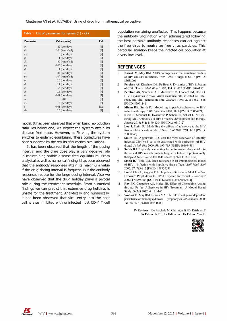
population remaining unaffected. This happens because the antibody vaccination when administered following the best possible antibody responses can act against the free virus to neutralize free virus particles. This particular situation keeps the infected cell population at a very low level.
REFERENCES 1 Nowak M, May RM. AIDS pathogenesis: mathematical models
of HIV and SIV infections. AIDS 1993; 7 Suppl 1: S3-18 [PMID: 8363800]
2 Perelson AS, Kirschner DE, De Boer R. Dynamics of HIV infection of CD4+ T cells. Math Biosci 1993; 114: 81-125 [PMID: 8096155]
3 Perelson AS, Neumann AU, Markowitz M, Leonard JM, Ho DD. HIV-1 dynamics in vivo: virion clearance rate, infected cell life-span, and viral generation time. Science 1996; 271: 1582-1586 [PMID: 8599114]
4 Miron RE, Smith RJ. Modelling imperfect adherence to HIV induction therapy. BMC Infect Dis 2010; 10: 6 [PMID: 20064271]
5 Klein F, Mouquet H, Dosenovic P, Scheid JF, Scharf L, Nussen-zweig MC. Antibodies in HIV-1 vaccine development and therapy. Science 2013; 341: 1199-1204 [PMID: 24031012]
6 Lou J, Smith RJ. Modelling the effects of adherence to the HIV fusion inhibitor enfuvirtide. J Theor Biol 2011; 268: 1-13 [PMID: 20888346]
7 Smith RJ, Aggarwala BD. Can the viral reservoir of latently infected CD4(+) T cells be eradicated with antiretroviral HIV drugs? J Math Biol 2009; 59: 697-715 [PMID: 19165438]
8 Smith RJ. Explicitly accounting for antiretroviral drug uptake in theoretical HIV models predicts long-term failure of protease-only therapy. J Theor Biol 2008; 251: 227-237 [PMID: 18191950]
9 Smith RJ, Wahl LM. Drug resistance in an immunological model of HIV-1 infection with impulsive drug effects. Bull Math Biol 2005; 67: 783-813 [PMID: 15893553]
10 Lou J, Chen L, Ruggeri T. An Impulsive Differential Model on Post Exposure Prophylaxis to HIV-1 Exposed Individual. J Biol Syst 2009; 17: 659-683 [DOI: 10.1142/S0218339009002934]
11 Roy PK, Chatterjee AN, Majee SB. Effect of Chemokine Analog through Perfect Adherence in HIV Treatment: A Model Based Study. IJAMA 2012; 4: 121-145
12 Wodarz D, May RM, Nowak MA. The role of antigen-independent persistence of memory cytotoxic T lymphocytes. Int Immunol 2000; 12: 467-477 [PMID: 10744648]
P- Reviewer: De Paschale M, Ghiringhelli PD, Krishnan T S- Editor: Ji FF L- Editor: A E- Editor: Yan JL
model. It has been observed that when basic reproduction ratio lies below one, we expect the system attain its disease free state. However, at R0 > 1, the system switches to endemic equilibrium. These conjectures have been supported by the results of numerical simulations.
It has been observed that the length of the dosing interval and the drug dose play a very decisive role in maintaining stable disease free equilibrium. From analytical as well as numerical finding it has been observed that the antibody responses attain its maximum value if the drug dosing interval is frequent. But the antibody responses reduce for the large dosing interval. Also we have observed that the drug holiday plays a pivotal role during the treatment schedule. From numerical findings we can predict that extensive drug holidays is unsafe for the treatment. Analytically and numerically, it has been observed that viral entry into the host cell is also inhibited with uninfected host CD4+ T cell
November 12, 2015|Volume 4|Issue 4|WJV|www.wjgnet.com 364
Table 1 List of parameters for system (1) - (2)
Parameter Value (units) Ref.
b 42 (per day) [6]β 1 10-5 (/mm3/d) [6]μ G 5 (per day) [9]p 1 (per day) [6]ST 80 (/mm3/d) [9]μ T 0.05 (per day) [6]d1 0.4 (per day) [6]α 25 (per day) [6]β 2 10-5 (/mm3/d) [6]q 0.6 (per day) [6]d2 0.4 (per day) [6]κ 0.6 (per day) [6]dI 0.5 (per day) [9]δ 0.01 (per day) [7]n 540 [6]μV 3 (per day) [7]ι 0.01 (per day) [12]dA 0.5 (per day) [7]
Chatterjee AN et al . HIV/AIDS: Using of drug from mathematical perceptive

Yupin Tong, Bonita E Lee, Xiaoli L Pang
Yupin Tong, Department of Medicine, University of Alberta, Edmonton, Alberta T6G 2J2, Canada
Bonita E Lee, Department of Pediatrics, University of Alberta, Edmonton, Alberta T6G 2J2, Canada
Xiaoli L Pang, Department of Laboratory Medicine and Pathology, University of Alberta, Edmonton, Alberta T6G 2J2, Canada
Xiaoli L Pang, Provincial Laboratory for Public Health, University of Alberta Hospital, Edmonton, Alberta T6G 2J2, Canada
Author contributions: Tong Y performed the experiments, analyzed the data and wrote the manuscript; Lee BE contributed data and sample selections for the study and revised the manuscript; Pang XL designed the research and revised the manuscript.
Institutional review board statement: The study was reviewed and approved by the Provincial Laboratory for Public Health Edmonton Alberta (Provlab) Institutional Review Board.
Conflict-of-interest statement: None.
Data sharing statement: Technical appendix, clinical dataset is available from the corresponding author at [email protected].
Open-Access: This article is an open-access article which was selected by an in-house editor and fully peer-reviewed by external reviewers. It is distributed in accordance with the Creative Commons Attribution Non Commercial (CC BY-NC 4.0) license, which permits others to distribute, remix, adapt, build upon this work non-commercially, and license their derivative works on different terms, provided the original work is properly cited and the use is non-commercial. See: http://creativecommons.org/licenses/by-nc/4.0/
Correspondence to: Xiaoli L Pang, PhD, Provincial Laboratory for Public Health, University of Alberta Hospital, WMC 2B4.58, 8440 112 Street, Edmonton, Alberta T6G 2J2, Canada. [email protected]: +1-780-4073483
Fax: +1-780-4078984
Received: May 29, 2015Peer-review started: June 2, 2015First decision: June 18, 2015Revised: August 1, 2015Accepted: September 29, 2015 Article in press: September 30, 2015Published online: November 12, 2015
AbstractAIM: To develop a real-time reverse transcription-polymerase chain reaction (RT-PCR) assay to genotype rotavirus (G and P) in Alberta from January 2012 to June 2013.
METHODS: We developed and validated a different approach to perform rotavirus G and P genotyping using a two-step SYBR green RT-PCR (rt-gPCR) by selecting genotype-specific primers of published conventional RT nested PCR (cnRT-PCR) assay and optimizing the amplification conditions. cDNA was first synthesized from total RNA with SuperScript™ Ⅱ reverse trans-criptase kit followed by amplication step using monoplex SYBR green real-time PCR. After the PCR reaction, melting curve analysis was used to determine specific genotype. Sixteen samples previously genotyped using cnRT-PCR were tested using the new assay and the genotyping results were compared as sensitivity analysis. Assay specificity was evaluated by testing other gastroenteritis viruses with the new assay. The amplicon size of each available genotype was determined by gel-electrophoresis and DNA sequences were obtained using Sanger-sequencing method. After validation and optimization, the new assay was used to genotype 122 pediatric clinical stool samples previously tested positive for rotavirus using electron microscopy between January
365 November 12, 2015|Volume 4|Issue 4|WJV|www.wjgnet.com
Submit a Manuscript: http://www.wjgnet.com/esps/Help Desk: http://www.wjgnet.com/esps/helpdesk.aspxDOI: 10.5501/wjv.v4.i4.365
World J Virol 2015 November 12; 4(4): 365-371ISSN 2220-3249 (online)
© 2015 Baishideng Publishing Group Inc. All rights reserved.
World Journal of VirologyW J V
Rapid genotyping of human rotavirus using SYBR green real-time reverse transcription-polymerase chain reaction with melting curve analysis
Basic Study
ORIGINAL ARTICLE

2012 and June 2013.
RESULTS: The new rt-gPCR assay was validated and optimized. The assay detected G1 to G4, G9, G12 and P[4] and P[8] that were available as positive controls in our laboratory. A single and clear peak of melting curve was generated for each of specific G and P genotypes with a Tm ranging from 80 ℃ to 82 ℃. The sensitivity of rt-gPCR was comparable to cnRT-PCR with 100% correlation of the 16 samples with known G and P genotypes. No cross reaction was found with other gastroenteritis viruses. Using the new rt-gPCR assay, genotypes were obtained for 121 of the 122 pediatric clinical samples tested positive for rotavirus: G1P[8] (42.6%), G2P[4] (4.9%), G3P[8] (10.7%), G9P[8] (10.7%), G9P[4] (6.6%), G12P[8] (23.0%), and unknown GP[8] (0.8%). For the first time, G12 rotavirus strains were found in Alberta and G12 was the second most common genotype during the study period. Gel electrophoresis of all the genotypes showed expected amplicon size for each genotype. The sequence data of the two G12 samples along with other genotypes were blasted in NCBI BLAST or analyzed with Rota C Genotyping tool (http://rotac.regatools.be/). All genotyping results were confirmed to be correct.
CONCLUSION: rt-gPCR is a useful tool for the geno-typing and characterization of rotavirus. Monitoring of rotavirus genotypes is important for the identification of emerging strains and ongoing evaluation of rotavirus vaccination programs.
Key words: Rotavirus A; Melting temperature; Real-time polymerase chain reaction; SYBR green; Genotyping
© The Author(s) 2015. Published by Baishideng Publishing Group Inc. All rights reserved.
Core tip: Genotyping rotavirus is essential for monitoring strain shifts in rotavirus surveillance and vaccine evaluation. Current conventional semi-nested real-time reverse transcription-polymerase chain reaction (RT-PCR), the most commonly used rotavirus genotyping assay is a labor-intensive, complex multi-step procedure and has long turn around-time. The newly developed SYBR Green real time RT-PCR assay is simple, fast and has comparable sensitivity and specificity as conven-tional semi-nested RT-PCR. This new assay was used to genotype clinical samples which tested positive for rotavirus from January 2012 to June 2013 and new emerging G12 strains were identified in Alberta, Canada.
Tong Y, Lee BE, Pang XL. Rapid genotyping of human rotavirus using SYBR green real-time reverse transcription-polymerase chain reaction with melting curve analysis. World J Virol 2015; 4(4): 365-371 Available from: URL: http://www.wjgnet.com/2220-3249/full/v4/i4/365.htm DOI: http://dx.doi.org/10.5501/wjv.v4.i4.365
INTRODUCTION Rotavirus group A is a major cause of severe acute gastroenteritis in children worldwide, with most children contracting the infection by five years of age[1]. Rotavirus vaccines are most efficacious in protecting children from severe rotavirus gastroenteritis, especially in areas with very low or low child and adult mortality[2]. Two effective rotavirus vaccines, RotaTeq® (Merck and Co., Inc.), a bovine-human reassortant vaccine covering the G1-G4 genotypes along with P[8] and Rotarix® (GlaxoSmithKline, Inc.), a monovalent attenuated human G1P[8] vaccine, were recommended by the WHO in 2009 for the routine immunization of infant globally[3].
The viral genome consists of 11 double-stranded RNA segments, which encode six structure proteins (VP1-4, VP6 and VP7) and six non-structure proteins (NSP1-6). The traditional classification of rotavirus using serotyping has mostly been replaced with G and P genotyping that are based on the diversity of VP7 and VP4 gene sequences, respectively. Twenty-seven G genotypes (G1-G27) and 35 P genotypes (P[1]-P[35]) have been described and six G (G1-4, G9 and G12) and three P (P[4], P[6], and P[8]) genotypes predominate globally with some regional differences[4,5]. Introduction of vaccines or natural evolution of rotavirus may alter antigenic properties of circulating strains in the regions. The antigenic drift between G and P genotypes could result in a decrease of the effectiveness of vaccines against infection in the near future[6,7]. Therefore, understanding the presence and distribution of G and P genotypes and monitoring the emerging and recom-bination of rotavirus genotypes are very important prior to and after the introduction of rotavirus vaccines.
Various molecular methods have been developed for rotavirus genotyping. Conventional reverse transcriptase (RT) and nested polymerase chain reaction (cnRT-PCR) broadly used since the 90s[8-10] have been improved with more sensitive and specific primers[11,12]. These improved cnRT-PCR methods recognize a broad cluster of various rotavirus G and P genotypes with good specificity. However, there are several drawbacks of these type of assays including: (1) multi-step maneuvers and the requirement of two runs of PCR reactions and gel electrophoresis; (2) labor intensive protocols and a long turn-around time for final results; (3) difficulty with result interpretations due to non-specific amplicons or multiple amplicons in samples with co-infections; and (4) risk of cross contamination of PCR end-products because of the need for open tube maneuvers. With the 2010 recommendation of use of rotavirus vaccine for healthy infants in Canada[13], a rapid and accurate genotyping assay is needed for the evaluation of the vaccine programs in many provinces.
Recently, real time RT-PCR assays in a closed tube system with their rapid turn around-time, sensitivity and specificity and low risk of cross contamination have been implemented in both research and diagnostic
366 November 12, 2015|Volume 4|Issue 4|WJV|www.wjgnet.com
Tong Y et al . SYBR green RT-PCR assay for genotyping rotavirus

laboratories for the detection of rotavirus in stool samples associated with gastroenteritis[14-16]. To date, rotavirus genotyping using TaqMan real time PCR assay has been reported[17]. The protocol could successfully genotype G1P[8], G2P[4], G3P[8], G4P[8] and G9P[8] with only two multiplex reactions, however the other genotypes and combinations were still unable to be typed. Melting curve analysis uses the melting temperature of double-stranded PCR products to determine the identity of the PCR products and also can detect the presence of nonspecific PCR products or primer-dimers. Melting curve analysis is commonly used in SYBR green RT-PCR (rt-gPCR) to determine PCR products and omits the need for gel electrophoresis. SYBR green real-time PCR with melting curve analysis has been used to type Dengue virus as a genotyping tool[18]. This study is to develop and validate a simple and rapid rotavirus genotyping assay using SYBR Green RT-PCR to detect a broad range of rotavirus strains and to use the new assay to characterize the rotavirus strains circulated in Alberta from January 2012 to June 2013.
MATERIALS AND METHODSDevelopment of rt-gPCR assay Sample preparation, RNA extraction and RT reaction: Archived rotavirus positive pediatric stool samples (n = 16) obtained from the Provincial Laboratory for Public Health (ProvLab) and previously genotyed by the cnRT-PCR[10] were used for the development and validation of the rt-gPCR assay. Viral RNA was extracted from 200 μL of 10% stool suspension (10% W/V with PBS buffer) using MagaZorb® total RNA Mini-Prep kit (Promega, Madison, United States) on an automaton extractor, KingFisher™ mL Magnetic Particle Processors, (Thermo Scientific, Mississauga, Canada) according to manufacturer’s instructions. Using SuperScript™
Ⅱ Reverse Transcriptase kit and random primer (Life technologies, Ontario, Canada), cDNA was synthesized from 5 μL RNA at 42 ℃ for 1 h, 75 ℃ for 15 min after heating 5 min at 97 ℃ and stored at -20 ℃ before use.
rt-gPCR: A group of published primers used in cnRT-PCR for genotyping rotavirus G types (G1-4, G8, G9-10) and P types (P[4], P[6], P[8], P[9], P[10], and P[11] ) were selected for the new assay development[8-10] (Table 1). Another two sets of primers for genotyping G8, and G12 which were not previously used in our laboratory was also added in the new rt-gPCR assay[12]. 5 μL of 1:10 diluted cDNA was applied to 20 μL of reaction mixture containing 2 μL of LightCycler® FastStart DNA Master SYBR Green Ⅰ (Roche Diagnostics, Quebec, Canada), 4 mmol/L MgCl2, 0.2 μmol/L each of VP7-R and specific G-typing primers or 0.4 μmol/L each of VP4-F and specific P-typing primers. The rt-gPCR was performed using LightCylcer® 1.0 (Roche Diagnostics, Quebec, Canada) with four-step experimental run protocol: (1) denaturation program (10 min at 95 ℃); (2) 45 cycles of amplification program (10 s at 95 ℃; 10 s at 53 ℃ (P-typing), 57 ℃ (G-typing), and 25 s of extension at 72 ℃); (3) melting curve program (0 s at 95 ℃, 120 s at 65 ℃ and 0 s at 95 ℃ with ramp rate at 0.1 ℃ per second); and (4) cooling program down to 40 ℃. Since different genotype specific primers would yield amplicons of different size with various GC content percentages, the temperature (Tm) and melting curve of the amplicon of specific rotavirus G or P genotype would be different. Thus melting Tm profiles were used to identify specific rotavirus G or P genotypes in our design. For data analysis, the Tm, fluorescence (d[F1]/dT) under the melting curve window was selected as the parameters for evaluation. A sample would be assigned to a specific genotype when the reaction Tm matched with known genotype controls, and the fluorescence d[F1]/dT was
367 November 12, 2015|Volume 4|Issue 4|WJV|www.wjgnet.com
Table 1 Polymerase chain reaction primers used for G and P typing of human rotavirus by reverse transcriptase polymerase chain reaction
Primer Sequence (5’-3’) Amplicon size (bp) Direction Ref.
G-type VP7-R AAC TTG CCA CCA TTT TTT CC Antisense Iturriza-Gómara et al[10]
G1 CAA GTA CTC AAA TCA ATG ATG G 618 Sense Gouvea et al[8]
G2 CAA TGA TAT TAA CAC ATT TTC TGT G 521 Sense Gouvea et al[8]
G3 ACG AAC TCA ACA CGA GAG G 682 Sense Iturriza-Gómara et al[10]
G4 CGT TTC TGG TGA GGA GTT G 452 Sense Gouvea et al[8]
G8 TTR1 TCG CAC CAT TTG TAA TT 756 Sense Aladin et al[12]
G9 CTT GAT GTG ACT AY1A AAT AC 179 Sense Iturriza-Gómara et al[10]
G10 ATG TCA GAC TAC AR2A TAC TGG 266 Sense Gouvea et al[8]
G12 GGT TAT GTA ATC CGA TGG CG 396 Sense Aladin et al[12]
P-type VP4-F TAT GCT CCA GTN3 AAT TGG Sense Simmonds et al[11]
P[4] CTA TTG TTA GAG GTT AGA GTC 362 Antisense Gentsch et al[9]
P[6] TGT TGA TTA GTT GGA TTC AA 146 Antisense Gentsch et al[9]
P[8] TCT ACT GGR2 TTR2 ACN3 TGC 224 Antisense Iturriza-Gómara et al[10]
P[9] TGA GAC ATG CAA TTG GAC 270 Antisense Gentsch et al[9]
P[10] ATC ATA GTT AGT AGT CGG 462 Antisense Gentsch et al[9]
P[11] GTA AAC ATC CAG AAT GTG 191 Antisense Iturriza-Gómara et al[10]
1Y: C or T; 2R: A or G; 3N: A, G, C or T.
Tong Y et al . SYBR green RT-PCR assay for genotyping rotavirus

368 November 12, 2015|Volume 4|Issue 4|WJV|www.wjgnet.com
Data analysis SD for the Tm of each genotype was calculated to show the variations in Tm. The sequence data of amplicons of G1 to G4, G9, G12, P[4] and P[8] were blasted in NCBI or analyzed with Rota C Genotyping tool (http://rotac.regatools.be/).
RESULTSDevelopment of rt-gPCR assay All 16 positive control samples previously genotyped by cnRT-PCR were confirmed using the rt-gPCR assay with 100% concordant results. Using the rt-gPCR assay, a single and clear peak of melting curve was yielded for each of specific G and P genotypes with a Tm ranging from 80 ℃ to 82 ℃ (Figure 1). More than 30 of the G1, G3, G9, P[8] and more than 20 of the G2, G4, G12,P[4] replicates and/or different samples at different runs were used to calculate to mean Tm and SD. Each genotype Tm showed very small variation during different PCR runs among different samples. The mean Tm (℃ ± SD) for each of the genotypes were: G1 at 80.0 ℃± 0.20, G2 at 80.9 ℃ ± 0.49, G3 at 81.7 ℃ ± 0.22, G4 at 80.7 ℃ ± 0.20, G9 at 80.9 ℃ ± 0.45, G12 at 80.6 ℃± 0.37, P[4] at 80.7 ℃ ± 0.50, and P[8] at 80.0 ℃ ± 0.34 (Figure 2). Multiplex rt-gPCR could not be performed to simultaneously detect all G or P genotypes because the Tm generated from each genotype was very close. A lower Tm peak (< 77 ℃) indicating primer-dimer was observed in the amplification reactions. All rt-gPCR genotype products showed expected amplicon sizes in the gel electrophoresis (Figure 3).
In the sensitivity comparison by serial ten-fold dilution of positive samples, both rt-gPCR and cnRT-
above 1.0. The cycle threshold (Ct) of amplification curve was used to provide a relative quantification. Positive controls of specific G and P genotypes were included in each rt-gPCR run as reference genotype and for quality control.
Assay sensitivity and specificity: The sensitivity of rt-gPCR assay was compared with cnRT-PCR using ten-fold serial dilutions from neat (undiluted) to 10-6 of samples with known G and P genotypes. Other common gastroenteritis viruses including norovirus, sapovirus, adenovirus, and astrovirus were also tested using the G and P primers to determine the specificity of the rt-gPCR assay.
DNA sequencing: Six G-types including G1 to G4, G9, G12 and two P-types P[4], P[8] detected by the rt-gPCR assay were sequenced with modification as described previously[19]. Briefly, the positive PCR products were run in 2.0% agarose gel and purified with QIAquick Gel Extraction kit (Qiagen, Hilden, Germany) then sequenced using 3730 Genetic Analyzer (Applied biosystems, Foster City, United States) at University of Alberta.
Genotyping clinical samplesProvLab provides routine diagnostic testing of stool samples submitted for testing of gastroenteritis viruses using electronic microscope (EM). A total of 122 stool samples with rotavirus identified by EM between January 5, 2012 and June 8, 2013 were genotyped using the validated rt-gPCR assay. The purpose was to determine the performance of the assay for rapid genotyping of rotavirus in the clinical setting.
Figure 1 Melting cure analysis of different genotypes using reverse transcriptase polymerase chain reaction (A: G-typing: G1, G2, G4, G9, G12, G3; B: P-typing: P[8], P[4]). The peak of curve in X-axis is the melting temperature of each genotype DNA fragment; Y-axis indicates the SYB green fluorescence density.
-(d/
dT)
fluor
esce
nce
(530
)
Melting peaks
Temperature (℃)
5.686
4.686
3.686
2.686
1.686
0.686
65 70 75 80 85 90 95
A
G1
G1, G4, G9, G12
G3
-(d/
dT)
fluor
esce
nce
(530
)
Melting peaks
Temperature (℃)
5.328
4.328
3.328
2.328
1.328
0.328
66 68 70 72 74 76 78 80 82 84 86 88 90 92 94
B
P[8]P[4]
Tong Y et al . SYBR green RT-PCR assay for genotyping rotavirus
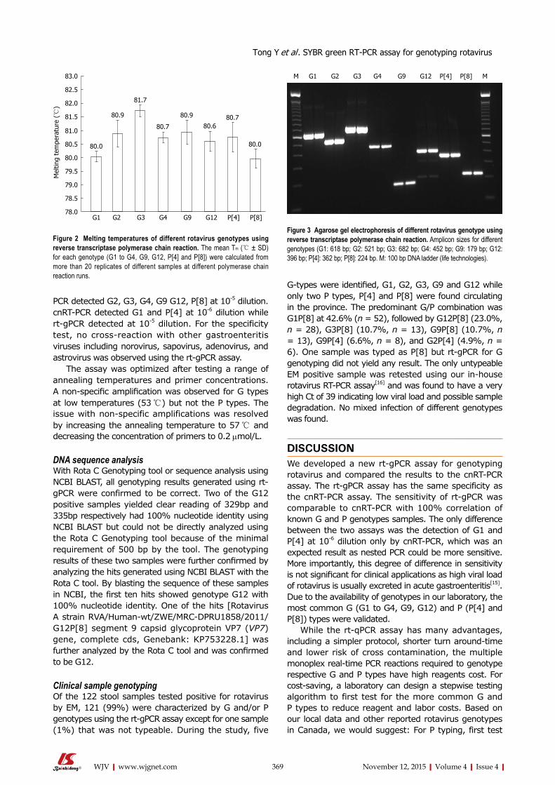
369 November 12, 2015|Volume 4|Issue 4|WJV|www.wjgnet.com
PCR detected G2, G3, G4, G9 G12, P[8] at 10-5 dilution. cnRT-PCR detected G1 and P[4] at 10-6 dilution while rt-gPCR detected at 10-5 dilution. For the specificity test, no cross-reaction with other gastroenteritis viruses including norovirus, sapovirus, adenovirus, and astrovirus was observed using the rt-gPCR assay.
The assay was optimized after testing a range of annealing temperatures and primer concentrations. A non-specific amplification was observed for G types at low temperatures (53 ℃) but not the P types. The issue with non-specific amplifications was resolved by increasing the annealing temperature to 57 ℃ and decreasing the concentration of primers to 0.2 μmol/L.
DNA sequence analysisWith Rota C Genotyping tool or sequence analysis using NCBI BLAST, all genotyping results generated using rt-gPCR were confirmed to be correct. Two of the G12 positive samples yielded clear reading of 329bp and 335bp respectively had 100% nucleotide identity using NCBI BLAST but could not be directly analyzed using the Rota C Genotyping tool because of the minimal requirement of 500 bp by the tool. The genotyping results of these two samples were further confirmed by analyzing the hits generated using NCBI BLAST with the Rota C tool. By blasting the sequence of these samples in NCBI, the first ten hits showed genotype G12 with 100% nucleotide identity. One of the hits [Rotavirus A strain RVA/Human-wt/ZWE/MRC-DPRU1858/2011/G12P[8] segment 9 capsid glycoprotein VP7 (VP7) gene, complete cds, Genebank: KP753228.1] was further analyzed by the Rota C tool and was confirmed to be G12.
Clinical sample genotypingOf the 122 stool samples tested positive for rotavirus by EM, 121 (99%) were characterized by G and/or P genotypes using the rt-gPCR assay except for one sample (1%) that was not typeable. During the study, five
G-types were identified, G1, G2, G3, G9 and G12 while only two P types, P[4] and P[8] were found circulating in the province. The predominant G/P combination was G1P[8] at 42.6% (n = 52), followed by G12P[8] (23.0%, n = 28), G3P[8] (10.7%, n = 13), G9P[8] (10.7%, n = 13), G9P[4] (6.6%, n = 8), and G2P[4] (4.9%, n = 6). One sample was typed as P[8] but rt-gPCR for G genotyping did not yield any result. The only untypeable EM positive sample was retested using our in-house rotavirus RT-PCR assay[16] and was found to have a very high Ct of 39 indicating low viral load and possible sample degradation. No mixed infection of different genotypes was found.
DISCUSSIONWe developed a new rt-gPCR assay for genotyping rotavirus and compared the results to the cnRT-PCR assay. The rt-gPCR assay has the same specificity as the cnRT-PCR assay. The sensitivity of rt-gPCR was comparable to cnRT-PCR with 100% correlation of known G and P genotypes samples. The only difference between the two assays was the detection of G1 and P[4] at 10-6 dilution only by cnRT-PCR, which was an expected result as nested PCR could be more sensitive. More importantly, this degree of difference in sensitivity is not significant for clinical applications as high viral load of rotavirus is usually excreted in acute gastroenteritis[15]. Due to the availability of genotypes in our laboratory, the most common G (G1 to G4, G9, G12) and P (P[4] and P[8]) types were validated.
While the rt-qPCR assay has many advantages, including a simpler protocol, shorter turn around-time and lower risk of cross contamination, the multiple monoplex real-time PCR reactions required to genotype respective G and P types have high reagents cost. For cost-saving, a laboratory can design a stepwise testing algorithm to first test for the more common G and P types to reduce reagent and labor costs. Based on our local data and other reported rotavirus genotypes in Canada, we would suggest: For P typing, first test
Mel
ting
tem
pera
ture
(℃
)
83.0
82.5
82.0
81.5
81.0
80.5
80.0
79.5
79.0
78.5
78.0G1 G2 G3 G4 G9 G12 P[4] P[8]
80.0
80.9
81.7
80.7
80.9
80.680.7
80.0
Figure 2 Melting temperatures of different rotavirus genotypes using reverse transcriptase polymerase chain reaction. The mean Tm (℃ ± SD) for each genotype (G1 to G4, G9, G12, P[4] and P[8]) were calculated from more than 20 replicates of different samples at different polymerase chain reaction runs.
M G1 G2 G3 G4 G9 G12 P[4] P[8] M
Figure 3 Agarose gel electrophoresis of different rotavirus genotype using reverse transcriptase polymerase chain reaction. Amplicon sizes for different genotypes (G1: 618 bp; G2: 521 bp; G3: 682 bp; G4: 452 bp; G9: 179 bp; G12: 396 bp; P[4]: 362 bp; P[8]: 224 bp. M: 100 bp DNA ladder (life technologies).
Tong Y et al . SYBR green RT-PCR assay for genotyping rotavirus

370 November 12, 2015|Volume 4|Issue 4|WJV|www.wjgnet.com
for P[8] and if P[8] is negative, then test for P[4]; for G typing, first test for G1 to be followed by G12 and G9, then G3, G2 +/- G4. This stepwise strategy would cover the range of G and P genotypes for most clinical samples.
The first G12 strain was identified in the Philippines in 1987 followed by reports of sporadic detection in other countries[20-22]. G12 is currently recognized as a globally emerging rotavirus genotype that appears to be spreading more rapidly in recent years[23]. Predominant of G12 was reported in Nepal in 2011, Cameroon in 2010/11 and rotavirus outbreak associated with G12 was found in United States in 2006-07[24-26]. In our study, G12 was detected as the second most prevalent genotype using rt-gPCR in Alberta. G12 could be detected by the new rt-gPCR assay but would be missed by the cnRT-PCR assay. We still believe that G12 was an emerging rotavirus genotype in 2012 as only 1.4% of EM rotavirus positive samples were untypeable from previous nine years in Alberta (data not shown). No G12 has been previously reported in Canada[27]. To our knowledge, this is the first report of G12 genotype detected in Canada. In addition, G9P[4] which was rarely detected in previous years increased dramatically in 2012. These findings emphasize the importance of ongoing surveillance for circulating rotavirus genotypes. In conclusion, the newly developed rt-gPCR assay with optimized primer selection enhanced the detection of broader genotypes which makes this assay a useful tool for the characterization and monitoring of strain shifts in rotavirus surveillance and the evaluation of vaccination program.
ACKNOWLEDGMENTSWe are grateful to ProvLab staff for providing routine diagnostic testing of gastroenteritis viruses by electron microscopy and thank Min Cao for her technical support with the study. We also thank Dr. Jutta K Preiksaitis for scientific discussions and partial financial support.
COMMENTSBackgroundGenotyping rotavirus is very important for the characterization and monitoring of strain shifts in rotavirus surveillance for the evaluation of vaccination program. The most commonly used rotavirus genotyping assay is a conventional nested reverse transcriptase (RT) polymerase chain reaction (cnRT-PCR) which has been used and revised for more than 30 years. The labor-intensive, complex multi-step procedure and high potential contamination risk due to nested PCR format make this method falling behind current demand.
Research frontiersAn accurate, easy-to-perform and rapid rotavirus genotyping tool is needed. Several published studies developed new genotyping PCR assays using multiplex Taqman real time PCR assay to replace conventional nested PCR but has limited sensitivity and can detect only a few G or P types.
Innovations and breakthroughsUsing SYBR Green based RT-PCR with melting curve analysis and melting temperature (Tm) to genotype rotavirus is simple, fast and provides accurate and
broad identifications of genotypes. Gel electrophoresis required by traditional conventional nested PCR genotyping assay is not needed and eliminates the risk of contamination from handling post-PCR products. The new assay showed similar sensitivity and specificity as the conventional nested RT-PCR.
ApplicationsThe new assay identified common genotypes circulating in Alberta, Canada as well as the emergence of rotavirus G12 in 2012-2013. G12 has never been reported in Canada. During the study, the most predominant rotavirus genotypes were: G1P[8] (42.6%), G12P[8] (23.0%), followed by G3P[8] (10.7%), G9P[8] (10.7%), G9P[4] (6.6%), and G2P[4] (4.9%). These new findings support the importance of ongoing surveillance and characterization of rotavirus genotypes.
TerminologyRotavirus G-typing is genotyping viral gene sequences diversity which encoding structure protein VP7; Rotavirus P-typing is genotyping viral gene sequences diversity which encoding structure protein VP4; SYBR green is a commonly used fluorescent DNA binding dye, binds all double-stranded DNA and detection is monitored by measuring the increase in fluorescence throughout the cycle. SYBR Green master mixes are designed for quantitative real-time PCR using a set of two PCR primers that flank the target region; Melting curve analysis is the temperature-dependent dissociation between two DNA-strands can be measured using a DNA-intercalating fluorophore such as SYBR green. Melting Tm can be used for determination of the identity of the target.
Peer-reviewThe article describes a new assay for genotyping the human rotavirus. The results presented seem pretty convincing and conclusions are valid.
REFERENCES1 Parashar UD, Nelson EA, Kang G. Diagnosis, management, and
prevention of rotavirus gastroenteritis in children. BMJ 2013; 347: f7204 [PMID: 24379214 DOI: 10.1136/bmj.f7204]
2 Rotavirus vaccines. WHO position paper - January 2013. Wkly Epidemiol Rec 2013; 88: 49-64 [PMID: 23424730]
3 Rotavirus vaccines: an update. Wkly Epidemiol Rec 2009; 84: 533-540 [PMID: 20034143]
4 Matthijnssens J, Van Ranst M. Genotype constellation and evolution of group A rotaviruses infecting humans. Curr Opin Virol 2012; 2: 426-433 [PMID: 22683209 DOI: 10.1016/j.coviro.2012.04.007]
5 Patton JT. Rotavirus diversity and evolution in the post-vaccine world. Discov Med 2012; 13: 85-97 [PMID: 22284787]
6 Hull JJ, Teel EN, Kerin TK, Freeman MM, Esona MD, Gentsch JR, Cortese MM, Parashar UD, Glass RI, Bowen MD. United States rotavirus strain surveillance from 2005 to 2008: genotype prevalence before and after vaccine introduction. Pediatr Infect Dis J 2011; 30: S42-S47 [PMID: 21183839 DOI: 10.1097/INF.0b013e3181fefd78]
7 Gentsch JR, Parashar UD, Glass RI. Impact of rotavirus vacci-nation: the importance of monitoring strains. Future Microbiol 2009; 4: 1231-1234 [PMID: 19995181 DOI: 10.2217/fmb.09.105]
8 Gouvea V, Glass RI, Woods P, Taniguchi K, Clark HF, Forrester B, Fang ZY. Polymerase chain reaction amplification and typing of rotavirus nucleic acid from stool specimens. J Clin Microbiol 1990; 28: 276-282 [PMID: 2155916]
9 Gentsch JR, Glass RI, Woods P, Gouvea V, Gorziglia M, Flores J, Das BK, Bhan MK. Identification of group A rotavirus gene 4 types by polymerase chain reaction. J Clin Microbiol 1992; 30: 1365-1373 [PMID: 1320625]
10 Iturriza-Gómara M, Kang G, Gray J. Rotavirus genotyping: keeping up with an evolving population of human rotaviruses. J Clin Virol 2004; 31: 259-265 [PMID: 15494266 DOI: 10.1016/j.jcv.2004.04.009]
11 Simmonds MK, Armah G, Asmah R, Banerjee I, Damanka S, Esona M, Gentsch JR, Gray JJ, Kirkwood C, Page N, Iturriza-Gómara M. New oligonucleotide primers for P-typing of rotavirus
COMMENTS
Tong Y et al . SYBR green RT-PCR assay for genotyping rotavirus
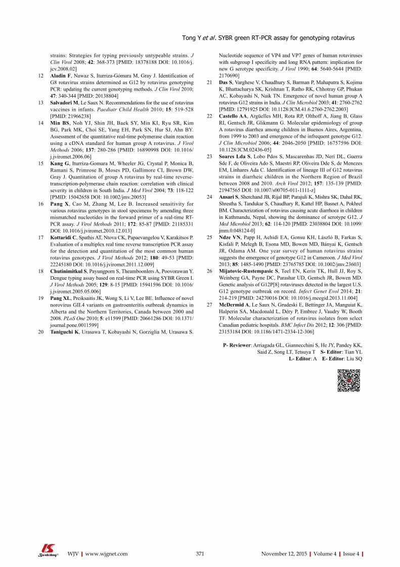
371 November 12, 2015|Volume 4|Issue 4|WJV|www.wjgnet.com
strains: Strategies for typing previously untypeable strains. J Clin Virol 2008; 42: 368-373 [PMID: 18378188 DOI: 10.1016/j.jcv.2008.02]
12 Aladin F, Nawaz S, Iturriza-Gómara M, Gray J. Identification of G8 rotavirus strains determined as G12 by rotavirus genotyping PCR: updating the current genotyping methods. J Clin Virol 2010; 47: 340-344 [PMID: 20138804]
13 Salvadori M, Le Saux N. Recommendations for the use of rotavirus vaccines in infants. Paediatr Child Health 2010; 15: 519-528 [PMID: 21966238]
14 Min BS, Noh YJ, Shin JH, Baek SY, Min KI, Ryu SR, Kim BG, Park MK, Choi SE, Yang EH, Park SN, Hur SJ, Ahn BY. Assessment of the quantitative real-time polymerase chain reaction using a cDNA standard for human group A rotavirus. J Virol Methods 2006; 137: 280-286 [PMID: 16890998 DOI: 10.1016/j.jviromet.2006.06]
15 Kang G, Iturriza-Gomara M, Wheeler JG, Crystal P, Monica B, Ramani S, Primrose B, Moses PD, Gallimore CI, Brown DW, Gray J. Quantitation of group A rotavirus by real-time reverse-transcription-polymerase chain reaction: correlation with clinical severity in children in South India. J Med Virol 2004; 73: 118-122 [PMID: 15042658 DOI: 10.1002/jmv.20053]
16 Pang X, Cao M, Zhang M, Lee B. Increased sensitivity for various rotavirus genotypes in stool specimens by amending three mismatched nucleotides in the forward primer of a real-time RT-PCR assay. J Virol Methods 2011; 172: 85-87 [PMID: 21185331 DOI: 10.1016/j.jviromet.2010.12.013]
17 Kottaridi C, Spathis AT, Ntova CK, Papaevangelou V, Karakitsos P. Evaluation of a multiplex real time reverse transcription PCR assay for the detection and quantitation of the most common human rotavirus genotypes. J Virol Methods 2012; 180: 49-53 [PMID: 22245180 DOI: 10.1016/j.jviromet.2011.12.009]
18 Chutinimitkul S, Payungporn S, Theamboonlers A, Poovorawan Y. Dengue typing assay based on real-time PCR using SYBR Green I. J Virol Methods 2005; 129: 8-15 [PMID: 15941596 DOI: 10.1016/j.jviromet.2005.05.006]
19 Pang XL, Preiksaitis JK, Wong S, Li V, Lee BE. Influence of novel norovirus GII.4 variants on gastroenteritis outbreak dynamics in Alberta and the Northern Territories, Canada between 2000 and 2008. PLoS One 2010; 5: e11599 [PMID: 20661286 DOI: 10.1371/journal.pone.0011599]
20 Taniguchi K, Urasawa T, Kobayashi N, Gorziglia M, Urasawa S.
Nucleotide sequence of VP4 and VP7 genes of human rotaviruses with subgroup I specificity and long RNA pattern: implication for new G serotype specificity. J Virol 1990; 64: 5640-5644 [PMID: 2170690]
21 Das S, Varghese V, Chaudhury S, Barman P, Mahapatra S, Kojima K, Bhattacharya SK, Krishnan T, Ratho RK, Chhotray GP, Phukan AC, Kobayashi N, Naik TN. Emergence of novel human group A rotavirus G12 strains in India. J Clin Microbiol 2003; 41: 2760-2762 [PMID: 12791925 DOI: 10.1128/JCM.41.6.2760-2762.2003]
22 Castello AA, Argüelles MH, Rota RP, Olthoff A, Jiang B, Glass RI, Gentsch JR, Glikmann G. Molecular epidemiology of group A rotavirus diarrhea among children in Buenos Aires, Argentina, from 1999 to 2003 and emergence of the infrequent genotype G12. J Clin Microbiol 2006; 44: 2046-2050 [PMID: 16757596 DOI: 10.1128/JCM.02436-05]
23 Soares Lda S, Lobo Pdos S, Mascarenhas JD, Neri DL, Guerra Sde F, de Oliveira Ado S, Maestri RP, Oliveira Dde S, de Menezes EM, Linhares Ada C. Identification of lineage III of G12 rotavirus strains in diarrheic children in the Northern Region of Brazil between 2008 and 2010. Arch Virol 2012; 157: 135-139 [PMID: 21947565 DOI: 10.1007/s00705-011-1111-z]
24 Ansari S, Sherchand JB, Rijal BP, Parajuli K, Mishra SK, Dahal RK, Shrestha S, Tandukar S, Chaudhary R, Kattel HP, Basnet A, Pokhrel BM. Characterization of rotavirus causing acute diarrhoea in children in Kathmandu, Nepal, showing the dominance of serotype G12. J Med Microbiol 2013; 62: 114-120 [PMID: 23038804 DOI: 10.1099/jmm.0.048124-0]
25 Ndze VN, Papp H, Achidi EA, Gonsu KH, László B, Farkas S, Kisfali P, Melegh B, Esona MD, Bowen MD, Bányai K, Gentsch JR, Odama AM. One year survey of human rotavirus strains suggests the emergence of genotype G12 in Cameroon. J Med Virol 2013; 85: 1485-1490 [PMID: 23765785 DOI: 10.1002/jmv.23603]
26 Mijatovic-Rustempasic S, Teel EN, Kerin TK, Hull JJ, Roy S, Weinberg GA, Payne DC, Parashar UD, Gentsch JR, Bowen MD. Genetic analysis of G12P[8] rotaviruses detected in the largest U.S. G12 genotype outbreak on record. Infect Genet Evol 2014; 21: 214-219 [PMID: 24270016 DOI: 10.1016/j.meegid.2013.11.004]
27 McDermid A, Le Saux N, Grudeski E, Bettinger JA, Manguiat K, Halperin SA, Macdonald L, Déry P, Embree J, Vaudry W, Booth TF. Molecular characterization of rotavirus isolates from select Canadian pediatric hospitals. BMC Infect Dis 2012; 12: 306 [PMID: 23153184 DOI: 10.1186/1471-2334-12-306]
P- Reviewer: Arriagada GL, Giannecchini S, He JY, Pandey KK, Said Z, Song LT, Tetsuya T S- Editor: Tian YL
L- Editor: A E- Editor: Liu SQ
Tong Y et al . SYBR green RT-PCR assay for genotyping rotavirus
Joana Ribeiro, Delfim Ferreira, Célia Arrabalde, Sandra Almeida, Inês Baldaque, Hugo Sousa
Joana Ribeiro, Célia Arrabalde, Sandra Almeida, Inês Baldaque, Hugo Sousa, Virology Service, Portuguese Institute of Oncology of Porto, 4200-072 Porto, Portugal
Joana Ribeiro, Delfim Ferreira, Hugo Sousa, Molecular Oncology and Viral Pathology Group, Portuguese Institute of Oncology of Porto, 4200-072 Porto, Portugal
Joana Ribeiro, Research Department, Portuguese League Against Cancer (Liga Portuguesa Contra o Cancro - Núcleo Regional do Norte), 4200-177 Porto, Portugal
Author contributions: Almeida S and Baldaque I performed the experimental tests for Adv and RV detection; Ribeiro J, Ferreira D, and Arrabalde C were responsible for the data collection; Ribeiro J and Sousa H analyzed data; Ribeiro J and Ferreira D wrote the paper; Sousa H supervised the study; all authors have provided the final approval of the manuscript.
Conflict-of-interest statement: All authors declare no conflicts of interest with any competing financial interest for the work described in this paper.
Data sharing statement: Not applied.
Open-Access: This article is an open-access article which was selected by an in-house editor and fully peer-reviewed by external reviewers. It is distributed in accordance with the Creative Commons Attribution Non Commercial (CC BY-NC 4.0) license, which permits others to distribute, remix, adapt, build upon this work non-commercially, and license their derivative works on different terms, provided the original work is properly cited and the use is non-commercial. See: http://creativecommons.org/licenses/by-nc/4.0/
Correspondence to: Hugo Sousa, MD, PhD, Virology Service, Portuguese Institute of Oncology of Porto, Rua Dr. António Bernardino Almeida, 4200-072 Porto, Portugal. [email protected]: +351-22-5084000Fax: +351-22-5084001
Received: May 8, 2015
Peer-review started: May 9, 2015First decision: June 3, 2015Revised: September 9, 2015Accepted: September 29, 2015 Article in press: September 30, 2015Published online: November 12, 2015
AbstractAIM: To characterize the prevalence of rotavirus (RV) and adenovirus (AdV) infections in immunocompromised patients with acute gastroenteritis.
METHODS: The presence of RV and AdV (serotypes 40 and 41) was evaluated in 509 stool samples obtained between January 2009 and December 2010 from 200 immunocompromised patients (83 females and 117 males; median age 21 years old, range 0-72. The diagnosis of infection was performed as a routine procedure and the presence of RV and AdV (serotypes 40 and 41) was determined by immunochromato-graphy using the RIDA® Quick Rota-Adeno-Kombi kit (r-Biopharm, Darmstadt, Germany). The data analysis and description of seasonal frequencies were performed using computer software IBM® SPSS® (Statistical Package for Social Sciences) Statistics version 20.0 for Mac. The frequencies of infection were compared into different age and gender groups by χ 2 test.
RESULTS: The study revealed 12.4% AdV positive samples and 0.8% RV positive samples, which corres-pond to a prevalence of 6.5% and 1.5%, respectively. AdV was more frequent between October 2009 and April 2010, while RV was identified in April 2010 and July 2010. The stool analysis revealed that from the 509 samples, 63 (12.4%) were positive for AdV and 4 (0.8%) positive for RV, which by resuming the information
372 November 12, 2015|Volume 4|Issue 4|WJV|www.wjgnet.com
Submit a Manuscript: http://www.wjgnet.com/esps/Help Desk: http://www.wjgnet.com/esps/helpdesk.aspxDOI: 10.5501/wjv.v4.i4.372
World J Virol 2015 November 12; 4(4): 372-376ISSN 2220-3249 (online)
© 2015 Baishideng Publishing Group Inc. All rights reserved.
World Journal of VirologyW J V
Prevalence of adenovirus and rotavirus infection in immunocompromised patients with acute gastroenteritis in Portugal
Retrospective Study
ORIGINAL ARTICLE

of each patient, lead to an overall prevalence of AdV and RV of 6.5% (13/200 patients) and 1.5% (3/200 patients), respectively. The stratification of the analy-sis regarding age groups showed a tendency to an increased prevalence of infection in paediatric patients between 0-10 years old. Considering the seasonal distribution of these infections, our study revealed that AdV infection was more frequent between October 2009 and April 2010, while RV infection was characterized by two distinct peaks (April 2010 and July 2010).
CONCLUSION: The overall prevalence of AdV and RV infection in immunocompromised patients with acute gastroenteritis was 8% and AdV was the most prevalent agent.
Key words: Viral gastroenteritis; Adenovirus; Rotavirus; Immunocompromised host; Stool samples
© The Author(s) 2015. Published by Baishideng Publishing Group Inc. All rights reserved.
Core tip: Acute gastroenteritis has been associated with significant rates of morbidity and mortality among immunocompromised patients. Rotavirus (RV) and adenovirus (AdV) are described as common agents of viral gastroenteritis causing acute diarrhoea. This is the first study in Portugal to characterize the prevalence and seasonal features of RV and AdV infections in immunocompromised patients with acute gastroenteritis. Results revealed 12.4% AdV positive samples and 0.8% RV positive samples, which correspond to a prevalence of 6.5% and 1.5%, respectively. Our results also demonstrate the importance of to add more screening methods for other emergent enteric viruses, in order to avoid the morbidity and mortality of the immunocompromised patients.
Ribeiro J, Ferreira D, Arrabalde C, Almeida S, Baldaque I, Sousa H. Prevalence of adenovirus and rotavirus infection in immunocompromised patients with acute gastroenteritis in Portugal. World J Virol 2015; 4(4): 372-376 Available from: URL: http://www.wjgnet.com/2220-3249/full/v4/i4/372.htm DOI: http://dx.doi.org/10.5501/wjv.v4.i4.372
INTRODUCTIONAcute gastroenteritis, one of the main causes of morbidity and mortality in the world, is a consequence of microbial infections, which in industrialized countries are mainly caused by viruses, such as rotavirus (RV), enteric adenovirus (AdV), astrovirus and human calicivirus[1-3].
RV is the most common cause of severe diarrhoea in children under 5 years of age, adults in close contact with infected children, hospitalised patients and the elderly[3,4]. Data from United States reveal that RV infection is responsible for approximately 3 million cases of acute diarrhoea in children each year[5]. RV infection is associated with high rates of morbidity throughout
the world and high rates of mortality in developing countries, where gastroenteritis caused by RV account for more than 800000 deaths per year due to poor nutrition and lack of health care[6]. In the majority of countries, RV infections occur dispersed throughout the year, nevertheless in temperate climates is characterized by a peak during the winter months[1-3].
AdV, especially serotypes 40 and 41 (enteric AdV), are frequently related with high morbidity and mortality in immunocompromised patients with the most susce-ptible groups to be children and patients submitted to transplantation[7-9]. Enteric AdV have been related with acute diarrhoea in variable frequency (ranging from 1.4% to 10%), depending on the geographic location and type of patients[10,11]. AdV is known for its large distribution worldwide and the majority of the studies refer it to be equally distributed during all the seasons of the year[8,9].
Immunocompromised patients constitute an impor-tant group for prevention of gastrointestinal infec-tions[7,12,13]. In fact, the incidence of infection-related post-transplant diarrhoea has been reported to be up to 40%, with viruses being the most common patho-gens[2,3,12]. Considering the importance of gastroenteritis prevention in immunocompromised individuals, the diagnosis of AdV and RV at early stages of the disease is extremely important, in order to reduce morbidity and mortality.
The aim of this study was to characterize the prevalence of RV and AdV infection in immunocom-promised patients with acute gastroenteritis from the North region of Portugal treated at the Portuguese Institute of Oncology of Porto.
MATERIALS AND METHODSPopulationThis study was performed as a cross-sectorial retro-spective hospital-based case study with 509 stool samples obtained between January 2009 and December 2010 from immunocompromised patients diagnosed with acute diarrhoea at Portuguese Institute of Oncology of Porto (IPO Porto). Samples were obtained from 200 immunocompromised patients with different hae-matological malignancies (median age 21 years old, range 0-72): 83 female (median age 15 years old, range 0-65) and 117 male (median age 39 years old, range 0-72).
RV/AdV detection The diagnosis of infection was performed as a routine procedure at the Virology Service of IPO Porto. The stool specimens were tested as soon as possible after collection and the presence of RV and AdV (serotypes 40 and 41) was determined by immunochromatography using the RIDA® Quick Rota-Adeno-Kombi kit (r-Biopharm, Darmstadt, Germany) according to manufacturer instructions. The faecal samples were diluted in the dilution buffer supplied with the kit. This is a ready-to-use test based on a nitrocellulose membrane sensitized with
373 November 12, 2015|Volume 4|Issue 4|WJV|www.wjgnet.com
Ribeiro J et al . Acute gastroenteritis by AdV and RV in immunocompromised patients
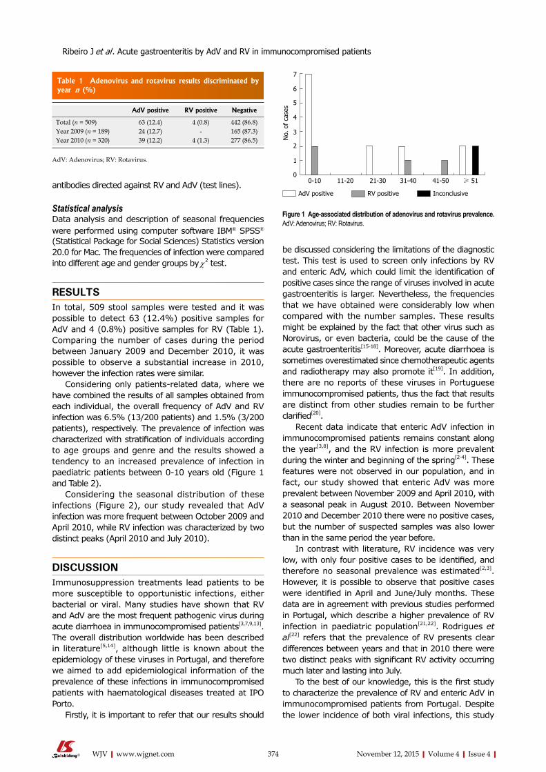
antibodies directed against RV and AdV (test lines).
Statistical analysisData analysis and description of seasonal frequencies were performed using computer software IBM® SPSS® (Statistical Package for Social Sciences) Statistics version 20.0 for Mac. The frequencies of infection were compared into different age and gender groups by χ2 test.
RESULTSIn total, 509 stool samples were tested and it was possible to detect 63 (12.4%) positive samples for AdV and 4 (0.8%) positive samples for RV (Table 1). Comparing the number of cases during the period between January 2009 and December 2010, it was possible to observe a substantial increase in 2010, however the infection rates were similar.
Considering only patients-related data, where we have combined the results of all samples obtained from each individual, the overall frequency of AdV and RV infection was 6.5% (13/200 patients) and 1.5% (3/200 patients), respectively. The prevalence of infection was characterized with stratification of individuals according to age groups and genre and the results showed a tendency to an increased prevalence of infection in paediatric patients between 0-10 years old (Figure 1 and Table 2).
Considering the seasonal distribution of these infections (Figure 2), our study revealed that AdV infection was more frequent between October 2009 and April 2010, while RV infection was characterized by two distinct peaks (April 2010 and July 2010).
DISCUSSIONImmunosuppression treatments lead patients to be more susceptible to opportunistic infections, either bacterial or viral. Many studies have shown that RV and AdV are the most frequent pathogenic virus during acute diarrhoea in immunocompromised patients[3,7,9,13]. The overall distribution worldwide has been described in literature[5,14], although little is known about the epidemiology of these viruses in Portugal, and therefore we aimed to add epidemiological information of the prevalence of these infections in immunocompromised patients with haematological diseases treated at IPO Porto.
Firstly, it is important to refer that our results should
be discussed considering the limitations of the diagnostic test. This test is used to screen only infections by RV and enteric AdV, which could limit the identification of positive cases since the range of viruses involved in acute gastroenteritis is larger. Nevertheless, the frequencies that we have obtained were considerably low when compared with the number samples. These results might be explained by the fact that other virus such as Norovirus, or even bacteria, could be the cause of the acute gastroenteritis[15-18]. Moreover, acute diarrhoea is sometimes overestimated since chemotherapeutic agents and radiotherapy may also promote it[19]. In addition, there are no reports of these viruses in Portuguese immunocompromised patients, thus the fact that results are distinct from other studies remain to be further clarified[20].
Recent data indicate that enteric AdV infection in immunocompromised patients remains constant along the year[3,8], and the RV infection is more prevalent during the winter and beginning of the spring[2-4]. These features were not observed in our population, and in fact, our study showed that enteric AdV was more prevalent between November 2009 and April 2010, with a seasonal peak in August 2010. Between November 2010 and December 2010 there were no positive cases, but the number of suspected samples was also lower than in the same period the year before.
In contrast with literature, RV incidence was very low, with only four positive cases to be identified, and therefore no seasonal prevalence was estimated[2,3]. However, it is possible to observe that positive cases were identified in April and June/July months. These data are in agreement with previous studies performed in Portugal, which describe a higher prevalence of RV infection in paediatric population[21,22]. Rodrigues et al[22] refers that the prevalence of RV presents clear differences between years and that in 2010 there were two distinct peaks with significant RV activity occurring much later and lasting into July.
To the best of our knowledge, this is the first study to characterize the prevalence of RV and enteric AdV in immunocompromised patients from Portugal. Despite the lower incidence of both viral infections, this study
374 November 12, 2015|Volume 4|Issue 4|WJV|www.wjgnet.com
No.
of
case
s
AdV positive RV positive Inconclusive
7
6
5
4
3
2
1
00-10 11-20 21-30 31-40 41-50 ≥ 51
Figure 1 Age-associated distribution of adenovirus and rotavirus prevalence. AdV: Adenovirus; RV: Rotavirus.
Table 1 Adenovirus and rotavirus results discriminated by year n (%)
AdV positive RV positive Negative
Total (n = 509) 63 (12.4) 4 (0.8) 442 (86.8)Year 2009 (n = 189) 24 (12.7) - 165 (87.3)Year 2010 (n = 320) 39 (12.2) 4 (1.3) 277 (86.5)
AdV: Adenovirus; RV: Rotavirus.
Ribeiro J et al . Acute gastroenteritis by AdV and RV in immunocompromised patients

375 November 12, 2015|Volume 4|Issue 4|WJV|www.wjgnet.com
the world, especially for immunocompromised patients. Immunosuppression treatments lead patients to be more susceptible to opportunistic infections, either bacterial or viral. Many studies have shown that rotavirus (RV) and adenovirus (AdV) are the most frequent pathogenic virus during acute diarrhoea in immunocompromised patients.
Research frontiersCharacterization of RV and AdV prevalence and seasonal distribution in immunocompromised patients with acute gastroenteritis.
Innovations and breakthroughsThe overall prevalence of AdV and RV infection in immunocompromised patients with acute gastroenteritis was 8%. AdV was the most prevalent with 6.5% (13/200 patients) followed by RV with a prevalence of 1.5% (3/200 patients). Results revealed a lower prevalence of RV and enteric AdV than expected.
ApplicationsThe lower incidence of RV and enteric AdV observed in the authors’ study pointed that it is extremely important to add more screening methods for other
emphasizes the importance of vigilance and prevention of viral infections in the gastrointestinal tract. Moreover, as the results reveal a lower incidence of RV and enteric AdV, it is extremely important to add more screening methods for other emergent enteric viruses, in order to avoid the morbidity and mortality of the immunocompromised patients.
ACKNOWLEDGMENTSAuthors are grateful to the Portuguese League Against Cancer (Liga Portuguesa Contra o Cancro - Núcleo Regional do Norte) for the grant of the first author.
COMMENTSBackgroundAcute gastroenteritis is one of the main causes of morbidity and mortality in
No.
of
posi
tive
case
s
20191817161514131211109876543210
Jan-
09
Feb-
09
Mar-0
9
Apr-0
9
May-0
9
Jun-
09Ju
l-09
Aug-
09
Sep-
09
Oct-09
Nov-0
9
Dec-0
9
Jan-
10
Feb-
10
Mar-1
0
Apr-1
0
May-1
0
Jun-
10Ju
l-10
Aug-
10
Sep-
10
Oct-10
Nov-1
0
Dec-1
0
50
45
40
35
30
25
20
15
10
5
0
No.
of
requ
ests
AdV positive RV positive No. of requests
Figure 2 Seasonal distribution of adenovirus and rotavirus infection throughout the study period, taking into account the number of requests. AdV: Adenovirus; RV: Rotavirus.
Table 2 Characterization adenovirus and rotavirus infection by age group and genre n (%)
AdV RV
Positive Negative IC Positive Negative ICTotal (n = 200) 13 (6.5) 185 (92.5) 2 (1.0) 3 (1.5) 196 (98.0) 1 (0.5)Age group 0-10 (n = 60) 7 (11.7) 53 (88.3) - 2 (3.3) 58 (96.7) - 11-20 (n = 21) - 21 (100.0) - - 21 (100.0) - 21-30 (n = 19) 2 (10.5) 17 (89.5) - - 19 (100.0) - 31-40 (n = 19) 2 (10.5) 17 (89.5) - 1 (5.3) 18 (94.7) - 41-50 (n = 19) - 18 (94.7) 1 (5.3) - 19 (100.0) - ≥ 51 (n = 62) 2 (3.2) 59 (95.2) 1 (1.6) - 61 (98.4) 1 (1.6)Genre Male (n = 117) 9 (7.7) 107 (91.4) 1 (0.9) 1 (0.9) 115 (98.2) 1 (0.9) Female (n = 83) 4 (4.8) 78 (94.0) 1 (1.2) 2 (2.4) 81 (97.6) -
AdV: Adenovirus; RV: Rotavirus; IC: Inconclusive.
COMMENTS
Ribeiro J et al . Acute gastroenteritis by AdV and RV in immunocompromised patients

376 November 12, 2015|Volume 4|Issue 4|WJV|www.wjgnet.com
emergent enteric viruses, in order to avoid the morbidity and mortality of the immunocompromised patients.
TerminologyThis study provides the first update on the prevalence of RV and AdV infection as agents of acute gastroenteritis in immunocompromised patients.
Peer-reviewThis is an interesting study on the cause of diarrhea by viral agents. The study is well-designed and the manuscript is well-written.
REFERENCES1 Wilhelmi I, Roman E, Sánchez-Fauquier A. Viruses causing
gastroenteritis. Clin Microbiol Infect 2003; 9: 247-262 [PMID: 12667234]
2 Carraturo A, Catalani V, Tega L. Microbiological and epidemi-ological aspects of rotavirus and enteric adenovirus infections in hospitalized children in Italy. New Microbiol 2008; 31: 329-336 [PMID: 18843886]
3 Akan H, Izbırak G, Gürol Y, Sarıkaya S, Gündüz TS, Yılmaz G, Hayran O, Vitrinel A. Rotavirus and adenovirus frequency among patients with acute gastroenteritis and their relationship to clinical parameters: a retrospective study in Turkey. Asia Pac Fam Med 2009; 8: 8 [PMID: 19943964 DOI: 10.1186/1447-056X-8-8]
4 Podkolzin AT, Fenske EB, Abramycheva NY, Shipulin GA, Sagalova OI, Mazepa VN, Ivanova GN, Semena AV, Tagirova ZG, Alekseeva MN, Molochny VP, Parashar UD, Vinjé J, Maleev VV, Glass RI, Pokrovsky VI. Hospital-based surveillance of rotavirus and other viral agents of diarrhea in children and adults in Russia, 2005-2007. J Infect Dis 2009; 200 Suppl 1: S228-S233 [PMID: 19817602 DOI: 10.1086/605054]
5 Elhag WI, Saeed HA, Omer el FE, Ali AS. Prevalence of rotavirus and adenovirus associated with diarrhea among displaced communities in Khartoum, Sudan. BMC Infect Dis 2013; 13: 209 [PMID: 23657114 DOI: 10.1186/1471-2334-13-209]
6 Kotloff KL, Nataro JP, Blackwelder WC, Nasrin D, Farag TH, Panchalingam S, Wu Y, Sow SO, Sur D, Breiman RF, Faruque AS, Zaidi AK, Saha D, Alonso PL, Tamboura B, Sanogo D, Onwuchekwa U, Manna B, Ramamurthy T, Kanungo S, Ochieng JB, Omore R, Oundo JO, Hossain A, Das SK, Ahmed S, Qureshi S, Quadri F, Adegbola RA, Antonio M, Hossain MJ, Akinsola A, Mandomando I, Nhampossa T, Acácio S, Biswas K, O’Reilly CE, Mintz ED, Berkeley LY, Muhsen K, Sommerfelt H, Robins-Browne RM, Levine MM. Burden and aetiology of diarrhoeal disease in infants and young children in developing countries (the Global Enteric Multicenter Study, GEMS): a prospective, case-control study. Lancet 2013; 382: 209-222 [PMID: 23680352 DOI: 10.1016/S0140-6736(13)60844-2]
7 Walls T, Shankar AG, Shingadia D. Adenovirus: an increasingly important pathogen in paediatric bone marrow transplant patients. Lancet Infect Dis 2003; 3: 79-86 [PMID: 12560192 DOI: 10.1016/S1473-3099(03)00515-2]
8 Ison MG. Adenovirus infections in transplant recipients. Clin Infect Dis 2006; 43: 331-339 [PMID: 16804849 DOI: 10.1086/505498]
9 Echavarría M. Adenoviruses in immunocompromised hosts. Clin Microbiol Rev 2008; 21: 704-715 [PMID: 18854488 DOI: 10.1128/CMR.00052-07]
10 Ramani S, Kang G. Viruses causing childhood diarrhoea in the developing world. Curr Opin Infect Dis 2009; 22: 477-482 [PMID: 19633550 DOI: 10.1097/QCO.0b013e328330662f]
11 Tran A, Talmud D, Lejeune B, Jovenin N, Renois F, Payan C, Leveque N, Andreoletti L. Prevalence of rotavirus, adenovirus, norovirus, and astrovirus infections and coinfections among hospitalized children in northern France. J Clin Microbiol 2010; 48: 1943-1946 [PMID: 20305010 DOI: 10.1128/JCM.02181-09]
12 Liakopoulou E, Mutton K, Carrington D, Robinson S, Steward CG, Goulden NJ, Cornish JM, Marks DI. Rotavirus as a significant cause of prolonged diarrhoeal illness and morbidity following allogeneic bone marrow transplantation. Bone Marrow Transplant 2005; 36: 691-694 [PMID: 16113671 DOI: 10.1038/sj.bmt.1705127]
13 Aggarwal V, Williams MD, Beath SV. Gastrointestinal problems in the immunosuppressed patient. Arch Dis Child 1998; 78: 5-8 [PMID: 9534668 DOI: 10.1136/adc.78.1.5]
14 Al-Thani A, Baris M, Al-Lawati N, Al-Dhahry S. Characterising the aetiology of severe acute gastroenteritis among patients visiting a hospital in Qatar using real-time polymerase chain reaction. BMC Infect Dis 2013; 13: 329 [PMID: 23865805 DOI: 10.1186/1471-2334-13-329]
15 Karst SM. Pathogenesis of noroviruses, emerging RNA viruses. Viruses 2010; 2: 748-781 [PMID: 21994656 DOI: 10.3390/v2030748]
16 Weber DJ, Rutala WA, Miller MB, Huslage K, Sickbert-Bennett E. Role of hospital surfaces in the transmission of emerging health care-associated pathogens: norovirus, Clostridium difficile, and Acinetobacter species. Am J Infect Control 2010; 38: S25-S33 [PMID: 20569853 DOI: 10.1016/j.ajic.2010.04.196]
17 Man SM. The clinical importance of emerging Campylobacter species. Nat Rev Gastroenterol Hepatol 2011; 8: 669-685 [PMID: 22025030 DOI: 10.1038/nrgastro.2011.191]
18 Rackoff LA, Bok K, Green KY, Kapikian AZ. Epidemiology and evolution of rotaviruses and noroviruses from an archival WHO Global Study in Children (1976-79) with implications for vaccine design. PLoS One 2013; 8: e59394 [PMID: 23536875 DOI: 10.1371/journal.pone.0059394]
19 Donaldson SS, Lenon RA. Alterations of nutritional status: impact of chemotherapy and radiation therapy. Cancer 1979; 43: 2036-2052 [PMID: 109184]
20 Fischer Walker CL, Perin J, Aryee MJ, Boschi-Pinto C, Black RE. Diarrhea incidence in low- and middle-income countries in 1990 and 2010: a systematic review. BMC Public Health 2012; 12: 220 [PMID: 22436130 DOI: 10.1186/1471-2458-12-220]
21 Rodrigues F, Iturriza M, Gray J, Januário L, Lemos L. Epidemiology of rotavirus in Portugal: G9 as a major cause of diarrhoea in non-hospitalised children. J Clin Virol 2007; 40: 214-217 [PMID: 17875400 DOI: 10.1016/j.jcv.2007.08.006]
22 Rodrigues F, Iturriza-Gómara M, Marlow R, Gray J, Nawaz S, Januário L, Finn A. The evolving epidemiology of rotavirus gastroenteritis in central Portugal with modest vaccine coverage. J Clin Virol 2013; 56: 129-134 [PMID: 23238239 DOI: 10.1016/j.jcv.2012.10.016]
P- Reviewer: Kamal SA, Mattner J S- Editor: Tian YL L- Editor: A E- Editor: Liu SQ
Ribeiro J et al . Acute gastroenteritis by AdV and RV in immunocompromised patients

© 2015 Baishideng Publishing Group Inc. All rights reserved.
Published by Baishideng Publishing Group Inc8226 Regency Drive, Pleasanton, CA 94588, USA
Telephone: +1-925-223-8242Fax: +1-925-223-8243
E-mail: [email protected] Desk: http://www.wjgnet.com/esps/helpdesk.aspx
http://www.wjgnet.com