world journal of - bsdwebstorage.blob.core.windows.net · giovanni dapri, brussels undurti n das,...
TRANSCRIPT
Published by Baishideng Publishing Group Inc
World Journal of DiabetesWorld J Diabetes 2016 December 15; 7(20): 572-630
ISSN 1948-9358 (online)
EDITORS-IN-CHIEFLu Qi, BostonJingbo Zhao, Aarhus
ASSOCIATE EDITORSGiovanni Dapri, BrusselsUndurti N Das, Federal WayMin Du, LaramieEdward B Jude, Ashton under Lyne Gregory I Liou, AugustaJuan F Navarro-Gonzalez, Santa Cruz de TenerifeKatarzyna Szkudelska, PoznanRichard Welbourn, TauntonSilvano Zanuso, Chatam Maritime
GUEST EDITORIAL BOARD MEMBERSJuei-Tang Cheng, TainanChih-Hsung Chu, KaohsiungLow-Tone Ho, TaipeiCheng-Cheng Hsiao, KeelungYung-Hsi Kao, TaoyuanChi-Feng Liu, TaipeiShing-Hwa Liu, TaipeiWayne HH Sheu, TaichungEing-Mei Tsai, KaohsiungChin-Hsiao Tseng, TaipeiWei-Chung V Yang, TaipeiWen-Chin Yang, TaipeiTzung-Hai Yen, Taipei
MEMBERS OF THE EDITORIAL BOARD
ArgentinaEduardo Spinedi, La Plata
AustraliaSof Andrikopoulos, HeidelbergHugh R Barrett, WesternBernhard T Baune, TownsvilleGrant D Brinkworth, AdelaideMelinda T Coughlan, MelbourneJosephine M Forbes, MelbournePaul A Fournier, PerthAngela Gialamas, AdelaideMark D Gorrell, SydneyGraeme J Hankey, PerthAnandwardhan A Hardikar, MelbourneMichael Horowitz, AdelaideKarin Jandeleit-Dahm, BalwynMartha Lappas, VictoriaPeter J Little, VictoriaXin Liu, BrisbaneDianna J Magliano, CaufieldLouise JM Maple-Brown, CasuarinaRobyn McDermott, AdelaideBeverly S Muhlhausler, SemaphoreChristopher J Nolan, CanberraLuciano Pirola, MelbourneKarly C Sourris, MelbourneGreg Tesch, VictoriaJack R Wall, PenrithOwen L Woodman, Victoria
AustriaChristian H Anderwald, ViennaHelmuth M Borkenstein, Graz
Latife Bozkurt, ViennaWalter H Horl, ViennaFriedrich Mittermayer, ViennaMarkus Paulmichl, SalzburgStefan Pilz, GrazThomas M Stulnig, ViennaLudwig Wagner, Vienna
BelgiumChristophe De Block, EdegemEkaterine Tskitishvili, LiegeF A Van Assche, LeuvenLuc F Van Gaal, Edegem
BrazilMonica L Andersen, Sao PauloClaudia RL Cardoso, Rio de JaneiroRicardo V Cohen, Sao PauloCassyano J Correr, CuritibaCintia C Curioni, Rio de JaneiroFreddy G Eliaschewitz, Sao PauloRodrigo Jorge, Ribeirao PretoLuciana A Naves, BrasiliaMatheus Roriz Cruz, Porto AlegreJúlio C Voltarelli, Sao PauloJacqueline N Zanoni, Maringá
CanadaJean-Luc Ardilouze, Sherbrooke
I
Editorial Board2016-2019
The World Journal of Diabetes Editorial Board now consists of 676 members, representing a team of worldwide experts in diabetes mellitus. They are from 56 countries, including Argentina (1), Australia (26), Austria (9), Belgium (5), Brazil (11), Canada (24), Chile (3), China (39), Cuba (1), Czech Republic (2), Denmark (12), Egypt (3), Finland (5), France (11), Germany (26), Greece (16), Hungary (4), Iceland (1), India (24), Iran (6), Iraq (2), Ireland (4), Israel (9), Italy (54), Japan (30), Jordan (1), Kuwait (3), Lebanon (1), Malaysia (1), Malta (1), Mexico (4), Netherlands (7), New Zealand (3), Nigeria (2), Norway (2), Oman (3), Pakistan (2), Poland (8), Portugal (1), Qatar (1), Romania (2), Singapore (4), Slovakia (1), South Africa (1), South Korea (15), Spain (25), Sweden (6), Switzerland (3), Thailand (4), Tunisia (1), Turkey (13), United Arab Emirates (3), United Kingdom (28), United States (199), Venezuela (2), and Yemen (1).
February 29, 2016WJD|www.wjgnet.com
World Journal of DiabetesW J D
Subrata Chakrabarti, LondonDavid ZI Cherney, TorontoMervyn Deitel, TorontoJean-Pierre Despres, QuébecDavid J Hill, OntarioTian-Ru Jin, TorontoArulmozhi D Kandasamy, AlbertaJennifer L Kuk, TorontoIsmail Laher, VancouverZhong-Cheng Luo, MontrealRoger S McIntyre, TorontoDavid Meyre, HamiltonJF Ndisang, SaskatoonRaj S Padwal, AlbertaCiriaco A Piccirillo, MontrealAM James Shapiro, Edmonton Guang Sun, St. John'sValerie Taylor, OntarioCory Toth, CalgaryAndré Tremblay, MontréalVVincent C Woo, ManitobaJames R Wright, Alberta Xi-Long Zheng, Calgary
ChileSebastian S Martin, ValparaisoArmando Rojas Rubio, TalcaLuis Sobrevia, Santiago
ChinaJie Chen, NanjingBernard Man Yung Cheung, Hong KongWilliam CS Cho, Hong KongTian-Pei Hong, BeijingQin Huang, ShanghaiPo-Sing Leung, Hong KongChao Liu, NanjingJian-Kang Liu, Xi’anLie-Gang Liu, WuhanRonald CW Ma, Hong KongZengchang Pang, QingdaoJin-Sheng Qi, ShijiazhuangJin-Xiong She, ShijiazhuangWing Y So, Hong KongCheuk C Szeto, Hong KongKathryn CB Tan, Hong KongCong-Yi Wang, WuhanYu Wang, Hong KongGuang-Da Xiang, WuhanBao-Feng Yang, HarbinShu-Yu Yang, XiamenXi-Lin Yang, Hong KongZai-Qing Yang, WuhanShan-Dong Ye, HefeiShi-Sheng Zhou, DalianZhi-Guang Zhou, Changsha
CubaLuis Sarmiento-Pérez, Havana
Czech RepublicMichal Krcma, PlzenTerezie Pelikanova, Prague
DenmarkCharlotte Brons, GentofteFlemming Dela, Copenhagen NKristine Faerch, GentofteLouise G Grunnet, GentofteR Scott Heller, GentofteFilip K Knop, HellerupHelle Markholst, MåløvOle H Mortensen, Copenhagen NOluf Pedersen, Copenhagen KEsben T Vestergaard, Aarhus NMilan Zdravkovic, Soborg
EgyptMamdouh MA Hssan, DokkiMoshira AH Rateb, CairoMona F Schaalan, Cairo
FinlandSiamak Bidel, HelsinkiGang Hu, HelsinkiThomas Kietzmann, OuluQing Qiao, EspooKaroliina Wehkalampi, Helsinki
FranceJean C Ansquer, DijonBertrand Cariou, NantesSylvie Dejager, Rueil-MalmaisonNaim A Khan, DijonJean-Philippe Lavigne, NimesMichel Marre, ParisMarie-Claude Morice, MassyRiccardo Perfetti, ParisGérard Said, ParisDidier Vieau, VilleneuveSophie Visvikis-Siest, Nancy
GermanyChrista Buechler, RegensburgRoland Büttner, HeidelbergMichael Froehner, DresdenIoanna Gouni-Berthold, CologneHammes Hans-Peter, MannheimNadja Herbach, MunichNadj Herbach, MunchenAndrea Icks, DüsseldorfThomas Jax, NeussMichael Kluge, MunichFlorian Lang, Tuebingen
Matthias Laudes, KolnRalf Lobmann, StuttgartRafael T Mikolajczyk, BremenAndreas S Mueller, HalleKarsten Müssig, TübingenNahid Parvizi, MarienseeThomas P Reinehr, DattelnMichael Ristow, JenaSven Schinner, DuesseldorfPeter EH Schwarz, DresdenOvidiu A Stirban, OeynhausenDiego J Walther, BerlinSilvia A Wein, KielChristian Wrede, BerlinDan Ziegler, Düsseldorf
GreeceGeorge P Chrousos, AthensMoses S Elisaf, IoanninaPanagiotis Georgoulias, LarissaNikolaos Kadoglou, ThessalonikiGerasimos E Krassas, KriniSpilios Manolakopoulos, AthensPeppa Melpomeni, HaIDariNikolaos Papanas, AlexandroupolisDimitrios Papazoglou, AlexandroupolisSokratis Pastromas, AthensChristina Piperi, GoudiNicholas K Tentolouris, AthensKonstantinos A Toulis, SalonikaApostolos Tsapas, ThessalonikiKonstantinos Tziomalos, ThessalonikiElias Zintzaras, Larissa
HungaryMária Bagyánszki, SzegedGyorgy Jermendy, BudapestKaroly Racz, BudapestGyula Soltesz, Pécs
IcelandSaher Hamed, Haifa
IndiaSarika Arora, New DelhiPitchai Balakumar, SivakasiMuthuswamy Balasubramanyam, ChennaiAnuradha Carani Venkatraman, NagarSubhabrata Chakrabarti, Hyderabad Abhay S Chakraborti, KolkataTapan K Chaudhuri, New DelhiKanwaljit Chopra, ChandigarhMalabika Datta, DelhiDebidas Ghosh, West BengalRavinder Goswami, New DelhiJothydev Kesavadev, KeralaKVS H Kumar, Lucknow
II February 29, 2016WJD|www.wjgnet.com

III February 29, 2016WJD|www.wjgnet.com
Anoop Misra, New DelhiAnalava Mitra, KharagpurViswanathan Mohan, ChennaiPallavi Panchu, BangaloreDeepak N Patel, MumbaiUsharani Pingali, HyderabadAmbady Ramachandran, ChennaiVadde Ramakrishna, KadapaRajat Sandhir, ChandigarhManju Sharma, New DelhiSuman B Sharma, Delhi
IranMohammad K Arababadi, RafsanjanLeila Azadbakht, IsfahanHamid Baradaran, TehranBehrooz Broumand, TehranMajid Ghayour-Mobarhan, MashhadMohsen Janghorbani, Isfahan
IraqSaad AR Hussain, BaghdadAbbas A Mansour, Basrah
IrelandAmar Agha, DublinMichael Aviram, HaifaRaymond E Farah, Safed Mark P Hehir, Dublin
IsraelGal Dubnov-Raz, HashomerShimon Efrat, Tel AvivOren Froy, RehovotFarid M Nakhoul, Lower GalileeOrit Pinhas-Hamiel, Ramat-GanEleazar Shafrir, JerusalemGerald H Tomkin, DublinHaim Werner, Tel AvivMarina S Zimlichman, Holon
ItalyLuigi A Angrisani, NapoliRoberto Baldelli, RomeGiuseppe Barbaro, RomeAlessandro Bartolomucci, ParmaGiuseppina Basta, PisaSimona Bertoli, MilanoFederico Bilotta, RomeFabio Broglio, TorinoRiccardo Calafiore, PerugiaSergio Coccheri, BolognaMassimo Collino, TorinoMarco A Comaschi, GenoaRenzo Cordera, GenovaFrancesco Dotta, Siena
Fiorucci Fiorucci, PerugiaMaurizio Galderisi, NaplesAmalia Gastaldelli, PisaEzio Ghigo, TurinCarla Giordano, PalermoPaolo Gisondi, VeronaRiccarda Granata, TurinGiorgio Iervasi, PisaClaudia Kusmic, PisaFrancesco Landi, RomeMonica R Loizzo, CosenzaPaolo Magni, MilanMariano Malaguarnera, CataniaMelania Manco, RomeGiulio M Marchesini, BolognaPiero Marchetti, PisaMassimo Massi-Benedetti, PerugiaMoschetta Moschetta, BariAntonio E Nicolucci, MilanoLucia Pacifico, RomeStefano Palomba, Reggio EmiliaGiampaolo Papi, CarpiRenato Pasquali, BolognaPiermarco M Piatti, MilanoDario Pitocco, RomeAntonio E Pontiroli, MilanoManfredi Rizzo, PalermoCarmelo L Rosa, CataniaRaffaella Rosso, GenoaGiuseppe Schillaci, PerugiaLeonardo A Sechi, SassariImad Sheiban, VeronaCesare R Sirtori, MilanoGiovanni Tarantino, NaplesGiovanni Targher, VeronaFrancesco G Tieh, ChietiDonadon Valter, PordenoneAlberto Verrotti, ChietiAndrea Viggiano, NapoliGian V Zuccotti, Milan
JapanMasato Asahina, ChibaTakuya Awata, TochigiYuichiro Eguchi, SagaGoji Hasegawa, KyotoSatoshi Inoue, TokyoEiji Ishimura, OsakaMasayuki Iwano, NaraTakashi Kadowaki, TokyoEisuke Kagawa, HiroshimaMasahito Katahira, NagoyaEiji N Kawasaki, NagasakiNoriyuki Koibuchi, GunmaKazuhiko Kotani, TochigiDaisuke Koya, IshikawaNorikazu Maeda, OsakaTakayuki Masaki, OitaYuji Matsuzawa, OsakaKazuaki Nishio, TokyoKenji Okumura, NagoyaMotoaki Saito, YonagoToshiyasu Sasaoka, Toyama
Michio Shimabukuro, OkinawaKohzo Takebayashi, SaitamaHiroyuki Tamemoto, AbikoTakashi Togo, YokohamaJun Udagawa, IzumoYoshinari Uehara, FukuokaTakuya Watanabe, TokyoToshihiko Yada, TochigiTohru Yorifuji, Kyoto
JordanYousef S Khader, Irbid
KuwaitKamal AAS Al-Shoumer, SurraIbrahim F Benter, SafatAbu S Mustafa, Safat
LebanonRamzi F Sabra, Beirut
MalaysiaMafauzy Mohamed, Kota Bharu
MaltaCharles Savona-Ventura, Msida
MexicoManuel Gonzalez-Ortiz, GuadalajaraFernando Guerrero-Romero, DgoJesus A Olivares-Reyes, MexicoRocío Salceda, Mexico
NetherlandsSander Kersten, WageningenNanne Kleefstra, ZwolleEdwin CM Mariman, MaastrichtFrans Pouwer, TilburgHan Roelofsen, GroningenSuat Simsek, AlkmaarMarcel T Twickler, Halsterseweg
New ZealandPaul Hofman, AucklandPeter E Lobie, GraftonElaine Rush, Auckland
NigeriaAdejuwon A Adeneye, IkejaAnthonia O Ogbera, Ikeja

IV February 29, 2016WJD|www.wjgnet.com
NorwayAkhtar Hussain, OsloOdd E Johansen, Hovik
OmanJumana S Saleh, MuscatMohammed A Shafaee, MuscatRadha Shenoy, Muscat
PakistanShahid Hameed, IslamabadJamil A Malik, Islamabad
PolandMarcin Baranowski, BialystokJerzy Beltowski, LublinAlicia H Dydejczyk, KrakowMaciej Owecki, PoznańEwa Pankowska, WarsawAgnieszka Piwowar, WroclawDorota A Zieba, Krakow
PortugalGraca M Pereira, Braga
QatarHong Ding, Doha
RomaniaElena Ganea, BucharestAdriana Georgescu, Bucharest
SingaporeThameem T Dheen, SingaporeYung-Seng Lee, SingaporeDaniel PK Ng, SingaporeRob M van Dam, Singapore
SlovakiaKatarína Šebeková, Bratislava
South AfricaMd S Islam, Durban
South KoreaHueng-Sik Choi, Gwangju
Kyung M Choi, SeoulWon M Hwang, SeoulEui-Bae Jeung, CheongjuJu-Hee Kang, IncheonSin-Gon Kim, Seongbuk-GuSung-Jin Kim, SeoulYoung-Gyu Ko, SeoulKang-Beom Kwon, ChonbukSangyeoup Lee, YangsanMyung Gull Lee, Gyeonggi-DoSoo Lim, SeongnamByung-Hyun Park, JeonbukSeungjoon Park, SeoulJeesuk Yu, Chungnam
SpainVivencio Barrios, MadridM. Luisa Bonet, Palma de MallorcaJusto P Castano, CordobaManuel A Diosdado, CádizJavier Espino, BadajozRicardo V García-Mayor, VigoJosé M Gómez-Sáez, BarcelonaOreste Gualillo, Santiago de CompostelaEmilio Herrera, MadridAmelia Marti, PamplonaNavarra JA Martínez, PamplonaMaria L Martinez-Chantar, DerioMerce Miranda, TarragonaAlberto Ortiz, MadridMaria J Ramirez, PamplonaEugenia Resmini, BarcelonaPedro Romero-Aroca, ReusJordi Salas-Salvado, ReusGines M Salido, CaceresVictor Sanchez-Margalet, SevilleHelmut Schroder, BarcelonaCarmen Segundo, CadizRafael Simo, BarcelonaManuel Vazquez-Carrera, Barcelona
SwedenJoanna Hlebowicz, MalmöPeter Lindgren, StockholmKaj S Stenlof, GöteborgAnn-Britt Wirehn, LinköpingWei-Li Xu, StockholmShao-Nian Yang, Stockholm
SwitzerlandKaspar Berneis, ZurichKim-Anne Le, LausanneChristian Toso, Geneva
ThailandNarattaphol Charoenphandhu, BangkokArthorn Riewpaiboon, Bangkok
Rawee Teanpaisan, Hat-YaiViroj Wiwanitkit, Bangkok
Tunisia Khaled Hamden, Sfax
TurkeyUgur Cavlak, DenizliTeoman Dogru, EtlikErsin Fadillioglu, AnkaraAbdurrahman F Fidan, AfyonkarahisarMuammer Karadeniz, Bornova-IzmirCevde Kaya, IstanbulFahrettin Kelestimur, KayseriAltan Onat, IstanbulSemir Ozdemir, AntalyaMustafa Sahin, AnkaraIlker Tasci, AnkaraBelma Turan, AnkaraSerap Yalin, Mersin
United Arab EmiratesErnest Akingunola Adeghate, Al AinMukesh M Agarwal, Al AinSamir M Awadallah, Sharjah
United KingdomNisreen Alwan, LeedsBing Chen, LiverpoolFay Crawford, EdinburghTimothy M Curtis, BelfastUmesh Dashora, EdinburghGareth W Davison, BelfastPeter Flatt, ColeraineKathleen M Gillespie, BristolPeter J Grant, LeedsLorna W Harries, ExeterNigel Hoggard, AberdeenNigel Irwin, ColerainePappachan Joseph, LondonAndreas F Kolb, AberdeenMoffat J Nyirenda, EdinburghJeetesh V Patel, BirminghamSnorri B Rafnsson, EdinburghThozhukat Sathyapalan, YorkshireLatika Sibal, NewcastleRajagopalan Sriraman, LincolnRamasamyiyer Swaminathan, LondonAbd A Tahrani, BirminghamNeil G Thomas, BirminghamCecil Thompson, LondonPaul H Whiting, Leicester
United StatesVarun Agrawal, Springfield

V February 29, 2016WJD|www.wjgnet.com
Pascale Alard, LouisvilleOmar Ali, MilwaukeeMohamed AS Al-Shabrawey, AugustaJudith Aponte, New YorkBalamurugan N Appakalai, LouisvilleHwyda A Arafat, PhiladelphiaCarl V Asche, Salt Lake CitySanford A Asher, PittsburghAnthony Atala, Winston-SalemSami T Azar, New YorkGeorge L Bakris, ChicagoAlistair J Barber, HersheyDaniel C Batlle, ChicagoDavid SH Bell, BirminghamRita Bortell, WorcesterSebastien G Bouret, Los AngelesDonald W Bowden, Winston-SalemDavid L Brown, Stony BrookJack D Caldwell, ErieAnna C Calkin, Los AngelesRoberto A Calle, GrotonKeith R Campbell, PullmanCarlos Campos, New BraunfelsHeping Cao, New OrleansKrista Casazza, BirminghamAaron B Caughey, PortlandEileen R Chasens, PittsburghMunmun Chattopadhyay, Ann ArborXiao-Li Chen, St PaulCraig I Coleman, HartfordRobert Conley, IndianapolisColleen Croniger, ClevelandDoyle M Cummings, GreenvilleWilliam C Cushman, MemphisPatricia Darbishire, West LafayetteGuillaume Darrasse-Jèze, New YorkRavi KM Dasu, SacramentoMichael H Davidson, ChicagoPrakash Deedwania, San FranciscoHong-Wen Deng, Kansas CityTeresa P DiLorenzo, BronxScot Dowd, LubbockSamuel Durso, BaltimoreKrystal Edwards, DallasAlexander M Efanov, IndianapolisAzza B El-Remessy, AugustaAmy Z Fan, AtlantaMelissa S Faulkner, TucsonGeorge S Ferzli, Staten IslandPaolo Fiorina, BostonJames E Foley, East HanoverSamuel N Forjuoh, TempleAlessia Fornoni, MiamiTrudy Gaillard, ColumbusPietro Galassetti, IrvineClaudia Gragnoli, HersheyJennifer B Green, DurhamAlok K Gupta, PiscatawayGary J Grover, PiscatawayWerner Gurr, New HavenSamy L Habib, San AntonioAbdel Hamad, BaltimoreTiffany Hilton, Pittsford
Michael F Holick, BostonZhaoyong Hu, HoustonRachel Hudacko, SuffernYasuo Ido, BostonBrian K Irons, LubbockPamela Itkin-Ansari, La JollaHieronim Jakubowski, NewarkHong-Lin Jiang, BlacksburgPing Jiao, ProvidenceShengkan Jin, PiscatawayArpita Kalla, St LouisRichard E Katholi, SpringfieldMelina R Kibbe, ChicagoBhumsoo Kim, Ann ArborTomoshige Kino, BethesdaJulienne K Kirk, Winston-SalemRenu A Kowluru, DetroitLewis H Kuller, PittsburghRajesh Kumar, TempleBlandine Laferrere, New YorkCong-Jun Li, BeltsvilleChing-Shwun Lin, San FranciscoJames F List, PrincetonDongmin Liu, BlacksburgZhen-Qi Liu, CharlottesvilleMaria F Lopes-Virella, CharlestonCai Lu, LouisvilleGeorge W Lyerly Jr, ConwayJian-Xing Ma, Oklahoma CityXin-Laing Ma, PhiladelphiaRong Ma, Fort WorthDavid Maggs, San DiegoKenneth Maiese, NewarkKevin C Maki, Glen EllynSridhar Mani, BronxSuresh Mathews, AuburnLauraar R McCabe, East LansingSarah Messiah, MiamiThomas O Metz, RichlandShannon Miller, OrlandoMurielle Mimeault, OmahaRaghu G Mirmira, IndianapolisPrasun J Mishra, BethesdaReema Mody, GrayslakeArshag D Mooradian, JacksonvilleMohammad-Reza Movahed, TucsonYingjun J Mu, RahwayNair G Muraleedharan, East LansingManuel F Navedo, SeattleCharles B Nemeroff, AtlantaJoshua J Neumiller, SpokaneSteven J Nicholls, ClevelandHirofumi Noguchi, DallasCraig S Nunemaker, CharlottesvillePatrick J O'Connor, MinneapolisWei-Hong Pan, Baton RougeNaushira Pandya, Fort LauderdaleMichael R Peluso, CorvallisInga Peter, New YorkAxel Pflueger, RochesterGretchen A Piatt, PittsburghJohn D Piette, Ann Arbor
Leonid Poretsky, New YorkParviz M Pour, OmahaWei Qiu, BostonTeresa Quattrin, BuffaloCristina Rabadán-Diehl, BethesdaRajendra S Raghow, MemphisSwapnil N Rajpathak, BronxArmin Rashidi, NorfolkMohammed S Razzaque, BostonBeverly AS Reyes, PhiladelphiaShuo L Rios, Los AngelesDavid Rodbard, PotomacHelena W Rodbard, RockvilleJune H Romeo, ClevelandRaul J Rosenthal, FloridaJuan M Saavedra, BethesdaFrank AJL Scheer, BostonRichard E Scranton, TivertonVallabh R Shah, AlbuquerqueAziz Shaibani, HoustonGuo-Ping Shi, BostonCarol A Shively, Winston-SalemAnders AF Sima, DetroitRajan Singh, Los AngelesPramil N Singh, Loma LindaDawn D Smiley, AtlantaMatthew D Solomon, StanfordRakesh K Srivastava, TylerBangyan L Stiles, Los AngelesErin St Onge, ApopkaYu-Xiang Sun, HoustonSalim Surani, Corpus ChristiArthur LM Swislocki, MartinezYa-Xiong Tao, AuburnJohn A Tayek, TorranceJohn G Teeter, New HavenCarlos M Telleria, VermillionChristophe G Thanos, ProvidenceRonald G Tilton, GalvestonSerena Tonstad, Loma LindaMichael Traub, Staten IslandMargrit Urbanek, ChicagoVladimir N Uversky, IndianapolisGabriel Uwaifo, Baton RougeVolker Vallon, San DiegoShambhu D Varma, BaltilmoreChengming Wang, AuburnHong-Jun Wang, BostonMark E Williams, BostonGuang-Yu Wu, New OrleansZhong-Jian Xie, San FrancisocoMing-Zhao Xing, BaltimoreHariom Yadav, BethesdaLijun Yang, GainesvilleRuojing Yang, RahwaySubhashini Yaturu, AlbanyJoseph Yeboah, CharlottesvilleDengping Yin, NashvilleYi-Sang Yoon, RochesterYi-Hao Yu, New YorkKevin CJ Yuen, PortlandIan S Zagon, Hershey

VI February 29, 2016WJD|www.wjgnet.com
Robert YL Zee, BostonCui-Lin Zhang, RockvilleJames X Zhang, RichmondSarah X Zhang, Oklahoma CityGuixiang Zhao, AtlantaYang Zhao, Carmel
Ming-Hui Zou, Oklahoma City
VenezuelaJosé F Arévalo, San Bernardino
Fuad Lechin, Caracas
YemenKhaled AA Ahmed, Ibb
-----=
Contents Monthly Volume 7 Number 20 December 15, 2016
December 15, 2016|Volume 7|Issue 20|WJD|www.wjgnet.com I
REVIEW572 Glucagon-likepeptide1inthepathophysiologyandpharmacotherapyofclinicalobesity
Anandhakrishnan A, Korbonits M
MINIREVIEWS599 Placeoftechnosphereinhaledinsulinintreatmentofdiabetes
Mikhail N
ORIGINAL ARTICLE
Basic Study
605 Evaluationofextractionprotocolsforanti-diabeticphytochemicalsubstancesfrommedicinalplants
Okoduwa SIR, Umar IA, James DB, Inuwa HM, Habila JD
Case Control Study
615 Brain-derivedneurotrophicfactorplasmalevelsandprematurecognitiveimpairment/dementiaintype2
diabetes
Murillo Ortíz B, Ramírez Emiliano J, Ramos-Rodríguez E, Martínez-Garza S, Macías-Cervantes H, Solorio-Meza S, Pereyra-
Nobara TA
Retrospective Study
621 DoublediabetesinSaudiArabia:Anewentityoranunderestimatedcondition
Braham R, Alzaid A, Robert AA, Mujammami M, Ahmad RA, Zitouni M, Sobki SH, Al Dawish MA
SYSTEMATIC REVIEWS627 Intermittentenergyrestrictionintype2diabetes:Ashortdiscussionofmedicationmanagement
Carter S, Clifton PM, Keogh JB

ContentsWorld Journal of Diabetes
Volume 7 Number 20 December 15, 2016
FLYLEAF
EDITORS FOR THIS ISSUE
Responsible Assistant Editor: Xiang Li Responsible Science Editor: Fang-Fang JiResponsible Electronic Editor: Dan Li Proofing Editorial Office Director: Xiu-Xia SongProofing Editor-in-Chief: Lian-Sheng Ma
NAMEOFJOURNALWorld Journal of Diabetes
ISSNISSN 1948-9358 (online)
LAUNCHDATEApril 15, 2010
FREQUENCYMonthly
EDITORS-IN-CHIEFLu Qi, MD, PhD, Assistant Professor, Department of Nutrition, Harvard School of Public Health, 665 Huntington Ave., Boston, MA 02115, United States
Jingbo Zhao, PhD, Associate Professor, Aalborg Hospital Science and Innovation Centre, Aalborg Hospital, Aarhus University Hospital, Aalborg 9000, Denmark
EDITORIALBOARDMEMBERSAll editorial board members resources online at http://www.wjgnet.com/1948-9358/editorialboard.htm
EDITORIALOFFICEXiu-Xia Song, DirectorFang-Fang Ji, Vice DirectorWorld Journal of DiabetesBaishideng Publishing Group Inc8226 Regency Drive, Pleasanton, CA 94588, USATelephone: +1-925-2238242Fax: +1-925-2238243E-mail: [email protected] Desk: http://www.wjgnet.com/esps/helpdesk.aspxhttp://www.wjgnet.com
PUBLISHERBaishideng Publishing Group Inc8226 Regency Drive, Pleasanton, CA 94588, USATelephone: +1-925-2238242Fax: +1-925-2238243E-mail: [email protected] Desk: http://www.wjgnet.com/esps/helpdesk.aspxhttp://www.wjgnet.com
PUBLICATIONDATEDecember 15, 2016
COPYRIGHT© 2016 Baishideng Publishing Group Inc. Articles published by this Open-Access journal are distributed under the terms of the Creative Commons Attribution Non-commercial License, which permits use, distribution, and reproduction in any medium, provided the original work is properly cited, the use is non-commercial and is otherwise in compliance with the license.
SPECIALSTATEMENTAll articles published in journals owned by the Baishideng Publishing Group (BPG) represent the views and opin-ions of their authors, and not the views, opinions or policies of the BPG, except where otherwise explicitly indicated.
INSTRUCTIONSTOAUTHORShttp://www.wjgnet.com/bpg/gerinfo/204
ONLINESUBMISSIONhttp://www.wjgnet.com/esps/
ABOUT COVER
December 15, 2016|Volume 7|Issue 20|WJD|www.wjgnet.com II
AIM AND SCOPE
EditorialBoardMemberofWorldJournalofDiabetes ,AmaliaGastaldelli,MSc,PhD,HeadofCardiometabolicRiskLaboratory,InstituteofClinicalPhysiology,NationalResearchCouncil,56100Pisa,Italy
World Journal of Diabetes (World J Diabetes, WJD, online ISSN 1948-9358, DOI: 10.4239), is a peer-reviewed open access academic journal that aims to guide clinical practice and improve diagnostic and therapeutic skills of clinicians.
WJD covers topics concerning α, β, δ and PP cells of the pancreatic islet, the effect of insulin and insulinresistance, pancreatic islet transplantation, adipose cells and obesity.
We encourage authors to submit their manuscripts to WJD. We will give priority to manuscripts that are supported by major national and international foundations and those that are of great clinical significance.
World Journal of Diabetes is now indexed in Emerging Sources Citation Index (Web of Science), PubMed, and PubMed Central.
I-Ⅵ EditorialBoard
INDEXING/ABSTRACTING
Ananthi Anandhakrishnan, Márta Korbonits
REVIEW
572 December 15, 2016|Volume 7|Issue 20|WJD|www.wjgnet.com
Glucagon-like peptide 1 in the pathophysiology and pharmacotherapy of clinical obesity
Ananthi Anandhakrishnan, Márta Korbonits, Centre for Endocrinology, William Harvey Research Institute, Barts and the London School of Medicine and Dentistry, Queen Mary University of London, London EC1M 6 BQ, United Kingdom
Author contributions: Anandhakrishnan A wrote the paper; Korbonits M had original idea and reviewed the paper.
Conflict-of-interest statement: Authors declare no conflict of interests for this article.
Open-Access: This article is an open-access article which was selected by an in-house editor and fully peer-reviewed by external reviewers. It is distributed in accordance with the Creative Commons Attribution Non Commercial (CC BY-NC 4.0) license, which permits others to distribute, remix, adapt, build upon this work non-commercially, and license their derivative works on different terms, provided the original work is properly cited and the use is non-commercial. See: http://creativecommons.org/licenses/by-nc/4.0/
Manuscript source: Invited manuscript
Correspondence to: Márta Korbonits, Professor of Endo-crinology and Metabolism, Centre for Endocrinology, William Harvey Research Institute, Barts and the London School of Medicine and Dentistry, Queen Mary University of London, Charterhouse Square, London EC1M 6 BQ, United Kingdom. [email protected]: +44-20-78826238Fax: +44-20-78826197
Received: June 14, 2016Peer-review started: June 27, 2016First decision: July 27, 2016Revised: September 27, 2016Accepted: October 17, 2016Article in press: October 18, 2016Published online: December 15, 2016
AbstractThough the pathophysiology of clinical obesity is un-
doubtedly multifaceted, several lines of clinical evidence implicate an important functional role for glucagon-like peptide 1 (GLP-1) signalling. Clinical studies assessing GLP-1 responses in normal weight and obese subjects suggest that weight gain may induce functional deficits in GLP-1 signalling that facilitates maintenance of the obesity phenotype. In addition, genetic studies implicate a possible role for altered GLP-1 signalling as a risk factor towards the development of obesity. As reductions in functional GLP-1 signalling seem to play a role in clinical obesity, the pharmacological replenishment seems a promising target for the medical management of obesity in clinical practice. GLP-1 analogue liraglutide at a high dose (3 mg/d) has shown promising results in achieving and maintaining greater weight loss in obese individuals compared to placebo control, and currently licensed anti-obesity medications. Generally well tolerated, provided that longer-term data in clinical practice supports the currently available evidence of superior short- and long-term weight loss efficacy, GLP-1 analogues provide promise towards achieving the successful, sustainable medical management of obesity that remains as yet, an unmet clinical need.
Key words: Obesity pathophysiology; Glucagon-like peptide 1 analogues; Glucagon-like peptide 1; Clinical obesity
© The Author(s) 2016. Published by Baishideng Publishing Group Inc. All rights reserved.
Core tip: Several lines of clinical evidence implicate an important functional role for glucagon-like peptide 1 (GLP-1) signalling in the pathophysiology of clinical obesity. Here we critically evaluate such findings in way that as yet has been unexplored; using the well established roles of GLP-1 as an incretin and meal to meal satiety signal to go some way toward explaining findings from interventional and observational clinical data that suggest functional deficits of GLP-1 to be a contributor to the obesity phenotype. We also explore
Submit a Manuscript: http://www.wjgnet.com/esps/Help Desk: http://www.wjgnet.com/esps/helpdesk.aspxDOI: 10.4239/wjd.v7.i20.572
World J Diabetes 2016 December 15; 7(20): 572-598ISSN 1948-9358 (online)
© 2016 Baishideng Publishing Group Inc. All rights reserved.
573 December 15, 2016|Volume 7|Issue 20|WJD|www.wjgnet.com
Anandhakrishnan A et al . GLP-1 and obesity
the promise shown by GLP-1 analogues in achieving and maintaining significant weight loss in obese individuals, and use findings to discuss to what extent they too may support a role for GLP-1 in obesity pathophysiology. We conclude by exploring what an association with functional GLP-1 deficit could mean for the clinical management of obesity; conducting cost and risk benefit analyses to evaluate the extent to which GLP-1 analogues may provide a successful and sustainable option for the medical management of obesity that remains as yet, an unmet clinical need.
Anandhakrishnan A, Korbonits M. Glucagon-like peptide 1 in the pathophysiology and pharmacotherapy of clinical obesity. World J Diabetes 2016; 7(20): 572-598 Available from: URL: http://www.wjgnet.com/1948-9358/full/v7/i20/572.htm DOI: http://dx.doi.org/10.4239/wjd.v7.i20.572
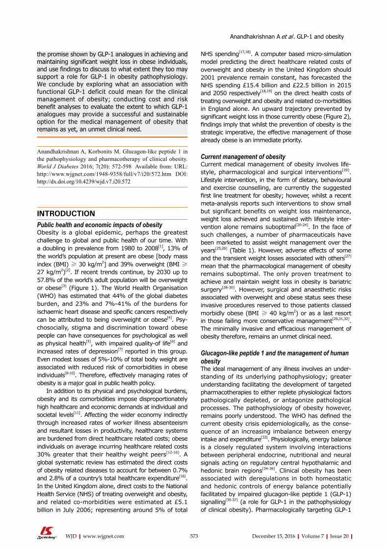
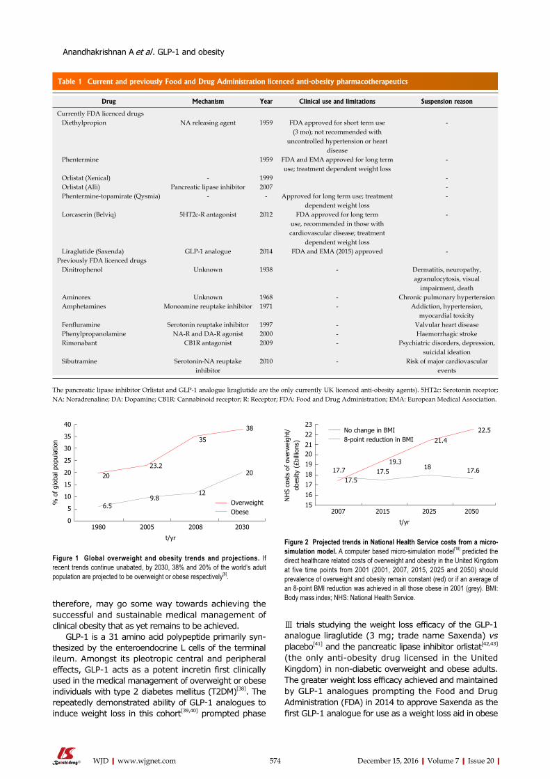
INTRODUCTIONPublic health and economic impacts of obesityObesity is a global epidemic, perhaps the greatest challenge to global and public health of our time. With a doubling in prevalence from 1980 to 2008[1], 13% of the world’s population at present are obese [body mass index (BMI) ≥ 30 kg/m2] and 39% overweight (BMI ≥ 27 kg/m2)[2]. If recent trends continue, by 2030 up to 57.8% of the world’s adult population will be overweight or obese[3] (Figure 1). The World Health Organisation (WHO) has estimated that 44% of the global diabetes burden, and 23% and 7%-41% of the burdens for ischaemic heart disease and specific cancers respectively can be attributed to being overweight or obese[4]. Psy-chosocially, stigma and discrimination toward obese people can have consequences for psychological as well as physical health[5], with impaired quality-of life[6] and increased rates of depression[7] reported in this group. Even modest losses of 5%-10% of total body weight are associated with reduced risk of comorbidities in obese individuals[8-10]. Therefore, effectively managing rates of obesity is a major goal in public health policy.
In addition to its physical and psychological burdens, obesity and its comorbidities impose disproportionately high healthcare and economic demands at individual and societal levels[11]. Affecting the wider economy indirectly through increased rates of worker illness absenteeism
and resultant losses in productivity, healthcare systems are burdened from direct healthcare related costs; obese individuals on average incurring healthcare related costs 30% greater that their healthy weight peers[12-16]. A global systematic review has estimated the direct costs of obesity related diseases to account for between 0.7% and 2.8% of a country’s total healthcare expenditure[16]. In the United Kingdom alone, direct costs to the National Health Service (NHS) of treating overweight and obesity, and related co-morbidities were estimated at £5.1 billion in July 2006; representing around 5% of total
NHS spending[17,18]. A computer based micro-simulation model predicting the direct healthcare related costs of overweight and obesity in the United Kingdom should 2001 prevalence remain constant, has forecasted the NHS spending £15.4 billion and £22.5 billion in 2015 and 2050 respectively[18,19] on the direct health costs of treating overweight and obesity and related co-morbidities in England alone. An upward trajectory prevented by significant weight loss in those currently obese (Figure 2), findings imply that whilst the prevention of obesity is the strategic imperative, the effective management of those already obese is an immediate priority.
Current management of obesity Current medical management of obesity involves life-style, pharmacological and surgical interventions[20]. Lifestyle intervention, in the form of dietary, behavioural and exercise counselling, are currently the suggested first line treatment for obesity; however, whilst a recent meta-analysis reports such interventions to show small but significant benefits on weight loss maintenance, weight loss achieved and sustained with lifestyle inter-vention alone remains suboptimal[20-24]. In the face of such challenges, a number of pharmaceuticals have been marketed to assist weight management over the years[25,26] (Table 1). However, adverse effects of some and the transient weight losses associated with others[27] mean that the pharmacological management of obesity remains suboptimal. The only proven treatment to achieve and maintain weight loss in obesity is bariatric surgery[28-30]. However, surgical and anaesthetic risks associated with overweight and obese status sees these invasive procedures reserved to those patients classed morbidly obese (BMI ≥ 40 kg/m2) or as a last resort in those failing more conservative management[20,31,32]. The minimally invasive and efficacious management of obesity therefore, remains an unmet clinical need.
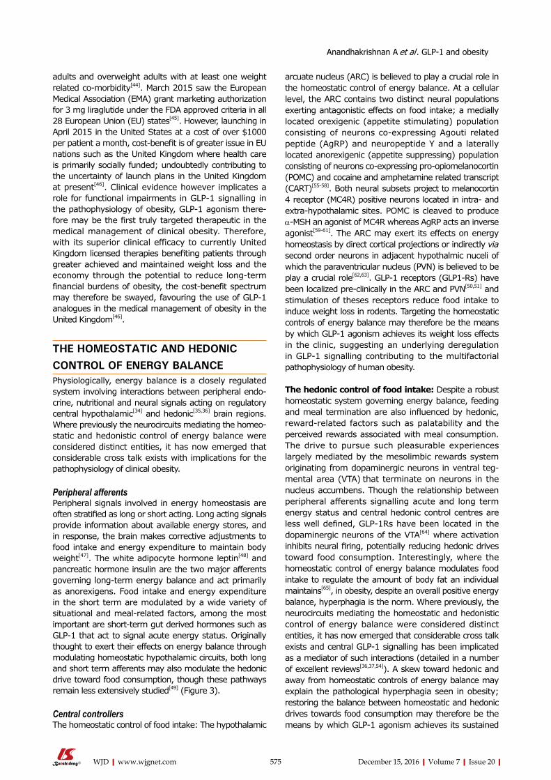
Glucagon-like peptide 1 and the management of human obesityThe ideal management of any illness involves an under-standing of its underlying pathophysiology; greater understanding facilitating the development of targeted pharmacotherapies to either replete physiological factors pathologically depleted, or antagonize pathological processes. The pathophysiology of obesity however, remains poorly understood. The WHO has defined the current obesity crisis epidemiologically, as the conse-quence of an increasing imbalance between energy intake and expenditure[33]. Physiologically, energy balance is a closely regulated system involving interactions between peripheral endocrine, nutritional and neural signals acting on regulatory central hypothalamic and hedonic brain regions[34-36]. Clinical obesity has been associated with deregulations in both homeostatic and hedonic controls of energy balance potentially facilitated by impaired glucagon-like peptide 1 (GLP-1) signalling[35-37] (a role for GLP-1 in the pathophysiology of clinical obesity). Pharmacologically targeting GLP-1
574 December 15, 2016|Volume 7|Issue 20|WJD|www.wjgnet.com
therefore, may go some way towards achieving the successful and sustainable medical management of clinical obesity that as yet remains to be achieved.
GLP-1 is a 31 amino acid polypeptide primarily syn-thesized by the enteroendocrine L cells of the terminal ileum. Amongst its pleotropic central and peripheral effects, GLP-1 acts as a potent incretin first clinically used in the medical management of overweight or obese individuals with type 2 diabetes mellitus (T2DM)[38]. The repeatedly demonstrated ability of GLP-1 analogues to induce weight loss in this cohort[39,40] prompted phase
Ⅲ trials studying the weight loss efficacy of the GLP-1 analogue liraglutide (3 mg; trade name Saxenda) vs placebo[41] and the pancreatic lipase inhibitor orlistat[42,43] (the only anti-obesity drug licensed in the United Kingdom) in non-diabetic overweight and obese adults. The greater weight loss efficacy achieved and maintained by GLP-1 analogues prompting the Food and Drug Administration (FDA) in 2014 to approve Saxenda as the first GLP-1 analogue for use as a weight loss aid in obese
40
35
30
25
20
15
10
5
01980 2005 2008 2030
t/yr
% o
f gl
obal
pop
ulat
ion
OverweightObese
2023.2
35
38
6.59.8
12
20
Figure 1 Global overweight and obesity trends and projections. If recent trends continue unabated, by 2030, 38% and 20% of the world’s adult population are projected to be overweight or obese respectively[8].
23
22
2120
19
18
17
16
152007 2015 2025 2050
t/yr
NH
S co
sts
of o
verw
eigh
t/ob
esity
(£b
illio
ns)
17.719.3
21.4
22.5
17.517.5
18 17.6
No change in BMI8-point reduction in BMI
Figure 2 Projected trends in National Health Service costs from a micro-simulation model. A computer based micro-simulation model[18] predicted the direct healthcare related costs of overweight and obesity in the United Kingdom at five time points from 2001 (2001, 2007, 2015, 2025 and 2050) should prevalence of overweight and obesity remain constant (red) or if an average of an 8-point BMI reduction was achieved in all those obese in 2001 (grey). BMI: Body mass index; NHS: National Health Service.
Drug Mechanism Year Clinical use and limitations Suspension reason
Currently FDA licenced drugs Diethylpropion NA releasing agent 1959 FDA approved for short term use
(3 mo); not recommended with uncontrolled hypertension or heart
disease
-
Phentermine 1959 FDA and EMA approved for long term use; treatment dependent weight loss
-
Orlistat (Xenical) - 1999 - Orlistat (Alli) Pancreatic lipase inhibitor 2007 - Phentermine-topamirate (Qysmia) - - Approved for long term use; treatment
dependent weight loss-
Lorcaserin (Belviq) 5HT2c-R antagonist 2012 FDA approved for long term use, recommended in those with cardiovascular disease; treatment
dependent weight loss
-
Liraglutide (Saxenda) GLP-1 analogue 2014 FDA and EMA (2015) approved -Previously FDA licenced drugs Dinitrophenol Unknown 1938 - Dermatitis, neuropathy,
agranulocytosis, visual impairment, death
Aminorex Unknown 1968 - Chronic pulmonary hypertension Amphetamines Monoamine reuptake inhibitor 1971 - Addiction, hypertension,
myocardial toxicity Fenfluramine Serotonin reuptake inhibitor 1997 - Valvular heart disease Phenylpropanolamine NA-R and DA-R agonist 2000 - Haemorrhagic stroke Rimonabant CB1R antagonist 2009 - Psychiatric disorders, depression,
suicidal ideation Sibutramine Serotonin-NA reuptake
inhibitor2010 - Risk of major cardiovascular
events
Table 1 Current and previously Food and Drug Administration licenced anti-obesity pharmacotherapeutics
The pancreatic lipase inhibitor Orlistat and GLP-1 analogue liraglutide are the only currently UK licenced anti-obesity agents). 5HT2c: Serotonin receptor; NA: Noradrenaline; DA: Dopamine; CB1R: Cannabinoid receptor; R: Receptor; FDA: Food and Drug Administration; EMA: European Medical Association.
Anandhakrishnan A et al . GLP-1 and obesity
575 December 15, 2016|Volume 7|Issue 20|WJD|www.wjgnet.com
adults and overweight adults with at least one weight related co-morbidity[44]. March 2015 saw the European Medical Association (EMA) grant marketing authorization for 3 mg liraglutide under the FDA approved criteria in all 28 European Union (EU) states[45]. However, launching in April 2015 in the United States at a cost of over $1000 per patient a month, cost-benefit is of greater issue in EU nations such as the United Kingdom where health care is primarily socially funded; undoubtedly contributing to the uncertainty of launch plans in the United Kingdom at present[46]. Clinical evidence however implicates a role for functional impairments in GLP-1 signalling in the pathophysiology of obesity, GLP-1 agonism there-fore may be the first truly targeted therapeutic in the medical management of clinical obesity. Therefore, with its superior clinical efficacy to currently United Kingdom licensed therapies benefiting patients through greater achieved and maintained weight loss and the economy through the potential to reduce long-term financial burdens of obesity, the cost-benefit spectrum may therefore be swayed, favouring the use of GLP-1 analogues in the medical management of obesity in the United Kingdom[46].
THE HOMEOSTATIC AND HEDONIC CONTROL OF ENERGY BALANCEPhysiologically, energy balance is a closely regulated system involving interactions between peripheral endo-crine, nutritional and neural signals acting on regulatory central hypothalamic[34] and hedonic[35,36] brain regions. Where previously the neurocircuits mediating the homeo-static and hedonistic control of energy balance were considered distinct entities, it has now emerged that considerable cross talk exists with implications for the pathophysiology of clinical obesity.
Peripheral afferentsPeripheral signals involved in energy homeostasis are often stratified as long or short acting. Long acting signals provide information about available energy stores, and in response, the brain makes corrective adjustments to food intake and energy expenditure to maintain body weight[47]. The white adipocyte hormone leptin[48] and pancreatic hormone insulin are the two major afferents governing long-term energy balance and act primarily as anorexigens. Food intake and energy expenditure in the short term are modulated by a wide variety of situational and meal-related factors, among the most important are short-term gut derived hormones such as GLP-1 that act to signal acute energy status. Originally thought to exert their effects on energy balance through modulating homeostatic hypothalamic circuits, both long and short term afferents may also modulate the hedonic drive toward food consumption, though these pathways remain less extensively studied[49] (Figure 3).
Central controllersThe homeostatic control of food intake: The hypothalamic
arcuate nucleus (ARC) is believed to play a crucial role in the homeostatic control of energy balance. At a cellular level, the ARC contains two distinct neural populations exerting antagonistic effects on food intake; a medially located orexigenic (appetite stimulating) population consisting of neurons co-expressing Agouti related peptide (AgRP) and neuropeptide Y and a laterally located anorexigenic (appetite suppressing) population consisting of neurons co-expressing pro-opiomelanocortin (POMC) and cocaine and amphetamine related transcript (CART)[55-58]. Both neural subsets project to melanocortin 4 receptor (MC4R) positive neurons located in intra- and extra-hypothalamic sites. POMC is cleaved to produce α-MSH an agonist of MC4R whereas AgRP acts an inverse agonist[59-61]. The ARC may exert its effects on energy homeostasis by direct cortical projections or indirectly via second order neurons in adjacent hypothalmic nuceli of which the paraventricular nucleus (PVN) is believed to be play a crucial role[62,63]. GLP-1 receptors (GLP1-Rs) have been localized pre-clinically in the ARC and PVN[50,51] and stimulation of theses receptors reduce food intake to induce weight loss in rodents. Targeting the homeostatic controls of energy balance may therefore be the means by which GLP-1 agonism achieves its weight loss effects in the clinic, suggesting an underlying deregulation in GLP-1 signalling contributing to the multifactorial pathophysiology of human obesity.
The hedonic control of food intake: Despite a robust homeostatic system governing energy balance, feeding and meal termination are also influenced by hedonic, reward-related factors such as palatability and the perceived rewards associated with meal consumption. The drive to pursue such pleasurable experiences largely mediated by the mesolimbic rewards system originating from dopaminergic neurons in ventral teg-mental area (VTA) that terminate on neurons in the nucleus accumbens. Though the relationship between peripheral afferents signalling acute and long term energy status and central hedonic control centres are less well defined, GLP-1Rs have been located in the dopaminergic neurons of the VTA[64] where activation inhibits neural firing, potentially reducing hedonic drives toward food consumption. Interestingly, where the homeostatic control of energy balance modulates food intake to regulate the amount of body fat an individual maintains[65], in obesity, despite an overall positive energy balance, hyperphagia is the norm. Where previously, the neurocircuits mediating the homeostatic and hedonistic control of energy balance were considered distinct entities, it has now emerged that considerable cross talk exists and central GLP-1 signalling has been implicated as a mediator of such interactions (detailed in a number of excellent reviews[36,37,54]). A skew toward hedonic and away from homeostatic controls of energy balance may explain the pathological hyperphagia seen in obesity; restoring the balance between homeostatic and hedonic drives towards food consumption may therefore be the means by which GLP-1 agonism achieves its sustained
Anandhakrishnan A et al . GLP-1 and obesity
576 December 15, 2016|Volume 7|Issue 20|WJD|www.wjgnet.com
weight loss effects in the clinic, suggesting an underlying deregulation in GLP-1 signalling contributing to the multi-factorial pathophysiology of human obesity.
GLP-1Synthesis, secretion and degradationGLP-1 is a 31 amino acid polypeptide derived from post-translational processing of the native 160 amino acid peptide proglucagon by the enzyme prohormone convertase 1 (PC1/3). Peripheral proglucagon gene
expression has been localized to the enteroendocrine L cells and pancreatic α-cells whilst centrally, proglucagon expressing neurons have been localized to brainstem regions such as the nucleus of the solitary tract (NTS)[66-68]. Tissue specific post-translational processing liberates different pro-glucagon derived peptides[69] depending on subtype of PC enzyme present. Figure 4 details the different post-translational products following PC1/3 and 2 cleavage.
GLP-1 is primarily synthesized by PC1/3 activity in the intestinal L cells[75]; open-type epithelial cells
Afferent Central processing Efferent
Liver
Nutrients
Adipose tissue
LeptinAdipokines
Pancreas
InsulinPP
EnterostatinAmylin
Glucagon
GI-tract
GhrelinCKKPYY
GLP-1OXM
ChemoreceptorsMechanoreceptors
BBB GLP-1R
GLP-1R
GLP-1R
Hedonic control VTA
Homeostatic control
ARC
NPYAgRP
POMCCART
Brain stem
Vagus nerve
PVN
CRH TRH
LHA
Orexin MCH
Pituitary Thyrotrophs
Pituitary Corticotrophs
Thyroid functionGrowth and reproduction
Sympathetic and parasympathetic
Pre-ganglionic neurons
Autonomic function, e.g. , adaptive thermogenesis
Cerebral cortexHypothalamusOlfactory areasBehavioural,e.g. , feeding patterns
Behavioural changes
e.g. , feeding
Food intake Energy expenditure
Energy homeostasis
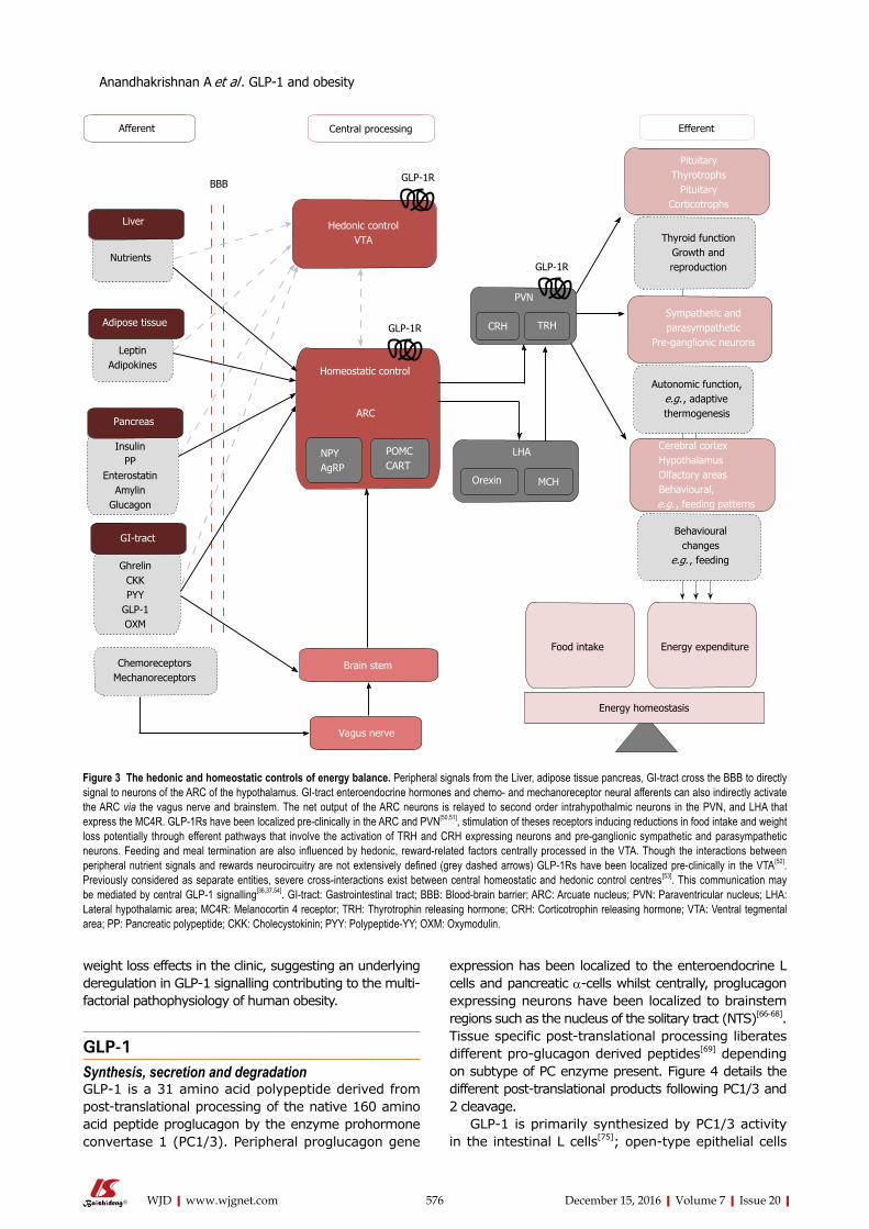
Figure 3 The hedonic and homeostatic controls of energy balance. Peripheral signals from the Liver, adipose tissue pancreas, GI-tract cross the BBB to directly signal to neurons of the ARC of the hypothalamus. GI-tract enteroendocrine hormones and chemo- and mechanoreceptor neural afferents can also indirectly activate the ARC via the vagus nerve and brainstem. The net output of the ARC neurons is relayed to second order intrahypothalmic neurons in the PVN, and LHA that express the MC4R. GLP-1Rs have been localized pre-clinically in the ARC and PVN[50,51], stimulation of theses receptors inducing reductions in food intake and weight loss potentially through efferent pathways that involve the activation of TRH and CRH expressing neurons and pre-ganglionic sympathetic and parasympathetic neurons. Feeding and meal termination are also influenced by hedonic, reward-related factors centrally processed in the VTA. Though the interactions between peripheral nutrient signals and rewards neurocircuitry are not extensively defined (grey dashed arrows) GLP-1Rs have been localized pre-clinically in the VTA[52]. Previously considered as separate entities, severe cross-interactions exist between central homeostatic and hedonic control centres[53]. This communication may be mediated by central GLP-1 signalling[36,37,54]. GI-tract: Gastrointestinal tract; BBB: Blood-brain barrier; ARC: Arcuate nucleus; PVN: Paraventricular nucleus; LHA: Lateral hypothalamic area; MC4R: Melanocortin 4 receptor; TRH: Thyrotrophin releasing hormone; CRH: Corticotrophin releasing hormone; VTA: Ventral tegmental area; PP: Pancreatic polypeptide; CKK: Cholecystokinin; PYY: Polypeptide-YY; OXM: Oxymodulin.
Anandhakrishnan A et al . GLP-1 and obesity
577 December 15, 2016|Volume 7|Issue 20|WJD|www.wjgnet.com
most densely located in the ileum and colon[76-78]. Long apical processes that extend toward the intestinal lumen[77] allow direct nutrient sensing by L cells, of which glucose has been implicated as the most potent GLP-1 secretagogue in both healthy and T2DM humans[79]. Being in close proximity to neurons of the enteric nervous system and the intestinal microvasculature[80,81], L cells also receive neural and hormonal signals that act as indirect nutrient sensors. Following synthesis, GLP-1 is secreted from the L-cells via secretory granules located in the basolateral membrane. GLP-1 secretion in re-sponse to nutrient sensing is biphasic; an initial rapid rise occurring within 10-15 min post-prandial, followed by a second longer phase peaking at 30-60 min[82]. The early phase of GLP-secretion has traditionally been attributed to signals from the parasympathetic vagal nerve and neurotransmitters such as gastrin-releasing peptide (GRP) and acetylcholine. However, more recently, GLP-1 secreting cells that show direct secretory responses to nutrient stimulation have been localised in significant numbers in the proximal small intestine implicating a role for this albeit sparser population of proximal GLP-1 releasing cells in the rapid postprandial rises of plasma GLP-1[83-85]. The second phase is mediated via direct nutrient contact with subsequent membrane depolarization or activation of second messenger systems mediating GLP-1 release. Figure 5 depicts the major nutrient, neural and hormonal secretagogues of GLP-1.
Secreted GLP-1 is rapidly degraded at its N-terminal residue by the ubiquitously expressed enzyme dipe-ptidyl peptidase Ⅳ (DPPV) to yield residues GLP-1 (9-36 amide) and GLP-1 9-37[88,89]. The majority of GLP-1 degradation is attributed to membrane-bound DPPV in the hepatic portal system resulting in an extremely short half-life (about 2 min)[81,90]. As such, only about 10%-15%
of GLP1 secreted from intestinal L cells reaches peripheral downstream targets. The amount of GLP-1 reaching potential central targets involved in energy balance is unknown. As parenteral administration of GLP-1 avoids the physiological first-pass effect of hepatic DPPV, the supraphysiological plasma concentrations achieved by subcutaneous (SC) administration may explain the weight loss efficacy achieved by 3 mg liraglutide in obese and overweight patients in the clinic. Findings also go some way to suggest either a reduction in secretion of, or sensitivity to, physiological GLP-1 secretion as a contributor to the multifactorial pathophysiology of human obesity.
Central and peripheral effects of GLP-1GLP-1 exerts its effects by intracellular signalling path-ways activated after binding to the G-protein coupled receptor GLP-1R[91]. The extensive central and peri-pheral expression of the GLP-R reflects the pleotropic physiological roles of GLP-1 that are summarised in Figure 6 and extensively reviewed elsewhere[70,86]. From this point onward the review will focus on exploring the evidence surrounding a role for physiological GLP-1 signalling in the regulation of energy balance and deregulations of this signalling as one contributor to the multifactorial pathophysiology of clinical obesity.
GLP-1 AND THE REGULATION OF ENERGY BALANCEEvidence: Effects of GLP-1 administration on food intake and energy expenditure in manNumerous clinical studies have examined the relationship between acute physiological and supraphysiological
Proglucagon
1 30 33 61 69 72 107 111 123 126 160
Proglucagon1 30 33 61 69 72 107 111 123 126 160
Intestine
Glycentin
Oxyntomodulin
GLP-1 (7-36)
GLP-1 (7-38)
IP2
GLP-2
1 69
33 69
72 108
78 108
107 111
126/128 158
Pancreas
Glucagon
IP-1
GRP1
MPGF
33 61
1 30
69
72 111 123 126 158
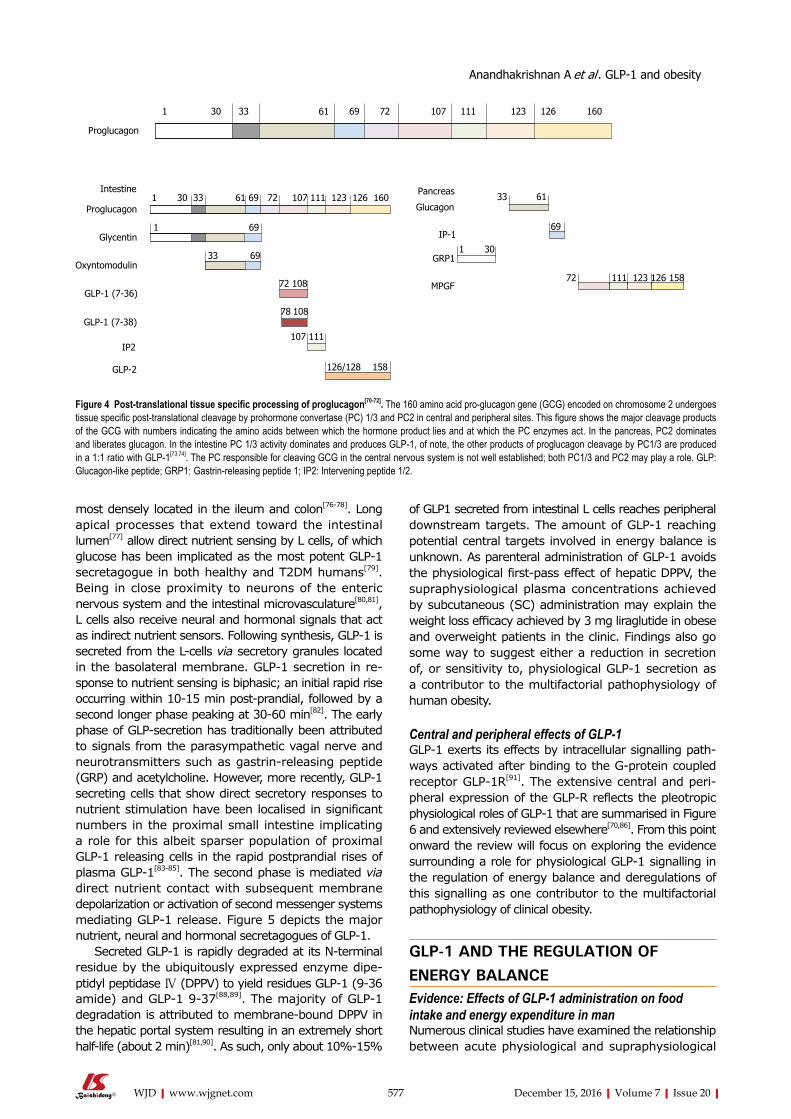
Figure 4 Post-translational tissue specific processing of proglucagon[70-72]. The 160 amino acid pro-glucagon gene (GCG) encoded on chromosome 2 undergoes tissue specific post-translational cleavage by prohormone convertase (PC) 1/3 and PC2 in central and peripheral sites. This figure shows the major cleavage products of the GCG with numbers indicating the amino acids between which the hormone product lies and at which the PC enzymes act. In the pancreas, PC2 dominates and liberates glucagon. In the intestine PC 1/3 activity dominates and produces GLP-1, of note, the other products of proglucagon cleavage by PC1/3 are produced in a 1:1 ratio with GLP-1[73,74]. The PC responsible for cleaving GCG in the central nervous system is not well established; both PC1/3 and PC2 may play a role. GLP: Glucagon-like peptide; GRP1: Gastrin-releasing peptide 1; IP2: Intervening peptide 1/2.
Anandhakrishnan A et al . GLP-1 and obesity

578 December 15, 2016|Volume 7|Issue 20|WJD|www.wjgnet.com
doses of GLP-1 with measurements of food intake and feelings of hunger and satiety in healthy normal weight and obese adults with and without T2DM[92-99]. The main findings of these studies have been summarized in Figure 7. Though individual studies are conflicting, a meta-analysis reports that acute GLP-1 infusion induces a mean 11.7% decrease in food intake when compared with saline control in man[100]. Interestingly, whilst supra-physiological doses of GLP-1 reduces appetite and food intake in both lean and obese subjects, physiological GLP-1 doses reduces appetite and food intake in only lean subjects[93,94,97,99]. Findings go some way to suggest
a role for resistance to physiological GLP-1 signalling as a factor contributing to obesity pathophysiology. Interestingly, whilst physiological GLP-1 infusions in obese subjects induce appetite reductions[92,95] similar to those observed in their lean peers, this is not translated into a reduction in food intake, suggesting pathological alterations of GLP-1 signalling in obesity that reinforce feeding despite a reduced physiological drive to food intake. One mechanism that this may be achieved is through a pathological skew toward hedonic and away from homeostatic controls of energy balance in obesity, potentially mediated by deregulated central GLP-1 sig-nalling (a role for GLP-1 in the pathophysiology of clinical obesity).
Whilst evidence from clinical interventional studies suggests that physiological GLP-1 contributes to negative energy balance by decreasing food intake. The effects of GLP-1 on energy expenditure are less clear. Fasting plasma GLP-1 concentrations have been positively asso-ciated with increased rates of energy expenditure in man[101]. Clinical evidence regarding the effects of acute GLP-1 administration on energy expenditure however is conflicting. Physiological infusions of GLP-1 have been reported to reduce energy expenditure in lean and non-diabetic obese patients[95,102] associated with reduced carbohydrate metabolism. Others, however, have observed that supraphysiological infusions of GLP-1 increase energy expenditure in lean individuals in an insulin dependent manner[103].
Interpretations: Implications for the clinicEvidence from clinical interventional studies suggests that acute post-meal rises in GLP-1 contribute to negative energy balance primarily through an anorexigenic effect. The long-acting GLP-1 analogue liraglutide (3 mg) has recently been approved as a once daily bolus SC injection for the medical management of obesity. The sustained anorectic effect of a long term agonist combined with supraphysiological dosing perhaps the mechanism of the clinical weight loss efficacy achieved by liraglutide 3 mg. Unfortunately, to date, clinical studies assessing the comparative efficacy of acute vs continuous GLP-1 administration on appetite reduction and weight loss remain scarce. Näslund et al[104] compared the effects of 4 doses of acute GLP-1 infused 30 min prior to meals [prandial subcutaneous infusion (PSI)] to an equivalent dose of continuous SC GLP-1 infusion (CSI) on food intake and weight loss in non diabetic obese patients. Though both acute and continuous GLP-1 infusion produced significant reductions in food intake when compared to placebo (P = 0.02 PSI and CSI), a statistically significant weight loss compared to placebo was only observed following PSI. With respect to the clinic, findings suggest that lowered dose; more frequent GLP-1 administration may prove more efficacious in inducing weight loss in obese patients. Nevertheless, in view of the negative impact of SC drug administration on patient adherence and the potential biases associated with the
Somatostatin
Leptin
Insulin
Acetylcholine
M1RM2R
GPCR 119
FFA derivative
Fats
FFABile
acids
Carbohydrates
Glucose
TGRSNa+
GPCR 40/120
SGLT-1
Protein
AA
Na+
AA transporter
L-type Ca2+ channelNa+ Na+
KATP channel
ΔΨ
ΔΨ[ATP]icAMP
AC
Ca2+
[Ca2+]i
MAPK PKC
Figure 5 Mechanisms of glucagon-like peptide 1 release from entero-endocrine L cell. Glucagon-like peptide 1 (GLP-1) release from L-cells is regulated by direct nutrient sensing via receptors and channels on apical processes or indirectly via neuro-hormonal mechanisms[70,71,86,87]. A: Nutrient signals. Carbohydrates: Glucose derived from carbohydrate metabolism is the most potent stimuli for GLP-1 secretion. Glucose can trigger GLP-1 release by two mechanisms: (1) the sodium-glucose cotransporter-1 (SGLT-1) couples the transport of glucose with Na ions. Na = influx leads to membrane depolarization (ΔΨ) (red arrows); and (2) glucose metabolism generates adenosine triphosphate (ATP). Elevated intracellular ATP concentrations [ATP]i close KATP channels and leads to membrane depolarization (ΔΨ) (green arrows). Both routes to membrane depolarisation increase intracellular Ca levels ([Ca2+]i) by opening L-type Ca channels. Elevated [Ca2+]i triggers the exocytosis of GLP-1 secretory granules located at the basolateral surface of the enteroendocrine L cell (dashed lines). Fats: Fats are potent stimuli for GLP-1 secretion. Free fatty acids (FFA) (blue arrows) interact with G-protein coupled receptors (GPCRs) that trigger Ca2+ release from internal stores and also activate protein kinase C (PKC). FFA derivates (purple arrows) interact with GPCRs that activate second messenger systems involving adenylate cyclase (AC) and cyclic AMP (cAMP) which increases [Ca2+]i. Bile acids (orange arrows) and short chained fatty acids (not shown) also increase [Ca2+]i by GPCR interactions. Proteins: Protein is a weak stimulator of GLP-1 release when compared with sugars and lipids. Amino acids (AA) derived from protein breakdown are transported intracelluarly with Na+ via Na+ dependent AA transporters. Na+ influx causes membrane depolarization and elevated [Ca2+]i with resultant GLP-1 exocytosis (pink arrows); B: Hormonal signals. Somatostatin inhibits GLP-1 release by blocking AC activation (light blue arrows). The peripheral adiposity signals leptin (yellow arrows) and insulin (brown arrows) are thought to stimulate GLP-1 release via activation of mitogen-activated protein kinase (MAPK) signalling pathway; C: Neural signals. Acetylcholine binding to muscarinic receptors (M1R, M2R) elevates [Ca2+]I stimulating GLP-1 release (grey arrows). GRP is though to stimulate GLP-1 release in association with the activation of mitogen activated protein kinase kinase (MAPKK) and subsequent phosphorylation and activation of MAPK (not shown).
Anandhakrishnan A et al . GLP-1 and obesity

579 December 15, 2016|Volume 7|Issue 20|WJD|www.wjgnet.com
significantly greater peak plasma GLP-1 concentrations achieved following PSI compared to CSI (269.4 pmol vs 88.7 pmol) once daily bolus administration at present, seems to be the most clinically efficacious means of therapeutic GLP-1 analogue delivery.
Interpretations: Potential effectors of GLP-1s negative energy balance effectsClinical and pre-clinical evidence suggests that targeting peripherally and centrally located GLP-1Rs may exert the anorectic effects of physiological GLP-1 signalling.
Peripheral effectors: Histological studies in man have shown GLP-1Rs to be expressed in cells of the gastric mucosa and in pancreatic islet cells[105,106]. Pre-clinically, stimulation of gastric and pancreatic GLP1-Rs are associated with reductions in food intake that
occurs alongside activation of hedonic and homeostatic brain regions[47,63,86]. Findings suggest physiological GLP-1 signalling may induce its anorectic effects in man by indirectly activating central controllers of appetite through gastric and pancreatic receptors.
Gastric mechanoreceptors are activated by gastric distension following acute nutrient intake, and gastric mechanoreceptor signalling plays an important role as a meal-to-meal satiety signal, activating the NTS which in turn modulates neural activity in both the ARC the VTA[107] (the homeostatic and hedonic control of energy balance). By relaying to the NTS, mechanoreceptor induced anorectic effects may therefore be exerted through modulation of both homeostatic and hedonic appetite control. The amount of gastric distension in response to a given meal is negatively associated with the rate of gastric emptying; delayed gastric emptying
Pancreas ↑ Insulin secretion ↓ Glucagon secretion ↑ β-cell proliferation ↓ β-cell apoptosis
CNS ↑ Satiety ↓ Appetite ↑ Energy expenditure
Hepatic ↑ Glycogen synthesis ↓ Hepatic glucose production ↓ Hepatic steatosis ↓ VLDL (ApoB100)
Kidney ↑ Natriuresis ↑ Diuresis
GLP-1
Cardiovascular system ↓ Ischeamia/reperfusion injury ↓ Blood pressure ↑ Heart rate
Fat ↑ Glucose uptake ↓ Inflammation
Muscle ↑ Glycogen synthesis ↑ Glucose oxidation
GIT ↓ Gastric emptying ↓ Acid secretion ↓ Chylomicrons (ApoB100)
Figure 6 Central and peripheral effects of glucagon-like peptide 1. Ex vivo and in vivo studies in rodents, and observational and interventional studies in man have allowed the characterization of numerous central and peripheral effects of GLP-1. Peripheral effects of GLP-1 may be classed broadly as pancreatic or extra-pancreatic. Pancreatic effects of GLP-1 act to promote insulin secretion (incretin effect). Extra-pancreatic effects of GLP-1 include: (1) regulation of energy metabolism and nutrient storage (liver, muscle and fat); (2) efficient nutrient handling (stomach and GIT); and (3) others: Cardiovascular repair, blood pressure control, diuresis[86]. VLDL: Very low-density lipoproteins; GLP-1: Glucagon-like peptide 1; GIT: Gastrointestinal tract; CNS: Central nervous system.
Flint et al [97], 1998Näslund et al [93], 1999Gutzwiller et al [94], 1999
Long et al [98], 1999
Gutzwiller et al [99], 1999
Cegla et al [96], 2014
Näslund et al [92], 1998Flint et al [95], 2001
Figure 7 Effects on Visual Analogue Scale assessed appetite scores and ad libitum food intake in lean and obese subjects following physiological and supraphysiological[92-99] infusions of glucagon-like peptide 1. Though individual studies report conflicting data, a meta-analysis of clinical studies evaluating the acute effects of GLP-1 infusion on food intake reports a mean 11.7% decrease when compared with saline control[100]. GLP-1: Glucagon-like peptide 1.
Appetite
Food intake
Appetite
Food intake
Appetite
Food intake
Appetite
Food intake
Appetite
Food intake
Anandhakrishnan A et al . GLP-1 and obesity

580 December 15, 2016|Volume 7|Issue 20|WJD|www.wjgnet.com
positively associated with increased satiety and fullness in both healthy and obese patients[108-112]. GLP-1 has been found to delay gastric emptying in healthy lean, obese and T2DM subjects, and histological studies in man have shown that GLP-1Rs are expressed in gastric mucosa[92,105,113-116]. Post-prandial GLP-1 secretion may therefore exert its anorectic effect through activating GLP-1Rs in gastric mucosa, which in turn increase mecha-noreceptor firing and signalling to the NTS. Though the neurotransmitters involved in relaying signals from the NTS to homeostatic and hedonic appetite controls remain to be defined, physiological gastric distension in rodents has been shown to up-regulate GLP-1 gene expression in the NTS associated with central proglucagon processing[117], implicating a role for centrally synthesised GLP-1.
In the fasted state, the stomach is empty and so gastric motility is reduced to basal levels. That reductions in appetite after GLP-1 administration have been observed in fasting human subjects[99], suggests that mechanisms other delaying gastric motility contribute to the physiological anorectic effect of GLP-1. The glu-coregulatory hormone insulin, traditionally viewed as an anorectic signal involved in the regulation of long-term energy balance[47,63], displays both basal and acute meal-related secretion[118]. With acute insulin administration associated with reduced ad libitum food intake in healthy lean individuals[119], findings implicate a role for insulin as an anorexigen involved in the regulation of short term energy balance. Insulin receptors are widely expressed in the ARC and VTA[120-122], thus modulation of both homeostatic and hedonic appetite control may be the means by which insulin exerts its anorectic effects on short-term energy balance.
The most extensively studied of GLP-1’s physiolo-gical roles is as a positive modulator of insulin secretion from pancreatic β-cells[123] (evidence: Effects of GLP-1 administration on food intake and energy expenditure in man). Whilst GLP-1 has been shown to increase energy expenditure in healthy lean individuals in an insulin dependent manner[103], no clinical evidence to date exists exploring the role of insulin as a mediator of GLP-1 anorexigenic signalling. Studies assessing the effects of GLP-1 interactions with the oriexigen ghrelin however, suggest that this may indeed be the case. Ghrelin receptors have been localised preclinically in Agrp/NpY neurons of the ARC and dopaminergic neurons of the VTA[124,125], with activation of neurons in either brain region producing orexigenic effects. GLP-1 infusion in healthy lean humans is associated with significant suppression of postprandial rises in ghrelin[126]; the decline in orexigenic signalling a potential indirect mediator of GLP-1s anorexigenic effect. Interestingly, the reductions in ghrelin concentration observed with GLP-1 infusion inversely correlate with coinciding rises in insulin concentration and elsewhere, insulin infusion has been shown to display a reciprocal relationship with ghrelin secretion in man[127]. Together, findings suggest that
GLP-1’s anorectic effects may be mediated secondary to its incretin effect that in turn that suppresses ghrelin release, thus orexigenic signalling.
Central controllers: Histological and in vivo studies in rodents have shown that GLP-1Rs are expressed in anorexigenic POMC/CART neurons of the ARC and in dopaminergic neurons of the VTA[59,61,18,128] where they stimulate and inhibit neural firing respectively. Preclinical studies have shown that the stimulation of the POMC/ARC neurons of the hypothalamus and inhibition of the dopaminergic neurons of the VTA reduce food intake. Findings suggest that GLP-1 may exert its negative energy balance effects in man through direct activation of central GLP-1 receptors in the ARC and VTA; activating the anorexigenic homeostatic and inhibiting the hedonic hyperphagic drives to food intake. With the development of neuroimaging techniques, in vivo clinical studies substantiate the effects of GLP-1 on brain regions involved in the homeostatic and hedonic controls of energy balance. Whether these effects are mediated by direct central GLP-1R activation or indirectly via peripherally located GLP-1Rs however, remain to be defined.
Using fluorodeoxyglucose positron emission to-mography Alvarez et al[129] demonstrate that GLP-1 infusion in lean individuals reduces glucose metabolism in the hypothalamus and brainstem. With patients fasted during the study and with no changes in peripheral hormone profiles observed, the effects of gastric mecha-noreceptor activation or other hormonal influences respectively on observed effects are negated. Elsewhere, correlations between PET assessed increases in hypo-thalamic blood flow and physiological post-prandial rises in serum GLP-1 have been observed[130]. Both findings may represent altered neural activity in brain regions associated with homeostatic energy balance secondary to direct or indirect GLP-1/GLP-1R signalling. The effects of this alteration in central neural activity on food intake and appetite however, have not been explored. Using functional magnetic resonance imaging (fMRI), De Silva et al[131] demonstrate that GLP-1 infusion in lean individuals attenuates neuronal activity in 6 brain regions involved in rewards processing and hedonic feeding accompanied with reductions in food intake. Though neither parameter reached statistical significance vs placebo, results support the idea that central GLP-1 signalling may at least in part exert its negative energy balance effects through modulations in hedonic appetite control centres, potentially by reducing the hedonic value associated with food and food-driven motivation.
Clinical evidence exists to suggest that the SNS modulates energy expenditure through increased thermogenesis assessed in vivo as muscle sympathetic nerve activity (MSNA)[132,133]; increased MSNA positively associated with increased short and longer term energy expenditure in otherwise healthy human subjects[134,135]. Peripheral GLP-1 infusion has been shown to significantly
Anandhakrishnan A et al . GLP-1 and obesity

581 December 15, 2016|Volume 7|Issue 20|WJD|www.wjgnet.com
increase MSNA in healthy human controls[136] and suggest that GLP-1 signalling may produce its negative energy balance effects not only through anorexigenic signalling, but also by increasing energy output.
A ROLE FOR GLP-1 IN THE PATHOPHYSIOLOGY OF CLINICAL OBESITYGeneticsGenetic analyses in man suggest clinical obesity is associated with a lack of functional GLP-1 signalling that may contribute to the development of the obesity phenotype.
Monogenic human obesity: Monogenic human obesity is a rare form of clinical obesity that shows Mendelian patterns of inheritance; the obesity phenotype attributed to the loss or gain of function in a single gene[137]. Two broad classes of Mendelian human obesity exist; syndromic obesity encompasses about 30 Mendelian disorders wherein obesity co-presents alongside chara-cteristic physical and developmental anomalies. Though causative genes have been identified, the mechanisms through which the genetic mutations induce obesity are not completely understood in all cases[138]. Non-syndromic obesity is characterized by a severe, early onset hyperphagic obesity attributed to loss of function mutations in 1 of 11 genes[139-141]. Interestingly, 8 of these genes have physiological roles in the central control of energy balance[142]. One such gene is PCSK1 encoding the enzyme PC1/3 involved in the proteolytic processing of proglucagon, to yield, amongst other peptides, GLP-1 (GLP-1). Six studies to date document the relationship between autosomal recessive, compound heterozygous or homozygous[143-148] mutations in PCSK1 in 21 probands associated with reduced or absent function of PC1/3. Table 2 details the phenotypes of probands, all of whom presented with an early onset hyperphagic obesity and malabsorptive diarrhoea with varying, though extensively
overlapping endocrine phenotypes. Though the cause of the obesity and endocrine pheno-
types associated with PCSK1 mutation are unknown, they may well be attributed to the loss of PC1/3 pro-hormone processing function. Signs of impaired in-testinal[146] pro-glucagon processing have been described in probands with PC1/3 deficiency and may contribute to the development of the obesity phenotype secondary to reduced GLP-1 synthesis. Disappointingly, only 2 of 6 studies detailing the phenotypes PCSK1 mutant probands assess post-prandial GLP-1 secretory responses and report conflicting results; whilst an oral glucose load (OGTT) yields significantly reduced GLP-1 response in three child probands compared to age matched con-trols, post-prandial responses in a 40-year-old proband match those of healthy age-matched controls. One interpretation of such findings may be that whilst other PCs may compensate for lacking PC1/3 to allow for GLP-1 synthesis in response to mixed nutrient secretagogues, PC1/3 is necessary and essential for GLP-1 synthesis in response to its most potent secretagogue, glucose. An alternative interpretation comes from observations that GLP-1 secretion following OGTT in the 3 child probands studied by Bandsma et al[146] seem to show an age dependent impairment improving with increasing age. Following follows reports by Parker et al[147] who observed that the pattern of endocrinopathy in probands with PSCK1 mutant monogenic obesity change with age, perhaps GLP-1 secretion too may show an age-dependent alteration, potentially compensated for over time. One way to test this hypothesis would be to histologically examine the enteroendocrine expression of GLP-1 in adult PCSK1-mutant probands; enteroendocrine expression of GLP-1 is significantly reduced compared to control in children with PCSK1 monogenic obesity[146], if indeed the normal post-prandial GLP-1 responses seen in adulthood are a reflection of the activation of redundant PC activity in intestinal cells up regulation of enteroendocrine GLP-1 expression would be observed. Though the cause of the hyperphagic obesity in PCSK1 mutant human monogenic obesity remains ill defined,
Ref. Jackson et al [144], 1997
Jackson et al [143], 2003
Farooqi et al [148], 2007
Frank et al [145], 2013
Parker et al [147], 2013
Bandsma et al [146], 2013
Obesity phenotypeHyperphagic, early-onset Yes Yes Yes Yes Yes YesEndocrine phenotypeAbnormal glucose metabolism Yes Yes Yes Yes YesHypogonadotrophic hypogonadism Yes Yes YesHypocortisolaemia Yes Yes Yes YesHypothyroidism Yes Yes YesCentral diabetes insipidus YesOthersEarly onset malabsorptive diarrhoea Yes Yes Yes Yes Yes Yes
Table 2 Obesity and endocrine phenotypes in probands with PCSK1 gene deletion monogenic obesity
Proband details: Jackson et al[144] (1997) - a 40-year-old Caucasian woman; Jackson et al[143] (2003) - female Caucasian infant, non consanguineous; Farooqi et al[148] (2007) - 6 North African boy, consanguineous; Frank et al[145] (2013) - male infant; Parker et al[147] (2013) - 13 children aged 3 to 17; Bandsma et al[146] (2013) - 2 children age 2 and 7 Arab, consanguineous, 2 children aged 1 and 10, African, non-consanguineous.
Anandhakrishnan A et al . GLP-1 and obesity
582 December 15, 2016|Volume 7|Issue 20|WJD|www.wjgnet.com
monogenic obesity implicates a role for deregulated GLP-1 signalling in the development of the obesity phenotype.
Polygenic obesity and Genome Wide Association Studies: Monogenic obesity is a rare form of clinical obesity, accounting for less than 1% of total cases of obesity worldwide. The obesity epidemic of the past 10-50 years has been largely attributed to environmental and societal changes facilitating a positive energy balance; “the obesogenic environment”[149,150]. However evidence from adoption, twin and family studies suggest the genetic contribution to BMI ranges between 60% and 84%[151]. As such, the current obesity epidemic may be defined as the interaction between a genetic predisposition and the “obesogenic environment”[149,150,152-154]. Genome wide association studies have identified 119 independent gene loci implicated as risk factors toward “common” obesity[155,156], as such today’s obesity epidemic may be referred to as a polygenic obesity. One such susceptibility gene is PCSK1 encoding the enzyme PC1/3 involved in the proteolytic processing of proglucagon, to yield, amongst other peptides, GLP-1.
Single-nucleotide polymorphisms (SNPs) at three independent PCSK1 loci have been consistently linked to an increased risk of obesity[157-161]. Though it is unclear how these minor alleles predispose to obesity, in vitro studies suggest that the encoded PC1/3 variants may not be as enzymatically active or physiologically available as the common form, potentially resulting in a partial PC1/3 deficiency. Decreased GLP-1 synthesis secondary to reduced proglucagon processing by PC1/3 in enter-oendocrine L cells may therefore be the mechanism by which identified PCSK1 SNPs confer an increased risk toward the obesity phenotype.
Intestinal neuroendocrine gene expression: Neuroendocrine signals from the gut play an important role in the physiological control of energy balance. Findings from a recent study by Ritze et al[162] studying the gene expression of several proteins in the intestinal neuroendocrine network go some way to suggest intestinal GLP-1 expression and/or function may be altered in obesity. Though GLP-1 was not directly tested in the study, the anorectic neuropeptide PYY shown to co-localise and be co-secreted with GLP-1 in enteroendocrine cells[163] was tested. Taking PYY levels as proxy measures of GLP-1, Ritze et al[162] report significant correlations between GLP-1 with the GLP-1R in non-obese subjects (suggesting physiological ligand-receptor signalling), a correlation lost in obese subjects and replaced by correlations with the orexigen ghrelin (P < 0.01). Ritze et al[162] also observed correlations between the long-term satiety signal leptin and GLP-1R in obese subjects not seen in their lean counterparts.
A recent in vitro study on human L cells has shown that ghrelin is a positive modulator of GLP-1 release[164]. Ghrelin levels have also been reported to be reduced in humans with obesity[165,166]. The correlations between
intestinal PYY (GLP-1) and ghrelin reported in obese subjects suggests that ghrelin decreases in obesity coincide with decreased GLP-1 levels, the latter potentially antagonising the anorexigenic effects of the former and may explain the difficulty to attain and maintain weight loss observed by many obese patients. Intestinal GLP-1 signalling has been suggested to promote small-intestinal motility in humans[167] and preclinically, central administration of leptin has been shown to increase the satiating effect of GLP-1, possibly through enhancing GLP-1R signalling. Correlations between the leptin and GLP-1R in obese subjects may therefore reflect a leptin-mediated enhancement of intestinal GLP-1/GLP-1R increasing intestinal motility to promote increased gastric emptying and reduced gastric mechanoreceptor activation in response to a given meal; the resultant decrease in anorexigenic signalling potentially explaining the persistent hyperphagia seen in obesity despite an overall positive energy balance.
Clinical studies in polygenic obesityInterventional and observational clinical evidence suggests that malfunctioning of GLP-1 contributes to the development and/or maintenance of the obesity phenotype, rationalizing the use of GLP-1 analogues as novel therapeutic agents in the medical management of obesity.
Post meal and oral glucose GLP-1 secretory responses: A number of clinical studies have assessed the effect of physiological GLP-1 secretion responses in obese and lean subjects following an oral 75 g glucose load (OGTT) or post-prandial following a balanced meal. Where an OGTT consistently demonstrates a reduced GLP-1 secretion in obese subjects compared to their lean control post-prandial GLP-1 responses are conflicting; some observing significant reductions and others no change[168-178] in obese subjects when compared to their lean counterparts (Figure 8). That oral glucose, the most powerful GLP-1 secretagogue consistently demonstrates reduced GLP-1 responses in obese subjects may suggest that the impaired GLP-1 response observed are secondary to a reduced L-cell glucose sensing capacity in obesity. Support for such a postulate comes from findings by Ranganath et al[170] who demonstrate that whilst GLP-1 secretion to an oral fat load remains intact, GLP-1 secretion in response to an oral carbohydrate load is decreased in obesity. However, reports by Adam et al[168] that demonstrate reductions in GLP-1 response in obesity to a balance meal with retained responses to an oral carbohydrate load challenge such an interpretation. Interestingly however, in their observational study, Adam et al[168] also demonstrate that whilst carbohydrates stimulate post-prandial GLP-1 release similarly in obese and non-obese subjects, these rises are positively correlated with increased satiety only in lean subjects and put forward an alternate hypothesis that rather than impaired GLP-1 secretion, downstream receptor resistance may be the root of GLP-1 dyshomeostasis in
Anandhakrishnan A et al . GLP-1 and obesity
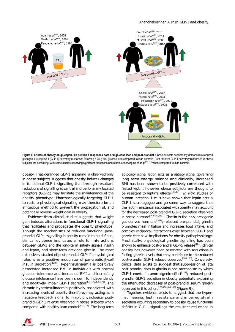
583 December 15, 2016|Volume 7|Issue 20|WJD|www.wjgnet.com
obesity. That deranged GLP-1 signalling is observed only in obese subjects suggests that obesity induces changes in functional GLP-1 signalling that through resultant reductions of signalling at central and peripherally located receptors (GLP-1) may facilitate the maintenance of the obesity phenotype. Pharmacologically targeting GLP-1 to restore physiological signalling may therefore be an efficacious method to prevent the propagation of, and potentially reverse weight gain in obesity.
Evidence from clinical studies suggests that weight gain induces alterations in functional GLP-1 signalling that facilitates and propagates the obesity phenotype. Though the mechanisms of reduced functional post-prandial GLP-1 signalling in obesity remain to be defined, clinical evidence implicates a role for interactions between GLP-1 and the long-term satiety signals insulin and leptin, and short-term orexigen ghrelin. The most extensively studied of post-prandial GLP-1’s physiological roles is as a positive modulator of pancreatic β-cell insulin secretion[123]. Hyperinsulinaemia is positively associated increased BMI in individuals with normal glucose tolerance and increased BMI and increasing glucose intolerance have been shown to independently and additively impair GLP-1 secretion[171-173,175,179]. The chronic hyperinsulinaemia positively associated with increasing levels of obesity therefore, may acting as a negative feedback signal to inhibit physiological post-prandial GLP-1 release observed in obese subjects when compared with healthy lean control[171-173]. The long term
adiposity signal leptin acts as a satiety signal governing long term energy balance and clinically, increased BMI has been shown to be positively correlated with fasted leptin, however obese subjects are thought to be resistant to leptin’s effects[180,181]. In vitro studies of human intestinal L-cells have shown that leptin acts a GLP-1 secretogogue and go some way to suggest that the leptin resistance associated with obesity may account for the decreased post-prandial GLP-1 secretion observed in obese humans[168-170,182]. Ghrelin is the only orexigenic gut derived hormone[183]; released pre-prandial, ghrelin promotes meal initiation and increases food intake, and complex reciprocal interactions exist between GLP-1 and ghrelin that have implications for obesity pathophysiology. Preclinically, physiological ghrelin signalling has been shown to enhance post-prandial GLP-1 release[164], clinical obesity has however been associated with reductions in fasting ghrelin levels that may contribute to the reduced post-prandial GLP-1 release observed[168-170]. Conversely, clinical data exists to suggest that suppression of late post-prandial rises in ghrelin is one mechanism by which GLP-1 exerts its anorexigenic effect[126]; reduced post-prandial GLP-1 secretion in obesity potentially explaining the attenuated decreases of post-prandial serum ghrelin observed in this cohort[168-170,184,185] (Figure 9).
Together, evidence exists to suggest that the hyper-insulinaemia, leptin resistance and impaired ghrelin secretion occurring secondary to obesity cause functional deficits in GLP-1 signalling; the resultant reductions in
Adam et al [168], 2005 Verdich et al [169], 2001Ranganath et al [170], 1996
Færch et al [171], 2015 Hussein et al [172], 2014 Muscelli et al [173], 2008Svedsen et al [174], 2012
Carroll et al [175], 2007Visboll et al [176], 2003 Toft-Nielsen et al [177], 2001 Näslund et al [178], 1998
Figure 8 Effects of obesity on glucagon-like peptide 1 responses post oral glucose load and post-prandial. Obese subjects consistently demonstrate reduced glucagon-like peptide 1 (GLP-1) secretory responses following a 75-g oral glucose load compared to lean controls. Post-prandial GLP-1 secretory responses in obese subjects are conflicting, with some studies observing significant reductions and others observing no change[168-178] when compared to lean controls.
75 g
Post OGTT GLP-1 Post-prandial GLP-1
Post-prandial GLP-1
Anandhakrishnan A et al . GLP-1 and obesity
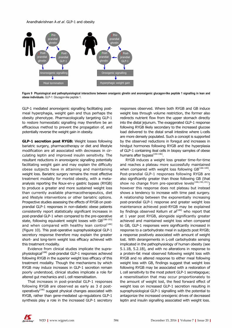
584 December 15, 2016|Volume 7|Issue 20|WJD|www.wjgnet.com
GLP-1 mediated anorexigenic signalling facilitating post-meal hyperphagia, weight gain and thus perhaps the obesity phenotype. Pharmacologically targeting GLP-1 to restore homeostatic signalling may therefore be an efficacious method to prevent the propagation of, and potentially reverse the weight gain in obesity.
GLP-1 secretion post RYGB: Weight losses following bariatric surgery, pharmacotherapy or diet and lifestyle modification are all associated with decreases in cir-culating leptin and improved insulin sensitivity. The resultant reductions in anorexigenic signalling potentially facilitating weight gain and may explain the difficulty obese subjects have in attaining and maintaining weight loss. Bariatric surgery remains the most effective treatment modality for morbid obesity, with a meta-analysis reporting the Roux-en-y gastric bypass (RYGB) to produce a greater and more sustained weight loss than currently available pharmacotherapeutics, diet and lifestyle interventions or other bariatric options. Prospective studies assessing the effects of RYGB on post-prandial GLP-1 responses in non-diabetic obese patients consistently report statistically significant increases in post-prandial GLP-1 when compared to the pre-operative state, following equivalent weight losses with GB[186-193] and when compared with healthy lean control[188] (Figure 10). This post-operative supraphysiological GLP-1 secretory response therefore may explain the greater short- and long-term weight loss efficacy achieved with this treatment modality.
Evidence from clinical studies implicate the supra-physiological[188] post-prandial GLP-1 responses achieved following RYGB in the superior weight loss efficacy of this treatment modality. Though the mechanisms by which RYGB may induce increases in GLP-1 secretion remain poorly understood, clinical studies implicate a role for altered gut mechanics and L cell resensitisation.
That increases in post-prandial GLP-1 responses following RYGB are observed as early as 3 d post-operatively[190] suggest physical changes associated with RYGB, rather than gene-mediated up-regulations GLP-1 synthesis play a role in the increased GLP-1 secretory
responses observed. Where both RYGB and GB induce weight loss through volume restriction, the former also redirects nutrient flow from the upper stomach directly into the distal jejunum. The exaggerated GLP-1 response following RYGB likely secondary to the increased glucose load delivered to the distal small intestine where L-cells are more densely populated. Such a concept is supported by the observed reductions in foregut and increases in hindgut hormones following RYGB and the hyperplasia of GLP-1 containing ileal cells in biopsy samples of obese humans after bypass[194-196].
RYGB induces a weight loss greater time-for-time and reaches a plateau more successfully maintained when compared with weight loss following GB[186-189]. Post-prandial GLP-1 responses following RYGB are also significantly greater than those following GB (that show no change from pre-operative levels[186,190,191]) however this response does not plateau but instead shows a tendency to increase with time past surgery. A relationship between the exponentially increasing post-prandial GLP-1 response and greater weight loss maintenance achieved post-RYGB may be explained by findings observed Kellum et al[189] who report that at 1 year post RYGB, alongside significantly greater achieved and maintained weight loss when compared to GB, GLP-1 responses were significantly increased in response to a carbohydrate meal in subjects post RYGB; a response positively associated with amount of weight lost. With derangements in L-cell carbohydrate sensing implicated in the pathophysiology of human obesity (see 5.1.1B, 5.2.1B), and with no alterations in response to a protein-fat meal observed following weight loss with RYGB and no altered response to either meal following weight loss with GB, findings suggest that weight loss following RYGB may be associated with a restoration of L cell sensitivity to the most potent GLP-1 secretagogue; a resensitisation that may occur proportionately to the amount of weight lost, the feed forward effect of weight loss on increased GLP-1 secretion resulting in supraphysiological GLP-1 signalling with the potential to antagonize the increased orexigenic drives of decreased leptin and insulin signalling associated with weight loss.
↑ Pre-prandial ghrelin
↓ Post-prandial ghrelin
↑ Post-prandial GLP-1
Anorexigenic signalling
Meal termination
↓ Pre-prandial ghrelin
↑ Post-prandial ghrelin
↓ Post-prandial GLP-1
Orexigenic signalling
Hyperphagic weight gain
Figure 9 Physiological and pathophysiological interactions between orexigenic ghrelin and anorexigenic glucagon-like peptide 1 signalling in lean and obese individuals. GLP-1: Glucagon-like peptide 1.
Anandhakrishnan A et al . GLP-1 and obesity
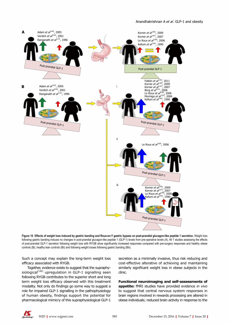
585 December 15, 2016|Volume 7|Issue 20|WJD|www.wjgnet.com
Such a concept may explain the long-term weight loss efficacy associated with RYGB.
Together, evidence exists to suggest that the supraphy-siological[188] upregulation in GLP-1 signalling seen following RYGB contributes to the superior short and long term weight loss efficacy observed with this treatment modality. Not only do findings go some way to suggest a role for impaired GLP-1 signalling in the pathophysiology of human obesity, findings support the potential for pharmacological mimicry of this supraphysiological GLP-1
secretion as a minimally invasive, thus risk reducing and cost-effective alterative of achieving and maintaining similarly significant weight loss in obese subjects in the clinic.
Functional neuroimaging and self-assessments of appetite: fMRI studies have provided evidence in vivo to suggest that central nervous system responses in brain regions involved in rewards processing are altered in obese individuals; reduced brain activity in response to the
Adam et al [168], 2005 Verdich et al [169], 2001Ranganath et al [170], 1996
Korner et al [186], 2009Korner et al [187], 2007Le Roux et al [188], 2006Kellum et al [189], 1990
Adam et al [168], 2005 Verdich et al [169], 2001Ranganath et al [170], 1996
Le Roux et al [188], 2006
Figure 10 Effects of weight loss induced by gastric banding and Roux-en-Y gastric bypass on post-prandial glucagon-like peptide 1 secretion. Weight loss following gastric banding induces no changes in post-prandial glucagon-like peptide 1 (GLP-1) levels from pre-operative levels (A). All 7 studies assessing the effects of post-prandial GLP-1 secretion following weight loss with RYGB show significantly increased responses compared with pre-surgery responses and healthy obese controls (Bi), healthy lean controls (Bii) and following weight losses following gastric banding (Biii).
A
B i
ii
iii
Post-prandial GLP-1
Korner et al [186], 2009Korner et al [187], 2007Le Roux et al [188], 2006Kellum et al [189], 1990
Post-prandial GLP-1Post-prandial GLP-1
Post-prandial GLP-1
Post-prandial GLP-1
Post-prandial GLP-1
Falkén et al [190], 2011 Korner et al [186], 2009Korner et al [187], 2007 Borg et al [191], 2006Le Roux et al [188], 2006Morínigo et al [192], 2006Kellum et al [189], 1990
Anandhakrishnan A et al . GLP-1 and obesity
586 December 15, 2016|Volume 7|Issue 20|WJD|www.wjgnet.com
consumption of, and increased activity in response to the anticipation of palatable food consistently observed when compared with healthy lean controls[197-199]. Interestingly, GLP-1 agonism reverses these functional brain changes to match those of lean control with associated reductions in ad libitum food intake[199], an effect prevented by pre-treatment with a GLP-1 antagonist[200] (Figure 11). Together, findings suggests that obesity induced de-creases in functional GLP-1 signalling contribute to altered rewards processing in obesity to facilitate hyperphagic weight gain despite an overall positive energy balance.
Further support for a role for GLP-1 and altered rewards processing in obesity pathophysiology comes from self-assessments of appetite. Subjectively ass-essed emotional eating scores have been defined as hedonic markers of appetite that display strong positive associations with the degree of human obesity, and, in obese subjects, relate to the extent to which GLP-1 receptor activation in brain regions involved in rewards processing are reduced[201-203]. Together, findings suggests that obesity induced decreases in functional GLP-1 sig-nalling creates a feed-forward loop of hyperphagic weight gain despite an overall positive energy balance, an effect perhaps secondary to a GLP-1 deficit mediated skew toward hedonic and away from homeostatic controls of food intake. Together, findings from fMRI and subjective appetite assessment scores implicate obesity-associated reductions in functional GLP-1/GLP-1R signalling in the pathophysiology of hyperphagic weight gain in obesity. As such, findings support the role of the GLP-1R as a novel therapeutic target in the medical management of obesity, providing rationale for the use of liraglutide 3 mg in the pharmacotherapy of obesity in the clinic.
A ROLE FOR GLP-1 IN THE PHARMACOTHERAPY OF CLINICAL OBESITYThe balance between drug efficacy and cost determines the selection of a pharmacological agent for use in the medical management of any disease; greater understanding of underlying disease pathophysiology facilitating the development of targeted therapeutics with the potential for greater efficacy. Several lines of clinical evidence implicate a role for altered GLP-1 function in the pathophysiology of human obesity and a number of recent clinical trials have validated the clinical efficacy of long-term once daily SC 3 mg liraglutide (Saxenda) as an adjunct to calorie-restriction and exercise counselling in obese and overweight individuals with at least one weight related comorbidity. Significant improvements in clinical outcome measures such as body weight, anthropometric and cardiometabolic parameters, and indices of glucose tolerance have been observed and recently reviewed elsewhere[204]. Though March 2015 saw the EMA grant marketing authorization for 3 mg liraglutide as a weight-management agent in all 28 EU states[45], cost-benefit of funding treatment on the NHS undoubtedly contributes
to the uncertainty of launch plans in the United Kingdom at present[46].
Weight loss efficacy of GLP-1 analoguesEvidence: One phase Ⅱ (NCT00422058)[43], and a number of phase Ⅲ multi-national double-blinded ran-domized control trials conducted in non-diabetic obese adults (NCT00480909)[42], overweight adults with at least one weight related co-morbidity (SCALE Obesity and Pre-diabetes, and SCALE Maintenance[205,206]), non-diabetic obese adults with obstructive sleep apnoea (OSA) (SCALE OSA[41]) and obese adults with T2DM (SCALE diabetes[207]) have established the efficacy of once daily 3 mg SC liraglutide as an adjunct to an energy-deficient low-calorie diet and physical activity counselling for weight management in this cohort. Results from the first study; a 20-wk phase Ⅱ trial in non-diabetic obese subjects showed that weight loss with liraglutide is dose-dependent up to 3.0 mg once daily[42,206]. Significantly more liraglutide 3 mg/d recipients than placebo or orlistat recipients achieved a 5% or 10% reduction of body weight at 20 wk. In a 2-year phase Ⅲ extension of the same study[42], double-blind treatment (liraglutide 1.2-3 mg/d) was continued until week 52, after which all liraglutide (< 2.4 mg/d) and placebo recipients were switched to liraglutide 2.4 mg, then 3.0 mg (week 70-96) based on 20-wk and 1-year results respectively (Figure 12) that indicated this was the optimal dosage. At 2 years, mean bodyweight reductions in those randomized to liraglutide were significantly greater than pancreatic lipase inhibitor orlistat, the only alternative licenced weight loss agent in the United Kingdom. Results from the SCALE maintenance and SCALE obesity and prediabetes[205,206] trials report similarly significant reductions in bodyweight in subjects randomized to 3 mg liraglutide when compared with placebo at 56 wk (P < 0.0001) alongside increased 5% and 10% responder rates. Findings are supported by results of the 32 wk SCALE Sleep Apnoea trial[41] in obese non-diabetic sub-jects with moderate to severe OSA and in the 56-68 wk SCALE Diabetes[207] trial in obese subjects with T2DM (Figure 13). With even modest losses of 5%-10% of total body weight associated with reduced risk of comorbidities in obese individuals[8-10], findings provide rationale for the licensing and funding of 3 mg liraglutide as an adjunct to lifestyle alteration as the first line anti-obesity pharmaceutical agent for weight management in obese and comorbid overweight adults in the United Kingdom.
Interpretations - obesity pathophysiology: Excessive consumption of palatable food can trigger neuroadaptive responses in brain reward circuits similar to that of alcohol and drugs of abuse[208] and clinical studies provide evidence to suggest that human obestiy is associated with altered rewards processing mediated in part by altered GLP-1 function that may render the hyperphagia of obesity the manifestation of a “food addiction”. Whilst 3 mg liraglutide has been shown to induce weight loss in man by reductions appetite pre-
Anandhakrishnan A et al . GLP-1 and obesity

587 December 15, 2016|Volume 7|Issue 20|WJD|www.wjgnet.com
clinically, liraglutide attenuates the reinforcing properties of alcohol in vivo[209,210]. As such, perhaps GLP-1 agonism may attenuate produce its weight loss effects in part, by attenuating the negative reinforcement of hyperphagia in obesity. Interestingly, though all aforementioned trials[41-43,205-207] advise participants to restrict food consumption throughout the treatment period, adherence rates are not reported. If indeed GLP-1 agonism induces its weight loss effects by modulating food related rewards that potentially reverses the negative reinforcement of hyperphagia in obesity, an increased adherence to caloric restriction would be expected. It would be interesting to see if this were the case.
Interpretations - cost-benefit of 3 mg liraglutide as an anti-obesity agent on the NHS: Follow-up period (FUP) assessments in the SCALE Maintenance, and SCALE Diabetes[205-207] trials suggest that weight loss with 3 mg liraglutide is treatment dependent; weight gains in excess to those seen in placebo control and subjects re-randomized to treatment observed in liraglutide treated participants upon treatment cessation (Figure 12).
Though reductions in bodyweight have been shown to significantly improve health outcomes and thereby reduce healthcare costs long term[211], in 2013, 24.9% of the United Kingdom adult population were classed obese and 90% of the £2.7 million T2DM adults in the United Kingdom were overweight. Following FDA and
EMA approval criteria, all these individuals are potential candidates for treatment with liraglutide 3 mg. Costing in excess of $1000 per patient a month, and with prevalence of obesity and overweight predicted to rise (introduction), prolonged treatment seems unsustainable. A potential solution comes from longitudinal observations from Astrup et al[43] who observed that subjects randomized to liraglutide 3 mg achieve maximal rates of weight loss in the initial 0-20 wk treatment period with a tendency toward weight gain beyond 36 wk[42,43]. Findings suggest that whilst initially treatment with a GLP-1 analogue may compensate for functional deficits in obesity, treatment beyond 20 wk may be associated with the development of treatment resistance, most apparent 36 wk from initiation (Figure 14). Based on this, perhaps treatment with liraglutide 3 mg should be prescribed for 20 to a maximum of 36 wk alongside behavioural therapies promoting lifestyle changes and developing strategies to combat the addiction driven hyperphagia implicated in obesity pathophysiology (GLP-1 secretion post RYGB); combing behavioural therapies in the initial 20 wk of drug induction where weight loss is most pronounced may act as a positive reinforcer of sustained behavioural change facilitating continuation of these behaviours. This approach, integrating the psychosocial empowerment associated with patient self-management of chronic illness, alongside cost benefits associated with limited in-treatment period seems an
Ai Aii
GLP-1
GLP-1Ex-9-39
12
B
C
Figure 11 Altered central nervous system responses in brain regions involved in rewards in obese subjects is reversed upon glucagon-like peptide 1 receptor activation. Central nervous system activity in regions involved in rewards processing show increased responses in obese subjects when exposed to food related images (Aii) associated with increased ad libitum food consumption when compared to healthy lean control (Ai). A response reversed upon administration of a GLP-1 analogue with concomitant reductions in ad libitum food intake (B), this effect antagonized by pre-treatment with GLP-1 antagonist Extendin 9-39 (EX9-39) (C). GLP-1: Glucagon-like peptide 1.
Anandhakrishnan A et al . GLP-1 and obesity

588 December 15, 2016|Volume 7|Issue 20|WJD|www.wjgnet.com
attractive one, especially if sustainable long-term weight losses with resultant reductions in the socioeconomic impacts of weight-related comorbidities can be achieved.
Glucose toleranceEvidence: Clinical studies in obese subjects with pre-diabetes consistently demonstrate a greater reversion to normal glycaemic control following treat-ment with liraglutide 3 mg coinciding reduced T2DM incidence[42,206,212].
Interpretations - obesity pathophysiology: Being overweight or obese is the main modifiable risk factor for T2DM and increasing BMI is positively associated with hyperinsulinaemia even in those with normal glycaemic control[175,179,213] suggesting a common pathophysiology to both conditions. That increased BMI and impaired glucose tolerance have been shown to independently and additively impair GLP-1 secretory responses following an OGTT[171-173] suggests that this common pathophysiology may lie in a functional deficit of GLP-1. Support for the existence of a common pathophysiology between obesity and T2DM comes from observations that treatment with the insulin sensitizer metformin (currently the first line pharmacotherapeutic agent in the management of T2DM) upregulates GLP-1 secretory response following
an OGTT, the restoration of physiological anorexigenic and incretin effects perhaps explaining the weight loss, and insulin sensitising properties of the drug seen in the clinic respectively[214,215].
Interpretations: Cost-benefit of 3 mg liraglutide as an anti-obesity agent on the NHS: Being overweight or obese is the main modifiable risk factor for T2DM[213] and T2DM is one of the major indirect financial burdens of obesity and overweight. Treating T2DM and its complications alone current costs the NHS £8.8 billion a year with indirect costs estimated at £13 billion[216]. With the incidence of obesity projected to rise (introduction), so too can be expected the incidence of T2DM, the management of which therefore, may become unsustainable on the NHS. Though costly, treatment with liraglutide 3 mg is associated with reductions in the rate of development of T2DM in overweight and obese subjects[42,206,212] and goes some way to suggest that treatment may reduce both direct burdens of obesity and overweight and the large indirect burden posed by new incidences of T2DM. However, weight loss in itself is associated with an improvement in glycaemic control. It may be argued therefore, that true cost-benefit of funding liraglutide 3 mg on the rationale of T2DM prevention in overweight and obese
PlaceboLiraglutide
80706050403020100
% s
tudy
sub
ject
s
SCAL
E ob
esity
and
pre
-di
abet
es
SCAL
E sl
eep
apno
ea
SCAL
E m
aint
enan
ce
SCAL
E di
abet
es
NCT
0048
0909
NCT
0042
2058
SCAL
E ob
esity
and
pre
-di
abet
es
SCAL
E sl
eep
apno
ea
SCAL
E m
aint
enan
ce
SCAL
E di
abet
es
NCT
0048
0909
NCT
0042
2058
≥ 5% body weight loss ≥ 10% body weight loss
Figure 12 Significantly greater 5% and 10% weight loss achieved following 3 mg subcutaneous liraglutide compared to placebo and orlistat. Five percent and 10% responder rates in NCT00480909 reported at 1 year (see text).
20
-2
-4-6-8
-10
% c
hang
e in
bod
y w
eigh
t fr
om b
asel
ine
56 wk68 wk
SCALE maintenance
LIR/PI Placebo LIR/PI Placebo LIR/PI PlaceboLIR/LIR
SCALE obesity and preDM SCALE T2DM
Figure 13 Body weight changes with liraglutide are treatment dependent. Following the 56-wk treatment period a 12-wk follow-up (FUP) was conducted in SCALE maintenance, SCALE obesity and pre-diabeties and SCALE diabetes trials. Twelve weeks FUP was an off-treatment period in SCALE Maintenance and SCALE diabetes. In SCALE Obesity and pre-diabetes FUP period involved a re-randomisation to either 3 mg liraglutide (LIR/LIR) or placebo (LIR/Pl) and though weight gain occurred in all three groups weight gain was significantly higher following cessation of liraglutide. T2DM: Type 2 diabetes mellitus.
Anandhakrishnan A et al . GLP-1 and obesity

589 December 15, 2016|Volume 7|Issue 20|WJD|www.wjgnet.com
subjects exists only if the improved glucose tolerance achieved following a given weight loss with liraglutide 3 mg exceeds those attained following other, arguably cheaper treatment modalities. Calculating correlation coefficients between percentage weight lost and percentage changes in glucose tolerance in non-diabetic obese subjects randomized to liraglutide 3 mg compared to those administered orlistat or placebo[41-43,205,206] in aforementioned phase Ⅲ trials may be one way in which this could be assessed. Figure 15 interprets the possible findings of such a test.
If indeed pharmacological GLP-1 agonism improves glucose tolerance independent of weight lost, the potential curb in prevalence, thus socio-economic burden of T2DM achieved with treatment provides a second rationale for the licensing and funding of liraglutide 3 mg as an adjunct to lifestyle alteration as the first line anti-obesity agent for weight management in obese and co-morbid overweight individuals in the United Kingdom.
Adverse drug eventsEvidence - in-treatment tolerability: Whilst evidence exists to suggest GLP-1 agonism may be a targeted agent with long term cost-benefit for use in the medical management of obesity, tolerability and safety are important considerations in determining the choice of any pharmaceutical, especially in the management of chronic disease. The safety and efficacy of liraglutide 3 mg has been evaluated in 5 phase Ⅲ double-blinded placebo controlled trials comprising 3384 overweight or obese subjects receiving liraglutide 3 mg and 1941 placebo controls for a treatment periods of 32, 52 and 56 wk[42,42,205-207]. In a pooled analysis of the 5 aforementioned trials, liraglutide 3 mg in obese and overweight subjects was generally well tolerated, with most adverse drug events gastrointestinal in nature, transient and of mild to moderate intensity[42,43,205-207]. However, 9.8% of liraglutide and 4.3% of placebo recipients discontinued treatment because of an adverse event[217]. Figure 16 details adverse reactions occurring with a higher incidence to placebo with an incidence of ≥ 10% in liraglutide 3 mg recipients, stratified by system.
Of interest, 0.6% of subjects receiving liraglutide
3 mg experienced increases in mean heart rate (an average baseline increase of 2.5 beats/min) compared to 0.1% of placebo recipients[218]. Potentially a manifestation of GLP-1 induced increases in SNS activity (potential effectors of GLP-1s negative energy balance effects: Central controllers) contributing to GLP-1 induced weight loss via increases energy expenditure, elsewhere tachy-cardia associated with 3 mg liraglutide treatment in non-diabetic obese subjects yields no associated increases in 24 h energy expenditure[209]. Thus, whilst the clinical significance of this finding remains to be determined, observations may warrant more intense monitoring in patients with pre-existing cardiovascular disease.
Interpretations - long term risk-benefit of liraglutide 3 mg as an anti-obesity agent on the NHS: Though generally well tolerated in the acute setting, safety concerns have been raised regarding the potential risk of pancreatitis and pancreatic and thyroid cancer with long-term use of GLP-1 analogues[219,220]. Confirmed cases of acute pancreatitis and papillary thyroid carcinoma were reported in 0.3% of liraglutide 3 mg treated compared to 0.2% of placebo treated participants, however the relative rarity of events means the relationship between treatment with disease incidence and severity remains to be defined. On-going clinical experience and thorough post-marketing surveillance should help clarify any such associations and also identify other potential adverse drug events. To this end, episodes of acute renal failure and medullary thyroid carcinoma (not observed during in-treatment and FUP period assessments[42,43,205-207]) have been reported in the post-marketing period, though again, insufficient data exists to establish or exclude a causal relationship. Figure 17 details other potentially serious medical conditions observed in during in-treatment and FUP assessments[42,43,205-207].
Though potentially associated with serious long-term adverse effects, the rarity of incidence and lack of causal relationship mean that current knowledge supports benefit over risk, supporting the licensing and funding of liraglutide 3 mg as the first line anti-obesity pharmaceutical agent for weight management in obese and overweight adults with at-least one weight related
987654
3210
Mea
n re
duct
ions
in
body
wei
ght
(kg)
Placebo Liraglutide3 mg
Orlistat Placebo Liraglutide3 mg
Orlistat Liraglutide3 mg
Orlistat
20 wk 56 wk 2 yr
Figure 14 Liraglutide 3 mg shows greater, dose dependent, reductions in body weight compared to placebo and orlistat with maximal effect between 0-20 wk of treatment. Red line dotted trace shows patters of weight loss from baseline in those subjects treated with 3 mg liraglutide. Greatest rate of weight loss was observed between 0-20 wk, reducing from 20-36 with a tendency toward weight gain from 36 wk and beyond.
Anandhakrishnan A et al . GLP-1 and obesity

590 December 15, 2016|Volume 7|Issue 20|WJD|www.wjgnet.com
co-morbidity in the United Kingdom.
CONCLUSIONObesity is a global epidemic, perhaps the greatest challenge to global and public health of our time. Whilst public health initiatives should continue to focus on curbing the projected upward trends in the incidence of obesity and overweight, effective management of those individuals already obese remains an important and as yet unmet clinical need. The current medical management of obesity in the United Kingdom is suboptimal, with the only treatment modality with proven long-term
efficacy being bariatric surgery. Both risky and costly, this treatment option is not viable for the widespread management of obesity, and remains reserved for ex-treme cases. The ideal medical management of any illness utilizes a targeted pharmacotherapy that either repletes physiological factors pathologically depleted, or antagonizes pathological processes, the development of such an agent requiring an understanding of the pathophysiology underpinning a disease. Though the pathophysiology of clinical obesity is undoubtedly multi-faceted, several lines of clinical evidence implicate a role for functional impairments in GLP-1 signalling. Whilst genetic studies implicate a role for primary altered GLP-1
1.2
1
0.8
0.6
0.4
0.2
0
% im
prov
emen
t in
glu
cose
to
lera
nce
1 2 3 4 5 6 7 8 9 10
% weight loss
PlaceboOrlistatLiraglutide 3 mg
1.2
1
0.8
0.6
0.4
0.2
0%
impr
ovem
ent
in g
luco
se
tole
ranc
e1 2 3 4 5 6 7 8 9 10
% weight loss
A B
Figure 15 Theoretic model to assess glucose tolerance as a factor of weight. Liraglutide 3 mg produces increased weight loss and increased improvements in glucose control compared to both placebo and control in non-diabetic obese and overweight subjects[41-43,205,206]. If the greater glucose sensitivity seen following treatment with liraglutide 3 mg is secondary to the greater weight loss achieved, the relationship between a given weight reduction and the percentage improvement in glucose tolerance should be the same in all treatment groups (A). If however, the relationship between a given weight loss and change in glucose tolerance is less strong (B) following treatment with liraglutide, findings would suggest that mechanisms beyond weight loss contribute to the greater improvements of glucose tolerance seen following GLP-1 agonism. One mechanism may be via activation of GLP-1R in the pancreas, where endogenous GLP-1 signalling has a well established incretin effect. GLP-1: Glucagon-like peptide 1.
454035302520151050
Inci
denc
e (%
of
stud
y po
pula
tion)
PlaceboSaxenda
Nau
sea
Dia
rrho
ea
Cons
tipat
ion
Vom
iting
Hyp
ogly
caem
ia
(in T
2DM
)
Dec
reas
ed a
ppet
ite
Hea
dach
eGI Metabolic CNS
Adverse drug reactions (by system)
Figure 16 Adverse drug reactions reported in ≥ 10% of liraglutide 3 mg recipients with a higher incidence than placebo stratified by system. GI: Dyspepsia, abdominal pain, dry mouth, gastritis, gastroesophageal reflux disease, flatulence, eructation and abdominal distension were also more prevalent in liraglutide treated participants but occurred with incidence ≤ 10%, not shown; 6.2% with Saxenda vs 0.8% with placebo discontinued treatment as a result of gastrointestinal adverse reactions; CNS: Dizziness, malaise and fatigue occurred were more also prevalent in liraglutide treated participants but occurred with incidence ≤ 10%, not shown; Metabolic: Liraglutide reduces blood glucose and thus, there is a potential for hypoglycaemia to occur. In the SCALE diabetes trial severe hypoglycemia occurred in 3 (0.7%) of 422 Saxenda-treated patients and in none of the 212 placebo-treated patients. In clinical trials involving patients without T2DM[42,43,205,206] no systematic reporting of hypoglycemia occurred but spontaneously reported symptomatic episodes potentially hypoglycemic in cause were reported by 1.6% (46/2962) of liraglutide 3 mg and 1.1% of (19/1729) placebo treated non diabetic patients. T2DM: Type 2 diabetes mellitus; GI: Gastrointestinal; CNS: Central nervous system.
Anandhakrishnan A et al . GLP-1 and obesity

591 December 15, 2016|Volume 7|Issue 20|WJD|www.wjgnet.com
signalling as a risk factor towards development of the obesity phenotype, clinical studies assessing physiological GLP-1 responses in normal weight and obese subjects suggest weight gain may induce functional deficits in GLP-1 that facilitates maintenance of the obesity phenotype. Whatever the relationship, cause or effect, reductions in functional GLP-1 signalling seems to play a role in clinical obesity, as such, the pharmacological replenishment of this functional deficit seems a promising target for the medical management of obesity in the clinic. Indeed, the GLP-1 analogue liraglutide 3 mg has shown promising results in achieving and maintaining greater weight loss in obese individuals when compared to control or currently licensed anti-obesity medication. Though results from extended phase Ⅲ and phase Ⅳ studies report the development of potentially fatal adverse drug events in those randomized to or prescribed liraglutide 3 mg respectively, the scarcity of incidence and lack of causal relationship sees such potential risks overshadowed by the proven superior weight loss efficacy of treatment. Cost-benefit, however, may pose a barrier toward viable NHS funding, though this may be overcome by strategic treatment delivery; combining short-term liraglutide 3 mg treatment (≤ 36 wk) with behavioural therapies targeted toward promoting healthy lifestyle changes. With drug induced weight loss potentially reinforcing adherence to long-term lifestyle changes, if successful, shortened in-treatment period alongside decreases in direct and indirect socioeconomic burdens of obesity and overweight secondary to achi-evement and maintenance of significant weight loss associates a long-term cost-benefit to funding treatment. Such a concept supports the use of liraglutide 3 mg as the first line anti-obesity agent on the NHS when conservative lifestyle management alone has failed in achieving clinically significant weight loss in comorbid overweight or obese adults.
REFERENCES1 Organisation TWH. Global health observatory data; Situation and
trend, 2016. Available from: URL: http://www.who.int/gho/ncd/risk_factors/obesity_text/en/
2 Organisation TWH. Obesity and Overweight fact sheet. Jun 2016. Available from: URL: http://www.who.int/mediacentre/factsheets/fs311/en/
3 Kelly T, Yang W, Chen CS, Reynolds K, He J. Global burden of obesity in 2005 and projections to 2030. Int J Obes (Lond) 2008; 32: 1431-1437 [PMID: 18607383 DOI: 10.1038/ijo.2008.102]
4 Guh DP, Zhang W, Bansback N, Amarsi Z, Birmingham CL, Anis AH. The incidence of co-morbidities related to obesity and overweight: a systematic review and meta-analysis. BMC Public Health 2009; 9: 88 [PMID: 19320986 DOI: 10.1186/1471-2458-9-88]
5 Puhl RM, Heuer CA. Obesity stigma: important considerations for public health. Am J Public Health 2010; 100: 1019-1028 [PMID: 20075322 DOI: 10.2105/AJPH.2009.159491]
6 Kolotkin RL, Meter K, Williams GR. Quality of life and obesity. Obes Rev 2001; 2: 219-229 [PMID: 12119993 DOI: 10.1046/j.1467-789X.2001.00040.x]
7 Luppino FS, de Wit LM, Bouvy PF, Stijnen T, Cuijpers P, Penninx BW, Zitman FG. Overweight, obesity, and depression: a systematic review and meta-analysis of longitudinal studies. Arch Gen Psychiatry 2010; 67: 220-229 [PMID: 20194822 DOI: 10.1001/archgenpsychiatry.2010.2]
8 Vidal J. Updated review on the benefits of weight loss. Int J Obes Relat Metab Disord 2002; 26 Suppl 4: S25-S28 [PMID: 12457296 DOI: 10.1038/sj.ijo.0802215]
9 Goldstein DJ. Beneficial health effects of modest weight loss. Int J Obes Relat Metab Disord 1992; 16: 397-415 [PMID: 1322866]
10 Blackburn G. Effect of degree of weight loss on health benefits. Obes Res 1995; 3 Suppl 2: 211s-216s [PMID: 8581779]
11 Grieve E, Fenwick E, Yang HC, Lean M. The disproportionate economic burden associated with severe and complicated obesity: a systematic review. Obes Rev 2013; 14: 883-894 [PMID: 23859626 DOI: 10.1111/obr.12059]
12 Trogdon JG, Finkelstein EA, Hylands T, Dellea PS, Kamal-Bahl SJ. Indirect costs of obesity: a review of the current literature. Obes Rev 2008; 9: 489-500 [PMID: 18331420 DOI: 10.1111/j.1467-789X.2008.00472.x]
13 Howard JT, Potter LB. An assessment of the relationships between overweight, obesity, related chronic health conditions and worker absenteeism. Obes Res Clin Pract 2014; 8: e1-15 [PMID: 24548572 DOI: 10.1016/j.orcp.2012.09.002]
14 Ricci JA, Chee E. Lost productive time associated with excess weight in the U.S. workforce. J Occup Environ Med 2005; 47: 1227-1234 [PMID: 16340703 DOI: 10.1097/01.jom.0000184871. 20901.c3]
15 Specchia ML, Veneziano MA, Cadeddu C, Ferriero AM, Mancuso A, Ianuale C, Parente P, Capri S, Ricciardi W. Economic impact of adult obesity on health systems: a systematic review. Eur J Public Health 2015; 25: 255-262 [PMID: 25320051 DOI: 10.1093/eurpub/cku170]
16 Withrow D, Alter DA. The economic burden of obesity world-
1.61.41.2
10.80.60.40.2
0
Inci
denc
e (%
of
stud
y po
pula
tion)
SaxendaPlacebo
Acut
e
Choli
thias
is
Chole
cysti
tisPT
C
Malign
ant C
RCBr
Ca
CCD
Hypote
nsion
Hypo-
ADR
Suici
dal id
eatio
n
Figure 17 Reported incidences of serious medical conditions from pooled analyses of 5 double-blinded placebo controlled trials studying the safety and efficacy of 3 mg liraglutide (Saxenda) in 3384 overweight or obese subjects receiving liraglutide and 1941 placebo controls. Hypotension associated adverse reactions (Hypo-ADR) refer to hypotension, orthostatic hypotension, circulatory collapse, and decreased blood pressure). One of the six (0.2%) liraglutide treated subjects reporting suicidal ideations attempted suicide. PTC: Papillary thyroid carcinoma; CRC: Colorectal carcinoma; Br Ca: Breast cancer; CCD: Cardiac conduction disorders.
Anandhakrishnan A et al . GLP-1 and obesity
592 December 15, 2016|Volume 7|Issue 20|WJD|www.wjgnet.com
wide: a systematic review of the direct costs of obesity. Obes Rev 2011; 12: 131-141 [PMID: 20122135 DOI: 10.1111/j.1467-789X.2009.00712.x]
17 Scarborough P, Bhatnagar P, Wickramasinghe KK, Allender S, Foster C, Rayner M. The economic burden of ill health due to diet, physical inactivity, smoking, alcohol and obesity in the UK: an update to 2006-07 NHS costs. J Public Health (Oxf) 2011; 33: 527-535 [PMID: 21562029 DOI: 10.1093/pubmed/fdr033]
18 McPherson K, Marsh TN. Foresight Tackling Obesities: Future Choices - Modelling Future Trends in Obesity and the Impact on Health (GOV UK). 2007
19 Collins B, Capewell S, O’Flaherty M, Timpson H, Razzaq A, Cheater S, Ireland R, Bromley H. Modelling the Health Impact of an English Sugary Drinks Duty at National and Local Levels. PLoS One 2015; 10: e0130770 [PMID: 26121677 DOI: 10.1371/journal.pone.0130770]
20 National Institute for Clinical Excellence T. Obesity: Identi-fication, assessment and management of overweight and obesity in children, young people and adults. 2014
21 Dombrowski SU, Knittle K, Avenell A, Araújo-Soares V, Sniehotta FF. Long term maintenance of weight loss with non-surgical interventions in obese adults: systematic review and meta-analyses of randomised controlled trials. BMJ 2014; 348: g2646 [PMID: 25134100 DOI: 10.1136/bmj.g2646]
22 Holzapfel C, Hauner H. [Weight maintenance after weight loss - how the body defends its weight]. Dtsch Med Wochenschr 2011; 136: 89-94 [PMID: 21225556 DOI: 10.1055/s-0030-1269445]
23 Loveman E, Frampton GK, Shepherd J, Picot J, Cooper K, Bryant J, Welch K, Clegg A. The clinical effectiveness and cost-effectiveness of long-term weight management schemes for adults: a systematic review. Health Technol Assess 2011; 15: 1-182 [PMID: 21247515 DOI: 10.3310/hta15020]
24 Greenway FL. Physiological adaptations to weight loss and factors favouring weight regain. Int J Obes (Lond) 2015; 39: 1188-1196 [PMID: 25896063 DOI: 10.1038/ijo.2015.59]
25 Derosa G, Maffioli P. Anti-obesity drugs: a review about their effects and their safety. Expert Opin Drug Saf 2012; 11: 459-471 [PMID: 22439841 DOI: 10.1517/14740338.2012.675326]
26 Kang JG, Park CY. Anti-Obesity Drugs: A Review about Their Effects and Safety. Diabetes Metab J 2012; 36: 13-25 [PMID: 22363917 DOI: 10.4093/dmj.2012.36.1.13]
27 Yanovski SZ, Yanovski JA. Obesity. N Engl J Med 2002; 346: 591-602 [PMID: 11856799 DOI: 10.1056/NEJMra012586]
28 Golomb I, Ben David M, Glass A, Kolitz T, Keidar A. Long-term Metabolic Effects of Laparoscopic Sleeve Gastrectomy. JAMA Surg 2015; 150: 1051-1057 [PMID: 26244446 DOI: 10.1001/jamasurg.2015.2202]
29 Hirth DA, Jones EL, Rothchild KB, Mitchell BC, Schoen JA. Laparoscopic sleeve gastrectomy: long-term weight loss outcomes. Surg Obes Relat Dis 2015; 11: 1004-1007 [PMID: 25980329 DOI: 10.1016/j.soard.2015.02.016]
30 Costa RC, Yamaguchi N, Santo MA, Riccioppo D, Pinto-Junior PE. Outcomes on quality of life, weight loss, and comorbidities after Roux-en-Y gastric bypass. Arq Gastroenterol 2014; 51: 165-170 [PMID: 25296074 DOI: 10.1590/S0004-28032014000300002]
31 Pasulka PS, Bistrian BR, Benotti PN, Blackburn GL. The risks of surgery in obese patients. Ann Intern Med 1986; 104: 540-546 [PMID: 3513685 DOI: 10.7326/0003-4819-104-4-540]
32 Dority J, Hassan ZU, Chau D. Anesthetic implications of obesity in the surgical patient. Clin Colon Rectal Surg 2011; 24: 222-228 [PMID: 23204937 DOI: 10.1055/s-0031-1295685]
33 Nishida C, Uauy R, Kumanyika S, Shetty P. The joint WHO/FAO expert consultation on diet, nutrition and the prevention of chronic diseases: process, product and policy implications. Public Health Nutr 2004; 7: 245-250 [PMID: 14972063 DOI: 10.1079/PHN2003592]
34 Woods SC, D’Alessio DA. Central control of body weight and appetite. J Clin Endocrinol Metab 2008; 93: S37-S50 [PMID: 18987269 DOI: 10.1210/jc.2008-1630]
35 Berthoud HR. Metabolic and hedonic drives in the neural control
of appetite: who is the boss? Curr Opin Neurobiol 2011; 21: 888-896 [PMID: 21981809 DOI: 10.1016/j.conb.2011.09.004]
36 Williams DL. Neural integration of satiation and food reward: role of GLP-1 and orexin pathways. Physiol Behav 2014; 136: 194-199 [PMID: 24650552 DOI: 10.1016/j.physbeh.2014.03.013]
37 Skibicka KP. The central GLP-1: implications for food and drug reward. Front Neurosci 2013; 7: 181 [PMID: 24133407 DOI: 10.3389/fnins.2013.00181]
38 Prasad-Reddy L, Isaacs D. A clinical review of GLP-1 receptor agonists: efficacy and safety in diabetes and beyond. Drugs Context 2015; 4: 212283 [PMID: 26213556 DOI: 10.7573/dic.212283]
39 Monami M, Dicembrini I, Marchionni N, Rotella CM, Mannucci E. Effects of glucagon-like peptide-1 receptor agonists on body weight: a meta-analysis. Exp Diabetes Res 2012; 2012: 672658 [PMID: 22675341 DOI: 10.1155/2012/672658]
40 Vilsbøll T, Christensen M, Junker AE, Knop FK, Gluud LL. Effects of glucagon-like peptide-1 receptor agonists on weight loss: systematic review and meta-analyses of randomised controlled trials. BMJ 2012; 344: d7771 [PMID: 22236411 DOI: 10.1136/bmj.d7771]
41 Collier A, Blackman A, Foster G, Zammit G, Rosenberg R, Wadden T, Aronne L, Claudius B, Jensen T, Mignot E. S28 Liraglutide 3.0 Mg Reduces Severity Of Obstructive Sleep Apnoea And Body Weight In Obese Individuals With Moderate Or Severe Disease: Scale Sleep Apnoea Trial. Thorax 2014; 69 Suppl 2: A16-A17 [DOI: 10.1016/j.jcjd.2015.01.139]
42 Astrup A, Carraro R, Finer N, Harper A, Kunesova M, Lean ME, Niskanen L, Rasmussen MF, Rissanen A, Rössner S, Savolainen MJ, Van Gaal L. Safety, tolerability and sustained weight loss over 2 years with the once-daily human GLP-1 analog, liraglutide. Int J Obes (Lond) 2012; 36: 843-854 [PMID: 21844879 DOI: 10.1038/ijo.2011.158]
43 Astrup A, Rössner S, Van Gaal L, Rissanen A, Niskanen L, Al Hakim M, Madsen J, Rasmussen MF, Lean ME. Effects of liraglutide in the treatment of obesity: a randomised, double-blind, placebo-controlled study. Lancet 2009; 374: 1606-1616 [PMID: 19853906 DOI: 10.1016/S0140-6736(09)61375-1]
44 Liraglutide 3.0 mg for Weight Management; briefing document. 2014
45 Novo Nordisk. Saxenda® approved in Europe for the treatment of obesity. 2015, March 23. Available from: URL: http://www.novonordisk.com/bin/getPDF.1905678.pdf
46 UK Medicines Information. New drugs online: Liraglutide. 201547 Gao Q, Horvath TL. Neurobiology of feeding and energy
expenditure. Annu Rev Neurosci 2007; 30: 367-398 [PMID: 17506645 DOI: 10.1146/annurev.neuro.30.051606.094324]
48 Zhang Y, Proenca R, Maffei M, Barone M, Leopold L, Friedman JM. Positional cloning of the mouse obese gene and its human homologue. Nature 1994; 372: 425-432 [PMID: 7984236 DOI: 10.1038/372425a0]
49 Volkow ND, Wang GJ, Baler RD. Reward, dopamine and the control of food intake: implications for obesity. Trends Cogn Sci 2011; 15: 37-46 [PMID: 21109477 DOI: 10.1016/j.tics.2010.11.001]
50 Secher A, Jelsing J, Baquero AF, Hecksher-Sørensen J, Cowley MA, Dalbøge LS, Hansen G, Grove KL, Pyke C, Raun K, Schäffer L, Tang-Christensen M, Verma S, Witgen BM, Vrang N, Bjerre Knudsen L. The arcuate nucleus mediates GLP-1 receptor agonist liraglutide-dependent weight loss. J Clin Invest 2014; 124: 4473-4488 [PMID: 25202980 DOI: 10.1172/JCI75276]
51 Katsurada K, Maejima Y, Nakata M, Kodaira M, Suyama S, Iwasaki Y, Kario K, Yada T. Endogenous GLP-1 acts on paraventricular nucleus to suppress feeding: projection from nucleus tractus solitarius and activation of corticotropin-releasing hormone, nesfatin-1 and oxytocin neurons. Biochem Biophys Res Commun 2014; 451: 276-281 [PMID: 25089000 DOI: 10.1016/j.bbrc.2014.07.116]
52 Koob GF, Volkow ND. Neurocircuitry of addiction. Neuropsychopharmacology 2010; 35: 217-238 [PMID: 19710631 DOI: 10.1038/npp.2009.110]
53 Figlewicz DP. Adiposity signals and food reward: expanding the
Anandhakrishnan A et al . GLP-1 and obesity

593 December 15, 2016|Volume 7|Issue 20|WJD|www.wjgnet.com
CNS roles of insulin and leptin. Am J Physiol Regul Integr Comp Physiol 2003; 284: R882-892 [PMID: 12626355 DOI: 10.1152/ajpregu.00602.2002]
54 Ulrich-Lai YM, Ryan KK. Neuroendocrine circuits governing energy balance and stress regulation: functional overlap and therapeutic implications. Cell Metab 2014; 19: 910-925 [PMID: 24630812 DOI: 10.1016/j.cmet.2014.01.020]
55 Broberger C, Johansen J, Johansson C, Schalling M, Hökfelt T. The neuropeptide Y/agouti gene-related protein (AGRP) brain circuitry in normal, anorectic, and monosodium glutamate-treated mice. Proc Natl Acad Sci USA 1998; 95: 15043-15048 [PMID: 9844012]
56 Hahn TM, Breininger JF, Baskin DG, Schwartz MW. Coexpression of Agrp and NPY in fasting-activated hypothalamic neurons. Nat Neurosci 1998; 1: 271-272 [PMID: 10195157 DOI: 10.1038/1082]
57 Millington GW. The role of proopiomelanocortin (POMC) neurones in feeding behaviour. Nutr Metab (Lond) 2007; 4: 18 [PMID: 17764572 DOI: 10.1186/1743-7075-4-18]
58 Dhillo WS, Small CJ, Stanley SA, Jethwa PH, Seal LJ, Murphy KG, Ghatei MA, Bloom SR. Hypothalamic interactions between neuropeptide Y, agouti-related protein, cocaine- and amphetamine-regulated transcript and alpha-melanocyte-stimulating hormone in vitro in male rats. J Neuroendocrinol 2002; 14: 725-730 [PMID: 12213133]
59 Haskell-Luevano C, Hendrata S, North C, Sawyer TK, Hadley ME, Hruby VJ, Dickinson C, Gantz I. Discovery of prototype peptidomimetic agonists at the human melanocortin receptors MC1R and MC4R. J Med Chem 1997; 40: 2133-2139 [PMID: 9216831 DOI: 10.1021/jm960840h]
60 Nijenhuis WA, Oosterom J, Adan RA. AgRP(83-132) acts as an inverse agonist on the human-melanocortin-4 receptor. Mol Endocrinol 2001; 15: 164-171 [PMID: 11145747 DOI: 10.1210/mend.15.1.0578]
61 Haskell-Luevano C, Monck EK. Agouti-related protein functions as an inverse agonist at a constitutively active brain melanocortin-4 receptor. Regul Pept 2001; 99: 1-7 [PMID: 11257308]
62 Broberger C, Hökfelt T. Hypothalamic and vagal neuropeptide circuitries regulating food intake. Physiol Behav 2001; 74: 669-682 [PMID: 11790430 DOI: 10.1016/S0031-9384(01)00611-4]
63 Cowley MA, Pronchuk N, Fan W, Dinulescu DM, Colmers WF, Cone RD. Integration of NPY, AGRP, and melanocortin signals in the hypothalamic paraventricular nucleus: evidence of a cellular basis for the adipostat. Neuron 1999; 24: 155-163 [PMID: 10677034 DOI: 10.1016/S0896-6273(00)80829-6]
64 Richard JE, Anderberg RH, Göteson A, Gribble FM, Reimann F, Skibicka KP. Activation of the GLP-1 receptors in the nucleus of the solitary tract reduces food reward behavior and targets the mesolimbic system. PLoS One 2015; 10: e0119034 [PMID: 25793511 DOI: 10.1371/journal.pone.0119034]
65 Kennedy GC. The role of depot fat in the hypothalamic control of food intake in the rat. Proc R Soc Lond B Biol Sci 1953; 140: 578-596 [PMID: 13027283]
66 Drucker DJ, Asa S. Glucagon gene expression in vertebrate brain. J Biol Chem 1988; 263: 13475-13478 [PMID: 2901414]
67 Lee YC, Brubaker PL, Drucker DJ. Developmental and tissue-specific regulation of proglucagon gene expression. Endocrinology 1990; 127: 2217-2222 [PMID: 2226310 DOI: 10.1210/endo- 127-5-2217]
68 Merchenthaler I, Lane M, Shughrue P. Distribution of pre-pro-glucagon and glucagon-like peptide-1 receptor messenger RNAs in the rat central nervous system. J Comp Neurol 1999; 403: 261-280 [PMID: 9886047]
69 Mojsov S, Heinrich G, Wilson IB, Ravazzola M, Orci L, Habener JF. Preproglucagon gene expression in pancreas and intestine diversifies at the level of post-translational processing. J Biol Chem 1986; 261: 11880-11889 [PMID: 3528148]
70 Baggio LL, Drucker DJ. Biology of incretins: GLP-1 and GIP. Gastroenterology 2007; 132: 2131-2157 [PMID: 17498508 DOI: 10.1053/j.gastro.2007.03.054]
71 Holst JJ. The physiology of glucagon-like peptide 1. Physiol
Rev 2007; 87: 1409-1439 [PMID: 17928588 DOI: 10.1152/physrev.00034.2006]
72 Pocai A. Unraveling oxyntomodulin, GLP1's enigmatic brother. J Endocrinol 2012; 215: 335-346 [PMID: 23019069 DOI: 10.1530/JOE-12-0368]
73 Orskov C, Holst JJ, Knuhtsen S, Baldissera FG, Poulsen SS, Nielsen OV. Glucagon-like peptides GLP-1 and GLP-2, predicted products of the glucagon gene, are secreted separately from pig small intestine but not pancreas. Endocrinology 1986; 119: 1467-1475 [PMID: 3530719 DOI: 10.1210/endo-119-4-1467]
74 Rocca AS, Brubaker PL. Role of the vagus nerve in mediating proximal nutrient-induced glucagon-like peptide-1 secretion. Endocrinology 1999; 140: 1687-1694 [PMID: 10098504 DOI: 10.1210/endo.140.4.6643]
75 Tucker JD, Dhanvantari S, Brubaker PL. Proglucagon processing in islet and intestinal cell lines. Regul Pept 1996; 62: 29-35 [PMID: 8738879 DOI: 10.1016/0167-0115(95)00167-0]
76 Bryant MG, Bloom SR, Polak JM, Hobbs S, Domschke W, Domschke S, Mitznegg P, Ruppin H, Demling L. Measurement of gut hormonal peptides in biopsies from human stomach and proximal small intestine. Gut 1983; 24: 114-119 [PMID: 6343197]
77 Eissele R, Göke R, Willemer S, Harthus HP, Vermeer H, Arnold R, Göke B. Glucagon-like peptide-1 cells in the gastrointestinal tract and pancreas of rat, pig and man. Eur J Clin Invest 1992; 22: 283-291 [PMID: 1499644 DOI: 10.1111/j.1365-2362.1992.tb01464.x]
78 Knudsen JB, Holst JJ, Asnaes S, Johansen A. Identification of cells with pancreatic-type and gut-type glucagon immunoreactivity in the human colon. Acta Pathol Microbiol Scand A 1975; 83: 741-743 [PMID: 1189925 DOI: 10.1111/j.1699-0463.1975.tb01407.x]
79 O’Donovan DG, Doran S, Feinle-Bisset C, Jones KL, Meyer JH, Wishart JM, Morris HA, Horowitz M. Effect of variations in small intestinal glucose delivery on plasma glucose, insulin, and incretin hormones in healthy subjects and type 2 diabetes. J Clin Endocrinol Metab 2004; 89: 3431-3435 [PMID: 15240627 DOI: 10.1210/jc.2004-0334]
80 Anini Y, Hansotia T, Brubaker PL. Muscarinic receptors control postprandial release of glucagon-like peptide-1: in vivo and in vitro studies in rats. Endocrinology 2002; 143: 2420-2426 [PMID: 12021207 DOI: 10.1210/endo.143.6.8840]
81 Hansen L, Deacon CF, Orskov C, Holst JJ. Glucagon-like peptide-1-(7-36)amide is transformed to glucagon-like peptide-1-(9-36)amide by dipeptidyl peptidase IV in the capillaries supplying the L cells of the porcine intestine. Endocrinology 1999; 140: 5356-5363 [PMID: 10537167 DOI: 10.1210/endo.140.11.7143]
82 Herrmann C, Göke R, Richter G, Fehmann HC, Arnold R, Göke B. Glucagon-like peptide-1 and glucose-dependent insulin-releasing polypeptide plasma levels in response to nutrients. Digestion 1995; 56: 117-126 [PMID: 7750665]
83 Pais R, Gribble FM, Reimann F. Signalling pathways involved in the detection of peptones by murine small intestinal enteroendocrine L-cells. Peptides 2016; 77: 9-15 [PMID: 26215048 DOI: 10.1016/j.peptides.2015.07.019]
84 Theodorakis MJ, Carlson O, Michopoulos S, Doyle ME, Juhaszova M, Petraki K, Egan JM. Human duodenal entero-endocrine cells: source of both incretin peptides, GLP-1 and GIP. Am J Physiol Endocrinol Metab 2006; 290: E550-E559 [PMID: 16219666 DOI: 10.1152/ajpendo.00326.2004]
85 Habib AM, Richards P, Cairns LS, Rogers GJ, Bannon CA, Parker HE, Morley TC, Yeo GS, Reimann F, Gribble FM. Overlap of endocrine hormone expression in the mouse intestine revealed by transcriptional profiling and flow cytometry. Endocrinology 2012; 153: 3054-3065 [PMID: 22685263 DOI: 10.1210/en.2011-2170]
86 Cho YM, Fujita Y, Kieffer TJ. Glucagon-like peptide-1: glucose homeostasis and beyond. Annu Rev Physiol 2014; 76: 535-559 [PMID: 24245943 DOI: 10.1146/annurev-physiol-021113-170315]
87 Lim GE, Brubaker PL. Glucagon-like peptide 1 secretion by the L-cell. Diabetes 2006; 55 Suppl 2: S70
88 Kieffer TJ, McIntosh CH, Pederson RA. Degradation of glucose-
Anandhakrishnan A et al . GLP-1 and obesity
594 December 15, 2016|Volume 7|Issue 20|WJD|www.wjgnet.com
dependent insulinotropic polypeptide and truncated glucagon-like peptide 1 in vitro and in vivo by dipeptidyl peptidase IV. Endocrinology 1995; 136: 3585-3596 [PMID: 7628397 DOI: 10.1210/endo.136.8.7628397]
89 Kieffer TJ, Habener JF. The glucagon-like peptides. Endocr Rev 1999; 20: 876-913 [PMID: 10605628 DOI: 10.1210/edrv.20.6.0385]
90 Deacon CF, Pridal L, Klarskov L, Olesen M, Holst JJ. Glucagon-like peptide 1 undergoes differential tissue-specific metabolism in the anesthetized pig. Am J Physiol 1996; 271: E458-E464 [PMID: 8843738]
91 Donnelly D. The structure and function of the glucagon-like peptide-1 receptor and its ligands. Br J Pharmacol 2012; 166: 27-41 [PMID: 21950636 DOI: 10.1111/j.1476-5381.2011.01687.x]
92 Baggio LL, Drucker DJ. Biology of incretins: GLP-1 and GIP. Gastroenterology 2007; 132: 2131-2157 [PMID: 17498508 DOI: 10.1053/j.gastro.2007.03.054]
93 Cho YM, Fujita Y, Kieffer TJ. Glucagon-like peptide-1: glucose homeostasis and beyond. Annu Rev Physiol 2014; 76: 535-559 [PMID: 24245943 DOI: 10.1146/annurev-physiol-021113-170315]
94 Näslund E, Gutniak M, Skogar S, Rössner S, Hellström PM. Glucagon-like peptide 1 increases the period of postprandial satiety and slows gastric emptying in obese men. Am J Clin Nutr 1998; 68: 525-530 [PMID: 9734726]
95 Flint A, Raben A, Astrup A, Holst JJ. Glucagon-like peptide 1 promotes satiety and suppresses energy intake in humans. J Clin Invest 1998; 101: 515-520 [PMID: 9449682 DOI: 10.1172/JCI990]
96 Cegla J, Troke RC, Jones B, Tharakan G, Kenkre J, McCullough KA, Lim CT, Parvizi N, Hussein M, Chambers ES, Minnion J, Cuenco J, Ghatei MA, Meeran K, Tan TM, Bloom SR. Coinfusion of low-dose GLP-1 and glucagon in man results in a reduction in food intake. Diabetes 2014; 63: 3711-3720 [PMID: 24939425 DOI: 10.2337/db14-0242]
97 Flint A, Raben A, Astrup A, Holst JJ. Glucagon-like peptide 1 promotes satiety and suppresses energy intake in humans. J Clin Invest 1998; 101: 515-520 [PMID: 9449682 DOI: 10.1172/JCI990]
98 Long SJ, Sutton JA, Amaee WB, Giouvanoudi A, Spyrou NM, Rogers PJ, Morgan LM. No effect of glucagon-like peptide-1 on short-term satiety and energy intake in man. Br J Nutr 1999; 81: 273-279 [PMID: 10999014]
99 Gutzwiller JP, Drewe J, Göke B, Schmidt H, Rohrer B, Lareida J, Beglinger C. Glucagon-like peptide-1 promotes satiety and reduces food intake in patients with diabetes mellitus type 2. Am J Physiol 1999; 276: R1541-R1544 [PMID: 10233049]
100 Verdich C, Flint A, Gutzwiller JP, Näslund E, Beglinger C, Hellström PM, Long SJ, Morgan LM, Holst JJ, Astrup A. A meta-analysis of the effect of glucagon-like peptide-1 (7-36) amide on ad libitum energy intake in humans. J Clin Endocrinol Metab 2001; 86: 4382-4389 [PMID: 11549680 DOI: 10.1210/jcem.86.9.7877]
101 Pannacciulli N, Bunt JC, Koska J, Bogardus C, Krakoff J. Higher fasting plasma concentrations of glucagon-like peptide 1 are associated with higher resting energy expenditure and fat oxidation rates in humans. Am J Clin Nutr 2006; 84: 556-560 [PMID: 16960169]
102 Flint A, Raben A, Rehfeld JF, Holst JJ, Astrup A. The effect of glucagon-like peptide-1 on energy expenditure and substrate metabolism in humans. Int J Obes Relat Metab Disord 2000; 24: 288-298 [PMID: 10757621]
103 Shalev A, Holst JJ, Keller U. Effects of glucagon-like peptide 1 (7-36 amide) on whole-body protein metabolism in healthy man. Eur J Clin Invest 1997; 27: 10-16 [PMID: 9041371]
104 Näslund E, Barkeling B, King N, Gutniak M, Blundell JE, Holst JJ, Rössner S, Hellström PM. Energy intake and appetite are suppressed by glucagon-like peptide-1 (GLP-1) in obese men. Int J Obes Relat Metab Disord 1999; 23: 304-311 [PMID: 10193877 DOI: 10.1038/sj.ijo.0800818]
105 Broide E, Bloch O, Ben-Yehudah G, Cantrell D, Shirin H, Rapoport MJ. GLP-1 receptor is expressed in human stomach mucosa: analysis of its cellular association and distribution within gastric glands. J Histochem Cytochem 2013; 61: 649-658 [PMID: 23803499 DOI: 10.1369/0022155413497586]
106 Tornehave D, Kristensen P, Rømer J, Knudsen LB, Heller RS. Expression of the GLP-1 receptor in mouse, rat, and human pancreas. J Histochem Cytochem 2008; 56: 841-851 [PMID: 18541709 DOI: 10.1369/jhc.2008.951319]
107 Travagli RA , Hermann GE, Browning KN, Rogers RC. Brainstem circuits regulating gastric function. Annu Rev Physiol 2006; 68: 279-305 [PMID: 16460274 DOI: 10.1146/annurev.physiol.68.040504.094635]
108 Hunt JN. A possible relation between the regulation of gastric emptying and food intake. Am J Physiol 1980; 239: G1-G4 [PMID: 7395999]
109 Di Lorenzo C, Williams CM, Hajnal F, Valenzuela JE. Pectin delays gastric emptying and increases satiety in obese subjects. Gastroenterology 1988; 95: 1211-1215 [PMID: 3169489 DOI: 10.1016/0016-5085(88)90352-6]
110 Clegg ME, Ranawana V, Shafat A, Henry CJ. Soups increase satiety through delayed gastric emptying yet increased glycaemic response. Eur J Clin Nutr 2013; 67: 8-11 [PMID: 23093339 DOI: 10.1038/ejcn.2012.152]
111 Hlebowicz J, Darwiche G, Björgell O, Almér LO. Effect of cinnamon on postprandial blood glucose, gastric emptying, and satiety in healthy subjects. Am J Clin Nutr 2007; 85: 1552-1556 [PMID: 17556692]
112 Hlebowicz J, Hlebowicz A, Lindstedt S, Björgell O, Höglund P, Holst JJ, Darwiche G, Almér LO. Effects of 1 and 3 g cinnamon on gastric emptying, satiety, and postprandial blood glucose, insulin, glucose-dependent insulinotropic polypeptide, glucagon-like peptide 1, and ghrelin concentrations in healthy subjects. Am J Clin Nutr 2009; 89: 815-821 [PMID: 19158209 DOI: 10.3945/ajcn.2008.26807]
113 Nauck MA, Niedereichholz U, Ettler R, Holst JJ, Orskov C, Ritzel R, Schmiegel WH. Glucagon-like peptide 1 inhibition of gastric emptying outweighs its insulinotropic effects in healthy humans. Am J Physiol 1997; 273: E981-E988 [PMID: 9374685]
114 Schirra J, Wank U, Arnold R, Göke B, Katschinski M. Effects of glucagon-like peptide-1(7-36)amide on motility and sensation of the proximal stomach in humans. Gut 2002; 50: 341-348 [PMID: 11839712 DOI: 10.1136/gut.50.3.341]
115 Little TJ, Pilichiewicz AN, Russo A, Phillips L, Jones KL, Nauck MA, Wishart J, Horowitz M, Feinle-Bisset C. Effects of intravenous glucagon-like peptide-1 on gastric emptying and intragastric distribution in healthy subjects: relationships with postprandial glycemic and insulinemic responses. J Clin Endocrinol Metab 2006; 91: 1916-1923 [PMID: 16492694 DOI: 10.1210/jc.2005-2220]
116 Meier JJ, Gallwitz B, Salmen S, Goetze O, Holst JJ, Schmidt WE, Nauck MA. Normalization of glucose concentrations and deceleration of gastric emptying after solid meals during intravenous glucagon-like peptide 1 in patients with type 2 diabetes. J Clin Endocrinol Metab 2003; 88: 2719-2725 [PMID: 12788879 DOI: 10.1210/jc.2003-030049]
117 Vrang N, Phifer CB, Corkern MM, Berthoud HR. Gastric distension induces c-Fos in medullary GLP-1/2-containing neurons. Am J Physiol Regul Integr Comp Physiol 2003; 285: R470-R478 [PMID: 12714357 DOI: 10.1152/ajpregu.00732.2002]
118 Polonsky KS, Given BD, Hirsch L, Shapiro ET, Tillil H, Beebe C, Galloway JA, Frank BH, Karrison T, Van Cauter E. Quantitative study of insulin secretion and clearance in normal and obese subjects. J Clin Invest 1988; 81: 435-441 [PMID: 3276729 DOI: 10.1172/JCI113338]
119 Benedict C, Kern W, Schultes B, Born J, Hallschmid M. Differential sensitivity of men and women to anorexigenic and memory-improving effects of intranasal insulin. J Clin Endocrinol Metab 2008; 93: 1339-1344 [PMID: 18230654 DOI: 10.1210/jc.2007-2606]
120 Niswender KD, Morrison CD, Clegg DJ, Olson R, Baskin DG, Myers MG, Seeley RJ, Schwartz MW. Insulin activation of phos-phatidylinositol 3-kinase in the hypothalamic arcuate nucleus: a key mediator of insulin-induced anorexia. Diabetes 2003; 52: 227-231 [PMID: 12540590 DOI: 10.2337/diabetes.52.2.227]
Anandhakrishnan A et al . GLP-1 and obesity

595 December 15, 2016|Volume 7|Issue 20|WJD|www.wjgnet.com
121 Watanabe M, Hayasaki H, Tamayama T, Shimada M. Histologic distribution of insulin and glucagon receptors. Braz J Med Biol Res 1998; 31: 243-256 [PMID: 9686147]
122 Iñiguez SD, Warren BL, Neve RL, Nestler EJ, Russo SJ, Bolaños-Guzmán CA. Insulin receptor substrate-2 in the ventral tegmental area regulates behavioral responses to cocaine. Behav Neurosci 2008; 122: 1172-1177 [PMID: 18823173 DOI: 10.1037/a0012893]
123 Meloni AR, DeYoung MB, Lowe C, Parkes DG. GLP-1 receptor activated insulin secretion from pancreatic β-cells: mechanism and glucose dependence. Diabetes Obes Metab 2013; 15: 15-27 [PMID: 22776039 DOI: 10.1111/j.1463-1326.2012.01663.x]
124 Wang Q, Liu C, Uchida A, Chuang JC, Walker A, Liu T, Osborne-Lawrence S, Mason BL, Mosher C, Berglund ED, Elmquist JK, Zigman JM. Arcuate AgRP neurons mediate orexigenic and glucoregulatory actions of ghrelin. Mol Metab 2014; 3: 64-72 [PMID: 24567905 DOI: 10.1016/j.molmet.2013.10.001]
125 Skibicka KP, Hansson C, Alvarez-Crespo M, Friberg PA, Dickson SL. Ghrelin directly targets the ventral tegmental area to increase food motivation. Neuroscience 2011; 180: 129-137 [PMID: 21335062 DOI: 10.1016/j.neuroscience.2011.02.016]
126 Hagemann D, Holst JJ, Gethmann A, Banasch M, Schmidt WE, Meier JJ. Glucagon-like peptide 1 (GLP-1) suppresses ghrelin levels in humans via increased insulin secretion. Regul Pept 2007; 143: 64-68 [PMID: 17434608 DOI: 10.1016/j.regpep.2007.03.002]
127 Saad MF, Bernaba B, Hwu CM, Jinagouda S, Fahmi S, Kogosov E, Boyadjian R. Insulin regulates plasma ghrelin concentration. J Clin Endocrinol Metab 2002; 87: 3997-4000 [PMID: 12161550 DOI: 10.1210/jcem.87.8.8879]
128 Wei Y, Mojsov S. Tissue-specific expression of the human receptor for glucagon-like peptide-I: brain, heart and pancreatic forms have the same deduced amino acid sequences. FEBS Lett 1995; 358: 219-224 [PMID: 7843404]
129 Alvarez E, Martínez MD, Roncero I, Chowen JA, García-Cuartero B, Gispert JD, Sanz C, Vázquez P, Maldonado A, de Cáceres J, Desco M, Pozo MA, Blázquez E. The expression of GLP-1 receptor mRNA and protein allows the effect of GLP-1 on glucose metabolism in the human hypothalamus and brainstem. J Neurochem 2005; 92: 798-806 [PMID: 15686481 DOI: 10.1111/j.1471-4159.2004.02914.x]
130 Pannacciulli N, Le DS, Salbe AD, Chen K, Reiman EM, Tataranni PA, Krakoff J. Postprandial glucagon-like peptide-1 (GLP-1) response is positively associated with changes in neuronal activity of brain areas implicated in satiety and food intake regulation in humans. Neuroimage 2007; 35: 511-517 [PMID: 17317222 DOI: 10.1016/j.neuroimage.2006.12.035]
131 De Silva A, Salem V, Long CJ, Makwana A, Newbould RD, Rabiner EA, Ghatei MA, Bloom SR, Matthews PM, Beaver JD, Dhillo WS. The gut hormones PYY 3-36 and GLP-1 7-36 amide reduce food intake and modulate brain activity in appetite centers in humans. Cell Metab 2011; 14: 700-706 [PMID: 22000927 DOI: 10.1016/j.cmet.2011.09.010]
132 Astrup A. Thermogenesis in human brown adipose tissue and skeletal muscle induced by sympathomimetic stimulation. Acta Endocrinol Suppl (Copenh) 1986; 278: 1-32 [PMID: 3464154]
133 Astrup A, Bülow J, Madsen J, Christensen NJ. Contribution of BAT and skeletal muscle to thermogenesis induced by ephedrine in man. Am J Physiol 1985; 248: E507-E515 [PMID: 3922230]
134 Welle S. Sympathetic nervous system response to intake. Am J Clin Nutr 1995; 62: 1118S-1122S [PMID: 7484930]
135 Spraul M, Ravussin E, Fontvieille AM, Rising R, Larson DE, Anderson EA. Reduced sympathetic nervous activity. A potential mechanism predisposing to body weight gain. J Clin Invest 1993; 92: 1730-1735 [PMID: 8408625 DOI: 10.1172/JCI116760]
136 Bharucha AE, Charkoudian N, Andrews CN, Camilleri M, Sletten D, Zinsmeister AR, Low PA. Effects of glucagon-like peptide-1, yohimbine, and nitrergic modulation on sympathetic and parasympathetic activity in humans. Am J Physiol Regul Integr Comp Physiol 2008; 295: R874-R880 [PMID: 18596108 DOI: 10.1152/ajpregu.00153.2008]
137 Huvenne H, Dubern B. Molecular Mechanisms Underpinning
the Development of Obesity; Monogenic Forms of Obesity. Switzerland: Springer International Publishing, 2014 [DOI: 10.1007/978-3-319-12766-8]
138 Chung WK. An overview of mongenic and syndromic obesities in humans. Pediatr Blood Cancer 2012; 58: 122-128 [PMID: 21994130 DOI: 10.1002/pbc.23372]
139 Doche ME, Bochukova EG, Su HW, Pearce LR, Keogh JM, Henning E, Cline JM, Saeed S, Dale A, Cheetham T, Barroso I, Argetsinger LS, O’Rahilly S, Rui L, Carter-Su C, Farooqi IS. Human SH2B1 mutations are associated with maladaptive behaviors and obesity. J Clin Invest 2012; 122: 4732-4736 [PMID: 23160192 DOI: 10.1172/JCI62696]
140 Mencarelli M, Dubern B, Alili R, Maestrini S, Benajiba L, Tagliaferri M, Galan P, Rinaldi M, Simon C, Tounian P, Hercberg S, Liuzzi A, Di Blasio AM, Clement K. Rare melanocortin-3 receptor mutations with in vitro functional consequences are associated with human obesity. Hum Mol Genet 2011; 20: 392-399 [PMID: 21047972 DOI: 10.1093/hmg/ddq472]
141 Yazdi FT, Clee SM, Meyre D. Obesity genetics in mouse and human: back and forth, and back again. PeerJ 2015; 3: e856 [PMID: 25825681 DOI: 10.7717/peerj.856]
142 Farooqi IS. Monogenic human obesity. Front Horm Res 2008; 36: 1-11 [PMID: 18230891 DOI: 10.1159/0000115333]
143 Jackson RS, Creemers JW, Farooqi IS, Raffin-Sanson ML, Varro A, Dockray GJ, Holst JJ, Brubaker PL, Corvol P, Polonsky KS, Ostrega D, Becker KL, Bertagna X, Hutton JC, White A, Dattani MT, Hussain K, Middleton SJ, Nicole TM, Milla PJ, Lindley KJ, O’Rahilly S. Small-intestinal dysfunction accompanies the complex endocrinopathy of human proprotein convertase 1 deficiency. J Clin Invest 2003; 112: 1550-1560 [PMID: 14617756 DOI: 10.1172/JCI18784]
144 Jackson RS, Creemers JW, Ohagi S, Raffin-Sanson ML, Sanders L, Montague CT, Hutton JC, O’Rahilly S. Obesity and impaired prohormone processing associated with mutations in the human prohormone convertase 1 gene. Nat Genet 1997; 16: 303-306 [PMID: 9207799 DOI: 10.1038/ng0797-303]
145 Frank GR, Fox J, Candela N, Jovanovic Z, Bochukova E, Levine J, Papenhausen PR, O’Rahilly S, Farooqi IS. Severe obesity and diabetes insipidus in a patient with PCSK1 deficiency. Mol Genet Metab 2013; 110: 191-194 [PMID: 23800642 DOI: 10.1016/j.ymgme.2013.04.005]
146 Bandsma RH, Sokollik C, Chami R, Cutz E, Brubaker PL, Hamilton JK, Perlman K, Zlotkin S, Sigalet DL, Sherman PM, Martin MG, Avitzur Y. From diarrhea to obesity in prohormone convertase 1/3 deficiency: age-dependent clinical, pathologic, and enteroendocrine characteristics. J Clin Gastroenterol 2013; 47: 834-843 [PMID: 24135795 DOI: 10.1097/MCG.0b013e 3182a89fc8]
147 Parker JA, McCullough KA, Field BC, Minnion JS, Martin NM, Ghatei MA, Bloom SR. Glucagon and GLP-1 inhibit food intake and increase c-fos expression in similar appetite regulating centres in the brainstem and amygdala. Int J Obes (Lond) 2013; 37: 1391-1398 [PMID: 23337772 DOI: 10.1038/ijo.2012.227]
148 Lake A, Townshend T. Obesogenic environments: exploring the built and food environments. J R Soc Promot Health 2006; 126: 262-267 [PMID: 17152319 DOI: 10.1177/1466424006070487]
149 Mackenbach JD, Rutter H, Compernolle S, Glonti K, Oppert JM, Charreire H, De Bourdeaudhuij I, Brug J, Nijpels G, Lakerveld J. Obesogenic environments: a systematic review of the association between the physical environment and adult weight status, the SPOTLIGHT project. BMC Public Health 2014; 14: 233 [PMID: 24602291 DOI: 10.1186/1471-2458-14-233]
150 Mackenbach JD, Rutter H, Compernolle S, Glonti K, Oppert JM, Charreire H, De Bourdeaudhuij I, Brug J, Nijpels G, Lakerveld J. Obesogenic environments: a systematic review of the association between the physical environment and adult weight status, the SPOTLIGHT project. BMC Public Health 2014; 14: 233 [PMID: 24602291 DOI: 10.1186/1471-2458-14-233]
151 Maes HH, Neale MC, Eaves LJ. Genetic and environmental factors in relative body weight and human adiposity. Behav Genet
Anandhakrishnan A et al . GLP-1 and obesity
596 December 15, 2016|Volume 7|Issue 20|WJD|www.wjgnet.com
1997; 27: 325-351 [PMID: 9519560]152 O’Rahilly S, Farooqi IS. Human obesity as a heritable disorder of
the central control of energy balance. Int J Obes (Lond) 2008; 32 Suppl 7: S55-S61 [PMID: 19136992 DOI: 10.1038/ijo.2008.239]
153 O’Rahilly S, Farooqi IS. Human obesity: a heritable neuro-behavioral disorder that is highly sensitive to environmental conditions. Diabetes 2008; 57: 2905-2910 [PMID: 18971438 DOI: 10.2337/db08-0210]
154 Hebebrand J, Hinney A. Environmental and genetic risk factors in obesity. Child Adolesc Psychiatr Clin N Am 2009; 18: 83-94 [PMID: 19014859 DOI: 10.1016/j.chc.2008.07.006]
155 Locke AE, Kahali B, Berndt SI, Justice AE, Pers TH, Day FR, Powell C, Vedantam S, Buchkovich ML, Yang J, Croteau-Chonka DC, Esko T, Fall T, Ferreira T, Gustafsson S, Kutalik Z, Luan J, Mägi R, Randall JC, Winkler TW, Wood AR, Workalemahu T, Faul JD, Smith JA, Hua Zhao J, Zhao W, Chen J, Fehrmann R, Hedman ÅK, Karjalainen J, Schmidt EM, Absher D, Amin N, Anderson D, Beekman M, Bolton JL, Bragg-Gresham JL, Buyske S, Demirkan A, Deng G, Ehret GB, Feenstra B, Feitosa MF, Fischer K, Goel A, Gong J, Jackson AU, Kanoni S, Kleber ME, Kristiansson K, Lim U, Lotay V, Mangino M, Mateo Leach I, Medina-Gomez C, Medland SE, Nalls MA, Palmer CD, Pasko D, Pechlivanis S, Peters MJ, Prokopenko I, Shungin D, Stančáková A, Strawbridge RJ, Ju Sung Y, Tanaka T, Teumer A, Trompet S, van der Laan SW, van Setten J, Van Vliet-Ostaptchouk JV, Wang Z, Yengo L, Zhang W, Isaacs A, Albrecht E, Ärnlöv J, Arscott GM, Attwood AP, Bandinelli S, Barrett A, Bas IN, Bellis C, Bennett AJ, Berne C, Blagieva R, Blüher M, Böhringer S, Bonnycastle LL, Böttcher Y, Boyd HA, Bruinenberg M, Caspersen IH, Ida Chen YD, Clarke R, Daw EW, de Craen AJ, Delgado G, Dimitriou M, Doney AS, Eklund N, Estrada K, Eury E, Folkersen L, Fraser RM, Garcia ME, Geller F, Giedraitis V, Gigante B, Go AS, Golay A, Goodall AH, Gordon SD, Gorski M, Grabe HJ, Grallert H, Grammer TB, Gräßler J, Grönberg H, Groves CJ, Gusto G, Haessler J, Hall P, Haller T, Hallmans G, Hartman CA, Hassinen M, Hayward C, Heard-Costa NL, Helmer Q, Hengstenberg C, Holmen O, Hottenga JJ, James AL, Jeff JM, Johansson Å, Jolley J, Juliusdottir T, Kinnunen L, Koenig W, Koskenvuo M, Kratzer W, Laitinen J, Lamina C, Leander K, Lee NR, Lichtner P, Lind L, Lindström J, Sin Lo K, Lobbens S, Lorbeer R, Lu Y, Mach F, Magnusson PK, Mahajan A, McArdle WL, McLachlan S, Menni C, Merger S, Mihailov E, Milani L, Moayyeri A, Monda KL, Morken MA, Mulas A, Müller G, Müller-Nurasyid M, Musk AW, Nagaraja R, Nöthen MM, Nolte IM, Pilz S, Rayner NW, Renstrom F, Rettig R, Ried JS, Ripke S, Robertson NR, Rose LM, Sanna S, Scharnagl H, Scholtens S, Schumacher FR, Scott WR, Seufferlein T, Shi J, Vernon Smith A, Smolonska J, Stanton AV, Steinthorsdottir V, Stirrups K, Stringham HM, Sundström J, Swertz MA, Swift AJ, Syvänen AC, Tan ST, Tayo BO, Thorand B, Thorleifsson G, Tyrer JP, Uh HW, Vandenput L, Verhulst FC, Vermeulen SH, Verweij N, Vonk JM, Waite LL, Warren HR, Waterworth D, Weedon MN, Wilkens LR, Willenborg C, Wilsgaard T, Wojczynski MK, Wong A, Wright AF, Zhang Q, Brennan EP, Choi M, Dastani Z, Drong AW, Eriksson P, Franco-Cereceda A, Gådin JR, Gharavi AG, Goddard ME, Handsaker RE, Huang J, Karpe F, Kathiresan S, Keildson S, Kiryluk K, Kubo M, Lee JY, Liang L, Lifton RP, Ma B, McCarroll SA, McKnight AJ, Min JL, Moffatt MF, Montgomery GW, Murabito JM, Nicholson G, Nyholt DR, Okada Y, Perry JR, Dorajoo R, Reinmaa E, Salem RM, Sandholm N, Scott RA, Stolk L, Takahashi A, Tanaka T, Van’t Hooft FM, Vinkhuyzen AA, Westra HJ, Zheng W, Zondervan KT, Heath AC, Arveiler D, Bakker SJ, Beilby J, Bergman RN, Blangero J, Bovet P, Campbell H, Caulfield MJ, Cesana G, Chakravarti A, Chasman DI, Chines PS, Collins FS, Crawford DC, Cupples LA, Cusi D, Danesh J, de Faire U, den Ruijter HM, Dominiczak AF, Erbel R, Erdmann J, Eriksson JG, Farrall M, Felix SB, Ferrannini E, Ferrières J, Ford I, Forouhi NG, Forrester T, Franco OH, Gansevoort RT, Gejman PV, Gieger C, Gottesman O, Gudnason V, Gyllensten U, Hall AS, Harris TB, Hattersley AT, Hicks AA, Hindorff LA, Hingorani AD, Hofman A, Homuth G, Hovingh GK, Humphries SE, Hunt SC, Hyppönen E, Illig T, Jacobs KB,
Jarvelin MR, Jöckel KH, Johansen B, Jousilahti P, Jukema JW, Jula AM, Kaprio J, Kastelein JJ, Keinanen-Kiukaanniemi SM, Kiemeney LA, Knekt P, Kooner JS, Kooperberg C, Kovacs P, Kraja AT, Kumari M, Kuusisto J, Lakka TA, Langenberg C, Le Marchand L, Lehtimäki T, Lyssenko V, Männistö S, Marette A, Matise TC, McKenzie CA, McKnight B, Moll FL, Morris AD, Morris AP, Murray JC, Nelis M, Ohlsson C, Oldehinkel AJ, Ong KK, Madden PA, Pasterkamp G, Peden JF, Peters A, Postma DS, Pramstaller PP, Price JF, Qi L, Raitakari OT, Rankinen T, Rao DC, Rice TK, Ridker PM, Rioux JD, Ritchie MD, Rudan I, Salomaa V, Samani NJ, Saramies J, Sarzynski MA, Schunkert H, Schwarz PE, Sever P, Shuldiner AR, Sinisalo J, Stolk RP, Strauch K, Tönjes A, Trégouët DA, Tremblay A, Tremoli E, Virtamo J, Vohl MC, Völker U, Waeber G, Willemsen G, Witteman JC, Zillikens MC, Adair LS, Amouyel P, Asselbergs FW, Assimes TL, Bochud M, Boehm BO, Boerwinkle E, Bornstein SR, Bottinger EP, Bouchard C, Cauchi S, Chambers JC, Chanock SJ, Cooper RS, de Bakker PI, Dedoussis G, Ferrucci L, Franks PW, Froguel P, Groop LC, Haiman CA, Hamsten A, Hui J, Hunter DJ, Hveem K, Kaplan RC, Kivimaki M, Kuh D, Laakso M, Liu Y, Martin NG, März W, Melbye M, Metspalu A, Moebus S, Munroe PB, Njølstad I, Oostra BA, Palmer CN, Pedersen NL, Perola M, Pérusse L, Peters U, Power C, Quertermous T, Rauramaa R, Rivadeneira F, Saaristo TE, Saleheen D, Sattar N, Schadt EE, Schlessinger D, Slagboom PE, Snieder H, Spector TD, Thorsteinsdottir U, Stumvoll M, Tuomilehto J, Uitterlinden AG, Uusitupa M, van der Harst P, Walker M, Wallaschofski H, Wareham NJ, Watkins H, Weir DR, Wichmann HE, Wilson JF, Zanen P, Borecki IB, Deloukas P, Fox CS, Heid IM, O’Connell JR, Strachan DP, Stefansson K, van Duijn CM, Abecasis GR, Franke L, Frayling TM, McCarthy MI, Visscher PM, Scherag A, Willer CJ, Boehnke M, Mohlke KL, Lindgren CM, Beckmann JS, Barroso I, North KE, Ingelsson E, Hirschhorn JN, Loos RJ, Speliotes EK. Genetic studies of body mass index yield new insights for obesity biology. Nature 2015; 518: 197-206 [PMID: 25673413 DOI: 10.1038/nature14177]
156 Choquet H, Meyre D. Genetics of Obesity: What have we Learned? Curr Genomics 2011; 12: 169-179 [PMID: 22043165 DOI: 10.2174/138920211795677895]
157 Benzinou M, Creemers JW, Choquet H, Lobbens S, Dina C, Durand E, Guerardel A, Boutin P, Jouret B, Heude B, Balkau B, Tichet J, Marre M, Potoczna N, Horber F, Le Stunff C, Czernichow S, Sandbaek A, Lauritzen T, Borch-Johnsen K, Andersen G, Kiess W, Körner A, Kovacs P, Jacobson P, Carlsson LM, Walley AJ, Jørgensen T, Hansen T, Pedersen O, Meyre D, Froguel P. Common nonsynonymous variants in PCSK1 confer risk of obesity. Nat Genet 2008; 40: 943-945 [PMID: 18604207 DOI: 10.1038/ng.177]
158 Choquet H, Kasberger J, Hamidovic A, Jorgenson E. Contribution of common PCSK1 genetic variants to obesity in 8,359 subjects from multi-ethnic American population. PLoS One 2013; 8: e57857 [PMID: 23451278 DOI: 10.1371/journal.pone.0057857]
159 Kilpeläinen TO, Bingham SA, Khaw KT, Wareham NJ, Loos RJ. Association of variants in the PCSK1 gene with obesity in the EPIC-Norfolk study. Hum Mol Genet 2009; 18: 3496-3501 [PMID: 19528091 DOI: 10.1093/hmg/ddp280]
160 Qi Q, Li H, Loos RJ, Liu C, Hu FB, Wu H, Yu Z, Lin X. Association of PCSK1 rs6234 with obesity and related traits in a Chinese Han population. PLoS One 2010; 5: e10590 [PMID: 20498726 DOI: 10.1371/journal.pone.0010590]
161 Chang YC, Chiu YF, Shih KC, Lin MW, Sheu WH, Donlon T, Curb JD, Jou YS, Chang TJ, Li HY, Chuang LM. Common PCSK1 haplotypes are associated with obesity in the Chinese population. Obesity (Silver Spring) 2010; 18: 1404-1409 [PMID: 19875984 DOI: 10.1038/oby.2009.390]
162 Ritze Y, Hengelhaupt C, Bárdos G, Ernst B, Thurnheer M, D’Haese JG, Bischoff SC, Schultes B. Altered intestinal neuroendocrine gene expression in humans with obesity. Obesity (Silver Spring) 2015; 23: 2278-2285 [PMID: 26381270 DOI: 10.1002/oby.21253]
163 Roth KA, Kim S, Gordon JI. Immunocytochemical studies suggest two pathways for enteroendocrine cell differentiation in the colon.
Anandhakrishnan A et al . GLP-1 and obesity
597 December 15, 2016|Volume 7|Issue 20|WJD|www.wjgnet.com
Am J Physiol 1992; 263: G174-G180 [PMID: 1514628]164 Gagnon J, Baggio LL, Drucker DJ, Brubaker PL. Ghrelin Is a
Novel Regulator of GLP-1 Secretion. Diabetes 2015; 64: 1513-1521 [PMID: 25412624 DOI: 10.2337/db14-1176]
165 Marzullo P, Verti B, Savia G, Walker GE, Guzzaloni G, Tagliaferri M, Di Blasio A, Liuzzi A. The relationship between active ghrelin levels and human obesity involves alterations in resting energy expenditure. J Clin Endocrinol Metab 2004; 89: 936-939 [PMID: 14764817 DOI: 10.1210/jc.2003-031328]
166 Rosická M, Krsek M, Matoulek M, Jarkovská Z, Marek J, Justová V, Lacinová Z. Serum ghrelin levels in obese patients: the relationship to serum leptin levels and soluble leptin receptors levels. Physiol Res 2003; 52: 61-66 [PMID: 12625808]
167 Jeppesen PB, Hartmann B, Thulesen J, Hansen BS, Holst JJ, Poulsen SS, Mortensen PB. Elevated plasma glucagon-like peptide 1 and 2 concentrations in ileum resected short bowel patients with a preserved colon. Gut 2000; 47: 370-376 [PMID: 10940274]
168 Adam TC, Westerterp-Plantenga MS. Glucagon-like peptide-1 release and satiety after a nutrient challenge in normal-weight and obese subjects. Br J Nutr 2005; 93: 845-851 [PMID: 16022753 DOI: 10.1079/BJN20041335]
169 Verdich C, Toubro S, Buemann B, Lysgård Madsen J, Juul Holst J, Astrup A. The role of postprandial releases of insulin and incretin hormones in meal-induced satiety--effect of obesity and weight reduction. Int J Obes Relat Metab Disord 2001; 25: 1206-1214 [PMID: 11477506 DOI: 10.1038/sj.ijo.0801655]
170 Ranganath LR, Beety JM, Morgan LM, Wright JW, Howland R, Marks V. Attenuated GLP-1 secretion in obesity: cause or consequence? Gut 1996; 38: 916-919 [PMID: 8984033]
171 Færch K, Torekov SS, Vistisen D, Johansen NB, Witte DR, Jonsson A, Pedersen O, Hansen T, Lauritzen T, Sandbæk A, Holst JJ, Jørgensen ME. GLP-1 Response to Oral Glucose Is Reduced in Prediabetes, Screen-Detected Type 2 Diabetes, and Obesity and Influenced by Sex: The ADDITION-PRO Study. Diabetes 2015; 64: 2513-2525 [PMID: 25677912 DOI: 10.2337/db14-1751]
172 Hussein MS, Abushady MM, Refaat S, Ibrahim R. Plasma level of glucagon-like peptide 1 in obese Egyptians with normal and impaired glucose tolerance. Arch Med Res 2014; 45: 58-62 [PMID: 24321596 DOI: 10.1016/j.arcmed.2013.10.012]
173 Muscelli E, Mari A, Casolaro A, Camastra S, Seghieri G, Gastaldelli A, Holst JJ, Ferrannini E. Separate impact of obesity and glucose tolerance on the incretin effect in normal subjects and type 2 diabetic patients. Diabetes 2008; 57: 1340-1348 [PMID: 18162504 DOI: 10.2337/db07-1315]
174 Svendsen PF, Jensen FK, Holst JJ, Haugaard SB, Nilas L, Madsbad S. The effect of a very low calorie diet on insulin sensitivity, beta cell function, insulin clearance, incretin hormone secretion, androgen levels and body composition in obese young women. Scand J Clin Lab Invest 2012; 72: 410-419 [PMID: 22708619 DOI: 10.3109/00365513.2012.691542]
175 Carroll JF, Kaiser KA, Franks SF, Deere C, Caffrey JL. Influence of BMI and gender on postprandial hormone responses. Obesity (Silver Spring) 2007; 15: 2974-2983 [PMID: 18198306 DOI: 10.1038/oby.2007.355]
176 Vilsbøll T, Krarup T, Sonne J, Madsbad S, Vølund A, Juul AG, Holst JJ. Incretin secretion in relation to meal size and body weight in healthy subjects and people with type 1 and type 2 diabetes mellitus. J Clin Endocrinol Metab 2003; 88: 2706-2713 [PMID: 12788877 DOI: 10.1210/jc.2002-021873]
177 Toft-Nielsen MB, Damholt MB, Madsbad S, Hilsted LM, Hughes TE, Michelsen BK, Holst JJ. Determinants of the impaired secretion of glucagon-like peptide-1 in type 2 diabetic patients. J Clin Endocrinol Metab 2001; 86: 3717-3723 [PMID: 11502801 DOI: 10.1210/jcem.86.8.7750]
178 Näslund E, Grybäck P, Backman L, Jacobsson H, Holst JJ, Theodorsson E, Hellström PM. Distal small bowel hormones: correlation with fasting antroduodenal motility and gastric emptying. Dig Dis Sci 1998; 43: 945-952 [PMID: 9590405]
179 McKeigue PM, Pierpoint T, Ferrie JE, Marmot MG. Relationship of glucose intolerance and hyperinsulinaemia to body fat pattern
in south Asians and Europeans. Diabetologia 1992; 35: 785-791 [PMID: 1511807]
180 Mannucci E, Ognibene A, Cremasco F, Bardini G, Mencucci A, Pierazzuoli E, Ciani S, Fanelli A, Messeri G, Rotella CM. Glucagon-like peptide (GLP)-1 and leptin concentrations in obese patients with Type 2 diabetes mellitus. Diabet Med 2000; 17: 713-719 [PMID: 11110504 DOI: 10.1046/j.1464-5491.2000.00367.x]
181 Zhang Y, Scarpace PJ. The role of leptin in leptin resistance and obesity. Physiol Behav 2006; 88: 249-256 [PMID: 16782141 DOI: 10.1016/j.physbeh.2006.05.038]
182 Anini Y, Brubaker PL. Role of leptin in the regulation of glucagon-like peptide-1 secretion. Diabetes 2003; 52: 252-259 [PMID: 12540594 DOI: 10.2337/diabetes.52.2.252]
183 Bewick GA, Kent A, Campbell D, Patterson M, Ghatei MA, Bloom SR, Gardiner JV. Mice with hyperghrelinemia are hyperphagic and glucose intolerant and have reduced leptin sensitivity. Diabetes 2009; 58: 840-846 [PMID: 19151202 DOI: 10.2337/db08-1428]
184 Daghestani MH. A preprandial and postprandial plasma levels of ghrelin hormone in lean, overweight and obese Saudi females. Journal of King Saud University Science 2009; 21: 119-124 [DOI: 10.1016/j.jksus.2009.05.001]
185 English PJ, Ghatei MA, Malik IA, Bloom SR, Wilding JP. Food fails to suppress ghrelin levels in obese humans. J Clin Endocrinol Metab 2002; 87: 2984 [PMID: 12050284 DOI: 10.1210/jcem.87.6.8738]
186 Korner J, Inabnet W, Febres G, Conwell IM, McMahon DJ, Salas R, Taveras C, Schrope B, Bessler M. Prospective study of gut hormone and metabolic changes after adjustable gastric banding and Roux-en-Y gastric bypass. Int J Obes (Lond) 2009; 33: 786-795 [PMID: 19417773 DOI: 10.1038/ijo.2009.79]
187 Korner J, Bessler M, Inabnet W, Taveras C, Holst JJ. Exaggerated glucagon-like peptide-1 and blunted glucose-dependent insulinotropic peptide secretion are associated with Roux-en-Y gastric bypass but not adjustable gastric banding. Surg Obes Relat Dis 2007; 3: 597-601 [PMID: 17936091 DOI: 10.1016/j.soard.2007.08.004]
188 le Roux CW, Aylwin SJ, Batterham RL, Borg CM, Coyle F, Prasad V, Shurey S, Ghatei MA, Patel AG, Bloom SR. Gut hormone profiles following bariatric surgery favor an anorectic state, facilitate weight loss, and improve metabolic parameters. Ann Surg 2006; 243: 108-114 [PMID: 16371744 DOI: 10.1097/01.sla.0000183349.16877.84]
189 Kellum JM, Kuemmerle JF, O'Dorisio TM, Rayford P, Martin D, Engle K, Wolf L, Sugerman HJ. Gastrointestinal hormone responses to meals before and after gastric bypass and vertical banded gastroplasty. Ann Surg 1990; 211: 763-770; discussion 770-771 [PMID: 2192696]
190 Falkén Y, Hellström PM, Holst JJ, Näslund E. Changes in glucose homeostasis after Roux-en-Y gastric bypass surgery for obesity at day three, two months, and one year after surgery: role of gut peptides. J Clin Endocrinol Metab 2011; 96: 2227-2235 [PMID: 21543426 DOI: 10.1210/jc.2010-2876]
191 Borg CM, le Roux CW, Ghatei MA, Bloom SR, Patel AG, Aylwin SJ. Progressive rise in gut hormone levels after Roux-en-Y gastric bypass suggests gut adaptation and explains altered satiety. Br J Surg 2006; 93: 210-215 [PMID: 16392104 DOI: 10.1002/bjs.5227]
192 Morínigo R, Moizé V, Musri M, Lacy AM, Navarro S, Marín JL, Delgado S, Casamitjana R, Vidal J. Glucagon-like peptide-1, peptide YY, hunger, and satiety after gastric bypass surgery in morbidly obese subjects. J Clin Endocrinol Metab 2006; 91: 1735-1740 [PMID: 16478824 DOI: 10.1210/jc.2005-0904]
193 Maggard MA, Shugarman LR, Suttorp M, Maglione M, Sugerman HJ, Sugarman HJ, Livingston EH, Nguyen NT, Li Z, Mojica WA, Hilton L, Rhodes S, Morton SC, Shekelle PG. Meta-analysis: surgical treatment of obesity. Ann Intern Med 2005; 142: 547-559 [PMID: 15809466]
194 Sarson DL, Scopinaro N, Bloom SR. Gut hormone changes after jejunoileal (JIB) or biliopancreatic (BPB) bypass surgery for morbid obesity. Int J Obes 1981; 5: 471-480 [PMID: 6796532]
195 Holst JJ, Sørensen TI, Andersen AN, Stadil F, Andersen B, Lauritsen KB, Klein HC. Plasma enteroglucagon after jejunoileal
Anandhakrishnan A et al . GLP-1 and obesity
598 December 15, 2016|Volume 7|Issue 20|WJD|www.wjgnet.com
bypass with 3: 1 or 1: 3 jejunoileal ratio. Scand J Gastroenterol 1979; 14: 205-207 [PMID: 432544 DOI: 10.3109/00365527909179871]
196 Buchan AM, Pederson RA, Koop I, Gourlay RH, Cleator IG. Morphological and functional alterations to a sub-group of regulatory peptides in human pancreas and intestine after jejuno-ileal bypass. Int J Obes Relat Metab Disord 1993; 17: 109-113 [PMID: 8095927]
197 Stice E, Spoor S, Bohon C, Small DM. Relation between obesity and blunted striatal response to food is moderated by TaqIA A1 allele. Science 2008; 322: 449-452 [PMID: 18927395 DOI: 10.1126/science.1161550]
198 Stice E, Spoor S, Bohon C, Veldhuizen MG, Small DM. Relation of reward from food intake and anticipated food intake to obesity: a functional magnetic resonance imaging study. J Abnorm Psychol 2008; 117: 924-935 [PMID: 19025237 DOI: 10.1037/a0013600]
199 van Bloemendaal L, Veltman DJ, Ten Kulve JS, Groot PF, Ruhé HG, Barkhof F, Sloan JH, Diamant M, Ijzerman RG. Brain reward-system activation in response to anticipation and consumption of palatable food is altered by glucagon-like peptide-1 receptor activation in humans. Diabetes Obes Metab 2015; 17: 878-886 [PMID: 26094857 DOI: 10.1111/dom.12506]
200 van Bloemendaal L, IJzerman RG, Ten Kulve JS, Barkhof F, Konrad RJ, Drent ML, Veltman DJ, Diamant M. GLP-1 receptor activation modulates appetite- and reward-related brain areas in humans. Diabetes 2014; 63: 4186-4196 [PMID: 25071023 DOI: 10.2337/db14-0849]
201 Witt AA, Raggio GA, Butryn ML, Lowe MR. Do hunger and exposure to food affect scores on a measure of hedonic hunger? An experimental study. Appetite 2014; 74: 1-5 [PMID: 24269255 DOI: 10.1016/j.appet.2013.11.010]
202 Singh M. Mood, food, and obesity. Front Psychol 2014; 5: 925 [PMID: 25225489 DOI: 10.3389/fpsyg.2014.00925]
203 van Bloemendaal L, Veltman DJ, ten Kulve JS, Drent ML, Barkhof F, Diamant M, IJzerman RG. Emotional eating is associated with increased brain responses to food-cues and reduced sensitivity to GLP-1 receptor activation. Obesity (Silver Spring) 2015; 23: 2075-2082 [PMID: 26331843 DOI: 10.1002/oby.21200]
204 Scott LJ. Liraglutide: a review of its use in the management of obesity. Drugs 2015; 75: 899-910 [PMID: 25985864 DOI: 10.1007/s40265-015-0408-8]
205 Pi-Sunyer X, Astrup A, Fujioka K, Greenway F, Halpern A, Krempf M, Lau DC, le Roux CW, Violante Ortiz R, Jensen CB, Wilding JP. A Randomized, Controlled Trial of 3.0 mg of Liraglutide in Weight Management. N Engl J Med 2015; 373: 11-22 [PMID: 26132939 DOI: 10.1056/NEJMoa1411892]
206 Wadden TA, Hollander P, Klein S, Niswender K, Woo V, Hale PM, Aronne L. Weight maintenance and additional weight loss with liraglutide after low-calorie-diet-induced weight loss: the SCALE Maintenance randomized study. Int J Obes (Lond) 2013; 37: 1443-1451 [PMID: 23812094 DOI: 10.1038/ijo.2013.120]
207 Davies MJ, Bergenstal R, Bode B, Kushner RF, Lewin A, Skjøth TV, Andreasen AH, Jensen CB, DeFronzo RA. Efficacy of Liraglutide for Weight Loss Among Patients With Type 2 Diabetes: The SCALE Diabetes Randomized Clinical Trial. JAMA 2015; 314: 687-699 [PMID: 26284720 DOI: 10.1001/jama.2015.9676]
208 Kenny PJ. Reward mechanisms in obesity: new insights and future directions. Neuron 2011; 69: 664-679 [PMID: 21338878 DOI: 10.1016/j.neuron.2011.02.016]
209 van Can J, Sloth B, Jensen CB, Flint A, Blaak EE, Saris WH. Effects of the once-daily GLP-1 analog liraglutide on gastric emptying, glycemic parameters, appetite and energy metabolism in obese, non-diabetic adults. Int J Obes (Lond) 2014; 38: 784-793 [PMID: 23999198 DOI: 10.1038/ijo.2013.162]
210 Vallöf D, Maccioni P, Colombo G, Mandrapa M, Jörnulf JW, Egecioglu E, Engel JA, Jerlhag E. The glucagon-like peptide 1 receptor agonist liraglutide attenuates the reinforcing properties of alcohol in rodents. Addict Biol 2016; 21: 422-437 [PMID: 26303264 DOI: 10.1111/adb.12295]
211 Bray GA. Why do we need drugs to treat the patient with obesity? Obesity (Silver Spring) 2013; 21: 893-899 [PMID: 23520198 DOI: 10.1002/oby.20394]
212 Pi-Sunyer FX, Astrup A, Fujioka K, Greenway FL, Halpern A, Krempf M, Lau DC, le Roux C, Ortiz RV, Jensen CB, Wilding J. Liraglutide 3.0 Mg Reduces the Prevalence of Prediabetes and Delays Onset of Type 2 Diabetes in Overweight and Obese Adults: Results from Scale Obesity and Prediabetes, a Randomized, Double-Blind and Placebo-Controlled 56-Week Trial. 2014. Available from: URL: http://press.endocrine.org/doi/abs/10.1210/endo-meetings.2014.OABA.19.PP05-4#sthash.adiubGqI.dpuf
213 Public Health England. Adult obesity and type 2 diabetes. 2014: p6-p17. [accessed 2015 Nov 17]. Available from: URL: https://www.gov.uk/government/uploads/system/uploads/attachment_data/file/338934/Adult_obesity_and_type_2_diabetes_.pdf
214 Mannucci E, Ognibene A, Cremasco F, Bardini G, Mencucci A, Pierazzuoli E, Ciani S, Messeri G, Rotella CM. Effect of metformin on glucagon-like peptide 1 (GLP-1) and leptin levels in obese nondiabetic subjects. Diabetes Care 2001; 24: 489-494 [PMID: 11289473 DOI: 10.2337/diacare.24.3.489]
215 Campbell IW, Howlett HC. Worldwide experience of metformin as an effective glucose-lowering agent: a meta-analysis. Diabetes Metab Rev 1995; 11 Suppl 1: S57-S62 [PMID: 8529486 DOI: 10.1002/dmr.5610110509]
216 Hex N, Bartlett C, Wright D, Taylor M, Varley D. Estimating the current and future costs of Type 1 and Type 2 diabetes in the UK, including direct health costs and indirect societal and productivity costs. Diabet Med 2012; 29: 855-862 [PMID: 22537247 DOI: 10.1111/j.1464-5491.2012.03698.x]
217 Novo Nordisk. Saxenda; injectable liraglutide for obesity. Drugs information leaflet ONLINE. Revised 2015. Available from: URL: https://www.saxenda.com/
218 European Medicines Agency. Assessment report: Saxenda. 2015. Available from: URL: http://www.ema.europa.eu/docs/en_GB/document_library/EPAR_-_Public_assessment_report/human/003780/WC500185788.pdf
219 Haluzík M, Mráz M, Svačina Š. Balancing benefits and risks in patients receiving incretin-based therapies: focus on cardiovascular and pancreatic side effects. Drug Saf 2014; 37: 1003-1010 [PMID: 25391858 DOI: 10.1007/s40264-014-0238-8]
220 Nauck MA, Friedrich N. Do GLP-1-based therapies increase cancer risk? Diabetes Care 2013; 36 Suppl 2: S245-S252 [PMID: 23882053 DOI: 10.2337/dcS13-2004]
P- Reviewer: Beltowski J, Tarantino G, Takebayashi K S- Editor: Kong JX L- Editor: A E- Editor: Li D
Anandhakrishnan A et al . GLP-1 and obesity

Nasser Mikhail
MINIREVIEWS
599 December 15, 2016|Volume 7|Issue 20|WJD|www.wjgnet.com
Place of technosphere inhaled insulin in treatment of diabetes
Nasser Mikhail, Department of Medicine, OliveView-UCLA Medical Center, Sylmar, CA 91342, United States
Author contributions: Mikhail N solely contributed to this paper.
Conflict-of-interest statement: The author has no conflict of interest to declare.
Open-Access: This article is an open-access article which was selected by an in-house editor and fully peer-reviewed by external reviewers. It is distributed in accordance with the Creative Commons Attribution Non Commercial (CC BY-NC 4.0) license, which permits others to distribute, remix, adapt, build upon this work non-commercially, and license their derivative works on different terms, provided the original work is properly cited and the use is non-commercial. See: http://creativecommons.org/licenses/by-nc/4.0/
Manuscript source: Invited manuscript
Correspondence to: Nasser Mikhail, MD, Department of Medicine, OliveView-UCLA Medical Center, 14445 Olive View Dr, Sylmar, CA 91342, United States. [email protected]: +1-818-3643205Fax: +1-818-3644573
Received: July 19, 2016 Peer-review started: July 21, 2016 First decision: September 5, 2016Revised: September 20, 2016 Accepted: October 17, 2016Article in press: October 18, 2016Published online: December 15, 2016
AbstractTechnosphere insulin (TI), Afrezza, is a powder form of short-acting regular insulin taken by oral inhalation with meals. Action of TI peaks after approximately 40-60 min and lasts for 2-3 h. TI is slightly less effective than subcutaneous insulin aspart, with mean hemoglobin A1c (HbA1c) reduction of 0.21% and 0.4%, respectively. When compared with technosphere inhaled placebo, the decrease in HbA1c levels was 0.8% and 0.4% with
TI and placebo, respectively. Compared with insulin aspart, TI is associated with lower risk of late post-prandial hypoglycemia and weight gain. Apart from hypoglycemia, cough is the most common adverse effect of TI reported by 24%-33% of patients vs 2% with insulin aspart. TI is contraindicated in patients with asthma and chronic obstructive pulmonary disease. While TI is an attractive option of prandial insulin, its use is limited by frequent occurrence of cough, need for periodic monitoring of pulmonary function, and lack of long-term safety data. Candidates for use of TI are patients having frequent hypoglycemia while using short-acting subcutaneous insulin, particularly late post-prandial hypoglycemia, patients with needle phobia, and those who cannot tolerate subcutaneous insulin due to skin reactions.
Key words: Afrezza; Efficacy; Safety; Technosphere insulin; Cough
© The Author(s) 2016. Published by Baishideng Publishing Group Inc. All rights reserved.
Core tip: Technosphere insulin is the only approved form of inhaled insulin. It is a short-acting insulin that can be taken with meals in patients with type 1 or type 2 diabetes. In this minireview, the author provides an appraisal of this new formulation of insulin to help determine its place in the management of diabetes.
Mikhail N. Place of technosphere inhaled insulin in treatment of diabetes. World J Diabetes 2016; 7(20): 599-604 Available from: URL: http://www.wjgnet.com/1948-9358/full/v7/i20/599.htm DOI: http://dx.doi.org/10.4239/wjd.v7.i20.599
INTRODUCTIONIn June 2014, the Food and Drug Administration (FDA) approved technosphere insulin (TI) under the trade name Afrezza (MannKind Corp., Valencia, CA) for use in
Submit a Manuscript: http://www.wjgnet.com/esps/Help Desk: http://www.wjgnet.com/esps/helpdesk.aspxDOI: 10.4239/wjd.v7.i20.599
World J Diabetes 2016 December 15; 7(20): 599-604ISSN 1948-9358 (online)
© 2016 Baishideng Publishing Group Inc. All rights reserved.

600 December 15, 2016|Volume 7|Issue 20|WJD|www.wjgnet.com
Mikhail N. Technosphere insulin
type 1 and type 2 diabetes[1]. Afrezza is a dry powder of recombinant human insulin adsorbed onto an inert excipient of fumaryl diketopiperazine (FDKP) particles[2]. In this powder, insulin and FDKP are present in a 1:9 ratio by dry weight[3]. The median aerodynamic diameter of technosphere microparticles is 2-2.5 µm, suited for deposition in distal lungs[3]. In initial studies, Afrezza was delivered by a device called MedTone. In 2010, the manufacturer produced a smaller and more efficient device called Gen2[4]. The main purpose of this article is to identify the best candidates for the use TI based on its pharmacodynamics, efficacy and safety. More emphasis will be placed on data derived from clinical trials using the current Gen2 device of TI.
SEARCH METHODOLOGYPubMed search was conducted until July 2016 to identify all humans studies related to efficacy and safety of TI published in the English, Spanish and French literature. The search included pertinent animal and in-vitro studies. Review articles, and prescribing information of afrezza are also reviewed. Search terms included “inhaled insulin”, “diabetes mellitus”, “technosphere”, “pulmonary safety”, “cough”, “lung cancer”.
ABSORPTION AND METABOLISMUpon inhalation, a mean of approximately 60% of the emitted dose of TI reaches the lungs. The remainder 40% is swallowed and enters the gastrointestinal tract[3]. The technosphere particles dissolve rapidly at the physiological pH of the lungs. This dissolution allows both insulin and the excipient FDPK to be independently absorbed across the alveolar wall to the systemic circulation with a time to maximum concentration (Tmax) of 15 and 10 min, respectively[1,3]. The inhaled insulin and its carrier FDKP are rapidly cleared from the lungs. Thus, clearance half-life of insulin and FDKP from the bronchoalveolar lavage is approximately 1 h. By 12 h post-inhalation, their concentrations in the bronchoalveolar lavage are below or near the limit of quantification[3]. After systemic absorption, the carrier FDKP is excreted unchanged in urine without any evidence of pharmacological activity[5].
PHARMACOKINETICS AND PHARMACODYNAMICSBioavailability of TI (calculated based on actual insulin content of the TI cartridge) is approximately 20%-27% relative to subcutaneous regular insulin, and 33% relative to lispro[6]. TI has rapid onset of action characterized by a sharp rise in serum concentrations reaching peak levels after 12-15 min[7]. The median time to maximum effect in 12 patients with type 1 DM was shown to be 53 min (SD 74 min) compared to approximately 90-120 min with insulin lispro[7]. In another study of 11 healthy volunteers, mean time for maximal effect of TI was 42-58 min for doses 25-100 units compared to 171 min with subcuta-neous regular insulin given in a single dose of 10 units[8].
Duration of action of TI is short and fades away by approximately 160 min or 2-3 h[7,9]. Median terminal half-life of TI is 28-39 min (for doses 4-32 units) vs 145 min for subcutaneous regular insulin (15 units)[7]. Based on the above pharmacodynamics, TI is considered an ultra-rapid insulin given at the start of a meal or within 20 min after starting a meal[10].
EFFICACY OF TIEffect on blood glucose concentrationsReduction in post-prandial hyperglycemia is the main target action of TI. In one placebo-controlled trial of patients with type 2 diabetes, patients randomized to TI had 43% more reduction in maximal postprandial glucose levels compared with inhaled placebo[11]. The study of Rosenstock et al[12] shed the light on timing of action of TI. Thus, compared with biaspart insulin injected 15 min before meals, the inhalation of TI within 90 s of meal ingestion was associated with significant decrease in self-monitored blood glucose (SMBG) at 1 h after meals (171 mg/dL vs 209 mg/dL with biaspart). Yet, 2 h after meals, blood glucose concentrations were similar between biaspart and TI, and after 2 h, postprandial glucose levels became higher in patients randomized to TI than biaspart[12]. The previous finding is in agreement with pharmacodynamics studies described above showing that peak action of TI occurs at approximately 1 h and fades away after 2 h[6,7].
Effect on hemoglobin A1c levelsOverall, available data suggest that TI is slightly less effective than subcutaneous insulin. In one meta-analysis of 8 clinical trials of type 1 and type 2 diabetes, mean hemoglobin A1c (HbA1c) reduction with subcutaneous insulin was slightly greater than TI, with net statistically significant difference of 0.16% (95%CI: 0.06%-0.25%)[13]. The same meta-analysis showed that mean HbA1c reduction compared to baseline was 0.55% (95%CI: 0.34%-0.78%) based on data compiled from 12 clinical trials using the older MedTone device and the current Gen-2 device[13]. The efficacy of the TI Gen2 device was published in 2 trials summarized in Table 1[9,10]. In one trial, the mean reduction in HbA1c levels among patients randomized to TI and aspart was 0.21% and 0.4%, respectively with a significant difference of 0.19% (95%CI: 0.02-0.36)[10]. In the second study, TI was superior to placebo, as expected, with HbA1c reduction of 0.8% vs 0.4%, respectively[9]. Although the MedTone device is no longer used, 2 randomized trials using this older device are presented in Table 2 because of their large size, and relatively long-duration (1-2 years)[12,14]. Studies that directly compare the efficacy of the 2 devices are not available. However, their short-term safety was compared in single head to head trial discussed in the safety section below[10].
SAFETY PROFILE OF TIHypoglycemiaIn clinical trials of TI-Gen 2, non-severe hypoglycemia

601 December 15, 2016|Volume 7|Issue 20|WJD|www.wjgnet.com
was defined as SMBG < 70 mg/dL and/or presence of symptoms of hypoglycemia, whereas severe hypogly-cemia was an event that required assistance of another person[9,10]. Compared with insulin aspart, incidence of all hypoglycemia was numerically lower in patients randomized to TI-Gen2, 96.0% and 99.4%, respectively (P = 0.062), and incidence of severe hypoglycemia was significantly lower with TI Gen2-treatment than with aspart, 18.4% and 29.2%, respectively[10]. Importantly, the timing of hypoglycemic events reported in patients treated with TI was consistent with its short duration of action. Hence, hypoglycemic event rates (events/patient-months) within 2 h after meals were similar in patients randomized to TI and insulin aspart. Meanwhile, 2-5 h after meals, those rates were 2-3 times less with the use of TI compared with insulin aspart[10]. On the other hand, when compared with inhaled placebo, the incidence of all
hypoglycemia was higher with TI-Gen 2 therapy (67.8%) compared with placebo (30.7%), (P < 0.0001), and incidence of severe hypoglycemia was 5.1% with TI vs 1.7% with placebo (P = 0.09)[9].
Cough and throat symptomsCough is the most common non-hypoglycemic adverse effect of TI reported by 24%-33% of patients randomized to TI compared to 2%-6% of patients randomized to subcutaneous insulin or usual diabetes care[9,10,13,14]. Cough induced by TI is characterized by several features. First, it is generally mild, described as severe in approximately 1% of patients[10]; second, it occurs within 10 min of inhalation[9]; third, the percentage of patients reporting cough is highest in the first week after treatment, then decreases gradually with time[10]; fourth, cough is reversible, and resolves within 1-2 d after drug discon-
Ref. Rosenstock et al[9] Bode et al[10]
Design Randomized, double-blind, placebo-controlled, 24 wk-duration
Randomized, open-label, 24 wk-duration
Type of diabetes Type 2 Type 1Intervention TI (n = 177) vs placebo (n = 177), both groups were on
oral agentsTI (n = 174) vs prandial aspart (n = 170).
Both groups received basal insulin (NPH or detemir, or glargine)
Mean HbA1c levels at baseline 8.26% 7.93%Reduction in HbA1c vs baseline -0.8% with TI and -0.4% with placebo -0.21% with TI vs -0.4% with aspartReduction in mean HbA1c with TI vs comparator -0.4% vs placebo (95%CI: -0.57 to -0.23) 0.19% vs aspart (95%CI: 0.02 to 0.36)Proportions of patients reaching HbA1c ≤ 7% 38% with TI vs 19% with placebo (P = 0.0005) 18% with TI vs 31% with aspart (P = 0.01)Proportions reporting adverse effects 61% TI vs 51.1% placebo 58% TI vs 43% aspartProportions of patients reporting hypoglycemia 67.8% TI vs 30.7% placebo (P < 0.0001) 96% TI vs 99.4% aspart (P = 0.06)Proportions of patients reporting cough 23.7% TI vs 19.9% placebo (difference not statistically
significant)31.6% TI vs 2.3% aspart P < 0.05
Withdrawal due to cough 1.1% with TI vs 3.4% with placebo 5.7% with TI vs 0% with aspartChange in mean weight + 0.5 kg TI vs -1.1 kg placebo (P < 0.0001) -0.4 kg with TI vs +0.9 kg aspart (P = 0.01)Change in mean FEV1 (L) - 013 L with TI vs -0.04 L with placebo -0.07 L with TI vs -0.04 L with aspart Withdrawal due to adverse effects 4% with TI vs with 5.1% placebo 9.2% with TI vs with 0% aspart
Table 1 Clinical trials of technosphere insulin using Gen2 device
FEV1: Forced expiratory volume in 1 s; TI: Technosphere insulin; HbA1c: Hemoglobin A1c.
Ref. Raskin et al[14] Rosensensk et al[12]
Design Randomized, open label, 2 yr-duration, pulmonary safety trial
Randomized, open-label, 52-wk duration
Type of diabetes Types 1 and 2 Type 2 Groups of subjects and intervention TI (n = 938), usual diabetes care (n = 951), control
subjects without diabetes (n = 164)Glargine qhs + prandial TI (n = 334) vs 1biaspart
insulin bid (n = 343)Proportions of patients with adverse effects 79% TI vs 71% usual care 84% TI vs 89% biaspartMean HbA1c at baseline 8.7% 8.7%Reduction in HbA1c vs baseline -0.59% with TI and -0.50% with usual care -0.68% with TI/glargine vs -0.76% with biaspartReduction in HbA1c with TI vs comparator 0.09% (not significant) 0.07% (not significant)Proportions of patients reporting hypoglycemia 39.5% TI vs 39.1% usual care 48% glargine/TI vs 69% biaspart. OR 0.4 (95%CI:
0.3-0.5)Proportions of patients reporting cough 27.8% TI vs 4.4% usual care 33% glargine/TI vs 6% biaspart Withdrawal due to cough 4.7% TI vs 0% usual care 2% glargine/TI vs 0% biaspartChange in mean weight Not reported + 0.9 kg glargine/TI vs +2.5 kg biaspart. Mean
difference 1.6 kg (95%CI: -2.4 to -0.7)Decline in mean FEV1 (liters) More decline in TI group vs usual care. Mean
difference 0.037 (95%CI: 0.017-0.06)-0.13 glargine/TI vs -0.09 biaspart (difference not
significant)Withdrawal due to adverse effects 11% TI vs 0.6% usual care 9% glargine/TI vs 4% biaspart
Table 2 Clinical trials of technosphere insulin using the Med-Tone device
1Biaspart insulin is pre-mixed insulin composed of 70% insulin protamine suspension + 30% insulin aspart. FEV1: Forced expiratory volume in 1 s; TI: Technosphere insulin; HbA1c: Hemoglobin A1c.
Mikhail N. Technosphere insulin
602 December 15, 2016|Volume 7|Issue 20|WJD|www.wjgnet.com
tinuation[9]; fifth, the occurrence of cough did not seem to be related to changes in pulmonary function as discussed below[14]; sixth, proportions of patients who reported cough was slightly higher in patients taking TI vs technosphere inhaled placebo powder: 23.7% (42 of 177) vs 19.9% (35 of 176), respectively[9]. The latter observation suggests that cough is mainly due to the inhaled excipient powder (FDKP), and that the insulin component contributes to a lesser extent to the development of cough. The exact mechanism of cough is unclear, and is probably due to stimulation of cough reflex by dry powder inhalation[9]. Unfortunately, frequency of cough associated with the use of the current Gen2 device is markedly greater than the older MedTone device, 31.6% and 22.5%, respectively[9]. This finding was attributed to the high amount of powder being inhaled in a single inhalation with Gen2, whereas with MedTone, the amount of powder inhaled per dose was distributed over 2 inhalations[9].
Throat pain or irritation occurred in 4.4% of patients with type 2 DM (n = 1991) compared with 0.9% of patients using comparator (non-inhaled) therapy (n = 1363)[7].
Effect of TI on pulmonary function testsThe effect of TI delivered by MedTone device on pulmonary function was studied in a large randomized trial composed of 3 groups of subjects followed for 2 years: Patients with type 1 or type 2 diabetes receiving TI (n = 730), patients with type 1 or type 2 diabetes receiving usual care n = 824), and a smaller group of subjects without diabetes not taking any medications (n = 145) (Table 2)[14]. After 3 mo, the authors recorded an initial decline among the 3 patient groups in all parameters of pulmonary functions studied including the forced expiratory volume in 1 second (FEV1) with the largest decline occurring in the TI-treated group. The difference in decline in FEV1 from baseline to 24 mo between the TI-treated group and usual care group was small but statistically significant: 0.037 liters (95%CI: 0.014 to 0.060)[14]. However, after 3 mo, the rate of change in respiratory parameters was not statistically different between patient groups. This suggests that worsening of pulmonary function in patients treated with TI occurred early in the first 3 month, and do not progress further up to 2 years of follow-up. The manufacturer recommends that pulmonary function tests (e.g., spirometry) should be assessed before treatment initiation, after 6 mo of therapy and annually thereafter[7]. If there is reduction of 20% or more in FEV1 compared to pre-treatment values, consideration should be given for drug-discontinuation[7]. Although Raskin et al[14] did not found a relationship between the changes in pulmonary function and the occurrence of cough, the manufacturer recommends more frequent monitoring of pulmonary function in patients with any pulmonary symptoms such as persistent cough, wheezing and breathing difficulties, and to discontinue the drug if symptoms persist[7]. Available data are insufficient regarding reversibility of pulmonary function abnormalities after discontinuation of long-term use of TI[7]. However, in a 24-wk trial, the
authors documented reversibility of FEV1 4 wk after discontinuation of TI[10]. The exact mechanisms of decline in pulmonary function after TI inhalation are unclear. Animal studies showed that the previous form of inhaled insulin (Exubera) forms amyloid aggregates in lungs of mice and may induce mitochondrial dysfunction leading to a significant reduction in pulmonary air flow[15].
Effect on weightThe effect of TI on weight gain was less pronounced compared with subcutaneous insulin formulations. Thus, when TI was compared with twice daily premixed biaspart, mean weight gain after 52 wk was 0.9 kg and 2.5 kg, respectively[12]. Moreover, the use of TI was associated with mean weight loss of 0.4 kg as opposed to a mean weight gain of 0.9 kg among patient randomized to prandial insulin aspart[10]. One meta-analysis has shown that TI was associated with less weight gain than subcutaneous insulin with a net difference of -1.1 kg (95%CI: -2.1 to -1.6 kg)[14]. Meanwhile, an average weight gain of 0.5 kg was recorded in patients randomized to TI vs a weight loss of 1.1 kg in patients randomized to placebo[9]. The reasons for low propensity of TI to cause weight gain are not entirely clear. Possible causes include its somewhat inferior efficacy and lower risk of causing late post-prandial hypoglycemia compared to subcutaneous insulin. The latter advantage might lead some patients to avoid “overeating” in an attempt to prevent hypoglycemia.
Diabetic ketoacidosisIn clinical trials of TI, no reports of ketoacidosis were reported[9-12,14]. However, a meta-analysis that examined regulatory documents reported a nearly 5 times higher incidence of DKA among patients treated with TI compared with prandial short-acting insulin[13]. Likewise, the manufacturer reports higher frequency of DKA in trials of type 1 diabetes among patients using TI vs subjects receiving comparators: 0.43% (n = 13) and 0.14% (n = 3), respectively[7]. The reasons for this increase in DKA with TI are not understood, but could be partly attributed to its ultra-short duration of action of TI creating times of day with relative insulin deficiency.
Lung cancerIn patients exposed to TI in clinical trials, the manu-facturer reported 2 cases of lung cancer (2 cases in 2750 patient-years of exposure) both having prior history of heavy tobacco abuse[7]. Two other cases (both squamous cell carcinoma) occurred in non-smokers after clinical trial completion. Thus, 4 cases of lung cancer were reported in patients exposed to TI vs none in control group[7]. Although the number of affected patients is too small to draw a valid conclusion, lung cancer is certainly a major concern of inhaled insulin, particularly that lung cancer rates were found to be increased in association with the previous inhaled insulin Exubera[16]. The long-term local effects of TI and its carrier on pulmonary cell are unknown. In vitro studies of lung cell line (Calu-3) showed that TI did not affect insulin transport, cell viability, and plasma membrane integrity[17]. Meanwhile,
Mikhail N. Technosphere insulin
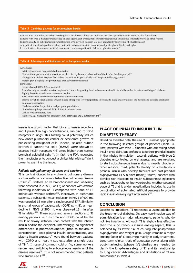
603 December 15, 2016|Volume 7|Issue 20|WJD|www.wjgnet.com
insulin is a growth factor that binds to insulin receptors and if present in high concentrations, can bind to IGF-I receptors in lungs. This binding could potentially induce new-onset pulmonary cancer or accelerate growth of pre-existing malignant cells. Indeed, isolated human bronchial carcinoma cells (H292) were shown to express insulin receptors 4-5 times higher than normal bronchial epithelial cells[18]. In fact, the FDA requested the manufacturer to conduct a clinical trial with sufficient power to examine this issue.
Patients with pulmonary diseases and smokersTI is contraindicated in any chronic pulmonary disease such as asthma or chronic obstructive pulmonary disease (COPD)[7]. Indeed, acute bronchospasm and wheezing were observed in 29% (5 of 17) of patients with asthma following inhalation of TI compared with none of 13 individuals without asthma[7]. Moreover, in asthmatic patients, a substantial mean reduction in FEV1 of 400 mL was recorded 15 min after a single dose of TI[7]. Similarly, in a small group of patients with COPD (n = 8), a mean decline in FEV1 of 200 mL was observed 18 min after TI inhalation[7]. These acute and severe reactions to TI among patients with asthma and COPD could be the result of airway irritation upon contact with the inhaled insulin and/or the excipient. Interestingly, no significant differences in pharmacokinetics (time to maximum concentration, peak plasma insulin concentrations, and plasma insulin exposure) were found between patients with COPD and healthy subjects after a single dose of TI[19]. In case of common cold or flu, some workers recommend switching to subcutaneous insulin until the disease resolves[4]. It is not recommended that patients who smoke use TI[7].
PLACE OF INHALED INSULIN TI IN DIABETES THERAPYBased on available data, the use of TI is most appropriate in the following selected groups of patients (Table 3). First, patients with type 1 diabetes who are taking basal insulin once daily, but prefers to take their prandial insulin in the inhaled formulation; second, patients with type 2 diabetes uncontrolled on oral agents, and are reluctant to start subcutaneous insulin due to needle phobia or other reasons; third, patients already on subcutaneous prandial insulin who develop frequent late post-prandial hypoglycemia (4-5 h after meals); fourth, patients who develop skin reactions to insulin subcutaneous injections such as lipoatrophy or lipohypertrophy. Another potential place of TI that is under investigations includes its use in combination of automated artificial pancreas to provide rapid insulin delivery right after meals[20].
CONCLUSIONDespite its limitations, TI represents a useful addition to the treatment of diabetes. Its easy non-invasive way of administration is a major advantage to patients who do not like injections. Although TI is slightly less effective than the subcutaneous insulin analog aspart, this is balanced by its lower risk of causing late postprandial hypoglycemia and weight gain. Cough remains a major limiting factor of TI occurring mainly in early treatment. Long-term clinical trials of adequate power along with post-marketing (phase IV) studies are needed to clarify the long-term safety of TI and its relationship to lung cancer. Advantages and limitations of TI are summarized in Table 4.
Patients with type 1 diabetes who are taking basal insulin once daily, but prefers to take their prandial insulin in the inhaled formulationPatients with type 2 diabetes uncontrolled on oral agents, and are reluctant to start subcutaneous insulin due to needle phobia or other reasonsPatients already on subcutaneous prandial insulin who develop frequent late post-prandial hypoglycemia (4-5 h after meals)Any patient who develops skin reactions to insulin subcutaneous injections such as lipoatrophy or lipohypertrophyIn combination of automated artificial pancreas to provide rapid insulin delivery right after meals[20]
Table 3 Candidate patients for technosphere insulin
Advantages Relatively easy and non-painful administration Flexible timing of administration either inhaled directly before meals or within 20 min after finishing a meal[10]
Hypoglycemia is less frequent than subcutaneous insulin, particularly late postprandial hypoglycemia Weight gain is slightly less pronounced than subcutaneous insulinLimitations Frequent cough (24%-33% of patients) Available only as prandial short-acting insulin. Hence, long-acting basal subcutaneous insulin should be added in patients with type 1 diabetes Slightly less effective than subcutaneous insulin Need for baseline and then serial pulmonary function testing Safer to switch to subcutaneous insulin in case of upper or lower respiratory infections to avoid exacerbation of the disease and possible unreliable pulmonary absorption No data available for pediatric and pregnant populations Limited strength options and difficult fine titration of doses Lack of long-term safety data High cost, e.g., average price of ninety 4-unit cartridges and 2 inhalers is $271[21]
Table 4 Advantages and limitations of technosphere insulin
Mikhail N. Technosphere insulin
604 December 15, 2016|Volume 7|Issue 20|WJD|www.wjgnet.com
REFERENCES1 Klonoff DC. Afrezza inhaled insulin: the fastest-acting FDA-
approved insulin on the market has favorable properties. J Diabetes Sci Technol 2014; 8: 1071-1073 [PMID: 25355710 DOI: 10.1177/1932296814555820]
2 Nuffer W, Trujillo JM, Ellis SL. Technosphere insulin (Afrezza): a new, inhaled prandial insulin. Ann Pharmacother 2015; 49: 99-106 [PMID: 25313261 DOI: 10.1177/1060028014554648]
3 Cassidy JP, Amin N, Marino M, Gotfried M, Meyer T, Sommerer K, Baughman RA. Insulin lung deposition and clearance following Technosphere® insulin inhalation powder administration. Pharm Res 2011; 28: 2157-2164 [PMID: 21491144 DOI: 10.1007/s11095- 011-0443-4]
4 Kugler AJ, Fabbio KL, Pham DQ, Nadeau DA. Inhaled technosphere insulin: a novel delivery system and formulation for the treatment of types 1 and 2 diabetes mellitus. Pharmacotherapy 2015; 35: 298-314 [PMID: 25809179 DOI: 10.1002/phar.1555]
5 Potocka E, Cassidy JP, Haworth P, Heuman D, van Marle S, Baughman RA. Pharmacokinetic characterization of the novel pulmonary delivery excipient fumaryl diketopiperazine. J Diabetes Sci Technol 2010; 4: 1164-1173 [PMID: 20920436 DOI: 10.1177/193229681000400515]
6 Heinemann L, Baughman R, Boss A, Hompesch M. Phar-macokinetic and Pharmacodynamic Properties of a Novel Inhaled Insulin. J Diabetes Sci Technol 2016; pii: 1932296816658055 [PMID: 27378794 DOI: 10.1177/1932296816658055]
7 Affreza. Prescribing Information. MannKind Corporation. Danbury, CT, June 2014
8 Rave K, Potocka E, Heinemann L, Heise T, Boss AH, Marino M, Costello D, Chen R. Pharmacokinetics and linear exposure of AFRESA compared with the subcutaneous injection of regular human insulin. Diabetes Obes Metab 2009; 11: 715-720 [PMID: 19476477 DOI: 10.1111/j.1463-1326.2009.01039.x]
9 Rosenstock J, Franco D, Korpachev V, Shumel B, Ma Y, Baughman R, Amin N, McGill JB. Inhaled Technosphere Insulin Versus Inhaled Technosphere Placebo in Insulin-Naïve Subjects With Type 2 Diabetes Inadequately Controlled on Oral Antidiabetes Agents. Diabetes Care 2015; 38: 2274-2281 [PMID: 26253730 DOI: 10.2337/dc15-0629]
10 Bode BW, McGill JB, Lorber DL, Gross JL, Chang PC, Bregman DB. Inhaled Technosphere Insulin Compared With Injected Prandial Insulin in Type 1 Diabetes: A Randomized 24-Week Trial. Diabetes Care 2015; 38: 2266-2273 [PMID: 26180109 DOI: 10.2337/dc15-0075]
11 Rosenstock J, Bergenstal R, Defronzo RA, Hirsch IB, Klonoff D, Boss AH, Kramer D, Petrucci R, Yu W, Levy B. Efficacy and safety of Technosphere inhaled insulin compared with Technosphere
powder placebo in insulin-naive type 2 diabetes suboptimally controlled with oral agents. Diabetes Care 2008; 31: 2177-2182 [PMID: 18678610 DOI: 10.2337/dc08-0315]
12 Rosenstock J, Lorber DL, Gnudi L, Howard CP, Bilheimer DW, Chang PC, Petrucci RE, Boss AH, Richardson PC. Prandial inhaled insulin plus basal insulin glargine versus twice daily biaspart insulin for type 2 diabetes: a multicentre randomised trial. Lancet 2010; 375: 2244-2253 [PMID: 20609970 DOI: 10.1016/S0140-6736(10)60632-0]
13 Pittas AG, Westcott GP, Balk EM. Efficacy, safety, and patient acceptability of Technosphere inhaled insulin for people with diabetes: a systematic review and meta-analysis. Lancet Diabetes Endocrinol 2015; 3: 886-894 [PMID: 26341170 DOI: 10.1016/S2213-8587(15)00280-6]
14 Raskin P, Heller S, Honka M, Chang PC, Boss AH, Richardson PC, Amin N. Pulmonary function over 2 years in diabetic patients treated with prandial inhaled Technosphere Insulin or usual antidiabetes treatment: a randomized trial. Diabetes Obes Metab 2012; 14: 163-173 [PMID: 21951325 DOI: 10.1111/j.1463- 1326.2011.01500.x]
15 Lasagna-Reeves CA, Clos AL, Midoro-Hiriuti T, Goldblum RM, Jackson GR, Kayed R. Inhaled insulin forms toxic pulmonary amyloid aggregates. Endocrinology 2010; 151: 4717-4724 [PMID: 20685871 DOI: 10.1210/en.2010-0457]
16 Bloomgarden ZT. Afrezza: some questions about a new approach to prandial insulin. J Diabetes 2014; 6: 489-490 [PMID: 25209874 DOI: 10.1111/1753-0407.12217]
17 Angelo R, Rousseau K, Grant M, Leone-Bay A, Richardson P. Technosphere insulin: defining the role of Technosphere particles at the cellular level. J Diabetes Sci Technol 2009; 3: 545-554 [PMID: 20144294 DOI: 10.1177/193229680900300320]
18 Mayer P, Reitzenstein U, Warnken M, Enzmann H, Racké K. Insulin action on H292 bronchial carcinoma cells as compared to normal bronchial epithelial cells. Pulm Pharmacol Ther 2012; 25: 104-114 [PMID: 22210006 DOI: 10.1016/j.pupt.2011.12.005]
19 Potocka E, Amin N, Cassidy J, Schwartz SL, Gray M, Richardson PC, Baughman RA. Insulin pharmacokinetics following dosing with Technosphere insulin in subjects with chronic obstructive pulmonary disease. Curr Med Res Opin 2010; 26: 2347-2353 [PMID: 20804443 DOI: 10.1185/03007995.2010.511971]
20 Zisser H, Dassau E, Lee JJ, Harvey RA, Bevier W, Doyle FJ. Clinical results of an automated artificial pancreas using technosphere inhaled insulin to mimic first-phase insulin secretion. J Diabetes Sci Technol 2015; 9: 564-572 [PMID: 25901023 DOI: 10.1177/1932296815582061]
21 Goldberg T, Wong E. Afrezza (Insulin Human) Inhalation Powder: A New Inhaled Insulin for the Management Of Type-1 or Type-2 Diabetes Mellitus. P T 2015; 40: 735-741 [PMID: 26609206]
P- Reviewer: Hill DJ, Masaki T S- Editor: Ji FF L- Editor: A E- Editor: Li D
Mikhail N. Technosphere insulin
Stanley Irobekhian Reuben Okoduwa, Ismaila A Umar, Dorcas B James, Hajara M Inuwa, James D Habila
ORIGINAL ARTICLE
605 December 15, 2016|Volume 7|Issue 20|WJD|www.wjgnet.com
Evaluation of extraction protocols for anti-diabetic phytochemical substances from medicinal plants
Basic Study
Stanley Irobekhian Reuben Okoduwa, Directorate of Research and Development, Nigerian Institute of Leather and Science Technology, Zaria 810004, Nigeria
Stanley Irobekhian Reuben Okoduwa, Ismaila A Umar, Dorcas B James, Hajara M Inuwa, Department of Biochemistry, Ahmadu Bello University, Zaria 811104, Nigeria
James D Habila, Department of Chemistry, Ahmadu Bello University, Zaria 811104, Nigeria
Author contributions: Okoduwa SIR and Umar IA got the concept and design of the study; Okoduwa SIR and Habila JD perform the study and data analysis; James DB, Inuwa HM and Habila JD participated in the interpretation of data; Okoduwa SIR wrote the first version of the manuscript; Umar IA, James DB, Inuwa HM and Habila JD critically revised the manuscript for important intellectual content; all the authors read and gave a final approval of the revised version of the manuscript to be published.
Institutional review board statement: Ethics of the study involving the use of animals were reviewed and approved by Institutional Animal Care and Use Committee of the Ahmadu Bello University and the Research Ethics Committee of Nigerian Institute of Leather and Science Technology, Zaria-Nigeria where the research was conducted.
Institutional animal care and use committee statement: The experimental protocol was review and approved by the Research Ethics Committee of the Institute. All experimental protocol was in conformity with the Institutional guidelines that are in compliance with National and International Laws and Guidelines for Care and Use of Laboratory Animals in Biomedical Research.
Conflict-of-interest statement: The authors declare no conflict of interest related to this study and publication.
Data sharing statement: No additional data are available.
Open-Access: This article is an open-access article which was selected by an in-house editor and fully peer-reviewed by external reviewers. It is distributed in accordance with the Creative Commons Attribution Non Commercial (CC BY-NC 4.0) license,
which permits others to distribute, remix, adapt, build upon this work non-commercially, and license their derivative works on different terms, provided the original work is properly cited and the use is non-commercial. See: http://creativecommons.org/licenses/by-nc/4.0/
Manuscript source: Unsolicited manuscript
Correspondence to: Stanley Irobekhian Reuben Okoduwa, Researcher, Directorate of Research and Development, Nigerian Institute of Leather and Science Technology, P.M.B. 1034, Zaria 810004, Nigeria. [email protected]: +234-909-9640143
Received: June 28, 2016 Peer-review started: July 1, 2016First decision: September 5, 2016Revised: September 16, 2016 Accepted: October 17, 2016Article in press: October 18, 2016Published online: December 15, 2016
AbstractAIMTo examine the efficacy of three extraction techniques: Soxhlet-extraction (SE), cold-maceration (CM) and micro-wave-assisted-extraction (MAE) using 80% methanol as solvent.
METHODSThe study was performed on each of 50 g of Vernonia amygdalina (VA) and Occimum gratissimum (OG) leaves respectively. The percentage yield, duration of extraction, volume of solvent used, qualitative and quantitative phytoconstituents present was compared. The biological activities (hypoglycemic effect) were investigated using albino wistar rat model of diabetes mellitus (n = 36) with a combined dose (1:1) of the two plants leaf ex-tracts (250 mg/kg b.w.) from the three methods. The
Submit a Manuscript: http://www.wjgnet.com/esps/Help Desk: http://www.wjgnet.com/esps/helpdesk.aspxDOI: 10.4239/wjd.v7.i20.605
World J Diabetes 2016 December 15; 7(20): 605-614ISSN 1948-9358 (online)
© 2016 Baishideng Publishing Group Inc. All rights reserved.

606 December 15, 2016|Volume 7|Issue 20|WJD|www.wjgnet.com
Okoduwa SIR et al . Effect of extraction methods on phytoconstituents
extracts were administered orally, once daily for 21 d.
RESULTSIn this report, the percentage VA extract yield from MAE was highest (20.9% ± 1.05%) within 39 min using 250 mL of solvent, when compared to the CM (14.35% ± 0.28%) within 4320 min using 900 mL of solvent and SE (15.75% ± 0.71%) within 265 min using 500 mL of solvent. The percentage differences in OG extract yield between: MAE vs SE was 41.05%; MAE vs CM was 46.81% and SE vs CM was 9.77%. The qualitative chemical analysis of the two plants showed no difference in the various phytoconstituents tested, but differs quantitatively in the amount of the individual phytoconstituents, as MAE had significantly high yield (P > 0.05) on phenolics, saponins and tannins. SE technique gave significantly high yield (P > 0.05) on alkaloid, while CM gave significant high yield on flavonoids. The extracts from CM exhibited a significantly (P > 0.05) better hypoglycemic activity within the first 14-d of treatment (43.3% ± 3.62%) when compared to MAE (36.5% ± 0.08%) and SE methods (33.3% ± 1.60%). However, the percentage hypoglycemic activity, 21 d post-treatment with 250 mg/kg b.w. extract from MAE was 72.6% ± 1.03% and it was more comparable to 10 mg/kg b.w. glibenclamide treated group (75.0% ± 0.73%), unlike the SE (69.5% ± 0.71%) and CM (69.1% ± 1.03%).
CONCLUSIONCM technique produces extract with better hypo-glycemic activity, whereas; MAE is a better option for high yield of phytoconstituents using less solvent within a short time.
Key words: Extraction techniques; Microwave-assisted-extraction; Maceration; Phytoconstituents; Medicinal plants; Soxhlet; Anti-diabetes
© The Author(s) 2016. Published by Baishideng Publishing Group Inc. All rights reserved.
Core tip: Extraction of active phytoconstituents from medicinal plants rely mostly on the use of appropriate extraction method. Different extraction techniques affect the yield and biological activity of phytocomponents. In this study, we observed that microwave assisted extraction produces significantly higher overall extract yield as well as in phenolic, saponin and tannin content. Cold maceration and soxhlet extraction produced higher flavonoid and alkaloid yield respectively. Maceration extracts exhibited significantly better hypoglycemic activities in diabetic rats compared to extracts from soxhlet and microwave assisted extraction. This study reveals that the choice of extraction protocol should depend primarily on the purpose of interest.
Okoduwa SIR, Umar IA, James DB, Inuwa HM, Habila JD. Evaluation of extraction protocols for anti-diabetic phytochemical substances from medicinal plants. World J Diabetes 2016; 7(20):
605-614 Available from: URL: http://www.wjgnet.com/1948-9358/full/v7/i20/605.htm DOI: http://dx.doi.org/10.4239/wjd.v7.i20.605
INTRODUCTIONDiabetes mellitus (DM) is one of the most common non-communicable diseases globally, affecting the quality of human life of all ages across the world[1,2]. The disease has become a global public health problem affecting the socio-economic status of the individual[3]. It is an age long, serious heterogeneous metabolic disorder characterized by hyperglycemia and glucose intolerance, due to endogenous insulin deficiency, impaired effec-tiveness of insulin action, or both[4]. With DM the body cannot regulate the amount of sugar in the blood. This leads to increased glucose in the body that causes deregulation of the metabolism, often accompanied by glycosuria, polydipsia, and polyuria[4].
According to International Diabetic Foundation report, every 6 s, a person dies from diabetes[5]. In 2013, 5.0 million deaths were recorded across the globe with a prevalence of 8.3%[5]. A total of 415 million people are affected with diabetes, worldwide as at December, 2015. The figure is estimated to rise above 642 million by 2040[5]. In Nigeria, 3921500 cases have been reported as at 2013 with a prevalence rate of 4.99%. This alarming rate calls for urgency to find better treatment and novel prevention strategies for the disease.
Several hypoglycemic drugs are available for manag-ing diabetes since it is incurable but they suffer from generally inadequate efficacy and number of serious adverse effects[6,7]. Hence, the shift to the use of plant source, a new hopeful approach that has long been authenticated by World Health Organization in its general assembly[8].
Plants are the major source of potential therapeutic agents worldwide. The uses of Plants for therapeutic purposes have been recorded to be as long as history. Plants which contain substances that could be used for medicinal purposes or which are precursors for the synthesis of useful drugs are considered as therapeutic plants[9]. According to the report of Farnsworth and Soejarto[10], there are between 35000 and 70000 plant species that have been used for medicinal purposes in the world[10]. The bioactive components present in plants can only be utilized in disease treatment/management after being extracted with suitable solvent and pre-pared into substances such as ointment, cream, gel, moisturizer, pills and so on[11,12].
Extraction is the partitioning of therapeutically dy-namic segments of plant utilizing suitably selective solvents through standard methods[12,13]. The basis of all extraction in medicinal plant research is to isolate the dissolvable plant metabolites, excluding the insoluble cell marc. The preliminary unrefined extracts obtained utilizing these strategies contain complex blend of numerous plant metabolites, for example, alkaloids,
607 December 15, 2016|Volume 7|Issue 20|WJD|www.wjgnet.com
glycosides, phenolics, terpenoids and flavonoids[12,14,15]. Some bioactive components present in plants are either heat sensitive or solvent specific hence the quality and composition of the extracts as well as their biological activities are affected by the type of extraction procedure. To get the most astounding biological efficacy and yield of plant extract, it is important to consider these limitations and utilize a standardized method for a specific bioactive molecule[14,16].
The needs of standardized extraction methodology for unrefined medications are to accomplish the remedially craved part and to dispose of the inactive portion by treatment with a specific solvent called menstruum. The extract consequently obtained might be prepared for use as a therapeutic agent in the form of tinctures and fluid extracts, it may be further processed to be fused in any dosage form such as tablets or capsules, or it might be fractionated to seclude singular synthetic substances, for example, vincristine, hyoscine and ajmalicine which are modem drugs. For that reason, standardization of extraction methodologies contributes fundamentally to the final nature of the medicinal drug[13,15].
However, with the expanding interest for natural therapeutic products and nutraceuticals for healthcare everywhere throughout the world, producers of medicinal plant extracts are in continuous search for the most suitable extraction strategy keeping in mind the end goal to produce extracts of characterized quality with minimal variability from batch to batch. Conventional extraction is generally carried-out using reflux, maceration, soxhlet and distillation techniques. These techniques which have been utilized for a long time are extremely tedious and require generally a lot of solvents. Extraction utilizing non-routine techniques, for instance, microwave tech-niques can produce high yield, within a shorter time utilizing a smaller amount of solvents[17,18]. Among the different customary and routine extraction systems, Soxhlet extraction has been the most generally utilized. Unfortunately, there is paucity of literature on the best extraction method for a specific bioactive molecule. The evaluation on some of these methods by most previous investigators focuses on either the yield or duration of extraction, without considering the effects of the protocol on the various bioactive entities that works in a synergic manner.
Soxhlet extraction serves not just as a method for extraction of phyto-constituents but additionally as a reference to look at more current extraction procedures. Previously, it has been suggested that the microwave-assisted-extraction (MAE), a present day extraction method is a superior method for extricating phyto-com-ponents from plants[19]. Several reports on the usefulness of the MAE as it concerns medicinal plants have been published[20-24].
Vernonia amygdalina (VA) commonly known as bitter leaf and Occimum gratissimum (OG) generally refers to as scent leaf, have been reported to have anti-diabetic properties[25]. The efficacy of the combined use of both
plants with respect to diabetes has been documented[26]. Also, their hypoglycemic activities have been attributed to the presence of flavonoids, alkaloids and saponins among others[27-29]. It is therefore imperative to examine the effect of different extraction methods on their biological activities.
From our insight, the extraction of phytoconstituents from VA and OG using MAE strategy has not yet been accounted for. These two plants were chosen as a reference point for other therapeutic plants owing to the fact that their anti-diabetic potentials have been established in recent publications[26-31]. Therefore, this study evaluated three extraction technologies, viz: MAE, Soxhlet extraction and cold maceration with an aim to present a comparison among the distinctive strate-gies utilized for extraction of hypoglycemic bioactive constituents from medicinal plants. The primary goal of the research is to give a successful and effective, straight-forward, safe and less time consuming with maximal yield strategy for extricating specific bioactive parts from therapeutic plants.
MATERIALS AND METHODSPlant preparationFresh specimen of VA and OG leaves were harvested in the month of May, 2015 from a local farm in Samaru, Zaria, Kaduna State, Nigeria. The plant samples were identified and authenticated by the Herbarium unit of the Department of Biological Science, Ahmadu Bello University, Zaria, Nigeria. A voucher specimen number 1166 and 1285 were deposited for VA and OG respectively. The leaves of the plants sample were dried under shade at room temperature to constant weights for seven days. The dried samples were then pulverized into powder using a laboratory milling machine (Thomas-Wiley Labo-ratory mill Model 4, United States). The powders were preserved in clean plastic containers, kept away from light, heat and moisture until use.
Reagents and chemicalsAll the chemicals and reagents used were of analytical reagent grade.
Experimental durationThe research was conducted between May and December, 2015.
ApparatusA conventional microwave oven (2450 MHz, Toshiba, and Tokyo, Japan) with variable power up to 1000 watts, a time controller, beam reflector and a stirring device was used.
ProcedureTwo conventional extraction techniques namely, Soxhlet and cold maceration were used in comparison with a new modern technology, the MAE technique.
Okoduwa SIR et al . Effect of extraction methods on phytoconstituents
608 December 15, 2016|Volume 7|Issue 20|WJD|www.wjgnet.com
Soxhlet extraction methodExhaustive Soxhlet extraction was performed using classical apparatus with accurately weighed 50 g of the powdered leaf samples of VA and OG respectively. Extraction was performed with 80% methanol as the extraction solvent. After extraction, the methanol solvent was evaporated by concentrating under vacuum with rotary evaporator (Senco Rotary Evaporator, Model RE 801) at 40 ℃ under reduced pressure. The solvent free methanol extract was thereafter evaluated.
Cold maceration methodMaceration was carried out in a closed conical flask for 72 h. In both case 50 g powdered VA and OG leaf sample and 80% methanol as the extraction solvent were used. The suspension after maceration was centrifuged and the supernatant evaporated under reduce pressure. The solvent free methanol extracts obtained were similarly evaluated.
Microwave assisted extraction methodAccurately weighed 50 g of the homogeneous powder leaf samples was mixed with 60 mL, 80% methanol. After allowing a preleaching time of 5 min the suspension was irradiated with microwave at optimized conditions[32-34]. The samples were treated under microwave irradiation in an intermittent way, i.e., Irradiation: cooling: irradiation. The microwave irradiation time was set at three minutes and cooling time of five minutes was allowed. After 5 repeats, the samples were centrifuged at 4000 rpm and the supernatant evaporated under pressure. The dried residue was evaluated accordingly.
Percentage recovery yield of extractionThe percentage extraction yield (w/w) by the three extraction methods was calculated using the formula: Percentage extraction yield for plant extract = [mass of extract (g)/mass of plant sample (g)] × 100
Aliquots of the extracts were stored in screwed cap vials at 4 ℃-8 ℃ until further use. The extracts were re-dissolved in distilled water when required and given orally through gastric intubations.
Phytochemical analysisStandard protocols were used in detecting the phyto-chemical constituents present in the two plants samples[35,36]. Tannins according the method describe by Markkar et al[37], Saponins as described by Bruneton[38], Alkaloids as described by Harbone[39], Flavonoids as described by Bohm et al[40].
Biological assayTo test biological activities of the plant extract from the different extraction techniques, thirty six albino wistar rats (150-200 g of either sex) fed with rat pellet diet (Grand Cereals Ltd, Nigeria) and water ad libitum were used. Animals were first acclimatized for two weeks before used. The study was conducted at the Research
and Development Laboratory of Nigerian Institute of Leather and Science Technology (NILEST), Zaria Nigeria. The anti-hyperglycemic effect of the extracts obtained from the three extraction methods were assessed using rat model of DM. The experimental protocol was approved by the Institutional Animal Ethic Committee. All experimental protocol was in conformity with the institutional guidelines that are in compliance with National and International Laws and Guidelines for Care and Use of Laboratory Animals in Biomedical Research. The rules and regulations in accordance to the Ethical Committee directive were strictly followed.
Induction of diabetesThe rat model of diabetes used for this study was developed as followed. First, the rats were fasted over-night after which they were given a single intra-peritoneal injection (ip) of 55 mg/kg b.w. of streptozotocin (STZ) (Adooq Bioscience, LLC, United States) dissolved in 0.1 mL fresh cold citrate buffer pH 4.5.
Confirmation of diabetes was done 72 h after STZ induction, using a One Touch Glucometer (Lifescan Inc 1995 Milpas, California, United States). Blood samples were obtained from the tail puncture of the rats. Animals with fasting blood glucose ≥ 200 mg/dL, after 10 d of STZ induction were considered diabetic and included in the study as diabetic animals[30].
Experimental designThirty six rats were divided into 6 groups of 6 rats per group. The treatments were as follows: (1) diabetic rats treated with extracts from cold maceration 250 mg/kg b.w.; (2) diabetic rats treated with extracts from soxhlet extraction (SEE) 250 mg/kg b.w.; (3) diabetic rats treated with extracts from MAE 250 mg/kg; (4) diabetic rats as positive control treated with standard drug (10 mg/kg b.w, glibenclamide); (5) diabetic rats as negative control. No treatment was given; and (6) non-diabetic rats as standard control (no induction, no treatment).
Route of administrationThe extracts were administered orally, once daily for 21 d using combined dose (1:1) of the two plants leaf extracts (250 mg/kg b.w.)
Blood sample collectionBlood sample was withdrawn from the tail vein and tested using glucose test strips and glucometer (On-Call Plus, Acon Laboratories Ins, United States) after an overnight fast.
Statistical analysisThe results obtained were expressed as mean ± SD where applicable. The data were analyzed using analysis of variance and significant differences among means were determined by Duncan’s multiple range test at P < 0.05 using Statistical Package for Social Sciences software version 20 for windows.
Okoduwa SIR et al . Effect of extraction methods on phytoconstituents

609 December 15, 2016|Volume 7|Issue 20|WJD|www.wjgnet.com
RESULTSThe efficacy of three extraction methods were compared by evaluating the anti-hyperglycemic effects of the two medicinal plants (VA and OG) leaf extracts. A flow chart illustrating the experimental design in detail is presented in Figure 1. The experiments were performed using the same quantity of plant samples (50 g each) and the biological activities were analyzed using rat model of diabetes with the same dose of extract (250 mg/kg body weight). A comparison of the extraction of the medicinal plants (VA and OG) using conventional microwaves, soxhlet extractor and cold maceration are shown in Table 1.
The yield of MAE extracts from VA for 39 min were higher (20.90% ± 1.05%) than that of soxhlet extract (15.75% ± 0.71%) for 265 min and that of maceration extract (14.35% + 0.28%) for 4320 min. Similarly, the yield of microwave extracts from OG (19.10% ± 1.67%) for 39 min were higher than that of soxhlet extract (11.25% ± 0.42%) for 255 min and that of maceration extract (10.15% ± 0.65%) for 4320 min. Regarding the extraction time, our result show that MAE appeared to be the fastest method since the extraction could be achieved within minutes. The MAE also consumed the least amount of solvent 250 mL when compared to the other methods, 500 mL and 900 mL for soxhlet and cold maceration respectively (Table 1).
The percentage differences in the recovery yield between two specific extraction methods are shown in the Table 2. It was observed that for VA, the difference in percentage recovery yield between MAE vs soxhlet extraction method was 24.59%; but with OG it was 41.05%. The difference in percentage yield between MAE vs cold maceration method was 31.29% for VA and 46.81% for OG. Although there were no significant differences between the soxhlet and cold maceration method for both plants, the percentage difference was
8.88% and 9.77 for VA and OG respectively (Table 2).The results from the present study showed that there
was no difference between the qualitative phytocon-stituents obtained by the various extraction technologies under investigation (Table 3).
However, the quantitative chemical analysis (Table 4) revealed a statistically higher yield in alkaloid from the soxhlet extraction when compared to the MAE and cold maceration for VA. But there was no significant difference in the alkaloid yield between the soxhlet and MAE method for OG. The cold maceration technology recorded the highest yield in flavonoid in both plant when compared to the MAE and soxhlet extraction method. The difference was statistically significant (P < 0.05). The MAE had the highest yield in phenolics, saponins and tannins from the two plants studied when compared to the conventional extraction techniques (soxhlet and cold maceration). However, there was no significant difference between MAE and soxhlet in the yield of tannin from VA.
Soxhlet, cold maceration and MAE extracts of VA and OG were tested and compared for anti-diabetic activities. All the extracts exhibited comparable anti-diabetic activities with that of standard drug (glibenclamide) under the same dose rate of 250 mg/kg body weight tested according to the method of Abdulazeez et al[26]. The cold maceration extract exhibited a better hypoglycemic effect within the first 7 (17.9%) and 14 (43.2%) d of treatment when compared to others. But the hypoglycemic activity of extract obtained from MAE was more comparable to the standard drug glibenclamide in reducing the blood glucose of the animals after 28 d post induction (21 d treatment).
The extract from MAE exhibited the least hypo-glycemic effect within the first 7 day of treatment. There was no significant difference between the hypoglycemic effects of extracts obtained from soxhlet and the cold maceration method at the end of the 21 d (Tables 5 and 6).
DISCUSSIONDeveloping nations are rich in therapeutic plants at the same time, because of trouble in getting reliably effective extraction equipments; esteem expansion to this rich bioresources are difficult. Usually and prevalently in extremely poor nations, the advancements utilized are improper and not efficient. The crucial setback is identified with the nature of the products. Primitive extraction techniques don’t promise a steady and top notch reliable quality and, sometimes, unseemly innovations and techniques result in creating defiled products which has low market value[41]. The present study, evaluated three different technologies for extraction of hypoglycemic compounds from medicinal plants, with a view to ascertain the best option for the isolation of specific bioactive entity.
In the study it was noticed that Soxhlet extraction and cold maceration spent longer time for complete extraction of same quantity of the plant sample studied. Also, the amount of the solvent utilized in MAE was
Dry under shadeSample collection and authentication Pulverized into powder
Concentration of extract
Soxhlet
Cold maceration
Microwave assisted
Extract Extract ExtractAnalysisPercentage yield of extract
Quantitative phytochemical
Qualitative phytochemical
Volume of solvent
Treatment with extract21 d
Fasting blood glucose
7 d
Confirmation of diabetes
3 d
STZ induction 55 mg/kg b.w.
Acclimitization2 wk
Rat purchase
Extraction(80% methanol)
Figure 1 Experimental design for the evaluation of three extraction methods. STZ: Streptozotocin.
Okoduwa SIR et al . Effect of extraction methods on phytoconstituents

610 December 15, 2016|Volume 7|Issue 20|WJD|www.wjgnet.com
less, proving that, MAE is indeed truly economical. Comparable results were reported by Vongsangnak et al[42], when contrasting routine extraction procedures with MAE throughout the isolation of saponins from cell culture of Panax notoginseng and analgesic substances from the roots of Ximenia americana[20]. Besides, micro-wave illumination technique has been reported to be extremely quick, dependable for making of Schiff bases[21]. Possible explanation observed in the various methods of extraction could be due to differences in rate of chemical reaction. For instance, in soxhlet extraction approach, the applied heat supplies the activation energy needed to rupture the plant tissues in order to release its phytoconstituents. Whereas in MAE, the activation energy is achieved due to oscillatory wave generated by the system and by the ionic conduction and dipole rotation of the molecules of the plant sample. This brings about increase in the cell pressure, thereby rupturing to release its contents on attaining its elastic limit. The heat in MAE is internally generated by the molecules unlike in soxhlet whereby the heat is externally generated. In cold maceration, the release of phytoconstituents from plant matrix is due to differences in ionic solvent concentration gradients, hence more solvent is require to create a positively dynamic concentration gradients. This account for the higher volume of solvent utilized by the cold maceration as observed in the present study. In a nutshell, with MAE, the plant cell tissues are effectively broken and separated to release phytoconstituents with ease, hence the greater yield as recorded.
The result of the quantitative phytochemical screening within the limit of the analyzed components in the pre-sent study suggests that the phytoconstituents of the
studied plants were not destroyed since the results obtained showed that the same phytoconstituents tested in all the extracts from the different procedures were the same. This finding is in agreement with earlier reports of Mandal et al[19], that, the plant components obtained from MAE are neither decomposed nor oxidized under optimized conditions. Several phytochemicals have been found to give an increase in their extractive yields when compared to their yields on subjection to conventional extraction techniques[21]. Chan et al[22] also recorded a higher yield in the extraction of anti-diabetic ingredient from herbal plant. In their experiment using 5 g sample with 150 mL solvent recorded a yield of 1.63 mg/g sample in 5 min using MAE as against 0.47 mg/g in 3 h obtained with soxhlet extraction technology. In a related study for extraction of caffeine and polyphenols from leaves of green tea, MAE achieved higher extraction yield within 4 min than any extraction methods at room temperature for 20 h[43]. Ginsenosides extraction yield from ginseng root was obtained in 15 min using focused MAE technique which was also better than other conventional solvent extraction technologies for 10 h[24,44]. The higher yield obtained from MAE and soxhlet extraction when compared to the cold maceration may be attributed to the heat exchange and mass transfer.
In soxhlet extraction, the heat exchange and the mass transfer are restricting variables contrast with MAE, where heat exchange happens from the focal point of
Vernonia amygdalina Ocimum gratissimumParameter MAE Soxhlet Maceration MAE Soxhlet MacerationSample (g) 50 50 50 50 50 50Solvent volume (mL) 250 500 900 250 500 900Extraction time (min) 39 265 4320 39 255 4320Recovery yield (g) 10.45 ± 0.53b 7.88 ± 0.35a 7.18 ± 0.14a 9.55 ± 0.84b 5.63 ± 0.21a 5.08 ± 0.32a
Percentage recovered (%) 20.90 ± 1.05b 15.75 ± 0.71a 14.35 ± 0.28a 19.10 ± 1.67b 11.25 ± 0.24a 10.15 ± 0.65a
Table 1 Comparison of percentage recovery yield, extraction time and volume of solvent used for the different extraction methods for the two plants
Values are mean ± SD of 3 replicate determinations; same superscript across the column under the same plant indicate no significant difference (P > 0.05). MAE: Microwave-assisted-extraction.
Vernonia amygdalina Ocimum gratissimumMAE Soxhlet Maceration
MAE 41.05% 46.81%Soxhlet 24.59% 9.77%Maceration 31.29% 8.88%
Table 2 Calculated percentage differences in recovery yield of extracts between the extraction methods
Percentage difference between two extraction methods = [(values from method with higher recovery yield - values from method with lower recovery yield)/values from method with higher recovery yield] × 100.
S/N Constituents Maceration Soxhlet Microwave
VA OG VA OG VA OG1 Carbohydrates + + + + + +2 Anthraquinones - - - - - -3 Glycosides + + + + + +4 Cardiac glycosides + + + + + +5 Saponins + + + + + +6 Steroids + + + + + +7 Triterpenes + + + + + +8 Tannins + + + + + +9 Flavonoids + + + + + +10 Alkaloid + + + + + +
Table 3 Comparison of some qualitative phytochemicals obtained with the three extraction methods for the two plants
The symbol (+): Indicate detected; (-): Not detected; VA: Vernonia amygdalina; OG: Ocimum gratisimum.
Okoduwa SIR et al . Effect of extraction methods on phytoconstituents

611 December 15, 2016|Volume 7|Issue 20|WJD|www.wjgnet.com
the specimens to the external colder environment, and volumetric warming impact prompts a speedier ascend in temperature. Additionally, the interior warming of in situ water inside of the plant material expands the plant cells and prompts the burst of the plant tissues[45]. This may be the explanation behind the higher extraction of phytochemicals from MAE when compared to soxhlet and cold maceration extracts observed in the present study. It is very obvious from these results, that micro-wave extraction represents a promising substitute for extracting hypoglycemic compounds from natural substrate.
The hypoglycemic activity of the 250 mg/kg b.w. cold
maceration extract was significantly better than others within the first 14 day of treatment. Surprisingly after 21 d treatment, the blood glucose lowering capacity of the MAE extract was almost the same with that of 10 mg/kg b.w. glibenclamide. The percentage change in blood glucose by MAE extract after the experimental duration of 21 d treatment was 72.6% whereas that obtained from the standard drug glibenclamide was 75.0%. The higher yield of alkaloid by soxhlet extraction may account for its better efficacy when compared to the cold maceration in the present study. Saponin, alkaloid and flavonoid are known to play a significant role in anti-diabetic action[27-29].
MAE had the highest yield in three different phyto-constituents viz phenolics, saponins and tannins. This suggests that microwave methanol extract could be used even at 250 mg/kg b.w. to complement currently available oral hypoglycemic drugs. The results suggest that microwave technology is a viable means for extracting valuable anti-diabetic components from medicinal plants. The reason for the observed differences in hypoglycemic activities by the various extract within the first 14 d and after the 21 d of experimental treatment could be due to the concentrations of the phyto-chemicals which vary in the extracts. It could also be possible that the different phytochemical exhibiting hypoglycemic effect have different rate of reaction in reducing the blood glucose level.
The main advantages of MAE over the conventional extraction techniques is that it reduces solvent con-sumption, it has a shorter operational time, modestly high recoveries, decent reproducibility and negligible
Vernonia amygdalina (mg/100 g) Ocimum gratissimum (mg/100 g)
MAE Soxhlet Maceration MAE Soxhlet MacerationAlkaloid 4.0 ± 0.65a 7.0 ± 0.57b 5.0 ± 0.46a 6.5 ± 0.75b 7.0 ± 0.15b 5.0 ± 0.90a
Flavonoid 13.0 ± 0.35b 9.0 ± 0.50a 15.0 ± 0.21c 10.0 ± 0.35b 8.0 ± 0.18a 12.0 ± 0.25c
Phenolic 17.5 ± 0.25c 15.0 ± 0.21b 12.0 ± 0.70a 15.0 ± 0.22c 14.0 ± 0.21b 11.0 ± 0.65a
Saponin 4.5 ± 0.45b 3.0 ± 0.30a 2.0 ± 0.75a 6.1 ± 0.75b 4.0 ± 0.92a 5.0 ± 0.20a
Tannin 14.0 ± 0.55b 13.0 ± 0.50b 10.0 ± 0.25a 12.0 ± 0.18b 9.0 ± 0.24a 9.1 ± 0.25a
Table 4 Comparison of the quantitative phytochemicals obtained with the three extraction methods for the two plants
Values are mean ± SD of 3 replicate readings. Same superscript across the column under the same plant signifies no significant difference at P > 0.05. MAE: Microwave-assisted-extraction.
Group Before induction 7 d after induction Days after treatment
7th 14th 21st
CME 79.4 ± 4.5 344.6 ± 10.3 282.8 ± 17.6 195.8 ± 18.3 106.5 ± 6.7SEE 85.3 ± 3.2 396.8 ± 15.7 342.5 ± 14.7 264.9 ± 16.8 121.1 ± 10.2MAE 82.7 ± 5.1 373.3 ± 13.6 331.3 ± 11.4 237.1 ± 8.9 102.4 ± 7.6PC 91.6 ± 4.5 389.4 ± 11.9 359.0 ± 12.6 266.3 ± 13.2 97.3 ± 5.8NC 80.6 ± 3.8 365.5 ± 9.8 395.1 ± 16.3 419.6 ± 10.2 448.4 ± 17.4HC 83.2 ± 6.2 84.8 ± 4.7 83.9 ± 7.8 84.7 ± 6.5 84.2 ± 7.1
Table 5 Effect of the extract from the different extraction methods on blood glucose in streptozotocin-induced diabetic rats
Values are mean ± SD (mg/dL) of readings from 6 rats per group. CME: Cold maceration extract; SEE: Soxhlet extraction extract; MAE: Microwave-assisted extraction extract; PC: Positive control; NC: Negative control; HC: Healthy control.
Group Initial blood glucose on day 0 (mg/dL)
Percentage change after treatment (%)
Day 7 Day 14 Day 21CME 344.6 ± 10.3 18.0 ± 2.66e 43.3 ± 3.62e 69.1 ± 1.03c
SEE 396.8 ± 15.7 13.7 ± 0.29d 33.3 ± 1.60c,d 69.5 ± 0.71c
MAE 373.3 ± 13.6 11.2 ± 0.18c,d 36.5 ± 0.08d 72.6 ± 1.03d
PC 389.4 ± 11.9 7.8 ± 0.42c 31.6 ± 1.30c 75.0 ± 0.73d
NC 365.5 ± 9.8 -8.1 ± 1.57a -14.8 ± 0.29a -22.7 ± 1.47a
HC 84.8 ± 4.7 1.4 ± 3.96b 0.1 ± 2.12b 0.7 ± 2.88b
Table 6 Calculated percentage change in blood glucose after treatment with the extracts in streptozotocin-induced diabetic rats
Same superscript down the column signifies no significant difference at P > 0.05. Percentage change in blood glucose = [(blood glucose before treatment - blood glucose after treatment)/blood glucose before treatment] × 100. CME: Cold maceration extract; SEE: Soxhlet extraction extract; MAE: Microwave-Assisted Extraction extract; PC: Positive control; NC: Negative control; HC: Healthy control.
Okoduwa SIR et al . Effect of extraction methods on phytoconstituents

612 December 15, 2016|Volume 7|Issue 20|WJD|www.wjgnet.com
specimen control for extraction process[46]. Several biologically active compounds have been extracted by application of MAE, such as extraction of azadiractine related limonoids from Azadirachta indica seed kernels[47], extraction of artemisinin from Artemisia annua[48] and ginsenosides extraction from roots of Panax ginseng[44], quercetin from herbal plant[22]. According to Pan et al[49], antioxidant activity of phenolic substances extricated from the peel of Dimocarpus Longan utilizing MAE was better than that of Soxhlet extraction. Besides, MAE of curcumin from Curcuma longa showed a better results and a higher extraction yield with noteworthy diminishing in the extraction time when compared to that of Soxhlet extraction, maceration and stirring extraction[50]. In the present study, there was higher yield in phenolics, saponins and tannins from both plants-VA and OG. This implies that MAE is a better technology for the extraction of these phytoconstituents. The soxhlet extraction technology showed a higher yield in alkaloid whereas maceration technology was best for the extraction of flavonoids.
We find the use of MAE leads to very fast extraction rate with high value of phytoconstituents compared to soxhlet and cold maceration technique. The findings obtained from the present research showed that the choice of extraction technology should be based primarily on the phytochemical entity of interest. For instance, with respect to the result from the present research, the use of soxhlet extraction technology would be recommended when alkaloid is the main phytoconstituent of interest, whereas, the cold maceration would be preferred for extraction of flavonoids. However, since there was no apparent destruction of any bioactive components by the extraction technologies studied, the MAE is recommended as the most suitable technology for routine extraction processes because it is faster, utilizes relatively less amount of solvent and saves more time. Nevertheless, since extraction efficiency differs from efficacy, no single method can be rated as best for extracting all forms of phyto-components. So, a further study on the isolation of the precise bioactive component(s) and its structural elucidation is recommended to ascertain the best technology for obtaining pure bioactive hypoglycemic compound(s) from medicinal plants.
ACKNOWLEDGMENTSThe authors say thanks to the management of SIRO-Nigeria Global Limited, Abuja for providing the basic logistics for the research. The Director and Staff of R and D, NILEST, Zaria for their technical assistance throughout the period of the research.
COMMENTSBackgroundAlthough several methods are available for extraction of active phytoconstituents from medicinal plants. The technique that produces higher yield of specific
phytoconstituents has not been reported. The biological activities of these active phytoconstituents are known to be affected by the extraction protocol employed. To address these issues, this study examines three different procedures of extracting anti-diabetic substances from medicinal plants and evaluated their hypoglycemic activities.
Research frontiersThere is active research in the field of investigation of a more effective extraction technique of active ingredient from medicinal plant. Hence, it is imperative to ascertain which extraction protocol produces a significant amount of specific phytoconstituents.
Innovations and breakthroughsThe hypoglycemic activity of anti-diabetic plant is revealed in this study to be subject to the extraction method employed. For the first time, extraction method producing significantly high yield of specific phytoconstituents is presented.
ApplicationsSince the biological efficacy of phytoconstituents is subject to the extraction protocol employed, the use of an appropriate extraction technique with respect to a specific active ingredient would enhance the process of drug development.
TerminologyPhytoconstituents are chemical compounds that occur naturally in plants. Some of which are responsible for the medicinal effect of the plant.
Peer-reviewThe authors provided the complete review of this issue. This manuscript provides the updated evidence to the readers.
REFERENCES1 Okoduwa SI, Umar IA, Ibrahim S, Bello F, Habila N. Age-
dependent alteration of antioxidant defense system in hypertensive and type-2 diabetes patients. J Diabetes Metab Disord 2015; 14: 32 [PMID: 25922827 DOI: 10.1186/s40200-015-0164-z]
2 Cheng Y, Malik U, Chang S. The risk factors of diabetic nephro-pathy in Taiwan, including old age, hypertension and aspirin therapy. Int J Diabetes Dev C 2013; 33 Suppl 2: 128 [DOI: 10.1007/s13410-013-0114-6]
3 Okoduwa SI, Umar IA, Ibrahim S, Bello F, Ndidi US. Socio-economic status of patients with type 2 diabetes and hypertension attending the Ahmadu Bello University Teaching Hospital, Zaria, North-West Nigeria. Glob J Health Sci 2015; 7: 280-287 [PMID: 25560354 DOI: 10.5539/gjhs.v7n1p280]
4 American Diabetes Association. Diagnosis and classification of diabetes mellitus. Diabetes Care 2014; 37 Suppl 1: S81-S90 [PMID: 24357215 DOI: 10.2337/dc14-S081]
5 International Diabetes Federation. IDF Diabetes Atlas, 7th ed. Brussels, Belgium: International Diabetes Federation, 2015. Available from: URL: http://www.diabetesatlas.org
6 Fowler MJ. Diabetes Treatment, Part 2: Oral agents for glycemic management. Clinical Diabetes 2007; 25: 131-134 [DOI: 10.2337/diaclin.25.4.131]
7 Wild S, Roglic G, Green A, Sicree R, King H. Global prevalence of diabetes: estimates for the year 2000 and projections for 2030. Diabetes Care 2004; 27: 1047-1053 [PMID: 15111519 DOI: 10.2337/diacare.27.5.1047]
8 Dey L, Attele AS, Yuan CS. Alternative therapies for type 2 diabetes. Altern Med Rev 2002; 7: 45-58 [PMID: 11896745]
9 Fagbohun ED, Asare, RR and Egbebi, AO. Chemical composition and antimicrobial activities of Urena lobata L. (Malvaceae). J Med Plants Res 2012; 6 Suppl 12: 2256-2260 [DOI: 10.5897/JMPR09.233]
10 Farnsworth NR, Soejarto, DD. Global Importance of Medicinal Plants. In: Akerele O, Heywood V, Synge H editors. The Conservation of Medicinal Plants Cambridge University Press,
COMMENTS
Okoduwa SIR et al . Effect of extraction methods on phytoconstituents
613 December 15, 2016|Volume 7|Issue 20|WJD|www.wjgnet.com
Cambridge, United Kingdom, 1991: 25-51 [DOI: 10.1017/CBO 9780511753312.005]
11 Handa SS. An Overview of Extraction Techniques for Medicinal and Aromatic Plants. In: Handa et al., (eds): Extraction technologies for medicinal and aromatic plants, 1st ed, no. 66. Italy: United Nations Industrial Development Organization and the International Centre for Science and High Technology. Trieste, 2008: 21-25
12 Azwanida NN. A Review on the Extraction Methods Use in Medicinal Plants, Principle, Strength and Limitation. Med Aromat Plants 2015; 4: 196 [DOI: 10.4172/2167-0412.1000196]
13 Handa SS, Khanuja SPS, Longo G, Rakesh DD. Extraction Technologies for Medicinal and Aromatic Plants, 1st ed, no. 66. Italy: United Nations Industrial Development Organization and the International Centre for Science and High Technology. Trieste, 2008
14 Prabhu KS, Lobo R, Shirwaikar AA, Shirwaikar A. Ocimum gratissimum: A Review of its Chemical, Pharmacological and Ethnomedicinal Properties. TOALTMED 2009; 1: 1-15 [DOI: 10.2174/1876391X00901010001]
15 Tiwari P, Kumar B, Kaur M, Kaur G, Kaur H. Phytochemical screening and extraction: a review. Internationale Pharmaceutica Sciencia 2011; 1 Suppl 1: 98-106
16 Ncube NS, Afolayan AJ, Okoh AI. Assessment techniques of antimicrobial property of natural compounds of plant origin. Current methods and future trend. Afr J Biotechnol 2008; 7 Suppl 12: 1797-1806 [DOI: 10.5897/AJB07.613]
17 Hijazi A, Bandar H, Rammal H, Hachem A, Saad Z, Badran B. Techniques for the Extraction of Bioactive Compounds from Lebanese Urtica dioica. AJPCT 2013; 1: Suppl 6: 507-513
18 Chemat F, Tomao V, Virot M. In: Otles, S. (Ed.), Handbook of Food Analysis Instruments. Ultrasound-Assisted Extraction in Food Analysis. CRC Press, 2008: 85-94
19 Mandal V, Mohan Y, Hemalatha S. Review Article Microwave Assisted Extraction an Innovative and Promising Extraction Tool for Medicinal Plant Research, Pharmacognosy Review. PHCOG Rev 2007; 1 Suppl 1: 7-18
20 Kenmogne SB, Ngassoum M, Tchatchueng JB, Vardamides JC, Dongmo. Microwave Assisted Extraction of Analgesic Compounds of the Root of Ximenia americana (Olacaceae). RJCS 2014; 4 Suppl 7: 7-10
21 Savalia RV, Patel AP, Trivedi PT, Gohel HR, Khetani DB. Rapid and Economic Synthesis of Schiff Base of Salicylaldehyde by Microwave Irradiation. RJCS 2013; 3 Suppl 10: 69-76
22 Chan CH, Yusoff R, Ngoh GC, Kung FW. Extraction of anti-diabtetic active ingredient, quercetin from herbal plant using microwave-assisted extraction (MAE) technique, International conference on Materials for Advanced Technologies. SUNTEC Singapore, 2011: KK-PO2-5 [DOI: 10.13140/2.1.3487.4885]
23 Tatke PA, Jirge, SS, Shukla TA. An extraction procedure of scopoletin from Convovuulus plaricuulis (Shankhapushpi). JMAPS 2010; 31: 126-126
24 Dhobi M, Mandal V, Hemalatha S. Optimization of microwave assisted extraction of bioactive flavonolignan silybinin. J Chem and Metrl 2009; 3: Suppl 1: 13-23
25 Mohammed YT, Okasha MA, Magaji RA, Yaro AH. Effects of aqueous leaves extract of Ocimum gratissimum on blood glucose levels of streptozotocin-induced diabetic wistar rats. Afr J Biotechnol 2007; 6 Suppl 18: 2087-2090 [DOI: 10.5897/AJB2007. 000-2323]
26 Abdulazeez MA , Ibrahim K, Bulus K, Babvoshia HB, Abdullahi Y. Effect of combined use of Ocimum gratissimum and Vernonia amygdalina extract on the activity of angiotensin converting enzyme, hypolipidemic and antioxidant parameters in streptozotocin-induced diabetic rats. AJBR 2013; 7 Suppl 9: 165-173
27 Taoying Z, Denghong L, xingyuan L, Yunbo. Hypoglycemic and hypolipidemic effects of flavonoids from lotus (Nelumbo nuficera Gaertn) leaf in diabetic mice. J Med Plants Res 2009; 3 Suppl 4: 290-293
28 Day C, Cartwright T, Provost J, Bailey CJ. Hypoglycaemic effect
of Momordica charantia extracts. Planta Med 1990; 56: 426-429 [PMID: 2077547 DOI: 10.1055/s-2006-961003]
29 Francis G, Kerem Z, Makkar HP, Becker K. The biological action of saponins in animal systems: a review. Br J Nutr 2002; 88: 587-605 [PMID: 12493081 DOI: 10.1079/BJN2002725]
30 Okon UA, Owo DU, Udokang NE, Udobang JA, Ekpenyong CE. Oral Administration of Aqueous Leaf Extract of Ocimum Gratissimum Ameliorates Polyphagia, Polydipsia and Weight Loss in Streptozotocin-Induced Diabetic Rats. AJMSM 2012; 2 Suppl 3: 45-49 [DOI: 10.5923/j.ajmms.20120203.04]
31 Modu S, Adeboye AE, Maisaratu A, Mubi BM. Studies on the administration of Vernonia amygdalina Del. (Bitter leaf) and glucophage on blood glucose level of alloxan - Induced diabetic rats. IJMPAM 2013; 1 Suppl 1: 013-019
32 Proestos C, Komaitis M. Application of microwave assisted extraction to the fast extraction of plant phenolic compounds. LWT-Food Sci Technol 2008; 41 Suppl 4: 652-659 [DOI: 10.1016/j.lwt.2007.04.013]
33 Gharekhani M, Rafiee Z, Ghorbani M, Jafari SM. Open vessel microwave system for extraction of analytes from medicine plants. Iran Patent 2009: 59321
34 Asghari J, Ondruschka B, Mazaheritehrani M. Extraction of bioactive chemical compounds from the medicinal Asian plants by microwave irradiation. J Med Plants Res 2011; 5 Suppl 4: 495-506
35 Evans WA. Plants in African traditional medicines. An over view. In Trease and Evans Pharmacognosy (15th ed). India: Saunders in Print Elsevier, 2005: 448-491
36 Harbone JB. Methods of extraction and isolation. In Phyto-chemical Methods. London: Chapman and Hall, 1998: 60-66
37 Markkar AOA, Goodchild AV. Quantification of tannins. A labo-ratory manual. Aleppo Syria: International Centre for Agriculture Research in the dry areas. (ICARDA), Aleppo, Syria, 1996: 55
38 Bruneton J. Pharmacognosy, phytochemistry, Medicinal plants. 2nd ed. Hamshire, UK: Intercept, 1999: 385-386
39 Harbone JB. Phytochemistry Methods: a guide to modern techniques of plants analysis. London: Chapman and Hall, 1973: 267-270
40 Bohm BA, Koupai-Abyazani. Flavonoids and condensed tannins from leaves of Hawaiian Vacinium reticulatum and V. calycinum (ericaceae). Pacific Science 1994; 48: 458-463
41 Fermeglia M. Role of Process Simulation in Extraction Tech-nologies for Medicinal and Aromatic Plants. In: Extraction technologies for medicinal and aromatic plants, 2008
42 Vongsangnak W, Gua J, Chauvatcharin S, Zhong, JJ. Toward Efficient Extraction of Notogingseng Saponins from Cultured Cells of Notoginseng. Biochem Eng J 2004; 18: 115-120 [DOI: 10.1016/S1369-703X(03)00197-9]
43 Pan X, Niu G, Liu H. Microwave-assisted extraction of tea polyphenols and tea caffeine from green tea leaves. Chem Eng Process 2003; 42 Suppl 2: 129-133 [DOI: 10.1016/S0255- 2701(02)00037-5]
44 Shu YY, Ko MY and Chang YS. Microwave assisted extraction of ginsenosides from ginseng root. Microchem J 2003; 74 Suppl 2: 131-139 [DOI: 10.1016/S0026-265X(02)00180-7]
45 Tatke P, Jaiswal Y. An overview of Microwave Assisted Extraction and its Applications in Herbal Drugs Research. Research Journal of Medicinal Plant 2011; 5 Suppl 1: 21-31 [DOI: 10.3923/rjmp. 2011.21.31]
46 Odabas HI, Koca I. Application of response surface methodology for optimizing the recovery of phenolic compounds from hazelnut skin using different extraction methods. Ind Crop Prod 2016; 91: 114-124 [DOI: 10.1016/j.indcrop.2016.05.033]
47 Dai J, Yaylayan, V, Raghavan G, Pare J. Extraction and colorimetric determination of azadirachtin related limonoids in neem seed kernel. J Agr Food Chem 1999; 47: 3738-3742 [DOI: 10.1021/jf990227h]
48 Hao JY, Han W, Huang SD, Xue BY, Deng X. Microwave assisted extraction of artemisinin from Artemisia annua L. Sep Purif Technol 2002; 28 Suppl 3: 191-196 [DOI: 10.1016/S1383-5866(02)00043-6]
49 Pan Y, Wang K, Huang S, Wang H, Mu X, He C, Ji X, Zang J, Huang F. Antioxidant activity of microwave assisted extract of
Okoduwa SIR et al . Effect of extraction methods on phytoconstituents

614 December 15, 2016|Volume 7|Issue 20|WJD|www.wjgnet.com
longan (Dimorcarpus logan) peel. Food Chem 2008; 106 Suppl 3: 1264-1270 [DOI: 10.1016/j.foodchem.2007.07.033]
50 Mandal V, Mohan Y, Hemalatha S. Microwave assisted extraction
of curcumin by sample-solvent dual heating mechanism using Taguchi L9 orthogonal design. J Pharmaceut Biomed 2008; 46: 322-327 [DOI: 10.1016/j.jpba.2007.10.020]
P- Reviewer: Liao KF, Papanas N S- Editor: Ji FF L- Editor: A E- Editor: Li D
Okoduwa SIR et al . Effect of extraction methods on phytoconstituents
Blanca Murillo Ortíz, Joel Ramírez Emiliano, Edna Ramos-Rodríguez, Sandra Martínez-Garza, Hilda Macías-Cervantes, Sergio Solorio-Meza, Texar Alfonso Pereyra-Nobara
ORIGINAL ARTICLE
615 December 15, 2016|Volume 7|Issue 20|WJD|www.wjgnet.com
Brain-derived neurotrophic factor plasma levels and premature cognitive impairment/dementia in type 2 diabetes
Case Control Study
Blanca Murillo Ortíz, Edna Ramos-Rodríguez, Sandra Martínez-Garza, Hilda Macías-Cervantes, Sergio Solorio-Meza, Texar Alfonso Pereyra-Nobara, Unidad de Investigación en Epidemiología Clínica, Unidad Médica de Alta Especialidad No. 1 Bajio, Instituto Mexicano del Seguro Social, Guanajuato 37320, Mexico
Joel Ramírez Emiliano, Departamento de Ciencias Médicas, División Ciencias de la Salud, Universidad de Guanajuato, Guanajuato 37320, Mexico
Author contributions: All the authors contributed to the paper.
Institutional review board statement: This protocol was approved by the local bioethics committee (R2014100188).
Informed consent statement: The informed consent was obtained from each volunteer.
Conflict-of-interest statement: The authors declare no conflicts of interest regarding this manuscript.
Data sharing statement: No data were created no data are available.
Open-Access: This article is an openaccess article which was selected by an inhouse editor and fully peerreviewed by external reviewers. It is distributed in accordance with the Creative Commons Attribution Non Commercial (CC BYNC 4.0) license, which permits others to distribute, remix, adapt, build upon this work noncommercially, and license their derivative works on different terms, provided the original work is properly cited and the use is noncommercial. See: http://creativecommons.org/licenses/bync/4.0/
Manuscript source: Invited manuscript
Correspondence to: Blanca Murillo Ortíz, PhD, Unidad de Investigación en Epidemiología Clínica, Unidad Médica de Alta Especialidad No. 1 Bajio, Instituto Mexicano del Seguro Social, López Mateos Esquina Insurgentes s/n Colonia Los Paraisos,
León, Guanajuato 37320, Mexico. [email protected]: +524777183039Fax: +524777183039
Received: June 21, 2016Peer-review started: June 24, 2016First decision: August 11, 2016Revised: August 27, 2016Accepted: October 1, 2016Article in press: October 9, 2016Published online: December 15, 2016
AbstractAIMTo assess the relationship of brain-derived neurotrophic factor (BDNF) with cognitive impairment in patients with type 2 diabetes.
METHODSThe study included 40 patients with diabetes mellitus type 2 (DM2), 37 patients with chronic kidney disease in hem dialysis hemodialysis therapy (HD) and 40 healthy subjects. BDNF in serum was quantified by ELISA. The Folstein Mini-Mental State Examination was used to evaluate cognitive impairment.
RESULTSThe patients with DM2 and the patients in HD were categorized into two groups, with cognitive impairment and without cognitive impairment. The levels of BDNF showed significant differences between patients with DM2 (43.78 ± 9.05 vs 31.55 ± 10.24, P = 0.005). There were no differences between patients in HD (11.39 ± 8.87 vs 11.11 ± 10.64 P = 0.77); interestingly, ferritin levels were higher in patients with cognitive impairment (1564 ± 1335 vs 664 ± 484 P = 0.001). The comparison
Submit a Manuscript: http://www.wjgnet.com/esps/Help Desk: http://www.wjgnet.com/esps/helpdesk.aspxDOI: 10.4239/wjd.v7.i20.615
World J Diabetes 2016 December 15; 7(20): 615-620ISSN 1948-9358 (online)
© 2016 Baishideng Publishing Group Inc. All rights reserved.
616 December 15, 2016|Volume 7|Issue 20|WJD|www.wjgnet.com
Murillo Ortíz B et al . BDNF premature cognitive impairment in type 2 diabetes
of BDNF values, using a Kruskal Wallis test, between patients with DM2, in HD and healthy controls showed statistical differences (P < 0.001).
CONCLUSIONLow levels of BDNF are associated with cognitive impair-ment in patients with DM2. The decrease of BDNF occurs early and progressively in patients in HD.
Key words: Diabetes mellitus type 2; Hemodialysis; Brain-derived neurotrophic factor; Folstein mini-mental; Premature cognitive impairment
© The Author(s) 2016. Published by Baishideng Publishing Group Inc. All rights reserved.
Core tip: The objective was to compare serum levels of brain-derived neurotrophic factor (BDNF) between patients with and without cognitive impairment, patients with diabetes mellitus type 2 (DM2) and chronic kidney disease patients on hemodialysis, in order to increase our knowledge on the possible role of BDNF in early cognitive impairment in DM2. We found differences in serum BDNF levels; they were lowest in patients with DM2 with cognitive impairment. In patients on hemodialysis, serum BDNF levels were lower than in patients with DM2 and healthy controls and ferritin levels were higher in patients with cognitive impairment.
Murillo Ortíz B, Ramírez Emiliano J, RamosRodríguez E, MartínezGarza S, MacíasCervantes H, SolorioMeza S, PereyraNobara TA. Brainderived neurotrophic factor plasma levels and premature cognitive impairment/dementia in type 2 diabetes. World J Diabetes 2016; 7(20): 615620 Available from: URL: http://www.wjgnet.com/19489358/full/v7/i20/615.htm DOI: http://dx.doi.org/10.4239/wjd.v7.i20.615
INTRODUCTIONBrain-derived neurotrophic factor (BDNF) is a growth factor that belongs to the neurotrophin family; its mature isoform binds specifically to the tropomyosin receptor kinase B, a tyrosine kinase receptor, whereas the precursor pro-BDNF binds to the pan-neurotrophin receptor p75NTR; each mediate different neurotrophic signals[1,2]. BDNF is also important for learning and memory processes, as it induces long-term potentiation in hippocampus and structural changes in synapses.
A positive correlation between brain BDNF con-centration and cognitive performance has been described, while decreased BDNF production has been proposed as one possible pathogenetic factor for Alzheimer’s disease and major depression[3,4]. Interestingly, plasma BDNF levels are decreased in patients with diabetes mellitus type 2 (DM2) and have been inversely correlated with plasma glucose and insulin resistance as assessed by homeostatic model assessment. Moreover, the output of plasma BDNF from the human brain is abrogated by hyperglycemia, but
it is not regulated by hyperinsulinemia[5]. Zhen et al[6] found both lower serum BDNF levels
and impaired cognitive functions in diabetic patients compared to controls; furthermore, a positive relation-ship between serum BDNF and delayed memory was observed in diabetic patients, suggesting a role for BDNF in cognitive deficit associated with DM2.
A longer duration of DM2 has been associated with a major risk of chronic kidney disease (CKD), and has been considered a possible new determinant of cognitive decline and dementia[7]. Most recent prospective studies have found an association between CKD and cognitive decline[8-11]. The Health, Aging, and Body Composition Study demonstrated that more advanced stages of CKD are associated with an increased risk for cognitive impairment[12]. BDNF plays a critical role in the func-tioning of the brain[13-20]. It has been observed that the concentration of serum BDNF reflects the changes in brain BDNF levels[21-23]; therefore, measuring the concentration of serum BDNF can be used to monitor its changes in the brain[24]. It was recently demonstrated that BDNF stimulates the production of prostacyclin in cerebral arteries[25]; it plays an important role in endo-thelium-dependent relaxation and has also antiplatelet, vasculoprotective, cardioprotective and anti-atherogenic properties[26-28]. Zoladz et al[29] demonstrated that the decrease in serum BDNF levels after hemodialysis is accompanied by elevated levels of F-isoprostanes and decreased plasma total antioxidant capacity, which might be caused by the increase in oxidative stress induced by hemodialysis.
The aim of the present study was to compare serum levels of BDNF and the results of the mini mental state examination between patients with DM2 and patients with CKD on hemodialysis, in order to obtain more information on the possible role of BDNF in premature cognitive impairment/dementia in type 2 diabetes. We also investigated whether BDNF predicted premature cognitive impairment, and if it was associated with any clinical parameters in a group of patients with chronic kidney disease.
MATERIALS AND METHODSA cross-sectional study was carried out in three groups of patients from the Unidad Médica de Alta Especialidad (UMAE) No. 1 Bajío, Instituto Mexicano del Seguro Social (IMSS), León, Guanajuato, México; the patients were matched by age.
Patients with DM2We selected 37 diabetic male patients, aged 39-59 year (mean age 50.57 ± 5.9 year) with a history of DM2 with a duration of 14.3 ± 6.22 year.
Patients with chronic kidney disease on hemodialysis We investigated 40 men, aged 18-67 year (mean ± SD, mean age 42.30 ± 12.8 years), with chronic kidney disease, who had started hemodialysis therapy (HD).
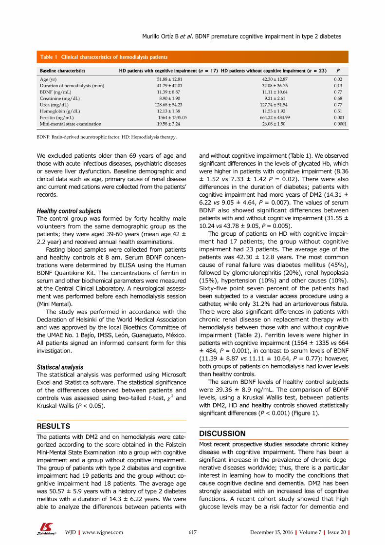
617 December 15, 2016|Volume 7|Issue 20|WJD|www.wjgnet.com
We excluded patients older than 69 years of age and those with acute infectious diseases, psychiatric diseases or severe liver dysfunction. Baseline demographic and clinical data such as age, primary cause of renal disease and current medications were collected from the patients’ records.
Healthy control subjectsThe control group was formed by forty healthy male volunteers from the same demographic group as the patients; they were aged 39-60 years (mean age 42 ± 2.2 year) and received annual health examinations.
Fasting blood samples were collected from patients and healthy controls at 8 am. Serum BDNF concen-trations were determined by ELISA using the Human BDNF Quantikine Kit. The concentrations of ferritin in serum and other biochemical parameters were measured at the Central Clinical Laboratory. A neurological assess-ment was performed before each hemodialysis session (Mini Mental).
The study was performed in accordance with the Declaration of Helsinki of the World Medical Association and was approved by the local Bioethics Committee of the UMAE No. 1 Bajío, IMSS, León, Guanajuato, México. All patients signed an informed consent form for this investigation.
Statiscal analysisThe statistical analysis was performed using Microsoft Excel and Statistica software. The statistical significance of the differences observed between patients and controls was assessed using two-tailed t-test, χ 2 and Kruskal-Wallis (P < 0.05).
RESULTSThe patients with DM2 and on hemodialysis were cate-gorized according to the score obtained in the Folstein Mini-Mental State Examination into a group with cognitive impairment and a group without cognitive impairment. The group of patients with type 2 diabetes and cognitive impairment had 19 patients and the group without co-gnitive impairment had 18 patients. The average age was 50.57 ± 5.9 years with a history of type 2 diabetes mellitus with a duration of 14.3 ± 6.22 years. We were able to analyze the differences between patients with
and without cognitive impairment (Table 1). We observed significant differences in the levels of glycated Hb, which were higher in patients with cognitive impairment (8.36 ± 1.52 vs 7.33 ± 1.42 P = 0.02). There were also differences in the duration of diabetes; patients with cognitive impairment had more years of DM2 (14.31 ± 6.22 vs 9.05 ± 4.64, P = 0.007). The values of serum BDNF also showed significant differences between patients with and without cognitive impairment (31.55 ± 10.24 vs 43.78 ± 9.05, P = 0.005).
The group of patients on HD with cognitive impair-ment had 17 patients; the group without cognitive impairment had 23 patients. The average age of the patients was 42.30 ± 12.8 years. The most common cause of renal failure was diabetes mellitus (45%), followed by glomerulonephritis (20%), renal hypoplasia (15%), hypertension (10%) and other causes (10%). Sixty-five point seven percent of the patients had been subjected to a vascular access procedure using a catheter, while only 31.2% had an arteriovenous fistula. There were also significant differences in patients with chronic renal disease on replacement therapy with hemodialysis between those with and without cognitive impairment (Table 2). Ferritin levels were higher in patients with cognitive impairment (1564 ± 1335 vs 664 ± 484, P = 0.001), in contrast to serum levels of BDNF (11.39 ± 8.87 vs 11.11 ± 10.64, P = 0.77); however, both groups of patients on hemodialysis had lower levels than healthy controls.
The serum BDNF levels of healthy control subjects were 39.36 ± 8.9 ng/mL. The comparison of BDNF levels, using a Kruskal Wallis test, between patients with DM2, HD and healthy controls showed statistically significant differences (P < 0.001) (Figure 1).
DISCUSSIONMost recent prospective studies associate chronic kidney disease with cognitive impairment. There has been a significant increase in the prevalence of chronic dege-nerative diseases worldwide; thus, there is a particular interest in learning how to modify the conditions that cause cognitive decline and dementia. DM2 has been strongly associated with an increased loss of cognitive functions. A recent cohort study showed that high glucose levels may be a risk factor for dementia and
Baseline characteristics HD patients with cognitive impairment (n = 17) HD patients without cognitive impairment (n = 23) P
Age (yr) 51.88 ± 12.81 42.30 ± 12.87 0.02Duration of hemodialysis (mon) 41.29 ± 42.01 32.08 ± 36-76 0.13BDNF (ng/mL) 11.39 ± 8.87 11.11 ± 10.64 0.77Creatinine (mg/dL) 8.90 ± 1.90 9.21 ± 2.61 0.68Urea (mg/dL) 128.68 ± 54.23 127.74 ± 51.54 0.77Hemoglobin (g/dL) 12.13 ± 1.38 11.53 ± 1.92 0.51Ferritin (ng/mL) 1564 ± 1335.05 664.22 ± 484.99 0.001Mini-mental state examination 19.58 ± 3.24 26.08 ± 1.50 0.0001
Table 1 Clinical characteristics of hemodialysis patients
BDNF: Brain-derived neurotrophic factor; HD: Hemodialysis therapy.
Murillo Ortíz B et al . BDNF premature cognitive impairment in type 2 diabetes
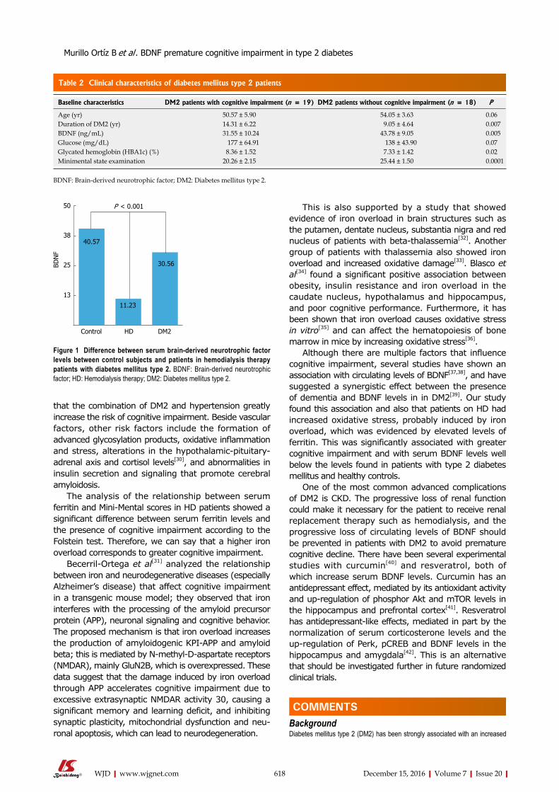
618 December 15, 2016|Volume 7|Issue 20|WJD|www.wjgnet.com
that the combination of DM2 and hypertension greatly increase the risk of cognitive impairment. Beside vascular factors, other risk factors include the formation of advanced glycosylation products, oxidative inflammation and stress, alterations in the hypothalamic-pituitary-adrenal axis and cortisol levels[30], and abnormalities in insulin secretion and signaling that promote cerebral amyloidosis.
The analysis of the relationship between serum ferritin and Mini-Mental scores in HD patients showed a significant difference between serum ferritin levels and the presence of cognitive impairment according to the Folstein test. Therefore, we can say that a higher iron overload corresponds to greater cognitive impairment.
Becerril-Ortega et al[31] analyzed the relationship between iron and neurodegenerative diseases (especially Alzheimer’s disease) that affect cognitive impairment in a transgenic mouse model; they observed that iron interferes with the processing of the amyloid precursor protein (APP), neuronal signaling and cognitive behavior. The proposed mechanism is that iron overload increases the production of amyloidogenic KPI-APP and amyloid beta; this is mediated by N-methyl-D-aspartate receptors (NMDAR), mainly GluN2B, which is overexpressed. These data suggest that the damage induced by iron overload through APP accelerates cognitive impairment due to excessive extrasynaptic NMDAR activity 30, causing a significant memory and learning deficit, and inhibiting synaptic plasticity, mitochondrial dysfunction and neu-ronal apoptosis, which can lead to neurodegeneration.
This is also supported by a study that showed evidence of iron overload in brain structures such as the putamen, dentate nucleus, substantia nigra and red nucleus of patients with beta-thalassemia[32]. Another group of patients with thalassemia also showed iron overload and increased oxidative damage[33]. Blasco et al[34] found a significant positive association between obesity, insulin resistance and iron overload in the caudate nucleus, hypothalamus and hippocampus, and poor cognitive performance. Furthermore, it has been shown that iron overload causes oxidative stress in vitro[35] and can affect the hematopoiesis of bone marrow in mice by increasing oxidative stress[36].
Although there are multiple factors that influence cognitive impairment, several studies have shown an association with circulating levels of BDNF[37,38], and have suggested a synergistic effect between the presence of dementia and BDNF levels in in DM2[39]. Our study found this association and also that patients on HD had increased oxidative stress, probably induced by iron overload, which was evidenced by elevated levels of ferritin. This was significantly associated with greater cognitive impairment and with serum BDNF levels well below the levels found in patients with type 2 diabetes mellitus and healthy controls.
One of the most common advanced complications of DM2 is CKD. The progressive loss of renal function could make it necessary for the patient to receive renal replacement therapy such as hemodialysis, and the progressive loss of circulating levels of BDNF should be prevented in patients with DM2 to avoid premature cognitive decline. There have been several experimental studies with curcumin[40] and resveratrol, both of which increase serum BDNF levels. Curcumin has an antidepressant effect, mediated by its antioxidant activity and up-regulation of phosphor Akt and mTOR levels in the hippocampus and prefrontal cortex[41]. Resveratrol has antidepressant-like effects, mediated in part by the normalization of serum corticosterone levels and the up-regulation of Perk, pCREB and BDNF levels in the hippocampus and amygdala[42]. This is an alternative that should be investigated further in future randomized clinical trials.
COMMENTSBackgroundDiabetes mellitus type 2 (DM2) has been strongly associated with an increased
Baseline characteristics DM2 patients with cognitive impairment (n = 19) DM2 patients without cognitive impairment (n = 18) P
Age (yr) 50.57 ± 5.90 54.05 ± 3.63 0.06Duration of DM2 (yr) 14.31 ± 6.22 9.05 ± 4.64 0.007BDNF (ng/mL) 31.55 ± 10.24 43.78 ± 9.05 0.005Glucose (mg/dL) 177 ± 64.91 138 ± 43.90 0.07Glycated hemoglobin (HBA1c) (%) 8.36 ± 1.52 7.33 ± 1.42 0.02Minimental state examination 20.26 ± 2.15 25.44 ± 1.50 0.0001
Table 2 Clinical characteristics of diabetes mellitus type 2 patients
BDNF: Brain-derived neurotrophic factor; DM2: Diabetes mellitus type 2.
BDN
F
50
38
25
13
Control HD DM2
40.57
11.23
30.56
P < 0.001
Figure 1 Difference between serum brain-derived neurotrophic factor levels between control subjects and patients in hemodialysis therapy patients with diabetes mellitus type 2. BDNF: Brain-derived neurotrophic factor; HD: Hemodialysis therapy; DM2: Diabetes mellitus type 2.
COMMENTS
Murillo Ortíz B et al . BDNF premature cognitive impairment in type 2 diabetes
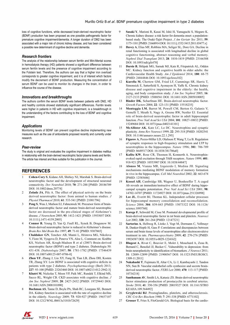
619 December 15, 2016|Volume 7|Issue 20|WJD|www.wjgnet.com
loss of cognitive functions, while decreased brain-derived neurotrophic factor (BDNF) production has been proposed as one possible pathogenetic factor for premature cognitive impairment/dementia. A longer duration of DM2 has been associated with a major risk of chronic kidney disease, and has been considered a possible new determinant of cognitive decline and dementia.
Research frontiersThe analysis of the relationship between serum ferritin and Mini-Mental scores in hemodialysis therapy (HD) patients showed a significant difference between serum ferritin levels and the presence of cognitive impairment according to the Folstein test. Therefore, the authors can say that a higher iron overload corresponds to greater cognitive impairment, and it is of interest which factors modify the decrement of BDNF production. Measuring the concentration of serum BDNF can be used to monitor its changes in the brain, in order to influence the course of the disease.
Innovations and breakthroughsThe authors confirm the serum BDNF levels between patients with DM2, HD and healthy controls showed statistically significant differences. Ferritin levels were higher in patients in HD with cognitive impairment, is a breakthrough in the understanding of the factors contributing to the loss of BDNF and cognitive impairment.
ApplicationsMonitoring levels of BDNF can prevent cognitive decline implementing new measures such as the use of antioxidants proposed recently and currently under research.
Peer-reviewThe study is original and evaluates the cognitive impairment in diabetes mellitus in relationship with the brain-derived neurotrophic factor plasma levels and ferritin. The article has interest and likes suitable for the publication in the Journal.
REFERENCES1 Cohen-Cory S, Kidane AH, Shirkey NJ, Marshak S. Brainderived
neurotrophic factor and the development of structural neuronal connectivity. Dev Neurobiol 2010; 70: 271288 [PMID: 20186709 DOI: 10.1002/dneu.20774]
2 Zoladz JA, Pilc A. The effect of physical activity on the brain derived neurotrophic factor: from animal to human studies. J Physiol Pharmacol 2010; 61: 533541 [PMID: 21081796]
3 Peng S, Wuu J, Mufson EJ, Fahnestock M. Precursor form of brainderived neurotrophic factor and mature brainderived neurotrophic factor are decreased in the preclinical stages of Alzheimer’s disease. J Neurochem 2005; 93: 14121421 [PMID: 15935057 DOI: 10.1111/j.14714159.2005]
4 Connor B, Young D, Yan Q, Faull RL, Synek B, Dragunow M. Brainderived neurotrophic factor is reduced in Alzheimer’s disease. Brain Res Mol Brain Res 1997; 49: 7181 [PMID: 9387865]
5 Chaldakov GN, Tonchev AB, Manni L, Hristova MG, Nikolova V, Fiore M, Vyagova D, Peneva VN, Aloe L. Comment on: Krabbe KS, Nielsen AR, KroghMadsen R et al (2007) Brainderived neurotrophic factor (BDNF) and type 2 diabetes. Diabetologia 50: 431438. Diabetologia 2007; 50: 17811782 [PMID: 17546439 DOI: 10.1007/s0012500707060]
6 Zhen YF, Zhang J, Liu XY, Fang H, Tian LB, Zhou DH, Kosten TR, Zhang XY. Low BDNF is associated with cognitive deficits in patients with type 2 diabetes. Psychopharmacology (Berl) 2013; 227: 93100 [PMID: 23263460 DOI: 10.1007/s0021301229423]
7 Khatri M, Nickolas T, Moon YP, Paik MC, Rundek T, Elkind MS, Sacco RL, Wright CB. CKD associates with cognitive decline. J Am Soc Nephrol 2009; 20: 24272432 [PMID: 19729443 DOI: 10.1681/ASN.2008101090]
8 Buchman AS, Tanne D, Boyle PA, Shah RC, Leurgans SE, Bennett DA. Kidney function is associated with the rate of cognitive decline in the elderly. Neurology 2009; 73: 920927 [PMID: 19657107 DOI: 10.1212/WNL.0b013e3181b72629]
9 Sasaki Y, Marioni R, Kasai M, Ishii H, Yamaguchi S, Meguro K. Chronic kidney disease: a risk factor for dementia onset: a populationbased study. The OsakiTajiri Project. J Am Geriatr Soc 2011; 59: 11751181 [PMID: 21668914 DOI: 10.1111/j.15325415.2011.03477.x]
10 Davey A, Elias MF, Robbins MA, Seliger SL, Dore GA. Decline in renal functioning is associated with longitudinal decline in global cognitive functioning, abstract reasoning and verbal memory. Nephrol Dial Transplant 2013; 28: 18101819 [PMID: 23166308 DOI: 10.1093/ndt/gfs470]
11 Darsie B, Shlipak MG, Sarnak MJ, Katz R, Fitzpatrick AL, Odden MC. Kidney function and cognitive health in older adults: the Cardiovascular Health Study. Am J Epidemiol 2014; 180: 6875 [PMID: 24844846 DOI: 10.1093/aje/kwu102]
12 Kurella M, Chertow GM, Fried LF, Cummings SR, Harris T, Simonsick E, Satterfield S, Ayonayon H, Yaffe K. Chronic kidney disease and cognitive impairment in the elderly: the health, aging, and body composition study. J Am Soc Nephrol 2005; 16: 21272133 [PMID: 15888561 DOI: 10.1681/ASN.2005010005]
13 Binder DK, Scharfman HE. Brainderived neurotrophic factor. Growth Factors 2004; 22: 123131 [PMID: 15518235]
14 Monteggia LM, Barrot M, Powell CM, Berton O, Galanis V, Gemelli T, Meuth S, Nagy A, Greene RW, Nestler EJ. Essential role of brainderived neurotrophic factor in adult hippocampal function. Proc Natl Acad Sci USA 2004; 101: 1082710832 [PMID: 15249684 DOI: 10.1073/pnas.0402141101]
15 McAllister AK, Katz LC, Lo DC. Neurotrophins and synaptic plasticity. Annu Rev Neurosci 1999; 22: 295318 [PMID: 10202541 DOI: 10.1146/annurev.neuro.22.1.295]
16 Figurov A, PozzoMiller LD, Olafsson P, Wang T, Lu B. Regulation of synaptic responses to highfrequency stimulation and LTP by neurotrophins in the hippocampus. Nature 1996; 381: 706709 [PMID: 8649517 DOI: 10.1038/381706a0]
17 Kafitz KW, Rose CR, Thoenen H, Konnerth A. Neurotrophinevoked rapid excitation through TrkB receptors. Nature 1999; 401: 918921 [PMID: 10553907 DOI: 10.1038/44847]
18 Alonso M, Vianna MR, Izquierdo I, Medina JH. Signaling mechanisms mediating BDNF modulation of memory formation in vivo in the hippocampus. Cell Mol Neurobiol 2002; 22: 663674 [PMID: 12585686]
19 Kossel AH, Cambridge SB, Wagner U, Bonhoeffer T. A caged Ab reveals an immediate/instructive effect of BDNF during hippocampal synaptic potentiation. Proc Natl Acad Sci USA 2001; 98: 1470214707 [PMID: 11724927 DOI: 10.1073/pnas.251326998]
20 Lee JL, Everitt BJ, Thomas KL. Independent cellular processes for hippocampal memory consolidation and reconsolidation. Science 2004; 304: 839843 [PMID: 15073322 DOI: 10.1126/science.1095760]
21 Karege F, Schwald M, Cisse M. Postnatal developmental profile of brainderived neurotrophic factor in rat brain and platelets. Neurosci Lett 2002; 328: 261264 [PMID: 12147321]
22 Sartorius A, Hellweg R, Litzke J, Vogt M, Dormann C, Vollmayr B, DankerHopfe H, Gass P. Correlations and discrepancies between serum and brain tissue levels of neurotrophins after electroconvulsive treatment in rats. Pharmacopsychiatry 2009; 42: 270276 [PMID: 19924587 DOI: 10.1055/s00291224162]
23 Blugeot A, Rivat C, Bouvier E, Molet J, Mouchard A, Zeau B, Bernard C, Benoliel JJ, Becker C. Vulnerability to depression: from brain neuroplasticity to identification of biomarkers. J Neurosci 2011; 31: 1288912899 [PMID: 21900567 DOI: 10.1523/JNEUROSCI. 130911.2011]
24 Nakahashi T, Fujimura H, Altar CA, Li J, Kambayashi J, Tandon NN, Sun B. Vascular endothelial cells synthesize and secrete brainderived neurotrophic factor. FEBS Lett 2000; 470: 113117 [PMID: 10734218]
25 Santhanam AV, Smith LA, Katusic ZS. Brainderived neurotrophic factor stimulates production of prostacyclin in cerebral arteries. Stroke 2010; 41: 350356 [PMID: 20019327 DOI: 10.1161/STRO KEAHA.109.564492]
26 Gryglewski RJ. Prostaglandins, platelets, and atherosclerosis. CRC Crit Rev Biochem 1980; 7: 291338 [PMID: 6771102]
27 Grosser T, Fries S, FitzGerald GA. Biological basis for the cardio
Murillo Ortíz B et al . BDNF premature cognitive impairment in type 2 diabetes
620 December 15, 2016|Volume 7|Issue 20|WJD|www.wjgnet.com
vascular consequences of COX2 inhibition: therapeutic challenges and opportunities. J Clin Invest 2006; 116: 415 [PMID: 16395396 DOI: 10.1172/JCI27291]
28 Chlopicki S, Swies J, Mogielnicki A, Buczko W, Bartus M, Lomnicka M, Adamus J, Gebicki J. 1Methylnicotinamide (MNA), a primary metabolite of nicotinamide, exerts antithrombotic activity mediated by a cyclooxygenase2/prostacyclin pathway. Br J Pharmacol 2007; 152: 230239 [PMID: 17641676 DOI: 10.1038/sj.bjp.0707383]
29 Zoladz JA, Śmigielski M, Majerczak J, Nowak ŁR, Zapart-Bukowska J, Smoleński O, Kulpa J, Duda K, Drzewińska J, Bartosz G. Hemodialysis decreases serum brainderived neurotrophic factor concentration in humans. Neurochem Res 2012; 37: 27152724 [PMID: 22903469 DOI: 10.1007/s1106401208626]
30 Passaro A, Dalla Nora E, Morieri ML, Soavi C, Sanz JM, Zurlo A, Fellin R, Zuliani G. Brainderived neurotrophic factor plasma levels: relationship with dementia and diabetes in the elderly population. J Gerontol A Biol Sci Med Sci 2015; 70: 294302 [PMID: 24621946 DOI: 10.1093/gerona/glu028]
31 Becerril-Ortega J, Bordji K, Fréret T, Rush T, Buisson A. Iron overload accelerates neuronal amyloid-β production and cognitive impairment in transgenic mice model of Alzheimer’s disease. Neurobiol Aging 2014; 35: 22882301 [PMID: 24863668 DOI: 10.1016/j.neurobiolaging.2014.04.019]
32 Qiu D, Chan GC, Chu J, Chan Q, Ha SY, Moseley ME, Khong PL. MR quantitative susceptibility imaging for the evaluation of iron loading in the brains of patients with β-thalassemia major. AJNR Am J Neuroradiol 2014; 35: 10851090 [PMID: 24578278 DOI: 10.3174/ajnr.A3849]
33 Karakas Z, Yilmaz Y, Celik DD, Annayev A, Demirel S, Kuruca SE. Total oxidant and antioxidant capacity in patients with transfusion dependent and nondependent beta thalassemia. Proceedings of the 57th ASH Annual Meeting Abstracts, 2015 Dec Vol 126, Issue 23: 4573
34 Blasco G, Puig J, DaunisIEstadella J, Molina X, Xifra G, FernándezAranda F, Pedraza S, Ricart W, PorteroOtín M, FernándezReal JM. Brain iron overload, insulin resistance, and cognitive performance in obese subjects: a preliminary MRI casecontrol study. Diabetes Care 2014; 37: 30763083 [PMID: 25125507 DOI: 10.2337/dc140664]
35 Lu WY, Zhao MF, Chai X, Meng JX, Xie F, Mu J, Zhu HB, Xu XN, Xiao X, Deng Q, Ma L. Iron Overload Impairs Hematopoiesis by Damaging MSCs Through ROS Signaling Pathway. Proceedings of the 54th ASH Annual Meeting Abstracts, 2012 Nov 16, Vol 120, Issue 21: 925172
36 Chai X, Li D, Cao X, Zhang Y, Mu J, Lu W, Xiao X, Li C, Meng J, Chen J, Li Q, Wang J, Meng A, Zhao M. ROSmediated iron overload injures the hematopoiesis of bone marrow by damaging hematopoietic stem/progenitor cells in mice. Sci Rep 2015; 5: 10181 [PMID: 25970748 DOI: 10.1038/srep10181]
37 Ono M, Ichihara J, Nonomura T, Itakura Y, Taiji M, Nakayama C, Noguchi H. Brainderived neurotrophic factor reduces blood glucose level in obese diabetic mice but not in normal mice. Biochem Biophys Res Commun 1997; 238: 633637 [PMID: 9299565 DOI: 10.1006/bbrc.1997.7220]
38 Ott A, Stolk RP, van Harskamp F, Pols HA, Hofman A, Breteler MM. Diabetes mellitus and the risk of dementia: The Rotterdam Study. Neurology 1999; 53: 19371942 [PMID: 10599761]
39 Gregg EW, Yaffe K, Cauley JA, Rolka DB, Blackwell TL, Narayan KM, Cummings SR. Is diabetes associated with cognitive impairment and cognitive decline among older women? Study of Osteoporotic Fractures Research Group. Arch Intern Med 2000; 160: 174180 [PMID: 10647755]
40 Franco-Robles E, CamposCervantes A, MurilloOrtiz BO, Segovia J, LópezBriones S, Vergara P, PérezVázquez V, SolísOrtiz MS, RamírezEmiliano J. Effects of curcumin on brainderived neurotrophic factor levels and oxidative damage in obesity and diabetes. Appl Physiol Nutr Metab 2014; 39: 211218 [PMID: 24476477 DOI: 10.1139/apnm20130133]
41 Liu D, Wang Z, Gao Z, Xie K, Zhang Q, Jiang H, Pang Q. Effects of curcumin on learning and memory deficits, BDNF, and ERK protein expression in rats exposed to chronic unpredictable stress. Behav Brain Res 2014; 271: 116121 [PMID: 24914461 DOI: 10.1016/j.bbr.2014.05.068]
42 Liu D, Xie K, Yang X, Gu J, Ge L, Wang X, Wang Z. Resveratrol reverses the effects of chronic unpredictable mild stress on behavior, serum corticosterone levels and BDNF expression in rats. Behav Brain Res 2014; 264: 916 [PMID: 24503118 DOI: 10.1016/j.bbr.2014.01.039]
P- Reviewer: GómezSáez JM, Li P, Tziomalos K S- Editor: Qiu S L- Editor: A E- Editor: Li D
Murillo Ortíz B et al . BDNF premature cognitive impairment in type 2 diabetes
Rim Braham, Aus Alzaid, Asirvatham Alwin Robert, Muhammad Mujammami, Rania Ahmad Ahmad, Monther Zitouni, Samia Hasan Sobki, Mohamed Abdulaziz Al Dawish
ORIGINAL ARTICLE
621 December 15, 2016|Volume 7|Issue 20|WJD|www.wjgnet.com
Double diabetes in Saudi Arabia: A new entity or an underestimated condition
Retrospective Study
Rim Braham, Aus Alzaid, Asirvatham Alwin Robert, Rania Ahmad Ahmad, Mohamed Abdulaziz Al Dawish, Department of Endocrinology and Diabetes, Prince Sultan Military Medical City, Riyadh 11159, Saudi Arabia
Muhammad Mujammami, Endocrinology and Diabetes Unit, Department of Medicine, King Khalid University Hospital, King Saud University, Riyadh 11461, Saudi Arabia
Monther Zitouni, Samia Hasan Sobki, Department of Pathology, Prince Sultan Military Medical City, Riyadh 11159, Saudi Arabia
Author contributions: All authors contributed to this work.
Institutional review board statement: The study protocol was approved by the Research and Ethics committee of Prince Sultan Military Medical City, Riyadh, Saudi Arabia.
Informed consent statement: During the informed consent process, study participants are assured that data collected will be used only for stated purposes and will not be disclosed or released to others without the consent of the participants.
Conflict-of-interest statement: Authors have no conflict of interests and the work was not supported or funded by any drug company.
Data sharing statement: No data sharing as this manuscript and the data were not published elsewhere.
Open-Access: This article is an open-access article which was selected by an in-house editor and fully peer-reviewed by external reviewers. It is distributed in accordance with the Creative Commons Attribution Non Commercial (CC BY-NC 4.0) license, which permits others to distribute, remix, adapt, build upon this work non-commercially, and license their derivative works on different terms, provided the original work is properly cited and the use is non-commercial. See: http://creativecommons.org/licenses/by-nc/4.0/
Manuscript source: Unsolicited manuscript
Correspondence to: Dr. Rim Braham, Department of Endo-
crinology and Diabetes, Prince Sultan Military Medical City, P.O. Box 7897, Riyadh 11159, Saudi Arabia. [email protected]: +966-1-4777714-43116
Received: June 2, 2016Peer-review started: June 6, 2016First decision: July 5, 2016Revised: September 15, 2016Accepted: October 17, 2016Article in press: October 18, 2016Published online: December 15, 2016
AbstractAIMTo determine the clinical and biological characteristics of double diabetes (DD) among young people in Saudi Arabia.
METHODSThis was a retrospective descriptive chart review study including 312 young newly diagnosed diabetic patients (aged 12-20 years), whom were admitted over a five year period (January 2009 to December 2013). Family history of diabetes mellitus (DM) (first degree), physical body mass index (BMI), acanthosis nigricans, history of auto-immune disease and laboratory information for glycosylated hemoglobin, basal C peptide level and diabetes autoantibody response (anti-GAD, anti-IA2 and anti-ICA) were collected from medical report. A mean follow-up of 3 years for these patients was performed.
RESULTSPatients were categorized into 4 groups, based on the autoantibody response (Ab+ or Ab-) and C-peptide secretion (β+ for fasting level 0.4-2.1 ng/mL and β- if < 0.4 ng/mL). Group1 (type 1a): Ab+ β- (21%), group 2 (type 1b): Ab- β- (9%), group 3 (DD): Ab+ β+ (31%)
Submit a Manuscript: http://www.wjgnet.com/esps/Help Desk: http://www.wjgnet.com/esps/helpdesk.aspxDOI: 10.4239/wjd.v7.i20.621
World J Diabetes 2016 December 15; 7(20): 621-626ISSN 1948-9358 (online)
© 2016 Baishideng Publishing Group Inc. All rights reserved.

622 December 15, 2016|Volume 7|Issue 20|WJD|www.wjgnet.com
Braham R et al . Double diabetes in Saudi Arabia
and group 4 (classic type 2 DM): Ab- β+ (39%). The mean age of the DD patients in our study was 15.1 ± 6.4 years. A total of 41% of the study population presented with diabetic ketoacidosis and 61% of the study popula-tion presented with positive family history of DM. The mean BMI was 26.8 kg/m2 with 64% of overweight or obese patients. Ninety two percent of the patients were started on insulin at the time of diagnosis. During a mean follow-up of 3 years, only 32% of the patients with DD required insulin and 78% were on metformin alone or with insulin.
CONCLUSIONOur findings enable us to arrive at the conclusion that almost one-third of the young Saudi diabetic patients reveal atypical forms of DM (double diabetes) express-ing features resulting from both T1D and T2D.
Key words: Double diabetes; Therapeutic approaches; Hybrid diabetes; Autoantibody response; Saudi Arabia
© The Author(s) 2016. Published by Baishideng Publishing Group Inc. All rights reserved.
Core tip: Almost one-third of the young Saudi diabetic patients reveal atypical forms of double diabetes (DD) expressing features resulting from both type 1 diabetes and type 2 diabetes. Therefore, identification of DD patients becomes important as this will give direction for the selection of the most apt diagnostic and therapeutic lines of treatment.
Braham R, Alzaid A, Robert AA, Mujammami M, Ahmad RA, Zitouni M, Sobki SH, Al Dawish MA. Double diabetes in Saudi Arabia: A new entity or an underestimated condition. World J Diabetes 2016; 7(20): 621-626 Available from: URL: http://www.wjgnet.com/1948-9358/full/v7/i20/621.htm DOI: http://dx.doi.org/10.4239/wjd.v7.i20.621
INTRODUCTIONAlmost one-third of the young Saudi diabetic patients reveal atypical forms of double diabetes (DD) expressing features resulting from both type 1 diabetes and type 2 diabetes. Therefore, identification of DD patients becomes important as this will give direction for the selection of the most apt diagnostic and therapeutic lines of treatment.
Diabetes mellitus (DM) is a chronic disease, recognized as a high-ranking and daunting health problem of the 21st century[1]. The rising incidence and prevalence of DM are becoming alarmingly evident irrespective of age groups or gender and occur in the developed and developing countries[1]. The International Diabetes Federation alert indicates that if lifestyle and health habits are not dra-stically and quickly changed, one-half of the Saudis will be diabetic by 2030. Also, as nearly 3 million (18%) Saudi children are overweight or obese, they will be most
vulnerable to acquiring DM[2].Type 1 diabetes is the presence of antibodies which
attack the insulin-producing pancreatic beta cells an indication that type 1 diabetes is an autoimmune dis-order. Whereas type 2 diabetes is characterized by insulin resistance and relative insulin deficiency, either or both of which may be present at the time diabetes is diagnosed. Traditionally, anyone exhibiting polyuria, polydipsia, and polyphagia, the classic symptoms of DM, and who also possess a family history of type 1 DM (T1D), obesity, acanthosis nigricans and the absence of both ketosis and diabetes-associated autoantibodies is recognized as a type 2 diabetic (T2D)[3]. However, T1D patients are most often thin and may have ketosis and diabetes-linked autoantibodies[4]. The DD patients exhibit characteristics of both T1D and T2D, which can be evident either during diagnosis or develop subsequently over time[5-7].
It was in 1991 that the nomenclature “double diabetes” was first given to T1D patients with a family history of T2D, as they were found to more likely be overweight and rarely had sufficient glycemic control, even on high insulin dosages[8]. The present classification makes it difficult to describe the type of heterogeneous DM affecting such young patients, whether to categorize them as T2D because of their obesity and insulin resis-tance, or as T1D due to the presence of auto-antibodies to the β cells[6]. Further, DD is quite hard to control, including the micro- and mostly macro-vascular typically T2D-associated complications[1,6].
Although the prevalence and incidence of DD is yet to be clearly defined, however nearly 25% of the T1D children showed obesity or were overweight[9]. Also, roughly 35% of children and adolescents with T2D possessed at least one diabetes-related antibody[9-11]. The rapid increase in the prevalence of T1D and T2D in the Kingdom caught the interest of the medical world soon after the rapid industrialization resulted in a dramatic spurt in the standard of living and incorporation of “Westernized” habits, including the selection of unhealthy dietary patterns, and reduction in physical activity[12]. In Saudi Arabia the prevalence of DM is at an alarming juncture and rising[1]. However, no study, to our knowledge, is currently available on the prevalence of DD in Saudi Arabia. Therefore, our objective is to ascertain the prevalence, clinical and biological features of DD among the young Saudi populace.
MATERIALS AND METHODSStudy design, setting and samplingThis is a retrospective descriptive study was conducted among 312 young newly diagnosed diabetic patients (aged 12-20 years) admitted over a 4-year period (January 2009 to December 2013) at Prince Sultan Military Medical City (PSMMC). PSMMC is a 1200-bed, tertiary medical center in Riyadh, Saudi Arabia, with round 40000 annual admissions (950000 active patients files) and 118000 emergency room visits per year from different region of the country. Patients selection of this

623 December 15, 2016|Volume 7|Issue 20|WJD|www.wjgnet.com
study was conducted using eligibility screening.
Data collectionFamily history of DM (first degree), physical body mass index (BMI), acanthosis nigricans, history of auto-immune disease, first degree family history of auto-immune disease (celiac disease, systemic lupus erythematosus, Sjögren’s syndrome, thyroid dysfunction (primary hypothyroidism, graves’ disease), psoriasis, Crohn’s disease, Addison’s disease, multiple sclerosis, and myasthenia gravis) and laboratory data for glycosylated hemoglobin (HbA1c), basal C-peptide level and diabetes autoantibody response (anti-GAD, anti-IA2 and anti-ICA) were collected.
BMIBMI was calculated by dividing the weight (kg) by the square of height in meters (BMI; kg/m2) and BMI z score (adjusted for child age and gender). The z score (standard deviation scores), was figured as per the formula (Xi-Mx)/SD, where Xi is the actual measurement, Mx is the mean value for that age and gender, and SD is the standard deviation corresponding to that age and gender[13].
Diabetic ketoacidosisDiabetic ketoacidosis (DKA) is defined as all three of the following must be present: (1) blood glucose level higher than > 250 mg/dL; (2) presence of urine ketones ++ or more; and (3) venous pH level lower than 7.30 and/or serum bicarbonate lower than 15 mEq/L[14].
HbA1c HbA1c ≥ 6.5% makes the diagnosis of DM. The HbA1c test was performed in our laboratory using a standard method (National Glycohemoglobin Standardization Program certified) and standardized to the diabetes control and complications trial assay. In the absence of unequivocal hyperglycemia, results were confirmed by repeat testing[15].
Statistical analysisData analysis was carried out using Microsoft Excel 2010, Microsoft Corporation, Seattle, WA, United States and the Statistical Package for Social Sciences version 20, SPSS Inc., Chicago, IL, United States. In addition to descriptive statistics t test and χ 2 analyses were carried out to compare between DD and others groups. A P-value of < 0.05 was considered to be statistically
significant.
RESULTSTable 1 lists the characteristics of the population studied. The mean age of patients presenting with DD was 15.1 ± 6.4 years with BMI 26.8 kg/m2, and 1:2 sex ratio (male vs female). The results indicated that 57% of the population studied had a family history, 64% showed obesity, 39% had DKA, 52% were positive for autoantibodies, 34% had acanthosis nigricans, 23% possessed a family history of autoimmune disease and 18 had a history of autoimmune disease.
The patients were divided on the basis of the auto-antibody response (Ab+ or Ab-) and C-peptide secretion (β+ for fasting level 0.4-2.1 ng/mL and β- if < 0.4 ng/mL) as listed in Figure 1. Depending on the autoantibody response (Ab+ or Ab-) and C-peptide secretion, the patients were segregated into four groups, viz., group1 (type 1a): Ab+ β- (21%); group 2 (type 1b): Ab- β-(9%), group 3 (DD): Ab+ β+ (31%) and group 4 (classic T2D): Ab- β+ (39%).
The characteristics of the patients are shown based on the presence or absence of auto-antibodies and the C-peptide secretion are shown in Table 2. More than 25% of the DD population had a family history of diabetes and the mean BMI of the DD population was 26.8 (kg/m2). Among the DD population, 28 (38.8%) had family history of auto immune disease, 17 (30.4%) had history of auto immune disease, ninety (31.4%) required insulin at diagnosis and thirty one (21%) required multiple dose of insulin injection during follow-up.
DISCUSSIONGlobally, as the DD population is steadily increasing in number it becomes harder to diagnose and treat because these individuals experience symptoms of both T1D and T2D with the hybrid diabetes[5,7,16]. Identifying DD in children and adolescents is crucial as it affects the diagnostic method and choice of treatment. Within the scope of our knowledge, no other study regarding the prevalence of DD in Saudi Arabia is currently available. Therefore, this study was performed to ascertain the prevalence, clinical and biological characteristics of DD among the young Saudi population at PSMMC, a tertiary medical center in Saudi Arabia.
The current study, using the autoantibody response and C-peptide secretion, showed 31% of the population studied with DD and 26.8 mean BMI. However, at present research is limited regarding the incidence and prevalence of DD[9]. The results from another study indicated that almost 25% of T1D children are either overweight or obese and have DD[11]. This condition usually develops insidiously and initially manifests as a rising requirement for more insulin to control the glucose levels. T2D patients too can be diagnosed with blood tests to identify the specific pancreas-attacking proteins. Some studies also recorded that nearly 35% of children and adolescents with T2D possessed at least one diabetes-related
Patients characteristics Yes % (n) No % (n)
Family history of diabetes 57 (178) 43 (134)Patients with overweight or obese 64 (199) 36 (113)Diabetic ketoacidosis at presentation 39 (122) 61 (190)Autoantibodies positivity 52 (162) 48 (150)Acanthosis nigricans 34 (106) 66 (206)Family history of auto-immune disease 23 (72) 77 (240)History of auto-immune disease 18 (56) 82 (256)
Table 1 Characteristics of the study population (n = 312)
Braham R et al . Double diabetes in Saudi Arabia

624 December 15, 2016|Volume 7|Issue 20|WJD|www.wjgnet.com
antibody[10]. Other studies reported that from among the patients newly diagnosed with diabetes, roughly one out of three children and adolescents suffer from DD[7]. DD can be produced by different factors, based on whether the individual initially has T1D or T2D[7]. If the patient commences with T1D and begins to gain excess or surplus weight, the individual may begin to be insulin resistant, implying that besides the body being ineffective in secreting insulin due to T1D, the typical insulin injections will no longer be effective as the patient becomes insulin-resistant, which causes the T2D. Such patients then develop DD necessitating medications plus insulin injections for blood sugar level control[9].
Several studies support the fact that DD is often seen in patients with a family history of T2D[8,17], a finding confirmed by the present study where 45% of the study population had a family history of DM. It is noteworthy that nearly 65% of the DD group required insulin, a discovery concurring with earlier reports in the literature[8,17]. During follow-up, nearly half the DD patients (46%) were managed solely on metformin (without necessitating insulin therapy).
Significantly, almost 64% of the population in this study was overweight or obese. This higher BMI per-centage may possibly be a result of the dramatic rise in the standard of living and the “Westernized” lifestyle habits
adopted in Saudi Arabia. Unhealthy and unwise dietary choices coupled with reduced physical activity have produced this situation[12,18-20]. The growing “obesogenic” state that induced insulin resistance could account for the development of islet cell autoimmunity via different mechanisms. Therefore, the trend of increasing obesity seems to play a prominent part in the rising incidence and changing phenotype of T1D among adolescents and children[7]. Some lifestyle changes and precautions can be incorporated to deter the development of DD for those with and without DM[12,18,19].
Diagnostic evaluation Universally applicable clinical diagnostic criteria as well as methods enabling the identification of DD, either at the time of onset of hyperglycemia or during the course of the disease process, must be established. In 2009, Pozzilli et al[9] introduced the idea of “metabolic load” to define the T2D characteristics and “autoimmune load” to define the T1D features. They revealed that in obese children with hyperglycemia, the presence of a high “metabolic load” and a low “autoimmune load” are indicators of DD[9]. Therefore, they presented some biochemical and clinical guidelines to identify DD, as listed: (1) evidence of the clinical characteristics of T2D, dyslipidemia, hypertension and higher BMI with increased
Auto-antibodies Ab+ Ab-
C-peptide secretion β- (G 1) β+ (double diabetes) β- (G2) β+Age of diagnosis 13.16a 15.3 16.6 17.02e
Family history of diabetes 24 (13.5%) 45 (25.3%) 14 (7.9%) 95 (53.4%)e
Family history of auto immune disease 32 (44.4%)a 28 (38.8%) 5 (6.9%) 7 (9.7%)e
History of auto immune disease 29 (51.7%)a 17 (30.4%) 2 (3.6%) 8 (14.3%)e
DKA at presentation 57 (46.7%)a 33 (27%) 5 (4%) 27 (22.1%)e
BMI (kg/m2) 21a 26.8 24.2 29.6e
HbA1c (%) 10.2a 8.9 10.8 11.7e
Patients requiring insulin at diagnosis 65 (22.6%)a 90 (31.4%) 28 (9.8%) 104 (36.2%)Patients requiring insulin multiple dose injection (follow-up) 65 (44.2%)a 31 (21%) 28 (19%)c 23 (15.6%)e
Patients on metformin only during follow-up 0a 48 (38.1%) 0c 78 (61.9%)e
Patients on metformin with insulin during follow-up 4 (6.1%)a 27 (41.5%) 2 (3.1%)c 32 (49.2%)
Table 2 Characteristics of the patients are shown based on the presence or absence of auto-antibodies and the C-peptide secretion
Groups compared by Students’t test and χ 2 test: aP < 0.05, Ab+ β+ vs Ab+ β-; cP < 0.05, Ab+ β+ vs Ab- β-; eP < 0.05, Ab+ β+ vs Ab- β+. DKA: Diabetic ketoacidosis; BMI: Body mass index.
Freq
uenc
y (%
, n)
45
40
35
30
25
20
15
10
5
0
21% (n = 65)
9% (n = 28)
31% (n = 97)
39% (n = 122)
Group 1 (type 1a, Ab+ β-)
Group 2 (type 1B, Ab- β-)
Group 3 (double diabetes, Ab+ β+)
Group 4 (classic type 2 diabetes)
Diabetes type
Figure 1 Patients categorized based on the autoantibody response (Ab+ or Ab-) and C-peptide secretion (β+ for fasting level 0.4-2.1 ng/mL and β- if < 0.4 ng/mL).
Braham R et al . Double diabetes in Saudi Arabia

625 December 15, 2016|Volume 7|Issue 20|WJD|www.wjgnet.com
cardiovascular risk, compared with children having classical T1D. Family history for T2D and T1D could be present; (2) a drop in the number of the clinical features of T1D, including polyuria and polydipsia, weight loss, formation of ketoacidosis; in this case insulin therapy is not the first line of treatment, unlike for patients with classical T1D; and (3) the number of autoantibodies to islet cells, although lesser in number and titer when compared with T1D, and sometimes a lower degree of risk linked with the MHC locus compared with T1D patients. Similar to T1D, where insulin resistance and obesity are not the usual characteristics, DD is always distinguished by an obese phenotype, besides the coexistence of β cell autoimmunity[9].
LimitationsThis study includes a few limitations such as the limited number of risk, social, and demographic factors studied and demonstrated in a single center. Further research, preferably conducted on a greater scale, is required to overcome these limitations. However, this study offers pertinent information regarding DD among the young Saudi population.
In conclusion, our findings enable us to arrive at the conclusion that almost one-third of the young Saudi diabetic patients reveal atypical forms of DM (double diabetes) expressing features resulting from both T1D and T2D. Therefore, identification of DD patients becomes crucial as this will give direction for the selection of the most apt diagnostic and therapeutic lines of treatment.
COMMENTSBackgroundThe double diabetes (DD) population is steadily increasing worldwide in number it becomes harder to diagnose and treat because these persons experience symptoms of both type 1 diabetes mellitus (DM) (T1D) and type 2 DM (T2D) with the hybrid diabetes. Classifying DD in children and adolescents is crucial as it affects the diagnostic method and choice of treatment.
Research frontiersThe present classification makes it difficult to describe the type of heterogeneous DM affecting such young patients, whether to categorize them as T2D because of their obesity and insulin resistance, or as T1D due to the presence of auto-antibodies to the β cells. Although the prevalence and incidence of DD is yet to be clearly defined, however nearly 25% of the T1D children showed obesity or were overweight. Also, roughly 35% of children and adolescents with T2D possessed at least one diabetes-related antibody. The rapid increase in the prevalence of T1D and T2D in the Saudi Arabia caught the interest of the medical world soon after the rapid industrialization resulted in a dramatic spurt in the standard of living and incorporation of “Westernized” habits, including the selection of unhealthy dietary patterns, and reduction in physical activity. In Saudi Arabia the prevalence of DM is at an alarming juncture and rising.
Innovations and breakthroughsNo study, to our knowledge, is currently available on the prevalence of DD in Saudi Arabia. Therefore, the authors ascertained the prevalence, clinical and biological features of DD among the young Saudi populace.
ApplicationsThe authors’ findings enable people to arrive at the conclusion that almost one-third of the young Saudi diabetic patients reveal atypical forms of DM (double
diabetes) expressing features resulting from both T1D and T2D. Therefore, identification of DD patients becomes crucial as this will give direction for the selection of the most apt diagnostic and therapeutic lines of treatment.
Peer-reviewThis study provides the prevalence, clinical and biological features of DD in Saudi Arabia. Therefore, the manuscript is good for the readership.
REFERENCES1 Robert AA, Al Dawish MA, Braham R, Musallam MA, Al Hayek
AA, Al Kahtany NH. Type 2 Diabetes Mellitus in Saudi Arabia: Major Challenges and Possible Solutions. Curr Diabetes Rev 2016 Jan 26; Epub ahead of print [PMID: 26813972 DOI: 10.2174/1573399812666160126142605]
2 Nakamura A, Osonoi T, Terauchi Y. Relationship between urinary sodium excretion and pioglitazone-induced edema. J Diabetes Investig 2010; 1: 208-211 [PMID: 24843434 DOI: 10.1111/j.1365- 2648.2009.05163.x]
3 American Diabetes Association. Diagnosis and classification of diabetes mellitus. Diabetes Care 2008; 31 Suppl 1: S55-S60 [PMID: 18165338 DOI: 10.2337/dc08-S055]
4 Reinehr T, Schober E, Wiegand S, Thon A, Holl R. Beta-cell autoantibodies in children with type 2 diabetes mellitus: subgroup or misclassification? Arch Dis Child 2006; 91: 473-477 [PMID: 16449253 DOI: 10.1136/adc.2005.088229]
5 Libman IM, Becker DJ. Coexistence of type 1 and type 2 diabetes mellitus: “double” diabetes? Pediatr Diabetes 2003; 4: 110-113 [PMID: 14655267 DOI: 10.1034/j.1399-5448.2003.00012.x]
6 Libman IM, Sun K, Foley TP, Becker DJ. Thyroid autoimmunity in children with features of both type 1 and type 2 diabetes. Pediatr Diabetes 2008; 9: 266-271 [PMID: 18466208 DOI: 10.1111/j.1399-5448.2008.00400.x]
7 Pozzilli P, Guglielmi C, Caprio S, Buzzetti R. Obesity, autoimmunity, and double diabetes in youth. Diabetes Care 2011; 34 Suppl 2: S166-S170 [PMID: 21525450 DOI: 10.2337/dc11-s213]
8 Teupe B, Bergis K. Epidemiological evidence for “double diabetes”. Lancet 1991; 337: 361-362 [PMID: 1671252 DOI: 10.1016/0140-6736(91)90988-2]
9 Pozzilli P, Guglielmi C. Double diabetes: a mixture of type 1 and type 2 diabetes in youth. Endocr Dev 2009; 14: 151-166 [PMID: 19293582 DOI: 10.1159/000207484]
10 Hathout EH, Thomas W, El-Shahawy M, Nahab F, Mace JW. Diabetic autoimmune markers in children and adolescents with type 2 diabetes. Pediatrics 2001; 107: E102 [PMID: 11389300 DOI: 10.1542/peds.107.6.e102]
11 Yki-Järvinen H. Acute and chronic effects of hyperglycaemia on glucose metabolism: implications for the development of new therapies. Diabet Med 1997; 14 Suppl 3: S32-S37 [PMID: 9272611 DOI: 10.1007/BF00400200]
12 Al Dawish MA, Robert AA, Braham R, Al Hayek AA, Al Saeed A, Ahmed RA, Al Sabaan FS. Diabetes Mellitus in Saudi Arabia: A Review of the Recent Literature. Curr Diabetes Rev 2015; 12: 259-368 [PMID: 26206092 DOI: 10.2174/1573399811666150724095130]
13 Al-Hayek AA, Robert AA, Abbas HM, Itani MB, Al-Saeed AH, Juhani AE, Al-Goudah HS, Al-Sabaan FS. Assessment of health-related quality of life among adolescents with type 1 diabetes mellitus in Saudi Arabia. Saudi Med J 2014; 35: 712-717 [PMID: 25028228]
14 Al-Hayek AA, Robert AA, Braham RB, Turki AS, Al-Sabaan FS. Frequency and associated risk factors of recurrent diabetic ketoacidosis among Saudi adolescents with type 1 diabetes mellitus. Saudi Med J 2015; 36: 216-220 [PMID: 25719588 DOI: 10.15537/smj.2015.2.10560]
15 Al-Rubeaan K. National surveillance for type 1, type 2 diabetes and prediabetes among children and adolescents: a population-based study (SAUDI-DM). J Epidemiol Community Health 2015; 69: 1045-1051 [PMID: 26085648 DOI: 10.1136/jech-2015-205710]
COMMENTS
Braham R et al . Double diabetes in Saudi Arabia

626 December 15, 2016|Volume 7|Issue 20|WJD|www.wjgnet.com
16 Pozzilli P, Buzzetti R. A new expression of diabetes: double diabetes. Trends Endocrinol Metab 2007; 18: 52-57 [PMID: 17208448 DOI: 10.1016/j.tem.2006.12.003]
17 Chillarón JJ, Flores-Le-Roux JA, Goday A, Benaiges D, Carrera MJ, Puig J, Cano-Pérez JF, Pedro-Botet J. [Metabolic syndrome and type-1 diabetes mellitus: prevalence and associated factors]. Rev Esp Cardiol 2010; 63: 423-429 [PMID: 20334808 DOI: 10.1016/S1885-5857(10)70091-8]
18 Sidawi B, Al-Hariri MT. The impact of built environment on diabetic patients: the case of Eastern Province, KIngdom of Saudi
Arabia. Glob J Health Sci 2012; 4: 126-138 [PMID: 22980349 DOI: 10.5539/gjhs.v4n4p126]
19 Sidawi B, Alhariri MT, Albaker WI. Creating a healthy built environment for diabetic patients: the case study of the eastern province of the Kingdom of Saudi Arabia. Glob J Health Sci 2014; 6: 136-147 [PMID: 24999135 DOI: 10.5539/gjhs.v6n4p136]
20 Al Hayek AA, Robert AA, Braham RB, Al Dawish MA. Frequency of Lipohypertrophy and Associated Risk Factors in Young Patients with Type 1 Diabetes: A Cross-Sectional Study. Diabetes Ther 2016; 7: 259-267 [PMID: 26979975]
P- Reviewer: Ali O, Hamad A, Zhao J S- Editor: Kong JX L- Editor: A E- Editor: Li D
Braham R et al . Double diabetes in Saudi Arabia
Sharayah Carter, Peter M Clifton, Jennifer B Keogh
SYSTEMATIC REVIEWS
627 December 15, 2016|Volume 7|Issue 20|WJD|www.wjgnet.com
Intermittent energy restriction in type 2 diabetes: A short discussion of medication management
Sharayah Carter, Peter M Clifton, Jennifer B Keogh, School of Pharmacy and Medical Sciences, University of South Australia, Adelaide, SA 5000, Australia
Author contributions: The short review was written by Carter S; Clifton PM and Keogh JB edited the paper; all authors have read and approved the paper.
Conflict-of-interest statement: All the authors declare that they have no competing interests. There was no funding.
Data sharing statement: No additional data are available.
Open-Access: This article is an open-access article which was selected by an in-house editor and fully peer-reviewed by external reviewers. It is distributed in accordance with the Creative Commons Attribution Non Commercial (CC BY-NC 4.0) license, which permits others to distribute, remix, adapt, build upon this work non-commercially, and license their derivative works on different terms, provided the original work is properly cited and the use is non-commercial. See: http://creativecommons.org/licenses/by-nc/4.0/
Manuscript source: Invited manuscript
Correspondence to: Peter M Clifton, Professor of Nutrition, School of Pharmacy and Medical Sciences, University of South Australia, GPO Box 2471, Adelaide, SA 5000, Australia. [email protected]: +61-403-197998
Received: July 1, 2016 Peer-review started: July 6, 2016 First decision: September 5, 2016Revised: September 16, 2016 Accepted: October 17, 2016Article in press: October 18, 2016Published online: December 15, 2016
AbstractAIMTo discuss type 2 diabetes mellitus (T2DM) medication
changes required during the popular 5:2 intermittent energy restriction (IER) diet.
METHODSA search was conducted in MEDLINE, EMBASE, AMED, CINAHL and Cochrane library for original research articles investigating the use of very low calorie diets (VLCD) in people with T2DM. The search terms used included “VLCD” or “very low energy diet” or “very low energy restriction” or “IER” or “intermittent fasting” or “calorie restriction” or “diabetes mellitus type 2” and “type 2 diabetes”. Reference lists of selected articles were also screened for relevant publications. Only research articles written in English, which also included an explanation of medication changes were included. A recent pilot trial using the 5:2 IER method, conducted by our research group, will also be summarized.
RESULTSA total of 8 studies were found that investigated the use of VLCD in T2DM and discussed medication management. Overall these studies indicate that the use of a VLCD for people with T2DM usually require the cessation of medication to prevent hypoglycemia. Therefore, the 5:2 IER method will also require medication changes, but as seen in our pilot trial, may not require total cessation of medication, rather a cessation on the 2 IER days only.
CONCLUSIONGuidelines outlined here can be used in the initial stages of a 2-d IER diet, but extensive blood glucose moni-toring is still required to make the necessary individual reductions to medications in response to weight loss.
Key words: Diabetes mellitus/therapy; Fasting; Caloric restriction; Diabetes complication; Intermittent energy restriction; Obesity; Very low calorie diet; Medication management; Type 2 diabetes mellitus
© The Author(s) 2016. Published by Baishideng Publishing Group Inc. All rights reserved.
Submit a Manuscript: http://www.wjgnet.com/esps/Help Desk: http://www.wjgnet.com/esps/helpdesk.aspxDOI: 10.4239/wjd.v7.i20.627
World J Diabetes 2016 December 15; 7(20): 627-630ISSN 1948-9358 (online)
© 2016 Baishideng Publishing Group Inc. All rights reserved.

628 December 15, 2016|Volume 7|Issue 20|WJD|www.wjgnet.com
Carter S et al . Medication management for intermittent energy restriction
Core tip: Use of the popular 5:2 intermittent energy restriction diet in people with type 2 diabetes requires careful manipulation of oral hypoglycemic agents and insulin to prevent poor blood glucose control. This short review fills a very important gap in the literature, reviewing necessary medication changes required in severe energy restriction and outlining how these changes may apply during the 5:2 diet by sharing our experiences from our recent 5:2 pilot trial.
Carter S, Clifton PM, Keogh JB. Intermittent energy restriction in type 2 diabetes: A short discussion of medication management. World J Diabetes 2016; 7(20): 627-630 Available from: URL: http://www.wjgnet.com/1948-9358/full/v7/i20/627.htm DOI: http://dx.doi.org/10.4239/wjd.v7.i20.627
INTRODUCTIONApproximately 80% of people with type 2 diabetes mellitus (T2DM) are overweight or obese[1]. Weight loss is known to reduce glycemia and increase insulin sensitivity[1] and large amounts of weight loss can lead to remission of T2DM[2]. However, weight loss for this population group is often difficult[3], with poor adherence to weight loss programs, suggesting people find continuous energy restriction (CER) difficult to maintain. Recently attention has been given to a new method of weight loss, known as intermittent energy restriction (IER), which in the overweight and obese populations, without diabetes, has shown to be comparable to CER in achieving weight loss[4,5]. IER uses short periods (usually 2 d) of severe energy restriction, 400800 kcal/d, followed by longer periods of habitual diet. There are however, very few studies comparing the effects of IER to daily CER in T2DM. Therefore, we have limited information on how to manage diabetes medications to prevent hypoglycemia, which is likely to occur during the short periods of severe energy restriction. We evaluated continuous very low calorie diet (VLCD) trials to provide a starting point for medication management and to provide guidance to future IER weight loss trials for people with T2DM.
MATERIALS AND METHODSA search was conducted in MEDLINE, EMBASE, AMED, CINAHL and Cochrane library for original research articles investigating the use of VLCD in people with T2DM. The search terms used included “VLCD” or “very low energy diet” or “very low energy restriction” or “IER” or “intermittent fasting” or “calorie restriction” or “diabetes mellitus type 2” and “type 2 diabetes”. Reference lists of selected articles were also screened for relevant publications. Only research articles written in English, which also included an explanation of medication changes were included.
VLCDs are defined as diets with an energy intake of
< 800 kcal (< 3344 kJ) per day with at least 50 g of highquality protein, essential fatty acids, daily requirements of vitamins and minerals as well as the addition of approximately 2 cups of nonstarchy vegetables to prevent constipation. VLCDs can be given as a complete liquid formula or if foodbased diets are used they often include a multivitamin supplement[6].
RESULTSSeven trials using continuous VLCD in participants with T2DM were found, and one controlled trial was found using intermittent VLCD (Table 1). In six trials, including the intermittent VLCD trial, all oral hypoglycemic agents (OHA) were discontinued before the start of the trial[2,711] regardless of the degree of glycemic control. In two trials, medications were reinitiated if blood glucose levels (BGL) were above a predetermined level[7,9]. In one trial, medications, including insulin, were restarted if the mean of two weekly fasting BGL averaged > 13.3 mmol/L for two weeks, dosages were increased thereafter on a casebycase basis[7]. In the second trial, medications were reinitiated at half the original dose if fasting BGLs increased > 13.9 mmol/L[9]. In the other four trials, there was no mention of reinitiating medications[2,8,10,11].
In the two remaining trials, diabetic medications and insulin were reduced by 50% at either the commencement of the VLCD treatment[12] or in accordance with selfmonitored BGLs[13]. In one trial, participants measured fasting and postprandial BGLs daily for two days before the start of the VLCD and if the mean result was < 8 and < 10 mmol/L, respectively, diabetic medications were halved[13]. Conversely, if levels were > 9 and > 11 mmol/L, respectively, medications were increased[13]. Medication changes occurred in the following order; insulin was decreased first followed by sulfonylureas and lastly metformin, and when increasing, medications were increased in reverse order[13]. In the second trial, medications were halved at the initiation of the VLCD and reduced further if the fasting weekly average was < 8.4 mmol/L or if participants experienced hypoglycemia (< 3.4 mmol/L) and increased if fasting weekly BGLs averaged > 8.4 mmol/L[12]. All changes to dosages occurred on a casebycase basis in both trials[12,13].
One trial used a VLCD in an overweight population with T2DM on an intermittent basis. The severe energy restriction was used at a frequency of either 1 d or 5 d per week over 20 wk. Oral glycemic agents were discontinued 2 wk before the start of the trial and people with fasting glucose > 16.7 mmol/L were excluded. People using insulin were also excluded from this trial. Medication was reinstated, at half the original dose, if fasting BGLs increased to > 13.9 mmol/L; participants were only required to measure their fasting BGLs levels twice per week.
We recently conducted a 3mo pilot trial testing the effects of a 2d IER compared to a CER diet in people with T2DM[14]. Our pilot trial demonstrated that 2 d of
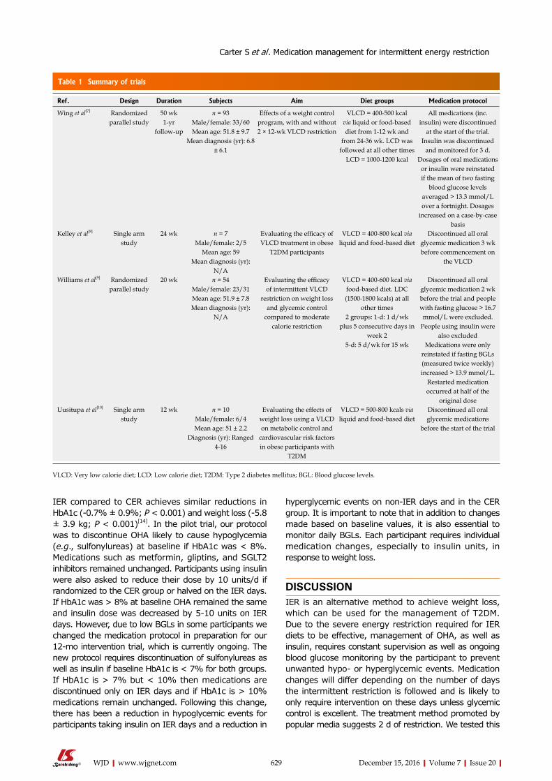
629 December 15, 2016|Volume 7|Issue 20|WJD|www.wjgnet.com
IER compared to CER achieves similar reductions in HbA1c (0.7% ± 0.9%; P < 0.001) and weight loss (5.8± 3.9 kg; P < 0.001)[14]. In the pilot trial, our protocol was to discontinue OHA likely to cause hypoglycemia (e.g., sulfonylureas) at baseline if HbA1c was < 8%. Medications such as metformin, gliptins, and SGLT2 inhibitors remained unchanged. Participants using insulin were also asked to reduce their dose by 10 units/d if randomized to the CER group or halved on the IER days. If HbA1c was > 8% at baseline OHA remained the same and insulin dose was decreased by 510 units on IER days. However, due to low BGLs in some participants we changed the medication protocol in preparation for our 12mo intervention trial, which is currently ongoing. The new protocol requires discontinuation of sulfonylureas as well as insulin if baseline HbA1c is < 7% for both groups. If HbA1c is > 7% but < 10% then medications are discontinued only on IER days and if HbA1c is > 10% medications remain unchanged. Following this change, there has been a reduction in hypoglycemic events for participants taking insulin on IER days and a reduction in
hyperglycemic events on nonIER days and in the CER group. It is important to note that in addition to changes made based on baseline values, it is also essential to monitor daily BGLs. Each participant requires individual medication changes, especially to insulin units, in response to weight loss.
DISCUSSIONIER is an alternative method to achieve weight loss, which can be used for the management of T2DM. Due to the severe energy restriction required for IER diets to be effective, management of OHA, as well as insulin, requires constant supervision as well as ongoing blood glucose monitoring by the participant to prevent unwanted hypo or hyperglycemic events. Medication changes will differ depending on the number of days the intermittent restriction is followed and is likely to only require intervention on these days unless glycemic control is excellent. The treatment method promoted by popular media suggests 2 d of restriction. We tested this
Ref. Design Duration Subjects Aim Diet groups Medication protocol
Wing et al[7] Randomized parallel study
50 wk1-yr
follow-up
n = 93Male/female: 33/60Mean age: 51.8 ± 9.7
Mean diagnosis (yr): 6.8 ± 6.1
Effects of a weight control program, with and without 2 × 12-wk VLCD restriction
VLCD = 400-500 kcal via liquid or food-based diet from 1-12 wk and
from 24-36 wk. LCD was followed at all other times
LCD = 1000-1200 kcal
All medications (inc. insulin) were discontinued
at the start of the trial. Insulin was discontinued
and monitored for 3 d. Dosages of oral medications
or insulin were reinstated if the mean of two fasting
blood glucose levels averaged > 13.3 mmol/L over a fortnight. Dosages
increased on a case-by-case basis
Kelley et al[8] Single arm study
24 wk n = 7Male/female: 2/5
Mean age: 59Mean diagnosis (yr):
N/A
Evaluating the efficacy of VLCD treatment in obese
T2DM participants
VLCD = 400-800 kcal via liquid and food-based diet
Discontinued all oral glycemic medication 3 wk before commencement on
the VLCD
Williams et al[9] Randomized parallel study
20 wk n = 54Male/female: 23/31Mean age: 51.9 ± 7.8Mean diagnosis (yr):
N/A
Evaluating the efficacy of intermittent VLCD
restriction on weight loss and glycemic control
compared to moderate calorie restriction
VLCD = 400-600 kcal via food-based diet. LDC (1500-1800 kcals) at all
other times 2 groups: 1-d: 1 d/wk
plus 5 consecutive days in week 2
5-d: 5 d/wk for 15 wk
Discontinued all oral glycemic medication 2 wk before the trial and people with fasting glucose > 16.7 mmol/L were excluded.
People using insulin were also excluded
Medications were only reinstated if fasting BGLs (measured twice weekly) increased > 13.9 mmol/L.
Restarted medication occurred at half of the
original doseUusitupa et al[10] Single arm
study12 wk n = 10
Male/female: 6/4Mean age: 51 ± 2.2
Diagnosis (yr): Ranged 4-16
Evaluating the effects of weight loss using a VLCD on metabolic control and
cardiovascular risk factors in obese participants with
T2DM
VLCD = 500-800 kcals via liquid and food-based diet
Discontinued all oral glycemic medications
before the start of the trial
Table 1 Summary of trials
VLCD: Very low calorie diet; LCD: Low calorie diet; T2DM: Type 2 diabetes mellitus; BGL: Blood glucose levels.
Carter S et al . Medication management for intermittent energy restriction

630 December 15, 2016|Volume 7|Issue 20|WJD|www.wjgnet.com
method and we suggest baseline medication changes based on HbA1c, as outlined in the second protocol above, as well as individual changes in response to weight loss. Participants, therefore, need to be willing to monitor their BGLs at least twice daily and report any episodes of hypo or hyperglycemia, which would indicate the need to further adjust medications.
ACKNOWLEDGMENTSThe authors would like to thank the participants in the 3mo 5:2 pilot trial and the ongoing 12mo 5:2 intervention trial.
COMMENTSBackgroundRecently attention has been given to a new method of weight loss, known as intermittent energy restriction (IER), which has demonstrated positive results for weight loss in overweight and obese populations.
Research frontiersFor this new diet method to be used safely in the type 2 diabetes mellitus (T2DM) population, medication management protocol must be established.
Innovations and breakthroughsVery low calorie diets used in the treatment of T2DM provide insight, but as seen from the research, medication changes may only be required on the IER treatment days and after weight loss.
ApplicationsIER is a successful treatment method for weight loss and glycemic control in T2DM, and with regular blood glucose levels monitoring, medications can be safely adjusted to limit unwanted episodes of hypo-or hyperglycemia.
Peer-reviewThe review though very short is written well. The authors state that intermittent energy restriction which requires severe energy restriction needs to be discussed as it is a developing concept.
REFERENCES1 Maggio CA, Pi-Sunyer FX. Obesity and type 2 diabetes. Endocrinol
Metab Clin North Am 2003; 32: 805-822, viii [PMID: 14711063 DOI: 10.1016/S0889-8529(03)00071-9]
2 Lim EL, Hollingsworth KG, Aribisala BS, Chen MJ, Mathers JC, Taylor R. Reversal of type 2 diabetes: normalisation of beta cell function in association with decreased pancreas and liver triacy-lglycerol. Diabetologia 2011; 54: 2506-2514 [PMID: 21656330 DOI: 10.1007/s00125-011-2204-7]
3 Wing RR, Marcus MD, Epstein LH, Salata R. Type II diabetic subjects lose less weight than their overweight nondiabetic spouses. Diabetes Care 1987; 10: 563-566 [PMID: 3677974 DOI: 10.2337/diacare.10.5.563]
4 Keogh JB, Pedersen E, Petersen KS, Clifton PM. Effects of inter-mittent compared to continuous energy restriction on short-term weight loss and long-term weight loss maintenance. Clin Obes 2014; 4: 150-156 [PMID: 25826770 DOI: 10.1111/cob.12052]
5 Harvie MN, Pegington M, Mattson MP, Frystyk J, Dillon B, Evans G, Cuzick J, Jebb SA, Martin B, Cutler RG, Son TG, Maudsley S, Carlson OD, Egan JM, Flyvbjerg A, Howell A. The effects of intermittent or continuous energy restriction on weight loss and metabolic disease risk markers: a randomized trial in young overweight women. Int J Obes (Lond) 2011; 35: 714-727 [PMID: 20921964 DOI: 10.1038/ijo.2010.171]
6 Baker S, Jerums G, Proietto J. Effects and clinical potential of very-low-calorie diets (VLCDs) in type 2 diabetes. Diabetes Res Clin Pract 2009; 85: 235-242 [PMID: 19560834 DOI: 10.1016/j.diabres.2009.06.002]
7 Wing RR, Blair E, Marcus M, Epstein LH, Harvey J. Year-long weight loss treatment for obese patients with type II diabetes: does including an intermittent very-low-calorie diet improve outcome? Am J Med 1994; 97: 354-362 [PMID: 7942937 DOI: 10.1016/0002-9343(94)90302-6]
8 Kelley DE, Wing R, Buonocore C, Sturis J, Polonsky K, Fitzsimmons M. Relative effects of calorie restriction and weight loss in noninsulin-dependent diabetes mellitus. J Clin Endocrinol Metab 1993; 77: 1287-1293 [PMID: 8077323 DOI: 10.1210/jcem.77.5.8077323]
9 Williams KV, Mullen ML, Kelley DE, Wing RR. The effect of short periods of caloric restriction on weight loss and glycemic control in type 2 diabetes. Diabetes Care 1998; 21: 2-8 [PMID: 9538962 DOI: 10.2337/diacare.21.1.2]
10 Uusitupa MI, Laakso M, Sarlund H, Majander H, Takala J, Penttilä I. Effects of a very-low-calorie diet on metabolic control and cardiovascular risk factors in the treatment of obese non-insulin-dependent diabetics. Am J Clin Nutr 1990; 51: 768-773 [PMID: 2333833]
11 Paisey RB, Frost J, Harvey P, Paisey A, Bower L, Paisey RM, Taylor P, Belka I. Five year results of a prospective very low calorie diet or conventional weight loss programme in type 2 diabetes. J Hum Nutr Diet 2002; 15: 121-127 [PMID: 11972741 DOI: 10.1046/j.1365-277X.2002.00342.x]
12 Collins RW, Anderson JW. Medication cost savings associated with weight loss for obese non-insulin-dependent diabetic men and women. Prev Med 1995; 24: 369-374 [PMID: 7479627 DOI: 10.1006/pmed.1995.1060]
13 Capstick F, Brooks BA, Burns CM, Zilkens RR, Steinbeck KS, Yue DK. Very low calorie diet (VLCD): a useful alternative in the treatment of the obese NIDDM patient. Diabetes Res Clin Pract 1997; 36: 105-111 [PMID: 9229194 DOI: 10.1016/S0168- 8227(97)00038-7]
14 Carter S, Clifton PM, Keogh JB. The effects of intermittent compared to continuous energy restriction on glycaemic control in type 2 diabetes; a pragmatic pilot trial. Diabetes Res Clin Pract 2016; 122: 106-112 [PMID: 27833048 DOI: 10.1016/j.diabres.2016.10.010]
P- Reviewer: Demonacos C, Liao KF, Sasikala M S- Editor: Ji FF L- Editor: A E- Editor: Li D
COMMENTS
Carter S et al . Medication management for intermittent energy restriction

© 2016 Baishideng Publishing Group Inc. All rights reserved.
Published by Baishideng Publishing Group Inc8226 Regency Drive, Pleasanton, CA 94588, USA
Telephone: +1-925-223-8242Fax: +1-925-223-8243
E-mail: [email protected] Desk: http://www.wjgnet.com/esps/helpdesk.aspx
http://www.wjgnet.com