woburn, ma 01801 usa perkit antibody mmae conjugation kit
TRANSCRIPT
CellMosaic, Inc. 10A Roessler Road
Woburn, MA 01801 USA
Phone: 781-463-0002
Fax: 781-998-4694 Email: [email protected]
Website: www.cellmosaic.com
1 CellMosaic |PerKit™ Antibody MMAE Conjugation Kit User Reference Guide, Rev F, 06/20, Doc#: DCM11409
PerKit™ Antibody MMAE Conjugation Kit (CM11409 and CM11409x3)
User Reference Guide Contents
Important Notes & Contact Information ...................................................................................................... 2
Kit Components............................................................................................................................................. 3
Safety Information ........................................................................................................................................ 4
Labeling Chemistry ........................................................................................................................................ 4
Support ......................................................................................................................................................... 5
Protocol ......................................................................................................................................................... 5
1. Lab Instrumentation Needed ............................................................................................................ 5
2. Prepare Site and MMAE for Labeling Experiment ............................................................................ 6
3. Preparation of Antibody Samples for Conjugation ........................................................................... 6
4. Antibody Reduction (Step 1 in Scheme 1) ........................................................................................ 8
5. Purification to Remove Excess Reagent A ......................................................................................... 9
6. MMAE Labeling (Step 2 in Scheme 1) ............................................................................................. 10
7. Purification of Conjugate ................................................................................................................ 10
Other Considerations .................................................................................................................................. 12
1. Concentration Determination for IgG Antibody (Unlabeled) ......................................................... 12
2. Concentration Determination for ADC ........................................................................................... 12
3. MW Calculation for ADC ................................................................................................................. 12
4. Drug-to-Antibody Ratio (DAR) and Characterization by UV ........................................................... 13
5. Characterization of ADC by HIC HPLC ............................................................................................. 13
6. Characterization of ADC by SEC HPLC ............................................................................................. 13
7. ADC Stabilizing Buffer ..................................................................................................................... 14
8. Recommended Storage Conditions ................................................................................................ 14
9. Sample Submission for HPLC Analysis ............................................................................................ 14
Appendix: Examples of MMAE ADC ............................................................................................................ 15
CellMosaic, Inc. 10A Roessler Road
Woburn, MA 01801 USA
Phone: 781-463-0002
Fax: 781-998-4694 Email: [email protected]
Website: www.cellmosaic.com
2 CellMosaic |PerKit™ Antibody MMAE Conjugation Kit User Reference Guide, Rev F, 06/20, Doc#: DCM11409
Important Notes & Contact Information
READ BEFORE USING ANY RESOURCES PROVIDED HEREIN
The information provided in this document and the methods included in this package are for information purposes only. CellMosaic provides no warranty of performance or suitability for the purpose described herein. The performance of this kit in labeling may be affected by many different variables, including but not limited to the purity and complexity of the starting materials, differences in preparation techniques, operator ability, and environmental conditions. Sample data are provided for illustration and example purposes only and represent a small dataset used to verify kit performance in the CellMosaic laboratory. Information about the chemicals and reagents used in the kit are provided as necessary. For Research Use Only. Not for Use in Diagnostic Procedures. The information in this document is subject to change without notice. CellMosaic assumes no responsibility for any errors that may appear in this document. In no event shall CellMosaic be liable, whether in contract, tort, warranty, or under any statute or on any other basis for special, incidental, indirect, punitive, multiple, or consequential damages in connection with or arising from this document, including but not limited to the use thereof.
NOTICE TO PURCHASER: LIMITED LICENSE
The purchase of this product includes a limited, non-transferable license to use this product to practice the labeling methods using the reagents solely for the purchaser’s research activities. The license granted herein is personal to the original purchaser and may not be transferred to any other party outside the purchaser’s company. No other right or license is conveyed or granted either expressly, by implication or by estoppel, to resell or repackage this product. Further information can be obtained by contacting:
Director of Licensing c/o CellMosaic, Inc. 10-A Roessler Road, Woburn, MA 01801.
Phone: 781-463-0002
Fax: 781-998-4694 E-mail: [email protected]

CellMosaic, Inc. 10A Roessler Road
Woburn, MA 01801 USA
Phone: 781-463-0002
Fax: 781-998-4694 Email: [email protected]
Website: www.cellmosaic.com
3 CellMosaic |PerKit™ Antibody MMAE Conjugation Kit User Reference Guide, Rev F, 06/20, Doc#: DCM11409
Kit Components
This kit provides materials to conjugate 1 to 3 mg of one (CM11409) or three (CM11409x3) antibody
samples (IgG) with monomethyl auristatin E (MMAE) using valine-citruline p-aminobenzylcarbamate
(VC-PAB) linker.
Upon receipt, please remove Box 1 and store in a freezer at or below -20°C. Store Box 2 in a refrigerator at 2-8°C.
Name Part #
Quantity (CM11409)
Quantity (CM11409x3)
Storage condition
Box 1
MC-VC-PAB-MMAE (red label) CM11001 0.11 mL 3 x 0.11 mL -20°C
Reagent A (blue label)
CM13004 1 unit 3 units
Box 2
Solution A (green label) CM01003 1.5 mL 6 mL
2-8°C
Reducing Buffer (orange label) CM02001 4 mL 12 mL
Labeling Buffer (indigo label) CM02005 4 mL 12 mL
Storage Buffer (1 x PBS buffer) (grey label)
CM02013 20 mL 60 mL
Centrifugal Filter Devices CM03CD050A 3 9
Desalting Column CM03SG10 1 3
Collection Tubes N/A 6 18
1.5 mL Centrifuge Tubes N/A 2 6
2.0 mL Centrifuge Tube(s) N/A 1 3
Hazardous Waste Bag(s) N/A 1 3
User Material
IgG Antibody N/A
NOT PROVIDED (User Supplied Material, 1-3 mg IgG needed per reaction)
Reaction Scale: The protocol is optimized for conjugating 3 mg of IgG antibody. If you have less than 3 mg of IgG, use the calculations in Steps B10, C3, D9, E2, F5, and F6 to obtain the correct volumes to be added in each step.
Drug-to-Antibody Ratio (DAR) Optimization: The reducing protocol is optimized for monoclonal IgG1 subtype to obtain an average 4 thiols per antibody (DAR = 4). For other IgG subtypes or polyclonal antibodies, the DAR may vary. For the best performance of the ADC and to obtain the desired DAR, you can purchase the Thiol Assay Kit with Purification (Product #: CM90005) separately and use it to perform an in-process thiol assay after the antibody reduction (Step C5 Note Section). The amount of reducing reagent can be adjusted based on the data to obtain your desired DAR.

CellMosaic, Inc. 10A Roessler Road
Woburn, MA 01801 USA
Phone: 781-463-0002
Fax: 781-998-4694 Email: [email protected]
Website: www.cellmosaic.com
4 CellMosaic |PerKit™ Antibody MMAE Conjugation Kit User Reference Guide, Rev F, 06/20, Doc#: DCM11409
Safety Information
Warning: some of the chemicals used can be potentially hazardous and can cause injury or illness. Please
read and understand the Safety Data Sheets (SDS) available at CellMosaic.com before you store, handle,
or use any of the materials.
Labeling Chemistry
The kit is designed to label any IgG antibody with monomethyl auristatin E (MMAE) using a valine-
citruline p-aminobenzylcarbamate (VC-PAB) linker. The user supplies the antibody. The kit includes
maleimide-activated VC-PAB-MMAE, which can be coupled directly to the antibody after reduction
through alkylation in a single step (a method developed by Seattle Genetics: Sun et al. 2005,
Bioconjugate Chem. 16, 1282-1290). The product is purified to remove any unreacted drugs.
Key features of this conjugation kit:
• Offers a simple and easy way to label IgG with MMAE with minimum exposure to the toxin
• Cathepsin B cleavable VC-PAB linkage (Ref. Doronina et al. 2008, Bioconjugate Chem. 19, 1960-
1963)
• Fast and easy preparation: 6 h preparation and <2 h hands-on time
• All reagents and supplies included for preparation and purification
• Easy to control the DAR if used together with the Thiol Assay Kit with Purification (Product #:
CM90005)
• More than 95% conjugated products (free of unreacted drug and less than 5% unreacted
antibody)
Drug Information:
• Name: Monomethyl auristatin E (MMAE) with MC-VC-PAB linkage
• CAS number: 646502-53-6
• Chemical formula: C68H105N11O15
• MW: 1316.65

CellMosaic, Inc. 10A Roessler Road
Woburn, MA 01801 USA
Phone: 781-463-0002
Fax: 781-998-4694 Email: [email protected]
Website: www.cellmosaic.com
5 CellMosaic |PerKit™ Antibody MMAE Conjugation Kit User Reference Guide, Rev F, 06/20, Doc#: DCM11409
Mechanism of action: Inhibits cell division by blocking the polymerization of tubulin, the VC-PAB linker is stable in extracellular fluid but cleaved by cathepsin B once inside the tumor cell, activating the antimitotic mechanism
• Activities: Antioxidant, anti-inflammatory, anti-cancer, and insecticidal activities
Requirement for antibody (IgG): 1. Preferably > 90% pure by gel electrophoresis 2. Total amount: 1-3 mg protein content as measured by UV. Note: the accuracy of your protein amount is the single most important factor to obtaining an optimized DAR. Please refer to the section Other Considerations in this manual to measure the protein amount.
Support
A customer can request a recommendation for the conjugation if the IgG has a special feature or less
than 1 mg of IgG to be labeled. CellMosaic provides other accessory tools, such as buffers, standards,
and reagents for ADC research. CellMosaic also provides fee-based support services to customers who
need help analyzing the final conjugates by HPLC and determining the DAR.
Protocol
Scheme 1. Schematic diagram of the workflow for preparing antibody-MMAE conjugates starting with 3
mg of IgG (volume of reagents varies if the amount of IgG is < 3 mg).
1. Lab Instrumentation Needed
• Vortex mixer, centrifuge (preferably refrigerated, 14,000 g capable), mini-centrifuge
• Pipettes and tips
• Timer
CellMosaic, Inc. 10A Roessler Road
Woburn, MA 01801 USA
Phone: 781-463-0002
Fax: 781-998-4694 Email: [email protected]
Website: www.cellmosaic.com
6 CellMosaic |PerKit™ Antibody MMAE Conjugation Kit User Reference Guide, Rev F, 06/20, Doc#: DCM11409
• Incubator or shaker set at 37°C and at RT
• Chemical hood
• Support stand, lab frame, or any support rod for desalting column
• Flask
• Personal protection equipment (lab coat, safety glasses, and chemical resistant nitrile
gloves)
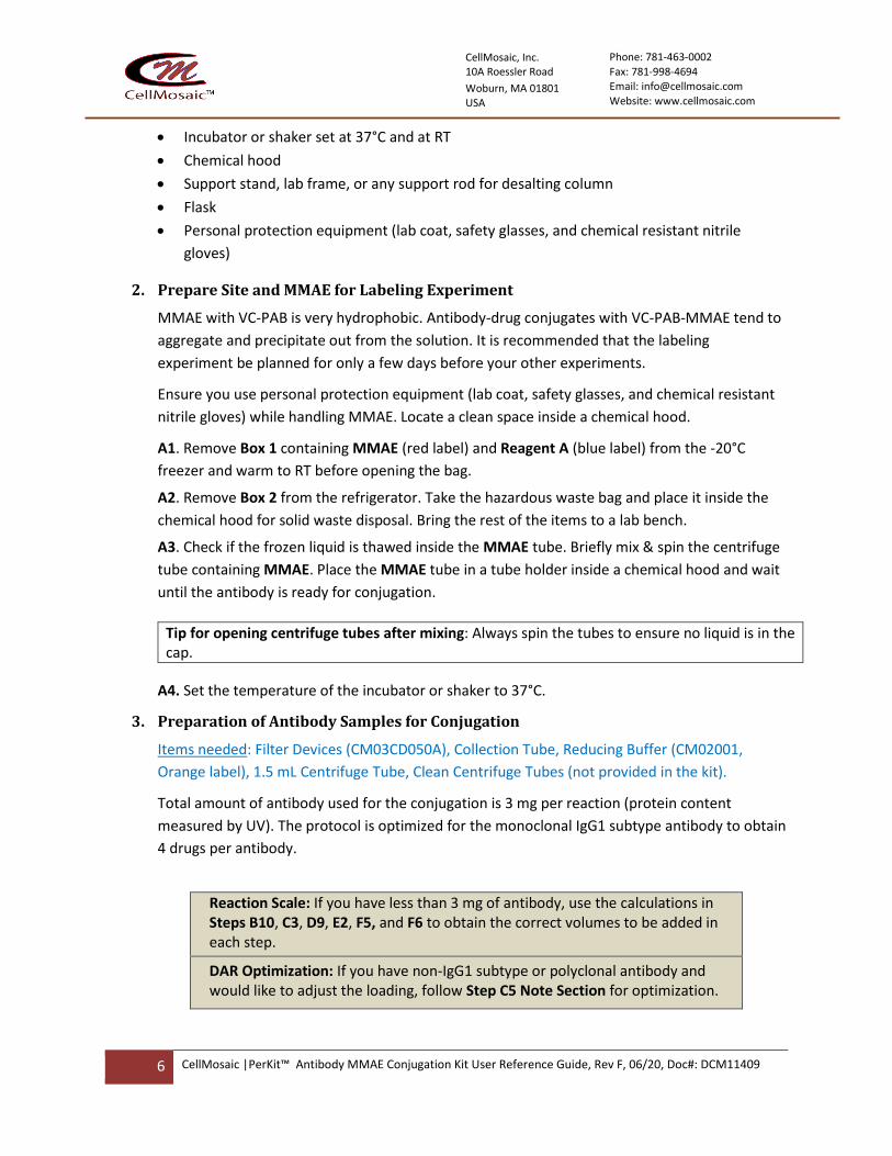
2. Prepare Site and MMAE for Labeling Experiment
MMAE with VC-PAB is very hydrophobic. Antibody-drug conjugates with VC-PAB-MMAE tend to
aggregate and precipitate out from the solution. It is recommended that the labeling
experiment be planned for only a few days before your other experiments.
Ensure you use personal protection equipment (lab coat, safety glasses, and chemical resistant
nitrile gloves) while handling MMAE. Locate a clean space inside a chemical hood.
A1. Remove Box 1 containing MMAE (red label) and Reagent A (blue label) from the -20°C
freezer and warm to RT before opening the bag.
A2. Remove Box 2 from the refrigerator. Take the hazardous waste bag and place it inside the
chemical hood for solid waste disposal. Bring the rest of the items to a lab bench.
A3. Check if the frozen liquid is thawed inside the MMAE tube. Briefly mix & spin the centrifuge
tube containing MMAE. Place the MMAE tube in a tube holder inside a chemical hood and wait
until the antibody is ready for conjugation.
Tip for opening centrifuge tubes after mixing: Always spin the tubes to ensure no liquid is in the cap.
A4. Set the temperature of the incubator or shaker to 37°C.
3. Preparation of Antibody Samples for Conjugation
Items needed: Filter Devices (CM03CD050A), Collection Tube, Reducing Buffer (CM02001,
Orange label), 1.5 mL Centrifuge Tube, Clean Centrifuge Tubes (not provided in the kit).
Total amount of antibody used for the conjugation is 3 mg per reaction (protein content
measured by UV). The protocol is optimized for the monoclonal IgG1 subtype antibody to obtain
4 drugs per antibody.
Reaction Scale: If you have less than 3 mg of antibody, use the calculations in Steps B10, C3, D9, E2, F5, and F6 to obtain the correct volumes to be added in each step.
DAR Optimization: If you have non-IgG1 subtype or polyclonal antibody and would like to adjust the loading, follow Step C5 Note Section for optimization.

CellMosaic, Inc. 10A Roessler Road
Woburn, MA 01801 USA
Phone: 781-463-0002
Fax: 781-998-4694 Email: [email protected]
Website: www.cellmosaic.com
7 CellMosaic |PerKit™ Antibody MMAE Conjugation Kit User Reference Guide, Rev F, 06/20, Doc#: DCM11409
B1. Insert the Filter Device into one of the provided collection tubes (micro-centrifuge tube with
the cap attached). Perform the step based on the following conditions.
✓ If your antibody is supplied as a lyophilized solid, dissolve the antibody in 500 μL of
deionized water and then transfer the entire contents to the Filter Device.
✓ If your antibody is supplied in < 500 μL buffer, transfer your antibody sample to the
Filter Device directly. Add Reducing Buffer to make up the total volume to 500 μL and
cap it.
✓ If the volume of your antibody sample is between 500 and 1000 μL, divide the volume
into two Centrifugal Filter Devices and add the antibody sample to the filter device. Add
Reducing Buffer to make up the total volume to 500 μL in each device and cap them.
✓ If the volume of your antibody sample is >1000 μL, add up to 500 μL of sample to the
two Filter Devices and cap them. Repeat Steps B1-B4 until all of the antibody sample is
transferred into the Filter Device. Move on to Step B5. Add Reducing Buffer to make up
the total volume to 500 μL in each device for the last refill.
B2. Place the capped Filter Device into the centrifuge rotor, aligning the cap strap toward the
center of the rotor; counterbalance with a similar device.
B3. Spin the Filter Device at 14,000 x g for 8 minutes (preferably cooled to 4°C) to concentrate
to < 100 µL (Spin time depends on many factors. The typical spin time for a 500 µL sample is
approximately 8 to 20 minutes. The typical volume is ~40 µL after spinning for 8 minutes on an
Eppendorf 5417R at 4°C).
B4. Remove the assembled device from the centrifuge and separate the Filter Device from the
collection tube. Transfer the filtrate from the collection tube to a clean centrifuge tube (not
provided). Save the filtrate until the experiments are done.
B5. Insert the Filter Device back into the collection tube. Add 400-450 μL of Reducing Buffer to
make up the total volume to 500 μL. Next, place the capped Filter Device into the centrifuge
rotor, aligning the cap strap toward the center of the rotor; counterbalance with a similar
device. Spin the device at 14,000 x g to concentrate to < 100 µL. Remove the assembled device
from the centrifuge and separate the Filter Device from the collection tube. Transfer the filtrate
from the collection tube to a clean centrifuge tube (not provided). Save the filtrate until the
experiments are done.
B6. Repeat Step B5 two more times.
B7. Transfer the concentrated sample from the Filter Device to a 1.5 mL micro-centrifuge tube
(use the pipetman to measure the approximate volume of the concentrated sample).
B8. Add 20-100 µL of Reducing Buffer to the Filter Device to rinse (actual volume of Reducing
Buffer added will depend upon the calculated total volume in Step B10). Stir it gently with a
pipet tip, then transfer the entire contents to the 1.5 mL micro-centrifuge tube from Step B7.
B9. Repeat Step B8 once.

CellMosaic, Inc. 10A Roessler Road
Woburn, MA 01801 USA
Phone: 781-463-0002
Fax: 781-998-4694 Email: [email protected]
Website: www.cellmosaic.com
8 CellMosaic |PerKit™ Antibody MMAE Conjugation Kit User Reference Guide, Rev F, 06/20, Doc#: DCM11409
B10. Add Reducing Buffer to the 1.5 mL micro-centrifuge tube from Step B9 to make up the
total volume of the sample to 300 5 μL and cap it.
Calculation 1 for Less Antibody (Ab):
𝑇𝑜𝑡𝑎𝑙 𝑣𝑜𝑙𝑢𝑚𝑒 𝑜𝑓 𝑡ℎ𝑒 𝑎𝑛𝑡𝑖𝑏𝑜𝑑𝑦 𝑖𝑛 𝑆𝑡𝑒𝑝 𝑩𝟏𝟎 (𝜇𝐿) = 𝐴𝑏 𝑖𝑛 𝑚𝑔 × 100
B11. Vortex the combined antibody sample for 30 seconds and then spin down.
4. Antibody Reduction (Step 1 in Scheme 1)
Items needed: Reagent A (CM13004, blue label), Solution A (CM01003, green label), Antibody
Solution from Step B11, Ice Bath.
C1. Spin the centrifuge tube containing Reagent A (blue label).
C2. Spin Solution A (green label) before opening it. Add 800 µL of Solution A to the tube with
Reagent A from Step C1. Vortex for 30 seconds to 1 minute to dissolve the reagent and then
spin.
C3. Add 7.5 µL of Reagent A solution from Step C2 to the centrifuge tube containing antibody
from Step B11 (Discard any unused Reagent A as hazardous chemical waste after completion of
all experiments).
Calculation 2 for Less Antibody (Ab):
𝑉𝑜𝑙𝑢𝑚𝑒 𝑜𝑓 𝑅𝑒𝑎𝑔𝑒𝑛𝑡 𝐴 𝑠𝑜𝑙𝑢𝑡𝑖𝑜𝑛 𝑡𝑜 𝑏𝑒 𝑡𝑟𝑎𝑛𝑠𝑓𝑒𝑟𝑟𝑒𝑑 𝑖𝑛 𝑆𝑡𝑒𝑝 𝑪𝟑 (𝜇𝐿) = 𝐴𝑏 𝑖𝑛 𝑚𝑔 × 2.5
C4. Vortex the solution for 30 seconds, and then spin to ensure no liquid is in the cap. Mix at
37°C for 2 h.
Tip for mixing: You can use a nutator, a shaker, a vortex, or an incubator shaker for mixing. If you are using end to end nutating, make sure the tube from step C4 is securely capped. If you don’t have any of this equipment, you can let the tube sit on the bench with manual mixing by pipetting every 20 minutes.
C5. Cool the reduced antibody solution to approximately 4oC by placing the tube on ice or
keeping it inside a refrigerator at 2-8°C for 5 minutes.
Note: Optimization of Thiol Content for non-IgG1 Subtype Antibody or Polyclonal Antibody
For the monoclonal IgG1 subtype, the average free thiol groups per antibody is 4 after reduction. If you have a polyclonal or other IgG subtype, you can purchase the Thiol Assay Kit with Purification (Product Number: CM90005) separately from CellMosaic to measure the free thiols while letting the reducing solution sit at 4°C in Step C5. Use 5 μL antibody solution from Step C5 and follow the protocol of CM90005. To calculate the number of thiols, please use the antibody concentration of 11.11 μM.
CellMosaic, Inc. 10A Roessler Road
Woburn, MA 01801 USA
Phone: 781-463-0002
Fax: 781-998-4694 Email: [email protected]
Website: www.cellmosaic.com
9 CellMosaic |PerKit™ Antibody MMAE Conjugation Kit User Reference Guide, Rev F, 06/20, Doc#: DCM11409
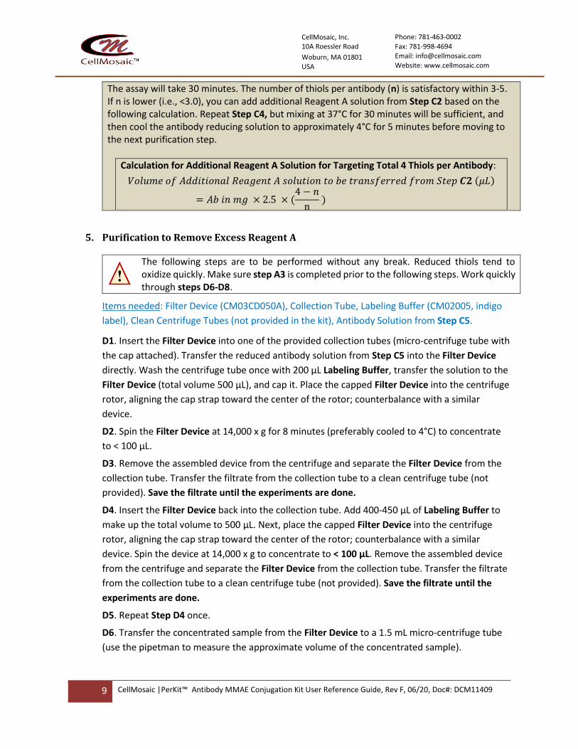
The assay will take 30 minutes. The number of thiols per antibody (n) is satisfactory within 3-5. If n is lower (i.e., <3.0), you can add additional Reagent A solution from Step C2 based on the following calculation. Repeat Step C4, but mixing at 37°C for 30 minutes will be sufficient, and then cool the antibody reducing solution to approximately 4°C for 5 minutes before moving to the next purification step.
Calculation for Additional Reagent A Solution for Targeting Total 4 Thiols per Antibody:
𝑉𝑜𝑙𝑢𝑚𝑒 𝑜𝑓 𝐴𝑑𝑑𝑖𝑡𝑖𝑜𝑛𝑎𝑙 𝑅𝑒𝑎𝑔𝑒𝑛𝑡 𝐴 𝑠𝑜𝑙𝑢𝑡𝑖𝑜𝑛 𝑡𝑜 𝑏𝑒 𝑡𝑟𝑎𝑛𝑠𝑓𝑒𝑟𝑟𝑒𝑑 𝑓𝑟𝑜𝑚 𝑆𝑡𝑒𝑝 𝑪𝟐 (𝜇𝐿)
= 𝐴𝑏 𝑖𝑛 𝑚𝑔 × 2.5 × (4 − 𝑛
n )
5. Purification to Remove Excess Reagent A
The following steps are to be performed without any break. Reduced thiols tend to oxidize quickly. Make sure step A3 is completed prior to the following steps. Work quickly through steps D6-D8.
Items needed: Filter Device (CM03CD050A), Collection Tube, Labeling Buffer (CM02005, indigo
label), Clean Centrifuge Tubes (not provided in the kit), Antibody Solution from Step C5.
D1. Insert the Filter Device into one of the provided collection tubes (micro-centrifuge tube with
the cap attached). Transfer the reduced antibody solution from Step C5 into the Filter Device
directly. Wash the centrifuge tube once with 200 μL Labeling Buffer, transfer the solution to the
Filter Device (total volume 500 μL), and cap it. Place the capped Filter Device into the centrifuge
rotor, aligning the cap strap toward the center of the rotor; counterbalance with a similar
device.
D2. Spin the Filter Device at 14,000 x g for 8 minutes (preferably cooled to 4°C) to concentrate
to < 100 µL.
D3. Remove the assembled device from the centrifuge and separate the Filter Device from the
collection tube. Transfer the filtrate from the collection tube to a clean centrifuge tube (not
provided). Save the filtrate until the experiments are done.
D4. Insert the Filter Device back into the collection tube. Add 400-450 μL of Labeling Buffer to
make up the total volume to 500 μL. Next, place the capped Filter Device into the centrifuge
rotor, aligning the cap strap toward the center of the rotor; counterbalance with a similar
device. Spin the device at 14,000 x g to concentrate to < 100 µL. Remove the assembled device
from the centrifuge and separate the Filter Device from the collection tube. Transfer the filtrate
from the collection tube to a clean centrifuge tube (not provided). Save the filtrate until the
experiments are done.
D5. Repeat Step D4 once.
D6. Transfer the concentrated sample from the Filter Device to a 1.5 mL micro-centrifuge tube
(use the pipetman to measure the approximate volume of the concentrated sample).

CellMosaic, Inc. 10A Roessler Road
Woburn, MA 01801 USA
Phone: 781-463-0002
Fax: 781-998-4694 Email: [email protected]
Website: www.cellmosaic.com
10 CellMosaic |PerKit™ Antibody MMAE Conjugation Kit User Reference Guide, Rev F, 06/20, Doc#: DCM11409
D7. Add 50-200 μL of Labeling Buffer to the Filter Device to rinse (actual volume of Labeling
Buffer added will depend upon the calculated total volume in Step D9). Stir it gently with a pipet
tip, then transfer the entire contents to the 1.5 mL micro-centrifuge tube from Step D6.
D8. Repeat Step D7 once.
D9. Add Labeling Buffer to make up the total volume of the sample to 640 10 μL.
Calculation 3 for Less Antibody (Ab):
𝑉𝑜𝑙𝑢𝑚𝑒 𝑜𝑓 𝑅𝑒𝑑𝑢𝑐𝑒𝑑 𝐴𝑛𝑡𝑖𝑏𝑜𝑑𝑦 𝑖𝑛 𝑆𝑡𝑒𝑝 𝑫𝟗 (𝜇𝐿) = 𝐴𝑏 𝑖𝑛 𝑚𝑔 × 213.3
D10. Vortex the combined antibody sample for 30 seconds and then spin down.
6. MMAE Labeling (Step 2 in Scheme 1)
Items needed: MMAE solution from step A3, Hazardous Waste Bag, Antibody Solution from step
D10.
E1. With personal protection equipment on, carefully open the centrifuge tube containing
MMAE from Step A3.
E2. Transfer the entire solution (110 μL total) to the centrifuge tube containing antibody from
Step D10. When you add the MMAE solution, place the pipette tip inside the antibody solution
and then dispense the MMAE slowly while swirling the pipette tip. Dispose of the pipette tip
and MMAE tube in the hazardous waste bag.
Calculation 4 for Less Antibody (Ab): 𝑉𝑜𝑙𝑢𝑚𝑒 𝑜𝑓 𝑀𝑀𝐴𝐸 𝑆𝑜𝑙𝑢𝑡𝑖𝑜𝑛 𝑡𝑜 𝑏𝑒 𝑇𝑟𝑎𝑛𝑠𝑓𝑒𝑟𝑟𝑒𝑑 𝑖𝑛 𝑆𝑡𝑒𝑝 𝑬𝟐 (𝜇𝐿) = 𝐴𝑏 𝑖𝑛 𝑚𝑔 × 36.7
Dispose of the remainder of the MMAE solution in the hazardous waste bag.
E3. Cap the centrifuge tube. Mix at 25°C or RT for 1 h.
Time-saving tip: While waiting for the reaction to complete, you can move on to Step F1 and equilibrate the column for purification.
7. Purification of Conjugate
Items needed: Desalting Column, Storage Buffer (1x PBS), 2.0 mL Centrifuge Tube, Hazardous
Waste Bag, Antibody Solution from Step E3.
F1. In a chemical hood, securely attach the Desalting Column to a support stand, lab frame, or
any support rod. Remove the top and bottom caps from the column and allow the excess liquid
to flow through by gravity. Collect the liquid in a flask.
F2. Add 5 mL of Storage Buffer and allow the buffer to completely enter the gel bed by gravity
flow.
F3. Repeat Step F2 twice.
F4. Spin the MMAE-labeled antibody solution from Step E3 before opening it. Add the entire
antibody solution to the column. Dispose of the centrifuge tube in the hazardous waste bag.
Wo
rk q
uic
kly

CellMosaic, Inc. 10A Roessler Road
Woburn, MA 01801 USA
Phone: 781-463-0002
Fax: 781-998-4694 Email: [email protected]
Website: www.cellmosaic.com
11 CellMosaic |PerKit™ Antibody MMAE Conjugation Kit User Reference Guide, Rev F, 06/20, Doc#: DCM11409
F5. Add 250 µL of Storage Buffer and allow the liquid to enter the gel bed completely (Note: this
elution buffer does not contain any of your product, you can let it drain to the waste).
Calculation 5 for Less Antibody (Ab):
𝑉𝑜𝑙𝑢𝑚𝑒 𝑜𝑓 𝑆𝑡𝑜𝑟𝑎𝑔𝑒 𝑏𝑢𝑓𝑓𝑒𝑟 𝑖𝑛 𝑆𝑡𝑒𝑝 𝑭𝟓 (𝜇𝐿) = 1000 − 𝐴𝑏 𝑖𝑛 𝑚𝑔 × 250
F6. Place a 2 mL centrifuge tube under the column. Add 1.25 mL of Storage Buffer to the
column. Collect the eluent by gravity and allow the buffer to enter the gel bed completely.
Calculation 6 for Less Antibody (Ab):
𝑉𝑜𝑙𝑢𝑚𝑒 𝑜𝑓 𝑆𝑡𝑜𝑟𝑎𝑔𝑒 𝑏𝑢𝑓𝑓𝑒𝑟 𝑖𝑛 𝑆𝑡𝑒𝑝 𝑭𝟔 (𝜇𝐿) = 500 + 𝐴𝑏 𝑖𝑛 𝑚𝑔 × 250
F7. Label the tube as your product. Store your conjugate at 4°C. Dispose of the Desalting
Column in the hazardous waste bag and seal the bag. Dispose of the waste following
regulations appropriate for your area.
Conjugate is Ready for Your Experiment
• Specifications of your product: MMAE-labeled antibodies with an average drug-to-
antibody ratio (DAR) of 4. A typical batch contains more than 95% conjugated products by
size exclusion chromatography (SEC) with less than 5% unreacted antibody and is free of any
unreacted drug. The approximate concentration of the ADC is 1.2 mg/mL in PBS buffer assuming
50% recovery. You can determine the concentration and estimated DAR of the ADC by UV/vis
spectrophotometry (see Other Considerations).

CellMosaic, Inc. 10A Roessler Road
Woburn, MA 01801 USA
Phone: 781-463-0002
Fax: 781-998-4694 Email: [email protected]
Website: www.cellmosaic.com
12 CellMosaic |PerKit™ Antibody MMAE Conjugation Kit User Reference Guide, Rev F, 06/20, Doc#: DCM11409
Other Considerations
1. Concentration Determination for IgG Antibody (Unlabeled)
The accuracy of the IgG amount is important for obtaining an optimized DAR in this protocol.
The simplest assay method for determining IgG concentration in solution is to measure the
absorbance of the IgG at 280 nm (UV range) (A1 mg/mL = 1.4).
If your antibody comes with a buffer that has no UV absorbance at 280 nm, you can measure
the UV absorbance prior to starting an experiment.
𝐶𝑜𝑛𝑐𝑒𝑛𝑡𝑟𝑎𝑡𝑖𝑜𝑛 (𝑚𝑔/𝑚𝐿) 𝑜𝑓 𝐼𝑔𝐺 =(𝐴280)
1.4
If your antibody comes with a buffer that has UV absorbance at 280 nm, you can determine the
concentration in step B11 after exchanging it with Reducing Buffer and assuming 95% recovery
of the IgG after buffer exchange. Reducing Buffer does not contain any substances that will
interfere with the UV measurement at 280 nm. The total volume of Reducing Buffer added in
Step B10 can be estimated based on the initially estimated amount of antibody and will not
affect the conjugation too much if the volume is off to some extent.
𝐶𝑜𝑛𝑐𝑒𝑛𝑡𝑟𝑎𝑡𝑖𝑜𝑛 (𝑚𝑔/𝑚𝐿) 𝑜𝑓 𝑆𝑡𝑎𝑟𝑡𝑖𝑛𝑔 𝐼𝑔𝐺 =(𝐴280)
1.4 × 0.95
After calculating the total amount, follow the calculations in Steps B10, C3, D9, E2, F5, and F6 to
obtain the correct volumes to be added in each step.
2. Concentration Determination for ADC
To determine the concentration of the ADC, dilute your conjugate from Step F7 with 1x PBS buffer. Measure the UV absorbance of the conjugate at 280 nm (A280) using a UV spectrometer and calculate the concentration based on the following formula:
𝐶𝑜𝑛𝑐𝑒𝑛𝑡𝑟𝑎𝑡𝑖𝑜𝑛 (𝑀) 𝑜𝑓 𝑡ℎ𝑒 𝑑𝑖𝑙𝑢𝑡𝑒 𝑠𝑎𝑚𝑝𝑙𝑒 =(𝐴280) × 4.7619
𝐿
𝐶𝑜𝑛𝑐𝑒𝑛𝑡𝑟𝑎𝑡𝑖𝑜𝑛 (𝑚𝑔/𝑚𝐿) 𝑜𝑓 𝑡ℎ𝑒 𝑑𝑖𝑙𝑢𝑡𝑒 𝑠𝑎𝑚𝑝𝑙𝑒 =(𝐴280) × 0.7143
𝐿
Where L is the UV cell path length (cm). If you are using a 1 cm UV cell, you can dilute the conjugate 4 times to obtain a good reading.
For a typical IgG with MW of 150,000, the molar extinction coefficient is 210,000 M-1cm-1.
3. MW Calculation for ADC
Calculation of the MW of the conjugate:
MW(ADC) = n x 1317+ 150000
Where n is the average molar ratio of MMAE per antibody. Use 4.0 if you do not have the hydrophobic interaction chromatography (HIC) profile of your conjugates.

CellMosaic, Inc. 10A Roessler Road
Woburn, MA 01801 USA
Phone: 781-463-0002
Fax: 781-998-4694 Email: [email protected]
Website: www.cellmosaic.com
13 CellMosaic |PerKit™ Antibody MMAE Conjugation Kit User Reference Guide, Rev F, 06/20, Doc#: DCM11409
4. Drug-to-Antibody Ratio (DAR) and Characterization by UV
In this kit, the target DAR is 4. Depending on your antibody, you may achieve a lower DAR.
To estimate the DAR, you can obtain the UV absorbance ratio (R) of your conjugate at 248 nm and 280 nm.
𝑅 =(𝐴248)
(𝐴280)⁄
The unlabeled antibody will have an R of 0.4 – 0.5. An MMAE-ADC with DAR of 3 – 5 will have an R of 0.65 – 0.80.
You can also use the following formula to calculate the estimated DAR (only for reference):
𝐷𝐴𝑅 =(21 × 𝑅 − 9)
(1.615 − 0.1425 × 𝑅)
Note: The UV absorbance of the MMAE in an ADC can vary greatly depending on many factors, such as aggregation and stacking. Therefore, the R value for an ADC can differ greatly for different antibodies and should be determined experimentally. The calculation for the DAR using this formula is for reference only.
5. Characterization of ADC by HIC HPLC
For ADCs prepared via a reduced thiol of the antibody, hydrophobic interaction chromatography
(HIC) HPLC is used to calculate the DAR and the heterogeneity of the ADCs. The conjugates are
separated based on hydrophobicity. Antibodies loaded with the same number of drugs (same
DAR) will have similar hydrophobicity and be eluted as a single peak. For a typical MMAE ADC,
multiple peaks represent various amounts of drug-loaded ADCs. You will find examples of HIC
HPLC profiles of MMAE ADCs of various antibodies in the Appendix.
CellMosaic offers an HIC buffer set (Product #: CM02025) for our customers to use with any HIC
column. The CM02025 product sheet contains all of the information and methodology needed
to run an HIC HPLC analysis.
If you do not have access to an HPLC facility, you can send your sample to CellMosaic for
analysis.
6. Characterization of ADC by SEC HPLC
VC-PAB-MMAE is very hydrophobic. This kit is designed to minimize the aggregation and
precipitation issues generally seen with MMAE labeling. However, you may still notice some
solid precipitate out or ADC aggregation during the reaction. The precipitate will be removed
during purification. Depending on the properties of your antibody, recovery will be 40-80%. If
you are concerned with the aggregation, you can use size exclusion chromatography (SEC) to
check the extent of aggregation. SEC separates the conjugates by apparent molecular weight
(MW) or size in aqueous solution. The larger the MW of the conjugate, the earlier it elutes. By
comparing the SEC profile of unlabeled IgG and the ADC, you can estimate how much
aggregation is in the ADC.

CellMosaic, Inc. 10A Roessler Road
Woburn, MA 01801 USA
Phone: 781-463-0002
Fax: 781-998-4694 Email: [email protected]
Website: www.cellmosaic.com
14 CellMosaic |PerKit™ Antibody MMAE Conjugation Kit User Reference Guide, Rev F, 06/20, Doc#: DCM11409
CellMosaic offers two SEC standards (Product #: CM92004 and CM92005) for our customers to
use with any SEC column. The CM92004 product sheet contains all of the information and
methodology you need to run an SEC HPLC analysis.
If you do not have access to an HPLC facility, you can send your sample to CellMosaic for
analysis.
7. ADC Stabilizing Buffer
CellMosaic’s proprietary ADC stabilizing PBS buffer (5x) (Product #: CM02022) contains 5x PBS
buffer and other stabilizers to prevent the hydrophobic drugs from interacting with each other
during storage, which would cause the ADCs to precipitate out. Stabilization buffer also helps
preserve the structure of the ADCs during lyophilization. The buffer is biocompatible and can be
used directly for any in vitro and in vivo studies. For more information on the stabilization
buffers, please check our website.
8. Recommended Storage Conditions
Unlike other ADCs labeled with hydrophobic drug, ADC with MMAE is relatively stable. Based on our preliminary data, the conjugate made with this kit can remain stable in PBS buffer for several weeks at 2-8°C. Do not freeze MMAE ADC.
The stability of your conjugate may be different due to your antibody and should be checked by
either HPLC or UV. If you need to store the ADCs for a longer period of time, you can purchase
the ADC stabilization PBS buffer separately. Dilute your ADC in Stabilization PBS buffer (5x).
Aliquot and store the conjugate in a < -20°C freezer or lyophilize to dryness. Avoid repeated
freeze and thaw cycles.
9. Sample Submission for HPLC Analysis
If you are submitting samples to CellMosaic for SEC and HIC analysis, please follow these instructions:
1) Go online: https://www.cellmosaic.com/hplc-analysis/, select SEC HPLC Analysis (Product# AS0023) and HIC HPLC Analysis (Product#: AS0025), choose the quantity (number of samples. Bulk discounts available for multiple samples) and submit the order. Alternatively, you can email [email protected] for a quote and to place the order.
2) Dilute your un-conjugated antibody in PBS buffer to 1 mg/mL, and then transfer 50 μL of the diluted solution to a 500 μL micro-centrifuge tube. Label the vial properly.
3) Transfer 50 μL of ADC (non-diluted solution) to a 500 μL micro-centrifuge tube and label the vial properly.
4) Ship your samples with a cold pack for overnight delivery.

CellMosaic, Inc. 10A Roessler Road
Woburn, MA 01801 USA
Phone: 781-463-0002
Fax: 781-998-4694 Email: [email protected]
Website: www.cellmosaic.com
15 CellMosaic |PerKit™ Antibody MMAE Conjugation Kit User Reference Guide, Rev F, 06/20, Doc#: DCM11409
Appendix: Examples of MMAE ADC
Example 1: MMAE Conjugation with Monoclonal Human IgG1 Subtype
ADC prepared at CellMosaic following the exact protocol without any adjustment.
Kit lot number: 5508.S9.020918
Scale of the reaction: 3 mg (CM11409)
Specification of the final conjugates:
Calculated average DAR: 4.86 (Multiple DAR products) Unreacted antibodies: 2.6% Unreacted MMAE: 0% ADC recovery: 81% Figure 1: HIC HPLC analysis of monoclonal human IgG1, Mal-VC-PAB-MMAE, and purified conjugates
Human IgG1
MMAE
MMAE ADC
Unreacted antibody (2.6%)
1 2 3
4 5 6-7

CellMosaic, Inc. 10A Roessler Road
Woburn, MA 01801 USA
Phone: 781-463-0002
Fax: 781-998-4694 Email: [email protected]
Website: www.cellmosaic.com
16 CellMosaic |PerKit™ Antibody MMAE Conjugation Kit User Reference Guide, Rev F, 06/20, Doc#: DCM11409
Example 2: MMAE Conjugation with Monoclonal Mouse IgG1 Subtype
ADC prepared by customer following the protocol with the adjusted volume.
Kit lot number: 5508.S9.020918
Scale of the reaction: 2.3 mg (CM11409)
Specification of the final conjugates:
Calculated average DAR: 3.8 (two DAR products) Unreacted antibodies: 0% Unreacted MMAE: 0% ADC aggregation: 0% Figure 2: HIC HPLC analysis of monoclonal mouse IgG1 and purified conjugates
Figure 3: SEC HPLC analysis of monoclonal mouse IgG1 and purified conjugate
Mouse IgG1
MMAE-ADC
DAR: 2
DAR: 4
Mouse IgG1
MMAE ADC

CellMosaic, Inc. 10A Roessler Road
Woburn, MA 01801 USA
Phone: 781-463-0002
Fax: 781-998-4694 Email: [email protected]
Website: www.cellmosaic.com
17 CellMosaic |PerKit™ Antibody MMAE Conjugation Kit User Reference Guide, Rev F, 06/20, Doc#: DCM11409
Example 3: MMAE Conjugation with Polyclonal IgG
ADC prepared by customer following the exact protocol without any adjustment.
Kit lot number: 5508.S9.032919
Scale of the reaction: 3 mg (CM11409) Specification of the final conjugates:
Calculated average DAR: 3.98 by A248/A280 (0.715) Unreacted antibodies: <2% Unreacted MMAE: 0% ADC aggregation: 6% Figure 4: HIC HPLC analysis of polyclonal IgG and purified conjugates
Figure 5: SEC HPLC analysis of polyclonal IgG and purified conjugate
Polyclonal IgG
MMAE ADC
Polyclonal IgG
MMAE ADC