what is the most successful method to culture
TRANSCRIPT

Li 1
What Is The Most Successful Method To Culture
Rhodobacter Sphaeroides To Yield The Greatest
Amount Of Polyhistidine-Tagged Proteins?
Rhodobacter Sphaeroides undergoing cell division(The University of Texas – Houston Health Science Center)
By: Timmy Li
Sponsor: Annie Chien
10th Grade Science Exhibition
Rubric: Science Experiment
Round 2, Final Draft
May 20th, 2005

Li 2
Abstract
Rhodobacter sphaeroides (commonly abbreviated as Rb. sphaeroides) are a species of
bacteria capable of performing photosynthesis. Various methods of culturing Rb.
sphaeroides to yield the greatest amount of photosynthetic reaction centers were
investigated in this experiment. Rb. sphaeroides were cultured under different conditions
(amount of light, temperature, oxygen, and time). After the culturing process, the
bacteria were separated from the nutrient media through the process of centrifugation.
The French Press Machine was used to break the bacteria’s cell membrane. After
breaking the cell membrane, the photosynthetic reaction centers were separated from the
membrane particles by using the centrifuge machine. Ultraviolet spectrophotometry was
performed to determine the photosynthetic reaction center concentration. The results of
this investigation determined that culturing Rb. sphaeroides without light, in a 34°
Celsius environment with oxygen for four days will yield the greatest amount of
photosynthetic reaction centers.
Introduction
Photosynthesis is the most important biological process on Earth. Virtually all
forms of life on Earth depend on photosynthesis for energy and oxygen. Photosynthesis
is the process in which green plants and certain bacteria use carbon dioxide, along with
water to convert solar energy into chemical energy and oxygen (Vermaas, 2004). Living
things that are able to produce their own energy are known as autotrophs; organisms
capable of absorbing solar radiation to create energy are called photoautotrophs
(California Polytechnic State University, n.d.).

Li 3
Rhodobacter sphaeroides, also known as Rb. sphaeroides are photoautotrophic
bacteria frequently used by biophysicists and scientists to study photosynthesis.
Photosynthesis occurs in the photosynthetic reaction center. Photosynthetic reaction
centers from Rb. sphaeroides are protein-pigment complexes called polyhistidine-tagged
proteins, which are located in the phospholipid bilayer – a two-layered structure that
consists of phosphate and lipid molecules that form the cell membrane of Rb. sphaeroides
(Gregory, 1989). A diagram showing the structure of the phospholipid bilayer is shown
below:
Structure of the Phospholipid Bilayer
Figure 1. This diagram shows the structure of the phospholipid bilayer. The large
structures are protein molecules. In the phospholipid bilayer of Rb. sphaeroides, the
protein molecules are called polyhistidine-tagged proteins. The round and tail-like
structures are phospholipids; the round “heads” are phosphate molecules and the
“tails” are lipid molecules.
Protein MoleculePhosphateMolecule Lipid
molecules

Li 4
The four major variables that can affect the amount of polyhistidine-tagged proteins in
Rb. sphaeroides are: amount of light, temperature, oxygen, and time. Scientists consider
Rb. sphaeroides to be valuable when large amounts of polyhistidine-tagged proteins are
present in the phospholipid bilayer of the bacteria because polyhistidine-tagged proteins
are used as the basis of photosynthetic research.
Scientists frequently use Rb. sphaeroides to study photosynthesis because it is a
simple bacterium that is easy to culture and work with. Rb. sphaeroides is also
inexpensive to grow; in addition, it can survive for a long period of time. Virtually all
nutrition and energy on Earth is the product of photosynthesis, therefore, understanding
this process and its applications to crop and food production, the environment,
electronics, and medicine is significant to human health. Thus, simple photosynthetic
model systems like the photosynthetic reaction centers (polyhistidine-tagged proteins)
from Rb. sphaeroides provide us the tools needed to study photosynthesis (Kaplan,
2005).
Studies indicate that the photosynthetic process is relatively inefficient. Of all the
solar energy absorbed by plants, only 1 to 2 % of it is converted into chemical energy.
Scientists believe sugar cane is the most efficient plant; sugar cane converts 8% of the
solar energy it absorbs into chemical energy. If scientists can fully understand how
photosynthetic reaction centers function, there is a great chance that the photosynthetic
process can be sped up. Speeding up the photosynthetic process will greatly benefit crop
and food production because an abundant amount of plants and vegetables can be grown
in a short amount of time. This will not only benefit agriculture, but will also be a
positive contribution to the economy (Gust, 1996).
Li 5
Increasing the efficiency of photosynthesis can also be a huge advantage to the
environment. Global warming is a topic of worldwide concern. Carbon dioxide in the
atmosphere helps keep Earth warm by preventing heat from escaping back out into space.
Due to the increase in the burning of fossil fuels, the amount of carbon dioxide in the
atmosphere has been increasing drastically in the last 200 years (Hallman, 2000). With
more carbon dioxide, more heat is trapped in the atmosphere, which will increase Earth’s
temperature, causing global warming. Photosynthesis helps remove carbon dioxide in the
atmosphere and replaces it with oxygen, thus, reducing the effects of global warming.
Therefore, increasing the rate in which plants perform photosynthesis can be a potential
solution to global warming.
Another application of photosynthesis is to electronics, especially
nanotechnology. At first glance, photosynthesis may seem to have nothing to do with
electronics; however, researchers at the Massachusetts Institute of Technology are trying
to power electronic devices, such as laptops and cell phones with photosynthetic reaction
centers. The group of researchers at the Massachusetts Institute of Technology isolated
photosynthetic reaction centers from spinach and placed it on top of a layer of organic
semiconductors. On top of the photosynthetic reaction centers were a layer of glass lined
with conductive material and a thin layer of gold. This set-up was structured like a
sandwich; the first layer is the organic semiconductors, second is the reaction centers, and
third is the glass with conductive material and gold. The researchers shined the
“sandwich” with a laser light and the “sandwich” generated a tiny electrical current.
Although the “sandwich” cannot produce sufficient amounts of energy, billions of them
working together can generate enough energy to power a laptop and other electronics

Li 6
(Zhang, 2004). The “sandwich” is extremely small in size; therefore, it can be used as a
power source for small gadgets, such as iPods and MP3 players.
Scientists are also using concepts of photosynthesis to benefit the medical
industry. Studies indicate that chlorophyll relatives (a type of photosynthetic reaction
center) naturally localize in cancerous tumors. Once the chlorophyll relatives enter the
human body, they naturally bond with cancerous tissues, and thus act as dyes that clearly
show the boundary between cancerous and healthy cells. Since chlorophyll relatives are
photosynthetic reaction centers, they absorb light; when the cancerous tissue (which are
bonded with reaction centers) are shined with light, they will absorb as much light as they
can. Excessive light absorption leads to tissue damage, which will destroy the cancerous
cells, but leaving the healthy cells unharmed because the healthy cells are not tagged with
reaction centers, they will not absorb as much light as the cancerous cells. This medical
application of photosynthesis is still at its early stages of study, therefore, more research
and studies needs to be performed (Gust, 1996).
A series of experiments to determine the most successful method to culture Rb.
sphaeroides to yield the greatest amount of polyhistidine-tagged proteins was conducted.
This investigation consists of four experiments. In the first experiment, Rb. sphaeroides
were cultured using different amounts of light. The second experiment involved
culturing Rb. sphaeroides in different temperatures. The third experiment involved
culturing the bacteria with and without oxygen. The last experiment used time as a
variable; Rb. sphaeroides were grown under different time periods.

Li 7
The hypothesis for this experiment is that culturing Rb. sphaeroides with light and
oxygen for four days in a 32° C. environment will yield the greatest amount of
polyhistidine-tagged proteins.
Methods
One hundred milligrams of YCC media was made using 0.5 grams of yeast
extract, 0.6 grams of Casamino acids, and 0.5 milliliters of solution C [for a complete
procedure on making YCC media, refer to Appendix C]. The pH of the media was
adjusted to 7.2 and separated into four glass media bottles; 25 milligrams in each bottle.
The media was sterilized using the autoclave procedure [refer to Appendix D for the
complete autoclave procedure]. After sterilization, the media was left alone for three
hours, in order to allow it to cool down. In order to eliminate all other unwanted bacteria
in the media, 25 microliters of tetracycline (an antibiotic; molecular formula:
C22H24N2O8,) was added into each media bottle using a sterilized pipette [refer to
Appendix E for the procedure on adding tetracycline]. The media bottle’s opening was
held over fire before and after the addition of tetracycline to preserve sterilization. After
the addition of tetracycline, a sterilized pipette was used to add 25 microliters of Rb.
sphaeroides into each media bottle [for the procedure on adding Rb. sphaeroides, refer to
Appendix F]. A total of 20 bottles of media were prepared following this procedure.
The first experiment, using light as a variable was started. Three bottles of
bacteria were used for this experiment. The first bottle, labeled “dark” was completely
wrapped with aluminum foil; this bottle received no light for the duration of this
experiment. The second bottle was labeled “half dark, half light”; the last bottle was

Li 8
labeled “light”. All of the bottles were placed in the incubator-shaker. The incubator-
shaker was set to rotate at a speed of 125 rotations per minute and a temperature of 34° C.
A 25 watt light was placed in front of the incubator-shaker, shining directly at the bottles.
After 48 hours, the bottle labeled “half dark, half light” was covered with aluminum foil.
The bacteria (media bottles) were kept in the incubator-shaker until they turned into a red
color. After the bacteria and media turned red, the bacteria and media were transferred to
big flasks filled with two liters of sterilized media (each bottle of bacteria was transferred
to a separate flask). The opening of the flasks were plugged with cotton and covered
with aluminum foil. The bacteria were kept in the big flasks until all of the contents in
the flask turned red. After turning red, the bacteria were separated from the nutrient
media through the process of centrifugation [instructions on using a centrifuge machine is
available at Appendix G]. After the media and bacteria were separated, the bacteria was
collected and put inside a test tube bottle. Each test tube bottle was labeled according to
the condition it received (“light”, “dark”, and “half dark-half light”). The test tube bottles
with the bacteria were stored in the freezer.
The second experiment, using temperature as a variable was started. A bottle of
bacteria was placed in the incubator-shaker set at a temperature of 30° C. The bacteria
and media were transferred to a big flask filled with two liters of sterilized media after the
bacteria turned red. The flask’s opening was plugged with cotton and covered with
aluminum foil. After this, the flask of bacteria and media were kept in the incubator-
shaker until the bacteria and media turned red. After turning red, the bacteria and media
underwent centrifugation. After centrifugation, the bacteria were put inside a test tube
bottles labeled “temperature experiment, 30° C” and stored in the freezer. This procedure

Li 9
was repeated two times, but the incubator-shaker was set at a different temperature each
time, 32° C, and 34° C.
The third experiment, using oxygen as a variable was started. Two bottles of
bacteria were used for this experiment. The two bottles were covered with five layers of
parafilm. After this, a tube connecting to a nitrogen tank was inserted into the bottles of
bacteria. An output hole was made on the parafilm using a needle simultaneously. The
nitrogen tank was turned on, and the tube connecting to the nitrogen tank was left in the
bacteria bottles for three minutes. This will force all of the air in the bacteria bottles out
because the pressure inside the bottle is much greater than the pressure outside. After
three minutes, the nitrogen tank was turned off and the tube connecting to the nitrogen
tank was removed. The bottles were covered with another layer of parafilm. The bottles
were capped and placed into the incubator shaker, set at a temperature of 34° C. A 25
watt light was placed in front of the incubator shaker, and one bottle was covered with
aluminum foil, in order to prevent light from entering. The bottles were left in the
incubator-shaker until they turned into a red color. After turning red, each bottle was
transferred into big flasks filled with two liters of sterilized media. The big flasks were
covered with five layers of parafilm. The nitrogen tube was inserted into the flasks; an
output hole was made with a needle simultaneously. The nitrogen tank was turned on
and the nitrogen tube was left in the flasks for ten minutes, in order to force all of the air
out. After ten minutes, the nitrogen tank was turned off; the tube was removed from the
flasks, and another layer of parafilm was used to seal up the flasks. The flask that
contains the bacteria that received no light initially was covered with aluminum foil.
After this, both of the big flasks were placed in the incubator-shaker until the bacteria and

Li 10
media turned into a red color. After turning red, the bacteria and media underwent
centrifugation. The bacteria were collected and put into test tubes. One test tube was
labeled “no oxygen” and another was labeled “no oxygen, no light”. Both of them were
stored in the freezer.
The last experiment, using time as a variable was started. Three bottles of
bacteria were used for this experiment. One bottle was labeled “two days”; another was
labeled “three days, control”, and the last was labeled “four days (the three-day bottle
will be used as the control)”. All of the bottles were put in the incubator-shaker until
they turned into a red color. After turning red, the bacteria were transferred to big flasks
filled with two liters of sterilized media. The flasks were all plugged with cotton and
covered with aluminum foil. They were all placed back into the incubator-shaker. The
flasks were kept in the incubator-shaker according to the amount of time (two days, three
days, and four days) labeled on the bottles. After the time labeled on the flasks/bottles
were up, the bacteria underwent centrifugation. Then they were placed in test tube
bottles labeled “two days/three days/four days” and stored in the freezer.
All of the test tube bottles containing the bacteria were retrieved from the freezer.
The French Press Machine was used to break the bacteria’s cell membrane [refer to
appendix I for the French Press procedure]. After breaking the cell membrane, the
bacteria were put in the centrifuge machine, to separate the membrane particles from the
photosynthetic reaction centers. After this, the reaction center concentrations were
determined by using the ultraviolet spectrophotometer [refer to appendix H for the
ultraviolet spectrophotometer procedure). The results were compared afterwards.
For a concept map that provides an overview of the methods and procedure, refer
to appendix B.

Li 11
Identification of Variables in this Experiment
Independent Variable: the variable that is purposely manipulated or changed. In this
experiment, the independent variables are the amount of light, oxygen, temperature and
time that the Rb. sphaeroides receives during the culturing process.
Dependent Variable: the variable that is being observed, which changes in response to the
independent variable. In this experiment, the dependent variable is the amount of
polyhistidine-tagged proteins yielded.
Control: subjects or procedures that permits comparison with the experimental results. In
this experiment, the control is the bacteria cultured without light in a 34° C. environment,
with oxygen for three days.
Constants: conditions or things in the experiment that remain the same. In this
experiment, the constants are the amount of tetracycline added into the media, amount of
Rb. sphaeroides added into the media, and the amount of time spent in the culturing
process.
Results
After the French Press process, an ultraviolet spectrophotometer was used to
determine the photosynthetic reaction center (polyhistidine-tagged protein)
concentrations.

Li 12
In the experiment that used light as a variable, the amount of polyhistidine-tagged
proteins yielded by the flask of bacteria that received light was undetermined. The flask
of bacteria that received no light yielded 4.85 micromoles of polyhistidine-tagged
proteins. The flask that received some light yielded 1.42 micromoles of polyhistidine-
tagged proteins.
In the experiment that used temperature as a variable, the flask of bacteria
cultured with a temperature of 30° C. yielded 3.07 micromoles of polyhistidine-tagged
proteins. The flask cultured with a temperature of 32° C. yielded 3.61 micromoles of
polyhistidine-tagged proteins. The flask of bacteria cultured with a temperature of 34° C.
yielded 4.85 micromoles of polyhistidine-tagged proteins.
In the oxygen experiment, the flask of bacteria that received no light and no
oxygen died; therefore, no polyhistidine-tagged proteins were yielded. The flask of the
amount of polyhistidine-tagged proteins yielded by the bacteria that received light and no
oxygen was undetermined.
In the time experiment, the flask of bacteria cultured for two days yielded 0.44
micromoles of polyhistidine-tagged proteins. The flask cultured for three days, which
was the control in this experiment yielded 0.819 micromoles of polyhistidine-tagged
proteins. The flask cultured for four days yielded 0.831 micromoles of polyhistidine-
tagged proteins.
The graphs shown on the next few pages were produced by the ultraviolet
spectrophotometer, which helps determine the reaction center (polyhistidine-tagged
protein) concentration.

Li 13
Comparison of Light and Dark Experiment Results
Figure 1. This is the graph produced by the ultraviolet spectrophotometer. The peaks on
the graph represent the reaction center (polyhistidine-tagged protein) concentrations. At
806 nanometers the line labeled “dark” has a higher peak than the line labeled
“light/dark”; therefore, the bacteria that was cultured in the dark has a higher reaction
center concentration than the bacteria cultured with some light.
Finding the Reaction Center Concentrations
Absorption at 806 nanometers ÷ 0.288 = Reaction Center Concentration in micromoles (µm)
Dark: Absorption peak = 1.39737 1.39737 ÷ 0.288 = 4.85 µm per gram
Light/Dark: Absorption Peak = 0.40944 0.40944 ÷ 0.288 = 1.42 µm per gram
Li 14
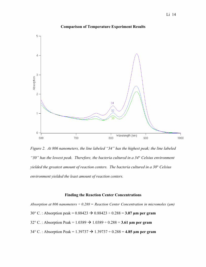
Comparison of Temperature Experiment Results
Figure 2. At 806 nanometers, the line labeled “34” has the highest peak; the line labeled
“30” has the lowest peak. Therefore, the bacteria cultured in a 34° Celsius environment
yielded the greatest amount of reaction centers. The bacteria cultured in a 30° Celsius
environment yielded the least amount of reaction centers.
Finding the Reaction Center Concentrations
Absorption at 806 nanometers ÷ 0.288 = Reaction Center Concentration in micromoles (µm)
30° C. : Absorption peak = 0.88423 0.88423 ÷ 0.288 = 3.07 µm per gram
32° C. : Absorption Peak = 1.0389 1.0389 ÷ 0.288 = 3.61 µm per gram
34° C. : Absorption Peak = 1.39737 1.39737 ÷ 0.288 = 4.85 µm per gram

Li 15
Comparison of Time Experiment Results
Figure 3. At 806 nanometers, the line labeled “4 days” has the highest peak and the line
labeled “2 days” has the lowest peak. Therefore, the bacteria cultured for four days
yielded the greatest amount of reaction centers. The bacteria cultured for two days
yielded the least amount of reaction centers.
Finding the Reaction Center Concentrations
Absorption at 806 nanometers ÷ 0.288 = Reaction Center Concentration in micromoles (µm)
2 Days: Absorption peak = 0.12648 0.12648 ÷ 0.288 = 0.44 µm per gram
3 Days: Absorption Peak = 0.2358 0.2358 ÷ 0.288 = 0.819 µm per gram
4 Days: Absorption Peak = 0.23931 0.23931 ÷ 0.288 = 0.831 µm per gram

Li 16
The table below summarizes the amount of polyhistidine-tagged proteins yielded.
Summary of Results
Conditions Received by Rb.
sphaeroides
Amount of Polyhistidine-tagged Proteins Yielded
Light Amount of polyhistidine-tagged protein is undetermined
No light 4.85 micromoles per gram/0.73 grams per milliliter
Some light 1.42 micromoles per gram/0.213 grams per milliliter
30° Celsius, no light 3.07 micromoles per gram/0.4605 grams per milliliter
32° Celsius, no light 3.61 micromoles per gram/0.5415 grams per milliliter
34° Celsius, no light 4.85 micromoles per gram/0.73 grams per milliliter
No light, no oxygen Died, no polyhistidine-tagged proteins yielded.
Light and no oxygen Amount of polyhistidine-tagged protein is undetermined
Two days, no light 0.44 micromoles per gram/0.066 grams per milliliter
Three days, no light (Control) 0.819 micromoles per gram/0.123 grams per milliliter
Four days, no light 0.831 micromoles per gram/0.125 grams per milliliter
Figure 4 . This table shows the amount of polyhistidine-tagged proteins yielded. The
bacteria that received “no oxygen with no light” died. The bacteria that were cultured
for three days with no light is the control in this experiment. The bacteria cultured with
no light in a 34 degrees Celsius environment yielded the greatest amount of
polyhistidine-tagged proteins (4.85 micromoles per gram). The bacteria cultured without
light for two days yielded the least amount of polyhistidine-tagged proteins (0.44
micromoles per gram).

Li 17
Comparison of Data
Figure 5. This graph is a visual representation of the data. The condition that yielded
the greatest amount of reaction centers (polyhistidine-tagged proteins) can clearly be
seen. The bacteria that were cultured with no light and 34° Celsius yielded the same
amount of reaction centers, as well as the greatest amount of reaction centers. The
bacteria cultured for two days yielded the least amount of reaction centers.
Comparison of Results
0
1
2
3
4
5
6
SomeLight
NoLight
30°C.
32°C.
34°C.
2Days
3Days
4Days
Conditions Received by Rb. sphaeroides
Am
ou
nt
of
RC
s Y
ield
ed
(mic
rom
ole
s p
er
gra
m)

Li 18
Conclusion
The hypothesis for this experiment was that culturing Rb. sphaeroides with light
and oxygen for four days in a 32° Celsius environment will yield the greatest amount of
polyhistidine-tagged proteins. This hypothesis was refuted by this investigation because
the reaction center concentration of the bacteria cultured with light was undetermined. It
was unable to be determined because no polyhistidine-tagged proteins were yielded;
instead, another type of protein was yielded.
The bacteria cultured in a 30° Celsius environment yielded the least amount of
polyhistidine-tagged proteins, while the bacteria cultured in a 34° Celsius environment
yielded the greatest amount. The bacteria cultured in a 32° Celsius environment yielded
more polyhistidine-tagged proteins that the 30° Celsius one, but less than the 34° Celsius.
Therefore, the lower the temperature is, the fewer polyhistidine-tagged proteins will be
yielded. 34° Celsius was determined to be the best temperature to culture Rb.
sphaeroides.
In the time experiment, the bacteria cultured for four days yielded the greatest
amount of reaction centers, while the bacteria cultured for two days yielded the least
amount. The bacteria that were cultured for three days yielded more polyhistidine-tagged
proteins than the bacteria cultured for two days, but less than the four days bacteria.
Therefore, culturing the bacteria for four days will yield high amounts of polyhistidine-
tagged proteins.
In the oxygen experiment, the bacteria cultured without light and oxygen died.
The amount of polyhistidine-tagged proteins yielded by the bacteria cultured with light

Li 19
but no oxygen is undetermined. Therefore, depriving Rb. sphaeroides of oxygen does not
help in yielding more polyhistidine-tagged proteins.
As a conclusion, culturing Rb. sphaeroides without light for four days in a 34°
Celsius environment with oxygen will yield the greatest amount of polyhistidine-tagged
proteins.
Discussion
The Rb. sphaeroides cultured in a 34° Celsius environment yielded high amounts
of polyhistidine-tagged proteins because bacteria usually thrive in warm temperatures.
Warmth is an essential element for bacterial growth because heat speeds up chemical
reactions, and thus speeds up the reproductive rate of bacteria. The Rb. sphaeroides
cultured with 30° Celsius yielded the least amount of polyhistidine-tagged proteins
because the low temperature slows down chemical reactions; therefore, it also slows
down the reproductive rate of bacteria.
The Rb. sphaeroides cultured for two days yielded the least amount of
polyhistidine-tagged proteins because the bacteria did not get enough time to mature; it is
still in its early stages of life, therefore, it cannot produce high amounts of proteins. The
bacteria cultured for four days had enough time to mature and absorb nutrients; as a
result, it was able to yield high amounts of polyhistidine-tagged proteins. However,
leaving the bacteria in the nutrient media for too long will lead to overgrowth, and
eventually death, because the bacteria will absorb all of the nutrients until the population
exceeds the carrying capacity allowed by the media, resulting in death.

Li 20
In the light experiment, the Rb. sphaeroides that received light turned into a
brown color, instead of the usual red color. The polyhistidine-tagged proteins from Rb.
sphaeroides are red; therefore, if polyhistidine-tagged proteins were yielded, the contents
in the flask should be red. However, the contents in the flask that received light was
brown, therefore, polyhistidine-tagged proteins were not yielded. A possible explanation
for this might be that the bacteria that received light yielded another type of protein. A
25 watt light bulb was placed in front of the incubator-shaker for about three days; the
bacteria received an abundance of light. Excessive light absorption might have damaged
the reaction centers and caused the bacteria to synthesize another type of protein (Zhang,
2005). The presence of light in the culturing process causes the formation of another type
of protein.
In the oxygen experiment, the bacteria cultured without light and oxygen died
because Rb. sphaeroides require light or oxygen to survive. With light, Rb. sphaeroides
can perform photosynthesis and make its own food. With oxygen, Rb. sphaeroides can
absorb nutrients from the media and break it down into energy by performing aerobic
respiration – the process in which cells use oxygen to break down food molecules and
nutrients to generate energy (Hunter, 2002). Therefore, Rb. sphaeroides can survive in
the presence of either light or oxygen. This explains why the Rb. sphaeroides cultured
without light and oxygen died; it was unable to perform photosynthesis because it had no
light and it was unable to utilize the nutrients from the media through aerobic respiration
because there was no oxygen. The flask of bacteria that received light but no oxygen
turned into a brown color. The results were similar to the flask of bacteria discussed in

Li 21
the light experiment; both of them turned brown because another type of protein was
synthesized.
***
All of the bacteria yielded more polyhistidine-tagged proteins than the control,
expect for the bacteria cultured for two days with no light. The control (three days, no
light), yielded 0.819 micromoles of polyhistidine-tagged proteins per gram. The bacteria
cultured for two days with no light, yielded 0.44 micromoles of polyhistidine-tagged
proteins per gram, which is less than the control.
***
There weren’t many errors in this experiment; however, there were certain aspects
of the experiment that were supposed to be kept constant. The amount of Rb.
sphaeroides added into each media bottle was supposed to be 25 microliters throughout
the experiment. However, 25 microliters of Rb. sphaeroides took too long to culture; it
took about one week for it to be ready for harvesting. Therefore, after the light
experiment, the amount of Rb. sphaeroides added into each media bottle was changed to
200 microliters, in order to speed up the culturing process. The amount of bacteria added
into each media bottle was supposed to be a constant; it should not have been changed,
but due to time constraints, it had to be altered. Altering this constant made this
investigation a little less scientific; it might have affected the results and made them not
as accurate.
The amount of time spent on the culturing process should also be kept constant –
three days. Three days is the average amount of time it takes for the bacteria to turn red,
or ready for harvesting. However, the bacteria were harvested according to the amount of

Li 22
time it takes to turn red, which varied from flask to flask. Altering the amount of time
spent on the culturing process can greatly affect the results because allowing the bacteria
to stay one more day in the nutrient media can result in more polyhistidine-tagged
proteins being yield.
If this experiment were to be performed again, the amount of Rb. sphaeroides
added into the media should be kept constant (25 microliters) and the time spent on the
culturing process should also be kept constant (three days).
***
The conclusion established by this investigation is not totally accurate or reliable
because this experiment was only performed once. Due to the amount of time allowed,
this experiment was unable to be performed a second time because this whole experiment
took about five months to complete; it would take another five months to perform this
experiment again. If the results of this experiment can be replicated, then the conclusion
established is accurate and reliable. Unfortunately, this cannot be done because of time
restraints.
***
Polyhistidine-tagged proteins are used as the basis of photosynthetic research;
scientists who conduct experiments on photosynthesis frequently use polyhistidine-
tagged proteins as photosynthetic models. Therefore, many experiments and
investigations can benefit from the conclusion established by this experiment. Scientists
should follow this method – culture Rb. sphaeroides without light in a 34° Celsius
environment with oxygen for four days if they want to perform photosynthetic research
with polyhistidine-tagged proteins.

Li 23
After this experiment, some questions that were aroused include: How does pH
affect the growth of Rb. sphaeroides? Is there a catalyst that can be added to the media
and bacteria during the culturing process to speed up the reproductive rate of Rb.
sphaeroides?
Since photosynthesis requires light, why weren’t any photosynthetic reaction
center yielded in the flask of bacteria that received light? Why was another type of
protein synthesized instead?
Li 24
Acknowledgements
I would like to acknowledge and thank the following people:
- Dr. Marilyn Gunner, professor of biophysics at the City College of the City
University of New York for allowing me to work at her biophysics science lab.
- Jennifer Madeo, biomedical engineer at the City College of New York for
helping clarify difficult biophysics questions.
- Xinyu Zhang, my mentor and biophysicist at the City College of New York for
her time and support. Not only did Xinyu supply me with all the materials and
equipment to perform this experiment, she also mentored me through this
experiment.
- Annie Chien, my science teacher and exhibition sponsor for hooking me up with
Xinyu. I am grateful for all the time Ms. Chien spent with me to revise and
improve this lab report.
- All of the staff at the Robert E. Marshak Science Building at the City College
of New York, especially Dr. David Calhoun, professor of chemistry and Sheila
Ehlinger for allowing me to use the French Press, Centrifuge and Autoclave
Machines at their labs.
- Brian Rayman, Chau Ngo, and Charlotte North, my math teacher and 9th grade
humanities teachers for taking their time to read over this lab report and giving me
constructive criticism on how to improve this paper.

Li 25
References
California Polytechnic State University, (n.d.). Retrieved Jan. 30, 2005, from
Photosynthesis Web site: http://www.calpoly.edu/~spuhl/.
Gregory, R. (1989). Biochemistry of Photosynthesis. 3rd ed. New York: John Wiley &
Sons Ltd.
Gust, D. (1996). Retrieved Mar. 19, 2005, from Why Study Photosynthesis? Web site:
http://photoscience.la.asu.edu/photosyn/study.html.
Hall, D. (1994). Photosynthesis: Studies in Biology. 5th ed. Ontario: Cambridge
University Press.
Hallman, R. (2000). The Living Environment: Biology. New York: Amsco School
Publications, Inc.
Hunter, S. (2002). Let's review: Biology-The Living Environment. 3rd ed. Hauppauge,
NY: Barron's Educational Series, Inc.
Kaplan, Samuel. "Significance of Rb. sphaeroides." E-mail to Timmy Li. Feb. 15,
2005.
Vermaas, W. (2004). Retrieved Jan. 30, 2005, from An Introduction to Photosynthesis
and Its Applications Web site:
http://photoscience.la.asu.edu/photosyn/education/photointro.html.
Zhang, S. (2004). Retrieved Mar. 20, 2005, from Electronics powered with
photosynthesis Web site: http://news.ninemsn.com.au/article.aspx?id=19008
Zhang, Xinyu. “Analysis and Questions about the Results.” E-mail to Timmy Li. May
12, 2005

Li 26
Appendix A
Complete List of Materials (with unfamiliar equipment explained)
1) Yeast extract
2) Casamino acids
3) Solution C (a mixture of various
irons and metals)
4) Rhodobacter sphaeroides
5) Distilled water
6) pH meter (apparatus used to measure
the pH of substances)
7) Hydrochloric acid (one molar
concentration)
8) Sodium hydroxide (one molar
concentration)
9) Autoclave machine (apparatus used
to sterilize solutions and substances
by using high temperature)
10) Centrifuge (apparatus used to
separate substance of different
densities)
11) French Press Machine (apparatus
that uses high pressure to break cell
membranes)
12) Ultraviolet spectrophotometer
(apparatus used to measure protein/
reaction center concentration)
13) Flash spectrophotometer (apparatus
used to measure the wavelength and
absorbance of a substance in its
excited state)
14) Digital balance (apparatus used to
measure the mass of substances)
15) Double beam balance (in this
experiment, the double beam balance
was used to ensure that the media
bottles had the same amount of
media and bacteria in it)
16) Pipettes (professional eye droppers
used to transfer solutions from one
place to another)
17) Sterilized disposable pipette tips
18) Non-sterilized disposable pipette tips
19) Eye droppers
20) Graduated cylinders
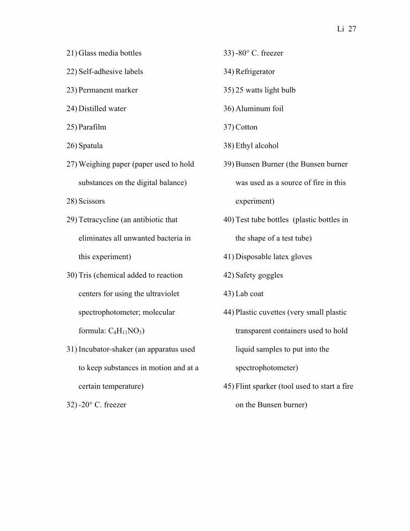
Li 27
21) Glass media bottles
22) Self-adhesive labels
23) Permanent marker
24) Distilled water
25) Parafilm
26) Spatula
27) Weighing paper (paper used to hold
substances on the digital balance)
28) Scissors
29) Tetracycline (an antibiotic that
eliminates all unwanted bacteria in
this experiment)
30) Tris (chemical added to reaction
centers for using the ultraviolet
spectrophotometer; molecular
formula: C4H11NO3)
31) Incubator-shaker (an apparatus used
to keep substances in motion and at a
certain temperature)
32) -20° C. freezer
33) -80° C. freezer
34) Refrigerator
35) 25 watts light bulb
36) Aluminum foil
37) Cotton
38) Ethyl alcohol
39) Bunsen Burner (the Bunsen burner
was used as a source of fire in this
experiment)
40) Test tube bottles (plastic bottles in
the shape of a test tube)
41) Disposable latex gloves
42) Safety goggles
43) Lab coat
44) Plastic cuvettes (very small plastic
transparent containers used to hold
liquid samples to put into the
spectrophotometer)
45) Flint sparker (tool used to start a fire
on the Bunsen burner)

Li 28
Appendix B
Concept Map of Methods
Rb. sphaeroides
High pressure andheat to sterilize themedia
involves making
Media
which uses
involves using a
the media and bacteriaare placed in theincubator-shaker
which is food for
AutoclaveMachine
the amount ofprotein can beinfluenced by
Temperature
after sterilization, Rb.sphaeroides is added intothe media
Centrifugation
Bacteria
Oxygen
Mediawhich separates the
Time
When the bacteriaready for harvesting,they undergo
from the
Light
After centrifugation, thebacteria is collected andplaced in the
Cell membrane
which usesto break thebacteria’s
After breaking the cellmembrane, the bacteria isplaced back into the
High pressure
French Press Machine
to separate the
From the
Polyhistidine-tagged Proteins
Cell membrane particles
Centrifuge Machine
Ultraviolet Spectrophotometer
To determine the
Which is placed in the
Reaction Center (polyhistidine-tagged protein) Concentration
Yielding Polyhistidine-tagged Proteins

Li 29
Appendix C
YCC Media Procedure
Note: The amount of ingredients in this procedure is intended for making 100 milliliters
of YCC media. To make more than 100 milliliters, determine the correct amount of
ingredients to use by using ratios.
1) Put a piece of weighing paper on the digital balance.
2) “Zero” the digital balance.
3) Use a speculum to transfer 0.5 grams of yeast extract onto the weighing paper.
4) Gently pour the 0.5 grams of yeast extract into a graduated cylinder by folding the
weighing paper into a spout.
5) Discard the weighing paper.
6) Put a new piece of weighing paper on the digital balance.
7) “Zero” the digital balance.
8) Use a new speculum to transfer 0.6 grams of Casamino acids onto the weighing
paper.
9) Gently pour the 0.6 grams of Casamino acids into the graduated cylinder with the
yeast extract.
10) Use a pipette with a non-sterilized tip to obtain 0.5 milliliters of solution C.
11) Instill the 0.5 milliliters of solution C into the graduated cylinder.
12) Fill the graduated cylinder up to 98 milliliters with distilled water.
13) Cut a piece of parafilm.

Li 30
14) Put the piece of parafilm over the graduated cylinder (wrap it tightly, make sure
the contents inside the graduated cylinder does not leak out).
15) Shake the graduated cylinder until all of the yeast extra, Casamino acids, and
Solution C are completely dissolved.
16) Calibrate the pH meter.
17) Adjust the pH of the contents inside of the graduated cylinder to 7.2 by using eye
droppers to add hydrochloric acid (to decrease the pH) or sodium hydroxide (to
increase the pH). (Note: use a separate eye dropper for the hydrochloric acid and
sodium hydroxide so the solutions don’t get contaminated).
18) After adjusting the pH of the contents in the graduated cylinder to 7.2, add
enough distilled water into the graduated cylinder to make the volume up to 100
milliliters.
19) Sterilize the media by following the autoclave procedure.

Li 31
Appendix D
Autoclave Procedure
1) Loosen the caps on the glass media bottles.
2) Put the glass media bottles in the autoclave plastic bin.
3) Put on autoclave gloves.
4) Open the autoclave machine door turning the knob counter clockwise.
5) Put the autoclave plastic bin with the glass media bottles into the autoclave
machine.
6) Close the door by turning the knob clockwise.
7) Set the time (45 minutes).
8) Press the “LIQUID” button.
9) After 45 minutes, the media is sterilized, but wait an additional 10 minutes to
allow the pressure to stabilize; after the pressure arrow reaches 0, open the door.
10) Take the media bottles out of the autoclave plastic bins.

Li 32
Appendix E
Procedure For Adding Tetracycline
1) Wipe the table with ethyl alcohol.
2) Get tetracycline out of the freezer.
3) Light-up the Bunsen burner.
4) Open the cap of one media bottle.
5) Hold the media bottle’s opening over the flame for 10 seconds.
6) Use a sterilized pipette tip to put 25 microliters of tetracycline into the media
bottle.
7) Hold the media bottle over the flame for 10 seconds.
8) Cap the media bottle back.

Li 33
Appendix F
Procedure For Adding Rb. Sphaeroides
1) Wipe the table with ethyl alcohol.
2) Get Rb. sphaeroides out of the freezer.
3) Light-up the Bunsen burner.
4) Open the cap of one media bottle.
5) Hold the media bottle’s opening over the flame for 10 seconds.
6) Use a sterilized pipette tip to put 25 microliters of Rb. sphaeroides into the media
bottle.
7) Hold the media bottle over the flame for 10 seconds.
8) Cap the media bottle back.

Li 34
Appendix G
Procedure For Using Centrifuge Machine
1) Distribute the flask of media and bacteria into 8 centrifuge bottles (only fill the
bottles up to 2/3 full).
2) Make sure the amount of media and bacteria in each centrifuge bottle are equal by
using the double beam balance.
3) Wipe the centrifuge machine and GSA rotor with alcohol.
4) Set the temperature to 4°C.
5) Set the speed to 9000 RPM (rotations per minute).
6) Set the rotor code to 10.
7) Set the time to 15 minutes.
8) Put the centrifuge bottles into the bottle compartments in the GSA rotor.
9) Lock the GSA rotor with the rotor top.
10) Wait until the centrifuge machine indicates that the temperature has lowered to
4°C.
11) After the temperature is down to 4°C, put the GSA rotor into the rotor
compartment of the centrifuge machine.
12) Close the door.
13) Press the “Start” button.
14) After 15 minutes is up, wait for another 5 minutes before opening the door.
15) Take the centrifuge bottles out of the GSA rotor.

Li 35
Appendix H
Ultraviolet Spectrophotometry Procedure
1) Prepare one milliliter of Tris with a pH of 8.
2) Prepare two ultraviolet cuvettes; put one milliliter of Tris in one, and one milliliter
of reaction centers in the other.
3) Cover the cuvette tops with parafilm.
4) Insert the cuvette with Tris (the will be the “baseline”) in the cuvette compartment
of the ultraviolet spectrophotometer.
5) Press “start” to determine the absorbance of the Tris.
6) After the absorbance is determined, take out the cuvette with Tris and put the
cuvette with the reaction centers in the cuvette compartment.
7) Press “auto-zero”.
8) Press “start”.

Li 36
Appendix I
French Press Procedure
1) Clean French Press Column with alcohol.
2) Uncap the French Press Column.
3) Pour sample into the French Press Column.
4) Cap the Column back.
5) Place the column into the French Press Machine.
6) Turn machine on.
7) Turn the Pressure Knob to “Medium”.
8) Wait 10 seconds.
9) Turn the Pressure Knob to “High”.
10) Place a centrifuge bottle in front of the output tube to collect bacteria.
11) After all of the bacteria have been collected, turn the Pressure knob to “Medium”.
12) Turn the Pressure Knob to “Low”.
13) Turn machine off.