weapons of mass destruction the health care professional’s role in preparation & response
TRANSCRIPT
WEAPONS OF MASS DESTRUCTION
The Health Care Professional’s Role in
Preparation & Response
Course Objectives• At the conclusion of this program, participants will be able to:• Define the term weapons of mass destruction (WMD) and categorize the types of WMD most likely to be used in acts of terrorism;• Discuss how acts of terrorism involving WMD have impacted the public health system and the practice of medicine;• Recognize the signs and symptoms of exposures to WMD; Utilize current treatment modalities for patients exposed to WMD;• Explain the health care practitioner’s role in syndromic surveillance and reporting; and use the Health Alert Network as an informational resource.• Identify personal protective equipment needed by health care practitioners when treating patients exposed to WMD;• Explain the procedures necessary to decontaminate a casualty.
Weapons of Mass Destruction The Threat
U.S. Threat increasing: Accessibility to these materials by an increasing number of potential adversaries:
International instability
Terrorist networks
Strength and perceived invulnerability of the United States’ homeland and military.
Changing dynamics of non-state violence.
Weapons of Mass Destruction The Threat
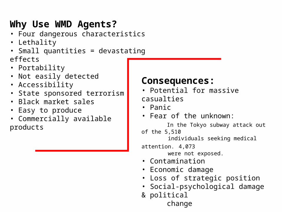
Why Use WMD Agents?• Four dangerous characteristics• Lethality• Small quantities = devastating effects• Portability • Not easily detected• Accessibility• State sponsored terrorism• Black market sales• Easy to produce• Commercially available products
Consequences:• Potential for massive casualties• Panic• Fear of the unknown: In the Tokyo subway attack out of the 5,510
individuals seeking medical attention. 4,073 were not exposed.
• Contamination• Economic damage• Loss of strategic position• Social-psychological damage & political change
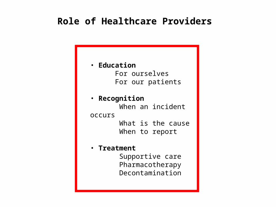
• Education For ourselves For our patients
• Recognition When an incident occurs What is the cause When to report
• Treatment Supportive care Pharmacotherapy Decontamination
Role of Healthcare Providers
Medical Aspects of
Nuclearand
Radiological Agents
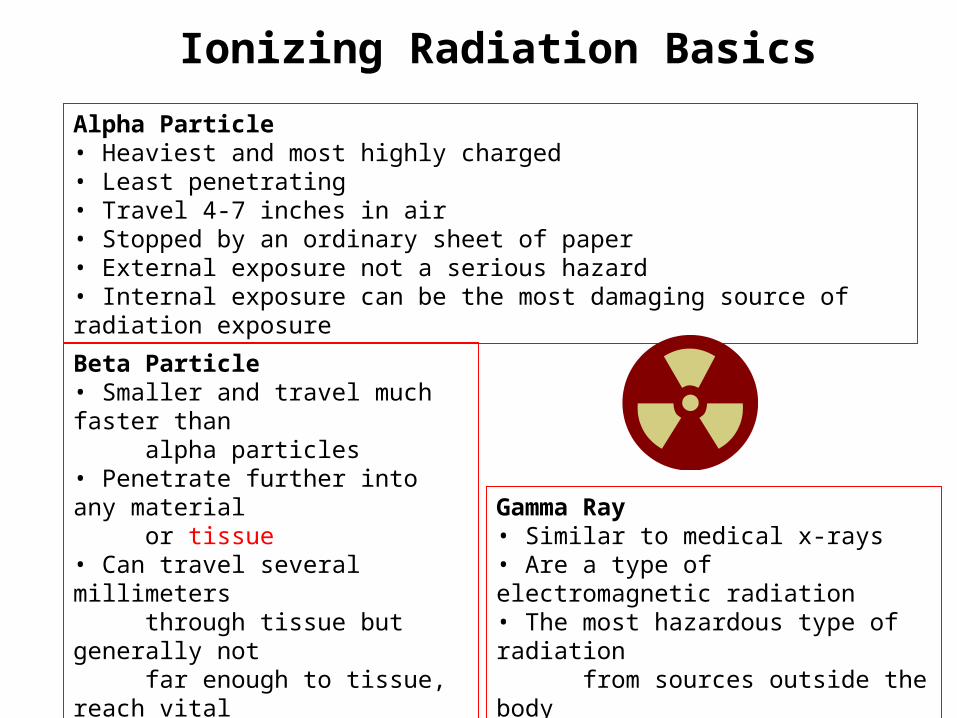
Ionizing Radiation Basics
Alpha Particle• Heaviest and most highly charged• Least penetrating• Travel 4-7 inches in air• Stopped by an ordinary sheet of paper• External exposure not a serious hazard• Internal exposure can be the most damaging source of radiation exposure
Beta Particle• Smaller and travel much faster than alpha particles• Penetrate further into any material or tissue• Can travel several millimeters through tissue but generally not far enough to tissue, reach vital organs• May be a major hazard when internal
Gamma Ray• Similar to medical x-rays• Are a type of electromagnetic radiation• The most hazardous type of radiation from sources outside the body• Greatest distances and penetration
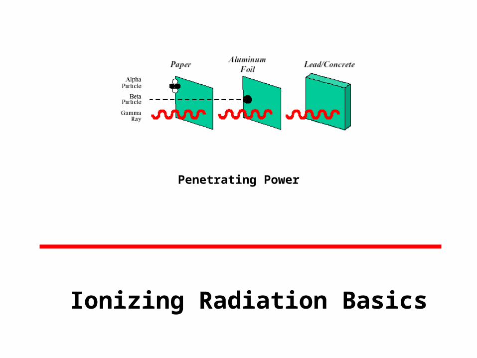
Penetrating Power
Ionizing Radiation Basics
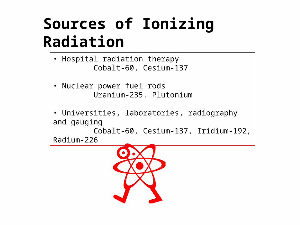
• Hospital radiation therapy Cobalt-60, Cesium-137
• Nuclear power fuel rods Uranium-235. Plutonium
• Universities, laboratories, radiography and gauging Cobalt-60, Cesium-137, Iridium-192, Radium-226
Sources of Ionizing Radiation
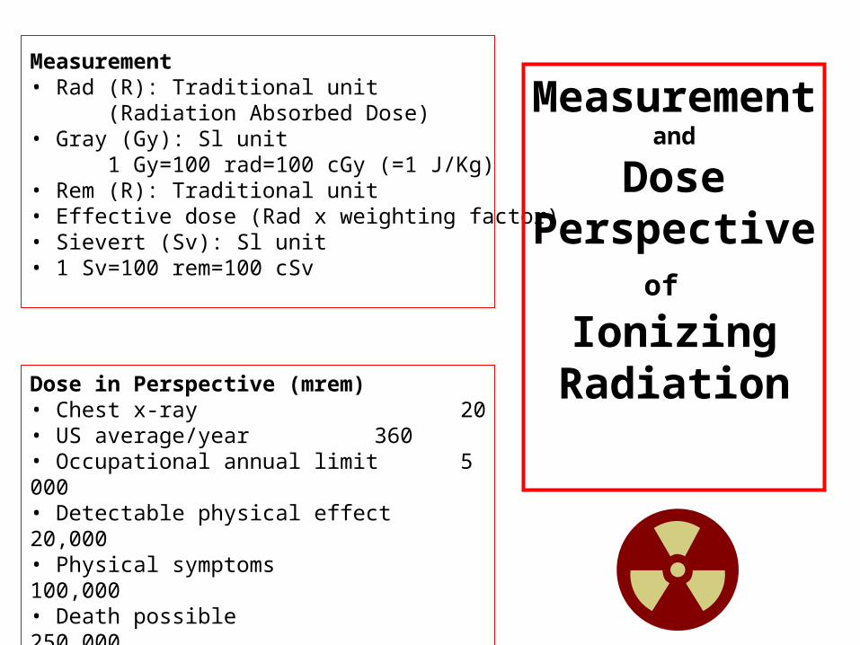
Measurement• Rad (R): Traditional unit (Radiation Absorbed Dose)• Gray (Gy): Sl unit 1 Gy=100 rad=100 cGy (=1 J/Kg)• Rem (R): Traditional unit• Effective dose (Rad x weighting factor)• Sievert (Sv): Sl unit• 1 Sv=100 rem=100 cSv
Dose in Perspective (mrem)• Chest x-ray 20• US average/year 360• Occupational annual limit 5 000• Detectable physical effect 20,000• Physical symptoms 100,000• Death possible 250,000• LD50 450,000
Measurementand
Dose Perspective
of Ionizing
Radiation
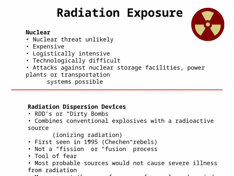
Radiation Exposure
Nuclear• Nuclear threat unlikely• Expensive• Logistically intensive• Technologically difficult• Attacks against nuclear storage facilities, power plants or transportation systems possible
Radiation Dispersion Devices• RDD’s or “Dirty Bombs”• Combines conventional explosives with a radioactive source (ionizing radiation)• First seen in 1995 (Chechen rebels) • Not a “fission” or “fusion” process• Tool of fear• Most probable sources would not cause severe illness from radiation• May prevent the use of an area for prolonged periods
• Radiation interacts with atoms, depositing energy and resulting in ionization
• Ionization can damage critical molecules or structures in a cell
• Directly hits a particularly sensitive atom or molecule in the cell
• Irreparable; the cell either dies or malfunctions
• Indirectly by interacting with water molecules
• Deposited energy leads to the creation of unstable, toxic hyperoxide molecules
Clinical Effects
Acute Radiation SicknessA combination of clinical syndromes occurring:
In stages (prodromal, latent, manifest illness)
During a period of hours to weeks after exposure, as injury to various tissues and organs isexpressed.
Hematopoietic Syndrome• Dose range: > 0.7 Gy• Effects: stem cells of all marrow cell lines• Prodromal Period Nausea, vomiting, anorexia, possible diarrhea Onset at 3-24 hours, duration <48 hours Severity = increases with dose• Latent Period Mostly asymptomatic, except mild weakness 3-4 weeks Hair loss/weight loss, about day 14 Manifest Illness Onset 3-5 weeks Bone marrow atrophy hemorrhage and infection
Clinical Effects

Gastrointestinal Syndrome• Dose range: >6 Gy• Effects: Gl stem cells and small vessels Prodromal Period Severe N/V diarrhea fever in 1-4 hours• Latent Period 5 – 7 days Manifest Illness Paralytic ileus, bloody diarrhea, severe vomiting, shock, sepsis
Cardiovascular / CNS Syndrome• Dose: >20 Gy• Effects: small blood vessels, especially in brain• Cerebral edema, death 2-3 days
Clinical Effects
Triage• By conventional injuries• Trauma• Burns• By radiation injury• Prodromal symptoms• N/V < / > 4 hours is the yardstick• Hematologic picture
Treatment• Standard emergency medical procedures• Decontaminate AFTER stabilized• Radiation injury NOT acutely life threatening• Supportive Care • Clean environment• Vascular integrity: IV fluids, blood products, stop losses• Prophylactic antibiotics• Definitive Treatments• Cytokines
Clinical Actions
Internal Contamination• Reduce intake and deposition• Increase elimination• Blocking and Diluting Agents• Kl, calcium, aluminum, barium, strontium salts• Chelating Agents• EDTA, DTPA, Deferoxamine, Penicillamine, Prussian Blue
Dosimetry• Initially• RADIACs• Nasal swabs (~5% of lung deposition)• Skin appearance• CBC q 4-6 h• Later• Nuclear medicine equipment• Bioassay: Excretion Sampling• Baseline and 24-hour urine/stool collections (according to nuclide)
Clinical Actions

Medical Aspects of
Chemical Agents
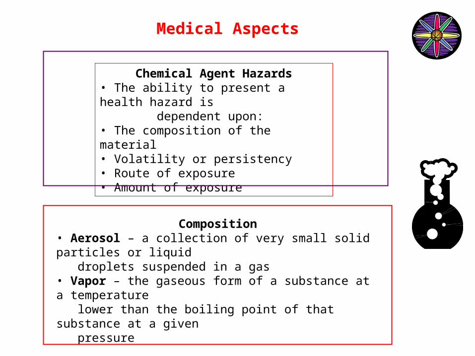
Chemical Agent Hazards• The ability to present a health hazard is dependent upon:• The composition of the material• Volatility or persistency• Route of exposure• Amount of exposure
Composition• Aerosol – a collection of very small solid particles or liquid droplets suspended in a gas• Vapor – the gaseous form of a substance at a temperature lower than the boiling point of that substance at a given pressure
Key Point: Aerosols settle faster than vapors
Medical Aspects
Volatility / Persistency
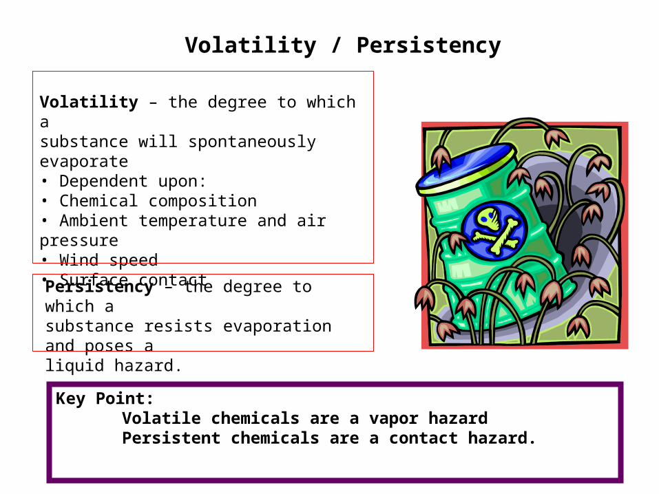
Volatility – the degree to which asubstance will spontaneously evaporate• Dependent upon:• Chemical composition• Ambient temperature and air pressure• Wind speed• Surface contact
Persistency – the degree to which asubstance resists evaporation and poses aliquid hazard.
Key Point: Volatile chemicals are a vapor hazard Persistent chemicals are a contact hazard.
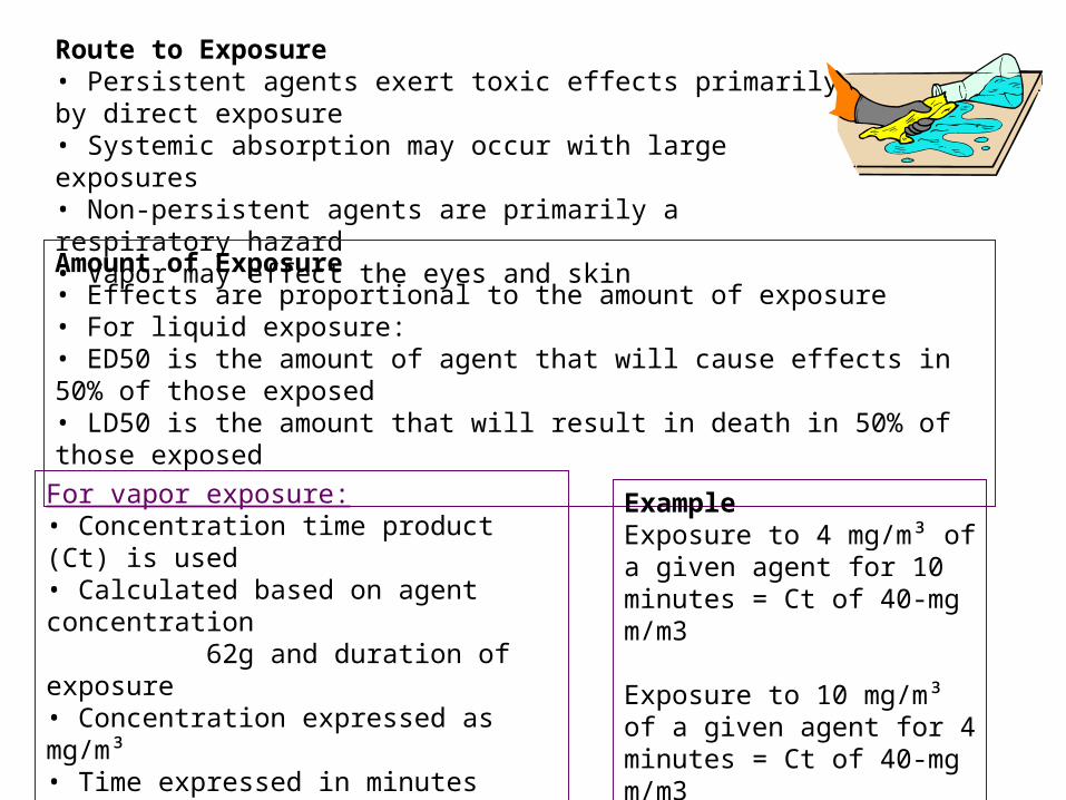
Route to Exposure• Persistent agents exert toxic effects primarily by direct exposure• Systemic absorption may occur with large exposures• Non-persistent agents are primarily a respiratory hazard• Vapor may effect the eyes and skin
Amount of Exposure• Effects are proportional to the amount of exposure• For liquid exposure:• ED50 is the amount of agent that will cause effects in 50% of those exposed• LD50 is the amount that will result in death in 50% of those exposed
For vapor exposure:• Concentration time product (Ct) is used• Calculated based on agent concentration 62g and duration of exposure• Concentration expressed as mg/m³• Time expressed in minutes• Does not take into account rate and depth of respiration
ExampleExposure to 4 mg/m³ of a given agent for 10 minutes = Ct of 40-mg m/m3
Exposure to 10 mg/m³ of a given agent for 4 minutes = Ct of 40-mg m/m3
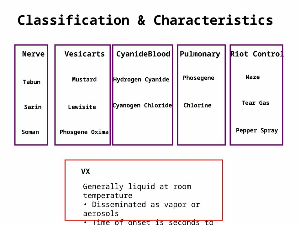
Classification & Characteristics
Nerve Vesicarts CyanideBlood Pulmonary Riot Control
Tabun
Sarin
Soman
Mustard
Lewisite
Phosgene Oxima
Hydrogen Cyanide
Cyanogen Chloride
Phosegene
Chlorine
Maze
Tear Gas
Pepper Spray
Generally liquid at room temperature• Disseminated as vapor or aerosols• Time of onset is seconds to hours
VX
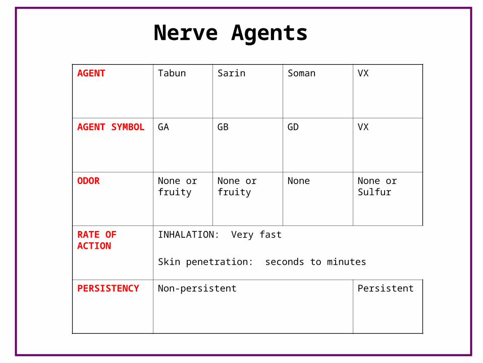
Nerve Agents
AGENT Tabun Sarin Soman VX
AGENT SYMBOL
GA GB GD VX
ODOR None or fruity
None or fruity None None or Sulfur
RATE OF ACTION
INHALATION: Very fast
Skin penetration: seconds to minutes
PERSISTENCY Non-persistent Persistent
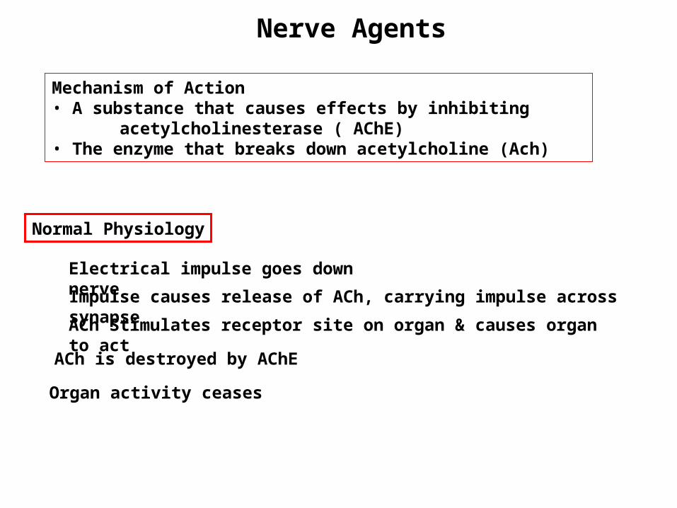
Mechanism of Action• A substance that causes effects by inhibiting acetylcholinesterase ( AChE)• The enzyme that breaks down acetylcholine (Ach)
Normal Physiology
Electrical impulse goes down nerve
Impulse causes release of ACh, carrying impulse across synapse
ACh Stimulates receptor site on organ & causes organ to act
ACh is destroyed by AChE
Organ activity ceases
Nerve Agents
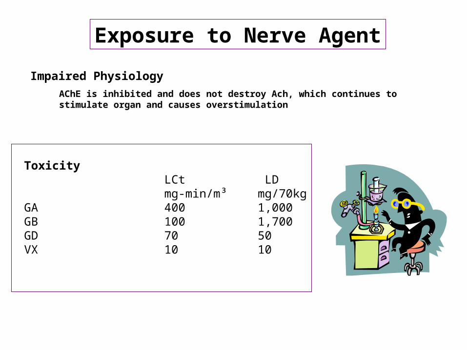
Exposure to Nerve Agent
Impaired Physiology
AChE is inhibited and does not destroy Ach, which continues to stimulate organ and causes overstimulation
ToxicityLCt LDmg-min/m³ mg/70kg
GA 400 1,000GB 100 1,700GD 70 50VX 10 10
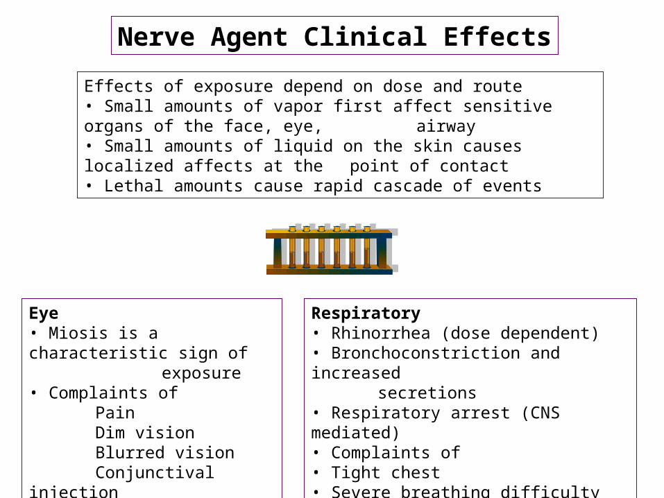
Nerve Agent Clinical Effects
Effects of exposure depend on dose and route• Small amounts of vapor first affect sensitive organs of the face, eye, airway• Small amounts of liquid on the skin causes localized affects at the
point of contact• Lethal amounts cause rapid cascade of events
Eye• Miosis is a characteristic sign of exposure• Complaints of Pain Dim vision Blurred vision Conjunctival injection
Respiratory• Rhinorrhea (dose dependent)• Bronchoconstriction and increased
secretions• Respiratory arrest (CNS mediated)• Complaints of• Tight chest• Severe breathing difficulty• Gasping, irregular breathing
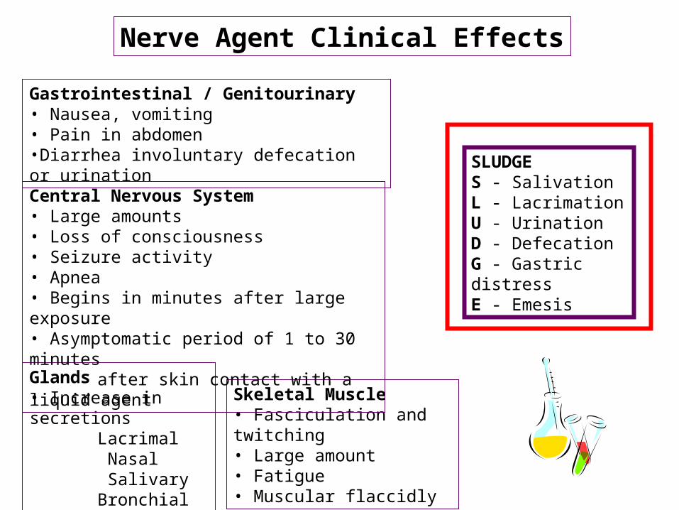
Gastrointestinal / Genitourinary• Nausea, vomiting• Pain in abdomen•Diarrhea involuntary defecation or urination
Glands• Increase in secretions Lacrimal Nasal Salivary Bronchial
Skeletal Muscle• Fasciculation and twitching• Large amount• Fatigue• Muscular flaccidly
Central Nervous System• Large amounts• Loss of consciousness• Seizure activity• Apnea• Begins in minutes after large exposure• Asymptomatic period of 1 to 30 minutes
after skin contact with a liquid agent
SLUDGES - SalivationL - LacrimationU - UrinationD - DefecationG - Gastric distressE - Emesis
Nerve Agent Clinical Effects

Laboratory Findings• Decreased RBC-ChE activity• Poor correlation between degree of enzyme inhibition and amount of
exposure or physical signs • Severe systemic effects generally indicate inhibition of 70 – 80%• Wide inter- and intrapersonal variability• Other laboratory findings will relate to complications
Medical ManagementManagement of a casualty with nerve agent intoxication consists of decontamination, ventilation, administration of antidotes, and supportive therapy. The condition of the patient dictates the need for each of these and the order in which they are instituted.
Ventilation• Requirement may last from 0.5 to 3 hours• Airway resistance is high (50-70cm of water)• Bronchoconstriction and secretions require vigorous pulmonary toilet
Antidotes• Atropine• Pralidoxime chloride•Benzodiazepenes
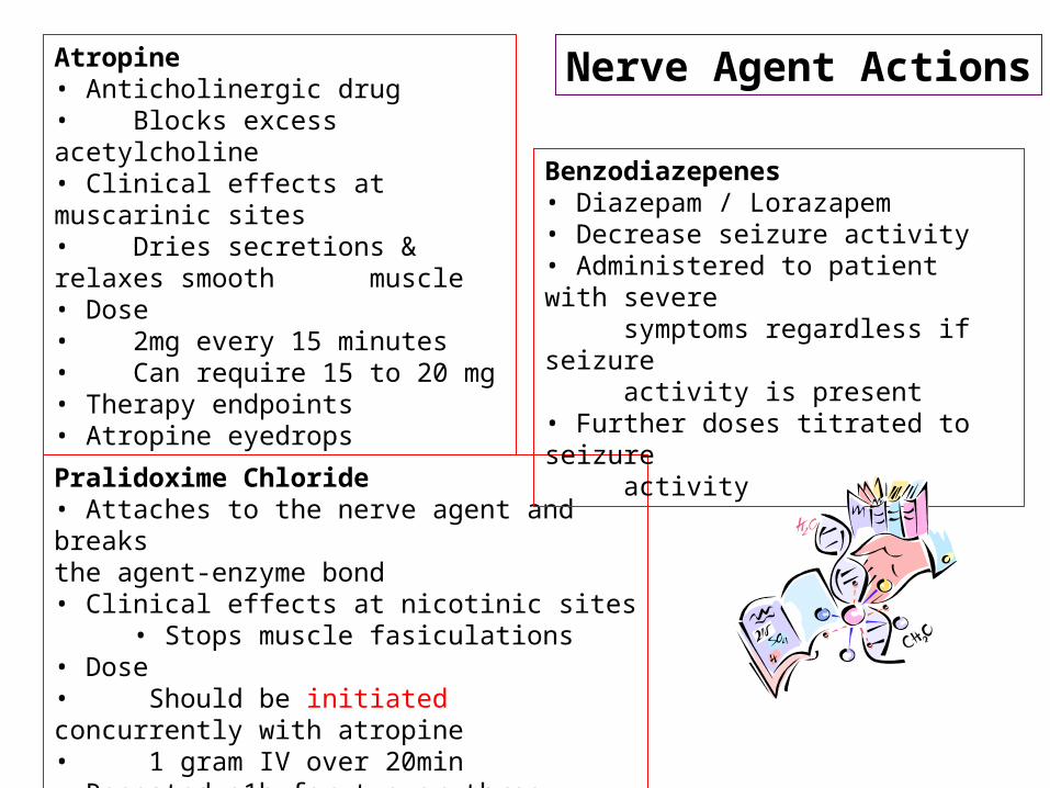
Atropine• Anticholinergic drug• Blocks excess acetylcholine• Clinical effects at muscarinic sites• Dries secretions & relaxes smooth muscle• Dose• 2mg every 15 minutes• Can require 15 to 20 mg• Therapy endpoints• Atropine eyedrops
Pralidoxime Chloride• Attaches to the nerve agent and breaksthe agent-enzyme bond• Clinical effects at nicotinic sites • Stops muscle fasiculations• Dose• Should be initiated concurrently with atropine• 1 gram IV over 20min• Repeated q1h for two or three additional doses
Benzodiazepenes• Diazepam / Lorazapem• Decrease seizure activity• Administered to patient with severe symptoms regardless if seizure activity is present• Further doses titrated to seizure activity
Nerve Agent Actions
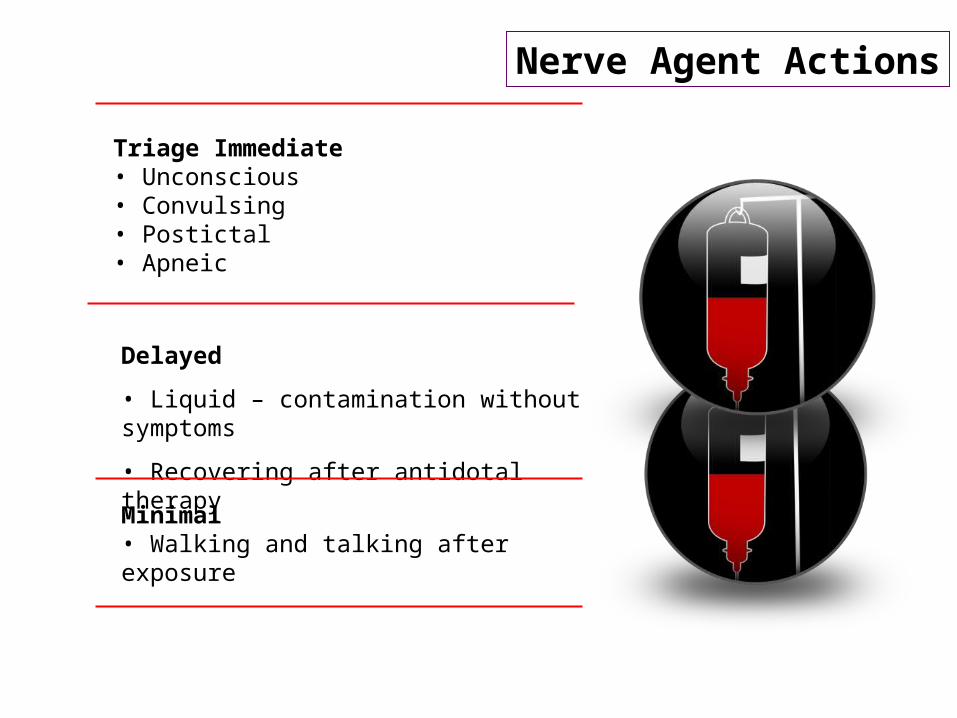
Triage Immediate • Unconscious • Convulsing • Postictal • Apneic
Minimal• Walking and talking after exposure
Delayed
• Liquid – contamination without symptoms
• Recovering after antidotal therapy
Nerve Agent Actions
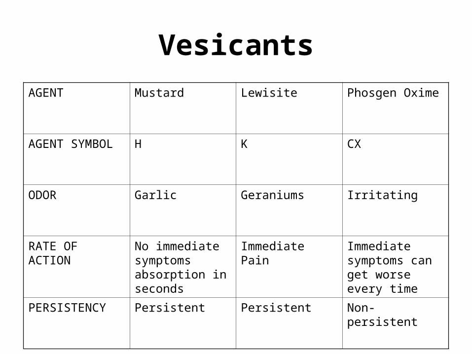
Vesicants
AGENT Mustard Lewisite Phosgen Oxime
AGENT SYMBOL H K CX
ODOR Garlic Geraniums Irritating
RATE OF ACTION
No immediate symptoms absorption in seconds
Immediate Pain Immediate symptoms can get worse every time
PERSISTENCY Persistent Persistent Non-persistent

Mechanism of Action
• Cause of death by interfering with DNA and cellular function (radiomimetic)
• Primarily a liquid threat may become vapor at higher temperatures
• Agents– Mustard (H, HD)– Lewisite (L)
Toxicity
• Through skin surfaces within 2 minutes– Cellular interaction: 1 to 2 minutes– Clinical effects: 2 to 48 hours (avg. 4-8)
• Penetration is enhanced by moisture, heat, and thin skin
Liquid• Blister 10μg• Death 100 mg/kg 7 gm/70 kg
Clinical Effects of Nerve Agents
• Have local and systemic effects
• Effects dependent on:– Ambient temperature and humidity– Site exposed
Skin
• Erythema (appears 2 – 48hrs)
• Small vesicles; later coalesce
• Blisters (12 – 24hrs)
• Blister fluid is clear, and does not contain mustard
• Weeks to months for complete healing
Clinical Effects of Nerve Agents
Airway
• Upper Airway– Burning & irritation of nose, sinuses & pharynx;
laryngitis and nonproductive cough– Damage to the trachea and upper bronchi lead to
productive cough• Lower Airway
– Increasingly severe productive cough– Distal airways & alveoli only affected in terminal
event: pulmonary edema not usually seen• Death by respiratory failure
– Mechanical obstruction & laryngospasm– Secondary pneumonia
Clinical Effects of Nerve Agents
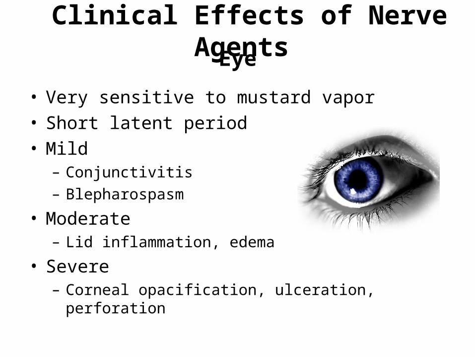
Eye
• Very sensitive to mustard vapor• Short latent period• Mild
– Conjunctivitis– Blepharospasm
• Moderate– Lid inflammation, edema
• Severe– Corneal opacification, ulceration, perforation
Clinical Effects of Nerve Agents
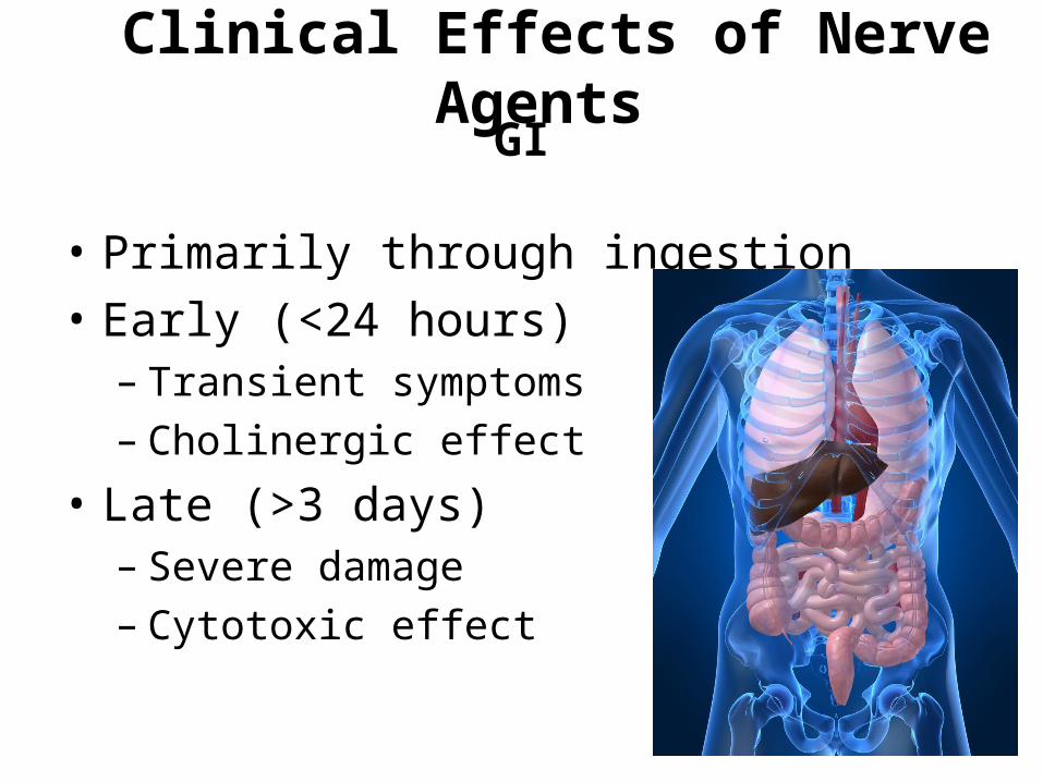
GI
• Primarily through ingestion
• Early (<24 hours)– Transient symptoms– Cholinergic effect
• Late (>3 days)– Severe damage– Cytotoxic effect
Clinical Effects of Nerve Agents

CNS
• CNS effects remain poorly defined
• Animal work demonstrates mustards are convulsants
• Several human case reports describe neurological effects with large amounts
Clinical Effects of Nerve Agents
Hematopoetic
• Bone marrow depression– Severe cases of skin and inhalation exposure– Usually irreversible
Clinical Effects of Nerve Agents

Diagnostic Studies
• Differential diagnosis includes contact dermatitis, drug eruption, or severe sunburn
• CBC
- Early leukocytosis followed by leukopenia
• Early chemical pneumonitis– Fever, WBC, chest x- ray
• Pneumonia: sputum exam / culture
Medical Management: Skin
• Soothing cream/lotion (0.25% camphor and menthol, calamine)
• Small blisters (under 1-2cm) left intact
• Systemic analgesics
• Appropriate IV fluids and electrolytes
Medical Management: Eyes
• Irrigation• Artificial tears• Topical mydriatics• Topical antibiotics• Vaseline on lid edges• Topical analgesics (nsaid drops)• Avoid topical anesthetics• Sunglasses
Medical Management: Airway
• Steam, cough suppressants• Oxygen• Bronchodilators, steroids• Early intubation• Assisted ventilation, early use of PEEP or
CPAP• Bronchoscopy• Antibiotics AFTER organism identified
Medical Management: GI
• Antiemetics
• Fluid therapy
• Electrolyte replacement
Antidotes
• British Anti-lewisite (BAL)– Initial dose: 0.5cc per 25 lbs IM (up to 4cc)– May be repeated at 4, 8, 12 hours after initial
dose
• Must consider toxicity of treatment– Hypertension– Nausea/vomiting
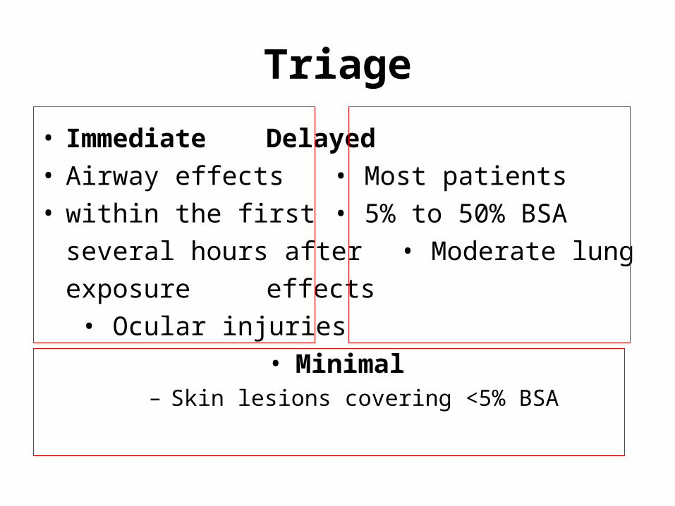
Triage
• Immediate Delayed• Airway effects • Most patients• within the first • 5% to 50% BSA
several hours after • Moderate lung
exposure effects
• Ocular injuries• Minimal
– Skin lesions covering <5% BSA
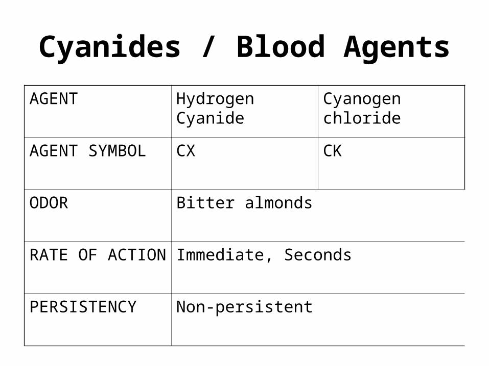
Cyanides / Blood Agents
AGENT Hydrogen Cyanide Cyanogen chloride
AGENT SYMBOL CX CK
ODOR Bitter almonds
RATE OF ACTION Immediate, Seconds
PERSISTENCY Non-persistent
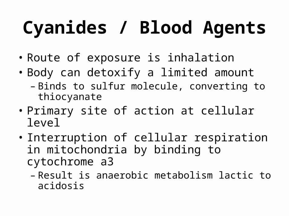
Cyanides / Blood Agents
• Route of exposure is inhalation• Body can detoxify a limited amount
– Binds to sulfur molecule, converting to thiocyanate
• Primary site of action at cellular level• Interruption of cellular respiration in
mitochondria by binding to cytochrome a3– Result is anaerobic metabolism lactic to
acidosis
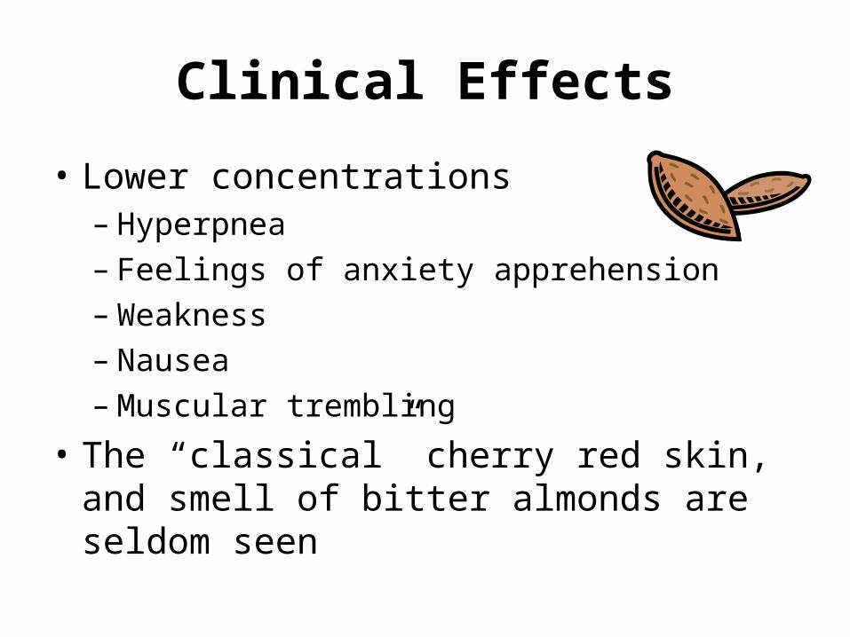
Clinical Effects
• Lower concentrations– Hyperpnea– Feelings of anxiety apprehension– Weakness– Nausea– Muscular trembling
• The “classical” cherry red skin, and smell of bitter almonds are seldom seen
Clinical Effects
• Higher concentrations
• Effects manifest primarily in the CNS
• Transient hyperpnea followed by convulsions ~ 15 seconds after exposure
• Respiratory activity ceases 2 – 3 minutes
• Cardiac activity ceases within 6 – 8 minutes
Diagnostic Studies
• Blood cyanide levels• Mild effects at 0.5 mcg/ml – 1.0 mcg/ml• Coma, convulsions and death at greater
than 2.5 mcg/ml• Anion gap acidosis• Venous blood gas analysis
• Higher than normal venous oxygen content
Medical Management
• General supportive therapy
• Ventilatory support
• Circulatory support (crystalloids/vasopressors)
• Treat metabolic acidosis (hyperventilation and bicardbonate administration)
• Specific antidotal therapy
• Cleaves cyanide-cytochrome a3 bond
• Sulfur donor
Antidotal Therapy
• Amyl nitrite• Given by inhalation by crushing vials
• 15 minutes on; 15 minutes off• Converts Hb02 (Fe2+) to metHb (Fe3+), but inhalation
leads to variable metHb levels • Sodium nitrite
• Converts Hb02 (Fe2+) to metHb (Fe3+)• 300 mg IV of a 3% soln (30 mg/mL) = 10
mL• Sodium thiosulfate
• Sulfur donor• 12.5 g IV of a 25% soln (250 mg/mL) = 50mL
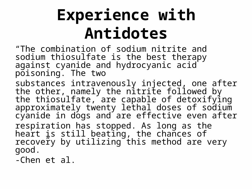
Experience with Antidotes
“The combination of sodium nitrite and sodium thiosulfate is the best therapy against cyanide and hydrocyanic acid poisoning. The twosubstances intravenously injected, one after the other, namely the nitrite followed by the thiosulfate, are capable of detoxifying approximately twenty lethal doses of sodium cyanide in dogs and are effective even afterrespiration has stopped. As long as the heart is still beating, the chances of recovery by utilizing this method are very good.”-Chen et al.
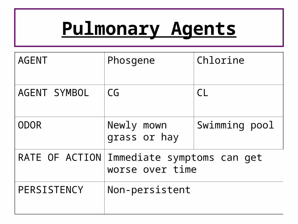
Pulmonary Agents
AGENT Phosgene Chlorine
AGENT SYMBOL CG CL
ODOR Newly mown grass or hay
Swimming pool
RATE OF ACTION Immediate symptoms can get worse over time
PERSISTENCY Non-persistent
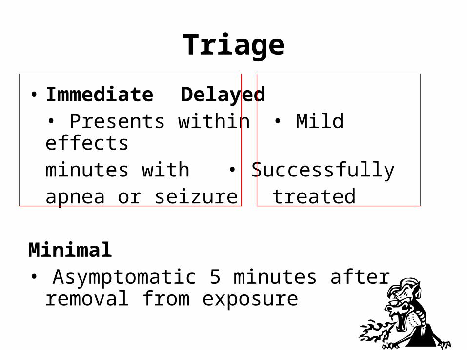
Triage
• Immediate Delayed• Presents within • Mild effects
minutes with • Successfullyapnea or seizure treated
Minimal• Asymptomatic 5 minutes after removal
from exposure
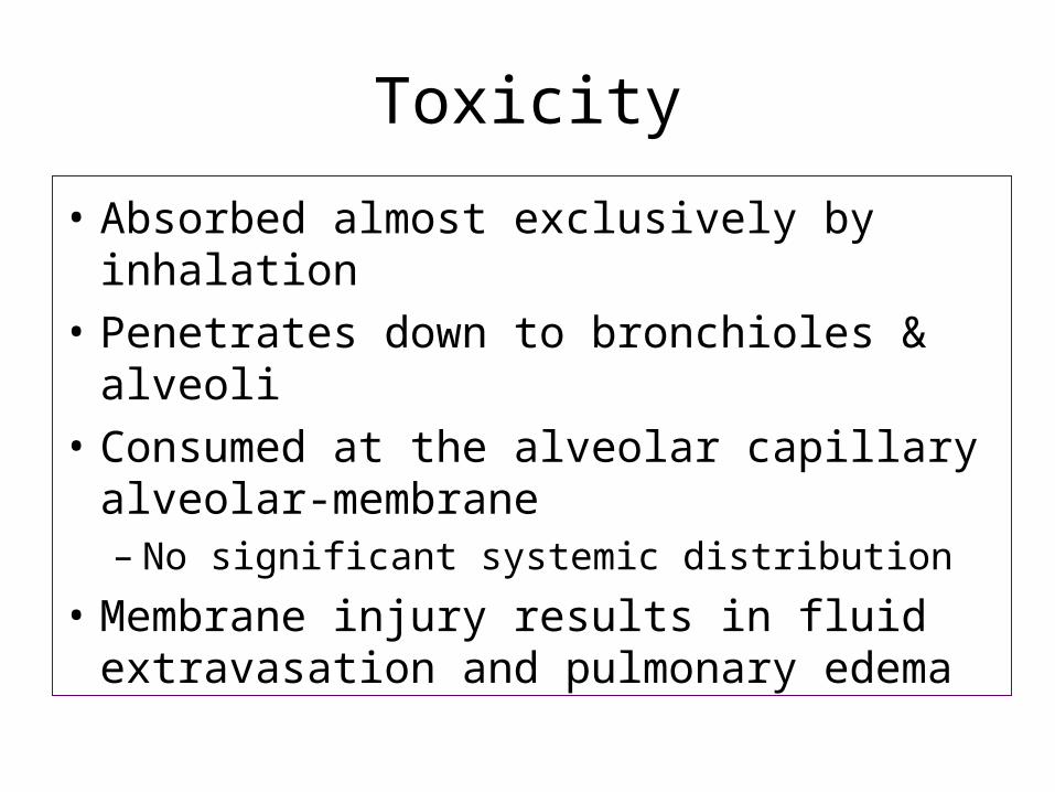
Toxicity
• Absorbed almost exclusively by inhalation
• Penetrates down to bronchioles & alveoli
• Consumed at the alveolar capillary alveolar-membrane– No significant systemic distribution
• Membrane injury results in fluid extravasation and pulmonary edema
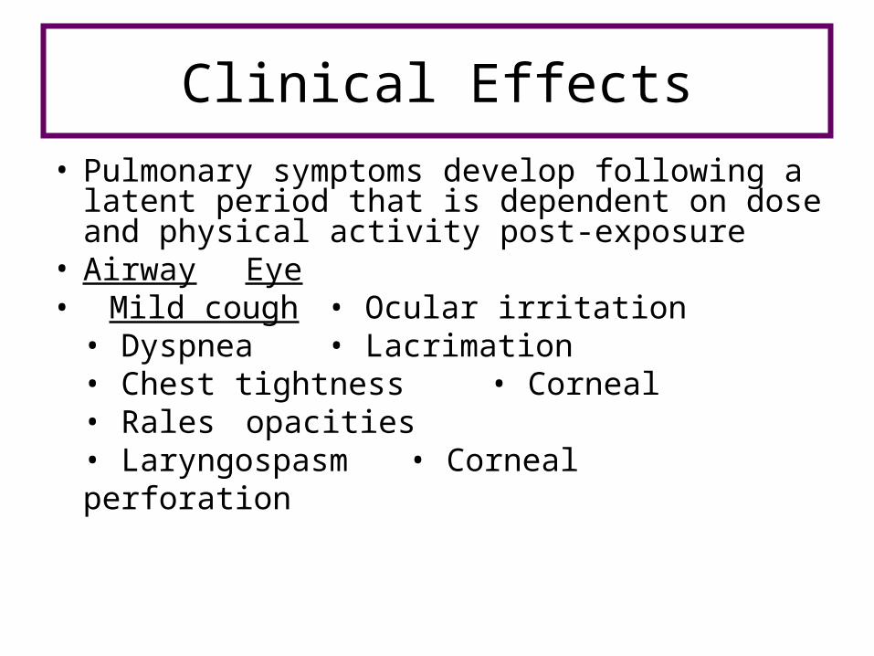
Clinical Effects
• Pulmonary symptoms develop following a latent period that is dependent on dose and physical activity post-exposure
• Airway Eye• Mild cough • Ocular irritation
• Dyspnea • Lacrimation• Chest tightness • Corneal• Rales opacities• Laryngospasm • Corneal
perforation
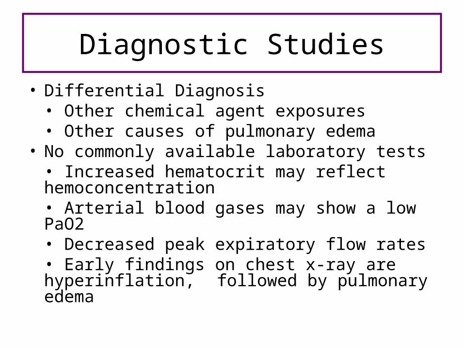
Diagnostic Studies
• Differential Diagnosis• Other chemical agent exposures• Other causes of pulmonary edema
• No commonly available laboratory tests• Increased hematocrit may reflect hemoconcentration• Arterial blood gases may show a low PaO2• Decreased peak expiratory flow rates• Early findings on chest x-ray are hyperinflation,
followed by pulmonary edema

Medical Management
• Supportive care• Strict bed rest• O2, PPV with PEEP / CPAP to maintain
PaO2• Bronchodilators for bronchospasm• IV fluids for hypotension (3rd spacing)• Surveillance cultures• Antibiotics when indicated
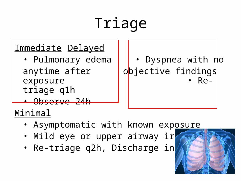
Triage
Immediate Delayed• Pulmonary edema • Dyspnea with no
anytime after objective findings exposure • Re-triage q1h
• Observe 24hMinimal
• Asymptomatic with known exposure• Mild eye or upper airway irritation• Re-triage q2h, Discharge in 12h
Summary
Weapons of Mass Destruction can include:
• Nuclear and Radiologic Agents
• Chemical Agents
• Nerve Agents
• Pulmonary Agents
Triage and Treatments
• Vary by type of agent and exposure