wagner syndrome: anatomic, functional and genetic ... ·...
TRANSCRIPT

GENETICS
WAGNER syndrome: anatomic, functional and geneticcharacterization of a Portuguese family
Joana R. Araújo1 & João Tavares-Ferreira1 & Sérgio Estrela-Silva1 & Paulo Rocha1 &
Elisete Brandão1 & Pedro Alves Faria1,2 & Fernando Falcão-Reis1,2 &
Amândio Rocha-Sousa1,2
Received: 14 April 2017 /Revised: 28 August 2017 /Accepted: 8 September 2017 /Published online: 25 October 2017# Springer-Verlag GmbH Germany 2017
AbstractPurpose To report the clinical (anatomic and functional) andgenetic findings of Wagner Syndrome (WS) in a Portuguesefamily.Methods Nine members of the family agreed to be examined.All had complete clinical eye examinations. The proband andselected patients underwent color fundus photography, spec-tral domain optical coherence tomography (SD-OCT), auto-matic static white-on-white computerized perimetry, and elec-trophysiology assessment (flash ERG, multifocal(mf) ERGand dark adaptometry). A pedigree was constructed basedon interviews with known affected subjects. Genomic DNA
samples derived from venous blood were collected from allaffected family members examined.Results Twenty-eight family members are affected. This fam-ily has the typical features of Wagner Syndrome, namely anempty vitreous cavity with veils, mild myopia and cataract.Four examined patients underwent vitreoretinal surgery due toabnormal peripheral vitreoretinal adhesions with peripheralretinal traction (n = 3). Retinal detachment was observed in5 of the examined subjects. Four of them occurred betweenthe ages of 5 and 15 years. Chorioretinal atrophy is also afrequent finding which results in moderate to severe visualfield and advanced rod-cone dystrophy from younger ages,also confirmed by absence of scotopic function on dark adap-tation. The macular dysfunction on mfERG was profound andof early onset. A heterozygous mutation in intron 7 of theVCAN gene (c.4004-1G > A) was found.Conclusions We described a rare autosomal dominantvitreoretinopathy with near complete penetrance in aPortuguese family. Abnormal peripheral vitreoretinal adhe-sions, retinal detachment and chorioretinal atrophy are presentinmost of the examined individuals at young ages. Early onsetof advanced visual field and electrophysiologic abnormalitieswere observed in this family. We also added relevant informa-tion to the literature by reporting our experience in surgicalmanagement of Wagner Syndrome patients with, and at riskof, retinal detachment.
Keywords Wagner syndrome .Hereditary vitreoretinopathy .
Stickler syndrome . VCAN . Retinal detachment
AbbreviationsWS Wagner SyndromeSD-OCT Spectral domain optical coherence
tomography
* Joana R. Araú[email protected]
João [email protected]
Sérgio [email protected]
Paulo [email protected]
Elisete Brandã[email protected]
Pedro Alves [email protected]
Fernando Falcã[email protected]; [email protected]
Amândio [email protected]; [email protected]
1 Ophthalmology Department, S. João Hospital, Porto, Portugal2 Department of Sense Organs, University of Porto, Porto, Portugal
Graefes Arch Clin Exp Ophthalmol (2018) 256:163–171DOI 10.1007/s00417-017-3800-0

ERG ElectroretinogramsISCEV International Society for Clinical
Electrophysiology of VisionBCVA Best corrected visual acuityIOL Intraocular lensPPV Pars plana vitrectomyRD Retinal detachmentNLP No light perceptionGAG Glycosaminoglycan
Introduction
Wagner syndrome is a rare, dominantly inherited vitreoretinopathywith near complete penetrance [1] first described in 1938by Wagner in a Swiss family [2]. The prevalence estimateof Wagner syndrome is less than 1:1,000,000.
The hallmark feature of affected individuals is an opticallyempty vitreous with strands, membranes and/or veils. Retinaldetachment (secondary to abnormal peripheral vitreoretinaladhesions), progressive night blindness, chorioretinal atrophy,myopia and pre-senile cataract are other common features. Nosystemic abnormalities have been described [3]. Severe visionloss can occur and is due to progressive chorioretinal atrophyand/or retinal detachment. Retinal detachment varies from afew percentage to 75% in some pedigrees [4].
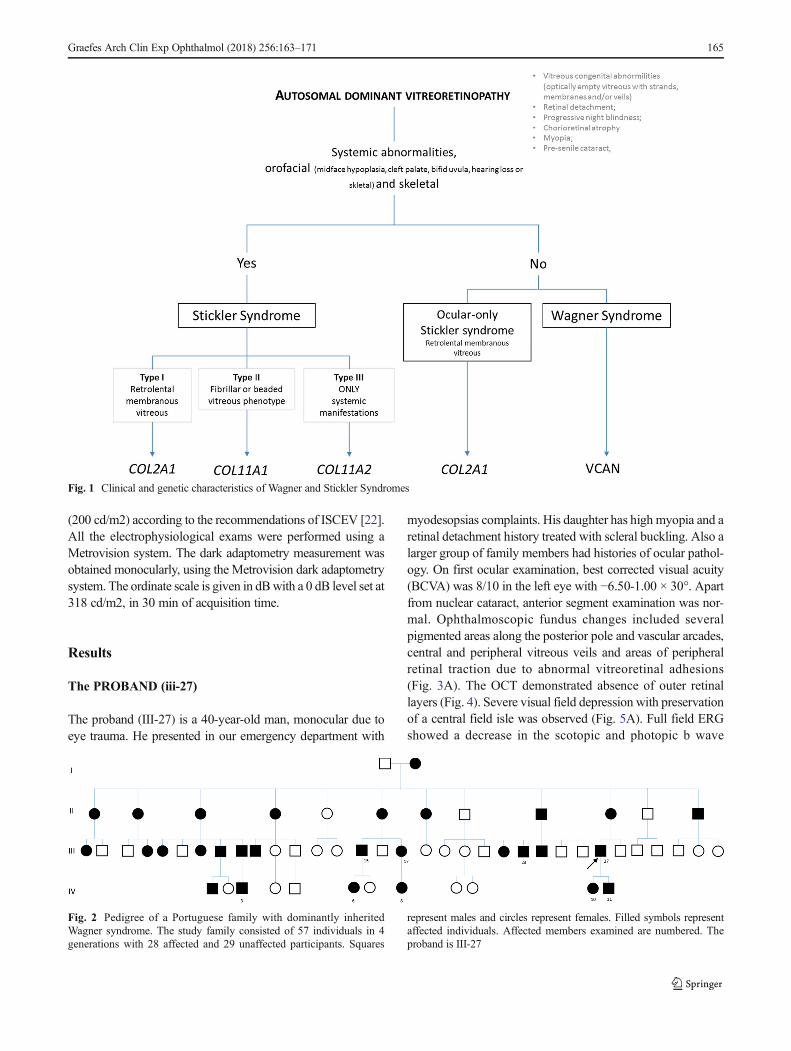
In 1965-1967 Stickler and associates described a connec-tive tissue dysplasia characterized by ocular findings, similarto those of Wagner syndrome, associated with orofacial andjoint problems, now known as Stickler Syndrome [5, 6]. Bothare vitreoretinopathies, which are disorders characterized byan abnormal vitreous gel structure and associated retinalchanges, and both are autosomal dominant. It can be particu-larly difficult to distinguish these two vitreoretinopathiesbased only on ocular phenotype, however vitreous phenotypecan help to distinguish subtypes of Stickler syndrome [7]. Thesystemic features, present only in Stickler syndrome, helpwith differentiation [8]. There is, however, a variant ofStickler syndrome devoid of systemic findings, the so-calledocular-only Stickler syndrome [9, 10]. These two syndromesare genetically distinct: (1) Wagner Syndrome is caused bymutation of the VCAN gene in chromosome 5q (previouslyknown as CSPG2); (2) Stickler Syndrome results from muta-tions in at least three collagen genes in chromosome 12q,mutation in COL2A1 gene being the most frequent [7, 8](Fig. 1). Jansen Syndrome and Erosive VitreoretinopathySyndrome are two other chromosome 5q retinopathies thatshare clinical and allelic features with Wagner syndrome[11–14]. Retinal detachment is predominant in JansenSyndrome [12]. Erosive vitreoretinopathy includes the clinicalfindings seen in Wagner disease, with the addition of progres-sive nyctalopia and visual field constriction due to a muchmore marked chorioretinal atrophy [14].
Versican is an extracellular matrix protein encoded by theVCAN gene and is a major component of vitreous [15], so islikely to be important in its structural integrity. Four transcript/protein isoforms of versican are known, which result from thealternative splicing of exons 7 and 8 and are found in manytissues including the eye [16, 17]. These two exons (7 and 8)contain the glycosaminoglycan attachment sites that supportchondroitin sulfate side chain aggregation. The glycoproteinbinds hyaluronate and link protein to form large aggregatesthat support vitreous integrity [7, 15]. Until now, all identifiedmutations of Wagner patients affect either the conserved ac-ceptor splice site of intron 7 or the donor splice site of intron 8of the VCAN gene [11, 16, 18–21].
This study describes a Portuguese family with WagnerSyndrome with mutation in the VCAN gene. A detailed fam-ily pedigree was constructed. Herein we describe anatomicand functional findings of this family with a rare disease.
Methods
This study describes a family whose proband had vitreousveils and signs of peripheral retinal traction due to abnormalperipheral vitreoretinal adhesions. A pedigree was constructedbased on interviews with known affected subjects and re-search of existing clinical records (Fig. 2). Eleven subjectswere examined, 9 affected and 2 unaffected. In addition tostandard ophthalmic history, health histories included ques-tions regarding systemic abnormalities, such as orofacial andskeletal abnormalities. The clinical evaluation includedSnellen equivalent visual acuity assessment, intraocular pres-sure, slit-lamp biomicroscopy and detailed fundus examina-tion. Genomic DNA samples derived from venous blood werecollected from all affected family members examined.
The proband and selected patients underwent color fundusphotography, spectral domain optical coherence tomography(SD-OCT HRA + OCT System Heidelberg Engineering,Heidelberg, Germany), automatic static white-on-whitecomputerized perimetry (program 30-2, SITA standard:Humphrey Instruments, Dublin, CA), and electrophysiologyassessment. Electrophysiological evaluation included full-fieldelectroretinograms (ERG) using a Ganzfeld dome, multifocalERG (mf ERG) and dark adaptometry curve assessment. TheERG testing was performed according to the protocol of theInternational Society for Clinical Electrophysiology of Vision(ISCEV) [22]. Briefly, under dilation and after dark adaptation(30 min), a dim white flash of 0.01-0.05 cd·s/m2 was used forthe scotopic (rod) response and a single white bright-flash (3 cd·s/m2) for the combined response. After light adaptation(10 min; 25 cd/m2), a brief white flash (3 cd·s/m2) wassuperimposed for the photopic response. The 30-Hz ERG wasobtained in the same conditions using a 30-Hz flickering stim-ulation. ThemfERGwas acquired using an array of 61 stimulus
164 Graefes Arch Clin Exp Ophthalmol (2018) 256:163–171
(200 cd/m2) according to the recommendations of ISCEV [22].All the electrophysiological exams were performed using aMetrovision system. The dark adaptometry measurement wasobtained monocularly, using the Metrovision dark adaptometrysystem. The ordinate scale is given in dBwith a 0 dB level set at318 cd/m2, in 30 min of acquisition time.
Results
The PROBAND (iii-27)
The proband (III-27) is a 40-year-old man, monocular due toeye trauma. He presented in our emergency department with
myodesopsias complaints. His daughter has high myopia and aretinal detachment history treated with scleral buckling. Also alarger group of family members had histories of ocular pathol-ogy. On first ocular examination, best corrected visual acuity(BCVA) was 8/10 in the left eye with −6.50-1.00 × 30°. Apartfrom nuclear cataract, anterior segment examination was nor-mal. Ophthalmoscopic fundus changes included severalpigmented areas along the posterior pole and vascular arcades,central and peripheral vitreous veils and areas of peripheralretinal traction due to abnormal vitreoretinal adhesions(Fig. 3A). The OCT demonstrated absence of outer retinallayers (Fig. 4). Severe visual field depression with preservationof a central field isle was observed (Fig. 5A). Full field ERGshowed a decrease in the scotopic and photopic b wave
Fig. 1 Clinical and genetic characteristics of Wagner and Stickler Syndromes
Fig. 2 Pedigree of a Portuguese family with dominantly inheritedWagner syndrome. The study family consisted of 57 individuals in 4generations with 28 affected and 29 unaffected participants. Squares
represent males and circles represent females. Filled symbols representaffected individuals. Affected members examined are numbered. Theproband is III-27
Graefes Arch Clin Exp Ophthalmol (2018) 256:163–171 165

amplitude, with a profound delay in both a and b wave implicittimes. The mfERG displayed an amplitude reduction and im-plicit time delay in both N1 and P1 waves, in all five ringanalyses. The patient has an absence of scotopic function,without its slope, in the dark adaptometry curve (Figs. 6 and 7).
Venous blood was collected for genomic DNA isolation anda heterozygous mutation in intron 7 of the VCAN gene(c.4004-1G > A) was found. This mutation results in the acti-vation of the cryptic downstream splice acceptor site of exon 8.
A myopic shift of −12 diopters(D) with decrease in left eyeBCVA (4/10 with -18D) occurred during the first follow-upyear due to nuclear cataract progression. Uncomplicated cat-aract surgery by phacoemulsification with placement of a pos-terior chamber intraocular lens (IOL) associated with prophy-lactic scleral buckling was performed. Until now the BCVAremains 9/10 and there is no evidence of retinal detachment.
Family clinical features
This family consisted of 4 generations with 28 affected and 29unaffected individuals (Figure 5). Of the 28 affected individ-uals, 9 underwent clinical examination and of the 29 unaffect-ed individuals, 2 underwent clinical examination. Membersexamined ranged from 6 to 40 years of age. The pertinentocular findings of all affected patients examined (n = 9) aresummarized in Table 1. None of the affected family membershad systemic clinical features of Stickler syndrome.
Anatomical features Empty vitreous cavity with veils, mildto highmyopia and cataract were consistent features present in
this family. Chorioretinal atrophy with pigmentation is also afrequent finding (Fig. 3B-C).
Abnormal peripheral vitreoretinal adhesions with peripher-al retinal traction, present in three eyes of three patients, led toperforming prophylactic scleral buckling procedures (patientIV-11, III-15, and III-23). Subsequent additionally pars planavitrectomy (PPV) was needed in one patient to relieve thetraction (patient III-15). Until now (follow-up time rangesfrom 1 to 2 years) there is no evidence of retinal detachment.
Five cases had retinal detachment (RD), four of them occur-ring between 5 and 15 years of age. Patient IV-10 had left eyeinferior rhegmatogenous RD at 9 years of age and was treatedwith scleral buckling. An asymptomatic nasal retinal detach-ment was diagnosed in a child (IV-6) and scleral buckling wasperformed. Patients III-23 and III-15 were treated for RD byscleral buckling and vitrectomy, respectively. Until now all pa-tients are stable with no retinal detachment. Patient IV-3 had lefteye congenital tractional RD with no light perception (NLP).
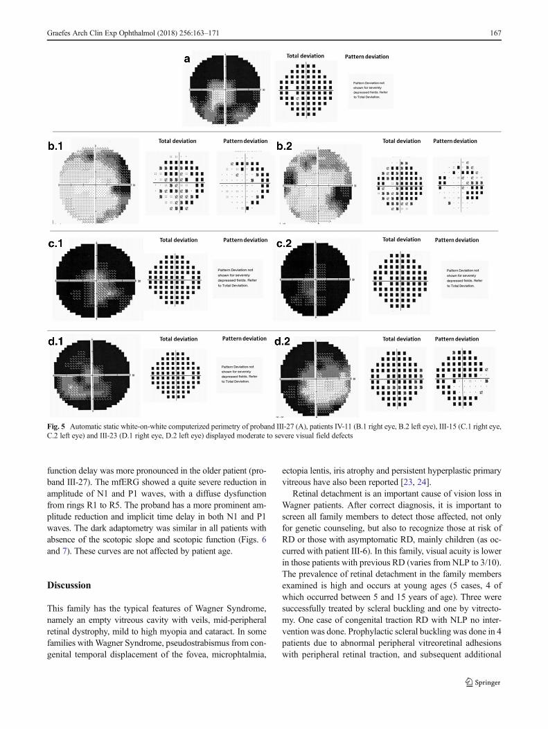
Functional features Visual acuity ranges from NLP to 9/10.In patients with previous RD, visual acuity ranges from NLPto 3/10. Some patients preserve relatively good central visualacuity but have moderate to severe constriction of the visualfield (n = 3) (Fig. 5B-D).
Electrophysiological evaluation was performed in 4 patients(Proband III-27, IV-6, IV-10, IV-11) showing similar function-al changes. The full field ERG displayed a decrease in thescotopic and photopic b wave amplitude, with a profound de-lay in both a and b wave implicit times. Patient IV-10 has aprofound decrease in photopic a and b waves, while the otherswere more affected in their scotopic function. The photopic
b.2
a
Fig. 3 Fundus photographs ofthe proband III-27 left eye (A),patient IV-6 (B.1 left eye, B.2right eye) and III-23 right eye (C)show peripheral vitreous veils(arrows) with underlying retinalpigmentary (arrowheads) andareas of chororetinal atrophy(stars)
Fig. 4 Proband III-27 SD OCTimaging of the left eye showingabsence of outer retinal layers
166 Graefes Arch Clin Exp Ophthalmol (2018) 256:163–171
function delay was more pronounced in the older patient (pro-band III-27). The mfERG showed a quite severe reduction inamplitude of N1 and P1 waves, with a diffuse dysfunctionfrom rings R1 to R5. The proband has a more prominent am-plitude reduction and implicit time delay in both N1 and P1waves. The dark adaptometry was similar in all patients withabsence of the scotopic slope and scotopic function (Figs. 6and 7). These curves are not affected by patient age.
Discussion
This family has the typical features of Wagner Syndrome,namely an empty vitreous cavity with veils, mid-peripheralretinal dystrophy, mild to high myopia and cataract. In somefamilies with Wagner Syndrome, pseudostrabismus from con-genital temporal displacement of the fovea, microphtalmia,
ectopia lentis, iris atrophy and persistent hyperplastic primaryvitreous have also been reported [23, 24].
Retinal detachment is an important cause of vision loss inWagner patients. After correct diagnosis, it is important toscreen all family members to detect those affected, not onlyfor genetic counseling, but also to recognize those at risk ofRD or those with asymptomatic RD, mainly children (as oc-curred with patient III-6). In this family, visual acuity is lowerin those patients with previous RD (varies from NLP to 3/10).The prevalence of retinal detachment in the family membersexamined is high and occurs at young ages (5 cases, 4 ofwhich occurred between 5 and 15 years of age). Three weresuccessfully treated by scleral buckling and one by vitrecto-my. One case of congenital traction RD with NLP no inter-vention was done. Prophylactic scleral buckling was done in 4patients due to abnormal peripheral vitreoretinal adhesionswith peripheral retinal traction, and subsequent additional
Fig. 5 Automatic static white-on-white computerized perimetry of proband III-27 (A), patients IV-11 (B.1 right eye, B.2 left eye), III-15 (C.1 right eye,C.2 left eye) and III-23 (D.1 right eye, D.2 left eye) displayed moderate to severe visual field defects
Graefes Arch Clin Exp Ophthalmol (2018) 256:163–171 167

PPV was needed in one patient to relieve the traction. Untilnow, all patients are stable.
Retinal detachment in Wagner Syndrome can be tractional[4, 11] or rhegmatogenous [16] and is caused by shrinkage ofthe preretinal membranes and the vitreous strands and veils. Inthe first reports, the retinal detachment in Wagner Syndromewas described in older ages [2, 4]. A recent report indicates that
detachment can occur earlier [20]. Also, in this family patientswith RD are young, which also supports the recent report.Prophylactic treatment for RD in patients with Wagner diseasehas not yet been well defined. More studies exist about effec-tiveness of prophylactic intervention in Stickler syndrome typeI, the most common inherited vitreoretinopathy. However, noconsensus exists: prophylactic cryopexy is performed by some
Fig. 6 Full field ERG, mfERG and dark adaptometry (DA) acquired onproband III-27 and patient IV-10, IV-11, and IV-6. In the ERG (leftcolumn). All have a rod-cone dystrophy, even the young ones. In thedark adaptometry (middle column) all have an absence of scotopic slop
and decrease of scotopic function. In the mfERG (right column) all thepatients have a profound retinal dysfunction, more severe in the proband(III-27)
Fig. 7 Ring analyses of the mf ERG N1 and P1 amplitude. There is a profound amplitude reduction in all rings, more pronounced in the older patient(proband III-27). The normal range is plotted with standard error bars
168 Graefes Arch Clin Exp Ophthalmol (2018) 256:163–171

Tab
le1.
Ocularclinicalfeatures
of9family
mem
berswith
WagnerSyndrom
e
Patient
Age
GenderEye
BCVA
Refractive
error
Error
spherical
equivalent,D
RecentP
ast
(Before
Surgery)
Lens
Status
Retinal
detachment
Age,y
Surgery
Optically
empty
vitreous
Vitreous
avascular
mem
brane
/veils
Retinal
tractio
nChorioretinal
atrophy
Retinal
Pigm
entary
changes
Ocular
alignm
ent
VisualF
ield
III-27
40M
OD
OS
9/10
NLPa
−1.75
−6.50NSC
No
CE/IOL+SB
Yes
Yes
Yes
Yes
Yes
NA
Severe
constrictio
n
IV-10
12F
OD
OE
9/10
1/10
−9.00
−12.00
NA
NA
Clear
Clear
No
Yes,9
years
No
SB
Yes
Yes
Yes
Yes
No
No
No
No
Yes
Yes
Ortho
Ortho
NA
NA
IV-11
14M
OD
OE
6/10
7/10
−8.00
−8.25
−7.50
−7.50Clear
Clear
No
No
No
SB
Yes
Yes
Yes
Yes
No
Yes
No
No
No
No
Ortho
Ortho
Bilateral
moderate
constrictio
n
III-15
34M
OD
OE
6/10
CF1M
−1.75
−0.25
NA
NA
PSC
PCIO
LNo
Yes,28
SB+CE/IOL+
PPV
CE/IOL+PPVb
Yes
Post-PP
VYes
Post-PPV
Yes
No
Yes
Yes
Yes
Yes
Ortho
Ortho
Bilateral
severe
constrictio
n
III-17
29F
OD
OE
6/10
4/10
−3.00
−1.25
NA
NA
Clear
Clear
No
No
No
No
Yes
Yes
Yes
Yes
No
No
Yes
Yes
Yes
Yes
PseudoXT
NA
NA
IV-6
6F
OD
OE
6/10
5/10
1.50
2.75
1.50
1.75
Clear
Clear
No
Yes,6
No
SB
Yes
Yes
Yes
Yes
Yes
Yes
Yes
Yes
Yes
Yes
XTc
ETc
NA
NA
III-23
24M
OD
OE
3/10
1/10
−0.50
−2.00
NA
NA
PCIO
LPC
IOL
Yes
d,
15years
No
SB+CE/IOLd
SB+CE/IOLd
Yes
Yes
Yes
Yes
No
No
Yes
Yes
Yes
Yes
XTd
Bilateral
Severe
constrictio
n
IV-3
15M
OD
OE
8/10
PL
−6.50
NA
NA
NA
Clear
PSC
No
Yes,6yearse
No
No
Yes
Yes
Yes
Yes
Yes
Yes
Yes
Yes
Yes
Yes
XT
NA
NA
IV-8
8F
OD
OE
5/10
6/10
−2,50
−2.75
NA
NA
Clear
Clear
No
No
No
No
Yes
Yes
Yes
Yes
No
No
No
No
Yes
Yes
Ortho
Ortho
NA
NA
Abb
reviations:BCVA,bestcorrectedvisual
acuity;D,diopters;HM,hand
motions;NA,notavailable;
NSC,nuclearsclerotic
cataract;NLP,
nolig
htperceptio
n;CF,
countfingers;PSC,posterior
subcapsularcataract;P
CIO
L,posterior
cham
berintraocularlens;C
E/IOL,cataractextraction/intraocularlens;S
B,scleralbuckle;P
PV,parsplanavitrectomy;
XT,
exotropia
aPrevioushistoryof
perforatingocular
traumaatage20
bWith
previous
historyof
CE/IOL+PP
Vin
otherHospitald
ueto
RetinalDetachm
ent
cWith
previous
historyof
exotropiasubm
itted
tostrabism
surgeryat4years-oldin
anotherhospital.EsotropiaafterSB
dWith
previous
historyof
exotropiasubm
itted
tostrabism
surgeryat10
years-old.Allsurgeriesweredone
inanotherhospital
eCongenitaltractionalretinaldetachmentw
ithno
light
perceptio
ndiagnosedat6yearsold.Nosurgerywas
done
Graefes Arch Clin Exp Ophthalmol (2018) 256:163–171 169

groups, while peripheral laser retinopexy is favored by others[25]. Recently, a retrospective comparative case series analysiswith four hundred eighty seven patients with type 1 Sticklersyndrome evaluated the long-term safety and efficacy of a stan-dardized prophylactic cryotherapy. The researchers concludedthat the Cambridge prophylactic cryotherapy protocol is safeand markedly reduces the risk of retinal detachment arisingfrom giant retinal tears in type 1 Stickler Syndrome [26].
ERG is useful in diagnosing chorioretinal atrophy and eval-uating its progression. ERG can be normal in early stages, canshow reduction in the scotopic b-wave or diffuse cone–rod lossin later stages or can even become extinguished. The visualfield can show a ring scotoma or advanced loss as thechorioretinal atrophy progresses [4]. Chorioretinal atrophy isalso a frequent finding in this family, with consequent moderateto severe visual field constriction and reduction in rod and coneresponses. Previous studies have demonstrated progressive in-volvement of rods, and later of cones, inWagner syndrome.Weperformed full field ERG and dark adaptometry in 3 children(ages ranged from 6 to 14 years) and in the proband (40 yearsold). Due to the progressive nature of the Wagner syndrome,the features identified in children may be different from thosemanifested by adults. However, in this family we found a rod-cone dysfunction even in the younger patients, while coneretinal dysfunction was more pronounced in the older. Also,in the mf ERG there is a profound reduction of both N1 and P1amplitude affecting R1 to R5 rings, which is more prominent inthe older patient. In the mfERG we did not observe differencesbetween the younger patients, which may be explained by thesmall amplitude of the obtainedwaves. In this family, advancedretinal dysfunction is present from young ages.
All mutations known to cause Wagner Syndrome occur ingene VCAN, which encodes the large extracellular matrix pro-teoglycan versican [11, 16, 18, 19, 21, 27]. In ten of twelveWagner families reported, sequence analysis of the entireVCAN coding region and flanking introns identified mutations;in two families no mutation was found [11, 16, 18, 19]. Untilnow, VCANmutations associated withWagner syndrome havebeen in the splice acceptor or splice donor sites of introns 7 and8, respectively. Four variants of versican protein (V0; V1; V2;V3) are determined by the presence or absence of two largeexons, 7 and 8, that encode the middle section of the protein.The isoform V2 contains only exon 7 and the V3 lacks bothexons. Mutations responsible for Wagner syndrome yield aquantitative imbalance between isoforms, with increasedamounts of V2 and V3 and haploinsufficiency of V0 and V1[11, 18]. It is thought that Glycosaminoglycan (GAG) partici-pates in formation of the vitreous gel and is post-translationallyattached to those protein domains encoded by exons 7 and 8[15]. The number of GAG side chains varies, being higher inisoform V0 followed by V1 > V2, and V3 has none [15].
A blood sample of the index case was collected and a het-erozygous mutation in intron 7 of the VCAN gene (c.4004-
1G > A) was found, which confirmed the diagnosis of WagnerSyndrome. This mutation results in the activation of the crypticdownstream splice acceptor site of exon 8. Themutation presentin this family contains exon 7 and lacks exon 8, which leads toan increased amount of isoform V2. V2 isoform has quite anumber of GAG side chains, which may explain the ocularphenotype in this family: high prevalence at younger ages ofRD and/or peripheral retinal lesions that predispose to RD, aswell as advanced chorioretinal atrophy which results in ad-vanced functional retinal dysfunction.
We have described a rare autosomal dominant vitreoretinopathywith near complete penetrance in a Portuguese family.Abnormal peripheral vitreoretinal adhesions, retinal de-tachment and chorioretinal atrophy are present in most ofexamined individuals at young ages. Early onset of ad-vanced abnormalities on visual field and electrophysiologywere observed. We also added relevant information to theliterature by reporting our experience in surgical manage-ment of Wagner Syndrome patients with, and at risk of,retinal detachment.
Funding No funding was received for this research.
Compliance with ethical standards
Conflict of interest All authors certify that they have no affiliationswith or involvement in any organization or entity with any financialinterest (such as honoraria; educational grants; participation in speakers’bureaus; membership, employment, consultancies, stock ownership, orother equity interest; and expert testimony or patent-licensing arrange-ments), or non-financial interest (such as personal or professional rela-tionships, affiliations, knowledge or beliefs) in the subject matter or ma-terials discussed in this manuscript.
Ethical approval All procedures performed in studies involving hu-man participants were in accordance with the ethical standards of theinstitutional and/or national research committee and with the 1964Helsinki declaration and its later amendments or comparable ethicalstandards.
Informed consent Informed consent was obtained from all individualparticipants included in the study.
Financial Support None
References
1. Hinton DR (2006) Basic clinical science and inherited retinal dis-eases. Philadelphia. PA: Elsevier Mosby 519-538
2. Wagner H (1938) Ein bisher unbekanntes des auges (degenerationhyaloideo-retinalis hereditaria), beobachtet im Kanton Zurich. KlinMonatsbl Augenheilkd 100:840–857
3. Kloeckener-Gruissem B, Amstutz C (2009) VCAN-relatedvitreoretinopathy. In: Pagon RA, Bird TD, Dolan CR, StephensK, Adam MP (eds) Gene Reviews. University of Washington,Seattle (WA)
170 Graefes Arch Clin Exp Ophthalmol (2018) 256:163–171
4. Graemiger RA, Niemeyer G, Schneeberger SA, Messmer EP(1995)Wagner vitreoretinal degeneration. Follow-up of the originalpedigree. Ophthalmology 102:1830–1839
5. Stickler GB, Belau PG, Farrell FJ et al (1965) Hereditary progres-sive arthroophthalmopathy. Mayo Clin Proc 40:433
6. Stickler GB, Pugh DG (1967) Hereditary progressivearthroophthalmopathy. II. Additional observations on vertebral ab-normalities, a hearing defect and a report of a similar case. MayoClin Proc 42:495
7. Edwards AO (2008) Clinical features of the congenitalvitreoretinopathies. Eye 22:1233–1242
8. Snead MP, Yates JR (1999) Clinical and molecular genetics ofStickler syndrome. J Med Genet 36(5):353–359
9. McAlinden A, Majava M, Bishop PN et al (2008) Missense andnonsense mutations in the alternatively-spliced exon 2 of COL2A1cause the ocular variant of Stickler syndrome. Hum Mutat 29(1):83–90
10. Richards AJ, Martin S, Yates JR et al (2000) COL2A1 Exon 2mutations: relevance to the Stickler and Wagner syndromes. Br JOphthalmol 84(4):364–371
11. Mukhopadhyay A, Nikopoulos K, Maugeri A et al (2006) Erosivevitreoretinopathy and Wagner disease are caused by intronic muta-tions in CSPG2/Versican that result in an imbalance of splice vari-ants. Invest Ophthalmol Vis Sci 47:3565–3572
12. Jansen LM (1962) Degeratio hyaloideo-retinalis herditaria.Ophthalmologica 144:348–363
13. Kloeckener-Gruissem B, Amstutz C (2009) VCAN-relatedVitreoretinopathy. GeneReviews® [internet]. Seattle (WA): univer-sity of Washington. Seattle 1993-2014
14. Perveen R, Hart-Holden N, Dixon MJ et al (1999) Refined geneticand physical localization of the Wagner disease (WGN1) locus andthe genes CRTL1 and CSPG2 to a 2- to 2.5-cM region of chromo-some 5q14.3. Genomics 57(2):219–226
15. Theocharis DA, Skandalis SS, Noulas AVet al (2008) Hyaluronanand chondroitin sulfate proteoglycans in the supramolecular orga-nization of the mammalian vitreous body. Connect Tissue Res 49:124–128
16. Miyamoto T, Inoue H, Sakamoto Y et al (2005) Identification of anovel splice site mutation of the CSPG2 gene in a Japanese familywith Wagner syndrome. Invest Ophthalmol Vis Sci 46:2726–2735
17. Zhao X, Russell P (2005) Versican splice variants in human trabec-ular meshwork and ciliary muscle. Mol Vis 11:603–608
18. Kloeckener-Gruissem B, Bartholdi D, Abdou MT, ZimmermannDR, Berger W (2006) Identification of the genetic defect in theoriginal Wagner syndrome family. Mol Vis 12:350–355
19. Meredith SP, Richards AJ, Flanagan DW, Scott JD, Poulson AV,Snead MP (2007) Clinical characterisation and molecular analysisof Wagner syndrome. Br J Ophthalmol 91:655–659
20. Ronan SM, Tran-Viet KN, Burner EL, Metlapally R, Toth CA,Young TL (2009) Mutational hot spot potential of a novel base pairmutation of the CSPG2 gene in a family with Wagner syndrome.Arch Ophthalmol 127:1511–1519
21. Brezin AP, Nedelec B, Barjol A, Rothschild PR, Delpech M,Valleix S (2011) A new VCAN/versican splice acceptor site muta-tion in a French Wagner family associated with vascular and in-flammatory ocular features. Mol Vis 17:1669–1678
22. Hood DC, Bach M, Brigell M, Keating D, Kondo M, Lyons JS,Marmor MF, McCulloch DL, Palmowski-Wolfe AM (2012)ISCEV standard for clinical multifocal electroretinography (2011edition). Doc Ophthalmol 124:1–13
23. Brown DM, Kimura AE, Weingeist TA, Stone EM (1994) Erosivevitreoretinopathy: a new clinical entity. Ophthalmology 101(4):694–704
24. Maumenee IH, Stoll HU, Mets MB (1982) The Wagner syndromeversus hereditary arthroophthalmopathy. Trans Am OphthalmolSoc 80:349–365
25. Carroll C, Papaioannou D, Rees A, Kaltenthaler E (2011) The clin-ical effectiveness and safety of prophylactic retinal interventions toreduce the risk of retinal detachment and subsequent vision loss inadults and children with Stickler syndrome: a systematic review.Health Technol Assess 15(16):iii–xiv 1-62
26. Fincham GS, Pasea L, Carroll C, McNinch AM, Poulson AV,Richards AJ, Scott JD, Snead MP (2014) Prevention of retinaldetachment in Stickler syndrome: the Cambridge prophylacticCryotherapy protocol. Ophthalmology, pii S0161-6420(14):00186–00189
27. Black GC, Perveen R, Wiszniewski W, Dodd CL, Donnai D,McLeod D (1999) A novel hereditary developmentalvitreoretinopathy with multiple ocular abnormalities inheritedvitreoretinopathies localizing to a 5-cM region of chromosome5q13–q14. Ophthalmology 106:2074–2081
Graefes Arch Clin Exp Ophthalmol (2018) 256:163–171 171