volume 17 number 7 7 april 2017 pages 1169–1362 lab on a …bebc.xjtu.edu.cn/paper...
TRANSCRIPT

rsc.li/loc
ISSN 1473-0197
Lab on a ChipMiniaturisation for chemistry, physics, biology, materials science and bioengineering
PAPERHui Yang, Feng Xu et al.A fully disposable and integrated paper-based device for nucleic acid extraction, amplification and detection
Volume 17 Number 7 7 April 2017 Pages 1169–1362

Lab on a Chip
PAPER
Cite this: Lab Chip, 2017, 17, 1270
Received 27th December 2016,Accepted 2nd March 2017
DOI: 10.1039/c6lc01586g
rsc.li/loc
A fully disposable and integrated paper-baseddevice for nucleic acid extraction, amplificationand detection†
Ruihua Tang,abd Hui Yang,*ab Yan Gong,cde MinLi You,cde Zhi Liu,df Jane Ru Choi,d
Ting Wen,e Zhiguo Qu,f Qibing Meiab and Feng Xu*cd
Nucleic acid testing (NAT) has been widely used for disease diagnosis, food safety control and environmen-
tal monitoring. At present, NAT mainly involves nucleic acid extraction, amplification and detection steps
that heavily rely on large equipment and skilled workers, making the test expensive, time-consuming, and
thus less suitable for point-of-care (POC) applications. With advances in paper-based microfluidic technol-
ogies, various integrated paper-based devices have recently been developed for NAT, which however re-
quire off-chip reagent storage, complex operation steps and equipment-dependent nucleic acid amplifica-
tion, restricting their use for POC testing. To overcome these challenges, we demonstrate a fully
disposable and integrated paper-based sample-in-answer-out device for NAT by integrating nucleic acid
extraction, helicase-dependent isothermal amplification and lateral flow assay detection into one paper de-
vice. This simple device allows on-chip dried reagent storage and equipment-free nucleic acid amplifica-
tion with simple operation steps, which could be performed by untrained users in remote settings. The
proposed device consists of a sponge-based reservoir and a paper-based valve for nucleic acid extraction,
an integrated battery, a PTC ultrathin heater, temperature control switch and on-chip dried enzyme mix
storage for isothermal amplification, and a lateral flow test strip for naked-eye detection. It can sensitively
detect Salmonella typhimurium, as a model target, with a detection limit of as low as 102 CFU ml−1 in
wastewater and egg, and 103 CFU ml−1 in milk and juice in about an hour. This fully disposable and inte-
grated paper-based device has great potential for future POC applications in resource-limited settings.
1. Introduction
Nucleic acids have been extensively used as molecular bio-markers in various applications such as medicaldiagnostics,1–3 food safety control4,5 and environmental moni-toring.6 Nucleic acid testing (NAT), which generally involvesthe steps of extraction, amplification and detection, is cur-rently labor-intensive, expensive, time-consuming and equip-
ment-dependent. With increasing incidence of infectious dis-eases, food and waterborne illnesses, especially in developingareas, point-of-care testing (POCT) has received significantlyincreasing attention.3,7–9 Recent advances in paper-basedmicrofluidics make it possible to achieve robust and cost-effective NAT in resource-limited settings. At present, variouspaper-based devices are being developed to extract nucleicacid from various biological samples,10 such as commercialfilter paper (e.g., FTA card,11 FTA elute card extraction12), thefiltration isolation of nucleic acid (FINA) method13 and paperorigami-based extraction.14 To address the limitations of largeequipment-dependent polymerase chain reaction (PCR), sev-eral studies have also demonstrated paper-based isothermalamplification by using a water bath or a small external heater,including paper-based loop-mediated isothermal amplifica-tion (LAMP),15 isothermal helicase-dependent amplification(HDA)16 and recombinase polymerase amplification (RPA).17
However, off-chip nucleic acid extraction and equipment-based amplification are still required and the amplificationand detection have been separately performed, restrictingtheir applications in POC settings.
1270 | Lab Chip, 2017, 17, 1270–1279 This journal is © The Royal Society of Chemistry 2017
a School of Life Sciences, Northwestern Polytechnical University, Xi'an 710072, P.R.
China. E-mail: [email protected] Key Laboratory for Space Bioscience and Biotechnology, Northwestern
Polytechnical University, Xi'an 710072, P.R. Chinac The Key Laboratory of Biomedical Information Engineering of Ministry of
Education, School of Life Science and Technology, Xi'an Jiaotong University, Xi'an
710049, P.R. China. E-mail: [email protected] Bioinspired Engineering and Biomechanics Center (BEBC), Xi'an Jiaotong
University, Xi'an 710049, P.R. Chinae Xi'an Diandi Biotech Company, Xi'an 710049, P.R. Chinaf Key Laboratory of Thermo-Fluid Science and Engineering of Ministry of Educa-
tion, School of Energy and Power Engineering, Xi'an Jiaotong University, Xi'an
710049, P.R. China
† Electronic supplementary information (ESI) available. See DOI: 10.1039/c6lc01586g
Lab Chip, 2017, 17, 1270–1279 | 1271This journal is © The Royal Society of Chemistry 2017
To address this, several studies have attempted to inte-grate nucleic acid extraction, amplification and detection intoone single paper-based sample-to-answer device.18–20 For in-stance, a “paper machine” has been developed to performFTA card-based nucleic acid extraction, in situ amplificationand fluorescent detection by a sliding motion of the device.21
To achieve simple colorimetric readout, an integrated paper-based sample-to-answer device has been reported combiningFTA card-based extraction, glass fiber-based amplificationand lateral flow assay (LFA).20 A fully integrated paper fluidicdevice has also been developed based on polyethersulfone(PES)-based DNA/RNA extraction, in situ amplification andLFA detection.18,19 These paper-based devices have dramati-cally reduced the detection time as compared to conventionalmethods (from ∼4 hours to ∼1 hour). However, the existingtechnologies still require off-chip reagent storage (e.g., −20°C) and complicated operation steps (e.g., adding fluids tomultiple parts of the device before extraction and amplifica-tion). Besides, the need for external reusable equipment foramplification (e.g., external incubator, water bath or heater)has not only added to the cost and complexity of the assaybut also increased the risk of cross-contamination and dis-ease transmission. Hence, there is an urgent need to developa fully integrated paper-based sample-to-answer device withincreased portability and disposability and simple operationsteps for nucleic acid testing at the POC.
In this study, we demonstrated a fully disposable and inte-grated paper-based device with simple user steps for nucleicacid extraction, helicase-dependent isothermal amplificationand LFA detection. We utilized a sponge-based reservoir anda paper-based valve to achieve nucleic acid extraction, anintegrated battery, a positive temperature coefficient (PTC)ultrathin heater, temperature control switch and on-chipdried enzyme mix storage for isothermal amplification, and alateral flow test strip for colorimetric signal detection. Thedevice could sensitively detect Salmonella typhimurium (as amodel analyte) with a detection limit of 102, 103, 103, and 102
CFU ml−1 in spiked wastewater, milk, juice and egg, respec-tively. The good specificity of the device was demonstrated bythe only positive result shown in the Salmonella typhimuriumsample whereas other samples (e.g., Vibrio parahaemolyticus,Listeria monocytogenes, Escherichia coli, Shigella, Staphylococ-cus aureus and SSC buffer) showed negative results. This fullydisposable and integrated device holds great promise for de-tection of infectious diseases, food contamination and envi-ronmental pollution in resource-limited settings.
2. Materials and methods2.1 Materials
Filter paper and Fusion 5 were purchased from Whatman(Inc., Florham Park, USA). Primers were synthesized fromSangon Biotech (ShangHai) Co., Ltd. IsoAmp® II UniversaltHDA kit was purchased from Biohelix (NEB, USA) Co., LTD.XH-RJ202010 ultrathin ceramic PTC heating tablet (20 mm ×20 mm, R = 5 Ω) and SAFTTY BW-BCM 65 °C temperature
control switch (15.1 mm × 6.4 mm × 0.3 mm) were purchasedfrom SINHE Electronic Technology (Jiangsu) Co., LTD. Cop-per sheet (dimension: 90 mm × 8 mm × 0.35 mm) was pur-chased from a local hardware store. All chemicals used inthis study were of analytical reagent grade.
2.2 Bacterial culture
Salmonella typhimurium were cultured on a Luria-Bertaniplate and incubated at 37 °C overnight. A single colony waspricked and placed in 10 mL of Luria-Bertani medium andcultured at 37 °C overnight. The concentration of bacteriawas measured using a Perkin Elmer Lambda 35 UV/visspectrophotometer (International Equipment Trading Ltd.).Then, the bacteria were diluted with Luria-Bertani mediumto concentrations in the range of 100–106 CFU ml−1.
2.3 Fabrication of the disposable and integrated device
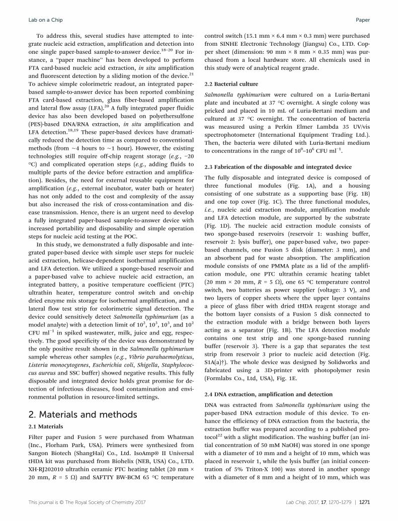
The fully disposable and integrated device is composed ofthree functional modules (Fig. 1A), and a housingconsisting of one substrate as a supporting base (Fig. 1B)and one top cover (Fig. 1C). The three functional modules,i.e., nucleic acid extraction module, amplification moduleand LFA detection module, are supported by the substrate(Fig. 1D). The nucleic acid extraction module consists oftwo sponge-based reservoirs (reservoir 1: washing buffer,reservoir 2: lysis buffer), one paper-based valve, two paper-based channels, one Fusion 5 disk (diameter: 3 mm), andan absorbent pad for waste absorption. The amplificationmodule consists of one PMMA plate as a lid of the amplifi-cation module, one PTC ultrathin ceramic heating tablet(20 mm × 20 mm, R = 5 Ω), one 65 °C temperature controlswitch, two batteries as power supplier (voltage: 3 V), andtwo layers of copper sheets where the upper layer containsa piece of glass fiber with dried tHDA reagent storage andthe bottom layer consists of a Fusion 5 disk connected tothe extraction module with a bridge between both layersacting as a separator (Fig. 1B). The LFA detection modulecontains one test strip and one sponge-based runningbuffer (reservoir 3). There is a gap that separates the teststrip from reservoir 3 prior to nucleic acid detection (Fig.S1A(a)†). The whole device was designed by Solidworks andfabricated using a 3D-printer with photopolymer resin(Formlabs Co., Ltd, USA), Fig. 1E.
2.4 DNA extraction, amplification and detection
DNA was extracted from Salmonella typhimurium using thepaper-based DNA extraction module of this device. To en-hance the efficiency of DNA extraction from the bacteria, theextraction buffer was prepared according to a published pro-tocol22 with a slight modification. The washing buffer (an ini-tial concentration of 50 mM NaOH) was stored in one spongewith a diameter of 10 mm and a height of 10 mm, which wasplaced in reservoir 1, while the lysis buffer (an initial concen-tration of 5% Triton-X 100) was stored in another spongewith a diameter of 8 mm and a height of 10 mm, which was
Lab on a Chip Paper
1272 | Lab Chip, 2017, 17, 1270–1279 This journal is © The Royal Society of Chemistry 2017
placed in reservoir 2. Next, an asymmetric thermophilichelicase-dependent amplification (tHDA) method was utilizedfor the amplification of the nucleic acid sequence extractedfrom Salmonella typhimurium. The primers were designedfrom the fimA gene sequence of Salmonella typhimuriumaccording to the instruction of the tHDA primer design.23
The details of the primers and probes are stated in ESI† Ta-ble S1. The amplification reaction volume was 50 μL, includ-ing 5 μL of 10× annealing buffer II, 2 μL of 100 mM MgSO4, 4μL of 500 mM NaCl, 3.5 μL of IsoAmp dNTP solution, 5 μL of100 μM forward primer, 1 μL of 5 μM reverse primer, 5 μL ofIsoAmp Enzyme Mix and 24.5 μL of ddH2O. All amplificationreagents were freeze-dried and stored in glass fiber (5 mm ×7 mm), which was attached to the upper copper layer (Fig.S1A(d)†). Following the extraction step, the Fusion 5 diskwith extracted DNA was moved to the amplification zone thatcontains a heating tablet. After the amplification, the coppersheet with the amplicons was moved to the detection zone,connecting both the test strip and sponge-based runningbuffer (reservoir 3) for the detection via LFA. After 15 min,the result was observed from the vision window of the detec-tion module by the naked eye. The images of the test line ofLFA were captured by an iPhone 6S, and the optical densitiesof the test strips were measured by the Image-Pro Plus 6.0software.
2.5 Synthesis of gold nanoparticles (AuNPs) and AuNP–DP(detector probe) conjugates
AuNPs with an average diameter of 13 nm were preparedaccording to a reported method.24 AuNPs modified with theSalmonella typhimurium detector probe were designedaccording to the fimA gene sequence of Salmonellatyphimurium.23 The detailed sequences can be found in ESI†Table S1. The AuNP–detector probe conjugates were preparedaccording to a reported method with a slight modification.24
2.6 Optimization of the disposable and integrated device
To achieve the optimum result, we firstly used an AppliedBiosystems Veriti 96 well thermal cycler (USA) to optimize thesample volume, including 10 μL, 30 μL, 50 μL, 70 μL and 90μL according to our previous studies. Secondly, we optimizedthe amplification temperature range of tHDA (58, 60, 63, 65,68 °C) for 90 min. Then, we optimized the reaction time ofamplification (30, 45, 60 min). To achieve the cost-effectiveness and integration of this device, we used freeze-dried technology to store the amplification reagents. 1% bo-vine serum albumin (BSA), 5% Ficoll 400 and 6% Raffinoseas protein protectants were respectively used to investigatethe effect of the freeze-dried reagents. Meanwhile, 104 CFUml−1 of the bacteria was utilized to investigate the storage
Fig. 1 The schematic of a disposable and integrated paper-based device for nucleic acid extraction, amplification and detection. (A) The differentfunctional modules of the device, including nucleic acid extraction, amplification by the battery heat blocker and LFA detection. (B) The substrateand a bridge that was used to separate the upper layer copper sheet from the bottom layer to prevent the lysis buffer and washing buffer fromflowing to the dry powder paper. (C) The top cover of the integrated paper-based device. (D) The integration platform of the different functionalmodules and the substrate. (E) The model of this device.
Lab on a ChipPaper

Lab Chip, 2017, 17, 1270–1279 | 1273This journal is © The Royal Society of Chemistry 2017
effect for one, two and three months. Additionally, we alsoused an Agilent 34907A data acquisition/Switch Unit (USA) tomeasure the temperature of the PTC ultrathin ceramicheating tablet at different ranges of voltage (2.8 V, 3.0 V, 3.2V) for 2 hours. Considering the varied conditions in real ap-plications (different temperatures, different humidities, anddifferent airflows), we tested the temperature of the PTCheating tablet under different conditions with a fixed voltageof 3 V, including different ambient humidities (20%, 30%,40%, 50%, 60%, 70%, 80%, 90% RH) at 20 °C indoor, differ-ent ambient temperatures (5, 10, 15, 20, 25, 40 °C) with 40%RH indoor, and different airflows (indoor, outdoor simulatedby using a fan) at 20 °C with 40% RH for 2 hours.
2.7 Mathematical simulation
To investigate the heating process using the PTC ultrathinceramic heating tablet, we performed a mathematical analy-sis of the natural convective heat transfer in the amplifi-cation chamber with two open gates (Fig. 4B(b)). The sizesof the chamber sides (e.g., Lx, Ly and Lz), the heating tab-let edges (e.g., ax and ay) and the gate width (w) weremeasured from our device (Fig. S2†). For simplification,the airflow inside the chamber is considered to be incom-pressible, laminar and Newtonian, and the Boussinesq ap-proximation is imposed on the density of the air.25 Theair density is given as follows,
ρ = ρ0(1 − β·(T − T0)) (1)
where T0, β, and ρ0 are the room temperature, the volume ex-pansion coefficient and the air density corresponding to T0,respectively. The governing equations coupled with conserva-tion of mass, momentum and energy equations are used todeal with this heat transfer problem and can be expressed as,
(2)
(3)
(4)
(5)
(6)
where u, v and w are the velocity in the direction of x, y,and z, respectively; μ, p, λ, and cP are the viscosity, pres-sure, thermal conductivity and specific heat of air, respec-
tively. The heating tablet provides a constant surface tem-perature (Th). The pressures at the two open gates are equalto the atmospheric pressure. Adiabatic and non-slip boundary con-ditions are applied to the remaining bounding walls. All theparameters used in the modeling are listed in Table S2.†The geometrical model of the square chamber was builtwith a non-uniform grid by the GAMBIT software, while themathematical model was solved by using the FLUENTsoftware.
2.8 Testing of various biological samples and the specificityof Salmonella typhimurium LFA
We spiked Salmonella typhimurium into wastewater, milk,juice and egg samples with a concentration range from 100 to106 CFU ml−1. Wastewater was obtained from sewage, andmilk, juice and egg were purchased from a grocery store.Additionally, we used 100 nM of the sequences of Salmonellatyphimurium, Vibrio parahaemolyticus, Listeria monocytogenes,Escherichia coli, Shigella, Staphylococcus aureus and SSCbuffer to detect the specificity of the Salmonella typhimuriumLFA method.
2.9 Statistical analysis
One-way ANOVA was used to compare the data among differ-ent groups. P < 0.05 was reported as statistically significant.
3. Results and discussion3.1 Design of the fully disposable and integrated paper-basedsample-to-answer device
To overcome the drawbacks of the existing nucleic acid test-ing technologies (e.g., need external reusable amplificationequipment, complex operations, off-chip reagent storage),we developed a fully disposable and integrated paper-basedsample-to- answer device for nucleic acid testing (Fig. 1).The device is composed of three functional modules(Fig. 1A) and a housing containing one substrate (Fig. 1B)and one top cover (Fig. 1C). The three functional moduleswere developed for nucleic acid extraction, amplificationand LFA detection, respectively. The integration platform asshown in Fig. 1D contains the three functional modulessupported by the substrate, and the top cover was thenmounted to the integration platform to create a fully inte-grated device (Fig. 1E).
The nucleic acid extraction module is composed of twosponge-based reservoirs (one lysis solution reservoir and onewashing solution reservoir), two paper-based channels (onelysis channel and one washing channel), one paper-basedvalve and one Fusion 5 disk (Fig. 1A). Two sponges were sepa-rately placed in the two reservoirs to store the lysis bufferand washing buffer, respectively. Meanwhile, two sealing rub-bers connected to the button were placed on top of thesponge to prevent reagent evaporation (Fig. 1A). The buttonwas used to turn on the nucleic acid extraction process byallowing the buffer to flow from the reservoirs to the two
Lab on a Chip Paper

1274 | Lab Chip, 2017, 17, 1270–1279 This journal is © The Royal Society of Chemistry 2017
paper-based channels (i.e., sample lysis channel and washingchannel) through their respective small holes (Fig. S1A(a)†).Briefly, one end of the lysis channel is connected with thesmall hole of the sponge-based lysis buffer whereas the otherend is connected to the Fusion 5 disk. The Fusion 5 disk iscomposed of silica-based glass fiber, and it absorbs DNAbased on the combination of high affinity between the nega-tively charged DNA and the positively charged glass fiber.26
The absorbent pad is placed under the Fusion 5 disk forwaste absorption (Fig. S1A(b)†). One end of the washingchannel (with “∟” shape) is connected to the small hole ofthe sponge-based washing buffer, while the other end isconnected to the paper-based valve to control fluid flowthrough the paper-based channel. The integrated coppersheets consist of two layers (Fig. 1A), the upper layer (amplifi-cation layer) consists of a piece of glass fiber that stores thedried amplification reagent which will be further describedin the next paragraph, whereas the bottom layer consists of aFusion 5 disk for nucleic acid extraction. During the nucleicacid extraction process, both layers are separated by a bridge(Fig. S1A(c)†) to prevent the extraction reagent from flowinginto the amplification layer, which may affect the entiresample-to-answer process.
The nucleic acid amplification module is composed of twobatteries, one ultrathin ceramic heating tablet, one 65 °Ctemperature control switch, two layers of copper sheets andone PMMA plate. The batteries were mounted on the basesubstrate to power the heating tablet. The 65 °C temperaturecontrol switch was mounted on the back of the PTC heatingtablet to control the temperature of the PTC heating tablet.During the heating process, the temperature switch will auto-matically disconnect when the temperature of the PTCheating tablet is higher than 65 °C. The aforementioned cop-per sheets were placed between the heating tablet and thePMMA plate (Fig. S1A(d)†), where the PMMA plate was usedto prevent sample evaporation and cross-contamination. Asmall piece of insulation pad was integrated into the device,which acts as a “switch” that isolates the battery powersource from the electrical circuit prior to nucleic acid amplifi-cation (Fig. S1A(e)†). Briefly, following the nucleic acid extrac-tion, the extracted nucleic acid was attached to the Fusion 5disk, which was used for the subsequent amplification pro-cess. The insulation pad was then removed to switch on thepower supply for the heating tablet. During the amplificationprocess, the two copper sheets were moved to the amplifica-tion zone between the PTC ultrathin ceramic heating tabletand the PMMA plate (Fig. 2B).
The nucleic acid detection module contains one lateralflow test strip and one sponge-based running buffer. Priorto nucleic acid detection, the test strip and running bufferwere separated by a gap (Fig. S1A(a)†). When the two coppersheets were moved to the detection zone, it occupied thegap and connected both the test strip and sponge-basedrunning buffer. The buffer and sample automatically flowedinto the test strip as driven by the capillary force. In thewhole device, the two copper sheets were used to connect
the extraction module, amplification module and LFA detec-tion module. The upper layer was labeled with two lines,amplification line and detection line, to indicate the posi-tion of both amplification and detection zones for user con-venience (Fig. 2B).
Considering that the capacity and life of the battery couldaffect the final temperature, in this device, we used new bat-teries to provide the power for the PTC heating tablet duringthe amplification process. After the amplification, the batteryvoltage was stable 3 V as measured by a multimeter, indicat-ing that the battery can be also reused for other householdappliances to avoid the waste and high cost. Additionally, thePTC heating tablet can also be recycled and used in the de-vice again. But other components (e.g., paper-based channel,reagents and LFA test strip) cannot be used again.
3.2 The whole operation process
In this study, we used detergent-based and alkaline extractionmethods to extract nucleic acid, asymmetric tHDA to amplifythe nucleic acid and LFA to detect the target. Although TritonX-100 is not necessarily lytic to bacteria, TritonX-100 andNaOH can be used to isolate DNA from blood according to areported study.22 Hence, considering the universality of thisdevice in the future, in this study, we also used TritonX-100and NaOH for DNA extraction from bacteria. As compared tothe reported study,22 in our device, 5% of TritonX-100 and 50mM NaOH as the initial concentrations of the extractionbuffer were respectively utilized to lyse the sample and washaway the debris and PCR inhibitors, because the efficiency ofDNA extraction has been shown to increase with increasingconcentration of the extraction reagent.27 The Fusion 5 diskand dry powder paper were separately used to capture theDNA and store the dried tHDA reagents, whereas the lateralflow test strip was utilized to detect the amplicons. Com-pared to the conventional process (Fig. 2A), the whole opera-tion process of our device is much simpler and only requiresa few steps, including sample addition, nucleic acid extrac-tion, removal of the insulation pad, amplification and LFAdetection (Fig. 2B, ESI† Movie S1). The detailed process is asfollows (Fig. 2C): firstly, the sample was added into the sam-ple area by manual pipetting or dropping. Secondly, the startbutton was manually pressed to start the extraction process,where the lysis buffer firstly flowed into the paper-basedchannel via small holes (Fig. S1A(a)†) and reached the sam-ple area to lyse the sample. Then, the washing buffer acti-vated the paper-based valve on the washing channel to con-nect to the lysed channel and flowed through both thesample area and Fusion 5 disk. The Fusion 5 disk was usedto capture the DNA, which was connected to an absorptionpad for waste removal (the whole extraction is shown in Fig.S3†). Following the DNA extraction, the two copper sheetsthat consist of the Fusion 5 disk with the DNA template andglass fiber with tHDA reagents were simultaneously moved tothe amplification zone (Fig. 2B), and the insulation pad wasremoved to start the amplification process. Finally, after the
Lab on a ChipPaper

Lab Chip, 2017, 17, 1270–1279 | 1275This journal is © The Royal Society of Chemistry 2017
amplification, the copper sheets were moved to the LFA de-tection module (Fig. 2B), and the target and running bufferflowed into the test strip as driven by the capillary force. Thecolorimetric signals produced by LFA were then observed bythe naked eye.
3.3 Optimization parameters of the disposable andintegrated device
To obtain the optimum detection result, we optimized thesample volume, amplification temperature and amplificationperiod used for the device using a fixed diameter of Fusion 5disk (diameter of 3 mm) (Fig. 3). We observed that with 10μL of sample, the optical density of the test zone was signifi-cantly lower than other sample volumes (30 μL, 50 μL, 70 μL,90 μL), Fig. 3A. There was no significant difference in opticaldensities of test zones using 30 μL, 50 μL, 70 μL and 90 μL ofsamples, suggesting that the Fusion 5 disk with a diameter of3 mm had the maximum DNA capturing capability when thesample volume was 30 μL. This result was further confirmedby the electrophoresis test, where we observed the higher op-tical density of the band for the case of 30 μL, 50 μL, 70 μLand 90 μL (Fig. S1B†). Since the temperature may affect theamplification efficiency, we also investigated the effect of am-
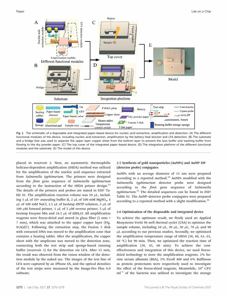
plification temperature (58 °C, 60 °C, 63 °C, 65 °C, 68 °C) onLFA with a 60 min amplification period, Fig. 3B. We foundpositive results for 63 °C, 65 °C and 68 °C as reflected by thered color shown in the test zone, but not for 58 °C and 60 °C.By comparing the optical density of the test zone at differenttemperatures, the color and optical density of the test zone of65 °C was significantly higher than those of 63 and 68 °C(Fig. 3B), indicating that the optimum amplification tempera-ture was 65 °C, which was further confirmed by the electro-phoresis result (Fig. S1C†). Furthermore, we investigated theeffect of the amplification period (30 min, 45 min and 60min) on the detection limit of bacteria in PBS (Fig. 3C). Wefound that the result was negative for the case of 30 min, thedetection limit for 45 min was 106 CFU ml−1, while the detec-tion limit for 60 min was significantly improved to 102 CFUml−1. This result indicated that the optimum amplificationperiod was 60 min.
To increase the portability of the device and eliminate thecold-chain requirement in resource-limited settings, westored the tHDA amplification reagents on the device byusing the freeze-drying method. To evaluate the reagent pres-ervation effect of different protein protectants for POC use,we added different protein protectants (1% bovine serum al-bumin (BSA), 5% Ficoll 400 and 6% Raffinose) to the tHDA
Fig. 2 The whole operation process of the fully disposable and integrated paper-based sample-to-answer device. Samples from wastewater, milk,fruit juice and egg. (A) The conventional operation process of nucleic acid extraction, amplification and detection. (B) Schematic diagram of theoperation of the disposable and integrated paper-based sample-to-answer device, including sample addition, DNA extraction, tHDA amplificationand LFA detection. (C) The internal diagram of the device with different steps. (D) The schematic diagram of the main parts of each step.
Lab on a Chip Paper

1276 | Lab Chip, 2017, 17, 1270–1279 This journal is © The Royal Society of Chemistry 2017
reaction reagent, which was then stored by the freeze-dryingmethod. We found that the result of 1% BSA was significantbetter than those of 5% Ficoll 400 and 6% Raffinose basedon the color and optical density of the test zone (Fig. S1D†).The stabilization was probably due to the hydrogen bondingbetween BSA and the dried proteins.28 To evaluate the stabil-ity of on-chip reagent storage at different storing periods andtemperatures for future POC applications, we evaluated theirperformance after one-, two- and three-month storage at dif-ferent temperatures (−20 °C, 4 °C, 25 °C, 37 °C, 45 °C, 56 °C).We vacuum-packed the disposable and integrated device afterplacing the lysis and washing buffers in the sponges and dry-ing tHDA reagent on paper. We observed that after onemonth of storage, the positive signal shown by the samplesfrom −20 °C was significantly higher than those from 4 °Cand 25 °C, whereas after two months of storage only the sam-ples from −20 °C and 4 °C showed a positive result, whichshowed a negative result for three months of storage. Other
temperatures (37, 45, 56 °C) showed negative results afterone to three months of storage, which may be due to the re-duced protein activity with increasing temperature29 (Fig. 4).To achieve room temperature storage and avoid the need forexternal storage unit (e.g., fridge) during shipping or in POCsettings, further optimization would be performed in the fu-ture in collaboration with a commercial partner.
To avoid the use of external reusable amplification equip-ment and improve the portability of the device, we utilizedthe PTC heating tablet and temperature control switch com-bined with the battery to provide the optimum amplificationtemperature. During the heating process, the PTC heating tab-let, as a kind of self-regulating heating element, was utilizedfor performing tHDA amplification because the fixed resis-tance PTC heating tablet is an automatic constant tempera-ture device.30 The temperature switch was used to control thePTC heating tablet when the temperature of the PTC heatingtablet was higher than 65 °C. The PTC heating tablet could
Fig. 3 Optimization of extraction and amplification parameters. (A) Different sample volumes, 10 μL, 30 μL, 50 μL, 70 μL, 90 μL, were used foroptimizing the extraction parameter, (B) different temperatures, 58 °C, 60 °C, 63 °C, 65 °C and 68 °C, were utilized for optimizing the tHDAamplification parameter, (C) different times, 30 min, 45 min and 60 min, were utilized for optimizing the amplification period of our device (NC –
negative control).
Lab on a ChipPaper

Lab Chip, 2017, 17, 1270–1279 | 1277This journal is © The Royal Society of Chemistry 2017
provide different temperatures through adjusting the voltagesand we achieved a stable condition at 65 °C up to 2 hourswhen the voltage was 3 V (Fig. 4B(a)). Considering the variedconditions in real applications, we also checked the tempera-ture of the PTC heating tablet with a fixed voltage of 3 Vacross different ambient conditions of humidity, temperatureand airflow (Fig. S4†). We found that the temperature of thePTC heating tablet was stable at 65 °C for all conditions of hu-midity except 20% RH (Fig. S4A†). Similarly, the PTC tempera-ture was stable at 65 °C when the ambient temperature was inthe range of 10–40 °C (Fig. S4B†). However, the PTC tempera-ture was unstable under simulated outdoor airflow conditions(Fig. S4C†). At present, this device prototype is not yet readyfor use anywhere except in the range of humidities (30–90%RH), temperatures (10–40 °C) and indoor condition. To fur-ther expand the application range of this device, our futurework would focus on development of a disposable and porta-ble device with stable environmental conditions to provide anoptimum condition for nucleic acid-based testing.
To investigate the temperature of the chamber during theheating process using the PTC ultrathin ceramic heating tab-let, we also performed a numerical analysis of the tempera-ture field in the amplification chamber by using the FLUENTsoftware. From the result, we found that the temperature of
the main area of the amplification chamber remains stable at65 °C though the temperature of the two open gates of theamplification chamber is less than 65 °C (Fig. 4B(b)). Thesimulation results indicate that the isothermal amplificationcould be well performed without considering the position er-ror of the copper sheet.
3.4 Testing of various biological samples
To verify the potential application of our fully disposable andintegrated paper-based sample-to-answer device for POC test-ing, different concentrations of Salmonella typhimurium rang-ing from 100 to 106 CFU ml−1 were spiked into different sam-ples, including wastewater, milk, juice and egg. After testingwith the paper-based device, we found that the detectionlimit of bacteria was 102, 103, 103, 102 CFU ml−1 in wastewa-ter, milk, juice and egg, respectively (Fig. 5), which is similarto a reported paper-based device with a detection limit of 103
CFU ml−1.31 As for wastewater and egg, the detection limitwas similar with that of PBS, which was lower than those ofbacteria in milk and juice. This is because milk and juicecontain some inhibitors (e.g., protease, calcium ions and ad-ditives) which can reduce the amplification efficiency. Addi-tionally, we also used 100 nM of the sequence of Salmonella
Fig. 4 Optimization of on-chip reagents' storage period and ultrathin heater tablet temperature parameter. (A) The stability test result of tHDAmixes at different storage temperatures and times: (a) one month, (b) two months, (c) three months. (B) (a) Temperature stability of the ultrathinheater tablet inside the device with different voltages, (b) the simulation temperature field of the amplification chamber (PC – positive control, NC– negative control).
Lab on a Chip Paper

1278 | Lab Chip, 2017, 17, 1270–1279 This journal is © The Royal Society of Chemistry 2017
typhimurium, Vibrio parahaemolyticus, Listeria monocytogenes,Escherichia coli, Shigella, Staphylococcus aureus and SSCbuffer to verify the specification of the Salmonellatyphimurium LFA method, which showed a positive result.Other investigations showed negative results (Fig. S1E†), indi-cating the good specificity of our device.
We successfully demonstrated that our prototype couldachieve sample-in-answer-out testing using simple operations,which could be performed by untrained users. To achieve thesimple and integrated nucleic acid testing, in this study, wedeveloped a paper-based DNA extraction module in the inte-grated device. As compared to the existing integrated paper-based device that combines FTA card-based extraction, glassfiber-based amplification and LFA,20 our device solved the ex-traction and amplification reagents' storage problem. As com-pared to the fully integrated paper fluidic device based onpolyethersulfone (PES)-based DNA/RNA extraction, in situ am-plification and LFA detection,18,19 our device achievedequipment-free amplification using an on-chip heating tablet.Additionally, when the bacteria target was directly used fortHDA amplification, we found that the results were negativebecause the inhibitor of the bacteria sample could affect thetHDA amplification. On the other hand, we also used a sim-ple filtration method to collect the bacteria and then re-moved inhibitors by washing, which gave positive results.
However, the simple filtration method and washing steps stillneed external multiplex operations, increasing the complexityof nucleic acid testing. Furthermore, our device decreases thecost, reduces the operation steps and realizes the fully porta-ble and disposable feature. Thus, our device offers great po-tential to meet the ASSURED criteria suggested by WHO fornucleic acid testing in POC settings.
4. Conclusion
In summary, we developed a fully disposable and integratedpaper-based device by integrating paper-based nucleic acidextraction, paper-based isothermal amplification and LFAinto one paper device, and achieved nucleic acid testing inabout an hour. This device only needs simple operations forthe entire sample-to-answer nucleic acid testing, includingadding the sample, pressing the button to begin the nucleicacid extraction, moving the copper sheet to the amplificationzone for tHDA amplification and moving the copper sheet tothe detection zone for LFA detection. This prototype could bedirectly utilized in resource-limited settings without specialtraining, external equipment (e.g., thermal cycler, refrigera-tor) and complex operation. In comparison with existing inte-grated paper-based sample-to-answer devices, our device
Fig. 5 Fully disposable and integrated paper-based device for various biological sample tests. Salmonella typhimurium from 100 to 106 CFU ml−1
were spiked into (A) waste water, (B) milk, (C) juice and (D) egg and utilized for the validation of this assay with a detection limit of 102, 103, 103,102 CFU ml−1, respectively (NC – negative control).
Lab on a ChipPaper

Lab Chip, 2017, 17, 1270–1279 | 1279This journal is © The Royal Society of Chemistry 2017
further decreases the cost, reduces the operation step and re-alizes the fully portable and disposable feature.
According to reported studies and our study, TritonX-100and NaOH can be used for blood sample and bacteria sampletesting. To further enhance the universality of this device,our ongoing work would focus on extracting nucleic acid(e.g., DNA or RNA) from various biological samples such asGram positive bacteria, cells, viruses and so on, which canfurther expand the application of this device at the point-of-care. We envision that this disposable and integrated paper-based device would be a powerful tool for nucleic acid testingin resource-limited settings.
Declaration of interest
The authors declare that they have no conflict of interest.
Acknowledgements
This work was supported by the Natural Science Foundationof China (11472224, 11672246) and National InstrumentationProgram (No. 2013YQ190467).
References
1 C. D. Chin, Y. K. Cheung, D. Steinmiller, V. Linder, H. Parsa,J. Wang, H. Moore, R. Rouse, E. Karita, L. Mwambarangwe,S. L. Braunstein, J. van de Wijgert, R. Sahabo, W. El-Sadrand S. K. Sia, Nat. Med., 2011, 17, 1015–1019.
2 K. Pardee, A. A. Green, M. K. Takahashi, D. Braff, G.Lambert, J. W. Lee, T. Ferrante, D. Ma, N. Donghia, M. Fan,N. M. Daringer, I. Bosch, D. M. Dudley, D. H. O'Connor, L.Gehrke and J. J. Collins, Cell, 2016, 165, 1255–1266.
3 D. Kagan, S. Campuzano, S. Balasubramanian, F. Kuralay,G. U. Flechsig and J. Wang, Nano Lett., 2011, 11, 2083–2087.
4 T. Denes and M. Wiedmann, Curr. Opin. Biotechnol.,2014, 26, 45–49.
5 A. Niemz, T. M. F and D. S. Boyle, Trends Biotechnol.,2011, 29, 240–250.
6 K. Bohmann, A. Evans, M. T. Gilbert, G. R. Carvalho, S.Creer, M. Knapp, D. W. Yu and M. de Bruyn, Trends Ecol.Evol, 2014, 29, 358–367.
7 S. K. Vashist, P. B. Luppa, L. Y. Yeo, A. Ozcan and J. H.Luong, Trends Biotechnol., 2015, 33, 692–705.
8 J. R. Choi, R. H. Tang, S. Q. Wang, W. A. Wan Abas, B. Pingguan-Murphy and F. Xu, Biosens. Bioelectron., 2015, 74, 427–439.
9 M. Medina-Sanchez, B. Ibarlucea, N. Perez, D. D.Karnaushenko, S. M. Weiz, L. Baraban, G. Cuniberti andO. G. Schmidt, Nano Lett., 2016, 16, 4288–4296.
10 R. H. Tang, H. Yang, J. R. Choi, Y. Gong, S. S. Feng, B.Pingguan-Murphy, Q. S. Huang, J. L. Shi, Q. B. Mei and F.Xu, Crit. Rev. Biotechnol., 2016, 1–18.
11 W. Lu, J. Wang, Q. Wu, J. Sun, Y. Chen, L. Zhang, C. Zheng,W. Gao, Y. Liu and X. Jiang, Biosens. Bioelectron., 2016, 75,28–33.
12 V. Wolfgramm Ede, F. M. de Carvalho, V. R. Aguiar, M. P.Sartori, G. C. Hirschfeld-Campolongo, W. M. Tsutsumidaand I. D. Louro, Forensic Sci. Int.: Genet., 2009, 3, 125–127.
13 S. R. Jangam, D. H. Yamada, S. M. McFall and D. M. Kelso,J. Clin. Microbiol., 2009, 47, 2363–2368.
14 A. V. Govindarajan, S. Ramachandran, G. D. Vigil, P. Yagerand K. F. Bohringer, Lab Chip, 2012, 12, 174–181.
15 J. R. Choi, J. Hu, Y. Gong, S. Feng, W. A. Wan Abas, B.Pingguan-Murphy and F. Xu, Analyst, 2016, 141, 2930–2939.
16 J. C. Linnes, A. Fan, N. M. Rodriguez, B. Lemieux, H. Kongand C. M. Klapperich, RSC Adv., 2014, 4, 42245–42251.
17 M. S. Cordray and R. R. Richards-Kortum, Malar. J.,2015, 14, 472.
18 N. M. Rodriguez, J. C. Linnes, A. Fan, C. K. Ellenson, N. R.Pollock and C. M. Klapperich, Anal. Chem., 2015, 87,7872–7879.
19 N. M. Rodriguez, W. S. Wong, L. Liu, R. Dewar and C. M.Klapperich, Lab Chip, 2016, 16, 753–763.
20 J. R. Choi, J. Hu, R. Tang, Y. Gong, S. Feng, H. Ren, T. Wen,X. Li, W. A. Wan Abas, B. Pingguan-Murphy and F. Xu, LabChip, 2016, 16, 611–621.
21 J. T. Connelly, J. P. Rolland and G. M. Whitesides, Anal.Chem., 2015, 87, 7595–7601.
22 S. M. McFall, R. L. Wagner, S. R. Jangam, D. H. Yamada, D.Hardie and D. M. Kelso, J. Virol. Methods, 2015, 214, 37–42.
23 H. J. Cohen, S. M. Mechanda and W. Lin, Appl. Environ.Microbiol., 1996, 62, 4303–4308.
24 R. Tang, H. Yang, J. R. Choi, Y. Gong, J. Hu, S. Feng, B.Pingguan-Murphy, Q. Mei and F. Xu, Talanta, 2016, 152,269–276.
25 D. Akrour, R. Bennacer and D. Kalahe, Int. J. Numer.Methods Fluids, 1983, 3, 249–264.
26 K.-H. Esser, W. H. Marx and T. Lisowsky, Nat. Methods,2006, 3, i–ii.
27 S. C. Tan and B. C. Yiap, J. Biomed. Biotechnol., 2009, 2009,574398–574407.
28 T. Arakawa, Y. Kita and J. F. Carpenter, Pharm. Res.,1991, 08, 285–291.
29 L. Nisius and S. Grzesiek, Nat. Chem., 2012, 4, 711–717.30 X. Wang, L. Zhang and G. Chen, Anal. Bioanal. Chem.,
2011, 401, 2657–2665.31 W. Wu, J. Li, D. Pan, J. Li, S. Song, M. Rong, Z. Li, J. Gao
and J. Lu, ACS Appl. Mater. Interfaces, 2014, 6, 16974–16981.
Lab on a Chip Paper