lab on a chip - me-web.engin.umich.edume-web.engin.umich.edu/ibbl/pdf/2015_labonchip_moraes.pdf ·...
TRANSCRIPT

Lab on a Chip
Publ
ishe
d on
29
July
201
5. D
ownl
oade
d by
Uni
vers
ity o
f M
ichi
gan
Lib
rary
on
15/0
9/20
15 2
0:31
:17.
PAPER View Article OnlineView Journal | View Issue
3760 | Lab Chip, 2015, 15, 3760–3765 This journal is © The R
aDepartment of Chemical Engineering, McGill University, 3610 University Street,
Montreal, QC H3A 2B2, CanadabDepartment of Biomedical Engineering, College of Engineering, University of
Michigan, 2200 Bonisteel Blvd, Ann Arbor, MI 48109, USA.
E-mail: [email protected] Biointerfaces Institute, University of Michigan, NCRC, 2800 Plymouth Road, MI
48109-2800, USAdDepartment of Mechanical Engineering, University of Michigan, Ann Arbor,
Michigan 48109, USAeMacromolecular Science and Engineering Center, College of Engineering,
University of Michigan, 2300 Hayward St., Ann Arbor, MI 48109, USA
† Electronic supplementary information (ESI) available. See DOI: 10.1039/c5lc00722d
Cite this: Lab Chip, 2015, 15, 3760
Received 25th June 2015,Accepted 29th July 2015
DOI: 10.1039/c5lc00722d
www.rsc.org/loc
Supersoft lithography: candy-based fabrication ofsoft silicone microstructures†
Christopher Moraes,abc Joseph M. Labuz,bc Yue Shao,d Jianping Fubd andShuichi Takayama*bce
We designed a fabrication technique able to replicate microstructures in soft silicone materials (E < 1 kPa).
Sugar-based ‘hard candy’ recipes from the confectionery industry were modified to be compatible with sil-
icone processing conditions, and used as templates for replica molding. Microstructures fabricated in soft
silicones can then be easily released by dissolving the template in water. We anticipate that this technique
will be of particular importance in replicating physiologically soft, microstructured environments for cell
culture, and demonstrate a first application in which intrinsically soft microstructures are used to measure
forces generated by fibroblast-laden contractile tissues.
Introduction
The ability to engineer microstructures at the length scale ofcells and tissues has played an important role in understand-ing how biological cells interact mechanically with their envi-ronment.1,2 Matrix mechanics are emerging as important reg-ulators of development, homeostasis, and disease in variousbiological systems. Hence, recapitulating the stiffness of tis-sues (Young's modulus E ≈ 0.1–100's of kPa) in vitro is nowconsidered an important strategy in tissue engineering, regen-erative medicine, and fundamental studies of cell biology.3
However, ‘soft lithography’, the primary enabling technologyin fabricating micron-scale structures is typically limited tosupraphysiologically stiff elastomeric materials such as con-ventional polydimethylsiloxane (PDMS; E ≈ 1000 kPa), and ischallenging to apply when working with very soft materials.Here, we leverage techniques originating in the confectioneryindustry to develop ‘supersoft lithography’, a simple, robust,and versatile approach to replicate microstructures in soft sili-cone materials (E ≈ 0.1 kPa). We then apply this approachtowards fabricating soft PDMS templates capable of
measuring both long-term and real-time contractile forcesgenerated by toroid-shaped engineered microtissues.
Existing methods to microfabricate intrinsically soft micro-structures suffer some severe limitations. Both top-down tech-niques such as microscale laser machining, and bottom-upprinting and photopatterning techniques are expensive, requirespecialized equipment, and/or are challenging to scale updevice production.4 Replica molding presents a viable alterna-tive to these strategies, and has to date been applied to micro-fabricate structures in soft hydrogel materials such ascollagen,5–8 polyacrylamide,9–11 and polyethylene glycol.12,13
However, use of these low-toughness materials limits the verti-cal dimensions of these fabricated structures, due to challengesin mechanically removing the template from the hydrogel. Assuch, previous studies with these approaches have been limitedto working with feature thicknesses of tens of microns, and fea-ture resolutions of several hundred microns.9 Furthermore,hydrogels undergo large degrees of stiffness-dependent swell-ing,9,14 up to 1200% for very soft gels,14 making it challengingto replicate high-fidelity microscale features in soft materials.In contrast, soft silicones such as Sylgard 527 (Dow Corning,E ≈ 1 kPa) exhibit low shrinkage/swelling ratios in aqueousmedium,15–20 and can be blended with conventional Sylgard184 to match stiffnesses across the physiological range.21 Ourmeasurements of baseline Sylgard 527 stiffness by shearrheometry indicates that with a monomer : crosslinker ratio of1 : 1, Sylgard 527 has a Young's modulus of 1.5 kPa, threeorders of magnitude less than conventional Sylgard 184 PDMS(Fig. 1A). This material modulus varied by <0.1 kPa overstrains up to 250% (Fig. 1B). The modulus was further reducedto <0.1 kPa when the component ratios were altered by a verysmall degree (5 : 4; Fig. 1A), and exhibited no measurablechange at high strains (Fig. 1A and B).
oyal Society of Chemistry 2015
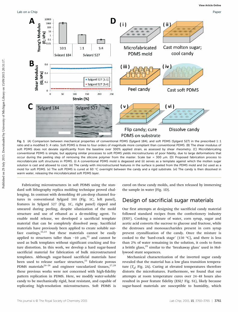
Fig. 1 (A) Comparison between mechanical properties of conventional PDMS (Sylgard 184), and soft PDMS (Sylgard 527) in the prescribed 1 : 1ratio and a modified 5 : 4 ratio. Soft PDMS is three to four orders of magnitude more compliant than conventional PDMS. (B) The shear modulus ofsoft PDMS does not deviate significantly from the baseline over 300% applied strain, as assessed by shear rheometry. (C) Microfabricatingconventional PDMS is simple, but applying similar processes to soft PDMS yields microstructures of poor fidelity, due to large deformations thatoccur during the peeling step of removing the silicone polymer from the master. Scale bar = 300 μm. (D) Proposed fabrication process tomicrofabricate soft structures in PDMS. (i) A conventional PDMS mold is degassed and (ii) serves as a template against which the molten sugarsolution is cast and allowed to cool. (iii) The candy with microstructured features in the surface is peeled from the PDMS mold and (iv) used as amold for soft PDMS. (v) The soft PDMS is cured at 60 °C overnight between the candy and a rigid substrate. (vi) The candy is then dissolved inwarm water, releasing the microfabricated soft PDMS layer.
Lab on a Chip Paper
Publ
ishe
d on
29
July
201
5. D
ownl
oade
d by
Uni
vers
ity o
f M
ichi
gan
Lib
rary
on
15/0
9/20
15 2
0:31
:17.
View Article Online
Fabricating microstructures in soft PDMS using the stan-dard soft lithography replica molding technique proved chal-lenging. In contrast with demolding 40 μm-deep channel fea-tures in conventional Sylgard 184 (Fig. 1C, left panel),features in Sylgard 527 (Fig. 1C, right panel) ripped andsmeared during peeling, despite silanization of the moldstructure and use of ethanol as a de-molding agent. Toenable mold release, we developed a sacrificial templatematerial that can be completely dissolved away. Sacrificialmaterials have previously been applied to create soluble sur-face coatings,22,23 but these materials cannot be easilyapplied to structures taller than ~10 μm,22 and cannot beused as bulk templates without significant cracking and fea-ture distortion. In this work, we develop a hard sugar-basedsacrificial material for fabrication of bulk microstructuredtemplates. Although sugar-based sacrificial materials havebeen used to release surface structures,22 fabricate porousPDMS materials24–26 and engineer vascularized tissues,27,28
these previous works were not concerned with high-fidelitypattern replication in PDMS. Here, we modify water-solublecandy to be mechanically rigid, heat resistant, and capable ofreplicating high-resolution microstructures. Soft PDMS is
This journal is © The Royal Society of Chemistry 2015
cured on these candy molds, and then released by immersingthe sample in water (Fig. 1D).
Design of sacrificial sugar materials
Our first attempts at designing the sacrificial candy materialfollowed standard recipes from the confectionery industry(ESI†). Cooking a mixture of water, corn syrup, sugar andacetic acid converts the sucrose to glucose and fructose, whilethe dextroses and monosaccharides present in corn syrupprevent crystallization of the candy. Once the mixture iscooked to the ‘hard-crack stage’ (150 °C), and there is lessthan 2% of water remaining in the solution, it cools to forma brittle glass,29 similar to the ‘breakaway glass’ used in Hol-lywood stunt sequences.
Mechanical characterization of the inverted sugar candyrevealed that the material has a low glass transition tempera-ture (Tg; Fig. 2A). Curing at elevated temperatures thereforedistorts the microfeatures. Furthermore, we found that ourattempts at room temperature cures over 24–48 hours alsoresulted in poor feature fidelity (ESI,† Fig. S1), likely becausesugar-based materials are susceptible to humidity, which
Lab Chip, 2015, 15, 3760–3765 | 3761

Fig. 2 Characterization of the supersoft lithography process. (A) Modulus of candy fabricated with the conventional inverted sugar process (redline) and the non-inverted sugar process (blue line) as a function of temperature. The inverted sugar process produces candy with a low glass tran-sition temperature, making it unsuitable for supersoft lithography. In contrast, the non-inverted sugar maintained rigidity over the range of temper-atures necessary to process PDMS. (B) Scanning electron microscopy reveals that candy cast against PDMS templates retains microscale featureson release (scale bar = 100 μm, inset scale bar = 10 μm). (C) Patterns transferred into soft PDMS of modulus 1.5 kPa. Scale bar = 100 μm, and (D)deep microwells patterned into softer PDMS of modulus 0.1 kPa, and imaged using confocal microscopy of surface adsorbed fluorescent dye. Toppanel = plan view, blue dashed line = cut-section shown in bottom panel (side view). Scale bar = 400 μm.
Lab on a ChipPaper
Publ
ishe
d on
29
July
201
5. D
ownl
oade
d by
Uni
vers
ity o
f M
ichi
gan
Lib
rary
on
15/0
9/20
15 2
0:31
:17.
View Article Online
causes feature degradation over extended time periods. Forexample, cotton candy (similar to our material) completelycollapses within 6 hours at 45% humidity.30 Furthermore,these numbers reflect bulk material changes, and surfacedamage of microfeatures is likely to occur much more rapidly.Hence, to circumvent humidity-driven template degradation,rapid (and therefore, high temperature) curing protocols arerequired. Raising the cure temperature to 60 °C would alsoreduce relative humidity by 85% (assuming a baseline indoorhumidity of 50%). This temperature difference would thereforedrive an ~100% decrease in sugar moisture content,31 and acorresponding 100× increase in template rigidity.29 Hence,optimizing the temperature resistance of the candy templatesis of critical importance in the success of this technique.
To increase Tg, we minimized the amount of sucroseinverted by maintaining a neutral pH, and sped up cooktimes. Sugar and light corn syrup in a 2 : 1 w/w ratio weremicrowaved until the sugar caramelized. Cook time variesdepending on microwave power, and so the process wasmonitored by observing color changes from clear to brownand the distinct aroma of caramelizing sugar. These mea-sures reduce Tg by minimizing the glucose and fructosemonosaccharides in the candy.32 Since extendedcaramelization is known to raise Tg by polymerizing mono-saccharide molecules,33 all samples were caramelized to a
3762 | Lab Chip, 2015, 15, 3760–3765
medium-brown color (ESI† Fig. S2). The non-inverted candyhad significantly improved mechanical rigidity and a Tg > 60°C (Fig. 2A), making it suitable for PDMS processing.
Fabrication of soft siliconemicrostructures
The molten sugar was cast against a Sylgard 184 PDMS moldcontaining microfabricated features. Candy pucks containingmicrofabricated features could be peeled from the PDMSmold, and were then stored in a vacuum desiccator until use.The supersoft lithography process is best performed in low-humidity environments, as the candy is hygroscopic andloses feature fidelity on exposure to moisture. Scanningelectron microscopy (SEM) analysis of the replicated sugarindicated that microscale features were transferred into andmaintained in the candy (Fig. 2B). No features were detectedon the flat portions of the sugar mold, and minor nanoscaleroughness present in the sidewalls of the SU-8 master werefaithfully replicated. To demonstrate the versatility of thistechnique for even smaller structures, arrays of micron-scaleposts were also faithfully replicated and maintained in thecandy (ESI,† Fig. S3).
Using this process, microfeatures were easily transferredfrom the candy molds into the Sylgard 527 PDMS (Fig. 2C;
This journal is © The Royal Society of Chemistry 2015

Lab on a Chip Paper
Publ
ishe
d on
29
July
201
5. D
ownl
oade
d by
Uni
vers
ity o
f M
ichi
gan
Lib
rary
on
15/0
9/20
15 2
0:31
:17.
View Article Online
E = 1.5 kPa) with feature resolutions down to 20 μm (ESI,†Fig. S1). Furthermore, tall microstructures (>400 μm) werefabricated in even softer PDMS (Fig. 2D; E = 0.1 kPa),although working with such soft materials was challenging asthe soft structures collapse easily (ESI,† Fig. S4).
Application: measuring microtissueforces
As a first biological application of this technology, we devel-oped intrinsically soft features to measure the mechanicalforces exerted during contractile tissue formation. The forcesinvolved in tissue remodeling are crucial features of develop-ment and morphogenesis, and previous approaches byLegant et al., measured these by developing microfabricated
This journal is © The Royal Society of Chemistry 2015
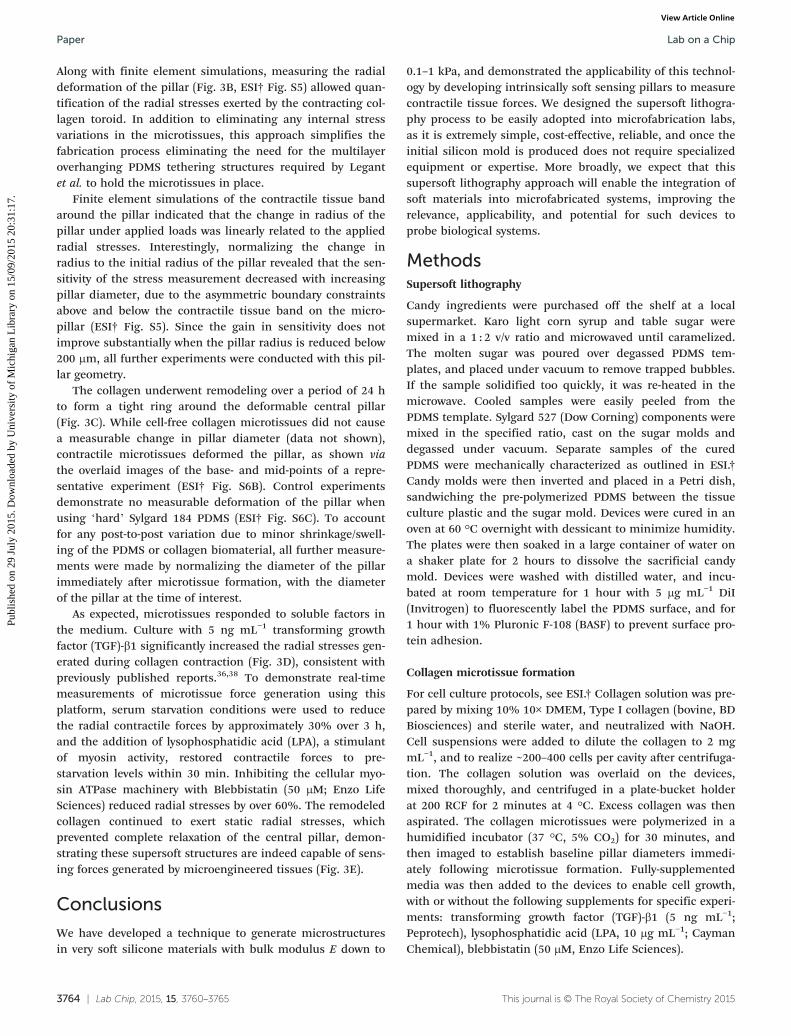
Fig. 3 Microstructured pillars in soft PDMS were designed to measure contfabrication and contraction process workflow: (i) a neutralized collagen geland (ii) centrifuged to drive the cells into the chamber. (iii) Excess collagen°C. (iv) Cell culture media is replaced and the collagen matrix is allowed tovia finite element analysis. (C) Toroid-shaped microtissues form over a perpillar structure. (red = DiI-stained PDMS, green = cellular actin; scale barsthe tissue maturation process increases stresses generated by the toroidal3 hours reduces microtissue-generated radial stresses. Adding lysophosstarvation levels, and adding blebbistatin (50 μM) to inhibit myosin II activitin the time-course, n = 15), demonstrating the ability to monitor real-time s
PDMS cantilevers that tether a contractile dog-bone shapedmicrotissue.34 The effective stiffness of the cantilever isreduced by decreasing the geometric cross-section of the can-tilever, enabling the measurement of small contractile forces.Although extremely useful,35,36 the dog-bone shaped tissuesmay exhibit strong necking profiles37 leading to varied cross-sectional areas, and thus, significant variations in internaltissue stresses.
To address this issue, we used the supersoft lithographyprocess to fabricate 400 μm tall pillar structures in softSylgard 527 PDMS, which were fluorescently labelled andaround which toroid-shaped microtissues of uniform cross-section can be formed (Fig. 3A). Contraction of microtissuesby a model stromal fibroblast cell line (HS-5; ATCC) gener-ated compressive mechanical forces on the central pillar.
Lab Chip, 2015, 15, 3760–3765 | 3763
ractile forces generated by a toroid-shaped microtissue. (A) Microtissuesolution containing cells is dispensed into the toroid-shaped chambers,is aspirated, and the remaining solution is allowed to polymerize at 37contract. (B) Changes in pillar radius are related to applied radial stressiod of 24 hours, creating a band of collagen around the central micro-= 200 μm). (D) The presence of soluble factors such as TGF-β1 duringmicrotissues (* p < 0.001, n = 13–15). (E) Starving the cells of serum forphatidic acid (LPA, 10 μg mL−1) increased stress generation to pre-y reduced stresses by 60% (* p < 0.001 against the previous treatmenttresses using this fabricated platform.
Lab on a ChipPaper
Publ
ishe
d on
29
July
201
5. D
ownl
oade
d by
Uni
vers
ity o
f M
ichi
gan
Lib
rary
on
15/0
9/20
15 2
0:31
:17.
View Article Online
Along with finite element simulations, measuring the radialdeformation of the pillar (Fig. 3B, ESI† Fig. S5) allowed quan-tification of the radial stresses exerted by the contracting col-lagen toroid. In addition to eliminating any internal stressvariations in the microtissues, this approach simplifies thefabrication process eliminating the need for the multilayeroverhanging PDMS tethering structures required by Legantet al. to hold the microtissues in place.
Finite element simulations of the contractile tissue bandaround the pillar indicated that the change in radius of thepillar under applied loads was linearly related to the appliedradial stresses. Interestingly, normalizing the change inradius to the initial radius of the pillar revealed that the sen-sitivity of the stress measurement decreased with increasingpillar diameter, due to the asymmetric boundary constraintsabove and below the contractile tissue band on the micro-pillar (ESI† Fig. S5). Since the gain in sensitivity does notimprove substantially when the pillar radius is reduced below200 μm, all further experiments were conducted with this pil-lar geometry.
The collagen underwent remodeling over a period of 24 hto form a tight ring around the deformable central pillar(Fig. 3C). While cell-free collagen microtissues did not causea measurable change in pillar diameter (data not shown),contractile microtissues deformed the pillar, as shown viathe overlaid images of the base- and mid-points of a repre-sentative experiment (ESI† Fig. S6B). Control experimentsdemonstrate no measurable deformation of the pillar whenusing ‘hard’ Sylgard 184 PDMS (ESI† Fig. S6C). To accountfor any post-to-post variation due to minor shrinkage/swell-ing of the PDMS or collagen biomaterial, all further measure-ments were made by normalizing the diameter of the pillarimmediately after microtissue formation, with the diameterof the pillar at the time of interest.
As expected, microtissues responded to soluble factors inthe medium. Culture with 5 ng mL−1 transforming growthfactor (TGF)-β1 significantly increased the radial stresses gen-erated during collagen contraction (Fig. 3D), consistent withpreviously published reports.36,38 To demonstrate real-timemeasurements of microtissue force generation using thisplatform, serum starvation conditions were used to reducethe radial contractile forces by approximately 30% over 3 h,and the addition of lysophosphatidic acid (LPA), a stimulantof myosin activity, restored contractile forces to pre-starvation levels within 30 min. Inhibiting the cellular myo-sin ATPase machinery with Blebbistatin (50 μM; Enzo LifeSciences) reduced radial stresses by over 60%. The remodeledcollagen continued to exert static radial stresses, whichprevented complete relaxation of the central pillar, demon-strating these supersoft structures are indeed capable of sens-ing forces generated by microengineered tissues (Fig. 3E).
Conclusions
We have developed a technique to generate microstructuresin very soft silicone materials with bulk modulus E down to
3764 | Lab Chip, 2015, 15, 3760–3765
0.1–1 kPa, and demonstrated the applicability of this technol-ogy by developing intrinsically soft sensing pillars to measurecontractile tissue forces. We designed the supersoft lithogra-phy process to be easily adopted into microfabrication labs,as it is extremely simple, cost-effective, reliable, and once theinitial silicon mold is produced does not require specializedequipment or expertise. More broadly, we expect that thissupersoft lithography approach will enable the integration ofsoft materials into microfabricated systems, improving therelevance, applicability, and potential for such devices toprobe biological systems.
MethodsSupersoft lithography
Candy ingredients were purchased off the shelf at a localsupermarket. Karo light corn syrup and table sugar weremixed in a 1 : 2 v/v ratio and microwaved until caramelized.The molten sugar was poured over degassed PDMS tem-plates, and placed under vacuum to remove trapped bubbles.If the sample solidified too quickly, it was re-heated in themicrowave. Cooled samples were easily peeled from thePDMS template. Sylgard 527 (Dow Corning) components weremixed in the specified ratio, cast on the sugar molds anddegassed under vacuum. Separate samples of the curedPDMS were mechanically characterized as outlined in ESI.†Candy molds were then inverted and placed in a Petri dish,sandwiching the pre-polymerized PDMS between the tissueculture plastic and the sugar mold. Devices were cured in anoven at 60 °C overnight with dessicant to minimize humidity.The plates were then soaked in a large container of water ona shaker plate for 2 hours to dissolve the sacrificial candymold. Devices were washed with distilled water, and incu-bated at room temperature for 1 hour with 5 μg mL−1 DiI(Invitrogen) to fluorescently label the PDMS surface, and for1 hour with 1% Pluronic F-108 (BASF) to prevent surface pro-tein adhesion.
Collagen microtissue formation
For cell culture protocols, see ESI.† Collagen solution was pre-pared by mixing 10% 10× DMEM, Type I collagen (bovine, BDBiosciences) and sterile water, and neutralized with NaOH.Cell suspensions were added to dilute the collagen to 2 mgmL−1, and to realize ~200–400 cells per cavity after centrifuga-tion. The collagen solution was overlaid on the devices,mixed thoroughly, and centrifuged in a plate-bucket holderat 200 RCF for 2 minutes at 4 °C. Excess collagen was thenaspirated. The collagen microtissues were polymerized in ahumidified incubator (37 °C, 5% CO2) for 30 minutes, andthen imaged to establish baseline pillar diameters immedi-ately following microtissue formation. Fully-supplementedmedia was then added to the devices to enable cell growth,with or without the following supplements for specific experi-ments: transforming growth factor (TGF)-β1 (5 ng mL−1;Peprotech), lysophosphatidic acid (LPA, 10 μg mL−1; CaymanChemical), blebbistatin (50 μM, Enzo Life Sciences).
This journal is © The Royal Society of Chemistry 2015

Lab on a Chip Paper
Publ
ishe
d on
29
July
201
5. D
ownl
oade
d by
Uni
vers
ity o
f M
ichi
gan
Lib
rary
on
15/0
9/20
15 2
0:31
:17.
View Article Online
Image collection and statistics
Scanning electron microscopy images were collected on aPhilips XL30 FEG. Fluorescent images were collected usingeither a confocal (Nikon A1) or epifluorescent (Nikon TE300)inverted microscope with a 20× objective. All image analysiswas performed in ImageJ (see ESI†). All data reported asmeans ± standard deviation. Statistical analysis conducted byANOVA in SigmaStat 3.5 (Systat Software Inc.; San Jose, CA,USA). The Tukey method was used for post-hoc comparisons.
Acknowledgements
We thank Youngri Kim and Michael Solomon for technicalassistance with shear rheometry measurements; and GaryLuker for the gift of HS-5 cells. We gratefully acknowledgesupport from the Natural Sciences and Engineering ResearchCouncil of Canada, and the Banting postdoctoral fellowshipprograms to CM, and US Department of Education GAANNand NIH MBSTP (NIH T32 EB005582) fellowships to JML. Thiswork was supported by the NSF (CBET 1149401 to JF) andNIH (CA 170198 and AI116482 to ST).
References
1 Kshitiz, P. Park, W. Kim, A. J. Helen, A. Engler, A. Levchenko
and D.-H. Kim, Integr. Biol., 2012, 4, 1008–1018.2 C. Moraes, Y. Sun and C. A. Simmons, Integr. Biol., 2011, 3,
959–971.3 F. M. Watt and W. T. S. Huck, Nat. Rev. Mol. Cell Biol.,
2013, 14, 467–473.4 M. L. McCain, A. Agarwal, H. W. Nesmith, A. P. Nesmith and
K. K. Parker, Biomaterials, 2014, 35(21), 5462–5471.5 M. D. Tang, A. P. Golden and J. Tien, J. Am. Chem. Soc.,
2003, 125, 12988–12989.6 C. M. Kraning-Rush, S. P. Carey, M. C. Lampi and C. A.
Reinhart-King, Integr. Biol., 2013, 5, 606.7 C. M. Nelson, M. M. VanDuijn, J. L. Inman, D. A. Fletcher
and M. J. Bissell, Science, 2006, 314, 298–300.8 Y. Zheng, P. W. Henderson, N. W. Choi, L. J. Bonassar, J. A.
Spector and A. D. Stroock, Biomaterials, 2011, 32, 5391–5401.9 J. M. Charest, J. P. Califano, S. P. Carey and C. A. Reinhart‐
King, Macromol. Biosci., 2012, 12, 12–20.10 S. Al-Haque, J. W. Miklas, N. Feric, L. L. Y. Chiu, W. L. K.
Chen, C. A. Simmons and M. Radisic, Macromol. Biosci.,2012, 12, 1342–1353.11 C. M. Kraning-Rush and C. A. Reinhart-King, Cell Adh. Migr.,
2012, 6, 274–279.12 K. Y. Suh, J. Seong, A. Khademhosseini, P. E. Laibinis and R.
Langer, Biomaterials, 2004, 25, 557–563.13 S. Kobel, M. Limacher, S. Gobaa, T. Laroche and M. P.
Lutolf, Langmuir, 2009, 25, 8774–8779.14 A. Buxboim, K. Rajagopal, A. E. X. Brown and D. E. Discher,
J. Phys.: Condens. Matter, 2010, 22, 194116.15 C. Moraes, Y. Sun and C. A. Simmons, J. Micromech.
Microeng., 2009, 19, 065015.This journal is © The Royal Society of Chemistry 2015
16 B. Trappmann, J. E. Gautrot, J. T. Connelly, D. G. T. Strange,
Y. Li, M. L. Oyen, M. A. C. Stuart, H. Boehm, B. Li, V. Vogel,J. P. Spatz, F. M. Watt and W. T. S. Huck, Nat. Mater.,2012, 11, 642–649.17 Y. Sun, K. M. A. Yong, L. G. Villa-Diaz, X. Zhang, W. Chen,
R. Philson, S. Weng, H. Xu, P. H. Krebsbach and J. Fu, Nat.Mater., 2014, 13, 599–604.18 Y. Sun, L.-T. Jiang, R. Okada and J. Fu, Langmuir, 2012, 28,
10789–10796.19 M. Mayer, R. Rabindranath, J. Borner, E. Horner, A. Bentz, J.
Salgado, H. Han, H. Bose, J. Probst, M. Shamonin, G. J.Monkman and G. Schlunck, PLoS One, 2013, 8, e76196.20 S. Calve and H.-G. Simon, FASEB J., 2012, 26, 2538–2545.
21 R. N. Palchesko, L. Zhang, Y. Sun and A. W. Feinberg, PLoSOne, 2012, 7, e51499.22 V. Linder, B. D. Gates, D. Ryan, B. A. Parviz and G. M.
Whitesides, Small, 2005, 1, 730–736.23 J. M. Goffin, P. Pittet, G. Csucs, J. W. Lussi, J. J. Meister and
B. Hinz, J. Cell Biol., 2006, 172, 259–268.24 A. D. Lantada, H. A. Inesta, B. P. Sanchez and J. P. Garcia-
Ruiz, Adv. Mater. Sci. Eng., 2014, 2014, e612976.25 M. G. King, A. J. Baragwanath, M. C. Rosamond, D. Wood
and A. J. Gallant, Procedia Chem., 2009, 1, 568–571.26 S.-J. Choi, T.-H. Kwon, H. Im, D.-I. Moon, D. J. Baek, M.-L.
Seol, J. P. Duarte and Y.-K. Choi, ACS Appl. Mater. Interfaces,2011, 3, 4552–4556.
27 L. M. Bellan, S. P. Singh, P. W. Henderson, T. J. Porri, H. G.
Craighead and J. A. Spector, Soft Matter, 2009, 5, 1354.28 J. S. Miller, K. R. Stevens, M. T. Yang, B. M. Baker, D.-H. T.
Nguyen, D. M. Cohen, E. Toro, A. A. Chen, P. A. Galie, X. Yu,R. Chaturvedi, S. N. Bhatia and C. S. Chen, Nat. Mater.,2012, 11, 768–774.29 R. W. Hartel and A. Hartel, Candy Bites, Springer New York,
New York, NY, 2014.30 K. M. Leinen and T. P. Labuza, J. Zhejiang Univ., Sci., B,
2006, 7, 85–89.31 J. E. Maudru and T. E. Paxson, Proc. Am. Soc. Sugar Beet,
1950, 6.32 J.-A. Seo, S. J. Kim, H.-J. Kwon, Y. S. Yang, H. K. Kim and
Y.-H. Hwang, Carbohydr. Res., 2006, 341, 2516–2520.33 B. Jiang, Y. Liu, B. Bhandari and W. Zhou, J. Agric. Food
Chem., 2008, 56, 5138–5147.34 W. R. Legant, A. Pathak, M. T. Yang, V. S. Deshpande, R. M.
McMeeking and C. S. Chen, Proc. Natl. Acad. Sci. U. S. A.,2009, 106, 10097–10102.35 R. Zhao, C. S. Chen and D. H. Reich, Biomaterials, 2014, 35,
5056–5064.36 A. R. West, N. Zaman, D. J. Cole, M. J. Walker, W. R. Legant,
T. Boudou, C. S. Chen, J. T. Favreau, G. R. Gaudette, E. A.Cowley and G. N. Maksym, Am. J. Physiol., 2013, 304, L4–L16.37 H. Wang, A. A. Svoronos, T. Boudou, M. S. Sakar, J. Y. Schell,
J. R. Morgan, C. S. Chen and V. B. Shenoy, Proc. Natl. Acad.Sci. U. S. A., 2013, 201313662.38 C. Moraes, A. B. Simon, A. J. Putnam and S. Takayama,
Biomaterials, 2013, 34, 9623–9631.Lab Chip, 2015, 15, 3760–3765 | 3765