viruses - umassmed.edu · viruses obligate intracellular parasites virus particle (virion) protect...
TRANSCRIPT

Viruses Obligate Intracellular Parasites Virus Particle (Virion) protect genome facilitate entry
Replication--1 virus can yield 10-100,000 progeny

Lecture 1 Introduction/history of virology Lecture 2 vertebrate virus families and virus classification structure virus-receptor interactions Lecture 3 Entry Lecture 4 Replication Lecture 5 Assembly and Release

Steps in Infection of Cells Overview
1. Encounter--Virus in proximity of susceptible cells: lecture 2
2. Attachment--Virus Binding to Cell Surfaces: lecture 2
3. Entry--Insertion of Viral Genome: lecture 3 4. Replication--Synthesis of components of Virions: lecture 4
5. Assembly--formation of progeny virions: lecture 5
6. Release--release of progeny virus from infected cells: lecture 5

General Principles Model Systems---used to Illustrate Principles Picornaviruses (small, RNA viruses) nonenveloped positive-stranded RNA Influenza viruses enveloped negative-stranded RNA
Principles of Structure, Virus Binding, Cell Entry, Replication, and Assembly

Virus Classification APPROXIMATELY 36 FAMILIES OF VIRUSES (vertebrate) CLASSIFICATION BASED ON: VIRION STRUCTURE VIRUS REPLICATION CYCLES

VIRION STRUCTURE All Viruses have two components Genome Protein (capsid or nucleocapsid)
Some Viruses have, in addition, a membrane.
Non-enveloped (no membrane)
Enveloped (membrane)
Genome inside capsid

Examples of Non-enveloped Viruses
Rotavirus Adenovirus Hepatitis A

Examples of Enveloped Viruses
influenza Ebola Hepatitis B
Vaccinia (small pox vaccine)
rabies
HIV
SARS

Togavirus Flavivirus
Semliki Forest Virus (like EEE)
Dengue virus (like West Nile)
Enveloped but structure very ordered

Relative Sizes of Viruses

Virion Structure: Genomes RNA a. single stranded positive stranded negative stranded b. double stranded (reoviruses) segmented or non-segmented DNA double stranded (most) single stranded (parvoviruses)

Genomes Size variable RNA 7.5-30 kb DNA 3.2 (HBV), 7.9 (HPV), 375 kbpairs (Herpes)
Predicts coding capacity
Expand coding capacity splicing Different ORFS use both DNA strands for gene expression

RNA Virus Families
* * *
*
* nonenveloped
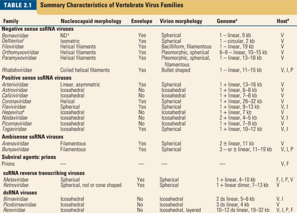

DNA Virus Families
* *
*
*nonenveloped

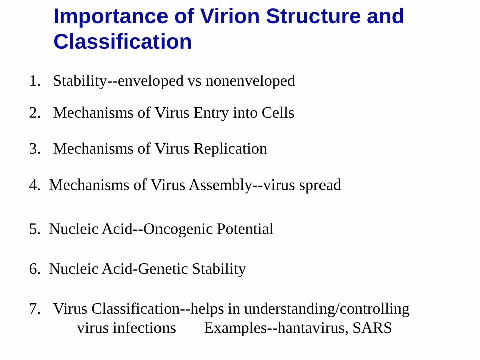
Importance of Virion Structure and Classification
1. Stability--enveloped vs nonenveloped
2. Mechanisms of Virus Entry into Cells
4. Mechanisms of Virus Assembly--virus spread
3. Mechanisms of Virus Replication
5. Nucleic Acid--Oncogenic Potential
6. Nucleic Acid-Genetic Stability
7. Virus Classification--helps in understanding/controlling virus infections Examples--hantavirus, SARS

Details of Virus Structure
Nonenveloped Enveloped

STRUCTURE OF NONENVELOPED VIRUSES Capsid---icosahedron 20 faces--each icosahedral triangle 12 vertices Repeating subunits (capsomeres) Simplest= 60 capsomeres Example--Picornaviruses simple-60 capsomeres capsomere=VP1, VP2, VP3, VP4 Example--Adenovirus much more complex Hexons, pentons (base, fiber, knob) hexon capsomere=3 hexons 180 capsomeres 12 pentons (one at each vertix) Example--reoviruses multiple capsids (shell within a shell)

edges
faces
vertices
Icosahedral structure of capsids

Picornavirus Structure Example of a simple, non-enveloped virus

Picornaviruses (small, RNA) Rhinoviruses---over 100 serotypes Enteroviruses polioviruses 3 serotypes Coxsackie viruses many serotypes Echoviruses many serotypes Enteroviruses several serotypes Aphthoviruses (FMDV) Cardioviruses (mice) Hepatoviruses--Hepatitis A
REACH

Picornavirus Structure Capsomere=VP1, VP2, VP3, VP4 Virion 60 capsomeres RNA 7.5 kb coding capacity?? single stranded positive polarity

Rhinovirus 14 VP1=blue VP2=green VP3=red VP4 inside

Cut away Inside VP4--yellow

Enveloped Virus Structure
Spikes--glycoproteins Membrane or matrix protein (some) Lipid Bilayer
Membrane:
Core: Capsid=iscosahedron eg flaviviruses, herpesviruses or Nucleocapsid=helical structure eg. Influenza, paramyxoviruses

Enveloped Virus Structure
Helical nucleocapsid Icosahedral
capsid
glycoproteins
genome
Lipid bilayer
Lipid bilayer

Enveloped virus with capsid core example togavirus

Helical nucleocapsid (inside envelope)
Intact virion Disrupted virion
Enveloped Virus with Helical Core

Enveloped Negative-stranded, RNA Genome Segmented Genome ***
30 INF/Morrison
Influenza—Structure

Influenza virus Envelope
Lipid Bilayer--membrane Four Proteins HA--hemagglutinin NA--neuraminidase M1--matrix M2--ion channel
HA
NA
M2
M1

Hemagglutinin--HA
Attachment to cell receptors sialic acid
Membrane Fusion--entry
Target of neutralizing antibody
Determinant of Organ Tropism--cleavage site HA0 cleaved to HA1 +HA2
32 INF/Morrison

Neuraminidase--NA
Cleaves sialic acid from glycoproteins and glycolipids
Important role in virus release
Target of antiviral drugs
33 INF/Morrison

M2
M1
Role in virus entry Ion channel
Small hydrophobic protein
Structural connection of envelope with core
Important in virus classification
Lines inner surface of membrane
34 INF/Morrison

Copyright ©2006 by the National Academy of Sciences
Harris, Audray et al. (2006) Proc. Natl. Acad. Sci. USA 103, 19123-19127
Distributions and shape-based differentiation of HA and NA spikes (cryo EM)

Influenza Virus Core RNA genome 8 segments of negative stranded RNA Associated Proteins NP (structural) PA, PB1, PB2 (virion associated polymerase)

Copyright ©2006 by the National Academy of Sciences
Harris, Audray et al. (2006) Proc. Natl. Acad. Sci. USA 103, 19123-19127
Central slices through influenza virions containing (7 + 1) solenoid-like RNP configurations

Organization of Genome in Influenza Virions
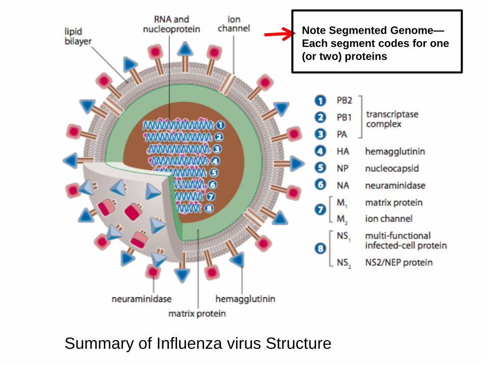
Diagram of Influenza Virions 39
Note Segmented Genome— Each segment codes for one (or two) proteins
Summary of Influenza virus Structure

Classification of Influenza Viruses
A B C
Pandemics broad host range humans, pigs, birds, seals
Epidemics humans only No pandemics
Endemic humans only
Important for epidemiology (and vaccination)
Classification based on NP and M proteins

Further Classification of Influenza A Viruses
Based on HA and NA
In nature 17 different HA genes (H1—H17) 9 different NA genes (N1-N9) Subtypes designated by which HA and which NA are present eg. H1N1 or H3N2
All exist in wild birds (H17, bats)
Divided into subtypes or species
Each subtype has many antigenic variants

Unique Epidemiology of Influenza: 1. Periodic Epidemics (regional) (seasonal flu) 2. Periodic Pandemics (world wide epidemics) (pandemic flu)
20-50 million deaths world wide 675,000 deaths in US
Early Pandemics? 1761-62 1833-37
1890 1900 1918 1957 1968 (1977) 2009
Documented Pandemics
42 INF/Morrison

Influenza A and Pandemics (pandemics caused by new subtype)
1890 1900 1918 1957 1968 2009
H2N2 H3N8
H1N1
H2N2
H3N2
Antigenic Shift
H1N1 43
INF/Morrison (Discuss H5N1, H7,N9 viruses)

Classification of Influenza B Viruses
No subtypes--same HA and NA
Many antigenic variants

Infection of Cells
1. Encounter--Virus in proximity of susceptible cells
2. Attachment--Virus Binding to Cell Surfaces
3. Entry--Insertion of Viral Genome into cells 4. Replication--Synthesis of components of Virions
5. Assembly--formation of progeny virions
6. Release--release of progeny virus from infected cells
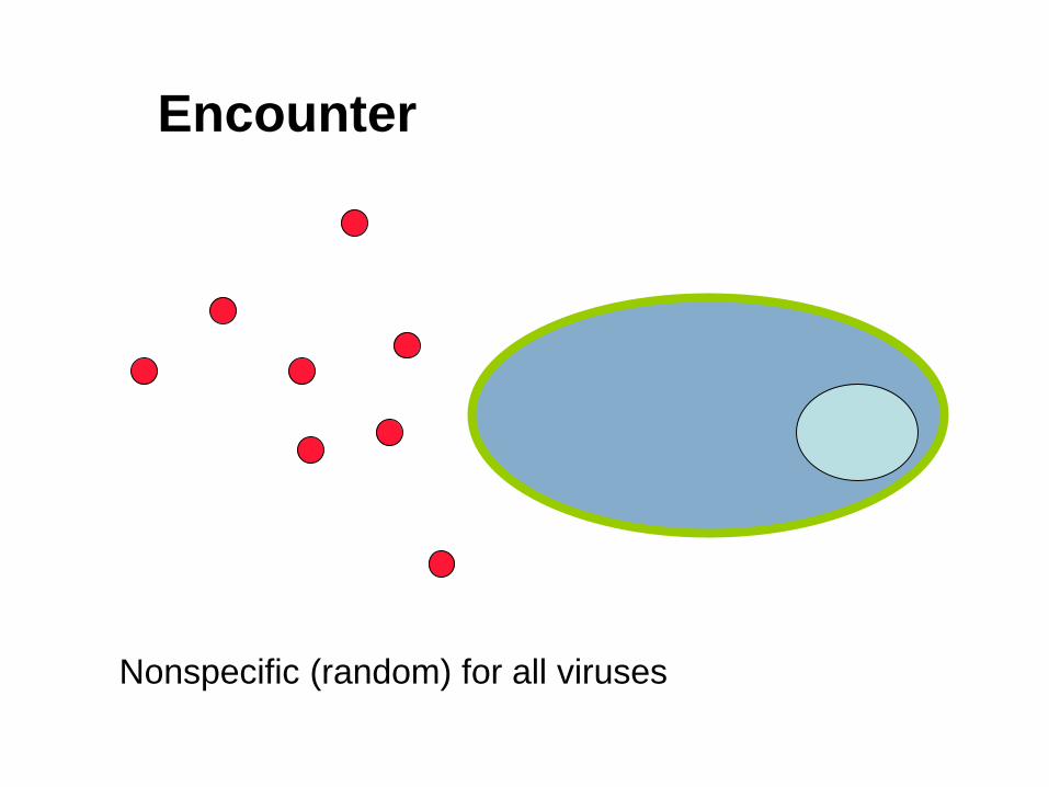
Encounter
Nonspecific (random) for all viruses

Encounter: Picornaviruses
vs Influenza viruses

Attachment Specific Interaction
VAP (virus attachment protein) nonenveloped-a capsid protein (Picornavirus—VP1) enveloped—a spike (glycoprotein) (Influenza virus—HA) Cell Receptor (specific to virus)--distribution determines (in part): species specificity of virus infection organ specificity of virus infection
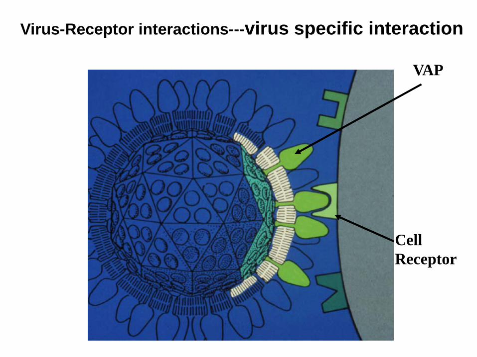
VAP
Cell Receptor
Virus-Receptor interactions---virus specific interaction

How are VAPs identified?
1. Antibodies
2. Mutation
3. Soluble protein
Picornavirus VAP=VP1 Influenza VAP=HA (hemagglutinin)
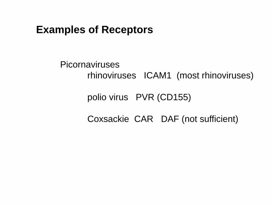
Examples of Receptors
Picornaviruses rhinoviruses ICAM1 (most rhinoviruses) polio virus PVR (CD155) Coxsackie CAR DAF (not sufficient)


Importance of Receptors Determinant of Species Specificity example polio virus (CD155—primate only) influenza A 2,3 linkage (avian) vs 2,6 linkage (human) of sialic acid Determinant of Organ specificity example HIV --CD4 expressing cells

How are receptors identified?
1. Antibodies
3. Soluble proteins 4. Inhibition of expression
Essential tool: permissive and nonpermissive cells (cell line expressing the receptor and cells not expressing the receptor) How identify?? 1st step in identifying potential receptors—ID potential candidates—Methods? Then, Verify. Tools:
2. cDNAs of candidates
Complications: co-receptors, attachment factors

Proof of that a receptor candidate is a receptor
1. Expression of candidate in permissive cells but not resistant cells
2. Transfection of resistant cells with vector expressing the candidate converts cells to permissive cells 3. Inhibition of expression of receptor candidate blocks virus infection/binding of virus 4. Direct interaction between VAP and receptor candidate
5. Soluble receptor competes with cells for virus binding

VAP-receptor Interactions A. Species and organ specificity B. VAP and Neutralizing antibody (definition) C. Neutralizing antibody binding vs receptor binding definition of serotype

Virus--antibody complexes
Neutralizing Antibody blocks attachment

Serotype--defined by neutralizing antibody
Virus A + anti-A Virus B+anti-B
Virus A+anti-B
No infection No infection Infection
All viruses inhibited by anti-A are same serotype (serotypeA) All viruses inhibited by anti-B are the same serotype (serotype B) which is different from serotype A

Implications of Different Serotypes of a Virus
1. Significant variability on the surfaces of virions
2. Infection due to one serotype will not protect against subsequent infection with another serotype For example, individuals may be infected with rhinoviruses many times since there are multiple serotypes of this virus
What does this mean structurally??

Rhinovirus 14 VP1=blue VP2=green VP3=red VP4 inside

Poliovirus
Depth cued VP1

Soluble ICAM1 bound to rhinovirus 14

Virus-receptor Interactions
Attachment to cell receptors

Rhinovirus 14 antigenic sites in pink (sites of Ab binding)

Virus-Neutralizing Antibody Interactions
Attachment to cell receptors
Ab neutralization
Ab blocks cell receptor-virion interactions

Evidence for Site of Antibody Binding/ Antibody Neutralization
Antibody escape mutants Directed mutations Crystallography

Hemagglutinin--HA is VAP
Attachment to cell receptors sialic acid
Membrane Fusion--entry
Target of neutralizing antibody
Determinant of Organ Tropism--cleavage site HA0 cleaved to HA1 +HA2
Influenza Virus-Receptor Interactions

Sialic acid binding Site in blue
HA of four different subtypes of Influenza (trimer of HA)
trimer

Neutralizing Ab (green) bound to HA trimer (blue and red)

Antibody binding sites vs sialic acid binding site
Influenza HA
trimer
Monomer Monomer

Antigenic Drift
Antigenic variants of one subtype
Cause of epidemics (not pandemics)

Antigenic Drift
Neutralizing antibody binding sites Amino acid change in each of five sites (a-d)= Antigenic variant (drift)
HA trimer HA Monomer
72 INF/Morrison

Pandemics Due to Antigenic Shift (completely new HA introduced into human population ) Mechanism will be defined in Lectures 4 and 5
Reading Fields Virology (edition 5) Chapters 2, 3 (pages 59-82), and 4 (pages 99-105) Chapters 24 (pages 796-808), 47 (pages 1647-1653)