viruses viruses are nucleic acids (dna/rna) wrapped in protein typically the protein coat, or...
Post on 18-Dec-2015
221 views
TRANSCRIPT
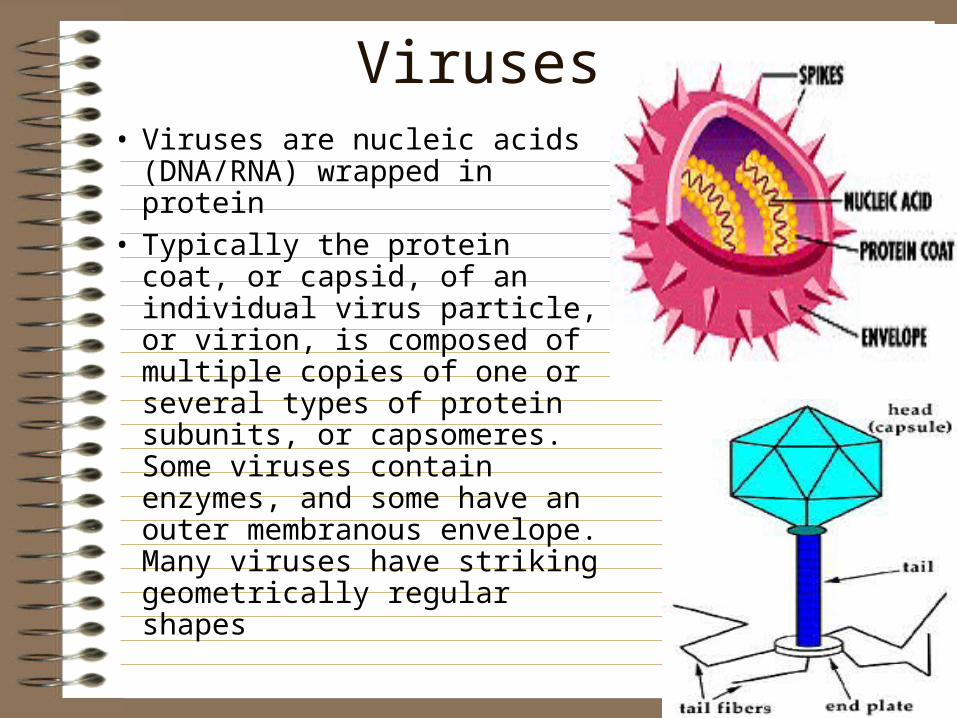
Viruses• Viruses are nucleic acids
(DNA/RNA) wrapped in protein
• Typically the protein coat, or capsid, of an individual virus particle, or virion, is composed of multiple copies of one or several types of protein subunits, or capsomeres. Some viruses contain enzymes, and some have an outer membranous envelope. Many viruses have striking geometrically regular shapes

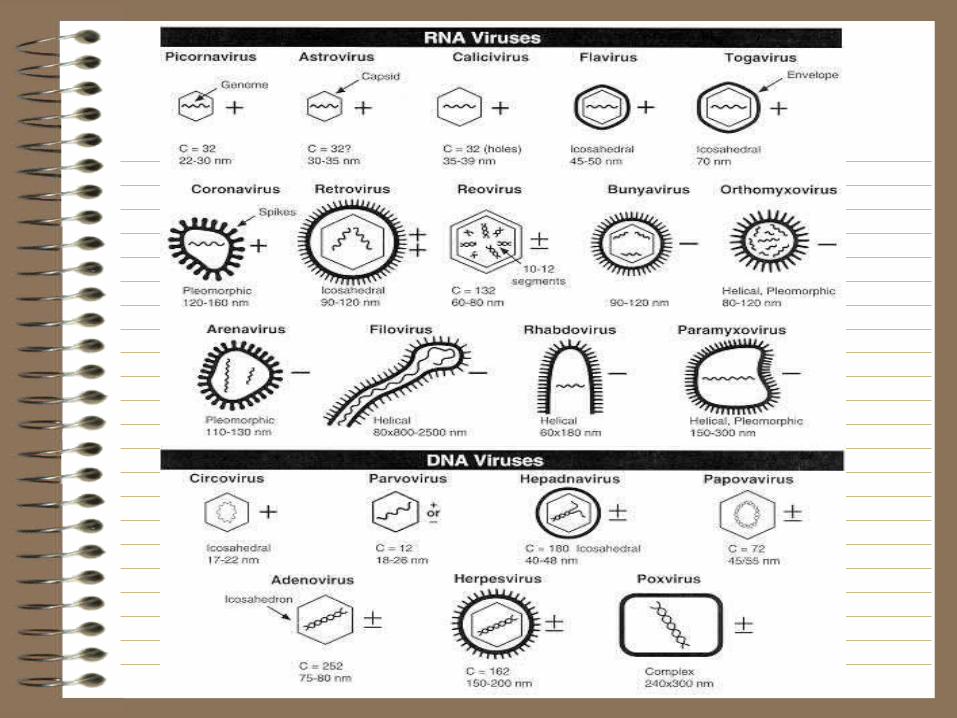
Virus Shapes3 main shapes:• Icosohedron/polyhedron: 20 triangular
sections (HIV)• Spiral: RNA surrounded by capsomere
proteins• Bacteriophage: spaceship• Others (ebola)

Types of Viruses• Viruses depend on the
host cells that they infect to reproduce. When found outside of host cells, viruses exist as a protein coat or capsid, sometimes enclosed within a membrane. The capsid encloses either DNA or RNA which codes for the virus elements.

Sizes of Viruses

Nucleic AcidPlus/Minus strand designation mRNA = +polarity
mRNA 5’ GAC UCG AGC 3’+DNA 5’ GAC TCG AGC 3’-DNA 5’ CTG AGC TCG 3’+RNA 5’ GAC UCG AGC 3’ (operates like mRNA)
-RNA 5’ CUG AGC UCG 3’ (euk. cells don’t have enz)
ssDNA(+)-DNA+mRNARetrovirus= RNAdsDNA using reverse transcriptase

Reverse transcription of retroviruses
Reverse Transcriptase 2 Classification of Viruses (reference)
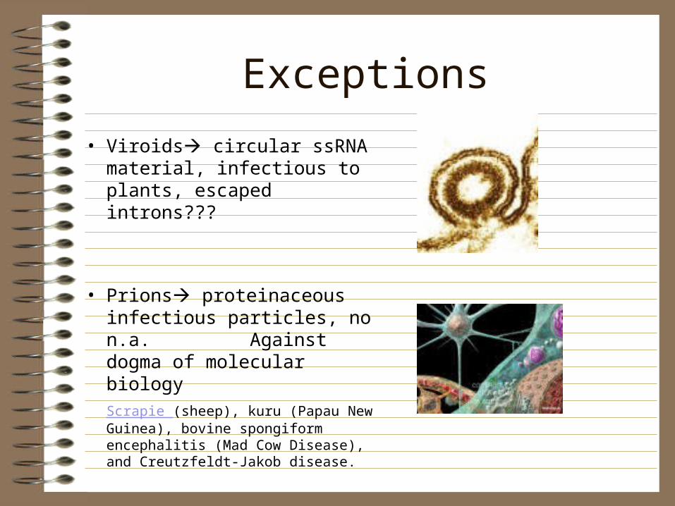
Exceptions
• Viroids circular ssRNA material, infectious to plants, escaped introns???
• Prions proteinaceous infectious particles, no n.a. Against dogma of molecular biology
Scrapie (sheep), kuru (Papau New Guinea), bovine spongiform encephalitis (Mad Cow Disease), and Creutzfeldt-Jakob disease.

Harmful Viruses• Viruses are notorious for
the plethora of diseases they cause, including influenza, rabies, AIDS, polio, herpes, ebola, measles, mumps, chicken pox, warts, small pox -->
Lewandowsky-Lutz/ 2Epidermodysplasia verruciformis,
herpes Warts

Helpful Viruses• Viruses carry out natural "genetic
engineering": by incorporating genetic material into its host
• This is known as transduction, and in some cases it may serve as a means of evolutionary change
• Certain varieties of flowers have been developed using viruses to alter the genetic code.
• Dr Patrick Lee uses reovirus to kill brain cancer cells transplanted into laboratory mice, while sparing normal, healthy cells. Clinical trials involving reovirus in people are now underway.
• Virus Rap

Helpful Viruses• A good virus• Most of us go out of our way to avoid viruses. But
Dr Patrick Lee - formerly of the University of Calgary and now at Dalhousie University - spends a lot of time in the company of a very common virus known as a reovirus. Normally this bug causes nothing more serious than a mild infection. But Dr Lee’s team discovered that the reovirus has the ability to kill brain cancer cells transplanted into laboratory mice, while sparing normal, healthy cells. Clinical trials involving reovirus in people are now underway.
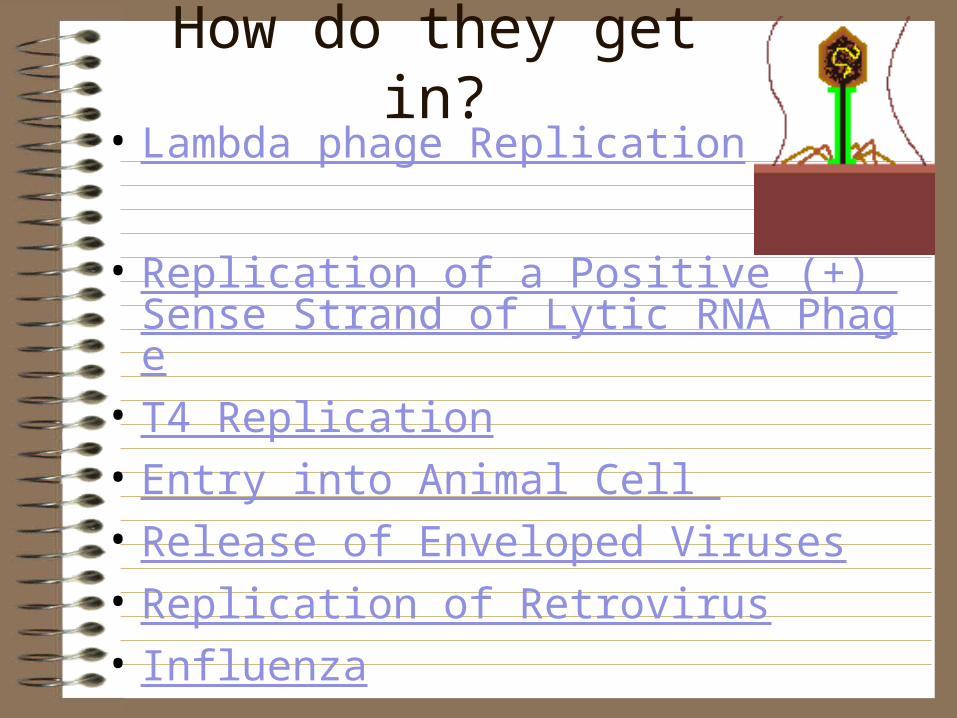
How do they get in?• Lambda phage Replication
• Replication of a Positive (+) Sense Strand of Lytic RNA Phage
• T4 Replication• Entry into Animal Cell • Release of Enveloped Viruses• Replication of Retrovirus• Influenza

Essential knowledge 3.C.3: Viral replication results in genetic variation, and viral infection can introduce
genetic variation into the hosts.
b. The reproductive cycles of viruses facilitate transfer of genetic information.1. Viruses transmit DNA or RNA when they infect a host cell.
• Transduction in bacteria (Specialized Transduction)• Transposons present in incoming DNA
2. Some viruses are able to integrate into the host DNA and establish a latent (lysogenic) infection. These latent viral genomes can result in new properties for the host such as increased pathogenicity in bacteria.
LO 3.29 The student is able to construct an explanation of how viruses introduce genetic variation in host organisms.
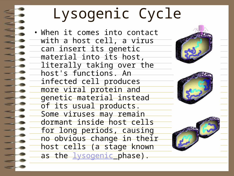
Lysogenic Cycle• When it comes into contact
with a host cell, a virus can insert its genetic material into its host, literally taking over the host's functions. An infected cell produces more viral protein and genetic material instead of its usual products. Some viruses may remain dormant inside host cells for long periods, causing no obvious change in their host cells (a stage known as the lysogenic phase).
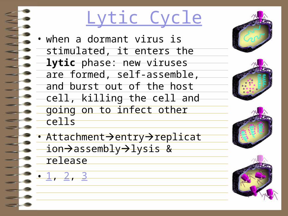
Lytic Cycle• when a dormant virus is
stimulated, it enters the lytic phase: new viruses are formed, self-assemble, and burst out of the host cell, killing the cell and going on to infect other cells
• Attachmententryreplicationassemblylysis & release
• 1, 2, 3

Essential knowledge 3.C.3: Viral replication results in genetic variation, and viral infection can introduce
genetic variation into the hosts.
a. Viral replication differs from other reproductive strategies and generates genetic variation via various mechanisms.
1. Viruses have highly efficient replicative capabilities that allow for rapid evolution and acquisition of new phenotypes. Viral Recombination , 1918 Spanish Flu, 2, 3
2. Viruses replicate via a component assembly model allowing one virus to produce many progeny simultaneously via the lytic cycle.
3. Virus replication allows for mutations to occur through usual host pathways.4. RNA viruses lack replication error-checking mechanisms, and thus have higher rates
of mutation.5. Related viruses can combine/recombine information if they infect the same host cell. (
Antigenic Shift)6. HIV is a well-studied system where the rapid evolution of a virus within the host
contributes to the pathogenicity of viral infection.
LO 3.30 The student is able to use representations and appropriate models to describe how viral replication introduces genetic variation in the viral population.
Life Cycle of HIV HIV Life Cycle 2 HIV Replication Future of HIV

Vaccines
• Constructing a Vaccine
• Engineering the Avian Flu
• 1918 Flu
• Malarial Vaccine • Virus Rap
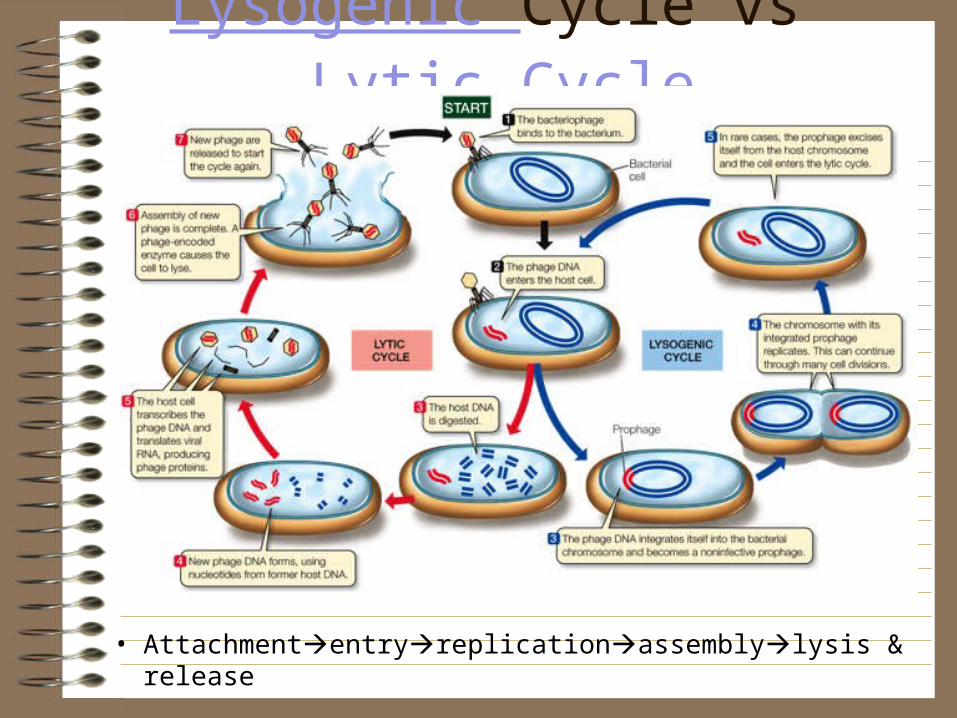
Lysogenic Cycle vs Lytic Cycle
• Attachmententryreplicationassemblylysis & release
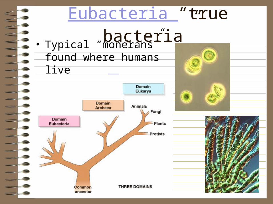
Eubacteria “true bacteria”• Typical “monerans” found
where humans live• Evolution of the 3 Domains

Archaea “archaic/old”extremophiles
• Methanogens• Thermophiles• Acidophiles• Halophiles• Alkaliphiles• Psychrophiles• Xerophiles• Barophiles
• Archae vs Bacteria

Bacteria Characteristics• Bacteria are
distinguished from other living things because of their cell structure:
• All bacterial cells have a cell wall surrounding a cell membrane, inside of which lies the unbound nuclear matter and other material.

• Bacteria have extra genomic DNA that is round and called a “plasmid”
• Plasmid Cloning

Bacteria: Classified by Shape• There are three types of bacterial cells, based on
shape: spherical (coccus), rodlike (bacillus), and spiral (spirillum).
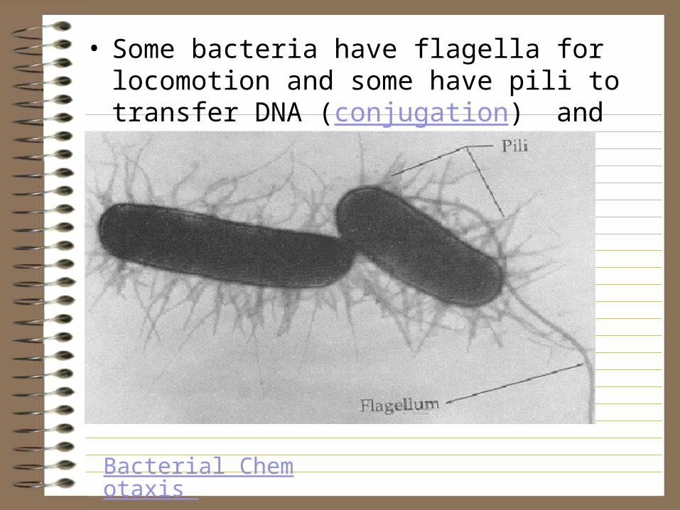
• Some bacteria have flagella for locomotion and some have pili to transfer DNA (conjugation) and to stick to substrates of host cells
Bacterial Chemotaxis

Harmful Bacteria• A number of bacteria
cause disease, these are called pathogenic bacteria.
• They can cause diseases of plants, animals, fungi, protists and other bacteria
• E. coli infection
• Salmonella infection
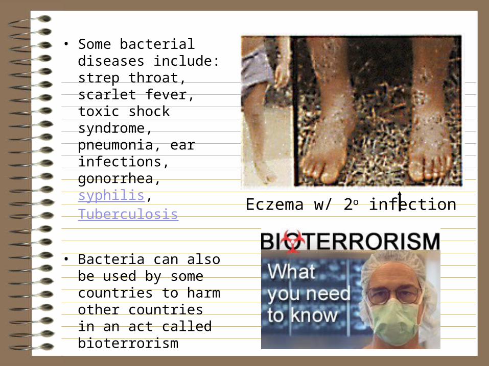
• Some bacterial diseases include: strep throat, scarlet fever, toxic shock syndrome, pneumonia, ear infections, gonorrhea, syphilis, Tuberculosis
• Bacteria can also be used by some countries to harm other countries in an act called bioterrorism
Eczema w/ 2o infection

Helpful Bacteria• actinomycetes,
produce antibiotics such as streptomycin and nocardicin
• live symbiotically in the guts of animals
• put the tang in yogurt and the sour in sourdough bread, cheese = spoiled milk
Symbiotic bacteria assist in digestion
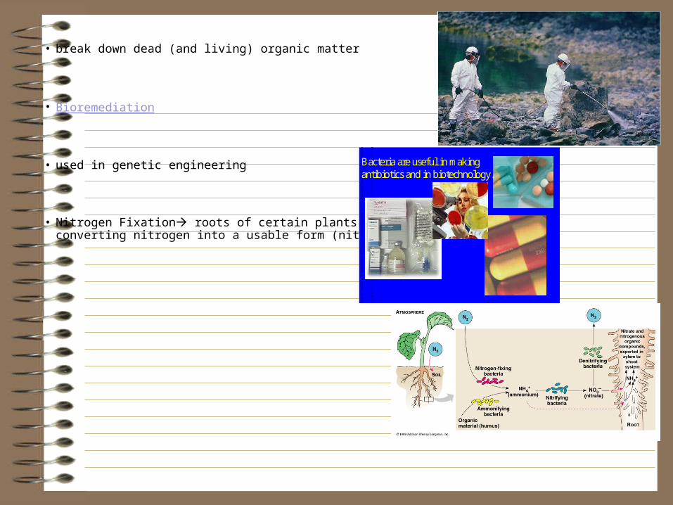
• break down dead (and living) organic matter
• Bioremediation
• used in genetic engineering
• Nitrogen Fixation roots of certain plants, converting nitrogen into a usable form (nitrate).
Wound infectionDefinition
of
wound
infection•1992
US
Centre
for
Disease
Control
•Defined
the
following:
•Surgical
site
infections
•Superficial
incisional
infection
•Deep
incisional
infections
•Organ
space
infections
•Surgical
site
infections
must
fulfill
the
following
criteria
•Infection
must
occur
within
30
days
of
surgery
•Infection
must
involve
only
the
skin
and
subcutaneous
tissue
•There
must
be
at
least
one
of
the
following
•Purulent
discharge
from
a
superficial
infection
•Organisms
isolated
from
aseptically
obtained
wound
culture
•Must
be
at
least
one
of
the
following
signs
of
infection
•Pain
or
tenderness
•Localised
swelling
•Redness
or
heat
Predisposing
factors•General
factors
•Age,
obesity,
malnutrition
•Endocrine
and
metabolic
disorders
•Hypoxia,
anaemia
•Malignant
disease
•Immunosupression
•Local
factors
•Necrotic
tissue
•Foreign
bodies
•Tissue
ischaemia
•Haematoma
formation
•Poor
surgical
technique
•Microbiological
contamination
•Type
and
virulence
of
organism
•Size
of
bacte
riological
dose
•Antibiotic
resistance
Aerobic
pathogens
in
wound
infections•Staphylococcus
aureus
(17%)
•Enterococci
(13%)
•Coagulase-negative
staphylococci
(12%)
•Escherichia
coli
(10%)
•Pseudomonas
aeruginosa
(8%)
•Enterobacter
species
(8%)
•Proteus
mirabilis
(4%)
•Klebsiella
pneumoniae
(3%)
•Candida
species
(2%)
Prevention
of
wound
infection•Exogenous
•Sterilisation
of
instruments,
sutures
etc
•Positive
pressure
ventilation
of
operating
theatres
•Laminar
air
flow
in
high
risk
areas
•Exclusion
of
staff
with
infections
•Endogenous
•Skin
preparation
•Mechanical
bowel
preparation
•Antibiotic
prophylaxis
•Good
surgical
technique
Wound
infection
rates•Risk
of
wound
infection
varies
with
type
of
surgery
•Infection
rate
can
be
reduced
with
antibiotic
prophylaxis
Clean
surgery•No
viscus
opened
(e.g.
hernia
repair)
•Infection
rate
typically
1-2%
Clean-contaminated•Viscus
opened
but
no
spillage
of
gut
contents
(e.g.
right
hemicolectomy)
•Infection
rate
usually
<10%
Contaminated•Viscus
opened
with
inflammation
or
spillage
of
contents
(e.g.
colectomy
for
obstruction)
•Infection
rate
15-20%
Dirty•Intraperitoneal
abscess
formation
or
visceral
perforation
•Infection
rate
40%
Antibiotic
prophylaxis
•Prophylaxis
is
the
use
of
antibiotics
to
prevent
infection
•Treatment
is
their
use
to
eradicate
established
sepsis.
•Prophylaxis
important
in:
•Surgery
with
a
high
incidence
of
post-operative
infection
(e.g.
colonic
surgery)
•Surgery
where
infection
would
be
hazardous
(e.g.
prosthetic
valves)
•Need
to
consider:
•The
use
of
an
appropriate
antibiotic
based
on
likely
bacteria
and
tissue
penetration
•Cefuroxime
&
metronidazole
for
colonic
surgery
•Benzylpenicillin
for
peripheral
vascular
surgery
•Timing
and
duration
of
administration
•Intravenous
administration
at
induction
•Number
of
doses
-
usually
no
more
than
three
doses
BibliographyHoran
T
C,
Gaynes
R
P,
Martone
W
J,
Jarvis
W
R,
Emon
T
G.
CDC
definitions
of
nosocomial
surgical
site
infections,
1992:
a
modification
of
CDC
definitions
of
surgical
wound
infections.
Am
J
Infect
Control
1992;
20:
271-274.McDonald
M,
Grabsch
E,
Marshall
C,
Forbes
A.
Single-versus
multiple-dose
antimicrobial
prophylaxis
for
major
surgery:
a
systematic
review.
Aust
N
Z
J
Surg
1998;
68:
388-396.
Bacteria are useful in making antibiotics and in biotechnology.

Producers in Geothermal Vents
Reproduction
• Bacteria reproduce asexually by binary fission• Bacterial Conjugation (lateral/horizontal gene exchange)
• Bacterial Transformation (lateral/horizontal gene exchange)
• Bacteria life cycle

BB CheckpointBB#1SB1a. Explain the role of cell organelles for both prokaryotic and eukaryotic cells, including the cell membrane, in maintaining homeostasis and cell reproduction.
BB#8 SB2e. Compare the advantages of sexual reproduction and asexual reproduction in different situations
BB#10 SB3b. Compare how structures and function vary between the six groups (archaebacteria, eubacteria, protists, fungi, plants, and animals).

Kingdom Protista• All protists are eukaryotes.
This means that their cells contain a nucleus, a membrane-bounded structure that encloses the
cell's genetic material. • Some protists are
autotrophs like plants, others are consumers like animals. Unlike plants and animals, however, protists do not have cells organized into specialized tissues.

Protista Classified by Nutrition• The first detailed descriptions of
protists were made in 1676 by the inventor of the microscope, Dutch naturalist Leewenhoek.
• The classification is currently based on the structure and organization of the cell, the presence of organelles, and the pattern of reproduction or life cycles. The five-kingdom system divides the Protista into 27 phyla. However, classifications based on DNA sequences suggest that many protist phyla may be sufficiently large and diverse to be classified as kingdoms.
• Gallimaufry, cornucopia, hodge-podge, potpourri

• Auto trophic Protists are called “Algae”. Scientists believe they gave rise to the kingdome Plantae
• Ingestive Heterotrophic protists are called “Proto zoa”. Scientists believe they gave rise to the kingdom Animalia
• Absorptive heterotrophic protists are called “Slimemolds”. Scientists believe they gave rise to the kingdom Fungi
• Protist Rap

Harmful Protists• Produce a nerve poison in
shellfish that kills humans and fish in red tide
• Cause diseases: Chaga’s disease, Malaria, 2, Lyme disease, diarrhea, toxoplasmosis, dysentary, Trypanosomaisis, 2, Leishmaniasis, 2, Toxoplasma, Cryptospiridium, Leishmaniasis, Brain Amoeba
• Cause mold and mildew which can spoil food and cause allergic reactions
• Cause algal blooms which can result in eutrophication

Beneficial Protists• Used as insect pathogens
• Used in ice cream, soups, nori (seaweed in sushi), jello, agar, vitamin supplements, or eaten as a sea vegetable
• Ancient dinoflagellates formed oil deposits
• Bioluminescent
• Diatoms mined for fine abrasives in silver polish and toothpaste and as packing in air and water filters
• Marine phytoplankton make up ~70% of the oxygen on the planet
• Forensic uses: Diatom Detectives
• Algae for Biofuel

BB CheckpointBB#9SB3a. Explain the cycling of energy through the processes of photosynthesis and respiration.
BB#10 SB3b. Compare how structures and function vary between the six groups (archaebacteria, eubacteria, protists, fungi, plants, and animals).

Fungi: Multicellular absorptive heterotrophs
• Though they grow in soil like plants, they are not autotrophic.
• The have cell walls made of the polysaccharide chitin
• What are the cell walls of plants made of? Bacteria?

Fungi AnatomyHyphae basic structural unit of a fungus made up of branching filaments
Mycelium tangled network of fibers
Fruiting body reproductive structure. In Phylum Basidiomycota it is the mushroom itself

Fungi: Classified by ReproductionDivision Zygomycota form zygospores i.e. Rhizopus (bread mold)
Division Ascomycota form ascospores i.e. yeast, morels, ergot, Dutch elm disease
Division Basidiomycota Most commonly known, forms basidiospores i.e. shelf fungi, mushrooms
Division Deuteromycota/ Imperfecti sexual reproduction unknown i.e. Penicillium, Aspergillus

Fungi Life Cycle
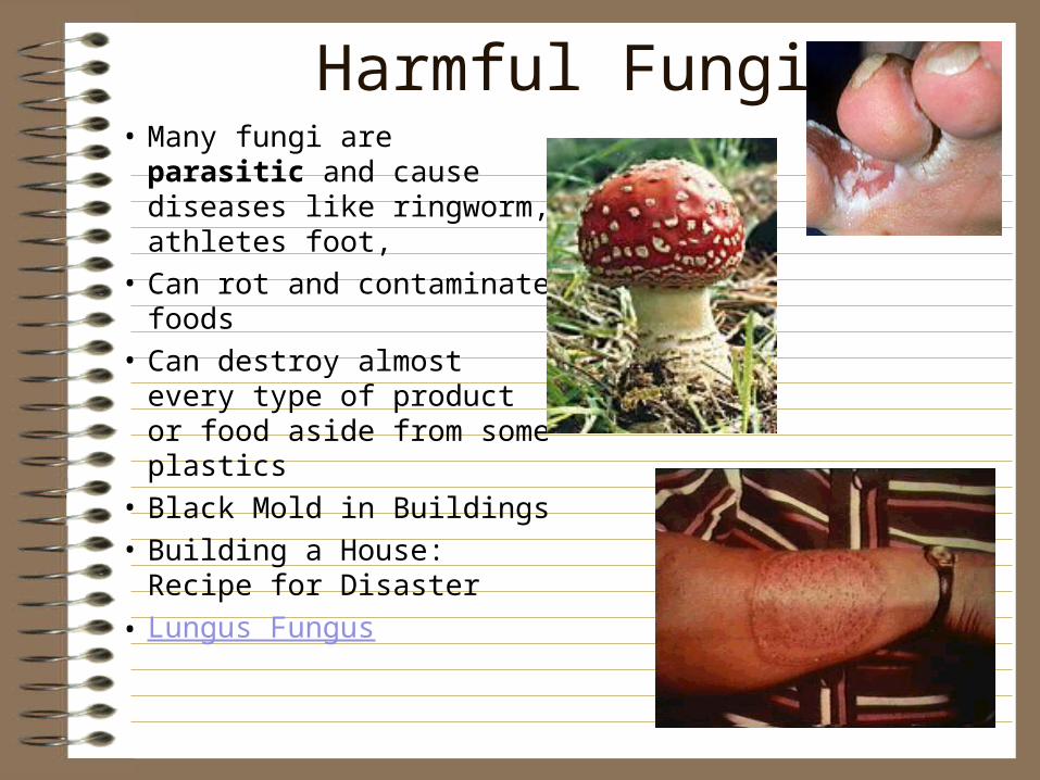
Harmful Fungi• Many fungi are parasitic
and cause diseases like ringworm, athletes foot,
• Can rot and contaminate foods
• Can destroy almost every type of product or food aside from some plastics
• Black Mold in Buildings
• Building a House: Recipe for Disaster
• Lungus Fungus
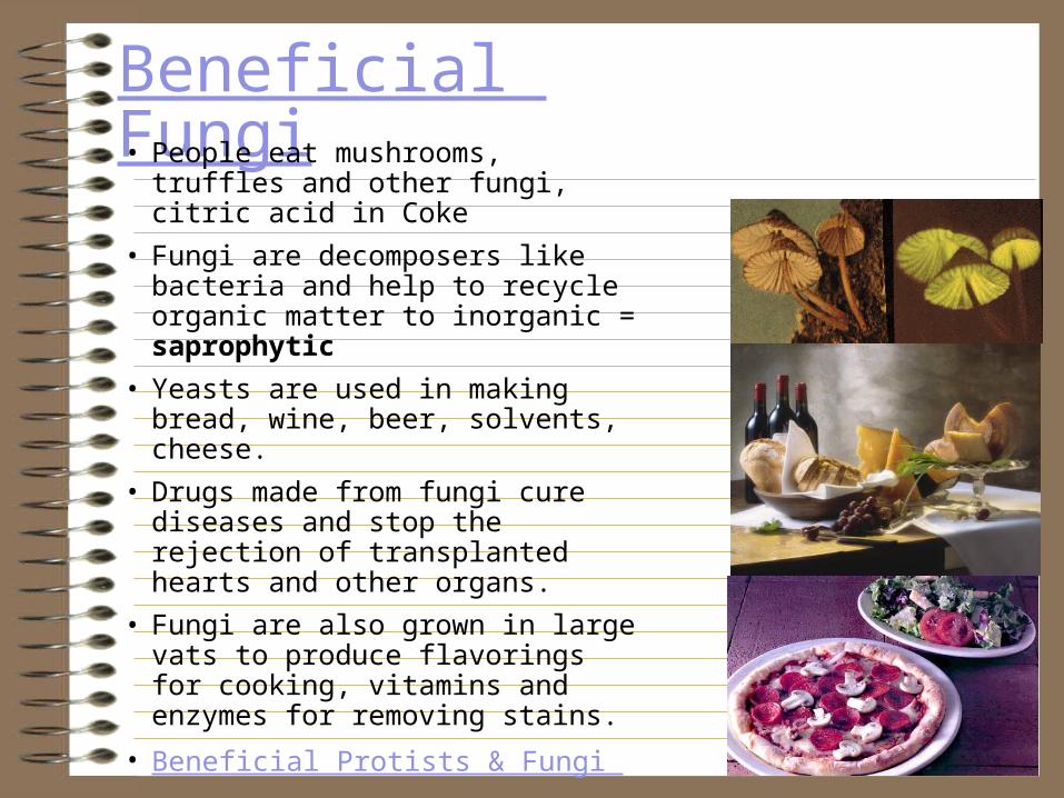
Beneficial Fungi• People eat mushrooms, truffles and
other fungi, citric acid in Coke
• Fungi are decomposers like bacteria and help to recycle organic matter to inorganic = saprophytic
• Yeasts are used in making bread, wine, beer, solvents, cheese.
• Drugs made from fungi cure diseases and stop the rejection of transplanted hearts and other organs.
• Fungi are also grown in large vats to produce flavorings for cooking, vitamins and enzymes for removing stains.
• Beneficial Protists & Fungi

Fungi Engage in Symbiosis
• Parasitic +/-: Mind control
• Mutualistic +/+: Lichens, a pioneer organism, a fungus and algae living together
• The mycorrhizal fungi live as partners with plants, helping them absorb nutrients
• Predatory +/-: Arthrobotrys, a deuteromycete

BB CheckpointBB#9SB3a. Explain the cycling of energy through the processes of photosynthesis and respiration.
BB#10 SB3b. Compare how structures and function vary between the six groups (archaebacteria, eubacteria, protists, fungi, plants, and animals).
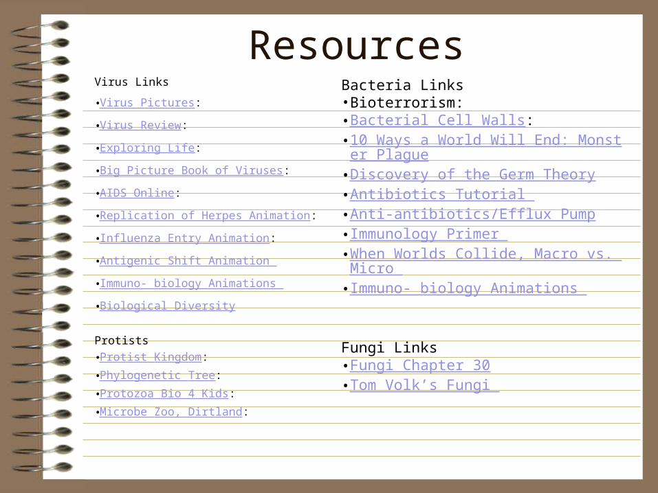
ResourcesVirus Links
•Virus Pictures:
•Virus Review:
•Exploring Life:
•Big Picture Book of Viruses:
•AIDS Online:
•Replication of Herpes Animation:
•Influenza Entry Animation:
•Antigenic Shift Animation
•Immuno- biology Animations
•Biological Diversity
Protists •Protist Kingdom: •Phylogenetic Tree: •Protozoa Bio 4 Kids: •Microbe Zoo, Dirtland:
Bacteria Links•Bioterrorism: •Bacterial Cell Walls: •10 Ways a World Will End: Monster Plague•Discovery of the Germ Theory•Antibiotics Tutorial •Anti-antibiotics/Efflux Pump•Immunology Primer •When Worlds Collide, Macro vs. Micro •Immuno- biology Animations
Fungi Links•Fungi Chapter 30•Tom Volk’s Fungi
Virus LinksVirus Pictures:
Virus Review:
Exploring Life:
Big Picture Book of Viruses:
AIDS Online:
Replication of Herpes Animation:
Influenza Entry Animation:
Antigenic Shift Animation
Immuno- biology Animations
Chapter 6 Viruses
Introduction to Plasmids & Viruses
Bozeman Viral ReplicationSuper Flu: Antigenic shift in influenza