viral genome replication || herpesvirus genome replication
TRANSCRIPT

Chapter 13Herpesvirus Genome Replication
Sandra K. Weller
The Herpesviruses
The Herpesviridae are a large family of enveloped, double-stranded DNA virusesthat are responsible for many human and veterinary diseases. Herpesviruses caninfect mammals, birds, and reptiles, and so far, eight distinct family members havebeen found which infect humans including herpes simplex virus type 1 (HSV-1) andherpes simplex virus type 2 (HSV-2), Epstein–Barr virus (EBV), cytomegalovirus(HCMV), varicella-zoster virus (VZV), human herpesvirus 6 (HHV-6), human her-pesvirus 7 (HHV-7), and Kaposi’s sarcoma herpesvirus (KSHV). All members ofthe family are capable of both lytic and latent infections although they differ greatlyin tissue tropism and in many aspects of their interactions with their hosts. Theseviruses share many aspects of virion structure (they all are T = 16), genomic orga-nization, mechanisms of DNA replication and life cycle. Herpes simplex viruses 1and 2 (HSV-1 and HSV-2) are the most extensively studied of all the herpesviruses,in part because they are most amenable to genetic and biochemical approaches. Thischapter will focus primarily on herpes simplex virus type 1 (HSV-1); however, otherhuman herpesviruses will be discussed when their replication strategy differs in sig-nificant detail from simplex viruses.
Overall Virus Life Cycle
HSV initiates viral infection by specific binding of viral glycoproteins to cell sur-face glycosaminoglycans and cellular receptors (Spear 2004). Following entry, cap-sids have been shown to translocate along microtubules (Sodeik et al. 1997) tothe nuclear pores where they dock and presumably eject their genomes into thenucleus (Ojala et al. 2000). A tightly regulated cascade of gene expression occurs
S.K. Weller (B)Department of Molecular, Microbial and Structural Biology, University of Connecticut HealthCenter, 263 Farmington Avenue, Farmington, CT 06030-3205, USAe-mail: [email protected]
C.E. Cameron et al. (eds.), Viral Genome Replication,DOI 10.1007/b135974 13, C© Springer Science+Business Media, LLC 2009
249
250 S.K. Weller
consisting of three well-defined kinetic classes of genes: immediate early, early,and late. Viral gene expression, DNA replication, and encapsidation occur withinglobular domains in the nucleus termed replication compartments (Knipe 1989;Lamberti and Weller 1996). Herpesvirus genomes replicate in the nucleus throughthe formation of longer-than-unit length concatemers. Monomeric units are cleavedfrom concatemers during a packaging reaction which occurs in conjunction withthe uptake of genomes into preassembled capsids. DNA-containing capsids exitthe nucleus by budding through the nuclear membrane, and they acquire their finalenvelope and mature glycoproteins by a series of envelopment and de-envelopmentsteps (reviewed in [Baines and Weller 2004]). Unlike other enveloped viruses, her-pesviruses are known to package approximately 20–30 proteins between the capsidand the envelope in a region termed the tegument or “skin”. Many tegument pro-teins are known to play active roles in the earliest stages of infection as they arebrought in with infecting virions into newly infected cells and play regulatory rolesin shut off of host protein synthesis and the stimulation of immediate-early viralgene expression.
Herpesvirus Genomic Structure
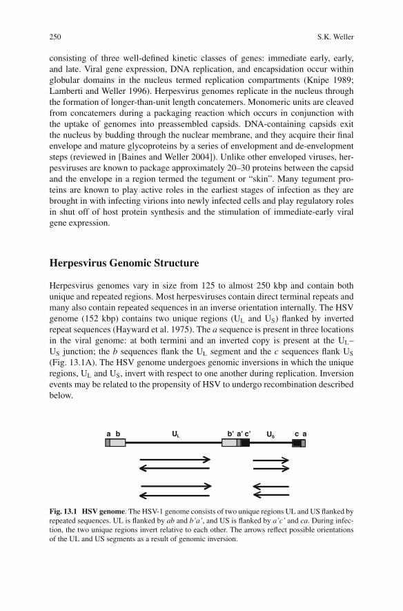
Herpesvirus genomes vary in size from 125 to almost 250 kbp and contain bothunique and repeated regions. Most herpesviruses contain direct terminal repeats andmany also contain repeated sequences in an inverse orientation internally. The HSVgenome (152 kbp) contains two unique regions (UL and US) flanked by invertedrepeat sequences (Hayward et al. 1975). The a sequence is present in three locationsin the viral genome: at both termini and an inverted copy is present at the UL–US junction; the b sequences flank the UL segment and the c sequences flank US
(Fig. 13.1A). The HSV genome undergoes genomic inversions in which the uniqueregions, UL and US, invert with respect to one another during replication. Inversionevents may be related to the propensity of HSV to undergo recombination describedbelow.
aa c'bb c'a'UL US
Fig. 13.1 HSV genome. The HSV-1 genome consists of two unique regions UL and US flanked byrepeated sequences. UL is flanked by ab and b’a’, and US is flanked by a’c’ and ca. During infec-tion, the two unique regions invert relative to each other. The arrows reflect possible orientationsof the UL and US segments as a result of genomic inversion.
13 Herpesvirus Genome Replication 251
Gene Expression and Regulation
HSV-1 is believed to encode over 80 open reading frames, and gene expression isvery tightly temporally controlled. Viral genes are classified as immediate early,early, or late and are transcribed from both strands of the viral genome. Most ofthe immediate-early genes encode regulatory proteins while the early gene productsare primarily involved in viral DNA replication. Many of the structural proteinsare encoded as late genes which are not expressed until viral DNA synthesis hasoccurred.
Cis- and Trans-Acting DNA Replication Factors
Origins of Replication
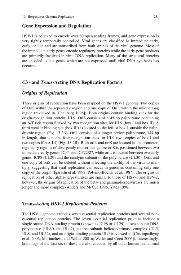
Three origins of replication have been mapped on the HSV-1 genome: two copiesof OriS within the repeated c region and one copy of OriL within the unique longregion (reviewed in [Challberg 1996]). Both origins contain binding sites for theorigin-recognition protein, UL9. OriS consists of a 45-bp palindrome containingan A/T-rich region flanked by two recognition sites for UL9 (box I and box II). Athird weaker binding site (box III) is located to the left of box 1 outside the palin-dromic region (Fig. 13.2A). OriL consists of a longer perfect palindrome, 144 bpin length, that contains four recognition sites for UL9 (two copies of box I andtwo copies if box III) (Fig. 13.2B). Both oriL and oriS are located in the promoter-regulatory regions of divergently transcribed genes: oriS is positioned between twoimmediate-early genes, ICP4 and ICP22/27, while oriL is located between two earlygenes, ICP8 (UL29) and the catalytic subunit of the polymerase (UL30). OriL andone copy of oriS can be deleted without affecting the ability of the virus to mul-tiply, suggesting that viral replication can occur in genomes containing only onecopy of the origin (Igarashi et al. 1993; Polvino-Bodnar et al. 1987). The origins ofreplication of other alpha-herpesviruses are similar to those of HSV-1 and HSV-2;however, the origins of replication of the beta- and gamma-herpesviruses are muchlonger and more complex (Anders and McCue 1996; Yates 1996).
Trans-Acting HSV-1 Replication Proteins
The HSV-1 genome encodes seven essential replication proteins and several non-essential replication proteins. The seven essential replication proteins include asingle-strand DNA-binding protein (known as ICP8 or UL29), a two subunit DNApolymerase (UL30 and UL42), a three subunit helicase/primase complex (UL5,UL8, and UL52), and an origin-binding protein UL9 (reviewed in [Chattopadhyayet al. 2006; Marintcheva and Weller 2001a; Weller and Coen 2006]). Interestingly,homologs of the first six of these are also encoded by all other human and animal
252 S.K. Weller
B
A
Fig. 13.2 Origins of DNA replication. A. OriS can be depicted with three recognition sites forUL9, the origin-binding protein (boxes I, II and III, marked in gray) and an AT-rich linker (markedin black) positioned between boxes I and II. OriS is positioned between two immediate-early tran-scripts. B. OriL is positioned between two divergently transcribed early mRNAs. Recognition sitesfor UL9 are boxed in gray, and the AT-rich regions are boxed in black. Since OriL is a perfectpalindrome, the recognition sequences are designated I and III on each side of the palindrome.
herpesviruses and are considered to function as the “core” replication proteins withall the necessary enzymatic activities to stimulate DNA replication on a primed invitro replication substrate. The conservation of functions suggests that the overallstrategy of lytic viral DNA replication is conserved within this family. The mecha-nism and regulation of initiation of viral DNA synthesis, however, is probably sharedonly by the alpha-herpesviruses. As mentioned above, the origins of replication forbeta- and gamma-herpesviruses are more complex, and in addition, no clear UL9homologs have been identified.
In addition to the six core replication proteins and the origin-recognition protein,HSV encodes a number of proteins which are not essential for viral DNA synthe-sis including enzymes involved in nucleotide biosynthesis and DNA metabolismsuch as thymidine kinase, ribonucleotide reductase, uracil–DNA–glycosylase,
13 Herpesvirus Genome Replication 253
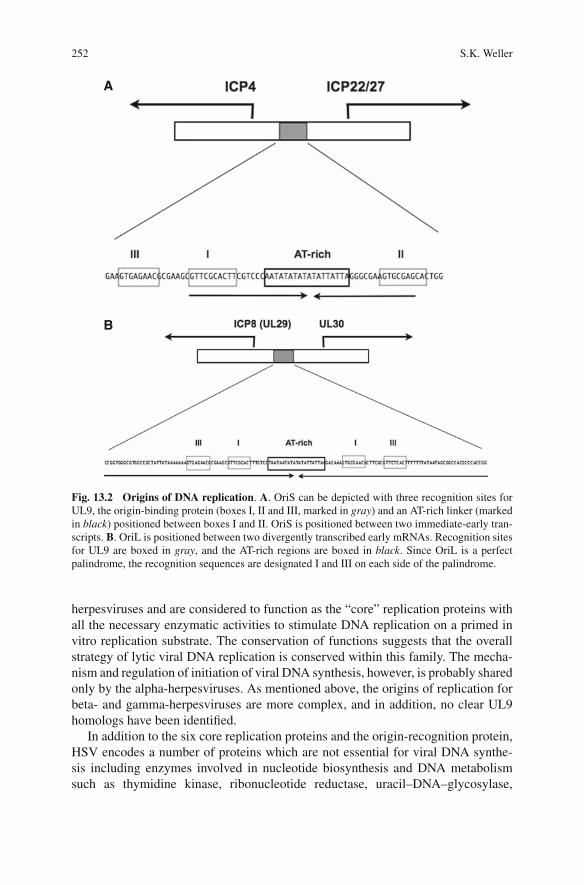
Table 13.1 Auxiliary HSV DNA replication genes
Gene Alternate abbreviation Major function
Essential for DNAreplication incultured cells?
UL23 TK Thymidine kinase NoUL39 RR1 Large subunit of
ribonucleotide reductaseNo
UL40 RR2 Small subunit ofribonucleotide reductase
No
UL2 UNG Uracil-DNA glycosylase NoUL50 dUTPase Deoxyuridine
triphosphataseNo
UL12 Alkaline exonuclease Putative viral recombinasesubunit
No
deoxyuridine triphosphatase, and the alkaline nuclease (Table 13.1) (reviewed in[Weller and Coen 2006]). Ribonucleotide reductase consists of two subunits, RR1and RR2, and both are needed for enzymatic activity whose function is to producedNTPs used in DNA synthesis. Recent reports raise the interesting possibility thatthe HSV RR1 subunit may also act as a chaperone perhaps to prevent the induc-tion of apoptosis and/or to promote the assembly of the translational machinery(Chabaud et al. 2003; Langelier et al. 2002; Perkins et al. 2002; Walsh and Mohr2006). The deoxyuridine triphosphatase (dUTPase) and uracil–DNA–glycosylasemay be important in preventing misincorporation of uracil residues into the viralgenome. The HSV alkaline nuclease (UL12) is a 5′ to 3′ exonuclease; in combina-tion with ICP8, UL12 is capable of a strand exchange activity in vitro, and these twoproteins may play a role in single-strand annealing (SSA) during infection (Reuvenet al. 2004a, 2003) (see below).
HSV-1 Origin-Binding Protein, UL9 (94 kDa)
Genetic analysis indicates that the origin-binding protein UL9 is essential for viralDNA replication (Carmichael et al. 1988). The analysis of temperature-sensitive (ts)mutants indicates, however, that UL9 is required early in HSV-1 infection but notlate in infection, once DNA synthesis has initiated (Schildgen et al. 2005). Activitiesassociated with UL9 include nucleoside triphosphatase, DNA helicase on partiallydouble stranded substrates, ability to form dimers in solution and ability to bindcooperatively to viral origins (reviewed in (Chattopadhyay et al. 2006; Weller &Coen 2006)). The seven conserved helicase motif characteristic of the SF2 familyof helicases reside in the N-terminal domain (residues 1–534) while the domainresponsible for specific origin binding has been mapped to the C-terminal one-third of UL9 (residues 564–832) (reviewed in [Chattopadhyay et al. 2006]). Geneticanalysis demonstrated that the conserved helicase motifs are essential for both the
254 S.K. Weller
in vivo and the in vitro ATPase and helicase activities of UL9 (Malik and Weller1996; Marintcheva and Weller 2003a, b, 2001b). UL9 has been reported to interactwith several other viral proteins including ICP8, UL8 (a component of the trimerichelicase/primase), and UL42 (the DNA polymerase accessory protein) (reviewed in[Weller and Coen 2006]).
UL30/UL42
HSV DNA polymerase comprises a catalytic subunit (UL30) and an accessory sub-unit which stimulates processivity (UL42) (Anders and McCue 1996). The catalyticsubunit, UL30, is a member of the alpha-like DNA polymerase family and pos-sesses a 3′–5′ exonuclease activity. Thus, UL30 contains motifs conserved in otherpolymerases, and mutations in these motifs have been shown to affect binding ofdNTPs and/or their incorporation (Huang et al. 1999). The crystal structure of UL30has been solved confirming that this protein resembles other alpha-like DNA poly-merases (Liu et al. 2006).
The processivity subunit UL42 functions by an unusual mechanism. While otherprocessivity subunits such as PCNA operate as “sliding clamps”, which form mul-timeric rings in solution and are loaded onto DNA with the aid of clamp loaders,UL42 binds to DNA by itself as a monomer with relatively high affinity (Randell andCoen 2004; Weisshart et al. 1999). UL42 can diffuse linearly (slide) on DNA despiteits high affinity for DNA (Randell and Coen 2001) and has recently been shown toaffect replication fidelity (Jiang et al. 2007). Interestingly, the crystal structure ofUL42 suggests that it shares a similar overall structural fold with the processivityfactors which function as sliding clamps (Zuccola et al. 2000).
UL5/UL8/UL52
The HSV-1 helicase/primase is a heterotrimer consisting of the products of the UL5,UL8, and UL52 genes, and genetic data indicate that all three are essential for viralDNA replication in cell culture (reviewed in [Marintcheva and Weller 2001a; Wellerand Coen 2006]). The HSV-1 UL5/8/52 complex exhibits DNA-dependent ATPase,primase, and helicase activities (reviewed in [Chattopadhyay et al. 2006; Weller andCoen 2006]). UL5 and UL52 together possess all known enzymatic activities whileUL8 appears to play a stimulatory role in addition to being able to interact withother members of the replication machinery including UL9, HSV Pol, and ICP8(reviewed in [Chattopadhyay et al. 2006; Weller and Coen 2006]). The sequence ofUL5 reveals seven motifs found in a large helicase superfamily, SF1, and geneticanalysis indicates that these motifs are essential for viral DNA replication (Zhu andWeller 1992) and for helicase and ATPase activities in vitro (Graves-Woodwardet al. 1997; Graves-Woodward and Weller 1996). UL52 contains a DXD signa-ture motif conserved in many primases, and this motif is essential for viral DNA
13 Herpesvirus Genome Replication 255
replication and for primase activity in vitro (Dracheva et al. 1995; Klinedinst andChallberg 1994). The C-terminus of HSV-1 UL52 contains a putative zinc-bindingmotif, which is also present in prokaryotic and eukaryotic primases (Carrington-Lawrence and Weller 2003; Chen et al. 2005, 2007). The presence of these motifssuggests that the helicase and primase activities of the complex likely reside in theUL5 and UL52 subunits, respectively; however, several lines of evidence suggest amore complex interaction between these two subunits. Mutations in the putative zincfinger at the C-terminus of UL52 have been shown to abrogate not only primase butalso ATPase, helicase, and DNA-binding activities of the UL5/UL52 subcomplex(Biswas and Weller 1999, 2001; Chen et al. 2005). These results suggest that UL52binding to DNA via the zinc finger may be necessary for loading UL5 onto DNA.Alternatively, it is possible that UL5 and UL52 share a DNA-binding site created byinteraction between the two subunits.
UL29 or ICP8
ICP8, encoded by the UL29 gene, is the HSV major single-strand DNA-bindingprotein and is essential for DNA replication. ICP8 (SSB) is a 130-kDa zincmetalloprotein that preferentially binds ssDNA in a non-sequence specific and coop-erative manner. ICP8 exhibits helix-destabilizing activities which are thought toplay a role in unwinding duplex DNA during DNA synthesis. ICP8 interacts withmany viral and cellular proteins (reviewed in [Challberg 1996]). In particular, ICP8has been reported to enhance biochemical activities of UL9, the helicase/primase,and polymerase ([Arana et al. 2001; Boehmer 1998; Hamatake et al. 1997] andrefs therein). ICP8 also interacts with the viral nuclease UL12 and together theseproteins exhibit a strand-annealing reaction ([Reuven and Weller 2005] and refstherein). ICP8 has also been reported to regulate viral gene expression by repress-ing transcription from the parental genome and stimulating late gene expressionfrom progeny genomes ([McNamee et al. 2000] and refs therein).
The structure of ICP8 lacking its C-terminal 60 residues was recently solved,revealing two separate domains, the N- (aa 9–1038) and the C- (aa 1049–1129) ter-minal domain, connected by a short linker region of loose electron density (Mapelliet al. 2005). The N-terminal domain forms three regions: the head, neck, and shoul-der which in turn are formed from non-contiguous secondary structural elements.
Overview of HSV DNA Replication
Although several cis- and trans-acting elements have been shown to be requiredfor HSV DNA replication as described above, very little is known about the actualmechanisms of HSV DNA replication. The model presented below is for the mostpart consistent with existing data; however, validation will require additional exper-imental support.

256 S.K. Weller
Fate of Incoming Viral DNA
HSV-1 DNA genomes enter the nucleus following docking of the capsid at a nuclearpore. Once the linear viral DNA is released it is thought that these linear DNAmolecules lose their free ends by a process that does not require de novo protein syn-thesis (Poffenberger and Roizman 1985). The simplest interpretation of these resultsis that the viral genome loses its free ends through the formation of a covalently closedcircular molecule leading to the model that linear virion DNA is rapidly circularizedin infected cells. Aspects of this model have recently been challenged experimentally(Jackson and DeLuca 2003), and considerable controversy still surrounds the fate ofthe incoming viral DNA soon after infection (Strang and Stow 2005). It is possiblethat the viral genome adopts an endless configuration by an intra- or intermolecu-lar homologous recombination event; however, additional experimentation will beneeded to determine the precise fate of the incoming viral genome.
Initiation of Viral DNA Replication
Current models suggest that the origin-recognition protein, UL9, binds one or moreof the origins of replication and recruits the rest of the replication machinery tothe origin, stimulating DNA synthesis. Although DNA synthesis is probably initi-ated by a UL9-dependent step, replication at the origins of HSV alone would notbe sufficient to generate the observed head-to-tail concatemers; therefore, we haveproposed that HSV DNA replication occurs in two stages (Fig. 13.3) (Weller andCoen 2006; Wilkinson and Weller 2003). We have proposed that UL9 is essentialduring the first stage; however, later in infection DNA replication appears to pro-ceed in an origin-independent manner (Blumel et al. 2000; Blumel and Matz 1995),which may proceed by rolling circle replication and/or a recombination-dependentreplication step (Wilkinson and Weller 2003). The involvement of recombinationwould be analogous to the replication program of the bacteriophages T4, lambda,and many other linear dsDNA bacteriophage.
The precise role of UL9 in the initiation of HSV DNA replication remains uncer-tain. Although UL9 is known to possess ATPase and helicase activities in addi-tion to its origin-binding properties, it is unable to unwind blunt-ended linear orcircular double-stranded DNA containing an origin of replication. The failure ofUL9 to unwind duplex origin DNA remains a major impediment to the estab-lishment of an in vitro DNA replication system. Although the details are not yetknown, it is believed that UL9 acts in conjunction with ICP8 to distort or perturbthe region, followed by the recruitment of the HSV-1 helicase/primase (H/P) com-plex (Fig. 13.4). We have demonstrated that an active primase is needed to recruitthe HSV DNA polymerase to viral foci in infected cells. Once the HSV DNA poly-merase is recruited to the fork, it is believed to be responsible for both leading andlagging strand DNA syntheses. The first stage of DNA synthesis may be bidirec-tional proceeding from one or more of the three origins of replication. The end prod-ucts of DNA replication are longer-than-unit-length head-to-tail DNA concatemers.

13 Herpesvirus Genome Replication 257
Stage 1
Stage 2
Fig. 13.3 Model for UL9-dependent and UL9-independent HSV DNA replication. Accordingto this model the first stage of HSV DNA replication involves origin unwinding and bidirectionalDNA replication (Stage I). In this step, UL9 likely acts in conjunction with ICP8. Following theopening, the helicase/primase can be recruited to the complex followed by either recombination-dependent or rolling circle replication (or both) (Stage II). Light gray symbols represent UL9 anddark gray symbols represent ICP8, the single-strand DNA-binding protein.
Fig. 13.4 HSV-1 replication fork. An HSV-1 replication fork would be expected to contain thehelicase/primase complex (UL5/UL52/UL8) at the fork: UL5 would be expected to unwind duplexDNA ahead of the fork and UL52 would be expected to lay down RNA primers which could thenbe extended by the two subunit DNA polymerase (UL30 and UL42). The HSV-1 pol would alsobe expected to carry out leading strand synthesis. ICP8 (UL29, SSB) would be expected to bind tossDNA generated during HSV DNA synthesis.

258 S.K. Weller
Replication Intermediates Are Complex and Branched
Although direct evidence is lacking, several lines of evidence suggest thatrecombination-dependent replication occurs during the second stage of viral DNAsynthesis. (i) We and others have shown that replication intermediates in HSV-1-infected cells are present in a non-linear structure which cannot enter a pulsedfield gel, even after digestion with a restriction enzyme which recognizes asingle-restriction site within the HSV genome (reviewed in [Wilkinson and Weller2003]). This complex, perhaps branched, structure is consistent with recombination-dependent replication. (ii) Inversion of the unique regions of the HSV genome hasoccurred at the earliest times that replicated DNA can be detected (Lamberti andWeller 1996). (iii) Severini et al. (1996) isolated DNA from the well of a pulsedfield gel, and following restriction enzyme digestion, fragments were subjected totwo-dimensional gel electrophoresis; both Y-shaped arches and X-shaped junctionswere observed (Severini et al. 1996). (iv) It has been shown that SV40 DNA repli-cates by theta replication, resulting in interlocked circles. SV40 DNA replicated bythe HSV-1 core replication machinery in infected cells, however, adopts a complex-branched DNA indistinguishable from that of replicating HSV DNA (Blumel et al.2000). These results taken together suggest that replicating DNA adopts a complex,most likely branched, structure consistent with the involvement of recombination-dependent replication.
The demonstration that HSV encodes a two subunit complex consisting ofICP8 and UL12 which can perform strand exchange is also consistent with arecombination-dependent replication mechanism of DNA replication. This two sub-unit complex may play a role analogous to that of the red recombinase systemencoded by bacteriophage lambda (Reuven et al. 2003; Reuven and Weller 2005;Reuven et al. 2004b). Furthermore, ICP8 has been reported to interact either directlyor indirectly with over 50 cellular and viral proteins, some of which play roles, suchas repair and recombination (Taylor and Knipe 2004). In addition, we have shownthat UL12 interacts with Nbs1, a component of the MRN complex known to play animportant role in cellular homologous recombination (Balasubramanian and Weller,manuscript in preparation). Taken together, the ability of ICP8 and UL12 to mediatestrand annealing and strand transfer and to interact with viral and cellular repair andrecombination proteins is consistent with the suggestion that DNA recombinationplays a role in HSV DNA replication (reviewed in [Wilkinson and Weller 2003]).Further experimental evidence will be required to test this model.
HSV DNA Replication at the Cellular Level
HSV-1 DNA replication occurs in large globular replication compartments (RCs)in the nucleus of infected cells, and viral gene expression and DNA replicationare thought to occur within these domains (Knipe 1989). ICP8 is believed toplay a major role in nuclear events leading to the formation of RCs (Taylor and
13 Herpesvirus Genome Replication 259
Knipe 2003), and recent work by Everett and colleagues has led to a more refinedmodel of the earliest steps of infection (Everett and Murray 2005; Everett et al. 2007,2004). HSV genomes enter the nucleus and appear to cause the recruitment of cel-lular proteins of cellular ND10 proteins. ND10 (nuclear domains 10, also known aspromyelocytic leukemia nuclear bodies or PODs) are defined by the accumulationof PML and many other cellular proteins involved in growth control, gene expres-sion, and possibly DNA recombination and the DNA damage response (Dellaire andBazett-Jones 2007, 2004). The genomes of several DNA viruses which replicate inthe nucleus including SV40, adenoviruses, and other herpesviruses such as HCMValso appear to recruit ND10 proteins (reviewed in [Everett 2006]). The formationof ND10-like foci at viral genomes may reflect an antiviral cellular mechanism torepress expression of the viral genome, consistent with reports that many ND10 pro-teins are transcriptional repressors and are found at silenced regions of the chromatin(regions of heterochromatin) (Fernandez-Capetillo et al. 2003; Turner 2007; Turneret al. 2005). In support of this notion, PML was found to play a role in mediatingthe antiviral effects of IFN treatment in both HSV and HCMV infections (Everett2006; Tavalai et al. 2006). Alternatively or in addition, it is possible that ND10components are attracted to viral genomes because the genome is seen as a dam-aged DNA molecule in need of repair; many ND10 proteins have functions in DNArepair and recombination (Dellaire and Bazett-Jones 2007, 2004). Thus ND10 com-ponents may play dual roles in responding to damage and induction of silencing.
HSV Induces Disruption of ND10s
HSV is believed to counter the antiviral action of ND10 recruitment by disruptingND10 by the action of ICP0, an immediate-early gene product. ICP0 is an E3 ubiq-uitin ligase (Boutell et al. 2003, 2002), and its ability to disrupt ND10 is thoughtto be a result of its ability to degrade sumoylated isoforms of PML and SP100,two components of ND10. The disruption of ND10 and the removal of at leastsome ND10 proteins may relieve the repressive activities of some ND10 proteinsand thus provide an environment conducive to viral gene expression and viral DNAreplication.
Prereplicative Sites
Replication compartments (RC) form rapidly during infection, and the only wayto identify subassemblies of viral proteins important for RC formation is to freezethe progression of infection either by infection with viruses bearing mutations inreplication proteins or in the presence of pharmacological agents which inhibitviral DNA synthesis (Burkham et al. 1998; Carrington-Lawrence and Weller 2003;Wilkinson and Weller 2004). We have defined five stages of infection based on theintracellular localization of viral and cellular proteins which are believed to play a
260 S.K. Weller
role in the formation of RCs. Stage I is defined by the recruitment of ND10 pro-teins to viral genomes as described above, and during this stage, no ICP8 foci canbe detected by immunofluorescence microscopy. In cells that are in Stage II, ND10have been disrupted and ICP8 can be detected. Although we originally reported thatICP8 is diffusely localized in Stage II, more sensitive microscopy has now revealedthat microfoci of ICP8 are present at this stage and that these ICP8 microfoci arepositioned adjacent to ICP4 foci (Livingston et al. 2008). Cells in Stage III con-tain a limited number of ICP8-containing foci whose formation is dependent on thepresence of UL5, UL8, UL9, and UL52 (Burkham et al. 1998). Stage III can bedivided into two: Stage IIIa foci contain the five viral proteins ICP8, UL5, UL8,UL9, and UL52, whereas Stage IIIb foci contain these five proteins along withHSV Pol, UL42, and PML. As mentioned above, the recruitment of the polymeraseholoenzyme to the five protein scaffold requires the presence of an active primasesubunit (Carrington-Lawrence and Weller 2003). If replication is allowed to pro-ceed, replication compartments are observed which can be detected with both theICP8 and the PML antibodies (stage IV). Thus, it appears that viral and cellularproteins assemble to prereplicative sites in an ordered manner to initiate viral DNAreplication.
Host–Cell Interactions
Although the viral cis- and trans-acting factors necessary for viral replication havebeen identified, we know very little about the role of host proteins in viral DNAreplication. Several cellular proteins are rearranged following viral infection: someare recruited into replication compartments and some are sequestered in foci adja-cent to RCs called VICE (virus-induced chaperone-enriched) domains (Burch andWeller 2004; Wilkinson and Weller 2006). Several questions remain, however. Dothe cellular proteins which are recruited to RCs such as RPA, Rad51, MRN proteins,and hsp90 play a direct role in viral DNA replication (Wilkinson and Weller 2006)?What is the function of the VICE domains, and why are some cellular proteinssequestered there, including the phosphorylated form of RPA, the ATR interactionprotein (ATRIP), and the heat-shock protein hsc70? What are the roles of host pro-teins which have been identified as interaction partners for viral-replication proteins,such as the transcriptional coactivator HCF-1 (T. Kristie, personal communication),polymerase alpha-primase (Lee et al. 1995), and a neural F-box protein NFB42which may play a role in ubiquitin-dependent degradation (Eom et al. 2004; Eomand Lehman 2003)? The biological significance of these interactions is not clear. Itis possible that several cellular proteins play direct roles in either the initiation or thelater stages of viral DNA replication. It is intriguing to speculate that the reason ithas not been possible to recapitulate origin-dependent HSV DNA synthesis in vitrois that one or more host cellular proteins may be required. Cellular proteins whichinteract with viral proteins may be co-opted by the virus for various purposes suchas subversion of antiviral defenses or the prevention of apoptosis. With the advent

13 Herpesvirus Genome Replication 261
of siRNA technology, it should be possible to address some of these unansweredquestions about the involvement of host proteins in viral DNA replication.
Encapsidation of Viral Genomes
Viral DNA packaging into virions is a multistep process involving resolution ofreplication and/or recombination intermediates, specific cleavage events, packaginginto preassembled capsids. The steps involved in this process are highly analogousto those of the more extensively studied DNA bacteriophages including (i) the for-mation of a procapsid intermediate consisting of a capsid shell initially supportedby an internal scaffold, (ii) replacement of the internal scaffold with viral DNA,(iii) insertion of DNA through a unique portal vertex, and (iv) generation of unitlength molecules by endonucleolytic cleavage of complex DNA concatemers by theactivity of a two-component terminase. Several HSV gene products are involved inthese steps including a terminase composed of the UL15 and UL28 proteins and aportal protein (UL6), which forms an oligomeric ring through which the viral DNAis taken up during the packaging reaction (reviewed in [Baines and Weller 2004]).
Summary
Although cis- and trans-acting viral proteins have been identified and their functionsdetermined, many questions about the actual mechanism of HSV DNA replicationand the involvement of host proteins remain unanswered. It is important to addressthese questions in part because viral proteins required for viral DNA replicationprovide very attractive targets for antiviral chemotherapy, and agents such as acy-clovir and its derivatives which target the viral thymidine kinase and the viral DNApolymerase have been very successful. As with many other therapies, however, drugresistance is a very real threat which limits efficacy. Because most of the replicationproteins discussed in this chapter are common to all the Herpesviridae, it is antic-ipated that new information generated here will be applicable to all herpesviruses.The helicase/primase has already been exploited as an antiviral target: two classesof highly potent helicase/primase inhibitors have been reported recently (Kleymann,2003 #1608).
References
Anders, D. G., and L. A. McCue. 1996. The human cytomegalovirus genes and proteins requiredfor DNA synthesis. Intervirology 39: 378–388.
Arana, M. E., B. Haq, N. Tanguy Le Gac, and P. E. Boehmer. 2001. Modulation of the herpessimplex virus type-1 UL9 DNA helicase by its cognate single-strand DNA-binding protein,icp8. J Biol Chem 276: 6840–6845.
Baines, J., and S. K. Weller. 2004. Cleavage and packaging of herpes simplex virus 1 DNA. In:C. Catalano (ed.) Virus packaging No. in press. Landes Bioscience, Georgetown.

262 S.K. Weller
Biswas, N., and S. K. Weller. 1999. A mutation in the c-terminal putative zn2+ finger motif ofUL52 severely affects the biochemical activities of the hsv-1 helicase-primase subcomplex.J Biol Chem 274: 8068–8076.
Biswas, N., and S. K. Weller. 2001. The UL5 and UL52 subunits of the herpes simplex virus type1 helicase-primase subcomplex exhibit a complex interdependence for DNA binding. J BiolChem 276: 17610–17619.
Blumel, J., S. Graper, and B. Matz. 2000. Structure of simian virus 40 DNA replicated by herpessimplex virus type 1. Virology 276: 445–454.
Blumel, J., and B. Matz. 1995. Thermosensitive UL9 gene function is required for early stages ofherpes simplex virus type 1 DNA synthesis. J Gen Virol 76 (Pt 12): 3119–3124.
Boehmer, P. E. 1998. The herpes simplex virus type-1 single-strand DNA-binding protein, ICP8,increases the processivity of the UL9 protein DNA helicase. J Biol Chem 273: 2676–2683.
Boutell, C., A. Orr, and R. D. Everett. 2003. Pml residue lysine 160 is required for the degra-dation of pml induced by herpes simplex virus type 1 regulatory protein ICP0. J Virol 77:8686–8694.
Boutell, C., S. Sadis, and R. D. Everett. 2002. Herpes simplex virus type 1 immediate-early proteinICP0 and is isolated ring finger domain act as ubiquitin e3 ligases in vitro. J Virol 76: 841–850.
Burch, A. D., and S. K. Weller. 2004. Nuclear sequestration of cellular chaperone and proteasomalmachinery during herpes simplex virus type 1 infection. J Virol 78: 7175–7185.
Burkham, J., D. M. Coen, and S. K. Weller. 1998. Nd10 protein pml is recruited to herpes simplexvirus type 1 prereplicative sites and replication compartments in the presence of viral DNApolymerase. J Virol 72: 10100–10107.
Carmichael, E. P., M. J. Kosovsky, and S. K. Weller. 1988. Isolation and characterization of herpessimplex virus type 1 host range mutants defective in viral DNA synthesis. J Virol 62: 91–99.
Carrington-Lawrence, S. D., and S. K. Weller. 2003. Recruitment of polymerase to herpes simplexvirus type 1 replication foci in cells expressing mutant primase (UL52) proteins. J Virol 77:4237–4247.
Chabaud, S. et al. 2003. The R1 subunit of herpes simplex virus ribonucleotide reductase haschaperone-like activity similar to hsp27. FEBS Lett 545: 213–218.
Challberg, M. 1996. Herpesvirus DNA replication. In: M. DePamphilis (ed.) DNA replication ineukaryotic cells. p 721–750. Cold Spring Harbor Press, Cold Spring Harbor.
Chattopadhyay, S., Y. Chen, and S. K. Weller. 2006. The two helicases of herpes simplex virustype 1 (HSV-1). Front Biosci 11: 2213–2223.
Chen, Y., S. D. Carrington-Lawrence, P. Bai, and S. K. Weller. 2005. Mutations in the putativezinc-binding motif of UL52 demonstrate a complex interdependence between the UL5 andUL52 subunits of the human herpes simplex virus type 1 helicase/primase complex. J Virol 79:9088–9096.
Chen, Y., C. M. Livingston, S. D. Carrington-Lawrence, P. Bai, and S. K. Weller. 2007. A mutationin the human herpes simplex virus type 1 UL52 zinc finger motif results in defective primaseactivity but can recruit viral polymerase and support viral replication efficiently. J Virol 81:8742–8751.
Dellaire, G., and D. P. Bazett-Jones. 2004. Pml nuclear bodies: Dynamic sensors of DNA damageand cellular stress. Bioessays 26: 963–977.
Dellaire, G., and D. P. Bazett-Jones. 2007. Beyond repair foci: Subnuclear domains and the cellularresponse to DNA damage. Cell Cycle 6: 1864–1872.
Dracheva, S., E. V. Koonin, and J. J. Crute. 1995. Identification of the primase active site of theherpes simplex virus type 1 helicase-primase. J Biol Chem 270: 14148–14153.
Eom, C. Y., W. D. Heo, M. L. Craske, T. Meyer, and I. R. Lehman. 2004. The neural f-box proteinnfb42 mediates the nuclear export of the herpes simplex virus type 1 replication initiator protein(UL9 protein) after viral infection. Proc Natl Acad Sci USA 101: 4036–4040.
Eom, C. Y., and I. R. Lehman. 2003. Replication-initiator protein (UL9) of the herpes simplexvirus 1 binds nfb42 and is degraded via the ubiquitin-proteasome pathway. Proc Natl Acad SciUSA 100: 9803–9807.

13 Herpesvirus Genome Replication 263
Everett, R. D. 2006. Interactions between DNA viruses, ND10 and the DNA damage response.Cell Microbiol 8: 365–374.
Everett, R. D., and J. Murray. 2005. ND10 components relocate to sites associated with herpessimplex virus type 1 nucleoprotein complexes during virus infection. J Virol 79: 5078–5089.
Everett, R. D., J. Murray, A. Orr, and C. M. Preston. 2007. Herpes simplex virus type 1 genomesare associated with ND10 nuclear sub-structures in quiescently infected human fibroblasts.J Virol. 81: 10991–11004.
Everett, R. D., G. Sourvinos, C. Leiper, J. B. Clements, and A. Orr. 2004. Formation of nuclear fociof the herpes simplex virus type 1 regulatory protein ICP4 at early times of infection: Local-ization, dynamics, recruitment of ICP27, and evidence for the de novo induction of ND10-likecomplexes. J Virol 78: 1903–1917.
Fernandez-Capetillo, O. et al. 2003. H2ax is required for chromatin remodeling and inactivation ofsex chromosomes in male mouse meiosis. Dev Cell 4: 497–508.
Graves-Woodward, K. L., J. Gottlieb, M. D. Challberg, and S. K. Weller. 1997. Biochemical analy-ses of mutations in the hsv-1 helicase-primase that alter ATP hydrolysis, DNA unwinding, andcoupling between hydrolysis and unwinding. J Biol Chem 272: 4623–4630.
Graves-Woodward, K. L., and S. K. Weller. 1996. Replacement of gly815 in helicase motif valters the single-stranded DNA-dependent ATPase activity of the herpes simplex virus type 1helicase-primase. J Biol Chem 271: 13629–13635.
Hamatake, R. K., M. Bifano, W. W. Hurlburt, and D. J. Tenney. 1997. A functional interaction ofICP8, the herpes simplex virus single-stranded DNA-binding protein, and the helicase-primasecomplex that is dependent on the presence of the UL8 subunit. J Gen Virol 78 (Pt 4): 857–865.
Hayward, G. S., R. J. Jacob, S. C. Wadsworth, and B. Roizman. 1975. Anatomy of herpes simplexvirus DNA: Evidence for four populations of molecules that differ in the relative orientationsof their long and short components. Proc Natl Acad Sci USA 72: 4243–4247.
Huang, L. et al. 1999. The enzymological basis for resistance of herpesvirus DNA polymerasemutants to acyclovir: Relationship to the structure of alpha-like DNA polymerases. Proc NatlAcad Sci USA 96: 447–452.
Igarashi, K., R. Fawl, R. J. Roller, and B. Roizman. 1993. Construction and properties of a recom-binant herpes simplex virus 1 lacking both s-component origins of DNA synthesis. J Virol 67:2123–2132.
Jackson, S. A., and N. A. DeLuca. 2003. Relationship of herpes simplex virus genome configura-tion to productive and persistent infections. Proc Natl Acad Sci USA 100: 7871–7876.
Jiang, C., Y. T. Hwang, J. C. Randell, D. M. Coen, and C. B. Hwang. 2007. Mutations that decreaseDNA binding of the processivity factor of the herpes simplex virus DNA polymerase reduceviral yield, alter the kinetics of viral DNA replication, and decrease the fidelity of DNA repli-cation. J Virol 81: 3495–3502.
Klinedinst, D. K., and M. D. Challberg. 1994. Helicase-primase complex of herpes simplex virustype 1: A mutation in the UL52 subunit abolishes primase activity. J Virol 68: 3693–3701.
Knipe, D. M. 1989. The role of viral and cellular nuclear proteins in herpes simplex virus replica-tion. Adv Virus Res 37: 85–123.
Lamberti, C., and S. K. Weller. 1996. The herpes simplex virus type 1 UL6 protein is essential forcleavage and packaging but not for genomic inversion. Virology 226: 403–407.
Langelier, Y. et al. 2002. The R1 subunit of herpes simplex virus ribonucleotide reductase protectscells against apoptosis at, or upstream of, caspase-8 activation. J Gen Virol 83: 2779–2789.
Lee, S. S., Q. Dong, T. S. Wang, and I. R. Lehman. 1995. Interaction of herpes simplexvirus 1 origin-binding protein with DNA polymerase alpha. Proc Natl Acad Sci USA 92:7882–7886.
Livingston, C. M., N. Deluca, D. E. Wilkinson, and S. K. Weller. 2008. The formation of foci ofICP8, the single strand DNA binding protein of HSV-1, requires the oligomerization of ICP4.J. Virol. 82: 6324–6336.
Liu, S. et al. 2006. Crystal structure of the herpes simplex virus 1 DNA polymerase. J Biol Chem281: 18193–18200.
264 S.K. Weller
Malik, A. K., and S. K. Weller. 1996. Use of transdominant mutants of the origin-binding protein(UL9) of herpes simplex virus type 1 to define functional domains. J Virol 70: 7859–7866.
Mapelli, M., S. Panjikar, and P. A. Tucker. 2005. The crystal structure of the herpes simplex virus1 ssdna-binding protein suggests the structural basis for flexible, cooperative single-strandedDNA binding. J Biol Chem 280: 2990–2997.
Marintcheva, B., and S. K. Weller. 2001a. A tale of two hsv-1 helicases: Roles of phage and animalvirus helicases in DNA replication and recombination. Prog Nucleic Acid Res Mol Biol 70:77–118.
Marintcheva, B., and S. K. Weller. 2001b. Residues within the conserved helicase motifs of UL9,the origin-binding protein of herpes simplex virus-1, are essential for helicase activity but notfor dimerization or origin binding activity. J Biol Chem 276: 6605–6615.
Marintcheva, B., and S. K. Weller. 2003a. Existence of transdominant and potentiating mutants ofUL9, the herpes simplex virus type 1 origin-binding protein, suggests that levels of UL9 proteinmay be regulated during infection. J Virol 77: 9639–9651.
Marintcheva, B., and S. K. Weller. 2003b. Helicase motif Ia is involved in single-strand DNA-binding and helicase activities of the herpes simplex virus type 1 origin-binding protein, UL9.J Virol 77: 2477–2488.
McNamee, E. E., T. J. Taylor, and D. M. Knipe. 2000. A dominant-negative herpesvirus pro-tein inhibits intranuclear targeting of viral proteins: Effects on DNA replication and late geneexpression. J Virol 74: 10122–10131.
Ojala, P. M., B. Sodeik, M. W. Ebersold, U. Kutay, and A. Helenius. 2000. Herpes simplex virustype 1 entry into host cells: Reconstitution of capsid binding and uncoating at the nuclear porecomplex in vitro. Mol Cell Biol 20: 4922–4931.
Perkins, D., E. F. Pereira, M. Gober, P. J. Yarowsky, and L. Aurelian. 2002. The herpes simplexvirus type 2 R1 protein kinase (ICP10 pk) blocks apoptosis in hippocampal neurons, involvingactivation of the mek/mapk survival pathway. J Virol 76: 1435–1449.
Poffenberger, K. L., and B. Roizman. 1985. A noninverting genome of a viable herpes simplexvirus 1: Presence of head-to-tail linkages in packaged genomes and requirements for circular-ization after infection. J Virol 53: 587–595.
Polvino-Bodnar, M., P. K. Orberg, and P. A. Schaffer. 1987. Herpes simplex virus type 1 oril isnot required for virus replication or for the establishment and reactivation of latent infection inmice. J Virol 61: 3528–3535.
Randell, J. C., and D. M. Coen. 2001. Linear diffusion on DNA despite high-affinity binding by aDNA polymerase processivity factor. Mol Cell 8: 911–920.
Randell, J. C., and D. M. Coen. 2004. The herpes simplex virus processivity factor, UL42, bindsDNA as a monomer. J Mol Biol 335: 409–413.
Reuven, N. B., S. Antoku, and S. K. Weller. 2004a. The UL12.5 gene product of herpes simplexvirus type 1 exhibits nuclease and strand exchange activities but does not localize to the nucleus.J Virol 78: 4599–4608.
Reuven, N. B., A. E. Staire, R. S. Myers, and S. K. Weller. 2003. The herpes simplex virus type 1alkaline nuclease and single-stranded DNA binding protein mediate strand exchange in vitro.J Virol 77: 7425–7433.
Reuven, N. B., and S. K. Weller. 2005. Herpes simplex virus type 1 single-strand DNA bindingprotein ICP8 enhances the nuclease activity of the UL12 alkaline nuclease by increasing itsprocessivity. J Virol 79: 9356–9358.
Reuven, N. B., S. Willcox, J. D. Griffith, and S. K. Weller. 2004b. Catalysis of strand exchange bythe hsv-1 UL12 and ICP8 proteins: Potent ICP8 recombinase activity is revealed upon resectionof dsdna substrate by nuclease. J Mol Biol 342: 57–71.
Schildgen, O., S. Graper, J. Blumel, and B. Matz. 2005. Genome replication and progeny virionproduction of herpes simplex virus type 1 mutants with temperature-sensitive lesions in theorigin-binding protein. J Virol 79: 7273–7278.
Severini, A., D. G. Scraba, and D. L. J. Tyrrel. 1996. Branched structures in the intracellular DNAof herpes simplex virus type 1. J. Virol. 70: 3169–3175.
13 Herpesvirus Genome Replication 265
Sodeik, B., M. W. Ebersold, and A. Helenius. 1997. Microtubule-mediated transport of incomingherpes simplex virus 1 capsids to the nucleus. J Cell Biol 136: 1007–1021.
Spear, P. G. 2004. Herpes simplex virus: Receptors and ligands for cell entry. Cell Microbiol 6:401–410.
Strang, B. L., and N. D. Stow. 2005. Circularization of the herpes simplex virus type 1 genomeupon lytic infection. J Virol 79: 12487–12494.
Tavalai, N., P. Papior, S. Rechter, M. Leis, and T. Stamminger. 2006. Evidence for a role of thecellular ND10 protein pml in mediating intrinsic immunity against human cytomegalovirusinfections. J Virol 80: 8006–8018.
Taylor, T. J., and D. M. Knipe. 2003. C-terminal region of herpes simplex virus ICP8 proteinneeded for intranuclear localization. Virology 309: 219–231.
Taylor, T. J., and D. M. Knipe. 2004. Proteomics of herpes simplex virus replication compartments:Association of cellular DNA replication, repair, recombination, and chromatin remodeling pro-teins with ICP8. J Virol 78: 5856–5866.
Turner, J. M. 2007. Meiotic sex chromosome inactivation. Development 134: 1823–1831.Turner, J. M. et al. 2005. Silencing of unsynapsed meiotic chromosomes in the mouse. Nat Genet
37: 41–47.Walsh, D., and I. Mohr. 2006. Assembly of an active translation initiation factor complex by a viral
protein. Genes Dev 20: 461–472.Weisshart, K., C. S. Chow, and D. M. Coen. 1999. Herpes simplex virus processivity factor UL42
imparts increased DNA-binding specificity to the viral DNA polymerase and decreased disso-ciation from primer-template without reducing the elongation rate. J Virol 73: 55–66.
Weller, S. K., and D. M. Coen. 2006. Herpes simplex virus. In: M. L. DePamphilis (ed.) DNAreplication and human disease. p 663–686. Cold Spring Harbor Laboratory Press, Cold SpringHarbor, NY.
Wilkinson, D. E., and S. K. Weller. 2003. The role of DNA recombination in herpes simplex virusDNA replication. IUBMB Life 55: 451–458.
Wilkinson, D. E., and S. K. Weller. 2004. Recruitment of cellular recombination and repair proteinsto sites of herpes simplex virus type 1 DNA replication is dependent on the composition ofviral proteins within prereplicative sites and correlates with the induction of the DNA damageresponse. J Virol 78: 4783–4796.
Wilkinson, D. E., and S. K. Weller. 2006. Herpes simplex virus type I disrupts the atr-dependentDNA-damage response during lytic infection. J Cell Sci 119: 2695–2703.
Yates, J. L. 1996. Epstein-Barr virus DNA replication. In: M. L. DePamphilis (ed.) DNA replicationin eukaryotic cells. p 751–773. Cold Spring Harbor Laboratory Press, Cold Spring Harbor,New York.
Zhu, L. A., and S. K. Weller. 1992. The six conserved helicase motifs of the UL5 gene product, acomponent of the herpes simplex virus type 1 helicase-primase, are essential for its function.J Virol 66: 469–479.
Zuccola, H. J., D. J. Filman, D. M. Coen, and J. M. Hogle. 2000. The crystal structure of anunusual processivity factor, herpes simplex virus UL42, bound to the c terminus of its cognatepolymerase. Mol Cell 5: 267–278.