herpesvirus replication compartments originate with single ... · nome organization and the ability...
TRANSCRIPT

Herpesvirus Replication Compartments Originate with SingleIncoming Viral Genomes
O. Kobiler, P. Brodersen,* M. P. Taylor, E. B. Ludmir,* and L. W. Enquist
Department of Molecular Biology and the Princeton Neuroscience Institute, Princeton University, Princeton, New Jersey, USA
* Present address: P. Brodersen, SABS IDC, University of Oxford, Oxford, United Kingdom; E. B. Ludmir, Duke University School of Medicine, Durham, North Carolina, USA
ABSTRACT Previously we described a method to estimate the average number of virus genomes expressed in an infected cell. Byanalyzing the color spectrum of cells infected with a mixture of isogenic pseudorabies virus (PRV) recombinants expressingthree fluorophores, we estimated that fewer than seven incoming genomes are expressed, replicated, and packaged into progenyper cell. In this report, we expand this work and describe experiments demonstrating the generality of the method, as well asproviding more insight into herpesvirus replication. We used three isogenic PRV recombinants, each expressing a fluorescentlytagged VP26 fusion protein (VP26 is a capsid protein) under the viral VP26 late promoter. We calculated a similar finite limit onthe number of expressed viral genomes, indicating that this method is independent of the promoter used to transcribe the fluo-rophore genes, the time of expression of the fluorophore (early versus late), and the insertion site of the fluorophore gene in thePRV genome (UL versus US). Importantly, these VP26 fusion proteins are distributed equally in punctate virion assembly struc-tures in each nucleus, which improves the signal-to-noise ratio when determining the color spectrum of each cell. To understandhow the small number of genomes are distributed among the replication compartments, we used a two-color fluorescent in situhybridization assay. Most viral replication compartments in the nucleus occupy unique nuclear territories, implying that theyarose from single genomes. Our experiments suggest a correlation between the small number of expressed viral genomes and thelimited number of replication compartments.
IMPORTANCE Herpesviruses use nuclear factors and architecture to replicate their DNA genomes in the host nuclei. Viral replica-tion compartments are distinct nuclear foci that appear during productive infection. We have recently developed a method thatuses three viral recombinants (each expressing a different fluorescent protein) to quantify the number of incoming viral ge-nomes that are expressed and replicated in each cell. We found that fewer than seven herpesvirus genomes can be expressed andreplicated. Here we have expanded and improved upon our method and demonstrated that the phenomenon of limited genomeexpression is independent of the recombinants used. We correlated the small number of genomes expressed to the limited num-ber of replication compartments by demonstrating that most replication compartments originate with a single genome. The dis-tinction among replication compartments is maintained even when most of the nucleus is filled with viral DNA, implying thatnuclear architecture constrains the compartments.
Received 22 November 2011 Accepted 28 November 2011 Published 20 December 2011
Citation Kobiler O, Brodersen P, Taylor MP, Ludmir EB, Enquist LW. 2011. Herpesvirus replication compartments originate with single incoming viral genomes. mBio 2(6):e00278-11. doi:10.1128/mBio.00278-11.
Editor Glen Nemerow, The Scripps Research Institute
Copyright © 2011 Kobiler et al. This is an open-access article distributed under the terms of the Creative Commons Attribution-Noncommercial-Share Alike 3.0 UnportedLicense, which permits unrestricted noncommercial use, distribution, and reproduction in any medium, provided the original author and source are credited.
Address correspondence to L. W. Enquist, [email protected].
The family Herpesviridae comprises a set of large DNA virusesthat replicate in the nucleus of the cell and form similar virion
structures. The alphaherpesvirus subfamily shares a common ge-nome organization and the ability to establish lifelong quiescent(latent) infections in neurons. This subfamily contains importanthuman and agricultural pathogens, including herpes simplex 1and 2 (HSV-1 and HSV-2), varicella zoster virus (VZV), and pseu-dorabies virus (PRV) (1).
Viral infection begins with the attachment of viral particles tothe host cellular membrane, where the nucleocapsids are releasedinto the cytoplasm and transported toward the cell nuclei. Viralgenomes enter the cell nuclei at the nuclear pores and begin toexpress immediate-early proteins, and these in turn allow expres-sion of the early proteins. The early genes initiate viral genome
replication in distinct foci known as replication compartments orreplication centers (RCs) (2). Late gene transcription occurs afterviral DNA replication commences (3, 4). The structural capsidproteins are late gene products that form distinct foci in the nuclei,known as assemblons, where newly synthesized viral genomes arepackaged into nucleocapsids (5).
The structure and distribution of RCs in the nucleus are drivenby interactions of viral DNA with viral and host proteins (6). Someof the host proteins are derived from nuclear domain 10 (ND10)complexes (7, 8). Although ND10 proteins have a role in silencingforeign DNA, viral genomes associated with ND10 proteins pref-erentially progress to form viral RCs (9). Both the HSV-1immediate-early protein ICP0 and its PRV early protein homolog,
RESEARCH ARTICLE
November/December 2011 Volume 2 Issue 6 e00278-11 ® mbio.asm.org 1
on February 9, 2019 by guest
http://mbio.asm
.org/D
ownloaded from
EP0, inactivate host silencing mechanisms and induce formationof active RCs (10, 11).
Originally, RCs were visualized using indirect immunofluores-cence with antibodies directed to viral immediate early proteins(2, 12). Later, other methods, including in situ hybridization toviral DNA (8, 13, 14), fluorescence-tagged proteins (15), and in-corporation of labeled nucleotides (16), were also described. Stud-ies with HSV-1 suggested that the number of RCs early after in-fection is fewer than the number of infectious units added per cell(multiplicity of infection [MOI]) and these early RCs are distrib-uted as distinct foci within the nucleus (13–16). At later timespostinfection, these compartments coalesce to form a single largereplication body occupying most of the nuclear space (15, 17).Early replication sites also colocalize with transcription sites (16),suggesting that only distinct areas inside the nucleus allow initia-tion of viral replication and expression.
Discrete nuclear structures containing proteins of both matureand immature capsids were identified as assemblons (5). Thesestructures are located adjacent to the RCs and are involved incapsid maturation and viral genome packaging. The constructionof capsid proteins tagged with fluorescent proteins (FPs) allowedlive visualization of the formation of assemblons (18, 19). Thesestructures may be related to the accumulation of capsids incrystal-like arrays within the nucleus that were recognized by elec-tron microscopy for many herpesviruses (20).
PRV is a swine alphaherpesvirus commonly used for studies ofthe molecular biology and pathogenesis of alphaherpesviruses(21). Recently we used three isogenic PRV recombinants, eachexpressing a different fluorescent protein from the immediate-early cytomegalovirus (CMV) promoter, to estimate the numberof incoming viral genomes actively participating in the infectionprocess (22). By monitoring the color distribution of infectedcells, we found that only a limited number of incoming genomesare expressed per cell and that in individual cells the expressedgenomes are the same genomes that are replicated and packaged.We developed a mathematical model to calculate that on averagefewer than seven incoming genomes are replicated per cell (22).
To determine if our results are influenced by the promoterused (CMV immediate-early promoter) or by the insertion site(the gG locus), we constructed a separate set of three isogenicrecombinants. These PRV Becker recombinants express uniquecapsid fusion proteins, each with different fluorescent tags:PRV180 (monomeric red fluorescent protein [RFP]) (23),PRV443 (enhanced green fluorescent protein [EGFP]) (24), andPRV543 (cyan fluorescent protein [CFP]) (unpublished results).All the FPs are expressed as VP26-XFP fusion proteins. Thesehybrid genes replace the normal VP26 open reading frame and areexpressed from the VP26 promoter. VP26 is a small capsid protein
present in 900 copies per capsid (24). Using this set of recombi-nants, we verified our previous findings that only a limited num-ber of viral genomes are expressed per cell.
We also tested the hypothesis that each RC originates from asingle genome, as was suggested previously (9). We established adual-color fluorescent in situ hybridization (FISH) assay for visu-alizing PRV RCs during coinfection with the two PRV Beckerrecombinants PRV151 (25) and PRV BeBlu (26), which differonly in a ~3,000-bp insert (egfp from the CMV promoter or lacZfrom the native gG promoter, respectively) at an identical locus(gG) in the viral genome. This assay enabled us to directly imagethe RCs from these two different strains in the same cell. We dem-onstrate that only a single genome was present in most RCs, sug-gesting that early RC formation initiates from a single incomingviral genome.
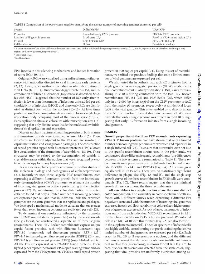
RESULTSGrowth properties of the three PRV recombinants expressingVP26 XFP fusion proteins. We have shown that only a limitednumber of incoming viral genomes are expressed and replicated ina single infected cell (22). To ensure that our results were not dueto the specific recombinant strains used in the prior study, weconstructed three different isogenic recombinants (the differencesbetween the two systems are summarized in Table 1). These re-combinants were previously constructed and characterized in ourlab: PRV180, PRV443, and PRV543 (Fig. 1A). All strains grewequally well in PK15 cells. There was no statistically significantdifference in plaque size (Fig. 1A and B), and the single-stepgrowth curves of the three recombinants in PK15 cells were com-parable (Fig. 1C). These results suggest that there are minimalgrowth differences among the three recombinants
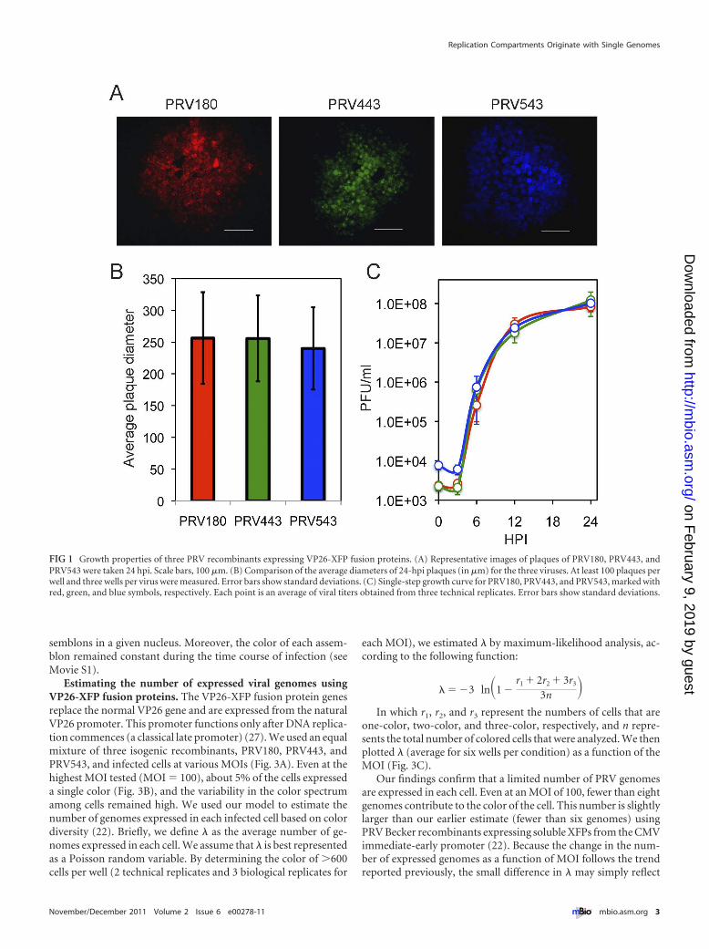
All assemblons in a single nucleus share the same distinctcolor composition. The variability in color spectrum of cells in-fected with 3 different viruses (each expressing a different FP)negatively correlated with the number of incoming viral genomesexpressed in each cell (low variability in color reflects higher num-bers of genomes expressed). A stock of an equal number of infec-tious units from each individual VP26-XFP recombinant (a 1:1:1mixture based on titer on PK15 cells) was prepared. We infectedcells at an MOI of 10 with this mixture (Fig. 2A; see also Movie S1in the supplemental material). The color spectrum of infected cellswas highly variable, corroborating our previous finding that only alimited number of viral genomes are expressed per cell (22). Eachgraph in Fig. 2B to H represents one nucleus, and the differentcolumns represent the relative ratios of colors in different fluores-cent nuclear foci (assemblons), as shown for cell B in Fig. 2B=. Ineach nucleus, all assemblons detected were the same color, sug-gesting that viral proteins are uniformly distributed among as-
TABLE 1 Comparison of the two systems used for quantifying the average number of incoming genomes expressed per cella
System Diffusible FPsb VP26-XFPc
Promoter Immediate-early CMV promoter PRV late VP26 promoterLocation of FP genes in genome In gG gene (US) Fused to VP26 coding region (UL)FP used RFP, YFP and CFP RFP, GFP, and CFPCell localization Diffuse Punctate in nucleusa A short summary of the major differences between the system used in this article and the system previously used (22). US and UL represent the unique short and unique longregions of the PRV genome, respectively (34).b See reference 22.c See this article.
Kobiler et al.
2 ® mbio.asm.org November/December 2011 Volume 2 Issue 6 e00278-11
on February 9, 2019 by guest
http://mbio.asm
.org/D
ownloaded from
semblons in a given nucleus. Moreover, the color of each assem-blon remained constant during the time course of infection (seeMovie S1).
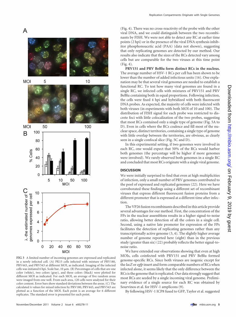
Estimating the number of expressed viral genomes usingVP26-XFP fusion proteins. The VP26-XFP fusion protein genesreplace the normal VP26 gene and are expressed from the naturalVP26 promoter. This promoter functions only after DNA replica-tion commences (a classical late promoter) (27). We used an equalmixture of three isogenic recombinants, PRV180, PRV443, andPRV543, and infected cells at various MOIs (Fig. 3A). Even at thehighest MOI tested (MOI � 100), about 5% of the cells expresseda single color (Fig. 3B), and the variability in the color spectrumamong cells remained high. We used our model to estimate thenumber of genomes expressed in each infected cell based on colordiversity (22). Briefly, we define � as the average number of ge-nomes expressed in each cell. We assume that � is best representedas a Poisson random variable. By determining the color of �600cells per well (2 technical replicates and 3 biological replicates for
each MOI), we estimated � by maximum-likelihood analysis, ac-cording to the following function:
� � �3 ln�1 �r1 � 2r2 � 3r3
3n �In which r1, r2, and r3 represent the numbers of cells that are
one-color, two-color, and three-color, respectively, and n repre-sents the total number of colored cells that were analyzed. We thenplotted � (average for six wells per condition) as a function of theMOI (Fig. 3C).
Our findings confirm that a limited number of PRV genomesare expressed in each cell. Even at an MOI of 100, fewer than eightgenomes contribute to the color of the cell. This number is slightlylarger than our earlier estimate (fewer than six genomes) usingPRV Becker recombinants expressing soluble XFPs from the CMVimmediate-early promoter (22). Because the change in the num-ber of expressed genomes as a function of MOI follows the trendreported previously, the small difference in � may simply reflect
FIG 1 Growth properties of three PRV recombinants expressing VP26-XFP fusion proteins. (A) Representative images of plaques of PRV180, PRV443, andPRV543 were taken 24 hpi. Scale bars, 100 �m. (B) Comparison of the average diameters of 24-hpi plaques (in �m) for the three viruses. At least 100 plaques perwell and three wells per virus were measured. Error bars show standard deviations. (C) Single-step growth curve for PRV180, PRV443, and PRV543, marked withred, green, and blue symbols, respectively. Each point is an average of viral titers obtained from three technical replicates. Error bars show standard deviations.
Replication Compartments Originate with Single Genomes
November/December 2011 Volume 2 Issue 6 e00278-11 ® mbio.asm.org 3
on February 9, 2019 by guest
http://mbio.asm
.org/D
ownloaded from
experimental variation. We conclude that this method, using cellcolor to count expressed genomes, does not depend on the pro-moter or insertion sites used to construct the test recombinants.
Detection of PRV replication compartments using two-colorFISH. HSV-1 has been shown to form a limited number of RCs(15, 16). Taken together with our finding that a limited number ofgenomes is expressed per cell, we predicted that each RC wouldoriginate with a single genome as was previously suggested (9). To
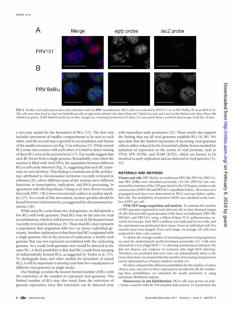
visualize PRV RCs, we used fluorescent in situ hybridization(FISH) probes to detect two PRV recombinants, PRV BeBlu andPRV151, each carrying a unique ~3,000-bp insertion, the lacZgene or the EGFP gene, respectively. To visualize PRV RCs, wesynthesized a unique set of PCR product probes directed to theunique sequence (egfp probes for PRV151 and lacZ probes forPRV BeBlu). At 4 h postinfection (hpi), PRV BeBlu- or PRV151-infected cells were fixed and hybridized with the two probes
FIG 2 Color analysis of nuclear assemblons. (A) Representative images of PK15 cells infected with a mixture of PRV180, PRV443, and PRV543 at 9 hpi with aMOI of 10. In cells (marked from B to H), individual assemblons were assayed for their fluorescence profiles. Scale bar, 10 �m. (B=) The marking of individualassemblons assayed for cell B are shown. (B to H) The graphs represent the normalized fluorescence composition of individual assemblons in a single nucleus.Each bar corresponds to a single assemblon, and each graph represents a single cell, as indicated by the letters.
Kobiler et al.
4 ® mbio.asm.org November/December 2011 Volume 2 Issue 6 e00278-11
on February 9, 2019 by guest
http://mbio.asm
.org/D
ownloaded from
(Fig. 4). There was no cross-reactivity of the probe with the otherviral DNA, and we could distinguish between the two recombi-nants by FISH. We were not able to detect any RC at earlier timepoints (2 hpi) or in the presence of the viral DNA synthesis inhib-itor phosphonoacetic acid (PAA) (data not shown), suggestingthat only replicating genomes are detected by our method. Ourresults also indicate that the sizes of the RCs detected vary amongcells but are comparable for the two viruses at this time point(Fig. 4).
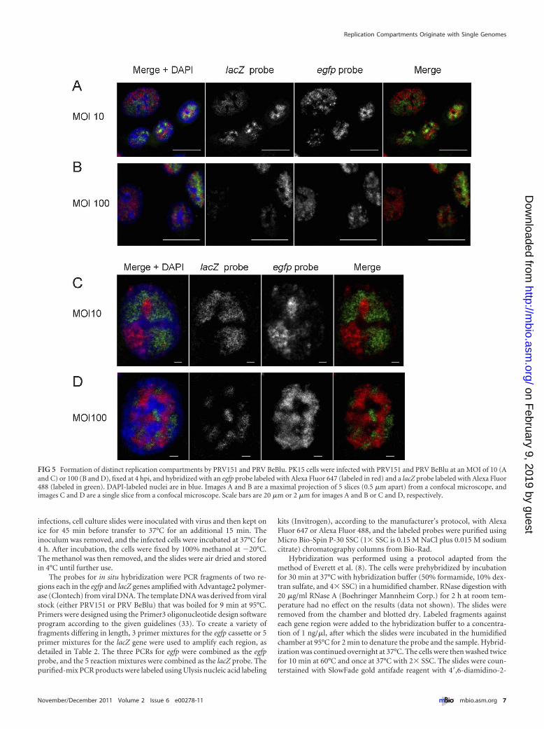
PRV151 and PRV BeBlu form distinct RCs in the nucleus.The average number of HSV-1 RCs per cell has been shown to belower than the number of added infectious units (16). One expla-nation may be that several viral genomes are needed to establish afunctional RC. To test how many viral genomes are found in asingle RC, we infected cells with mixtures of PRV151 and PRVBeBlu containing both in equal proportions. Following infection,the cells were fixed 4 hpi and hybridized with both fluorescentDNA probes. As expected, the majority of cells were infected withboth viruses (in experiments with both MOI of 10 and 100). Thedistribution of FISH signal for each probe was restricted to dis-crete foci with little colocalization of the two probes, suggestingthat most RCs contained only a single type of genome (Fig. 5A toD). Even in cells where the RCs coalesce and fill most of the nu-clear space, distinct territories, containing a single type of genomewith little overlap between the territories, are obvious, as clearlyseen in a single confocal slice (Fig. 5C and D).
In this experimental setting, if two genomes were involved ineach RC, one would expect that 50% of the RCs would harborboth genomes (the percentage will be higher if more genomeswere involved). We rarely observed both genomes in a single RCand concluded that most RCs originate with a single viral genome.
DISCUSSION
We were initially surprised to find that even at high multiplicitiesof infection, only a small number of PRV genomes contributed tothe pool of expressed and replicated genomes (22). Here we havecorroborated these findings using a different set of recombinantviruses that express different fluorescent fusion proteins from adifferent promoter that is expressed at a different time after infec-tion.
The VP26 fusion recombinants described in this article provideseveral advantages for our method. First, the concentration of theFPs in the nuclear assemblons results in a higher signal-to-noiseratio, allowing better detection of all the colors in a single cell.Second, using a native late promoter for expression of the FPsfacilitates the detection of replicating genomes rather than anytranscriptionally active genome (3, 4). The slightly higher averagenumber of genome reported here (eight) than in the previousstudy (greater than six) (22) probably reflects the better signal-to-noise ratio.
We have extended our observations showing that even at highMOIs, cells coinfected with PRV151 and PRV BeBlu formedgenome-specific RCs. Since both viruses are isogenic except forthe lacZ or egfp insert and form comparable numbers of RCs wheninfected alone, it seems likely that the only difference between theRCs is the genome that is replicated. Our data strongly suggest thatmost RCs are seeded by a single incoming viral genome. Prelimi-nary evidence of a single source for each RC was obtained bySourvinos et al. for HSV-1 amplicons (9).
By following HSV-1 ICP8 fused to GFP, Taylor et al. suggested
FIG 3 A limited number of incoming genomes are expressed and replicatedin a newly infected cell. (A) PK15 cells infected with mixture of PRV180,PRV443, and PRV543 at different MOI, as indicated. Imaging of the infectedcells was initiated 6 hpi. Scale bar, 10 �m. (B) Percentages of cells that are onecolor (white), two colors (gray), and three colors (black) were plotted fordifferent MOI as indicated. For each MOI, an average of five random areaswere imaged from one well. From each area, 120 cells were analyzed for theircolor content. Error bars show standard deviations between the areas. (C) Thecalculated � values for mixed infection by PRV180, PRV443, and PRV543 areplotted as a function of the MOI. Each point is an average for 6 differentreplicates. The standard error is presented for each point.
Replication Compartments Originate with Single Genomes
November/December 2011 Volume 2 Issue 6 e00278-11 ® mbio.asm.org 5
on February 9, 2019 by guest
http://mbio.asm
.org/D
ownloaded from
a two-step model for the formation of RCs (15). The first stepincludes movement of smaller compartments to be next to eachother, and the second step is growth by accumulation and fusionof the smaller structures (see Fig. 12 in reference 15). While severalRCs come into contact with each other, it is hard to detect mixingof these RCs even at the protein level (17). Our results suggest thateach RC forms from a single genome. Remarkably, even when thenucleus is filled with viral DNA, the separation between differentRCs is still easily observed (Fig. 5), suggesting that each RC main-tains its own territory. This finding is reminiscent of the architec-ture attributed to chromosomal territories (recently reviewed inreference 28), where different areas of the nucleus serve differentfunctions in transcription, replication, and RNA processing. Inagreement with this hypothesis, Chang et al. have shown recentlythat early HSV-1 RCs move toward and coalesce at nuclear speck-les (17). As a result of this movement, nuclear speckles should befound between viral territories, as suggested for chromosomal ter-ritories (28).
While most RCs arise from one viral genome, we did identify afew RCs with both genomes. Dual RCs may be the sites for viralrecombination, which is well known to occur in the herpesviruses(recently reviewed in reference 29). These dual RCs may representa population that originated with two (or more) individual ge-nomes. Another explanation is that these dual RCs originated witha single genome, but in the process of replication, a nearby viralgenome that was not expressed recombined with the replicatinggenome. As a result, both genomes now would be detected in thesame RC. A third possibility is that dual RCs result from mergingof independently formed RCs, as suggested by Taylor et al. (15).To distinguish these and other models for formation of mixedRCs, it will be important to develop real-time live visualization ofdifferent viral genomes in single nuclei.
Our findings correlate the known limited number of RCs withthe restriction of the number of expressed viral genomes. Thelimited number of RCs may also result from the restriction ofgenome expression, since this restriction can be detected even
with immediate-early promoters (22). These results also supportthe finding that not all viral genomes establish RCs (9, 30). Wespeculate that the limited expression of incoming viral genomesreflects either reduced levels of essential cellular factors needed forinitiation of expression or the action of viral proteins, such asVP16, EP0 (ICP0), and IE180 (ICP4), which are known to beinvolved in early replication and are detected in viral particles (31,32).
MATERIALS AND METHODSViruses and cells. PRV Becker recombinants PRV180, PRV443, PRV151,and PRV BeBlu were described previously (23–26). PRV543 was con-structed by insertion of the CFP gene fused to the VP26 gene, similar to theconstruction of PRV180 and PRV443 (unpublished data). All viruses werepropagated and titers were determined in PK15 (porcine kidney epithe-lial) cells. The multiplicity of infection (MOI) was calculated as the num-ber of PFU per cell.
VP26-XFP image acquisition and analysis. To estimate the numberof PRV genomes expressed in each infected cell, we first obtained imagesof cells infected with equal amounts of the three recombinants (PRV180,PRV443, and PRV543) using a Nikon Eclipse Ti-E epifluorescence in-verted microscope. Each MOI condition was replicated in two wells, andthe experiment was performed three times. From an individual well, fiverandom areas were imaged. From each image, on average 120 cells wereanalyzed for their color content.
To define the average number of incoming genomes being expressed,we used the mathematical model developed previously (22). Cells wereinfected for 6 h at a high MOI (�5), allowing synchronized infection. Wedid not observe any evidence of exclusion after high-MOI infection.Therefore, we concluded that each virus can independently infect a cell.Given these facts, we assumed that the number of incoming viral genomescan be represented as a Poisson random variable (�).
We then computed the different probabilities for the number of colors(that is, zero, one, two or three) expressed in an infected cell. By combin-ing these probabilities, we estimated the model parameter �, usingmaximum-likelihood analysis.
Fluorescence in situ hybridization. PK15 cells were grown on poly-L-lysine-coated 8-well Lab-Tek chamber slide systems. To synchronize the
FIG 4 Similar viral replication centers after infection with two PRV recombinants. PK15 cells were infected by PRV151 (A) or PRV BaBlu (B) at an MOI of 10.The cells were then fixed at 4 hpi and hybridized with an egfp probe labeled with Alexa Fluor 647 (labeled in red) and a lacZ probe labeled with Alexa Fluor 488(labeled in green). DAPI-labeled nuclei are in blue. Images are a maximal projection of 5 slices (0.5 �m apart) from a confocal microscope. Scale bar, 20 �m.
Kobiler et al.
6 ® mbio.asm.org November/December 2011 Volume 2 Issue 6 e00278-11
on February 9, 2019 by guest
http://mbio.asm
.org/D
ownloaded from
infections, cell culture slides were inoculated with virus and then kept onice for 45 min before transfer to 37°C for an additional 15 min. Theinoculum was removed, and the infected cells were incubated at 37°C for4 h. After incubation, the cells were fixed by 100% methanol at �20°C.The methanol was then removed, and the slides were air dried and storedin 4°C until further use.
The probes for in situ hybridization were PCR fragments of two re-gions each in the egfp and lacZ genes amplified with Advantage2 polymer-ase (Clontech) from viral DNA. The template DNA was derived from viralstock (either PRV151 or PRV BeBlu) that was boiled for 9 min at 95°C.Primers were designed using the Primer3 oligonucleotide design softwareprogram according to the given guidelines (33). To create a variety offragments differing in length, 3 primer mixtures for the egfp cassette or 5primer mixtures for the lacZ gene were used to amplify each region, asdetailed in Table 2. The three PCRs for egfp were combined as the egfpprobe, and the 5 reaction mixtures were combined as the lacZ probe. Thepurified-mix PCR products were labeled using Ulysis nucleic acid labeling
kits (Invitrogen), according to the manufacturer’s protocol, with AlexaFluor 647 or Alexa Fluor 488, and the labeled probes were purified usingMicro Bio-Spin P-30 SSC (1� SSC is 0.15 M NaCl plus 0.015 M sodiumcitrate) chromatography columns from Bio-Rad.
Hybridization was performed using a protocol adapted from themethod of Everett et al. (8). The cells were prehybridized by incubationfor 30 min at 37°C with hybridization buffer (50% formamide, 10% dex-tran sulfate, and 4� SSC) in a humidified chamber. RNase digestion with20 �g/ml RNase A (Boehringer Mannheim Corp.) for 2 h at room tem-perature had no effect on the results (data not shown). The slides wereremoved from the chamber and blotted dry. Labeled fragments againsteach gene region were added to the hybridization buffer to a concentra-tion of 1 ng/�l, after which the slides were incubated in the humidifiedchamber at 95°C for 2 min to denature the probe and the sample. Hybrid-ization was continued overnight at 37°C. The cells were then washed twicefor 10 min at 60°C and once at 37°C with 2� SSC. The slides were coun-terstained with SlowFade gold antifade reagent with 4=,6-diamidino-2-
FIG 5 Formation of distinct replication compartments by PRV151 and PRV BeBlu. PK15 cells were infected with PRV151 and PRV BeBlu at an MOI of 10 (Aand C) or 100 (B and D), fixed at 4 hpi, and hybridized with an egfp probe labeled with Alexa Fluor 647 (labeled in red) and a lacZ probe labeled with Alexa Fluor488 (labeled in green). DAPI-labeled nuclei are in blue. Images A and B are a maximal projection of 5 slices (0.5 �m apart) from a confocal microscope, andimages C and D are a single slice from a confocal microscope. Scale bars are 20 �m or 2 �m for images A and B or C and D, respectively.
Replication Compartments Originate with Single Genomes
November/December 2011 Volume 2 Issue 6 e00278-11 ® mbio.asm.org 7
on February 9, 2019 by guest
http://mbio.asm
.org/D
ownloaded from
phenylindole (DAPI) from Invitrogen and imaged in a Leica SP5 confocalmicroscope.
ACKNOWLEDGMENTS
We thank Roger Everett for sharing the FISH protocol. We thank allEnquist lab members for their comments.
O.K. is funded by the International Human Frontier Science Program.L.W.E. acknowledges support from NIH grants 1RC1NS068414 and P40RR 018604.
SUPPLEMENTAL MATERIALSupplemental material for this article may be found at http://mbio.asm.org/lookup/suppl/doi:10.1128/mBio.00278-11/-/DCSupplemental.
Movie S1, MOV file, 1.7 MB.
REFERENCES
1. Flint SJ, Enquist LW, Racaniello VR, Skalka AM. 2009. Principles ofvirology, 3rd ed. ASM Press, Washington, DC.
2. Quinlan MP, Chen LB, Knipe DM. 1984. The intranuclear location of aherpes simplex virus DNA-binding protein is determined by the status ofviral DNA replication. Cell 36:857– 868.
3. Holland LE, Anderson KP, Shipman C, Jr, Wagner EK. 1980. Viral DNAsynthesis is required for the efficient expression of specific herpes simplexvirus type 1 mRNA species. Virology 101:10 –24.
4. Jones PC, Roizman B. 1979. Regulation of herpesvirus macromolecularsynthesis. VIII. The transcription program consists of three phases duringwhich both extent of transcription and accumulation of RNA in the cyto-plasm are regulated. J. Virol. 31:299 –314.
5. Ward PL, Ogle WO, Roizman B. 1996. Assemblons: nuclear structures
TABLE 2 Primers used for making FISH probesa
PCR mix Primer name Orientation Starting codon Sequence from 5=egfp 1 egfp-L31 Forward 1 ATGGTGATGCGGTTTTGGegfp 1 egfp-R263 Reverse 232 TAGCGGATCTGACGGTTCegfp 1 egfp-L287 Forward 256 GCTCAAGCTTCGAATTCTGCegfp 1 egfp-R595 Reverse 564 TCGTGCTGCTTCATGTGGegfp 1 egfp-L598 Forward 567 TCTTCAAGTCCGCCATGCegfp 1 egfp-R757 Reverse 726 ATGTTGCCGTCCTCCTTGegfp 2 egfp-L807 Forward 776 ATGGCCGACAAGCAGAAGegfp 2 egfp-R985 Reverse 954 TTGGGGTCTTTGCTCAGGegfp 2 egfp-L1035 Forward 1004 ATCACTCTCGGCATGGACegfp 2 egfp-L1146 Forward 1115 TCCCCCTGAACCTGAAACegfp 2 egfp-R1297 Forward 1266 GAGTTTGGACAAACCACAACegfp 2 egfp-R1403 Reverse 1372 GCCGATTTCGGCCTATTGegfp 3 egfp-L1393 Forward 1362 CCGAAATCGGCAAAATCegfp 3 egfp-R1555 Reverse 1524 GGTGATGGTTCACGTAGTGGegfp 3 egfp-L1566 Forward 1535 TTTTTGGGGTCGAGGTGegfp 3 egfp-R1766 Reverse 1735 CTGTAGCGGCGCATTAAGegfp 3 egfp-L1783 Forward 1752 CTGTAGCGGCGCATTAAGegfp 3 egfp-R1923 Reverse 1892 TGGTTCTTTCCGCCTCAGlacZ 1 lacZ-L54 Forward 1 ATTCCGCCGATACTGACGlacZ 1 lacZ-R172 Reverse 118 ATTCCGCCGATACTGACGlacZ 1 lacZ-L189 Forward 135 AGCGGCTGATGTTGAACTGlacZ 1 lacZ-R342 Reverse 288 ACTGCCGCCTGTTTTGAClacZ 1 lacZ-L361 Forward 307 TTCTTGCGGCCCTAATCClacZ 1 lacZ-R547 Reverse 493 GCAGCATCAGGGGAAAAClacZ 2 lacZ-L690 Forward 636 CGCCAATGTCGTTATCCAGlacZ 2 lacZ-R910 Reverse 856 TCTGGCGGAAAACCTCAGlacZ 2 lacZ-L1048 Forward 994 TTGTGGAGCGACATCCAGlacZ 2 lacZ-R1226 Reverse 1172 CGCTGACGGAAGCAAAAClacZ 2 lacZ-L1286 Forward 1232 GGCGTATCGCCAAAATCAClacZ 2 lacZ-R1442 Reverse 1388 ACAGTCTTGGCGGTTTCGlacZ 3 lacZ-L1453 Forward 1399 GTGGGCGTATTCGCAAAGlacZ 3 lacZ-R1656 Reverse 1602 CGCTGGATCAAATCTGTCGlacZ 3 lacZ-L1659 Forward 1605 ACAGCGCGTCGTGATTAGlacZ 3 lacZ-R1816 Reverse 1762 CATGGTGCCAATGAATCGlacZ 3 lacZ-L1893 Forward 1839 TCGGATAATGCGAACAGClacZ 3 lacZ-R2071 Reverse 2017 GGTGCGGATTGAAAATGGlacZ 4 lacZ-L2086 Forward 2032 ATCGCAGGCTTCTGCTTClacZ 4 lacZ-R2252 Reverse 2198 TCGGCGGTGAAATTATCGlacZ 4 lacZ-L2278 Forward 2224 CGTTTCACCCTGCCATAAAGlacZ 4 lacZ-R2448 Reverse 2394 TTCCGTGACGTCTCGTTGlacZ 4 lacZ-L2457 Forward 2403 TCATCCGCCACATATCCTGlacZ 4 lacZ-R2556 Reverse 2502 CTGAGCGCATTTTTACGClacZ 5 lacZ-L2564 Forward 2510 TCAGACGGCAAACGACTGlacZ 5 lacZ-R2734 Reverse 2680 TGTTCCCACGGAGAATCClacZ 5 lacZ-L2739 Forward 2685 GCGGATTGACCGTAATGGlacZ 5 lacZ-R2917 Reverse 2863 CAGCCTGAATGGCGAATGlacZ 5 lacZ-L2916 Forward 2862 TGCGCAACTGTTGGGAAGlacZ 5 lacZ-R3057 Reverse 3003 TCACTGGCCGTCGTTTTACa The sequences of the PCR primers used for generating the FISH probes are listed. The position and orientation of each probe relative to the first probe for each gene are given.
Kobiler et al.
8 ® mbio.asm.org November/December 2011 Volume 2 Issue 6 e00278-11
on February 9, 2019 by guest
http://mbio.asm
.org/D
ownloaded from
defined by aggregation of immature capsids and some tegument proteinsof herpes simplex virus 1. J. Virol. 70:4623– 4631.
6. Taylor TJ, Knipe DM. 2004. Proteomics of herpes simplex virus replica-tion compartments: association of cellular DNA replication, repair, re-combination, and chromatin remodeling proteins with ICP8. J. Virol.78:5856 –5866.
7. Maul GG. 1998. Nuclear domain 10, the site of DNA virus transcriptionand replication. Bioessays 20:660 – 667.
8. Everett RD, Murray J. 2005. ND10 components relocate to sites associ-ated with herpes simplex virus type 1 nucleoprotein complexes duringvirus infection. J. Virol. 79:5078 –5089.
9. Sourvinos G, Everett RD. 2002. Visualization of parental HSV-1 ge-nomes and replication compartments in association with ND10 in liveinfected cells. EMBO J. 21:4989 – 4997.
10. Everett RD. 2006. Interactions between DNA viruses, ND10 and the DNAdamage response. Cell. Microbiol. 8:365–374.
11. Everett RD, Boutell C, McNair C, Grant L, Orr A. 2010. Comparison ofthe biological and biochemical activities of several members of the alpha-herpesvirus ICP0 family of proteins. J. Virol. 84:3476 –3487.
12. Knipe DM, Senechek D, Rice SA, Smith JL. 1987. Stages in the nuclearassociation of the herpes simplex virus transcriptional activator proteinICP4. J. Virol. 61:276 –284.
13. Ishov AM, Maul GG. 1996. The periphery of nuclear domain 10 (ND10)as site of DNA virus deposition. J. Cell Biol. 134:815– 826.
14. Maul GG, Ishov AM, Everett RD. 1996. Nuclear domain 10 as preexistingpotential replication start sites of herpes simplex virus type-1. Virology217:67–75.
15. Taylor TJ, McNamee EE, Day C, Knipe DM. 2003. Herpes simplex virusreplication compartments can form by coalescence of smaller compart-ments. Virology 309:232–247.
16. Phelan A, Clements JB. 1997. Functional domains within the nucleus ofa cell infected with HSV-1. Rev. Med. Virol. 7:229 –237.
17. Chang L, et al. 2011. Herpesviral replication compartments move andcoalesce at nuclear speckles to enhance export of viral late mRNA. Proc.Natl. Acad. Sci. U. S. A. 108:E136 –E144.
18. Desai P, Person S. 1998. Incorporation of the green fluorescent proteininto the herpes simplex virus type 1 capsid. J. Virol. 72:7563–7568.
19. de Oliveira AP, et al. 2008. Live visualization of herpes simplex virus type1 compartment dynamics. J. Virol. 82:4974 – 4990.
20. Miyamoto K. 1971. Mechanism of intranuclear crystal formation of her-
pes simplex virus as revealed by the negative staining of thin sections. J.Virol. 8:534 –550.
21. Pomeranz LE, Reynolds AE, Hengartner CJ. 2005. Molecular biology ofpseudorabies virus: impact on neurovirology and veterinary medicine.Microbiol. Mol. Biol. Rev. 69:462–500.
22. Kobiler O, Lipman Y, Therkelsen K, Daubechies I, Enquist LW. 2010.Herpesviruses carrying a Brainbow cassette reveal replication and expres-sion of limited numbers of incoming genomes. Nat. Commun. 1:146.
23. del Rio T, Ch’ng TH, Flood EA, Gross SP, Enquist LW. 2005. Hetero-geneity of a fluorescent tegument component in single pseudorabies virusvirions and enveloped axonal assemblies. J. Virol. 79:3903–3919.
24. Smith GA, Gross SP, Enquist LW. 2001. Herpesviruses use bidirectionalfast-axonal transport to spread in sensory neurons. Proc. Natl. Acad. Sci.U. S. A. 98:3466 –3470.
25. Smith BN, et al. 2000. Pseudorabies virus expressing enhanced greenfluorescent protein: a tool for in vitro electrophysiological analysis oftranssynaptically labeled neurons in identified central nervous system cir-cuits. Proc. Natl. Acad. Sci. U. S. A. 97:9264 –9269.
26. Banfield BW, Yap GS, Knapp AC, Enquist LW. 1998. A chicken embryoeye model for the analysis of alphaherpesvirus neuronal spread and viru-lence. J. Virol. 72:4580 – 4588.
27. McNabb DS, Courtney RJ. 1992. Identification and characterization ofthe herpes simplex virus type 1 virion protein encoded by the UL35 openreading frame. J. Virol. 66:2653–2663.
28. Cremer T, Cremer M. 2010. Chromosome territories. Cold Spring Harb.Perspect. Biol. 2:a003889.
29. Muylaert I, Tang KW, Elias P. 2011. Replication and recombination ofherpes simplex virus DNA. J. Biol. Chem. 286:15619 –15624.
30. Everett RD, Zafiropoulos A. 2004. Visualization by live-cell microscopyof disruption of ND10 during herpes simplex virus type 1 infection. J.Virol. 78:11411–11415.
31. Kramer T, Greco TM, Enquist LW, Cristea IM. 2011. Proteomic char-acterization of pseudorabies virus extracellular virions. J. Virol. 85:6427– 6441.
32. Michael K, Klupp BG, Mettenleiter TC, Karger A. 2006. Composition ofpseudorabies virus particles lacking tegument protein US3, UL47, orUL49 or envelope glycoprotein E. J. Virol. 80:1332–1339.
33. Rozen S, Skaletsky H. 2000. Primer3 on the WWW for general users andfor biologist programmers. Methods Mol. Biol. 132:365–386.
34. Szpara ML, et al. 2011. A wide extent of inter-strain diversity in virulentand vaccine strains of alphaherpesviruses. PLoS Pathog. 7:e1002282.
Replication Compartments Originate with Single Genomes
November/December 2011 Volume 2 Issue 6 e00278-11 ® mbio.asm.org 9
on February 9, 2019 by guest
http://mbio.asm
.org/D
ownloaded from