view - computer science at ubc
TRANSCRIPT
A Brief Survey of Central Mechanisms in Primate Visual Perception
Simon A. J. Winder
[email protected] June 21, 2002
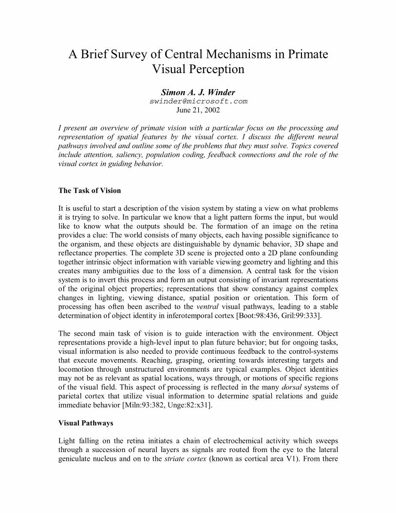
I present an overview of primate vision with a particular focus on the processing and representation of spatial features by the visual cortex. I discuss the different neural pathways involved and outline some of the problems that they must solve. Topics covered include attention, saliency, population coding, feedback connections and the role of the visual cortex in guiding behavior. The Task of Vision It is useful to start a description of the vision system by stating a view on what problems it is trying to solve. In particular we know that a light pattern forms the input, but would like to know what the outputs should be. The formation of an image on the retina provides a clue: The world consists of many objects, each having possible significance to the organism, and these objects are distinguishable by dynamic behavior, 3D shape and reflectance properties. The complete 3D scene is projected onto a 2D plane confounding together intrinsic object information with variable viewing geometry and lighting and this creates many ambiguities due to the loss of a dimension. A central task for the vision system is to invert this process and form an output consisting of invariant representations of the original object properties; representations that show constancy against complex changes in lighting, viewing distance, spatial position or orientation. This form of processing has often been ascribed to the ventral visual pathways, leading to a stable determination of object identity in inferotemporal cortex [Boot:98:436, Gril:99:333]. The second main task of vision is to guide interaction with the environment. Object representations provide a high-level input to plan future behavior; but for ongoing tasks, visual information is also needed to provide continuous feedback to the control-systems that execute movements. Reaching, grasping, orienting towards interesting targets and locomotion through unstructured environments are typical examples. Object identities may not be as relevant as spatial locations, ways through, or motions of specific regions of the visual field. This aspect of processing is reflected in the many dorsal systems of parietal cortex that utilize visual information to determine spatial relations and guide immediate behavior [Miln:93:382, Unge:82:x31]. Visual Pathways Light falling on the retina initiates a chain of electrochemical activity which sweeps through a succession of neural layers as signals are routed from the eye to the lateral geniculate nucleus and on to the striate cortex (known as cortical area V1). From there

they spread to extra-striate visual processing centers and eventually to other areas of the brain that perform polysensory or sensorimotor integration [Wise:97:323]. At each stage the signals are modified and transformed in order to make explicit certain features of the image that are relevant to the organism. Whereas neurons in the retina respond to brightness contrast at some well defined location in the scene [Troy:93:294], neurons in extra-striate cortex respond to specific classes of faces or to gestures no matter where they may appear in space [Roll:92:266]. Whereas neurons in the retina respond to the wavelength of incident light [DeMo:75:210], those in the cortex may respond to the perceived color of an object in a manner which is stable against illumination changes [Zeki:80:167]. Figure 1 shows a view of the primate brain; the cortical areas responsible for vision are highlighted. The cortical surface is a thin sheet and this “computing real estate” is divided up into regions that are allocated to particular types of processing: vision, audition, the tactile senses, motor control, behavior planning and so on. Multiple representations of retinal space are mapped onto the cortex in a manner that preserves visual topology – adjacent locations in the retina are also adjacent in the cortex. These representations define the visual modules: V1, V2, V4, MT, etc. with V1 being the first mapping of the retina onto the cortical surface [Zeki:78:12, Zeki:88:11]. Figure 2 gives an overview of the streams of processing in the vision system and exemplifies a common way of looking at the known anatomical connections as a feed-forward hierarchy in which each neural layer extends the processing carried out by layers below it [Desi:85:3]. In this context, the classical receptive field of any neuron is that region of the retina which, when stimulated in the correct manner, causes the neuron to respond. Since there is convergence of signals at each level in the hierarchy, the size of typical receptive fields grows as one examines neurons further up in the processing chain. By virtue of their connectivity, neurons combine activity (in retinal space) from preceding neurons with small receptive fields to create larger receptive fields [Roll:92:266]. This is how neurons in inferotemporal (IT) cortex become sensitive to stimuli at any point in space; their receptive fields cover the entire visual field. As well as the progression of receptive field sizes and increasingly complex feature selectivity of neurons as one ascends the chain of processing, there are also diverging streams of function [Unge:82:x31] . Starting from the retina certain portions of the pathway become more and more specialized according to task. Typical examples are the processing of color, motion and form [Zeki:92:71]. Beginning with a subset of retinal ganglion cells, the M-pathway utilizes myelination and fast conducting axons to expedite the processing of change in the visual field. This pathway is the start of a stream that is dedicated to computing information about motion. The slower conducting axons of the P-pathway belong to neurons that have smaller receptive fields and are sensitive to color; this pathway allows for sustained scrutiny, higher acuity, and color discrimination [Kapl:86:125, Livi:88:13]. In some respects these streams remain segregated - even at high level areas of the visual cortex - although not all researchers agree on the extent of coupling [Lenn:98:350].

Figure 1: A map of the cortical surface of the macaque monkey. The surface has been unfolded and laid out flat so that all areas are visible. A Cartesian Theatre? A common misunderstanding of vision is that somehow the scene must be reconstructed in the mind in order to be perceived. The evidence however points in the opposite direction: By recording from many individual neurons throughout the primate visual cortex we have come to see that a set of analyses are carried out in parallel – the image is broken down according to stimulus properties such as color, shape, motion, or location – and that specialized pathways and areas of the brain exist to process these [Zeki:92:71]. To a large extent, this information is never brought back together in a “Cartesian Theatre” or centralized location where it is integrated and perceived [Bart:98:320, Cric:98:327, Denn:91:b22]. Instead, it is provided when needed as the answer to specific questions, such as “what is the color of this object?” which involves checking the color centers, or “do I know this person?” which requires an answer from the cortical area specialized for representing faces. In this way the information can be made available as it is required by whatever system is involved in generating a specific human behavior.

Eye
V1
V2
V3/V3a
MT
MSTV4
InferotemporalCortex (IT)
STS
ParietalCortex
(7a, PO, VIP, LIP,AIP, MIP)
LGN SupCol Pulv
Incr
easin
g re
cept
ive
field
siz
e
Dor
sal s
tream
Vent
ral s
tream
Figure 2: Interconnections in the vision system. Sub-cortical sites are circles: LGN, lateral geniculate nucleus; SupCol, superior colliculus; Pulv, pulvinar nucleus. Boxes represent cortical areas: MT, middle temporal; MST, medial superior temporal; PO, parieto-occipital; VIP, LIP, MIP, AIP, medial, lateral, ventral, anterior intraparietal areas respectively; STS, superior temporal sulcus. Feedforward and Feedback Connections Latency studies have shown that, when the retina is suddenly presented with a stimulus, a succession of visual areas are stimulated in sequence according to their level in the hierarchy, and that after approximately 100ms visually evoked activity reaches the frontal cortex and other areas not directly involved with vision [Schm:98:x59, Lamm:00:321]. This feedforward processing is only part of the story because neurons at successively higher levels send information back down via feedback connections to influence those neurons from which their inputs derive. Neurons with large receptive fields in extra-striate V2 send modulatory feedback connections to neurons in V1 with spatially coincident receptive fields [Zeki:88:11]. Since this kind of arrangement is ubiquitous, the results of a computation in inferotemporal cortex can affect the behavior of V1 neurons and these changes are expressed back in IT cortex after a further delay.

Given that both backward and forward waves of activity are present after stimulus onset, with the feedforward processing being expressed more faithfully in the first 100ms, we would expect an evolution of neural selectivity over time. This is found to be the case. From the first firing of an action potential, neurons throughout the visual system have very specific optimal visual stimuli requirements but these are narrowed further as time progresses [Lamm:00:321]. Neurons selective for faces initially respond rapidly to anything that looks like a general face, but over time they become more selective for individual faces; the population of face selective neurons is initially very active but settles down with a much sparser number of neurons firing to represent specific facial identity [Suga:99:441]. In a similar scenario, neurons in V1 become modulated by figural contexts: the response of a neuron to a texture element is enhanced if that element forms part of the figure (foreground) as opposed to the ground (background) [Lee:98:336]. Lateral Connections Visual processing begins with the retina and there one finds lateral connections between adjacent photoreceptors [Ravi:75:x57]. Later, horizontal and amacrine cells also act to spread neural activity across the retina [Kolb:77:x58]. Beyond the retina, feedforward and feedback signals are not the only influences on the activity of a cortical neuron – lateral connections from other neurons at the same representational level form a major modulating influence [Gilb:89:426, Tso:86:439]. Such interactions are prevalent throughout the visual system. Since cortical areas usually express a retinotopic mapping, this means that lateral connections can be related to influences between different locations in visual space. Judging by the dense layers of horizontally spreading axon fibers in supragranular V1, these influences should form a major component of information processing [Arbi:97:b23]. Single-neuron recording experiments have demonstrated that a given cortical neuron will show influences from outside its classical receptive field, e.g. [Zhou:00:440]. These influences are likely to arise from a combination of feedback and horizontal connections which act to modulate a neuron’s response in relation to its spatial context, but may not be able to directly drive the neuron in the absence of its classical feedforward influences. Population Codes How does the pattern of neural firing represent the appearance of a stimulus? This is a difficult question because there are many ways to measure the “activity” of a neuron. The spike timing of action potentials may be important, or the activity may mean different things at different times (known as temporal coding) [Reic:00:443, Vict:00:445]. Frequently, experimenters report the average firing rate and correlate this with the dimensions of some stimulus presentation. For instance, in IT cortex, a neuron might fire more rapidly to the image of a face than to the image of a dog. We can say the neuron prefers the face, but this still may not be the optimal stimulus; many other shapes must be tested [Roll:92:266, Tana:97:337]. By performing such tests, experimenters have found that neurons often respond weakly to a class of stimuli but have a strong response to a more restricted subset. A different neuron in the same cortical area may also respond to

the same class, but prefer a different subset. We can therefore consider population codes in which a collection of neurons may respond to a given stimulus class and the pattern of their differential activity is able to allow discrimination between class members. It is useful to draw a distinction between sparse and dense or distributed population codes [Fold:95:438]. In a sparse code most neurons are inactive; a very restricted subset responds to a given stimulus. This means that the neurons are highly selective and this is why most remain silent (they require a very specific input.) In contrast, a dense code would have a large number of active nodes because the selectivity of each node is broad and one stimulus activates them all to a varying extent. The dense population response is still sufficient to allow discrimination. If we compare these codes we can see that the sparse code has an advantage when multiple stimuli can be present at the same time. In this case multiple activity peaks are easily separable because there is a close correspondence between stimulus identity and which neurons are active. On the other hand, the dense code merges the identity of multiple stimuli into one response destroying their separation, but for single stimuli it has the advantage of being more robust against noise since it is the average activity over many neurons that is important. It is clear that the brain uses a range of representations along the sparse-to-dense axis for different purposes. In modeling visual cortex we often consider the population code to be sparse [Olsh:97:414], but sparse codes need not be physically local and may still involve neurons distributed over a wide cortical area [Haxb:01:437, Isha:00:343]. If a population contains neurons whose selectivity varies with time then we may have a situation where a dense code could evolve into a sparse code. This is probably true in the case of face representation as mentioned previously. Saliency and Attention We say that a feature is salient if it stands out from the crowd and is likely to attract attention. Figure 3 shows examples of salient texture elements which are found to “pop out” of a background consisting of different elements [Berg:83:63, Jule:91:68, Jule:90:62]. They are characterized by differing from the surroundings in terms of low-level feature properties: brightness, orientation, size, color, corners, direction of motion and so on. Elements which are salient do not require a serial visual search to be detected – their presence can be verified at a glance – even among many distracters [Trei:86:226, Trei:80:x52]; such targets appear to have a higher subjective contrast than their neighbors. In experiments, V1 neurons show a reduced response to elements when similar elements are placed outside the neuron’s receptive field; the strongest responses are to isolated features or to features surrounded by dissimilar neighbors [Knie:92:220]. This could be the neural correlate of a saliency calculation and may involve competition via the extensive network of lateral connections between neurons with spatially adjacent receptive fields. Saliency provides a form of priority ranking for subsequent processing allocation. Pop-out features are likely to attract attention and to become the targets for saccadic eye movements which center the region onto the fovea for more detailed inspection. Sudden

change in the visual field is highly salient and is a potent attention attractor. In experiments where differences cues were masked, subjects were often blind to quite extreme changes in their visual input [Simo:97:x61]. For example an object may disappear from a photograph, but if the before and after shots are separated by a brief occluder, viewers are unable to determine what has changed [OReg:99:x60]. This implies that we rely on the outside world as a memory rather than keep an internal track of all the elements of a scene. We inspect the scene as we need specific information, and change detection allows us to keep abreast of updates.
Figure 3: Some texture elements are salient and naturally “pop out” of background clutter (top figures), whereas locating others require a serial search (bottom figures). The visual image may contain many objects but the brain’s ability to concurrently represent these is limited and therefore a mechanism has to exist for it to direct its processing resources [Cohe:93:b24]. Attention is the capability of ignoring distracting inputs and focusing on the subject of interest and this can occur covertly without the need for eye movements [Posn:80:x53] (although eye movements are a large part of visual attention in natural settings.) The attention “searchlight” is able to capture information over a wide or narrow area of the visual field or at different locations [Adin:92:173, Schr:01:317]. In addition, non-spatial dimensions can be attended to, such as the orientation, color or motion of a feature, as well as many other qualities [Kast:00:319]. Attention increases the amount of processing allocated to those elements that share the same attended location or feature properties.

Attention
Figure 4: Neurons representing the whole visual field (top) respond to stimulus properties such as color or shape without indicating which properties are related. Attention selects for information coming from a certain location and solves the binding problem by disambiguating the responses – the blue circle is ignored.
Figure 5: Selective attention to feature properties such as color or shape can allow groupings of elements to be seen for instance the ‘X’ or ‘∆’. The blue squares cannot be globally grouped in this way because attending to blue produces confusion with circles and attending to squares produces confusion with pink.

The requirement for attending to particular locations arises from the fact that there is complete spatial convergence within the vision system. Neurons in IT cortex have receptive fields that cover the entire visual field and this allows them to represent object properties in a manner which is independent of the size or position of the stimulus [Desi:85:3]. When there are multiple stimuli in their receptive fields (which is normally the case) the population of IT neurons will respond to a combination of features from all the stimuli and the visual system is unable to bind together an object with its properties. Attention processes control the bottom-up gating of information at various levels in the hierarchy such that information from one location is dominant and therefore the IT population represents the appearance of a specific target (Figure 4). When attention is shifted to feature qualities such as color or orientation then targets with these properties seem to pop out of scenes containing a mixture of elements; they become the “figure” and are selected for priority processing. It is then possible to see spatial groupings of these targets which would not be visible if the scene were viewed as a whole (Figure 5). Neurophysiological evidence shows that the activity of neurons in V4 can be modulated by attention. When the monkey attends to a stimulus in a neuron’s receptive field in the presence of a distracting stimulus, also placed within the receptive field, then the neuron’s activity is reduced if it was selective for the feature properties of the distracter and increased if it was selective for the attended target [Desi:90:165, Kast:00:319]. Neurons in V4 normally have large receptive fields, but in the presence of attention it is as though the receptive field shrinks to include only the target of interest. The population response of neurons representing a large area of the visual field comes to represent the identity of only one specific element, ignoring the remainder. The source of appropriate gating signals for attention is still contentious but the parietal cortex is strongly implicated [Hame:02:304, Hame:02:306, Desi:90:165]. Some images can be interpreted in more than one way (Figure 6). A typical example is the vase and faces illusion in which the figure and ground choice is arbitrary and perceptual bi-stability results. Another example is the Necker cube. This ambiguity rarely occurs in natural scenes, but gives a useful insight into the way that images are processed by the brain: it seems that two or more different interpretations can compete for ownership of the visual elements. We can use directed attention to force the choice of interpretation but we are not able to hold onto it for long because the alternative interpretation rapidly becomes more salient. Thus attention has a time limit beyond which other salient features gain precedence. Monkeys have been trained to indicate their interpretation of a bi-stable motion figure. Recordings from neurons in MT have shown that neural activity changes magnitude at the same time as a figure-ground switch is reported – an increase in activity being observed for neurons that were selective for features of the perceptual foreground [Brad:98:429]. Similar neural correlates of perceptual switching have been demonstrated using binocular rivalry [Shei:97:428].

Figure 6: Bistable patterns. Left: the Necker cube reverses in depth. Right: figure and ground can be assigned arbitrarily to the left or right regions of this figure. A Review of Feature Selectivity and Visual Pathways Having described some general aspects of vision, I now focus briefly on the feature selectivity of neurons at different levels of the visual hierarchy and summarize some of the more complex properties of later stages. The retina is well known for its center-surround receptive fields (Figure 7) [Kuff:53:x28]. Retinal ganglion cells respond as though they sum photoreceptor inputs over a small central area and this region provides excitation; a larger ring around this area provides an inhibitory influence. This configuration makes them sensitive to small spots of light, but if the size of the spot is increased then their response is reduced. This is the classical ‘on-center’ cell type. ‘Off-center’ units also exist in which the roles of the two regions are swapped and these cells are excited by dark spots on a light background. (In the primate there are also various cone-opponent color selectivity profiles [DeMo:75:210].) If ganglion cells are monitored while stimulating the retina with sine-wave gratings (regularly-spaced bars of light) then experimenters find a peak of response modulation at one spatial frequency (reciprocal of bar spacing). Neurons in the retina therefore act as band-pass spatial filters [Hube:60:44, Enro:66:38, Lins:82:32]. Retinal ganglion cells innervate the lateral geniculate nucleus of the thalamus (LGN) and the receptive fields of LGN cells are somewhat similar to those of the retina; most also exhibit a center-surround organization with on-center and off-center sub-types [Lenn:80:4, Wies:66:200]. However there are some key differences: The response of an on-center unit in the LGN to a large disk of light covering both the receptive field center and surround is often much lower than for retinal cells [Hube:61:33]. This implies an increase in inhibition from the surround and a more severe tuning for the size of a stimulus. Another interesting difference is that LGN neurons are end-stopped, that is they show a reduced response when an elongated bar of light is used as a stimulus when compared with a spot of light or a shorter bar [Jone:91:295]. This reduction is far greater than would be predicted from their center-surround configuration alone. The LGN is a
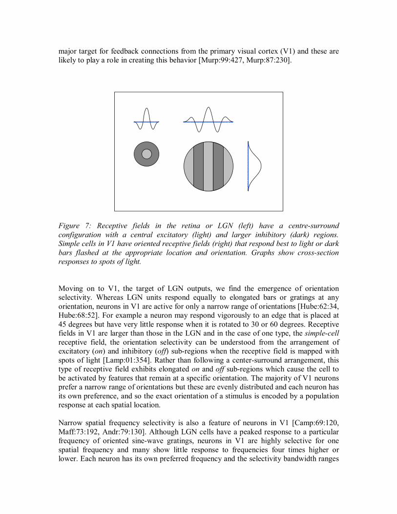
major target for feedback connections from the primary visual cortex (V1) and these are likely to play a role in creating this behavior [Murp:99:427, Murp:87:230].
Figure 7: Receptive fields in the retina or LGN (left) have a centre-surround configuration with a central excitatory (light) and larger inhibitory (dark) regions. Simple cells in V1 have oriented receptive fields (right) that respond best to light or dark bars flashed at the appropriate location and orientation. Graphs show cross-section responses to spots of light. Moving on to V1, the target of LGN outputs, we find the emergence of orientation selectivity. Whereas LGN units respond equally to elongated bars or gratings at any orientation, neurons in V1 are active for only a narrow range of orientations [Hube:62:34, Hube:68:52]. For example a neuron may respond vigorously to an edge that is placed at 45 degrees but have very little response when it is rotated to 30 or 60 degrees. Receptive fields in V1 are larger than those in the LGN and in the case of one type, the simple-cell receptive field, the orientation selectivity can be understood from the arrangement of excitatory (on) and inhibitory (off) sub-regions when the receptive field is mapped with spots of light [Lamp:01:354]. Rather than following a center-surround arrangement, this type of receptive field exhibits elongated on and off sub-regions which cause the cell to be activated by features that remain at a specific orientation. The majority of V1 neurons prefer a narrow range of orientations but these are evenly distributed and each neuron has its own preference, and so the exact orientation of a stimulus is encoded by a population response at each spatial location. Narrow spatial frequency selectivity is also a feature of neurons in V1 [Camp:69:120, Maff:73:192, Andr:79:130]. Although LGN cells have a peaked response to a particular frequency of oriented sine-wave gratings, neurons in V1 are highly selective for one spatial frequency and many show little response to frequencies four times higher or lower. Each neuron has its own preferred frequency and the selectivity bandwidth ranges

around 1 to 2 octaves [DeVa:82:184, Fost:85:123, Keef:98:325]. It can be argued that V1 computes a local Fourier analysis (or wavelet transform) of the visual scene, dividing each visible feature into its spatial frequency and orientation components [Marc:80:21, Daug:85:27]. The synaptic weights at the inputs to each neuron provide a local basis vector onto which the visual information is projected. This linear systems perspective is possible because simple cells are known to approximate linear spatial summation within their receptive fields [Movs:78:129, Andr:79:130, Lamp:01:354]. Another cell type in V1 is the complex cell. These neurons are just as orientation and spatial frequency selective as simple cells but their receptive fields do not show segregated on and off sub-regions [Movs:78:10]. Complex cells respond to moving gratings with a continuous elevated firing rather than the regular sine-wave synchronized pulses of activity that are characteristic of simple cells; they therefore signal the presence of an appropriate spatial feature without regard to its spatial phase (exact position). A common model that describes the derivation of a complex-cell response involves pooling together the rectified outputs of a number of simple cells, each responding to different phases of the sine-wave grating [Spit:88:275]. Both simple and complex cells are modulated by influences from outside their receptive fields. The term ‘end-stopping’ is used when a neuron shows a reduced response to an optimally oriented bar if that bar is longer than a certain length [Gilb:77:7, Sill:77:234, Rose:77:232]. In a similar way, side-stopped receptive fields are those for which an extended grating is a less effective stimulus than one which contains only one or two cycles of the sine-wave [DeVa:85:30]. Side-stopping and end-stopping may be present together, in which case the cell may respond more vigorously to a short isolated bar or to a corner than to an extended grating [DeAn:94:225]. Other complicated response properties exist in V1: Some complex cells in layer 5 are highly responsive to spots of light, but still show orientation selectivity to bars [Gilb:77:7, Hamm:94:293]. Many neurons show an increased response when they form part of a figure, or else are close to a figure-ground boundary that is defined solely by a texture discontinuity, such as a change in the orientation of a hatching pattern [Lee:98:336, Zhou:00:440]. Excitatory or inhibitory interactions can occur between neurons having different spatial frequency or orientation preferences [DeVa:83:6]. Some of these phenomena are related to contrast gain control and I describe this phenomenon in a later section. An exhaustive description of V1 receptive fields should also include their many color, motion and binocular properties [Livi:84:206, Naka:85:135, Bish:71:127, Lenn:90:205, DeVa:00:326, Lenn:80:4, Lenn:99:355], but I do not wish to include this detail here. V2, the second visual area, is a region of cortex with its own retinotopic map that surrounds V1 and is a significant target for fibers which leave there [Livi:87:x62]. V2 represents a second level in the cortical hierarchy of visual processing and is responsible for further integration of visual information. This integration takes the form of a strategic convergence of signals from previous levels to generate new receptive fields which have more complex selectivities and cover a greater area of visual space. Increasing the size of

the region over which an image is analyzed by each neuron allows for responsiveness to more abstract and less spatially localized features. This process in repeated as we move on to higher cortical areas. Receptive fields in V2 have not been characterized with the same detail as those in V1 but there are many similarities [Fost:85:123]. Populations of V2 neurons are know to be tuned to orientation and spatial frequency and there is a mix of simple and complex receptive field types [Levi:94:322, Fost:85:123]. Additionally, end-stopped cells are present as well as some neurons with large complex un-oriented receptive-fields [Hube:85:18]. The more interesting V2 neurons respond to illusory contours or texture boundaries. In natural scenes, object occlusion boundaries are rarely completely defined by light-dark transitions; instead there may simply be a discontinuity of texture statistics or even no discontinuity at all. Nevertheless we are able to detect figures that are defined largely in this way (Figure 8). Many V2 neurons respond both to these texture boundaries and to regular boundaries with the same orientation selectivity – a behavior that is not frequently attributed to V1 [Heyd:89:159, Shet:96:314]. Other cells respond to a moving bar when the passage of the bar across the receptive field is only implied by the surrounding elements and is not literally present as a change in brightness [Heyd:84:145, Pete:89:160].
Figure 8: Illusory shapes such as the Kaniza triangle or a circle defined by line ends are still visible despite the lack of continuous luminance contours. Another emerging behavior in V2 is the selectivity of neurons for the feature-phase of a stimulus. Boundaries are perceptually defined as edges (transitions from light to dark) or as light or dark bars [Burr:89:190], but neurons in V1 often respond to all these types with similar levels of activity and are activated primarily by the Fourier components that are present [DeVa:79:9, Albr:80:20, Albr:81:157]. In V2 however, Peterhans and von der Heydt encountered neurons that responded to a dark bar, for example, but responded weakly or not at all to light bars or edges [Pete:89:160]. They also found similar selectivity for edges of one polarity over bars or edges of the other contrast polarity, and

some neurons responded better to square wave patterns than to sine wave gratings. Whereas neurons in V1 are selective for the Fourier components in a stimulus, it appears that V2 may begin to synthesize a description in terms of edge and bar features with bar detection neurons that are not only stimulated by the fundamental sine wave component in a square wave grating, but also by the sharp step-edges, even when the fundamental component is missing. Beyond V2 the visual pathways become more segregated into the ventral stream of the temporal lobe (V3, V4, STS and IT) and the dorsal stream of the parietal lobe (V3a, MT, MST, PO, area 7, and areas of the intraparietal sulcus) although there are clearly many interactions between these processing steps [Dist:93:x63]. Area V4 is a gateway to the temporal lobe and its receptive field properties are interesting because of their complexity and because the neurons in this area show strong response modulation with attention. V4 receptive fields are large and frequently cover the center of vision, extending across the mid-line. Neurons are selective for a wide variety of stimuli: color, light and dark, edges, bars, oriented or non-oriented, moving or stationary, square wave and sine wave gratings of various spatial frequencies, the length and width of a light or dark region, binocular disparity, and the conformation of contour or corner features [Desi:87:228, Pasu:99:422, Hink:01:425, Pasu:01:424]. One consistent finding is that they have receptive field centers that are excited by some spatial pattern and very large inhibitory surrounds that are also sensitive to the same pattern. Maximum response is produced when the two regions are presented with different patterns or else the center is stimulated alone. Sensitivity to the relation between a stimulus and its spatial neighbors is thought to result in a number of useful perceptual effects (such as size constancy) [Jule:91:68]. Zeki found that V4 color selective neurons showed significant color constancy – perceived object color remains stable over wide variations in illumination chromaticity – a capacity which may be facilitated by a centre-surround arrangement [Zeki:80:167]. Another reason for this receptive field structure could be the generation of texture pop-out responses over greater length scales than is feasible in V1 or V2; an isolated object would appear to be more salient due to the lack of spatial competition with surrounding elements. Area IT of the temporal lobe contains neurons whose receptive fields cover the entire visual field and this area is the general target for outputs from V4. It contains many specialized neurons that are selective for faces or hands as well as those responding to a variety of juxtapositions of patterns, oriented segments, and light or dark regions [Tana:93:264, Tana:97:337, Roll:92:266]. In humans, different regions of IT cortex show object specific activity [Isha:00:343]. The constituent neurons are likely to form a distributed representation that is the basis for recognition because their responses have important properties: they are contrast invariant, size invariant and position invariant [Desi:85:3, Gros:92:265, Ito:95:x54, Desi:84:x55, Gros:72:x56]. For example, neurons respond quite exclusively to particular shapes without regard to their viewing distance or location in the visual field – and they can do this in the presence of significant clutter [Shei:01:342, Gril:99:333]. The behavior of IT neurons in primates has been reported to correspond closely to the human’s perception of what the monkey is looking at during an
experiment [Gros:92:265]. There is some indication that a neural sub-population may exist in IT that encodes for the short-term memory of a visual item; this population would be compared against a sensory population in tasks that require visual matching between a presented stimulus and one that is remembered [Mill:93:263]. Areas MT and MST are concerned with analyzing motion and depth in the visual field and these may facilitate the two perceptual capabilities of structure from motion and structure from stereopsis [Maun:83:55, Maun:83:56, Albr:84:54, Marr:82:b2, Ullm:79:110]. MT sends information to areas MST and hence to the parietal cortex. MST and area 7a neurons have been found to respond to full-field motions, including those which are relevant to visual locomotion, or to complex optical flow patterns such as rotation or expansion [Sieg:97:415]. STS neurons are thought to represent biologically relevant motions related to social behavior and may be part of a system that can infer the intentions of other individuals from their movement [Iaco:99:446, Rizz:96:x64]. The posterior parietal association cortex (area 7a, V6a/PO, VIP, LIP, AIP, MIP) has been shown to contain neurons involved with visual reaching, feeding behavior, grasping and manipulation [Culh:01:339, Snyd:00:329], as well as those that are able to re-map retinal space into body or head-centered coordinate systems, remove the effects of eye-movements, and distinguish self-induced motion from external motion [Colb:98:340, Ande:97:338, Colb:99:309]. Such capabilities are indicative of a system that is not just specialized for motion, but is also able to relate the positions of objects in three dimensions and direct physical interactions without necessarily involving recognition [Miln:93:382]. Two other areas of the brain should be mentioned: the pulvinar and superior colliculus. These sub-cortical nuclei are interconnected with many parts of the visual cortex [Feig:98:345]. The superior colliculus is involved in directing saccadic eye movements and contains a number of spatial maps that control the future direction of gaze [Kand:00:b25]. It receives information from visual and auditory areas and mediates reflexive gaze-shifts to targets that become salient by virtue of a sudden sound or movement. The pulvinar, like the LGN, is part of the thalamus and has a role in gating information flow between areas of the cortex. Its function is still undetermined, although there are many indications that the pulvinar is critical for attention control and the perceptual binding of an object’s visual properties to its spatial location [Desi:90:165]. The Cortical Microcircuit The grey matter of the cortex is a thin sheet densely packed with neurons. It has a laminar organization and in visual cortex these layers are numbered 1 to 6. One prevalent view of its structure concerns the canonical microcircuit. This is the idea that each tiny region of the cortical surface contains a stereotyped neural circuit that carries out computations of the same basic form no matter what kind of inputs that area receives [Doug:91:302]. A computational unit is thereby formed from a vertical column of cells penetrating through all the layers; many identical columns are packed side-by-side, interconnected by horizontal connections. This structure is present throughout the neocortex and includes a

stereotyped layered organization of input/output connections with other areas [Arbi:97:b23]. Figure 9 illustrates the general form of the excitatory connections known to exist in primate V1 [Call:98:331]. Ascending input from the LGN (of the thalamus) is received by layer 4C “spiny stellate” cells (which form the granular layer) and is relayed to neurons in layers 2-4B which send their outputs to other cortical areas. Layer 4C has reciprocal connections with “pyramidal” neurons in layer 6 and layers 2-4B maintains reciprocal connections with similar neurons in layer 5. Layer 6 is the origin of feedback to the LGN and layer 5 projects to the pulvinar nucleus of the thalamus and also to the superior colliculus [Feig:98:345]. Feedback connections from higher cortical areas send their inputs to layers 1 and 6 [Zeki:88:11]. This basic scheme is found in other areas of the cortex with layer 4 providing the feedforward input layer, the upper layers projecting axons to other cortical sites, and layers 5 and 6 projecting to sub-cortical nuclei. Additionally, layers 2/3 and layers 5/6 contain a dense network of horizontally spreading long-range connections that link neural activity between related cortical columns [Arbi:97:b23].
L1L2,3L4
L5,6
L1
L2
L3
L4AL4BL4C
L5
L6
Grey Matter(Six Neural Layers)
White Matter
Incoming andOutgoing Fibres
Long-range HorizontalConnections
Short-rangeConnections
Microcolumn
αβ
Inputs fromLGN
Feedback toLGN
Feedforward topulvinar and superior
colliculus
Feedback fromother cortical
areas
V2
V2, MT
Figure 9: A view of the striate cortex (V1) (left) and a cross section through the six neural layers (right). The surface of the cortex is divided into functionally distinct regions or microcolumns, each about 30µm in diameter, which each form a computational unit. Learning and Plasticity Far from being a static network, the nervous system is constantly updating its internal connectivity. Neurons are known to change their synaptic connection strengths during the course of experiments designed to stimulate learning and they can also grow new synapses or prune unused connections [Blis:73:x65]. The hippocampus and cerebellum are important areas that have been studied extensively and both long-term potentiation (LTP) and depression (LTD) of synaptic strengths have been demonstrated there [Crep:95:448]. In one form of LTP, Hebbian learning, the connection strength between

one neuron and the next is only found to increase when both source and target neurons are simultaneously active [Brow:95:447]. Many other different forms of LTP and LTD have been identified and often involve the excitatory glutamate-binding “NMDA” receptor [Koch:98:b26]. When pre- and post-synaptic neurons are simultaneously active, calcium ions enter the cell through the NMDA channel and set off a course of events that lead to increased glutamate sensitivity. Under these circumstances the NMDA receptor implements a Hebbian synapse [Chur:92:b27]. NMDA receptors are prevalent throughout the cortex and this may be part of the reason that changes in neural selectivity are constantly occurring. Other mechanisms of reorganization involve the growth of new axon connections. Receptive fields in sensory cortex slowly alter their receptive field sizes and stimulus preferences following amputations [Merz:84:x51] or lesions to nearby cortical sites [Dari:94:x50, Kaas:99:348]. When an animal is required to learn to make fine sensory distinctions the tuning of cellular receptive fields is found to become narrower with time reflecting the need for greater discrimination [Reca:93:x66, Buon:98:451]. These changes most likely occur because of appropriate alterations in synaptic connection strengths. Acetylcholine released into the cortex by brain-stem neurons is thought to have a role in gating the learning process so that it proceeds more rapidly when motivational factors are present [Pati:98:450, Hass:99:449]. Not only long-term but also short-term changes can occur in the brain. Short-term reductions in neural excitability include synaptic depression and spike-frequency adaptation. During synaptic depression, the ability of a particular synaptic input to cause excitatory currents is turned down with repeated stimulation [Abbo:97:x67, Nels:98:453, Gala:98:454]. Such depressed synapses rapidly recover if the incoming activity ceases. A different phenomenon, spike-frequency adaptation, involves a reduction in the rate of a cell’s firing over time during the application of a constant stimulus current [Koch:98:b26]. Both these mechanisms render neurons more responsive to changes in their input signals, de-emphasizing stimuli that remain constant and improving discrimination ability [Mull:99:349]. Contrast Gain Control The visual world can contain objects at many different contrasts but we can still recognize them providing they are not too contaminated with visual noise. This is an expression of contrast invariance and is an important aspect of recognition. When recordings are made from neurons at successive levels in the visual hierarchy their responses are found to rise as the contrast of a pattern is increased; however, this rise occurs rather slowly at lower levels of processing but very rapidly to the point of saturation at high levels [Scla:90:276]. This means that cortical regions representing abstract pattern information respond as soon as the stimulus has enough contrast to be visible without caring about any additional contrast. To achieve this behavior requires that biological networks implement a form of contrast gain control in which each step in the processing chain partially normalizes all of its responses so that increases in local contrast do not cause proportionally large increases in neural activity [Ohza:85:285]. One

possible mechanism for this gain control is thought to revolve around inhibitory feedback among local neural networks such that each neuron in the population responds to its preferred stimulus parameters at a level that is relative to the prevailing contrast [Heeg:92:273, Wils:93:217, Cara:97:307]. Other proposed mechanisms involve synaptic depression [Abbo:97:x67, Cara:97:452]. Successive stages of contrast gain control result in contrast invariance at the level of object representations.
Sub-corticalSensorySystems
PrimarySensory Areas
SecondaryAssociation
Areas
Frontal Lobes, Hippocampus, LimbicSystem
Sub-corticalMotor Systems
Cortex, Sub-cortical Areas,
Cerebellum
Cortical Pre-motor Areas,
Striatum
Spinal SensoryAssociation
Spinal MotorPools
Senses Muscles
Mor
e co
mpl
ex re
pres
enta
tions
Long
er p
roce
ssin
g de
lay
ReflexArc
CorrectiveInfluences
Stimulus Action
Long time-scalereflexes
Short time-scalereflexes
Figure 10: A layered view of the central nervous system. Reflexes with reducing speed and increasing complexity are mediated by structures at progressively higher processing levels. Each downward path provides a correction to ongoing lower-level reflexive behaviors. A Multi-Layer Control System To complete this overview I introduce a useful way to comprehend the integration of visual processing with other brain functions. In this view we think of the entire nervous system as a layered set of reflex arcs (Figure 10) [Fust:97:b21, Broo:91:134]. At the lowest level, the senses pick up environmental signals and with very little processing these are rapidly used to control muscles and obtain reflex behavior. This allows the organism to initiate a very fast response to a change in the environment without the delay

of a slow multistage neural computation. While this is happening, sensory input is analyzed to a greater extent and the results are used to modulate the ongoing behavior as they become available. A succession of additional layered control systems go on to create appropriate responses to the environment on different timescales and with different levels of abstraction. This viewpoint predicts that we would find sensorimotor integration at many levels of the vision system, and this is indeed the case: Low-level reflexes that involve short pathways between the retina, superior colliculus and other sub-cortical nuclei include the pupil light reflex and the visual startle reflex, while the extra abstraction achieved once visual information reaches the level of MT allows for reflexes that measure retinal slip velocity and keep our eyes fixated on moving targets [Born:00:455]. At higher levels, visual information is integrated with other sensory input to control locomotion or ongoing limb movements [Ande:97:338, Wise:97:323]. Finally, the frontal lobes, sitting at the top of the chain, combine visual information with episodic memories and overall motivational drives to plan appropriate sequences of behavior over an extended timescale. References [Abbo:97:x67] L. F. Abbott, K. Sen, J. Varela and S. B. Nelson. Synaptic depression
and cortical gain control. Science, 275:220--224, 1997. [Adin:92:173] Y. Adini and D. Sagi. Parallel processes within the "spot-light" of
attention. Spatial Vision, 6:61--77, 1992. [Albr:80:20] D. G. Albrecht, R. L. De Valois and L. G. Thorell. Visual cortical
neurons: Are bars or gratings the optimal stimuli? Science, 207:88--90, 1980.
[Albr:81:157] D. G. Albrecht and R. L. De Valois. Striate cortex responses to periodic patterns with and without the fundamental harmonics. Journal of Physiology, 319:497--514, 1981.
[Albr:84:54] T. D. Albright. Direction and orientation selectivity of neurons in visual area MT of the macaque. Journal of Neurophysiology, 52:1106--1130, 1984.
[Ande:97:338] R. A. Andersen, L. H. Snyder, D. C. Bradley and J. Xing. Multimodal representation of space in the posterior parietal cortex and its use in planning movements. Annual Reviews Neuroscience, 20:303--330, 1997.
[Andr:79:130] B. W. Andrews and D. A. Pollen. Relationship between spatial frequency selectivity and receptive field profile of simple cells. Journal of Physiology, 287:163--176, 1979.
[Arbi:97:b23] M. A. Arbib, P. Érdi and J. Szentágothai. Neural organization. MIT Press, Cambridge, MA, 1997.
[Bart:98:320] A. Bartels and S. Zeki. The theory of multistage integration in the visual brain. Proceedings of the Royal Society of London, Series B, 265:2327--2332, 1998.
[Berg:83:63] J. R. Bergen and B. Julesz. Rapid discrimination of visual patterns. IEEE Transactions on Systems, Man, and Cybernetics, 13:857--863,

1983. [Bish:71:127] P. O. Bishop, G. H. Henry and C. J. Smith. Binocular interaction fields
of single units in the cat striate cortex. Journal of Physiology, 216:39--68, 1971.
[Blis:73:x65] T. V. Bliss and T. Lomo. Long-lasting potentiation of synaptic transmission in the dentate area of the anaesthetized rabbit following stimulation of the perforant path. Journal of Physiology, 232:331--356, 1973.
[Boot:98:436] M. C. Booth and E. T. Rolls. View-invariant representations of familiar objects by neurons in the inferior temporal visual cortex. Cerebral Cortex, 8:510--523, 1998.
[Born:00:455] R. T. Born, J. M. Groh, R. Zhao and S. J. Lukasewycz. Segregation of object and background motion in visual area MT: Effects of microstimulation on eye movements. Neuron, 26:725--734, 2000.
[Brad:98:429] D. C. Bradley, G. C. Chang and R. A. Andersen. Encoding of 3D structure-from-motion by primate area MT neurons. Nature, 392:714--717, 1998.
[Broo:91:134] R. A. Brooks. New approaches to robotics. Science, 253:1227--1232, 1991.
[Brow:95:447] T. H. Brown and S. Chattarji. Hebbian synaptic plasticity. In M. A. Arbib editor, The Handbook of Brain Theory and Neural Networks, pages 454--459. MIT Press, Cambridge, MA, 1995.
[Buon:98:451] D. V. Buonomano and M. M. Merzenich. Cortical plasticity: From synapses to maps. Annual Reviews Neuroscience, 21:149--186, 1998.
[Burr:89:190] D. C. Burr, M. C. Morrone and D. Spinelli. Evidence for edge and bar detectors in human vision. Vision Research, 29:419--431, 1989.
[Call:98:331] E. M. Callaway. Local circuits in primary visual cortex of the macaque monkey. Annual Reviews Neuroscience, 21:47--74, 1998.
[Camp:69:120] F. W. Campbell, G. F. Cooper and C. Enroth-Cugell. The spatial selectivity of the visual cells of the cat. Journal of Physiology, 203:223--235, 1969.
[Cara:97:307] M. Carandini, D. J. Heeger and J. A. Movshon. Linearity and normalization in simple cells of the macaque primary visual cortex. Journal of Neuroscience, 17:8621-8644, 1997.
[Cara:97:452] M. Carandini and D. Ferster. A tonic hyperpolarization underlying contrast adaptation in cat visual cortex. Science, 276:949-952, 1997.
[Chur:92:b27] P. S. Churchland and T. J. Sejnowski, editors. The computational brain. MIT Press, Cambridge, MA, 1992.
[Cohe:93:b24] R. A. Cohen. The neuropsychology of attention. Plenum Press, New York, 1993.
[Colb:98:340] C. L. Colby. Action-oriented spatial reference frames in cortex. Neuron, 20:15--24, 1998.
[Colb:99:309] C. L. Colby and M. E. Goldberg. Space and attention in parietal cortex. Annual Reviews Neuroscience, 22:319--349, 1999.
[Crep:95:448] F. Crepel, N. Hemart, D. Jaillard and H. Daniel. Long-term depression in the cerebellum. In M. A. Arbib editor, The Handbook of Brain

Theory and Neural Networks, pages 560--563. MIT Press, Cambridge, MA, 1995.
[Cric:98:327] F. Crick and C. Koch. Consciousness and neuroscience. Cerebral Cortex, 8:97--107, 1998.
[Culh:01:339] J. C. Culham and N. G. Kanwisher. Neuroimaging of cognitive functions in human parietal cortex. Current Opinion in Neurobiology, 11:157--163, 2001.
[Dari:94:x50] C. Darian-Smith and C. D. Gilbert. Axonal sprouting accompanies functional reorganization in adult cat striate cortex. Nature, 368:737--740, 1994.
[Daug:85:27] J. G. Daugman. Uncertainty relation for resolution in space, spatial frequency, and orientation optimized by two-dimensional visual cortical filters. Journal of the Optical Society of America A, 2:1160--1169, 1985.
[DeMo:75:210] F. M. De Monasterio and P. Gouras. Functional properties of ganglion cells of the rhesus monkey retina. Journal of Physiology, 251:167--195, 1975.
[DeVa:85:30] R. L. De Valois, L. G. Thorell and D. G. Albrecht. Periodicity of striate-cortex-cell receptive fields. Journal of the Optical Society of America A, 2:1115--1123, 1985.
[DeVa:83:6] K. K. De Valois and R. B. H. Tootell. Spatial-frequency-specific inhibition in cat striate cortex cells. Journal of Physiology, 336:359--376, 1983.
[DeVa:79:9] K. K. De Valois, R. L. De Valois and E. W. Yund. Responses of striate cortex cells to grating and checkerboard patterns. Journal of Physiology, 291:483--505, 1979.
[DeVa:82:184] R. L. De Valois, D. G. Albrecht and L. G. Thorell. Spatial frequency selectivity of cells in macaque visual cortex. Vision Research, 22:545--559, 1982.
[DeAn:94:225] G. C. DeAngelis, R. D. Freeman and I. Ohzawa. Length and width tuning of neurons in the cat's primary visual cortex. Journal of Neurophysiology, 71:347--374, 1994.
[Denn:91:b22] D. C. Dennett. Consciousness explained. Little, Brown and Company, Boston, 1991.
[Desi:84:x55] R. Desimone, T. D. Albright, C. G. Gross and C. J. Bruce. Stimulus selective properties of inferior temporal neurons in the macaque. Journal of Neuroscience, 8:2051--2062, 1984.
[Desi:87:228] R. Desimone and S. J. Schein. Visual properties of neurons in area V4 of the macaque: Sensitivity to stimulus form. Journal of Neurophysiology, 57:835--868, 1987.
[Desi:90:165] R. Desimone, M. Wessinger, L. Thomas and W. Schneider. Attentional control of visual perception: Cortical and subcortical mechanisms. Cold Spring Harbor Symposia on Quantitative Biology, 55:963--971, 1990.
[Desi:85:3] R. Desimone, S. J. Schein, J. Moran and L. G. Ungerleider. Contour, color and shape analysis beyond the striate cortex. Vision Research,
25:441--452, 1985. [Dist:93:x63] C. Distler, D. Boussaoud, R. Desimone and L. G. Ungerleider.
Cortical connections of inferior temporal area TEO in macaque monkeys. Journal of Comparative Neurology, 334:125--150, 1993.
[Doug:91:302] R. J. Douglas and K. A. C. Martin. A functional microcircuit for cat visual cortex. Journal of Physiology, 440:735--769, 1991.
[Enro:66:38] C. Enroth-Cugell and J. G. Robson. The contrast sensitivity of retinal ganglion cells of the cat. Journal of Physiology, 187:517--552, 1966.
[Fold:95:438] P. Földiák and M. P. Young. Sparse coding in the primate cortex. In M. A. Arbib editor, The Handbook of Brain Theory and Neural Networks, pages 895--898. MIT Press, Cambridge, MA, 1995.
[Feig:98:345] S. Feig and J. K. Harting. Corticocortical communication via the thalamus: Ultrastructure studies of corticothalamic projections from area 17 to the lateral posterior nucleus of the cat and inferior pulvinar nucleus of the owl monkey. Journal of Comparative Neurology, 395:281--295, 1998.
[Fost:85:123] K. H. Foster, J. P. Gaska, M. Nagler and D. A. Pollen. Spatial and temporal frequency selectivity of neurones in visual cortical areas V1 and V2 of the macaque monkey. Journal of Physiology, 365:331--363, 1985.
[Fust:97:b21] J. M. Fuster. The prefrontal cortex. Lippincott-Raven, Philadelphia, October 1997.
[Gala:98:454] M. Galarreta and S. Hestrin. Frequency-dependent synaptic depression and the balance of excitation and inhibition in the neocortex. Nature Neuroscience, 1:587--594, 1998.
[Gilb:89:426] C. D. Gilbert and T. N. Wiesel. Columnar specificity of intrinsic horizontal and corticocortical connections in cat visual cortex. Journal of Neuroscience, 9:2432--2442, 1989.
[Gilb:77:7] C. D. Gilbert. Laminar differences in receptive field properties of cells in cat primary visual cortex. Journal of Physiology, 268:391--421, 1977.
[Gril:99:333] K. Grill-Spector, T. Kushnir, S. Edelman, G. Avidan, Y. Itzchak and R. Malach. Differential processing of objects under various viewing conditions in the human lateral occipital complex. Neuron, 24:187--203, 1999.
[Gros:72:x56] C. G. Gross, C. E. Rocha-Miranda and D. B. Bender. Visual properties of neurons in inferotemporal cortex of the macaque. Journal of Neuropsychology, 35:96--111, 1972.
[Gros:92:265] C. G. Gross. Representation of visual stimuli in inferior temporal cortex. Philosophical Transactions of the Royal Society of London, Series B, 335:3--10, 1992.
[Hame:02:304] S. Ben Hamed and J.-R. Duhamel. Ocular fixation and visual activity in the monkey lateral intraparietal area. Experimental Brain Research, 142:512--528, 2002.
[Hame:02:306] S. Ben Hamed, J.-R. Duhamel, F. Bremmer and W. Graf. Visual receptive field modulation in the lateral intraparietal area during
attentive fixation and free gaze. Cerebral Cortex, 12:234--245, 2002. [Hamm:94:293] P. Hammond and L. K. Fothergill. Quantification of excitatory
receptive fields of complex neurones in cat striate cortex. Experimental Brain Research, 99:170--174, 1994.
[Hass:99:449] M. E. Hasselmo. Neuromodulation: Acetylcholine and memory consolidation. Trends in Cognitive Science, 3:351--359, 1999.
[Haxb:01:437] J. V. Haxby, M. I. Gobbini, M. L. Furey, A. Ishai, J. L. Schouten and P. Pietrini. Distributed and overlapping representations of faces and objects in ventral temporal cortex. Science, 293:2425--2430, 2001.
[Heeg:92:273] D. J. Heeger. Normalisation of cell responses in cat striate cortex. Visual Neuroscience, 9:181--197, 1992.
[Heyd:84:145] R. von der Heydt, E. Peterhans and G. Baumgartner. Illusory contours and cortical neuron responses. Science, 224:1260--1262, 1984.
[Heyd:89:159] R. von der Heydt and E. Peterhans. Mechanisms of contour perception in monkey visual cortex. I. Lines of pattern discontinuity. Journal of Neuroscience, 9:1731--1748, 1989.
[Hink:01:425] D. A. Hinkle and C. E. Connor. Disparity tuning in macaque area V4. NeuroReport, 12:365--369, 2001.
[Hube:60:44] D. H. Hubel and T. N. Wiesel. Receptive fields of optic nerve fibres in the spider monkey. Journal of Physiology, 154:572--580, 1960.
[Hube:62:34] D. H. Hubel and T. N. Wiesel. Receptive fields, binocular interaction and functional architecture in the cat's visual cortex. Journal of Physiology, 160:106--154, 1962.
[Hube:85:18] D. H. Hubel and M. S. Livingstone. Complex-unoriented cells in a subregion of primate area 18. Nature, 315:325--327, 1985.
[Hube:68:52] D. H. Hubel and T. N. Wiesel. Receptive fields and functional architecture of monkey striate cortex. Journal of Physiology, 195:215-243, 1968.
[Hube:61:33] D. H. Hubel and T. N. Wiesel. Integrative action in the cat's lateral geniculate body. Journal of Physiology, 155:385--398, 1961.
[Iaco:99:446] M. Iacoboni, R. P. Woods, M. Brass, H. Bekkering, J. C. Mazziotta and G. Rizzolatti. Cortical mechanisms of human imitation. Science, 286:2526--2528, 1999.
[Isha:00:343] A. Ishai, L. G. Ungerleider, A. Martin and J. V. Haxby. The representation of objects in the human occipital and temporal cortex. Journal of Cognitive Neuroscience, 12-S2:35--51, 2000.
[Ito:95:x54] M. Ito, H. Tamura, I. Fujita and K. Tanaka. Size and position invariance of neuronal responses in monkey inferotemporal cortex. Journal of Neurophysiology, 73:218--226, 1995.
[Jone:91:295] H. E. Jones and A. M. Sillito. The length-response properties of cells in the feline dorsal lateral geniculate nucleus. Journal of Physiology, 91:329--348, 1991.
[Jule:91:68] B. Julesz. Early vision and focal attention. Reviews of Modern Physics, 63:735--771, 1991.
[Jule:90:62] B. Julesz. Early vision is bottom-up, except for focal attention. Cold Spring Harbor Symposia on Quantitative Biology, 55:973--978, 1990.

[Kaas:99:348] J. H. Kaas, S. L. Florence and N. Jain. Subcortical contributions to massive cortical reorganizations. Neuron, 22:657--660, 1999.
[Kand:00:b25] E. R. Kandel, J. H. Schwartz and T. M. Jessell, editors. Principles of neural science. McGraw-Hill Professional Publishing, 4th edition, 2000.
[Kapl:86:125] E. Kaplan and R. M. Shapley. The primate retina contains two types of ganglion cells, with high and low contrast sensitivity. Proceedings of the National Academy of Sciences USA, 83:2755--2757, 1986.
[Kast:00:319] S. Kastner and L. G. Ungerleider. Mechanisms of visual attention in the human cortex. Annual Reviews Neuroscience, 23:315--341, 2000.
[Knie:92:220] J. J. Knierim and D. C. Van Essen. Neuronal responses to static texture patterns in area V1 of the alert macaque monkey. Journal of Neurophysiology, 67:961--980, 1992.
[Koch:98:b26] C. Koch, editor. Biophysics of computation: Information processing in single neurons. Oxford University Press, 1998.
[Kolb:77:x58] H. Kolb. The organization of the outer plexiform layer in the retina of the cat: Electron microscopic observations. Journal of Neurocytology, 6:131--153, 1977.
[Kuff:53:x28] S. W. Kuffler. Discharge patterns and functional organisation of mammalian retina. Journal of Neurophysiology, 16:37--68, 1953.
[Lamm:00:321] V. A. F. Lamme and P. R. Roelfsema. The distinct modes of vision offered by feedforward and recurrent processing. Trends in Neuroscience, 23:571--579, 2000.
[Lamp:01:354] I. Lampl, J. S. Anderson, D. C. Gillespie and D. Ferster. Prediction of orientation selectivity from receptive field architecture in simple cells of cat visual cortex. Neuron, 30:263--274, 2001.
[Lee:98:336] T. S. Lee, D. Mumford, R. Romero and V. A. F. Lamme. The role of the primary visual cortex in higher level vision. Vision Research, 38:2429--2454, 1998.
[Lenn:99:355] P. Lennie. Color coding in the cortex. In K. R. Gegenfurtner and L. T. Sharpe editors, Color Vision: From Genes to Perception, pages 235-247. Cambridge University Press, 1999.
[Lenn:90:205] P. Lennie, J. Krauskopf and G. Sclar. Chromatic mechanisms in striate cortex of macaque. Journal of Neuroscience, 10:649--669, 1990.
[Lenn:98:350] P. Lennie. Single units and visual cortical organization. Perception, 27:889--935, 1998.
[Lenn:80:4] P. Lennie. Parallel visual pathways: A review. Vision Research, 20:561--594, 1980.
[Levi:94:322] J. B. Levitt, D. C. Kiper and J. A. Movshon. Receptive fields and functional architecture of macaque V2. Journal of Neurophysiology, 71:2517--2546, 1994.
[Lins:82:32] R. A. Linsenmeier, L. J. Frishman, H. G. Jakiela and C. Enroth-Cugell. Receptive field properties of X and Y cells in the cat retina derived from contrast sensitivity measurements. Vision Research, 22:1173--1183, 1982.
[Livi:84:206] M. S. Livingstone and D. H. Hubel. Anatomy and physiology of a

color system in the primate visual cortex. Journal of Neuroscience, 4:309--356, 1984.
[Livi:87:x62] M. S. Livingstone and D. H. Hubel. Connections between layer 4B of area 17 and the thick cytochrome oxidase stripes of area 18 in the squirrel monkey. Journal of Neuroscience, 7:3371--3377, 1987.
[Livi:88:13] M. Livingstone and D. Hubel. Segregation of form, color, movement and depth: Anatomy, physiology and perception. Science, 240:740--749, 1988.
[Mull:99:349] J. R. Müller, A. B. Metha, J. Krauskopf and P. Lennie. Rapid adaptation in visual cortex to the structure of images. Science, 285:1405--1408, 1999.
[Maff:73:192] L. Maffei and A. Fiorentini. The visual cortex as a spatial frequency analyser. Vision Research, 13:1255--1267, 1973.
[Marc:80:21] S. Marc. Mathematical description of the responses of simple cortical cells. Journal of the Optical Society of America, 70:1279--1300, 1980.
[Marr:82:b2] D. Marr. Vision. W. H. Freeman and Company, 1982. ISBN: 0-7167-1567-8.
[Maun:83:55] J. H. R. Maunsell and D. C. Van Essen. Functional properties of neurons in middle temporal visual area of the macaque monkey. I. Selectivity for stimulus direction, speed, and orientation. Journal of Neurophysiology, 49:1127--1147, 1983.
[Maun:83:56] J. H. R. Maunsell and D. C. Van Essen. Functional properties of neurons in middle temporal visual area of the macaque monkey. II. Binocular interactions and sensitivity to binocular disparity. Journal of Neurophysiology, 49:1148--1167, 1983.
[Merz:84:x51] M. M. Merzenich, R. J. Nelson, M. P. Stryker, M. S. Cynader, S. Schoppmann and J. M. Zook. Somatosensory cortical map changes following digit amputation in monkeys. Journal of Comparative Neurology, 224:591--605, 1984.
[Mill:93:263] E. K. Miller, L. Li and R. Desimone. Activity of neurons in anterior inferior temporal cortex during a short term memory task. Journal of Neuroscience, 13:1460--1478, 1993.
[Miln:93:382] A. D. Milner and M. A. Goodale. Visual pathways to perception and action. Progress in Brain Research, 95:317--337, 1993.
[Movs:78:10] J. A. Movshon, I. D. Thompson and D. J. Tolhurst. Receptive field organisation of complex cells in the cat's striate cortex. Journal of Physiology, 283:79--99, 1978.
[Movs:78:129] J. A. Movshon, I. D. Thompson and D. J. Tolhurst. Spatial summation in the receptive fields of simple cells in the cat's striate cortex. Journal of Physiology, 283:53--77, 1978.
[Murp:87:230] P. C. Murphy and A. M. Sillito. Corticofugal feedback influences the generation of length tuning in the visual pathway. Nature, 329:727--729, 1987.
[Murp:99:427] P. C. Murphy, S. G. Duckett and A. M. Sillito. Feedback connections to the lateral geniculate nucleus and cortical response properties. Science, 286:1552--1554, 1999.

[Naka:85:135] K. Nakayama. Biological image motion processing: A review. Vision Research, 25:625--660, 1985.
[Nels:98:453] S. B. Nelson and G. G. Turrigiano. Synaptic depression: A key player in the cortical balancing act. Nature Neuroscience, 1:539--541, 1998.
[Keef:98:325] L. P. O'Keefe, J. B. Levitt, D. C. Kiper, R. M. Shapley and J. A. Movshon. Functional organisation of owl monkey lateral geniculate nucleus and visual cortex. Journal of Neurophysiology, 80:594--609, 1998.
[OReg:99:x60] J. K. O'Regan, R. A. Rensink and J. J. Clark. Change blindness as a result of mudsplashes. Nature, 398:34, 1999.
[Ohza:85:285] I. Ohzawa, G. Sclar and R. D. Freeman. Contrast gain control in the cat's visual system. Journal of Neurophysiology, 54:651--667, 1985.
[Olsh:97:414] B. A. Olshausen and D. J. Field. Sparse coding with an overcomplete basis set: A strategy employed by V1? Vision Research, 37:3311--3325, 1997.
[Pasu:99:422] A. Pasupathy and C. E. Connor. Responses to contour features in macaque area V4. Journal of Neurophysiology, 82:2490-2502, 1999.
[Pasu:01:424] A. Pasupathy and C. E. Connor. Shape representation in area V4: Position-specific tuning for boundary conformation. Journal of Neurophysiology, 86:2505--2519, 2001.
[Pati:98:450] M. M. Patil, C. Linster, E. Lubenov and M. E. Hasselmo. Cholinergic agonist carbachol enables associative long-term potentiation in piriform cortex slices. Journal of Neurophysiology, 80:2467-2474, 1998.
[Pete:89:160] E. Peterhans and R. von der Heydt. Mechanisms of contour perception in monkey visual cortex. II. Contours bridging gaps. Journal of Neuroscience, 9:1749--1763, 1989.
[Posn:80:x53] M. I. Posner. Orientation of attention. Quarterly Journal of Experimental Psychology, 32:3--25, 1980.
[Ravi:75:x57] E. Raviola and N. B. Gilula. Intramembrane organization of specialized contacts in the outer plexiform layer of the retina: A freeze-fracture study in monkeys and rabbits. Journal of Cell Biology, 65:192--222, 1975.
[Reca:93:x66] G. H. Recanzone, C. E. Schreiner and M. M. Merzenich. Plasticity in the frequency representation of primary auditory cortex following discrimination training in adult owl monkeys. Journal of Neuroscience, 13:87--103, 1993.
[Reic:00:443] D. S. Reich, F. Mechler, K. P. Purpura and J. D. Victor. Interspike intervals, receptive fields, and information encoding in primary visual cortex. Journal of Neuroscience, 20:1964--1974, 2000.
[Rizz:96:x64] G. Rizzolatti, L. Fadiga, V. Gallese and L. Fogassi. Premotor cortex and the recognition of motor actions. Cognitive Brain Research, 3:131--141, 1996.
[Roll:92:266] E. T. Rolls. Neurophysiological mechanisms underlying face processing within and beyond the temporal cortical visual areas. Philosophical Transactions of the Royal Society of London, Series B,

335:11--21, 1992. [Rose:77:232] D. Rose. Responses of single units in cat visual cortex to moving bars
of light as a function of bar length. Journal of Physiology, 271:1--23, 1977.
[Schm:98:x59] M. T. Schmolesky, Y. C. Wang, D. P. Hanes, K. G. Thompson, S. Leutgeb, J. D. Schall and A. G. Leventhal. Signal timing across the macaque visual system. Journal of Neurophysiology, 79:3272--3278, 1998.
[Schr:01:317] C. E. Schroeder, A. D. Mehta and J. J. Foxe. Determinants and mechanisms of attentional modulation of neural processing. Frontiers in Bioscience, 6:672--684, 2001.
[Scla:90:276] G. Sclar, J. H. R. Maunsell and P. Lennie. Coding of image contrast in central visual pathways of the macaque monkey. Vision Research, 30:1--10, 1990.
[Shei:97:428] D. L. Sheinberg and N. K. Logothetis. The role of temporal cortical areas in perceptual organization. Proceedings of the National Academy of Sciences USA, 94:3408--3413, 1997.
[Shei:01:342] D. L. Sheinberg and N. K. Logothetis. Noticing familiar objects in real world scenes: The role of temporal cortical neurons in natural vision. Journal of Neuroscience, 21:1340--1350, 2001.
[Shet:96:314] B. R. Sheth, J. Sharma, S. C. Rao and M. Sur. Orientation maps of subjective contours in visual cortex. Science, 274:2110--2115, 1996.
[Sieg:97:415] R. M. Siegel and H. L. Read. Analysis of optic flow in the monkey parietal area 7a. Cerebral Cortex, 7:327--346, 1997.
[Sill:77:234] A. M. Sillito and V. Versiani. The contribution of excitatory and inhibitory inputs to the length preference of hypercomplex cells in layers II and III of the cat's striate cortex. Journal of Physiology, 273:775--790, 1977.
[Simo:97:x61] D. J. Simons and D. T. Levin. Change blindness. Trends in Cognitive Science, 1:261--267, 1997.
[Snyd:00:329] L. H. Snyder, A. P. Batista and R. A. Anderson. Intention-related activity in the posterior parietal cortex: A review. Vision Research, 40:1433--1441, 2000.
[Spit:88:275] H. Spitzer and S. Hochstein. Complex-cell receptive field models. Progress in Neurobiology, 31:285--309, 1988.
[Suga:99:441] Y. Sugase, S. Yamane, S. Ueno and K. Kawano. Global and fine information coded by single neurons in the temporal cortex. Nature, 400:869--873, 1999.
[Tana:93:264] K. Tanaka. Neuronal mechanisms of object recognition. Science, 262:685--688, 1993.
[Tana:97:337] K. Tanaka. Mechanisms of visual object recognition: Monkey and human studies. Current Opinion in Neurobiology, 7:523--529, 1997.
[Trei:86:226] A. Treisman. Features and objects in visual processing. Scientific America, 255:106--115, 1986.
[Trei:80:x52] A. M. Treisman and G. Gelade. A feature-integration theory of attention. Cognitive Psychology, 12:97--136, 1980.

[Troy:93:294] J. B. Troy and C. Enroth-Cugell. X and Y ganglion cells inform the cat's brain about contrast in the retinal image. Experimental Brain Research, 93:383--390, 1993.
[Tso:86:439] D. Y. Ts'o, C. D. Gilbert and T. N. Wiesel. Relationships between horizontal interactions and functional architecture in cat striate cortex as revealed by cross-correlation analysis. Journal of Neuroscience, 6:1160--1170, 1986.
[Ullm:79:110] S. Ullman. The interpretation of structure from motion. Proceedings of the Royal Society of London, Series B, 203:405--426, 1979.
[Unge:82:x31] L. G. Ungerleider and M. Mishkin. Two cortical visual systems. In D. J. Ingle, M. A. Goodale and R. J. W. Mansfield editors, Analysis of Visual Behavior, pages 549--586. MIT Press, Cambridge, MA, 1982.
[DeVa:00:326] R. L. De Valois, N. P. Cottaris, L. E. Mahon, S. D. Elfar and J. A. Wilson. Spatial and temporal receptive fields of geniculate and cortical cells and direction selectivity. Vision Research, 40:3685--3702, 2000.
[Vict:00:445] J. D. Victor. How the brain uses time to represent and process visual information. Brain Research, 886:33-46, 2000.
[Wies:66:200] T. N. Wiesel and D. H. Hubel. Spatial and chromatic interactions in the lateral geniculate body of the rhesus monkey. Journal of Neurophysiology, 29:1115--1156, 1966.
[Wils:93:217] H. R. Wilson and R. Humanski. Spatial frequency adaptation and contrast gain control. Vision Research, 33:1133--1149, 1993.
[Wise:97:323] S. P. Wise, D. Boussaoud, P. B. Johnson and R. Caminiti. Premotor and parietal cortex: Corticocortical connectivity and combinatorial computations. Annual Reviews Neuroscience, 20:25--42, 1997.
[Zeki:80:167] S. Zeki. The representation of colours in the cerebral cortex. Nature, 284:412--418, 1980.
[Zeki:92:71] S. Zeki. The visual image in mind and brain. Scientific America, 267(3):43--50, 1992.
[Zeki:88:11] S. Zeki and S. Shipp. The functional logic of cortical connections. Nature, 335:311--317, 1988.
[Zeki:78:12] S. M. Zeki. Functional specialisation in the visual cortex of the rhesus monkey. Nature, 274:423--428, 1978.
[Zhou:00:440] H. Zhou, H. S. Friedman and R. von der Heydt. Coding of border ownership in monkey visual cortex. Journal of Neuroscience, 20:6594--6611, 2000.