vibrational (ft-ir and ft-raman) spectra and quantum
TRANSCRIPT

Indian Journal of Pure & Applied Physics Vol. 55, January 2017, pp. 43-59
Vibrational (FT-IR and FT-Raman) spectra and quantum chemical studies on the molecular structure of p-hydroxy-N-(p-methoxy benzylidene) aniline
B Revathia, V Balachandrana*, B Rajab & K Anithac
aCentre for Research, Department of Physics, Arignar Anna Government Arts College, Musiri, Tiruchirappalli 621 211, India bDepartment of Physics, Government Arts College, Kulithalai, Karur 639 120, India
cDepartment of Physics, Bharathidasan University Constitutent College, Lalgudi, Tiruchirappalli 621 601, India
Received 10 June 2016; revised 28 September 2016; accepted 17 November 2016
The FT-IR and FT-Raman spectra of p-hydroxy-N-(p-methoxy benzylidene) aniline have been recorded in the region 4000-400 cm−1 and 3500-100 cm−1, respectively. The optimized molecular geometry, vibrational frequencies in ground state have been calculated using density functional B3LYP methods (DFT) with 6-31+G(d,p) and 6-311++G(d,p) basis sets. The observed FT-IR and FT-Raman vibrational frequencies have been analysed and compared with theoretically predicted vibrational frequencies. The geometries and normal modes of vibration obtained from B3LYP/6-311+G(d,p) and B3LYP/6-311++G(d,p) methods are reliable compared with the experimental data. The natural bonding orbital (NBO) analysis of the investigated molecule have been computed using DFT/ B3LYP/6-311++G(d,p) calculations. The calculated HOMO and LUMO energies show that charge transfer occurs within molecule.
Keywords: Vibrational spectra, p-Hydroxy-N-(p-methoxy benzylidene) aniline, First order hyperpolarizability, HOMO–LUMO, Charge transfer
1 Introduction Aromatic amines are very important in chemical
industries. Aniline and its derivatives have been widely used as starting materials in a vast amount of chemicals, pharmaceuticals, dyes, electro-optical and many other industrial processes1-5. The conducting polymers of aniline are used as diodes and transistors6-9. Particularly, aniline and its derivatives are used in the production of dyes, pesticides and antioxidants. 4-chloro-2-methylaniline and 4-chloro-3-methylaniline and its hydrochlorides were commercially used as intermediates in the manufacture of azo dyes, aromatic amino sulphonic acids and chlordane form an insecticide10,11. Some of the p-substituted derivatives of aniline are local anesthetics, and in these molecules the amino group plays an important role in the interaction with the corresponding receptor. The understanding of their structure, molecular properties as well as nature of reaction mechanism, they undergo, has great importance and has been the subject of many experimental and theoretical studies. A systematic study on the vibrational spectra of simple primary, secondary and tertiary anilines received considerable attention in the spectroscopic literature in view of their industrial significance. The introduction of one or more
substituents in aniline leads to the variation of charge distribution in the molecules, and consequently, this greatly affects the structural, electronic and vibrational parameters12. The methyl and amino groups are generally referred as electron donating substituents in aromatic ring systems.
The methyl group interacts with the aromatic ring system through hyper conjugation, while the amino group shares its lone pair electrons with the ring and induces delocalization of π-electrons. Both the effects imply electronic delocalization and are taken into account by the molecular orbital approach13,14. The position of the substituent in the benzene ring as well as its electron donor/acceptor capabilities plays a very important role on the structural and electronic properties of the molecules.
The molecular geometrical structure changes due to enhanced interaction between the aromatic ring, the methyl and amino groups15. The methyl group in compound makes it around 25 times more reactive than benzene in such pharmalogical action.
Vibrational assignment based FT-IR in the vapour solution liquid phases and the Raman spectra in the liquid state have been reported for aniline16. The molecular structure of aniline is also known in the gas phase from microwave spectroscopy17,18 and in the solid state from X-ray crystallography19.
______________ * Corresponding author (E–mail: [email protected])

INDIAN J PURE & APPL PHYS, VOL 55, JANUARY 2017
44
Vibrational analysis has been reported theoretically using semi-empirical20,21 ab initio methods20-23. Extensive recent studies on vibrational spectra of substituted anilines assigned24,25-29 complete vibrational mode and frequency analysis. Vibrational modes and frequencies analysis of m-methyl aniline have been studied by Altun et al.
24. Assignments of some bands observed in the infrared spectrum of p-methyl aniline are given in literature30-34. The vibrational spectra of fluoro methylaniline35 and chloro methylaniline36 have been reported. Shankar et al.
37 studied 2-chlro-6-methylaniline with polarized Raman and infrared spectra. Barluenga et al.
38 synthesized and studied 1H and13C NMR spectra of 2-chloro-N-methylaniline. Recently Rai et al.
39 performed IR, Raman spectral studied and DFT calculations of chlorine substituted anilines. Overtone spectra of 2-ethylaniline, N-methylaniline, N-ethylaniline, N-N-dimethyl aniline and N-N-di ethyl aniline have been studied in region 2500-1500 cm-1 by Rai et al.
40. Potentially useful NLO material N-(p-methoxy benzylidene) aniline has been studied by DFT computations and spectroscopic analysis by Balachandran et al.1.
In the present study, we have proposed the vibrational assignments of p-hydroxy-N-(p-methoxy benzylidene) aniline (pHNpMBA) according to the characteristics frequencies observed in FT-IR and FT-Raman spectra. Furthermore, we interpreted the calculated spectra in terms of potential energy distribution (PEDs) and also made the vibrational assignments based on these PED results. In addition to that the molecular species which are responsible for chemical stability and chemical reactivity of the molecule were also identified by NBO and MEP surface analysis, respectively. The electric dipole moment (µ) and first hyper polarizability (β) values of the investigated molecule were computed using density functional theory calculations. The calculated results also show that the pHNpMBA molecule might have microscopic non-linear optical (NLO) behaviour with non-zero values. Furthermore, the intensities of molecular vibrations at different temperatures were examined on basis of correlation graphs. To the best of our knowledge, these have been no other significant studies reported for considering of pHNpMBA so far. This inadequacy observed in the literature encouraged us to make the aforementioned studies in this work.
2 Experimental Study The molecule pHNpMBA was provided by the
Lancaster Chemical Company, (UK), and it was used as
such without further purification. The room temperature FT-IR spectrum of pHNpMBA was recorded in the frequency region 4000–400 cm−1 at a resolution of ±1 cm−1 using BRUKER IFS 66V FTIR spectrometer equipped with an MCT detector, a KBr beam splitter and globar source. The FT-Raman spectrum of MBA was recorded on a computer interfaced BRUKER IFS 66V model interferometer equipped with FRA-106 FT-Raman accessories. The spectrum was measured in the Stokes region 3500-100 cm−1 using Nd:YAG laser at 1064 nm of 200 mW output as the excitation source and with a liquid nitrogen-cooled Ge-diode detector with the powder sample in a capillary tube. 100 scans were accumulated to increase the signal-to-noise ratio with a total registration time of about 30 min. The reported wave numbers are expected to be accurate within ±1 cm−1. A correction according to the fourth-power scattering factor was performed, but no instrumental correction was made.
3 Computational Details
The density functional theory computations were performed with the aid of GAUSSIAN 09W software package41 with internally stored B3LYP/6-31+G(d,p) and B3LYP/6-311++G(d,p) basis sets. At first, the global minimum energy structure of the title molecule was optimized by both the aforesaid basis set methods. Subsequently, the vibrational normal mode wave numbers in association with the molecule were derived along with their IR intensity and Raman activity.
In our calculations, there were some deviations persist between the observed and calculated wave numbers due to the neglect of anharmonic effect at the beginning of frequency calculation and basis set deficiencies. In the present study, these deviations were overcome by a selective scaling procedure in the natural internal coordinate representation followed by other studies42,43. Transformations of the force field and the subsequent normal coordinate analysis including the least squares refinement of the scaling factors, calculation of potential energy distribution (PED) and IR and Raman intensities were done on a PC with the MOLVIB program (Version V7.0-G77) written by Sundius44–46. The PED elements provide a measure of each internal coordinate’s contribution to the normal coordinate. For the plots of simulated IR and Raman spectra, pure Lorentzian band shapes were used with a bandwidth of 10 cm-1 and the modified Raman activities during scaling procedure with MOLVIB were converted to relative Raman intensities using the following relationship derived from the basic theory of Raman scattering47–49. Finally, the converted

REVATHI et al.: MOLECULAR STRUCTURE OF p-HYDROXY-N-(p-METHOXY BENZYLIDENE) ANILINE
45
Raman intensities and the calculated infrared intensities were modified by assigning the highest intensity peak to 100%:
−−
−=
Tk
hcvv
svvfI
b
ii
iii
exp1
)( 40
where 0v is the exciting wave number (1064 nm =
9398 cm-1) of laser light source used while recording Raman spectra, iv the vibrational wave number of the
ith normal mode. h, c and Kb fundamental constants,
and f is a suitably chosen common normalization factor for all peak intensities of the Raman spectrum of the title molecule. In order to predict the reactive behaviour of the molecule, we have plotted MEP surface and derived electrostatic potential values and point charges at B3LYP/6-311++G(d,p) basis set. From the computed NBO results, the
stabilization energies of molecular species which are most responsible for the stability of molecule were identified. Furthermore, the highest occupied molecular orbital (HOMO) and the lowest unoccupied molecular orbital (LUMO) energies were predicted to interpret the orbital overlapping and the possibility of charge transfer within the molecule using B3LYP method with B3LYP/6-311++G(d,p) basis set combination.
4 Results and Discussion
4.1 Molecular geometry
In order to find most optimized geometry, the energy calculations are carried out for various possible conformers. The eight possible conformers of the title compound are shown in Fig. 1. The global minimum energy were carried out using B3LYP/6-31+G(d,p) and B3LYP/6-311++G(d,p) basis sets for eight conformers. The calculated energies for all conformers are presented in Table 1. From DFT calculations using B3LYP/6-311++G(d,p) basis set the conformer C4 (Fig. 2) is predicted to be -746.5029
Fig. 1 − Various possible conformers of p-hydroxy-N-(p-methoxybenzylidene)aniline

INDIAN J PURE & APPL PHYS, VOL 55, JANUARY 2017
46
Hartrees (-1959942.9675 kJ/mole) more stable than the other conformers. As clearly seen from the values given in Table 1, the basis set size effect on the calculated energies is not so much, but of considerable importance; use of the basis sets of larger sizes gives rise to slight increases in the differences; however, unstable conformers give rise to decreases in the differences between the calculated energies.
The geometry of the molecules under investigation is considered by possessing C1 point group symmetry. The 84 fundamental modes of vibrations of compound are distributed into the irreducible representations under C1 symmetry as 57 in-plane vibrations of and 27 out of plane vibrations of A'
species. Normal co-ordinate analysis is the mathematical
procedure that gives the normal co-ordinates their frequencies and force constants. The detailed description of vibrational modes can be given by means of normal co-ordinate analysis. The internal co-ordinates describe the position of the atoms in terms of distances, angles and dihedral angles with respect to an origin atom. The symmetry co-ordinates are constructed using the set of internal co-ordinates. In this study, the full set of 108 standard internal co-ordinates (containing 27 redundancies) for pHNpMBA was defined as given in Table 2. From those a non-redundant set of local symmetry co-ordinates were constructed by suitable linear combinations of internal co-ordinates are presented in Table 3.
The optimized molecular structure (conformer C4) of the pHNpMBA compound is shown in Fig. 2 with numbering of the atoms. The optimized bond lengths and bond angles of pHNpMBA calculated by B3LYP method using 6-31+G(d,p) and 6-311++G(d,p) basis set are listed in Table 2, in accordance with atom numbering scheme as shown in Fig. 1. Our optimized structural parameters are now compared with the exact experimental X-ray data for 4-chloro 2-methyl
aniline and 4-benzylidene amino antipyrine50,51 and the theoretical values of N-(p-methoxy benzylidene) aniline1 are shown in Table 4, it is shown that the various bond lengths are found to be more or less than experiment. This overestimation can be explained that the theoretical calculations belong to isolated molecule in gaseous phase and the experimental results belong to similar molecules in solid state.
Several researches52,53 have explained the changes in the frequency or bond length of the C–H bond on substitution due to a change in the charge distribution on the carbon atom of the benzene ring. The carbon atoms are bonded to the hydrogen atoms with σ bond in benzene and substitution of an aniline group for hydrogen reduces the electron density at the ring carbon atom. The ring carbon atoms in substituted benzenes exert a large attraction on the valance electron cloud of the hydrogen atom resulting in an increase in the C–H force constant and a decrease in the corresponding bond length. The reverse holds well on substitution with electron denoting groups. The actual change in the C–H bond length would be influenced by the combined effects of the inductive mesomeric interaction and the electric dipole field of the polar subsistent. In this study, the C–H bond lengths were calculated as 1.0856, 1.0814, 1.0834 and 1.0833 by B3LYP/6-311++G(d,p) for the ring.
The optimized parameters obtained by B3LYP method using different basis sets are approximately smaller than those observed experimentally. At, all levels reported here, the calculated bond length and angle at the very close to the experimental values, except for the ring angle at the point of substitution. With an electron donating and electron-with drawing substituents at the para positions the symmetry of the benzene ring is distorted, yielding ring angles smaller than 120˚ at the point substitution and slightly longer than 120° at the ortho and meta position. Similar values found to be present in other aniline derivatives which are m-methylaniline24, o-methyaniline54 and
Fig. 2 − Optimized molecular structure of p-hydroxy-N-(p-methoxybenzylidene)aniline
Table 1 − Calculated energy for p-hydroxy-N-(p-methoxybenzylidene) aniline by B3LYP/6-31+G(d,p) and B3LYP /6-311++G(d,p)
Energy B3LYP/6-31+G(d,p) B3LYP/6-311++G(d,p) Conformer
Hartrees kJ/mole Hartrees kJ/mole C1 -746.5024 -1959942.2987 -746.6555 -1960344.2427 C2 -746.5025 -1959942.3621 -746.6556 -1960344.2719 C3 -746.5027 -1959942.8768 -746.6557 -1960344.7778 C4 -746.5029 -1959942.9675 -746.6558 -1960344.8957 C5 -746.4920 -1959914.8675 -746.6452 -1960317.0719 C6 -746.4918 -1959914.3375 -746.6449 -1960316.4155 C7 -746.4918 -1959914.4728 -746.6450 -1960316.5746 C8 -746.4920 -1959914.9668 -746.6452 -1960317.1967
C4: Global minimum energy

REVATHI et al.: MOLECULAR STRUCTURE OF p-HYDROXY-N-(p-METHOXY BENZYLIDENE) ANILINE
47
p-methyl aniline. The C1–N13 and C14–N13 bond distance of ca 1.41 and 1.27 is just 0.01 Å lower than the reported experimental values of 1.42 Å. 4.2 First hyperpolarizability
NLO effects arise from the interactions of electromagnetic fields in various media to produce new fields altered in phase, frequency, amplitude or other propagation characteristics’ from the incident fields55. NLO is at the forefront of current research because of its importance in providing the key functions of frequency shifting, optical modulation, optical switching, optical logic, and optical memory for the emerging technologies in areas such as telecommunications, signal processing, and optical interconnections56–59.
The first hyperpolarizability (β) of this novel molecular system, and the related properties (β, α0 and ∆α) were calculated based on the finite-field approach. In the presence of an applied electric field, the energy of a system is a function of
the electric field. Polarizability and hyperpolarizability characterize the response of a system in an applied electric field60. They determine not only the strength of molecular interactions (long-range interaction, dispersion force, etc.) and the cross sections of different scattering and collision process but also the NLO properties of the system61,62. First hyperpolarizability is a third rank tensor that can be described by a 3×3×3 matrix. The 27 components of the 3D matrix can be reduced to 10 components due to the Kleinman symmetry63 and can be given in the lower tetrahedral format. It is obvious that the lower part of the 3×3×3 matrices is tetrahedral. The components are defined as the coefficients in the Taylor series, with expansion of the energy in the external electric field. When the external electric field is weak and homogeneous, this expansion becomes:
K+−−−= γβααβγβααβαα βαµ FFFFFFEE6
1
2
10
Table 2 − Internal coordinates p-hydroxy-N-(p-methoxybenzylidene)aniline No. (i) Symbol Type Definition 1-9 ti C-H C5-H8, C6-H7, C2-H12, C3-H11, C14-H15, C17-H25,C18-H24,C21-H22, C20-H23 10-12 ti C-H C27-H28, C27-H27,C27-H30 13-25 Ri C-C C1-C2, C2-C3, C3-C4, C4-C5, C5-C6, C6-C1, C14-C16, C16-C17, C17-C18, C18-C19, C19-C20, C20-C21, C21-C16 26-27 Pi C-N C1-N13, C14-N13 28-30 ri C-O C4-O9, C19-O26, C27-O26 31 Ti O-H O9-H10 32-37 αii Ring1 C1-C2-C3, C2-C3-C4, C3-C4-C5, C4-C5-C6, C5-C6-C1, C6-C1-C2 38-43 αii Ring2 C16-C17-C18, C17-C18-C19, C18-C19-C20, C19-C20-C21, C20-C21-C16, C21-C16-C17 44 βi C-O-H C4-O9-H10 45-52 γi C-C-H C4-C5-H8, C6-C5-H8, C5-C6-H7, C1-C6-H7, C1-C2-H12,C3-C2-H12, C2-C3-H11,C4-C3-H11 53-60 γi C-C-H C16-C17-H25, C18-C17-C25, C17-C18-H24, C19-C18-H24, C19-C20-H23, C21-C20-H23, C20-C21-H22, C16-C21-H22 61-62 δi C-C-N C2-C1-N13, C6-C1-N13 63 Πi C-C-N C16-C14-N13 64 ρi N-C-H N13-C14-H15 65 γi C-C-H C16-C14-H15 66-69 σi C-C-O C18-C19-C26, C20-C19-O26, C3-C4-O9, C5-C4-O9 70 ψi C-O-C C19-O26-C27 71-73 φi O-C-H O26-C27-H28, C26-C27-H29, C26-C27-H30 74-76 ∆i H-C-H H28-C27-H29, H28-C27-H30, H29-C27-H30 77-82 ωi Ring1 C1-C2-C3-C4, C2-C3-C4-C5, C3-C4-C5-C6, C4-C5-C6-C1,C5-C6-C1-C2, C6-C1-C2-C3 83-88 ωi Ring2 C16-C17-C18-C19, C17-C18-C19-C20, C18-C19-C20-C21, C19-C20-C21-C16, C20-C21-C16-C17, C21-C16-C17-C18 89-92 ωi C-H H7-C6-C5-C1, H12-C2-C1-C3, H11-C3-C2-C4, H8-C5-C6-C4 93-96 ωi C-H H25-C17-C16-C18, H24-C18-C17-C19, H23-C20-C19-C21, H22-C21-C20-C16 97-98 ωi O-C O9-C4-C5-C3 , O26-C19-C18-C20 99 ωi C-N N13-C1-C2-C6 100 ωi C14-N13-C1-C2(C6) 101 ωi C-H H15-C14-N13-C16 102-104 τi C-O C19-O26-C27-H28,C19-O26-C27-H29,C19-O26-C27-H30 105 ωi C-N C1-N13-C14-H15, 106 ωi C-C C17-C16-C14-H15 107 ωi C-N C1-N13-C14-C16 108 ωi O-H H10-O9-C6-C3(C5)

INDIAN J PURE & APPL PHYS, VOL 55, JANUARY 2017
48
where E0 is the energy of the unperturbed molecules Fα is the field at the origin, µα, ααβ and βαβγ are the components of the dipole moment, polarizability and the first hyperpolarizabilities, respectively. The total static dipole moment µ , the mean polarizability α, the anisotropy of the polarizability ∆α, and the mean first hyperpolarizability β, are defined as follows using the x, y, z components:
1/22 2 2+ +x y zµ µ µµ
=
3xx yy zzα α α
α+ +
=
( ) ( )( ) ( )
2 2
2 2 2 2
+ - +
- + 6 + +
2
xx yy xx zz
zz yy xz yz xy
-α α α α
α α α α α
1/2
α
∆ =
Table 3 − Local symmetry coordinates of p-hydroxy-N-(p-methoxybenzylidene)aniline
No. (i) Symbol Definition
-9 CH t1,t2,t3,t4,t5,t6,t7,t8,t9 10 CH3ss (t10+t11+t12)/ √3 11 CH3ips (2t10-t11-t12)/ √6 12 CH3ops (t11-t12)/ √2 13-25 CC R13, R14, R15, R16, R17, R18, R19, R20, R21, R22, R23, R24, R25 26-27 CN P26,P27 28-30 CO r 28,r29,r30 31 OH T31 32 R1trigd (α32- α33+ α34- α35+ α36- α37)/ √6 33 R1symd (-α32- α33+2 α34- α35- α36+2 α37)/ √2 34 R1asymd (α32- α33+ α35- α36)/ √2 35 R2trigd (α38- α39+ α40- α41+ α42- α43)/ √6 36 R2symd (-α38- α39+2α40- α41- α42+2α43)/ √12 37 R2asymd (α38- α39+ α41- α42)/ √2 38 CO β44 39-46 CH (γ45- γ46)/√2, ( γ47- γ48)/2,( γ49- γ50)/√2, (γ51- γ52)/√2, ( γ53- γ54)/√2, (γ55- γ56)/ √2,( γ57- γ58)/2,( γ59- γ60)/ √2 47 CN (δ61- δ62)/ √2 48 CN π63 49 CN ρ64 50 CH γ65 51-52 CC (σ66-σ67)/ √2, (σ68-σ69)/ √2 53 CO ψ70 54 CH3sb (-φ71-φ72-φ73+∆74+∆75+∆76)/ √6 55 Ch3ipb (-∆74+∆75+2∆70)/ √6 56 Ch3opb (-∆74-∆76)/ √2 57 CH3ipr (2φ71-φ72-φ73)/ √6 58 Ch3opr (φ72-φ73)/ √2 59 tR1trig (ω77+ω78+ω79+ω80+ω81+ω82)/ √6 60 tR1sym (ω77+ω79+ω80+ω82) 61 tR1asym (-ω77+2ω78-ω79+ω80+2ω81-ω82)√12 62 tR2trig (ω83+ω84+ω85+ω86+ω87+ω88)/ √6 63 tR2 sym (-ω83+ω85+ω86+ω88) 64 tR2 asym (-ω83+2ω84-ω85+ω80+2ω87-ω88)√12 65-72 ωCH ω89, ω90, ω91, ω92, ω93, ω94, ω95, ω96 73-74 ωOC ω97, ω98 75-76 ωCN ω99, ω100 77 ωCH ω101 78-80 τCO τ102, τ103, τ104 81 ωCN ω105 82 ωCC ω106 83 ωCN ω107 84 ωOH ω108

REVATHI et al.: MOLECULAR STRUCTURE OF p-HYDROXY-N-(p-METHOXY BENZYLIDENE) ANILINE
49
( ) ( ) ( ) 2/1222
++++++++= ββββββββββ
zyyzxxzzzyxxyzzyyyxzzxyyxxxtot
The total static dipole moment, the mean polarizability, the anisotropy of the polarizability and the mean first-order hyperpolarizability of the title compound has been calculated using B3LYP/6-311++G(d, p) level. The
conversion factor α, β in atomic and cgs units: 1 atomic unit (a.u.) =0.1482 x 10⁻²⁴ electrostatic unit (esu) for α; 1 a.u. = 8.6393×10⁻³³ esu for β.
The calculated dipole moment and hyperpolarizability values obtained from B3LYP with the basis set 6-311++G(d, p) method is collected in Table 5. Urea is
Table 4 − Optimized molecular geometrical parameters of p-hydroxy-N-(p-methoxy benzylidene)aniline
Bond lengths (Å) Parameters Bond angles (°) Parameters
Experimental* Ref.# A B Experimental* Ref.# A B
C1-C2 1.39 1.40 1.39 1.41 C2-C1-C6 117.5 120.16 118.74 118.14 C1-C6 1.40 1.39 1.40 C2-C1-N13 120.32 117.34 123.97 C1-N13 1.42 1.41 1.41 1.41 C6-C1-N13 118.33 123.90 117.83 C2-C3 1.39 1.39 1.38 1.39 C1-C2-C3 123.2 119.28 120.95 121.05 C2-H12 1.08 1.08 1.07 1.08 C1-C2-H12 120.3 119.17 118.35 119.78 C3-C4 1.43 1.39 1.39 1.40 C3-C2-H12 120.71 120.70 119.14 C3-H11 1.08 1.08 1.07 1.08 C2-C3-C4 119.39 119.63 119.98 C4-C5 1.39 1.38 1.40 C2-C3-H11 120.05 121.52 121.02 C4-O9 1.38 1.37 C4-C3-H11 119.31 118.84 118.99 C5-C6 1.39 1.39 1.39 1.39 C3-C4-C5 120.54 120.28 119.74 C5-H8 1.08 1.07 1.09 C3-C4-O9 116.97 117.51 C6-H7 1.07 1.07 1.08 C5-C4-O9 122.75 122.75 O9-H10 0.95 0.96 C4-C5-C6 120.05 119.79 120.01 N13-C14 1.27 1.27 1.28 C4-C5-H8 120.34 120.38 120.07 C14-H15 1.08 1.08 1.08 1.10 C6-C5-H8 119.31 119.82 119.92 C14-C16 1.46 1.46 1.47 1.46 C1-C6-C5 120.65 120.59 121.04 C16-C17 1.39 1.39 1.39 1.41 C1-C6-H7 120.2 119.17 120.26 118.51 C16-C21 1.35 1.40 1.40 1.40 C5-C6-H7 119.18 119.12 120.44 C17-C18 1.42 1.39 1.39 1.38 C4-O9-H10 114.82 109.67 C17-H25 1.08 1.08 1.07 1.08 C1-N13-C14 118.1 119.5 123.38 120.59 C18-C19 1.34 1.39 1.39 1.41 N13-C14-H15 120.9 121.59 121.59 C18-H24 1.08 1.08 1.07 1.08 N13-C14-C16 123.6 123.34 122.86 123.09 C19-C20 1.34 1.40 1.39 1.40 H15-C14-C16 115.66 115.54 115.31 C19-O26 1.36 1.37 1.36 C14-C16-C17 119.1 119.62 119.91 121.97 C20-C21 1.41 1.38 1.38 1.39 C14-C16-C21 121.4 121.94 121.55 119.76 C20-H23 1.08 1.08 1.07 1.08 C17-C16-C21 121.6 118.43 118.54 118.27 C21-H22 1.07 1.07 1.09 C16-C17-C18 117.5 121.51 121.38 120.79 O26-C27 1.42 1.43 1.42 C16-C17-H25 119.48 119.74 118.60 C27-H28 1.09 1.08 1.10 C18-C17-H25 118.99 118.88 120.61 C27-H29 1.08 1.08 1.09 C17-C18-C19 119.51 119.17 120.30 C27-H30 1.09 1.08 1.10 C17-C18-H24 121.23 119.60 121.34 C19-C18-H24 121.23 121.23 118.36 C18-C19-C20 121.4 119.84 120.23 119.77 C18-C19-O26 118.84 124.01 115.65 C20-C19-O26 115.75 124.58 C19-C20-C21 121.2 120.22 120.00 119.29 C19-C20-H23 118.45 118.36 121.18 C21-C20-H23 121.32 121.63 119.53 C16-C21-C20 120.73 120.68 121.59 C16-C21-H22 118.73 118.70 119.43 C20-C21-H22 120.53 120.63 118.98 C19-O26-C27 118.84 121.83 118.78 O26-C27-H28 111.22 105.52 111.33 O26-C27-H29 105.67 111.03 105.80 O26-C27-H30 111.22 111.03 111.32 H28-C27-H29 109.48 109.69 109.37 H28-C27-H30 109.65 109.69 109.56 H29-C27-H30 109.48 109.79 109.38 A: Calculated by B3LYP/6-31+G(d,p); B: Calculated by B3LYP/6-311++G(d,p) *Ref. 50, 51 #Ref. 1

INDIAN J PURE & APPL PHYS, VOL 55, JANUARY 2017
50
one of the prototypical molecules used in the study of the NLO properties of the molecular systems. Therefore it is used frequently as a threshold value for comparative purposes. The first order hyperpolarizability of pHNpMBA with B3LYP/6-311++G(d, p) basis set is 2.06×10⁻³⁰ esu which is five and half times greater than the value of urea (βtot = 0.372×10⁻³⁰ esu). From the computation, the high values of the hyperpolarizability of pHNpMBA are probably attributed to the nonlinear optical (NLO) property of the molecule. 4.3 HOMO-LUMO analysis
The analysis of frontier molecular orbitals describes one electron excitation from the highest occupied molecular orbital (HOMO) to the lowest unoccupied
molecular orbital (LUMO). The energy of HOMO is directly related to the ionization potential and the energy of LUMO is related to the electron affinity. The HOMO–LUMO energy gap is an important stability index and also it reflects the chemical activity of a molecule64. The MOs are defined as eigen functions of the Fock operator, which exhibits the full symmetry of the nuclear point group, and they necessarily form a basis for irreducible representations of full point-group symmetry. In the present study, the energies of HOMO, HOMO-1, HOMO-2, LUMO, LUMO+1 and LUMO+2 and their orbital energy gaps are calculated by B3LYP/6-311++G(d,p) method. The pictorial illustration of the frontier molecular orbitals and their respective positive and negative regions are shown in Fig. 3. In the HOMO
Fig. 3 − HOMO-LUMO plot of p-hydroxy-N-(p-methoxybenzylidene) aniline
Table 5 − The calculated electric dipole moment µ(D), the average polarizability α(esu) and the first hyperpolarizability βtot (esu)) of p-hydroxy-N-(p-methoxy benzylidene)aniline using B3LYP/6-311++G(d,p).
Parameters B3LYP/6-311++G (d, p) Parameters B3LYP/6-311++G (d, p)
Parameters B3LYP/6-311++G (d, p)
µx 0.667 αXX 175.6718 βxxx -13.167 µy -1.4841 αXY 34.15309 βxxy -39.7373 µz 0.001 αYY 342.4196 βxyy 4.496797 µ(Debye) 1.6271 αXZ -3.00914 βyyy 283.8209 αYZ 2.212319 βxxz 3.124805 αZZ 96.42178 βxyz 0.923486 α(esu) 3.0357E-23 βyyz 4.315452 ∆α(esu) 3.3416x10⁻²³ βxzz 35.69838 βyzz -7.47203 βzzz -5.20935 βtot (esu) 2.0575x 10⁻³⁰

REVATHI et al.: MOLECULAR STRUCTURE OF p-HYDROXY-N-(p-METHOXY BENZYLIDENE) ANILINE
51
surface, the bonding p orbitals spreading over the ring carbon atoms are helpful to hold the molecule together. It is worth mentioning here that the molecular orbital lobes which located on the chlorine atom of the HOMO surface is a non-bonding orbital. Hence, the electrons in chlorine atom act little like a lone pair of electrons in a Lewis structure. In contrast, the molecular orbital lobes on C3–H11 and C6–H7 bonds are σ orbitals and they have cylindrical symmetry about the inter-nuclear axis. The same type of σ orbital is also identified on the bond C2–H12 in LUMO + 1 surface. The molecular orbital lobes spreading over the HOMO - 2 and LUMO + 2 surfaces are of anti-bonding character because it has a node between adjacent nuclei with lobes of opposite sign (shown in different colors) and p-orbital on the carbon atom is a major contributor of HOMO - 2, LUMO + 2 surfaces. The energy gap between HOMO and LUMO explains the eventual charge transfer interaction within the molecule, which influences the biological activity of a molecule. In the case of pHNpMBA, the smallest energy gap of ∆E = −0.15558 a.u. is identified between the HOMO and LUMO with the help of B3LYP/6-311++G(d,p) method.
4.4 Natural bond orbital analysis
The natural bond orbital (NBO) analysis65 of pHNpMBA are being performed to estimate the delocalization pattern of electron density between the principle occupied Lewis-type (bond or lone pair) orbitals and uncoccupied non-Lewis (antibond or
Rydberg) orbitals. Table 6 lists the occupancies and energies of most interacting NBO’s along with their percentage of hybrid atomic orbital contribution. The interactions result is a loss of occupancy from the localized NBO of the idealized Lewis structure into an empty non-Lewis orbital. For each donor (i) and acceptor (j), the stabilization energy E(2) associated with the delocatziation i→ j is estimated as:
E
Fn
FnE
ij
∆−=
−−=
2
*
2
)2(σ
σσ εε
σσ
where σσ F or ij
F is the Fock matrix element
between the i and j NBO orbital, εσ and εσ* are the energies of σ and σ* NBO’s, and nσ is the population of the donor σ orbital. NBO analysis provides an efficient method for studying intra and intermolecular bonding and interaction among bonds, and also provides a convenient basis for investigating charge-transfer or conjugative interaction in molecular systems. Some electron donor orbital, acceptor orbital and the interacting stabilization energy resulted from the second-order micro-disturbance theory are reported66. The larger the E(2) value, the more intensive is the interaction between electron donor and electron acceptors, i.e., the more donating tendency from electron donors to electron acceptors and the greater the extent of conjugation of the whole system. NBO analysis has been performed on the molecular pHNpMBA to
Table 6 − Second-order perturbation theory analysis of Fock matrix in NBO basic corresponding to the intra molecular bonds of p-hydroxy-N-(p-methoxybenzylidine)aniline
Donor (i) ED (i) (e) Acceptor (j) ED (j) (e) aE(2) (kJ mol-1) bE(j) – E(i) (a.u.) cF(i,j) (a.u.) σ(C1−N13) 1.97309 σ*(C14−C16) 0.03824 5.31 1.10 0.068 σ(C4−O9) 1.99383 σ*(C2−C3) 0.01482 1.29 1.41 0.038 σ(C9−H10) 1.98929 σ *(C3−C4) 0.02322 3.37 1.33 0.060 σ(N13−C14) 1.98716 σ*(C1−C6) 0.02187 2.72 1.45 0.056 σ(C14−H15) 1.98219 σ*(C16−C17) 0.02305 2.67 1.13 0.037 σ(C14−C16) 1.97317 σ*(C1−N13) 0.02920 3.62 0.99 0.053 σ(C18−C19) 1.97762 σ*(C19−C20) 0.02634 4.01 1.29 0.064 σ(C19−C20) 1.97810 σ*(C18−C19) 0.02181 3.89 1.29 0.063 σ(C19−O26) 1.98919 σ*(C20−C21) 0.01522 1.41 1.40 0.040 σ(O26−C27) 1.99329 σ*(O27−H29) 0.00999 21.12 3.73 0.251 σ(C27−H28) 1.99335 σ*(C27−H30) 0.01770 26.48 5.48 0.341 σ(C27−H29) 1.99129 σ*(C27−H30) 0.01770 10.16 5.47 0.211 LP(1)O9 1.98289 σ*(C4−C5) 0.02449 4.69 1.18 0.067 LP(1)N13 1.92677 σ*(C14−H15) 0.04282 9.00 0.84 0.078 LP(1)O26 1.97189 σ*(C19−C20) 0.02634 4.89 1.17 0.068 LP(2)O9 1.89850 π*(C3−O4) 0.38876 18.87 0.38 0.081 LP(2)O26 1.85676 π*(C19−C20) 0.38795 23.60 0.33 0.084 E(2) means energy of hyperconjugative interactions. b Energy difference between donor and acceptor i and j NBO orbitals. cF(i,j) is the Fock matrix element between i and j NBO orbitals.

INDIAN J PURE & APPL PHYS, VOL 55, JANUARY 2017
52
identify and explain the formation of the strong intramolecular hydrogen bonding between toluene group and carbon in the molecule. The corresponding results have been tabulated in Table 6. The importance of hyper-conjugative interaction and electron density transfer (EDT) from lone electron pairs of the Y atom to the (X–H) anti-bonding orbital in the X–H ...Y system have been reported by Reed et al.
67. Table 6 shows calculated natural orbital occupancy (number of electron (or) “natural population” of the orbital). It is noted that the maximum occupancies 1.99383, 1.99335 and, 1.99329 and 1.9880 e are obtained for BD(C4–O9), BD(C27–H28) and BD(O26–C27), respectively. Therefore, the results suggest that the C4–O9, C1–N13 and C19–O26 bond lengths of these compounds are essentially controlled by the p character of these hybrids orbital’s and also by the nature of the C4–O9, C1–N13 and C19–O26 bonds.
4.5 Molecular electrostatic potential (MEP) Surface
Molecular electrostatic potential (MEP) at a point in space around a molecule gives information about the net electrostatic effect produced at that point by total charge distribution (electron + proton) of the molecule and correlates with dipole moments, electro-negativity, partial charges and chemical reactivity of the molecules. It provides a visual method to understand the relative polarity of the molecule. An electron density is surface mapped with electrostatic potential surface depicts the size, shape, charge density and site of chemical reactivity of the molecules.
The different values of the electrostatic potential at the surface are represented by different colors; red represents regions of most electro negative electrostatic
potential, blue represents regions of the most positive electrostatic potential and green represents region of zero potential. Potential increases in the order red < orange < yellow <green < blue. Such mapped electrostatic potential surface have been plotted for title molecule in B3LYP/6-311++G (d, p) basis set using the computer software Gauss view. Projections of these surfaces along the molecular plane and a perpendicular plane are given in Fig. 4. This figure provides a visual representation of the chemically active sites and comparative reactivity of atoms68.
4.6 Vibrational spectral analysis
The vibrational analysis obtained for pHNpMBA with unscaled B3LYP/6-31+G(d,p) and B3LYP/6-311++G(d,p) force field are generally somewhat greater than the experimental values due to neglect of anharmonicity in real system. These discrepancies can be corrected either by computing anharmonic corrections explicitly or by introducing a scaled field or directly scaling the calculated wavenumbers with proper scale factor. A tentative assignment is often made on the basis of the unscaled frequencies by assuming the observed frequencies so that they are in the same order as the calculated ones. Then for an easier comparison to the observed values, the calculated frequencies are scaled by the scale to less than 1, to minimize the overall deviation. For better agreement we have utilized different scaling factors (0.9345 and 0.9786) for all fundamental modes to obtain the scaled frequencies of the compound. The resultant scaled frequencies are also listed in Table 7. Figures 5 and 6, present the experimental and theoretical FT-IR and FT-Raman spectra of pHNpMBA, respectively.
Fig. 4 − Electrostatic potential surface of p-hydroxy-N-(p-methoxybenzylidene)aniline

REVATHI et al.: MOLECULAR STRUCTURE OF p-HYDROXY-N-(p-METHOXY BENZYLIDENE) ANILINE
53
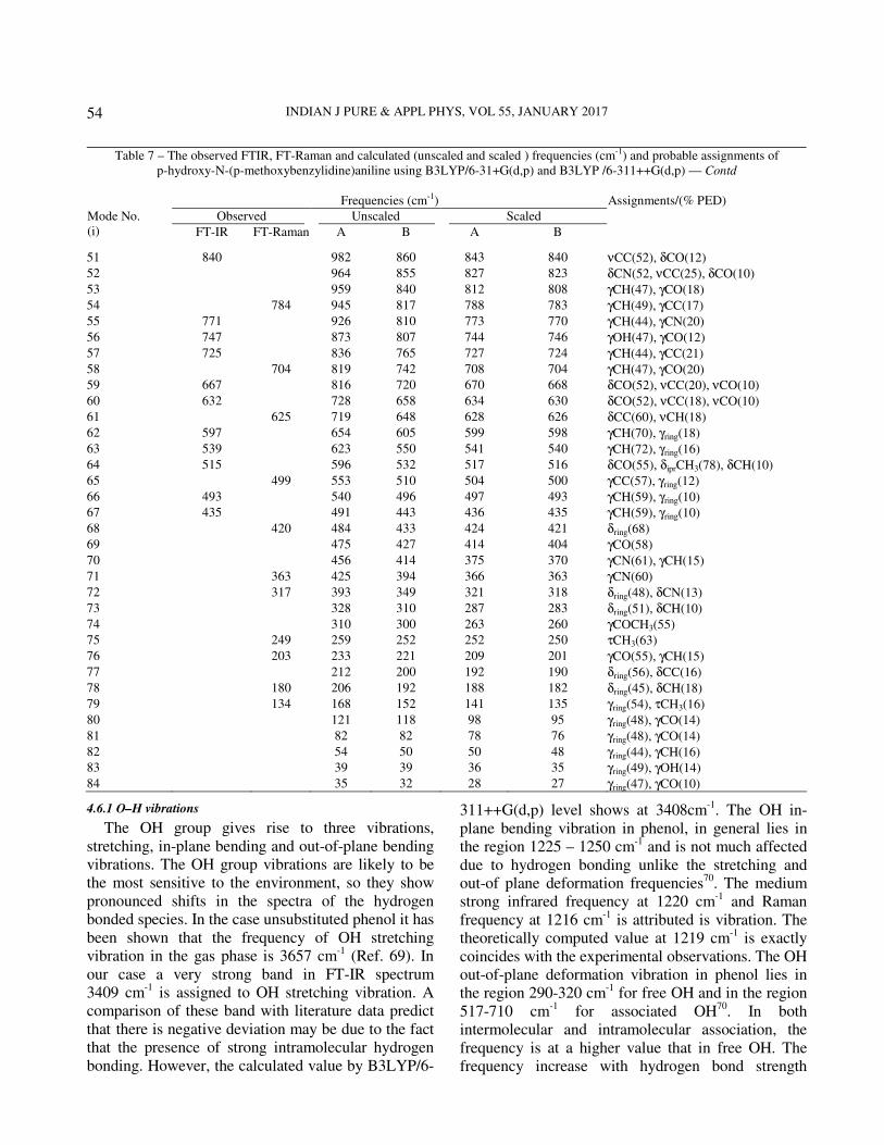
Table 7 – The observed FTIR, FT-Raman and calculated (unscaled and scaled ) frequencies (cm-1) and probable assignments of p-hydroxy-N-(p-methoxybenzylidine)aniline using B3LYP/6-31+G(d,p) and B3LYP /6-311++G(d,p)
Frequencies (cm-1)
Observed Unscaled Scaled Mode No. (i) FT-IR FT-Raman A B A B
Assignments/(% PED)
1 3409 4048 3837 3412 3408 νOH(95) 2 3068 3414 3206 3074 3068 νCH(99) 3 3045 3411 3202 3049 3044 νCH(98) 4 3000 3402 3192 3007 3002 νCH(97) 5 2977 3396 3190 2981 2977 νCH(99) 6 2955 3392 3186 2959 2954 νCH(96) 7 2932 3380 3174 2933 2932 νCH(96) 8 2909 3351 3160 2913 2910 νCH(96) 9 2886 3349 3150 2890 2885 νCH(98) 10 2841 3343 3137 2845 2840 νCH(96) 11 2750 3277 3068 2756 2751 νassCH3 (96) 12 2704 3230 3008 2705 2704 νassCH3(98)
13 2659 3204 2997 2663 2660 νssCH3(98)
14 1625 1884 1682 1628 1625 νCN(77), δCH(15) 15 1602 1822 1650 1607 1603 νCC(69), δCH(12) 16 1581 1815 1638 1586 1580 νCC(68), δCH(14) 17 1569 1788 1618 1572 1570 νCC(59), δOH(25), δCO(12) 18 1558 1557 1770 1603 1555 1558 νCC(58), δCO(20), νCN(10) 19 1512 1511 1702 1543 1516 1512 δCH(63), νCO(23), δCN(11) 20 1500 1694 1531 1507 1502 δCH(62), νCO(20), δCO(12) 21 1440 1443 1668 1505 1443 1440 δipbCH3(88)
22 1429 1661 1494 1435 1428 δopbCH3(85) 23 1627 1475 1427 1422 δsbCH3(87) 24 1417 1604 1462 1422 1418 δCH(62), δOH(18), νCC(10) 25 1406 1409 1591 1452 1412 1410 δCH(60), δOH(21), νCC(12) 26 1341 1548 1405 1345 1340 δCH(54), νCO(21), δCN(10) 27 1336 1484 1357 1338 1335 δOH(71), δCH(20) 28 1324 1474 1346 1323 1324 νCC(54), δCO(21), δCC(12) 29 1289 1284 1423 1328 1290 1285 νCC(58), δCO(20) 30 1267 1392 1311 1269 1270 νCC(59), δCH(26) 31 1255 1385 1279 1260 1256 νCN(63), νCO(18), νCC(10) 32 1220 1216 1373 1277 1223 1219 νCO(78), δCH(16), δCO(10) 33 1357 1265 1216 1212 νCC(52), δCO(19), δCC(11) 34 1197 1193 1324 1216 1198 1194 νCO(60), νCN(15), νCC(10) 35 1185 1313 1202 1189 1185 δoprCH3(82) 36 1309 1189 1171 1165 δCH(58), δOH(17), νCC(10) 37 1304 1186 1163 1159 δCH(52), δOH(21), νCN(12) 38 1276 1179 1147 1140 δCH(56), δOH(22), νCO(10) 39 1127 1125 1253 1168 1128 1126 δiprCH3(78), νCO(10) 40 1116 1236 1130 1120 1117 δCH(68), νCO(15), δCN(10) 41 1226 1121 1063 1060 δring (66), νCO(18), δCN(10) 42 1035 1171 1057 1040 1036 νCC(59), νCN(12) 43 1155 1023 1023 1020 νCC(54), δCO(17), δCC(10)
44 1142 1023 994 990 νCO(68), δCH(26)
45 979 977 1139 1003 980 978 δCH(56), δOH(21) 46 968 1130 992 971 968 δCH(58), δOH(24), νCO(10) 47 933 1129 959 936 933 δCH(58), δOH(22) 48 887 1114 945 889 886 νCC(52), δCO(18) 49 1106 940 871 864 νCC(58), δCO(12) 50 993 895 859 853 δCN(49), νCC(25) Contd—
INDIAN J PURE & APPL PHYS, VOL 55, JANUARY 2017
54
4.6.1 O–H vibrations
The OH group gives rise to three vibrations, stretching, in-plane bending and out-of-plane bending vibrations. The OH group vibrations are likely to be the most sensitive to the environment, so they show pronounced shifts in the spectra of the hydrogen bonded species. In the case unsubstituted phenol it has been shown that the frequency of OH stretching vibration in the gas phase is 3657 cm-1 (Ref. 69). In our case a very strong band in FT-IR spectrum 3409 cm-1 is assigned to OH stretching vibration. A comparison of these band with literature data predict that there is negative deviation may be due to the fact that the presence of strong intramolecular hydrogen bonding. However, the calculated value by B3LYP/6-
311++G(d,p) level shows at 3408cm-1. The OH in-plane bending vibration in phenol, in general lies in the region 1225 – 1250 cm-1 and is not much affected due to hydrogen bonding unlike the stretching and out-of plane deformation frequencies70. The medium strong infrared frequency at 1220 cm-1 and Raman frequency at 1216 cm-1 is attributed is vibration. The theoretically computed value at 1219 cm-1 is exactly coincides with the experimental observations. The OH out-of-plane deformation vibration in phenol lies in the region 290-320 cm-1 for free OH and in the region 517-710 cm-1 for associated OH70. In both intermolecular and intramolecular association, the frequency is at a higher value that in free OH. The frequency increase with hydrogen bond strength
Table 7 – The observed FTIR, FT-Raman and calculated (unscaled and scaled ) frequencies (cm-1) and probable assignments of p-hydroxy-N-(p-methoxybenzylidine)aniline using B3LYP/6-31+G(d,p) and B3LYP /6-311++G(d,p) — Contd
Frequencies (cm-1) Assignments/(% PED)
Observed Unscaled Scaled Mode No. (i) FT-IR FT-Raman A B A B
51 840 982 860 843 840 νCC(52), δCO(12) 52 964 855 827 823 δCN(52, νCC(25), δCO(10) 53 959 840 812 808 γCH(47), γCO(18) 54 784 945 817 788 783 γCH(49), γCC(17) 55 771 926 810 773 770 γCH(44), γCN(20) 56 747 873 807 744 746 γOH(47), γCO(12) 57 725 836 765 727 724 γCH(44), γCC(21) 58 704 819 742 708 704 γCH(47), γCO(20) 59 667 816 720 670 668 δCO(52), νCC(20), νCO(10) 60 632 728 658 634 630 δCO(52), νCC(18), νCO(10) 61 625 719 648 628 626 δCC(60), νCH(18) 62 597 654 605 599 598 γCH(70), γring(18) 63 539 623 550 541 540 γCH(72), γring(16) 64 515 596 532 517 516 δCO(55), δiprCH3(78), δCH(10) 65 499 553 510 504 500 γCC(57), γring(12) 66 493 540 496 497 493 γCH(59), γring(10) 67 435 491 443 436 435 γCH(59), γring(10) 68 420 484 433 424 421 δring(68) 69 475 427 414 404 γCO(58) 70 456 414 375 370 γCN(61), γCH(15) 71 363 425 394 366 363 γCN(60) 72 317 393 349 321 318 δring(48), δCN(13) 73 328 310 287 283 δring(51), δCH(10) 74 310 300 263 260 γCOCH3(55) 75 249 259 252 252 250 τCH3(63)
76 203 233 221 209 201 γCO(55), γCH(15) 77 212 200 192 190 δring(56), δCC(16) 78 180 206 192 188 182 δring(45), δCH(18) 79 134 168 152 141 135 γring(54), τCH3(16) 80 121 118 98 95 γring(48), γCO(14) 81 82 82 78 76 γring(48), γCO(14) 82 54 50 50 48 γring(44), γCH(16) 83 39 39 36 35 γring(49), γOH(14) 84 35 32 28 27 γring(47), γCO(10)

REVATHI et al.: MOLECULAR STRUCTURE OF p-HYDROXY-N-(p-METHOXY BENZYLIDENE) ANILINE
55
because of the larger amount of energy required to twist the OH bond out-of-plane 70. In our case the Raman band at 317 cm-1 is assigned as OH out-of-plane bending vibration. The calculated value of this vibration at 318 cm-1 is good agreement with experimental value.
4.6.2 C–H vibrations
For simplicity, modes of vibrations of aromatic compounds are considered as separate C−H or ring C−C vibrations. However, as with any complex molecules, vibrational interactions occur and these
levels only indicate the predominant vibration. Substituted benzenes have large number of sensitive bands, i.e., bands whose position is significantly affected by the mass and electronic properties, mesomeric or inductive of the substituents. According to the literature71,72, in infrared spectra, most mononuclear and polynuclear aromatic compounds have three or four peaks in the region 3000–3100 cm−1, these are due to the stretching vibrations of the ring CH bands. Accordingly, in the present study, the FT-IR bands identified at 3068, 3045, 3000, 2977,
Fig. 5 − Observed and calculated infrared spectrum of p-hydroxy-N-(p-methoxybenzylidene)aniline

INDIAN J PURE & APPL PHYS, VOL 55, JANUARY 2017
56
2950, 2932, 2909, 2886 and 2659cm−1 are assigned to C–H stretching vibrations of pHNpMBA. In B3LYP/6-311++G (d, p) method the values calculated at 3068, 3044, 3002, 2977, 2954, 2932, 2910, 2885, 2660 cm−1. The FT-IR bands at 1512, 1500, 1417, 1406, 1116, 979, 968 and 933 cm−1 and the FT Raman bands at1511, 1409, 1341, 977 cm−1 are assigned to C–H in-plane bending vibration of pHNpMBA. The C–H out-of-plane bending vibrations of the pHNpMBA are well indentified at 771,
725, 597, 539, 493, 435 cm−1 in the FT-IR and 784, 704 cm−1 in the FT-Raman spectra which are found to be well within their characteristic region.
4.6.3 Methyl group vibrations
The title compound under consideration possesses a CH3 group in the side-substituted chain. For the assignments of CH3 group frequencies, one can expect that nine fundamentals can be associated with each CH3 group, namely the symmetrical stretching in CH3
Fig. 6 − Observed and calculated Raman spectrum of p-hydroxy-N-(p-methoxybenzylidene)aniline

REVATHI et al.: MOLECULAR STRUCTURE OF p-HYDROXY-N-(p-METHOXY BENZYLIDENE) ANILINE
57
(CH3 symmetric stretch) and asymmetrical stretching (CH3 asymmetric stretch) in-plane stretching modes (i.e., in-plane hydrogen stretching mode); the symmetrical (CH3 symmetric deform) and asymmetrical (CH3 asymmetric deform) deformation modes; the in-plane rocking (CH3 ipr), out-of-plane rocking (CH3 opr), and twisting (tCH3) modes. Methyl groups are generally referred as electron-donating substituent in the aromatic ring system. The methyl hydrogen atoms are subjected simultaneously to hyper conjugation and back donation, which causes the decrease in stretching wave numbers and infrared intensities, as reported in literature73 for similar molecular system. For the O–CH3 group compounds74 the symmetric stretching mode appears in the range 2825–2870 cm–1, lower in magnitude compared with the value in CH3 compounds (2860–2935 cm–1), whereas the asymmetric stretching modes lie in the same region, 2924 and 2985 cm–1. In the present study, the asymmetric and symmetric stretching vibrations of CH3 group have been identified at 2840, 2751 and 2704 cm−1 by B3LYP/6-311++G (d, p) method. The recorded FT-IR spectrum show two weak bands observed at 2841, 2750 and 2704 cm−1, which are assigned to C–H asymmetric and symmetric vibrations of a CH3 group for the title compound. The in-plane and out-of-plane rocking vibrations for CH3 group have been calculated at 1126 and 1194 cm−1 by B3LYP/6-311++G(d, p) method. The asymmetrical CH3 deformation vibrations are computed by B3LYP++G(d, p) method at 1440 cm−1. Similarly, the symmetrical deformation vibration of CH3 group is computed at 1428 cm–1 for pHNpMBA. The bands observed at 1440 and 1443 cm−1 FT-IR and FT-Raman spectra, respectively. 4.6.4 C–C vibrations
The ring C=C and C–C stretching vibrations, known as semicircle stretching usually occur in the region 1400–1625 cm−. Hence in the present1,75,76
investigation, the FT-IR bands indentified at 1581, 1569, 1558, 1324, 1289, 1267 cm–1 and the FT-Raman bands at 1602, 1557, 1284 cm−1 are assigned to C–C stretching vibrations. The band ascribed at 625 and 499 cm–1 in FT-Raman spectrum have been designated to CC in-plane and out-of-plane bending mode. The calculated bending modes found at 626 cm−1 and 500 cm−1 in B3LYP/6-311++G(d, p) are assigned to C–C in-plane and out-of-plane bending vibrations, respectively.
4.6.5 O–CH3 vibrations
The O–CH3 mode is assigned to ∼ 1040 cm–1 for anisole77 and to the region 1000–1100 cm−1 for anisole and its derivatives78,79. This mode is assigned to 1026, 909 and 995 cm−1 for o-, m- and p-methoxy benzaldehyde, respectively. In the present investigation, the O–CH3 stretching mode is theoretically identified value at 990 cm−1 using B3LYP/6-311++G(d, p) basis set. The C–O–CH3 bending mode is assigned near 300 cm–1
for anisole by Owen and Hester80 and at 421 cm−1 for p-methoxy benzaldehyde by Compagnaro and Wood81. Ramana Rao and co-workers82 have proposed assignment for this mode in the region 300−670 cm–1 for anisole and its derivatives. As this mode lies in the region of the ring planar C–C–C angle bending modes, a strong mixing among these two modes and other planar modes is expected. Krishnakumar et al.
72 Assigned C–O–CH3 angle bending mode at 341, 382 and 430 cm−1 for the o- , m- and p-methoxy-benzaldehydes, respectively. In accordance with above we have assigned the theoretically computed values by B3LYP/6-311++G(d, p) method at 630 cm−1 for the title compound as C–O–CH3 angle bending mode, the value that coincides with 632 cm−1 band observed in FT-IR spectrum. The torsion mode of the O–CH3 group was observed for anisole at 100 cm−1 by some workers83. The computed value predicted by B3LYP/6-311++G(d, p) method at 260 cm−1 for O–CH3 torsion mode for pHNpMBA.
4.6.6 C–N and C=N vibrations
The identification of C–N and C=N stretching vibrations are a difficult task since the mixing of several modes are possible in the region. Silverstein83 assigned C–N and C=N stretching absorption in the region 1386–1266 cm−1 and 1690–1630 cm−1, respectively. Hence, in the present investigation, the band at 1625 cm−1 in FT-Raman spectrum assigned to C=N stretching vibration. The theoretically computed value at 1628 cm−1 and 1625 cm−1 by B3LYP/6-31+ G(d, p) and B3LYP/6-311++ G(d, p) methods, respectively for C=N stretching vibration. The band at 1255 cm−1 in FT-IR spectrum assigned to C–N stretching vibrations. The computed value for C–N stretching mode found at 1260 cm−1 and 1256 cm−1, respectively in B3LYP/6-31+ G(d, p) and B3LYP/6-311++ G(d, p) methods.
5 Conclusions The vibrational characteristics of pHNpMBA has been
investigated by the experimental (FT-IR and FT-Raman)

INDIAN J PURE & APPL PHYS, VOL 55, JANUARY 2017
58
and theoretical DFT quantum chemical methods. Scaled theoretical wave numbers and PED results were quite useful for the reliable assignments of normal modes of vibrations; moreover, the optimized geometrical parameters were calculated. On the basis of agreement between the calculated and experimental results, assignments of all the fundamental vibrational modes of the title compound are examined and proposed in this investigation. Therefore, the assignments made at higher level of theory with higher basis set with only reasonable deviations from the experimental values, seems to be correct. Based on calculated energy differences, the C4 conformer is found to be the most stable conformer. The electric dipole moments and first hyper polarizability of the compound studied have been calculated by DFT method with 6-311++G(d, p) basis set. The DFT calculated non-zero µ value of this legend shows that the pHNpMBA compound might have microscopic first hyperpolarizability with non-zero values obtained by the numerical second derivatives of the electric dipole moment according to the applied field strength. The NBO analysis performed in this study enabled us to know about the conjugative interactions and other type of interactions taking place within the molecular species. The second-order perturbation theory results show that the CH3 group behaves as separate unit with sufficient interaction energy. The mapped iso-density surfaces for the frontier molecular orbitals were also plotted and the smallest energy gap of -0.15558 a.u. is calculated between HOMO and LUMO orbitals. The lowering of HOMO−LUMO band gap supports bioactive property of the molecule. Acknowledgments
The authors are thankful to SAIF, Indian Institute of Technology, Chennai for getting recorded FT-IR and FT-Raman spectra. One of the authors (B Revathi) is grateful to the UGC-MRP [MRP-5128/149 (SERO/UGC)] for the financial assistance. References
1 Balachandran V, Santhi G, Karpagam V & Lakshmi A, J Mol Struct, 1047 (2013) 249.
2 Diaz F A, Sanchez C O, Del Valle M A, Torres J L & Tagle LH, Synth Met, 118 (2001) 25.
3 Ng S C & Xu L, Adv Mater, 10 (1998) 1525. 4 Misra R A, Dubey S, Prasad B M & Singh D, Indian J Chem,
38 (1999) 141. 5 Prevast V, Petit A & Pla F, Synth Met, 104 (1999) 79. 6 Kanungo M, Kumar A & Contractor A Q, J Electroanal
Chem, 528 (2002) 46.
7 Inzelt G & Kertesz V, Electro Chim Acta, 42 (1997) 229. 8 Dong Y & Mu S, Electro Chim Acta, 36 (1991) 2015. 9 Delongchamp D M & Hammond P T, Chem Mater, 16
(2004) 4799. 10 Whysner J, Vera L & Williams G M, Pharmacol Ther, 71
(1996) 107. 11 Hohenberg P & Kohn W, Phys Rev B, 136 (1964) 864. 12 Vaschetto M E, Retamal B A & Monkman A P, J Mol Struct,
468 (1999) 209. 13 Tzeng W B, Narayanan K, Lin J L & Tung C C, Spectro
Chim Acta, 55 (1999) 153. 14 Tzeng W B, Narayanan K, Shieh K C & Tung C C, J Mol
Struct, 428 (1998) 231. 15 Hohenberg P & Kohn W, Phys Rev B, 136 (1964) 864. 16 Evans J C, Spectro Chim Acta, 16 (1960) 428. 17 Roussy G & Nonat A, J Mol Spectrosc, 118 (1985) 180. 18 Lister G D, Tyler J K, Hog J H & Larsen N W, J Mol Struct,
23 (1974) 253. 19 Fukuyo M, Hirotsu K & Higuchi T, Acta Crystallogr, 38
(1982) 640. 20 Ventura M C & Kassab E, Spectro Chim Acta, 50 (1994) 69. 21 Gorse A D & Pesquer M, J Mol Struct, 21 (1993) 281. 22 Bock C W, George P & Trachtman M, Theor Chim Acta, 69
(1986) 235. 23 Wang Y, Saeb S & Pittman C U, J Mol Struct, 91 (1993)
281. 24 Altun A, Gokuk K & Kumru M, J Mol Struct, 625 (2003) 17. 25 Yudakul S & Sen A I, Vib Spectrosc 20 (1999) 27. 26 Alkalin E & Akyuz S, J Mol Struct, 175 (1999) 428. 27 Lopez Tocon I, Becucci M, Pietra Perzia G, Castelluchi E &
Otero J C, J Mol Struct, 421 (2001) 565. 28 Vaschettom M E, Retamal B A & Monkman A P, J Mol
Struct, 468 (1999) 209. 29 Palafox M A, Nunez J L & Gil M, J Mol Struct, 593 (2002)
101. 30 Tzeng W B & Narayanan K, J Mol Struct, 434 (1998) 247. 31 Engelter C, Thornton D A & Ziman M R, J Mol Struct, 49
(1978) 7. 32 Engelter C, Thornton D A & Ziman M R, J Mol Struct 119
(1976) 33 33 Johnson P R & Thornton D A, Chimia, 28 (7) (1974) 345. 34 Altun A, Golcuk K & Kumru M, J Mol Struct, 673 (2003)
155. 35 Sharma S J & Dwivedi C P D, Indian J Pure Appl Phys, 13
(1975) 570. 36 Sundaraganesan N, Saleem H, Mohan S & Ramalingam M,
Spectrochim Acta A, 61 (2005) 377. 37 Shankar R, Yadav R A, Singh I S & Singh O N, Indian J
Pure Appl Phys, 23 (1985) 339. 38 Barluenga J, Fananas F J, Sanz R & Fernandez Y, Chem Eur J,
8 (2002) 2034. 39 Rai A K, Kumar S & Rai A, Vib Spectrosc, 42(2006) 397. 40 Rai V K, Rai A, Rai D K & Rai S B, Spectrochim Acta, 60
(2004) 53. 41 Frisch M J, Trucks G W & Schlegel H B, Gaussian, (Inc,
Wallingford CT), 2009. 42 Pulay P, Fogarasi G, Pongor G, Boggs J E & Vargha A, J Am
Chem Soc, 105 (1983) 7037. 43 Rauhut G & Pulay P, J Phys Chem, 99 (1995) 3093. 44 Sundius T, J Mol Spectrosc, 82 (1980) 138. 45 Sundius T, J Mol Struc, 218 (1990) 321.

REVATHI et al.: MOLECULAR STRUCTURE OF p-HYDROXY-N-(p-METHOXY BENZYLIDENE) ANILINE
59
46 Sundius T, MOLVIB: A program for Harmonic Force Fields
Calculations, QCPE Program No 604, 1991. 47 Polavarapu P L, J Phys Chem, 94 (1990) 8106. 48 Keresztury G, Holly S, Varga J, Besenyei G, Wang A V &
Durig J R, Spectrochim Acta, 49 (1993) 2007. 49 Keresztury G, Chalmers J M & Griffiths P R, Handbook of
vibrational spectroscopy, Vol 1, (John Wiley & Sons, Ltd), 2002. 50 Roussy G & Nonat A, J Mol Spectrosc 118 (1986) 180 51 Schultz G, Portalone G, Ramondo F, Domenicano A &
Hargittai I, Struct Chem, 7 (1996) 59. 52 Prasad J V, Rai S B & Thakur S N, Chem Phys Lett, 164
(1989) 629. 53 Ahmed M K & Henry B R, J Phys Chem, 90 (1986) 629. 54 Tzeng W B, Narayan K, Lin J L & Tung C C, Spectrochim
Acta, A,55 (1999) 153. 55 Sun X, Hao Q L, Wei W X, Yu Z X, Lu D D, Wang X &
Wang Y S, J Mol Struct Theochem, 74 (2009) 904. 56 Andraud C, Brotin, Garcia T, Pelle F, Goldner P, Bigot B &
Collet A, J Am Chem Soc, 116 (1994) 2094. 57 Geskin V M, Lambert C & Bredas J L, J Am Chem Soc, 125
(2003) 15651. 58 Nalano M, Fujita H, Takahata M & Yamaguchi K, J Am
Chem Soc, 124 (2002) 9648. 59 Sajan D, Joe I H, Jayakumar V S & Zaleski J, J Mol Struct,
785 (2006) 43. 60 Zhang C R, Chen H S & Wang G H, Chem Res Chin Univ,
20 (2004) 640. 61 Sun Y, Chen X, Sun L, Guo X & Lu W, J Chem Phys Lett,
381 (2003) 397. 62 Christiansen O, Gauss J & Stanton J F, J Chem Phys Lett,
305 (1999)147. 63 Kleinman D A, Phys Rev, 126 (1962) 1977. 64 Srivastava A, Tandon P, Jain S & Asthana B P, Spectrochim
Acta A, 84 (2011) 144.
65 Glendening E D, Landis C R & Weinhold F, WIREs Comput
Mol Sci, 2 (2012) 1. 66 Liv J N, Chem Z R & Yuan S F, J Zhejiang Unit Sci B, 6
(2005) 584. 67 Reed A E, Curtiss L A & Weinhold F, Chem Rev, 88 (1988)
896. 68 Gupta V P, Sharma A, Virdi V & Ram VJ, Spectrochim Acta A,
64 (2006) 57. 69 Michalska D, Dienko D C, Abkowicz-Bienko A J & Latajka Z,
J Phys Chem, 100 (1996) 123. 70 Varasanyi G, Assignments of Vibrational spectra of seven
hundred benzene derivatives, (Wiley, New York), 1974. 71 Socrates G, Infrared and raman characteristic group
frequencies: 3rd Ed (Wiley, New York), 2001. 72 Krishna Kuamr V & Balachandran V, Spectrochim Acta,
63A (2006) 464. 73 Smith B, Infrared spectral interpretation, a systematic
approach, (CRC, Washington DC), 1999. 74 Singh D N, Singh I D & Yadav R A, Ind J Phys, 763 (2002)
307. 75 Krishnakumar V & John Xavier R, Indian J Pure Appl Phys,
41 (2003) 95. 76 Furic K, Mohacek V, Bonifacic M & Stefanic I, J Mol Struct,
267 (1992) 39. 77 Walter J & Balfour, Spectrochim Acta, 39 (1983) 795. 78 Owen N L & Hester R E, Spectrochim Acta, 25 (1969) 343. 79 Campangnaro G E & Wood J L, J Mol Struct, 6 (1970) 117. 80 Owen N L & Hester R E, Spectrochim Acta, 25 (1969) 343. 81 Campangnaro G E & Wood J L, J Mol Struct, 6 (1970) 117. 82 Venkkatram Reddy B, Ramana Rao G, Vib Spectro, 6 (1994)
231. 83 Silverstein M, Clayton Basseler G & Morill C, Spectrometric
identification of organic compounds, (Wiley, New York), 1981.