veterinary embryology - download.e-bookshelf.de · veterinary embryology t. a. mcgeady, mvb, ms,...
TRANSCRIPT

VETERINARY EMBRYOLOGY

VETERINARY EMBRYOLOGY
T. A. McGeady, MVB, MS, MSc, MRCVSFormer Senior Lecturer in Veterinary Anatomy, Histology and Embryology,Department of Veterinary Anatomy,Faculty of Veterinary Medicine,University College Dublin
P. J. Quinn, MVB, PhD, MRCVSProfessor Emeritus,Former Professor of Veterinary Microbiology and Parasitology,Faculty of Veterinary Medicine,University College Dublin
E. S. FitzPatrick, FIBMSDepartment of Veterinary Anatomy,Faculty of Veterinary Medicine,University College Dublin
M. T. Ryan, MSc, PhDMolecular Biology Laboratory,Faculty of Veterinary Medicine,University College Dublin
Illustrations byS. Cahalan, MVBFaculty of Veterinary Medicine,University College Dublin

© 2006 TA McGeady, PJ Quinn, ES FitzPatrick, MT Ryan and S Cahalan
Editorial Offices:Blackwell Publishing Ltd, 9600 Garsington Road, Oxford OX4 2DQ, UK
Tel: +44 (0)1865 776868Blackwell Publishing Professional, 2121 State Avenue, Ames, Iowa 50014-8300, USA
Tel: +1 515 292 0140Blackwell Publishing Asia, 550 Swanston Street, Carlton, Victoria 3053, Australia
Tel: +61 (0)3 8359 1011
The right of the Author to be identified as the Author of this Work has been asserted in accordance with the Copyright, Designs and Patents Act 1988.
All rights reserved. No part of this publication may be reproduced, stored in a retrieval system, or transmitted, in any form or by any means, electronic, mechanical, photocopying, recording or otherwise, except as permitted by the UK Copyright, Designs and Patents Act 1988, without the prior permission of the publisher.
First published 2006 by Blackwell Publishing Ltd
ISBN-10: 1-4051-1147-XISBN-13: 978-1-4051-1147-8
Library of Congress Cataloging-in-Publication Data
Veterinary embryology / T.A. McGeady . . . [et al.].; illustrations by S. Cahalan.— 1st ed.p. cm.
Includes bibliographical references and index.ISBN-13: 978-1-4051-1147-8 (pbk. : alk. paper)ISBN-10: 1-4051-1147-X (pbk. : alk. paper)1. Veterinary embryology. I. McGeady, T. A. (Thomas A.)
SF767.5.V48 2006636.089′264—dc22
2005022781
A catalogue record for this title is available from the British Library
For further information on Blackwell Publishing, visit our website:www.blackwellpublishing.com
Preface ixAcknowledgements xi
1 Division, growth and differentiation of cells 1The cell cycle 1Mitosis 1Meiosis 5
2 Gametogenesis 10Spermatogenesis 10Oogenesis 13
3 Fertilisation 17Capacitation 18Cellular events in the process of fertilisation 18Barriers to polyspermy 18Ovum activation 19In vitro fertilisation 21Comparative fertilisation rates 21Sex determination 22Parthenogenesis 22Sex ratio 23Chromosomes of domestic animals 23
4 Cleavage 25Cleavage in primitive chordates,
amphibians, avian species and mammals 25Stem cells 30
5 Gastrulation 34Primitive chordates 34Amphibians 34Avian species 34Mammals 36Establishment of left–right symmetry
in vertebrates 38Twinning 38
6 Aspects of cell signalling and gene functioning during development 42Cellular messengers and receptors 42Types of signalling 43Induction and competence 44Paracrine signalling during development 44Apoptosis 48
Morphogens 49Differentiation 49Gene structure and organisation 49X-chromosome inactivation 49DNA methylation and parental
imprinting in mammals 49Promoters, enhancers and silencers 50Transcription factors 50Gene systems essential for development 50Experimental measurement of gene
expression 53Experimental evaluation of gene function 53Concluding comments 53
7 Establishment of the basic body plan 54
8 Coelomic cavities 59Pleural and pericardial cavities 59Diaphragm 61Peritoneal cavity 64Omenta 65
9 Foetal membranes 66Development of the foetal membranes 68Birds 68Mammals 70
10 Forms of implantation and placentation 78Implantation 78Placentation in mammals 81Functional aspects of the placenta 101
11 Cardiovascular system 105Development of the cardiac tubes 106Molecular aspects of cardiac development 112Formation of the cardiac chambers 112Conducting system of the heart 117Developmental anomalies of the
cardiovascular system 130
12 Embryological and post-natal features ofhaematopoiesis 136Embryological aspects of haematopoiesis 136Cell differentiation and maturation during
haematopoiesis 139
Contents

CONTENTS
Stem cells in human adults and mature animals 146
Immunodeficiency 147Inherited defects in natural immunity 151
13 Nervous system 153Dorsal–ventral patterning of the neural tube 153Neural crest 154Differentiation of the cellular components
of the neural tube 155Spinal nerves 157Myelination of peripheral nerve fibres 161Changes in the relative positions of the
spinal cord and the developingvertebral column 161
Anomalies of the spinal cord 161Differentiation of the brain sub-divisions 163Ventricular system of the brain and
cerebrospinal fluid circulation 174Molecular aspects of brain development 175Brain anomalies 176Brain stem and spinal cord 177Cranial nerves 178Peripheral nervous system 178Autonomic nervous system 178Enteric nervous system 181Meninges 182
14 Muscular and skeletal systems 184Differentiation of somites 184Muscular system 184Skeletal muscle 184Cytodifferentiation of muscle 186Skeletal system 187Skeletal anomalies 203
15 Digestive system 205Molecular regulation of alimentary tract
development 207Oesophagus 209Stomach 209Liver 213Pancreas 213Spleen 216Development and rotation of the intestines
in domestic animals 217Comparative features of the intestines 217Hindgut 220Developmental anomalies of the
alimentary tract 221
16 Respiratory system 225Formation of the larynx 225Trachea, bronchi and lungs 225Molecular aspects of respiratory development 231Anomalies of the respiratory system 231
17 Urinary system 233Kidney 233Molecular basis of metanephros development 235Unilobar kidneys 238Multilobar kidneys 240Bladder 240Developmental anomalies of the urinary
system 240
18 Male and female reproductive systems 244Primordial germ cells 244Undifferentiated stage of gonad formation 245Differentiation and maturation of the testes 245Differentiation and maturation of the ovaries 245Features of equine gonadal development 248Genital ducts 249Formation of the genital fold 251External genitalia 252Factors which influence sexual
differentiation in mammals 253Sex determination 254Molecular aspects of gonadogenesis 254Influence of hormones on development
of genital ducts and external genitalia 255Sexual differentiation, associated brain
function and subsequent sexualbehaviour at puberty 257
Anomalies of sexual development 257Descent of the testes 259Ovarian migration 262Cryptorchidism 262Development of the mammary gland 263Comparative features of mammary gland
development in domestic animals 265
19 Structures in the head and neck 268Pharyngeal region 268Derivatives of the pharyngeal apparatus 269Face 270Nasal cavities 272Oral cavity 277Tongue 277Salivary glands 278Teeth 279Comparative aspects of dentition 281Molecular aspects of tooth development 282Development of the skull 282Congenital malformations of face and
oral cavity 283
20 Endocrine system 286Pituitary gland 286Pineal gland 289Adrenal glands 289Thyroid gland 291Parathyroid glands 291
vi
CONTENTS
Thymus 293Pancreatic islets 293
21 Eye and ear 295Eye 295Ear 304
22 Integumentary system 313Epidermis 313Dermis 314Hypodermis 315Hair 315Mammalian skin glands 318Avian skin 320Congenital and inherited defects of the skin 322Hooves and claws 322Horns and related structures 328
23 Age determination of the embryo and foetus 331
24 Genetic, chromosomal and environmental factors which adversely affect pre-natal development 337Mutations 337Chromosomal abnormalities 338Teratogens 339Therapeutic drugs and chemicals 339Cytotoxic drugs used for treating
neoplastic diseases 348Poisonous plants 348Infectious agents 349Assessing the aetiology of congenital disease 353
Glossary 355Index 364
vii

An understanding of the origin, development and matu-ration of cells in the developing embryo and, later, in thefoetus provides veterinary students with informationrelevant to organ primordia and development of bodysystems. A study of embryology offers the student anunderstanding of the development, structure, final formand relationships of tissues and organs. Developmentaldefects and the clinical conditions to which they give risecan be more completely understood through a knowledgeof the factors which control developmental processesand the deleterious affects of environmental teratogenson normal embryological development.
This book is primarily concerned with developmentalaspects of cells, tissues, organs and body systems of animals. Where feasible, comparative aspects of humanembryology are included. Drawings of cells, tissues andorgans, along with flow diagrams and tables, are used toprovide a clear understanding of information containedin the text.
There are 24 chapters in this book, each dealing withtopics which are fundamental to an understanding of the sequential stages of embryological and foetaldevelopment. Cell division, gametogenesis, fertilisation,cleavage and gastrulation are presented in sequentialchapters. Succeeding chapters are concerned with cellsignalling, establishment of a body plan, formation offoetal membranes and placentation. Body systems areconsidered in separate chapters and the embryologicalaspects of structures associated with special senses are
reviewed. Age determination and aspects of mutagenesisand teratogenesis are briefly reviewed in final chapters.
Although this book is intended primarily as a textbookfor undergraduate veterinary students, it may be of valueto colleagues engaged in teaching embryology, either aspart of a veterinary curriculum or in courses relating to animal science or developmental biology. Researchworkers engaged in projects on animal reproductionand allied topics may find particular chapters relevantto their fields of investigation.
Throughout the book, emphasis is placed on the originand differentiation of tissues and organs and their relationships to each other. This approach provides alogical foundation for acquiring an understanding ofthe form and relationships of cells, tissues, organs andstructures in defined regions of the body. Such know-ledge is a fundamental requirement for the appreciationof topographical anatomy, a cornerstone in the acquisi-tion of clinical skills, interpretation of diagnostic imag-ing and the implementation of surgical procedures.Molecular aspects of embryology provide an introduc-tion to genes and the transcription factors which promoteor regulate orderly development of the embryo and foetus. Developmental defects of clinical significanceare also included. The classification used throughout thebook generally conforms to the Nomina EmbryologicaVeterinaria system proposed in 1994. Selected reviewarticles and textbooks are listed in each chapter assources of additional information.
Preface

We wish to acknowledge the constructive commentsand advice of colleagues who reviewed chapters andproofread sections of the book or who offered technicalsupport and guidance during the completion and assem-bly of the text:
H. Bassett, S. Baynes, J. Cassidy, W.J.C. Donnelly, M. Dore, M. Gleeson, O. Golden, T. Grimes, P.J.Hartigan, S. Hogan, D. Hogg, E. Hughes, J. Irwin, A. Kelly, D. Kilroy, F. LeMatti, D. Maguire, G.McCarthy, T. McElligot, T. Miceli, E. Murphy, J. O’Donovan, K. O’Driscoll, E. O’Neill, P. O’Neill, J. Quinn, M. Quinn, C. Reid, J. Roche, M. Scanlon andT. Sweeney.
We are appreciative of the space and facilities madeavailable in the Departments of Veterinary Anatomy
and Veterinary Microbiology and Parasitology byProfessors S. Carrington and G. Mulcahy and by Dr B. Markey.
We wish to thank the library staff of the Faculty ofVeterinary Medicine, especially Ms G. Ryan, for thehelp and facilities provided.
Through her careful reading of the manuscript, MsMary Sayers, copy editor, improved the accuracy of thetext and the clarity of the illustrations. Ms SamanthaJackson and Ms Sally Rawlings and their colleagues atBlackwell Publishing provided advice on layout andstyle and offered encouragement as the book approachedthe end of its uncertain gestational period.
Dublin, September 2005
Acknowledgements

The mammalian body is composed of an array oforgans, tissues and individual cells which function in aspecialised and highly coordinated manner. Althoughthese cells, tissues and organs exhibit considerablediversity in both structure and function, they all derivefrom a single cell, a fertilised ovum. The fertilised ovumis a product of the fusion of two specialised reproductivecells, gametes, of male and female origin. Following fertilisation, the ovum undergoes a series of divisionswhich ultimately lead to the formation of pluripotentstem cells, from which all cells, tissues and organs of thebody arise. The study of this process of growth and dif-ferentiation, beginning with the fertilisation of an ovumand progressing to a fully formed individual animal, istermed embryology.
Cells associated with tissue formation and regenerationare described as somatic cells. Specialised reproductivecells, referred to as germ cells, include gametes of maleand female origin and their precursors.
Coordinated and regulated cell division is essential forembryological development. Somatic cell division con-sists of nuclear division, mitosis, followed by cytoplas-mic division, cytokinesis. In mitotic division of somaticcells, the daughter cells produced are genetically iden-tical. A form of cell division distinctly different frommitosis occurs in germ cells. In this form of cell division,referred to as meiosis, the cells produced contain halfthe number of chromosomes of the progenitor germ celland are not genetically identical. Somatic cell divisioncombined with other cellular processes such as pro-gressive differentiation, migration, adhesion, hypertro-phy and apoptosis are prerequisites for embryologicaldevelopment.
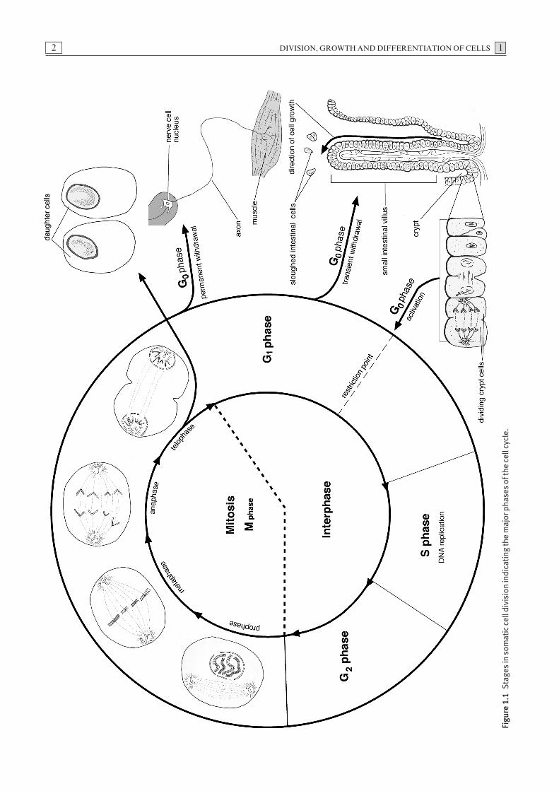
The cell cycle
Somatic cells undergo a series of molecular and morpho-logical changes as part of the cell cycle. These changesoccur in four sequential phases, namely G1, S, G2 andM, and also a quiescent phase, termed G0 (Fig. 1.1). TheG1 and G2 phases are termed resting phases. In these
phases, the cell is metabolically active, fulfilling its spe-cialised function preparatory to the next phase of thecycle, but DNA replication does not take place. Duringthe S phase, DNA synthesis occurs prior to chromo-somal replication. This is followed by mitosis whichoccurs during the M phase. Collectively, the G1, S andG2 phases constitute the interphase (Fig. 1.1). Cellswhich enter a G0 state may remain transiently or perma-nently in that state. Certain fully differentiated cells,such as neurons, do not divide, and continue to functionpermanently in a G0 state. Other cell types, such asepithelial cells and hepatocytes, can re-enter the cellcycle from G0 and proceed to mitotic division inresponse to appropriate stimuli.
A number of stimuli such as growth factors, mitogensand signals from other cells and from the extra-cellularmatrix can induce cells in a G0 state to re-enter the cell cycle near the end of the G1 phase. Growth factorswhich bind to cell surface receptors activate intra-cellular signalling pathways. In most mammalian cells,the activation of genes encoding cyclins and cyclin-dependent kinases (Cdks) specific to the G1 phase regu-late the cell cycle and commit the cell to enter the Sphase. This process is initiated at the restriction point, astage at which mammalian cells become committed toentering the S phase and are then capable of completingthe cell cycle independent of extra-cellular influences.
The rate of cell division varies in different cell types andat different stages of differentiation. Variations in cellcycle length are largely attributed to differences in thelength of the G1 phase, which can range from six hoursto several days. Early embryonic development is charac-terised by rapid cell division, but as cells become moredifferentiated during organ development, the rate of celldivision generally decreases.
Mitosis
The nuclei of somatic cells of each mammalian specieshave a defined number of chromosomes (Table 1.1). A somatic cell with a full complement of chromosomes
1 Division, Growth and Differentiation of Cells
DIVISION, GROWTH AND DIFFERENTIATION OF CELLS 12
Figu
re 1
.1St
ages
in s
omat
ic c
ell d
ivis
ion
indi
cati
ng th
e m
ajor
pha
ses
of th
e ce
ll cy
cle.

1 DIVISION, GROWTH AND DIFFERENTIATION OF CELLS
is referred to as diploid and given the designation 2n.The term mitosis is used to describe nuclear division of somatic cells, a process which usually results in the production of two cells, with the same chromosome complement as the progenitor cell from which theyderived. Mitosis is essential for embryonic growth anddevelopment and for repair and replacement of tissuethroughout life. The stages of mitosis occur as a distinctsequence of cytological events, which are part of the cellcycle.
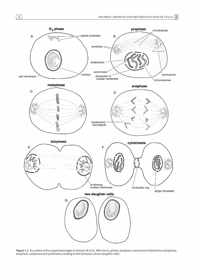
Stages of mitosis
Preparatory to mitosis, the chromosomes are replicatedin the S phase of the cell cycle forming sister chromatids.Within the nuclear envelope sister chromatids remainattached at a constricted region of the chromosome calleda centromere. Following the G2 phase, mitosis, whichcan be divided into four stages, prophase (Fig. 1.2B),metaphase (Fig. 1.2C), anaphase (Fig. 1.2D) and finallytelophase (Fig. 1.2E), begins. The stages of mitosis areusually followed by cytoplasmic division or cytokinesis(Fig. 1.2F).
Prophase
The first stage of mitosis is prophase (Fig. 1.2B). Dur-ing this period, the chromosomes, consisting of closelyassociated sister chromatids, condense. Outside thenucleus, the centrosomes, composed of paired centriolespreviously replicated during interphase, begin to form
microtubule spindles or asters. The spindles are respon-sible for the movement of the centrosomes to oppositepoles of the dividing cell.
Microtubules, an essential part of the mitotic apparatus,are visible microscopically only during the M phase. Indi-vidual microtubules are cylindrical structures, composedof 13 parallel protofilaments consisting of alternating α-tubulin and β-tubulin subunits. An individual micro-tubule may grow or shrink by a process of polymerisa-tion of α-tubulin and β-tubulin. A growing microtubulehas a structure referred to as a guanidine-triphosphate(GTP) cap. The β-subunit of a microtubule contains GTPcapable of being hydrolysed to guanidine-diphosphate(GDP). This, in turn, alters the conformation of the sub-units, resulting in shrinking of the microtubules. If GTPhydrolysis occurs more rapidly than subunit addition,the cap is lost and the microtubule shrinks. Shrinkingand growing are a dynamic process and these changesenable the microtubules to actively orientate and movechromosomes during mitosis and meiosis.
Metaphase
Events during the metaphase stage of mitosis can bedivided into two phases, pro-metaphase and metaphase.Disintegration of the nuclear envelope marks the beginning of pro-metaphase. A kinetochore, a proteincomplex which forms on the centromeres during lateprophase, acts as a platform for attachment to micro-tubules. Chromosomes attach to the microtubules viatheir kinetochores and the combination of these two latter structures is termed a kinetochore microtubule.The formation of the kinetochore microtubule enablesthe movement of chromosomes to take place. Duringmetaphase, the chromosomes are positioned midwaybetween the poles of the cell at a region termed themetaphase plate. Each sister chromatid is attached to thecentrosome by its kinetochore microtubule (Fig. 1.2C).
Anaphase
During the anaphase stage, the pairs of conjoined sisterchromatids synchronously separate as the centromeressplit and the attached kinetochore microtubules shorten.The newly separated chromatid sets are drawn towardsopposite poles of the cell (Fig. 1.2D).
Telophase
The two groups of identical chromosomes (former chro-matids) clustered at their respective poles, de-condenseand a nuclear envelope forms around each set. The for-mation of nuclear envelopes marks the end of mitosis, aprocess which results in equal and symmetrical divisionof the nucleus (Fig. 1.2E).
3
Table 1.1 The number of chromosomes in diploid human andanimal cells.
Species Number of chromosomes (2n)
Humans 46
Cats 38
Cattle 60
Chickens 78
Dogs 78
Donkeys 62
Goats 60
Horses 64
Pigs 38
Rabbits 44
Rats 42
Sheep 54
DIVISION, GROWTH AND DIFFERENTIATION OF CELLS 14
Figure 1.2 An outline of the sequential stages in mitosis (A to G). After the G2 phase, prophase commences followed by metaphase,anaphase, telophase and cytokinesis, leading to the formation of two daughter cells.
1 DIVISION, GROWTH AND DIFFERENTIATION OF CELLS
Cytokinesis
Following the formation of the nuclear envelope, a con-tractile ring of actin and myosin pinches the cell walland divides the cytoplasm, resulting in the formation oftwo daughter cells (Figs. 1.2F and G). This latter pro-cess, termed cytokinesis, typically results in the forma-tion of two equally-sized daughter cells. Occasionally,unequal amounts of cytoplasm or organelles may be distributed to the daughter cells during cytokinesis. Insome instances mitosis may occur without subsequentcytokinesis, resulting in the formation of binucleate or,occasionally, multinucleate cells.
In lower organisms such as amphibians, the cytokinesiswhich occurs early in development can generate daugh-ter cells in which the factors which direct the fate of thecells may not be uniformly distributed. This unequaldivision of fate determinants results in differing devel-opmental potential in individual daughter cells. Inmammals, experimental evidence suggests that cell divisions which give rise to totipotential cells occur earlyin development. This suggests that, in mammals, cyto-plasmic determinants are shared uniformly betweendaughter cells and that the initial stages of differentia-tion arise as a result of cell communication and micro-environmental factors.
Regulation of mitosis
The enzyme M-cyclin-dependent kinase (M-Cdk) has a central role in the initiation of mitosis following the G2 phase of the cell cycle. This heterodimeric protein,which is a complex of Cdk1 and M-cyclin, is activatedby the removal of inhibitory phosphate groups in thelate G2 phase. The M-Cdk protein induces events essen-tial for mitosis, including phosphorylation of the pro-teins which control microtubule dynamics, chromatincondensation, rearrangement of both the cytoskeletonand organelles and, finally, dissolution of the nuclearenvelope. Although the mitotic cell cycle is normallyhighly regulated, undesirable alterations in the func-tioning of the genes known as proto-oncogenes ortumour suppressor genes, responsible for the control of cell proliferation or differentiation, may lead tomalignant transformation of normal tissue. Typically,changes in two or more of these regulatory genes appear to be required for cells to undergo malignanttransformation.
Mitotic division in successive generations of cells derivedfrom a neoplastic cell continues to give rise to abnormalcells which are not subject to normal regulatory pro-cesses. Neoplastic conditions such as leukaemia, lym-phoma and myeloma can arise from gene alteration
within a single cell in the bone marrow or in peripherallymphoid tissue. With the accumulation of large popu-lations of abnormal cells, clinical effects of neoplasiabecome evident.
Meiosis
This process of cell division occurs only during gameto-genesis. Meiosis differs from mitosis in several respects:
(1) The resulting gametes are haploid and are given thedesignation ‘n’.
(2) There is a reciprocal exchange of genetic materialbetween non-sister chromatids (Fig. 1.3).
(3) The resulting gametes are a product of the randomsegregation of maternally-derived and paternally-derived chromatids.
Meiosis is divided into two stages, meiosis I and II.
The first meiotic division
Meiosis I consists of prophase I (Figs. 1.4B and C),metaphase I (Fig. 1.4D), anaphase I (Fig. 1.4E) andtelophase I (Fig. 1.4F). The amount of DNA in a cellentering prophase I doubles.
Prophase I
During prophase I, many crucial intracellular eventsoccur (Figs. 1.4B and C). This process can be further subdivided into five substages: leptotene, zygotene,pachytene, diplotene, and diakinesis. At the diakinesisstage, the chromosomes become short and thick, thecentrosomes are positioned at the poles and the nuclearmembrane begins to disintegrate.
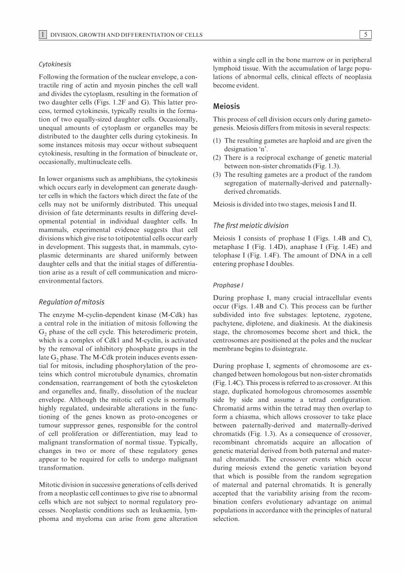
During prophase I, segments of chromosome are ex-changed between homologous but non-sister chromatids(Fig. 1.4C). This process is referred to as crossover. At thisstage, duplicated homologous chromosomes assembleside by side and assume a tetrad configuration.Chromatid arms within the tetrad may then overlap toform a chiasma, which allows crossover to take placebetween paternally-derived and maternally-derivedchromatids (Fig. 1.3). As a consequence of crossover,recombinant chromatids acquire an allocation ofgenetic material derived from both paternal and mater-nal chromatids. The crossover events which occur during meiosis extend the genetic variation beyond that which is possible from the random segregation of maternal and paternal chromatids. It is generallyaccepted that the variability arising from the recom-bination confers evolutionary advantage on animalpopulations in accordance with the principles of naturalselection.
5
DIVISION, GROWTH AND DIFFERENTIATION OF CELLS 1
Metaphase I
As in mitosis, homologous chromosome pairs attach viatheir kinetochores to the microtubules arising from thecentrosomes which are located at opposite poles of the
cell. During metaphase, the homologous chromosomepairs are positioned at the metaphase plate by the kine-tochore microtubules (Fig. 1.4D).
Anaphase I
During anaphase I, the tetrad splits into two dyads (half a tetrad), which move to opposite poles of the cell. Unlike the anaphase stage of mitosis, splitting of the centromeres does not occur because in this instance only one kinetochore forms on each dyad. The distribution of paternally derived and maternallyderived homologous chromosomes at this point is ran-dom, and it is this variable arrangement which under-lies the Mendelian principle of random assortment (Fig. 1.4E).
Telophase I
In telophase I, nuclear envelopes develop around theseparate chromosome sets and cytokinesis follows (Figs. 1.4F and G). In the formation of primary sperma-tocytes, progenitors of male gametes, the cytoplasm isdivided equally between the two cells. However, duringthe formation of oocytes, female gametes, one of thetwo resulting cells retains the greater portion of cyto-plasm. The smaller of the two cells is termed a polarbody. A short resting phase, termed interkinesis, followstelophase I and replication of DNA does not occur dur-ing this phase.
The second meiotic division
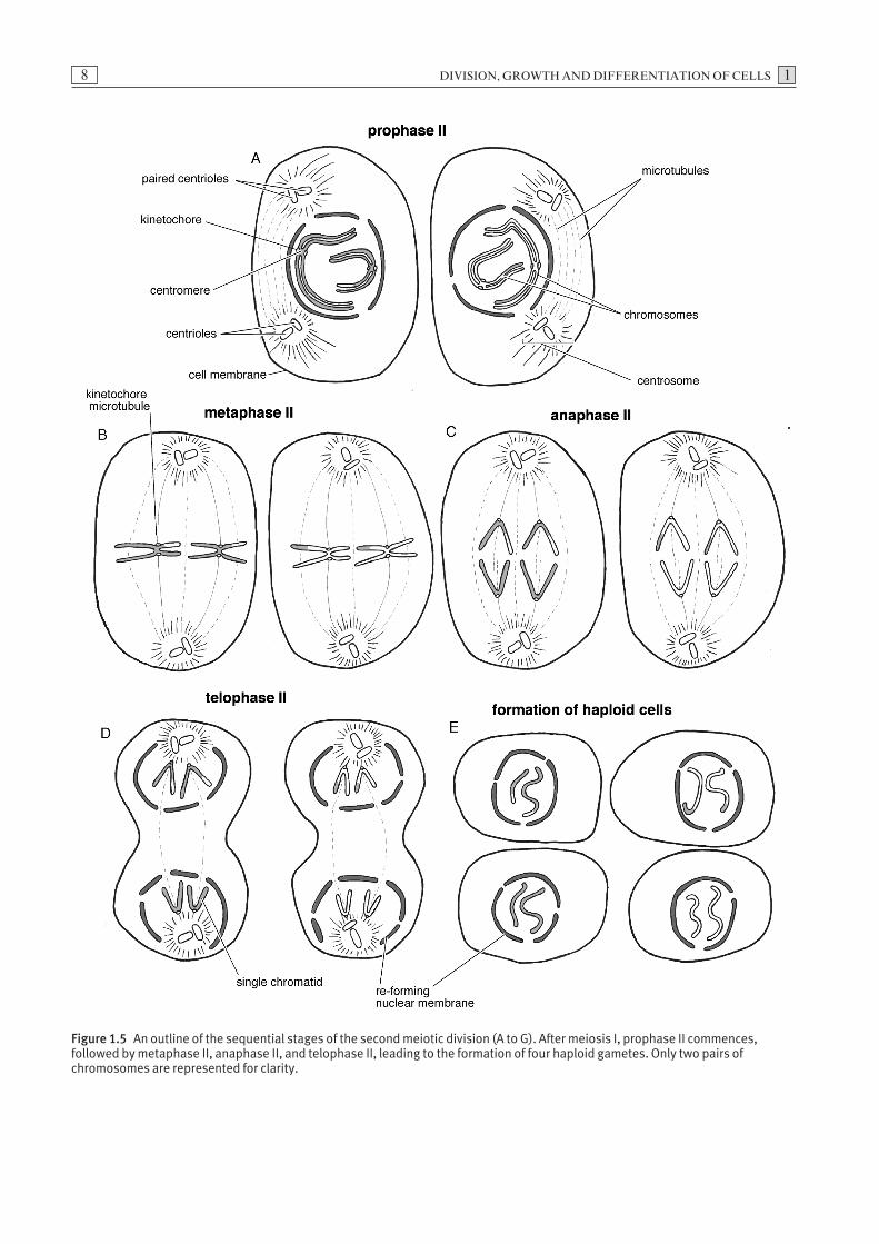
Prophase II
The events of prophase II are similar to prophase I. The nucleus contains a set of dyads each composed of a pair of chromatids connected by a shared centromere(Fig. 1.5A).
Metaphase II
The phase termed metaphase II is similar to metaphase I in that the chromosomes are positioned at the met-aphase plate by the kinetochore microtubules. In thisinstance, however, kinetochores form on each of theindividual chromatids. This allows the microtubules toattach separately to each chromatid (Fig. 1.5B).
Anaphase II
During anaphase II, the dyads are separated into indi-vidual chromatids by the kinetochore microtubules andthe sets of chromatids are drawn towards opposite polesof the dividing cell (Fig. 1.5C).
6
Figure 1.3 Chiasma formation and reciprocal exchange ofgenetic material between non-sister homologous chromatidsduring meiosis I.

1 DIVISION, GROWTH AND DIFFERENTIATION OF CELLS 7
Figure 1.4 An outline of the sequential stages of the first meiotic division (A to G). After the G2 phase, prophase I commencesfollowed by metaphase I, anaphase I and telophase I.
DIVISION, GROWTH AND DIFFERENTIATION OF CELLS 18
Figure 1.5 An outline of the sequential stages of the second meiotic division (A to G). After meiosis I, prophase II commences,followed by metaphase II, anaphase II, and telophase II, leading to the formation of four haploid gametes. Only two pairs ofchromosomes are represented for clarity.
1 DIVISION, GROWTH AND DIFFERENTIATION OF CELLS
Telophase II
At the end of telophase II, nuclear envelopes formaround each set of chromatids and the cytoplasmdivides again (Fig. 1.5D). As a consequence of meiosis Iand II, four haploid cells are formed from a singlediploid germ cell (Fig. 1.5E).
Consequences of non-dysjunction of chromosomesduring meiosis
The term non-dysjunction describes the failure of two homologous chromosomes in meiosis I, or sisterchromatids in meiosis II, to separate properly and tomove correctly to opposite poles. Meiosis depends onthe establishment of specialised interactions betweenchromosomes along with specific modifications to themitotic cell cycle regulatory processes. Errors in theseprocesses, which usually occur during meiosis I, canresult in defective segregation. Abnormalities arisingfrom this include numerical alteration and structural
defects in chromosomes. While chromosomal defectsassociated with germ cells generally lead to embryonicdeath, in some instances offspring may survive andexhibit developmental defects. Alterations of chromo-some numbers may involve either autosomes or sexchromosomes.
Further reading
Alberts, B., Johnson, A., Lewis, J., Raff, M., Roberts,K. and Walter, P. (2002) Molecular Biology of theCell, 4th edn. Garland Science, New York.
Klug, W.S. and Cummins, M.R. (1999) Essentials ofGenetics. Prentice-Hall, Upper Saddle River, NewJersey.
Levine, E.M. (2004) Cell cycling through development.Development 131, 2241–2246.
Marston, A.L. and Amon, A. (2004) Meiosis: cell-cyclecontrols shuffle and deal. Nature Reviews: Molecularand Cell Biology 5, 983–997.
9
The sequential stages in the differentiation and matura-tion of primordial germ cells into gametes in male and female animals are referred to as gametogenesis.Primordial germ cells in the endoderm of the yolk sacmigrate via the dorsal mesentery to the developinggonads. During migration these cells undergo mitosis,producing large numbers of germ cells which populatethe gonads. Germ cells undergo similar sequentialdevelopment in male and female animals.
Spermatogenesis
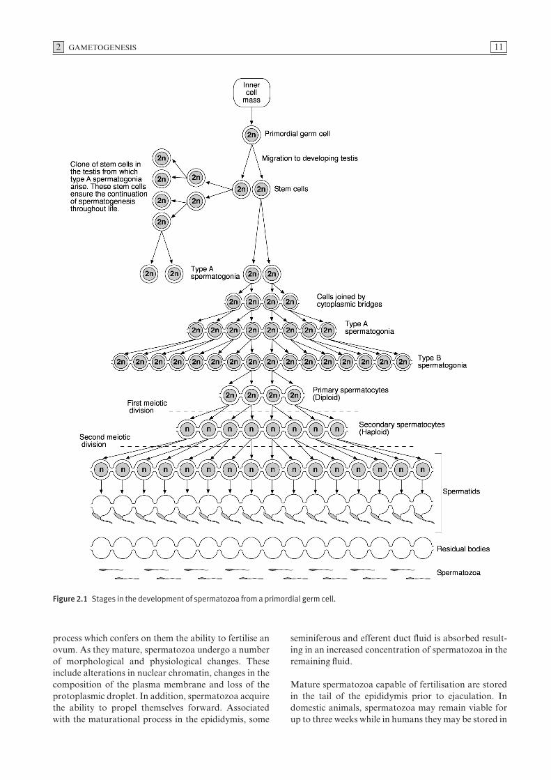
Primordial germ cells undergo a series of mitotic divisionsproducing stem cells which, in association with meso-dermal cells, form seminiferous cords in the develop-ing testis. In this location, they remain quiescent untilthe onset of puberty, when sexual maturation begins. At puberty, these dormant germ cells become activatedand, through a series of mitotic divisions, produce clonesof cells referred to as type A spermatogonia (Fig. 2.1).Subsequently, some type A cells divide, giving rise totype B spermatogonia, from which primary spermato-cytes arise.
The diploid primary spermatocytes undergo the firststage of meiotic division resulting in the formation ofhaploid secondary spermatocytes. When these haploidsecondary spermatocytes undergo the second stage of meiotic division, they form haploid spermatids (Fig. 2.1).
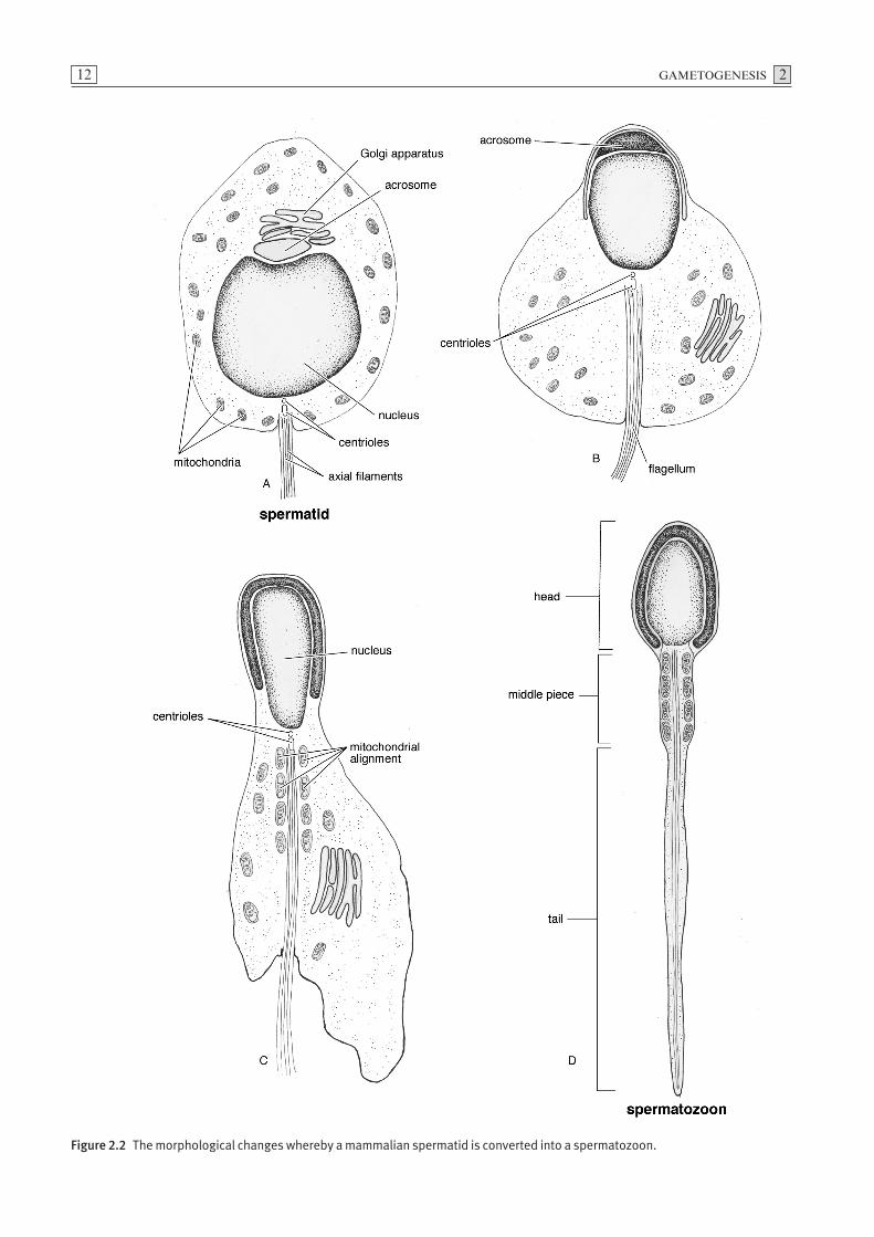
The process whereby a spermatid undergoes metamor-phosis into a spermatozoon is termed spermiogenesis(Fig. 2.2). Initially the spermatid has the organelles of atypical mammalian cell including a spherical nucleus, aGolgi complex, mitochondria, paired centrioles andendoplasmic reticulum. Granules, which are synthesisedin the Golgi complex, fuse forming a single large acroso-mal vesicle. When this vesicle covers the anterior aspectof the condensed nucleus, it is referred to as the acro-some. The centrioles, which migrate to the pole of thenucleus opposite the acrosome, form the axial filamentfrom which the tail of the spermatozoon develops.Mitochondria aggregate in the proximal region of thefilament forming the middle piece of the spermatozoon.
Excess portions of cytoplasm shed from individual sper-matids are collectively referred to as residual bodies. A unique feature of spermatogenesis is that the cyto-plasmic divisions of the dividing spermatogonia areincomplete as the spermatocytes remain attached bycytoplasmic bridges. The time required for the produc-tion of spermatozoa from type A spermatogonia mayrange from 40 to 60 days depending on the species.
As spermatogenesis proceeds, the spermatogenic cellsdevelop in close association with Sertoli cells in the seminiferous tubules. The germ cells are almost com-pletely surrounded by the cytoplasm of Sertoli cellswhich nourish and support them during differentiation.Tight junctions between adjacent Sertoli cells divideseminiferous tubules into basal compartments and adluminal compartments, thereby preventing the entryof cells involved in the generation of immunologicalresponses into the adluminal compartments. These junc-tions also prevent macromolecules from crossing fromthe adluminal compartments into the animal’s circula-tion. The structures which isolate the cells on the adlu-minal side of seminiferous tubules from the testicularvascular supply constitute the blood–testis barrier. Atthe completion of spermiogenesis, immature spermato-zoa are extruded from their intimate association withthe Sertoli cells into the lumen of the seminiferoustubules, a process referred to as spermiation. Prior totheir release, most of the cytoplasm of the immaturespermatozoa is shed and phagocytosed by Sertoli cells.At the time of its release into the lumen of the semini-ferous tubule, a small amount of cytoplasm, the proto-plasmic droplet, remains attached to the middle piece ofthe immature spermatozoon. The spermatozoa withinthe seminiferous tubules are immotile and are carriedpassively by the tubular fluid to the rete testis. From thislocation they are conveyed by ten to 20 efferent ductulesto the epididymis through the ciliary action of ductepithelium and the contractions of the smooth muscle of the duct wall.
The epididymis, which consists of a long, tightly con-voluted tube, is anatomically divided into three re-gions, head, body and tail. During their passage throughthe epididymis, spermatozoa undergo a maturational
2 Gametogenesis
2 GAMETOGENESIS
process which confers on them the ability to fertilise anovum. As they mature, spermatozoa undergo a numberof morphological and physiological changes. Theseinclude alterations in nuclear chromatin, changes in thecomposition of the plasma membrane and loss of theprotoplasmic droplet. In addition, spermatozoa acquirethe ability to propel themselves forward. Associatedwith the maturational process in the epididymis, some
seminiferous and efferent duct fluid is absorbed result-ing in an increased concentration of spermatozoa in theremaining fluid.
Mature spermatozoa capable of fertilisation are storedin the tail of the epididymis prior to ejaculation. Indomestic animals, spermatozoa may remain viable forup to three weeks while in humans they may be stored in
11
Figure 2.1 Stages in the development of spermatozoa from a primordial germ cell.
GAMETOGENESIS 212
Figure 2.2 The morphological changes whereby a mammalian spermatid is converted into a spermatozoon.
2 GAMETOGENESIS
the epididymis for only a few days before losing theirviability. Most of the unejaculated spermatozoa aregradually discharged into the urinary system; a smallpercentage which remain in the epididymis undergodegenerative change and are phagocytosed. The trans-port of spermatozoa through the epididymis, due tocontractions of the smooth muscle of the epididymalduct wall, takes up to 12 days in the bull and ram and upto 14 days in the boar and stallion. With increased fre-quency of ejaculation, transport time may be reduced.
Oogenesis
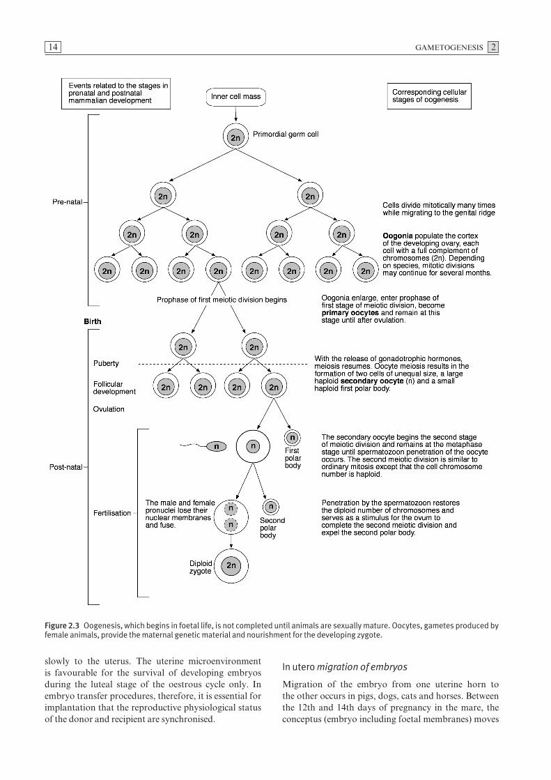
Oogonia, which arise from primordial germ cells in the endoderm, undergo repeated mitotic divisions in the foetal ovary. The duration of this period of mitosisvaries in individual species. Irrespective of species, themitotic phase of oogenesis ceases in mammals soon afterbirth. When they have completed their cycles of mitosis,oogonia enter the prophase of the first of two meioticdivisions and become primary oocytes which are diploid.Such diploid cells are given the designation 2n to indicatethat they contain a full complement of chromosomes.All primary oocytes are formed before puberty (Fig. 2.3).
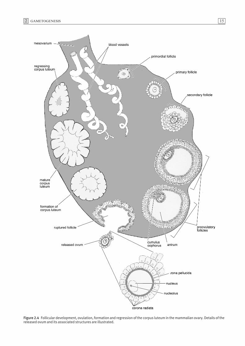
A primary oocyte surrounded by a single layer of squa-mous epithelial cells is known as a primordial follicle(Fig. 2.4). Primary oocytes do not complete the prophaseof the first meiotic division but enter a prolonged restingor dictyate stage until activated by gonadotrophic hor-mones which induce further development. During boththe proliferative and resting phases, a high proportionof primordial follicles undergo atresia. Completion ofthe initial stage of the first meiotic division follows hormonal stimulation. During puberty, the oocyteincreases in size and the surrounding epithelial follicularcells form a stratified layer around the oocyte. Thisstructure is now known as a primary follicle. Glyco-proteins, secreted primarily by the oocyte, condenseforming a prominent translucent acellular layer, thezona pellucida, located between the vitelline membraneof the oocyte and the follicular cells. As the follicleenlarges, the thickness of the zona pellucida increases.The oocyte and the follicular cells maintain contact bymeans of microvillous cytoplasmic processes which pen-etrate the zona. Gap junctions between the oocyte andthe cytoplasmic processes of follicular cells allow inter-cellular communication. As the follicle continues toincrease in size, small fluid-filled spaces appear betweenthe follicular cells which gradually coalesce forming afluid-filled cavity known as the antrum. The squamousfollicular cells, which become cuboidal, form stratifiedlayers and are referred to as granulosa cells. The oocyteremains attached to the follicular wall by an accumula-tion of granulosa cells termed the cumulus oophorus(Fig. 2.4). Those granulosa cells which surround the
oocyte in a radial fashion are referred to as the coronaradiata. The mature follicle is now referred to as a vesic-ular or Graafian follicle. The completion of the first mei-otic division results in the production of two haploidcells of unequal size. The cell which receives most of thecytoplasm is referred to as the secondary oocyte and theother, which receives a minimal amount of cytoplasm, isthe first polar body (Fig. 2.3). Following formation ofthe first polar body, the secondary oocyte commencesthe second meiotic division.
Ovulation
Release of the ovum from the follicle is referred to asovulation (Fig. 2.4). Prior to ovulation, the oocyte andcorona radiata detach from the cumulus oophorus andfloat in the follicular fluid. Rupture of the follicle isattributed to the formation of a blister-like area, thestigma, on the ovarian surface directly above the follicle.While it is accepted that the stigma arises from constric-tion of blood vessels as a result of hormonal or enzy-matic activity, the exact details of follicular rupture arepoorly understood.
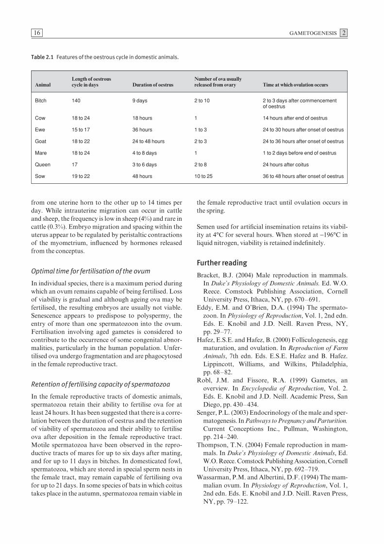
Although ovulation generally occurs near the end ofoestrus, the precise time at which it occurs differs amongdomestic species (Table 2.1). Ovulation occurs spon-taneously in most species (spontaneous ovulation). Incats, rabbits, ferrets and camels, however, ovulation isinduced by coitus (induced ovulation). The number ofova released, which is characteristic for a given species,is strongly influenced by genetic factors. In most mam-mals, ovulation occurs during the metaphase of the sec-ond meiotic stage of oogenesis. Exceptions include dogsand foxes, where ovulation usually occurs during themetaphase of the first meiotic division. Completion ofthe second meiotic division and formation of the secondpolar body occur after fertilisation.
Transport of ova in the uterine tube
After ovulation, the ovum enters the uterine tube, thesite of fertilisation in mammals. Tubal wall contractionsaided by the ciliary beat of the epithelium of the tube areresponsible for the transportation of ova along the tube.Whether or not they are fertilised, ova normally reachthe uterus within three to four days after ovulation.However, in domestic carnivores it may take up to sevendays for ova to reach the uterus. Fertilised ova of horsesand bats enter the uterus, whereas non-fertilised ova areretained at the isthmus of the uterine tube. In rabbits,opossums and dogs, a mucopolysaccharide coat formsaround the zona pellucida while the ovum is in the uter-ine tube. As the uterus provides a favourable environ-ment for the survival of spermatozoa but not for theblastocyst, it is essential that fertilised ova be transported
13
GAMETOGENESIS 214
Figure 2.3 Oogenesis, which begins in foetal life, is not completed until animals are sexually mature. Oocytes, gametes produced byfemale animals, provide the maternal genetic material and nourishment for the developing zygote.
slowly to the uterus. The uterine microenvironment is favourable for the survival of developing embryosduring the luteal stage of the oestrous cycle only. Inembryo transfer procedures, therefore, it is essential forimplantation that the reproductive physiological statusof the donor and recipient are synchronised.
In utero migration of embryos
Migration of the embryo from one uterine horn to the other occurs in pigs, dogs, cats and horses. Betweenthe 12th and 14th days of pregnancy in the mare, theconceptus (embryo including foetal membranes) moves
2 GAMETOGENESIS 15
Figure 2.4 Follicular development, ovulation, formation and regression of the corpus luteum in the mammalian ovary. Details of thereleased ovum and its associated structures are illustrated.
GAMETOGENESIS 2
from one uterine horn to the other up to 14 times perday. While intrauterine migration can occur in cattleand sheep, the frequency is low in sheep (4%) and rare incattle (0.3%). Embryo migration and spacing within theuterus appear to be regulated by peristaltic contractionsof the myometrium, influenced by hormones releasedfrom the conceptus.
Optimal time for fertilisation of the ovum
In individual species, there is a maximum period duringwhich an ovum remains capable of being fertilised. Lossof viability is gradual and although ageing ova may befertilised, the resulting embryos are usually not viable.Senescence appears to predispose to polyspermy, theentry of more than one spermatozoon into the ovum.Fertilisation involving aged gametes is considered tocontribute to the occurrence of some congenital abnor-malities, particularly in the human population. Unfer-tilised ova undergo fragmentation and are phagocytosedin the female reproductive tract.
Retention of fertilising capacity of spermatozoa
In the female reproductive tracts of domestic animals,spermatozoa retain their ability to fertilise ova for atleast 24 hours. It has been suggested that there is a corre-lation between the duration of oestrus and the retentionof viability of spermatozoa and their ability to fertiliseova after deposition in the female reproductive tract.Motile spermatozoa have been observed in the repro-ductive tracts of mares for up to six days after mating,and for up to 11 days in bitches. In domesticated fowl,spermatozoa, which are stored in special sperm nests inthe female tract, may remain capable of fertilising ovafor up to 21 days. In some species of bats in which coitustakes place in the autumn, spermatozoa remain viable in
the female reproductive tract until ovulation occurs inthe spring.
Semen used for artificial insemination retains its viabil-ity at 4°C for several hours. When stored at −196°C inliquid nitrogen, viability is retained indefinitely.
Further reading
Bracket, B.J. (2004) Male reproduction in mammals. In Duke’s Physiology of Domestic Animals. Ed. W.O.Reece. Comstock Publishing Association, CornellUniversity Press, Ithaca, NY, pp. 670–691.
Eddy, E.M. and O’Brien, D.A. (1994) The spermato-zoon. In Physiology of Reproduction, Vol. 1, 2nd edn.Eds. E. Knobil and J.D. Neill. Raven Press, NY,pp. 29–77.
Hafez, E.S.E. and Hafez, B. (2000) Folliculogenesis, eggmaturation, and ovulation. In Reproduction of FarmAnimals, 7th edn. Eds. E.S.E. Hafez and B. Hafez.Lippincott, Williams, and Wilkins, Philadelphia, pp. 68–82.
Robl, J.M. and Fissore, R.A. (1999) Gametes, anoverview. In Encyclopedia of Reproduction, Vol. 2.Eds. E. Knobil and J.D. Neill. Academic Press, SanDiego, pp. 430–434.
Senger, P.L. (2003) Endocrinology of the male and sper-matogenesis. In Pathways to Pregnancy and Parturition.Current Conceptions Inc., Pullman, Washington, pp. 214–240.
Thompson, T.N. (2004) Female reproduction in mam-mals. In Duke’s Physiology of Domestic Animals, Ed.W.O. Reece. Comstock Publishing Association, CornellUniversity Press, Ithaca, NY, pp. 692–719.
Wassarman, P.M. and Albertini, D.F. (1994) The mam-malian ovum. In Physiology of Reproduction, Vol. 1,2nd edn. Eds. E. Knobil and J.D. Neill. Raven Press,NY, pp. 79–122.
16
Table 2.1 Features of the oestrous cycle in domestic animals.
Length of oestrous Number of ova usuallyAnimal cycle in days Duration of oestrus released from ovary Time at which ovulation occurs
Bitch 140 9 days 2 to 10 2 to 3 days after commencement of oestrus
Cow 18 to 24 18 hours 1 14 hours after end of oestrus
Ewe 15 to 17 36 hours 1 to 3 24 to 30 hours after onset of oestrus
Goat 18 to 22 24 to 48 hours 2 to 3 24 to 36 hours after onset of oestrus
Mare 18 to 24 4 to 8 days 1 1 to 2 days before end of oestrus
Queen 17 3 to 6 days 2 to 8 24 hours after coitus
Sow 19 to 22 48 hours 10 to 25 36 to 48 hours after onset of oestrus