uva-dare (digital academic repository) the circadian ... · roche). bm purple ap substrate...
TRANSCRIPT

UvA-DARE is a service provided by the library of the University of Amsterdam (http://dare.uva.nl)
UvA-DARE (Digital Academic Repository)
The circadian systemBuijs, F.N.
Link to publication
Citation for published version (APA):Buijs, F. N. (2019). The circadian system: A regulatory feedback network of periphery and brain.
General rightsIt is not permitted to download or to forward/distribute the text or part of it without the consent of the author(s) and/or copyright holder(s),other than for strictly personal, individual use, unless the work is under an open content license (like Creative Commons).
Disclaimer/Complaints regulationsIf you believe that digital publication of certain material infringes any of your rights or (privacy) interests, please let the Library know, statingyour reasons. In case of a legitimate complaint, the Library will make the material inaccessible and/or remove it from the website. Please Askthe Library: http://uba.uva.nl/en/contact, or a letter to: Library of the University of Amsterdam, Secretariat, Singel 425, 1012 WP Amsterdam,The Netherlands. You will be contacted as soon as possible.
Download date: 27 May 2019

526835-L-bw-Buijs526835-L-bw-Buijs526835-L-bw-Buijs526835-L-bw-BuijsProcessed on: 14-12-2018Processed on: 14-12-2018Processed on: 14-12-2018Processed on: 14-12-2018 PDF page: 77PDF page: 77PDF page: 77PDF page: 77
CHAPTER 4
The NPY intergeniculate leaflet projections to the Suprachiasmatic nucleus transmit metabolic conditions.
Saderi NCazarez-Márquez FBuijs FNSalgado-Delgado RCGuzman-Ruiz MAdel Carmen Basualdo MEscobar CBuijs RM.
Neuroscience. 29;246:291-300. (2013)
4181206LAYOUTversie6.indd 77 13-12-18 15:00

526835-L-bw-Buijs526835-L-bw-Buijs526835-L-bw-Buijs526835-L-bw-BuijsProcessed on: 14-12-2018Processed on: 14-12-2018Processed on: 14-12-2018Processed on: 14-12-2018 PDF page: 78PDF page: 78PDF page: 78PDF page: 78
78
Chapter 4
AbstractThe Intergeniculate leaflet (IGL) is classically known as the area of the Thalamic Lateral Geniculate Complex providing the suprachiasmatic nucleus (SCN) non-photic information. In the present study we investigated whether this information might be related to the metabolic state of the animal. The following groups of male Wistar rats were used for analysis of NPY and c-Fos in the IGL and SCN. 1. Fed ad libitum. 2. Fasted for 48 hours. 3. fasted for 48 hours followed by refeeding for 3 hours. 4. monosodium glutamate-lesioned and 48h-fasted. 5. Electrolytic lesion in the IGL and 48h-fasted. The results were quantified by optical densitometry. Neuronal tracers were injected in two brain areas that receive metabolic information from the periphery, the Arcuate Nucleus and Nucleus of the Tractus Solitarius to investigate whether there is an anatomical relationship with the IGL. Lesion studies showed the IGL, and not the ARC, as origin of most NPY projections to the SCN. Fasting induced important changes in the NPY expression in the IGL, coinciding with similar changes of NPY/GAD projections of the IGL to the SCN. These changes revealed that the IGL is involved in the transmission of metabolic information to the SCN. In fasted animals IGL lesion resulted in a significant increase of c-Fos in the SCN as compared to intact fasted animals demonstrating the inhibitory influence of the IGL to the SCN in fasting conditions. When the animal after fasting was refed, an increase of c-Fos in the SCN indicated a removal of this inhibitory input. Together these observations show that in addition to increased inhibitory IGL input during fasting, the negative metabolic condition also results in increased excitatory input to the SCN via other pathways. Consequently the present observations show that at least part of the non-photic input to the SCN, arising from the IGL contains information about metabolic conditions.
181206LAYOUTversie6.indd 78 13-12-18 15:00

526835-L-bw-Buijs526835-L-bw-Buijs526835-L-bw-Buijs526835-L-bw-BuijsProcessed on: 14-12-2018Processed on: 14-12-2018Processed on: 14-12-2018Processed on: 14-12-2018 PDF page: 79PDF page: 79PDF page: 79PDF page: 79
79
NPY IGL projections to SCN transmit metabolic conditions
4
IntroductionThe Suprachiasmatic Nucleus (SCN) controls the phase of body physiology, mainly by imposing activity-resting cycles that are facilitated by time organized autonomic and hormonal changes (Buijs&Kalsbeek, 2001; Dibner et al, 2010). Several studies indicated that in order to execute a correct temporal organization of these behavioral and physiological functions the SCN needs to integrate the physiological state of the body (Hastings et al, 1997; Yannielli & Harrington, 2004). Several areas in the hypothalamus, forebrain and hindbrain up to the Raphe nuclei provide the SCN with “non-photic” information. Up till now very little is known about the functional meaning of this input, with exception of the input arising from the Intergeniculate Leaflet (IGL) Dorsal Raphe (DR) and Arcuate nucleus (ARC). While IGL and DR provide the SCN with information about activity (Janik & Mrososvsky, 1994, Kuroda et al, 1997) the ARC is proposed to provide the SCN metabolic information (Yi et al, 2006). On the basis of previous studies which demonstrated that phase advances induced by time and caloric feeding restriction are appreciably decreased in IGL-lesioned rats (Challet et al, 1996), we investigated whether in addition to the ARC, the IGL may also provide metabolic related information to the SCN. Our results showed that food deprivation up-regulates NPY expression in the IGL and increases NPY innervation density in the SCN. Lesion studies performed either in the ARC or in the IGL of fasted rats, attested that the NPY augmentation is dependent on IGL projections to the SCN. The increase in the number of c-Fos expressing cells in the SCN during refeeding suggests that food access removes a negative control on SCN neuronal activity and the concomitant decrease of NPY in the IGL suggests the direct involvement of the IGL in this process.
Materials and MethodsFood availability experiments (immunohistochemistry). Male Wistar rats, weighing 250-300g, were housed in a 12:12 light-dark cycle (light-on at 7:00 am) with free access to food and water, during one week previous to the beginning of the experimental procedures. Experiments were approved by the committee for ethical evaluation at the Universidad Nacional Autónoma de México, in strict accordance with the Mexican norms for animal handling, Norma Oficial Mexicana NOM-062-ZOO-1999.On the first day of the experiment, rats were randomly assigned to one of the following groups: 5 were fed ad libitum (CTR group); 5 were fasted for 48 hours (FST group) starting at ZT10; 5 were fasted for 48 hours starting at ZT7; then refed for 3 hours.All Animals were sacrificed at ZT10 of the third day of the experiment by intracardial perfusion with 4% paraformaldheyde (Sigma –Aldrich Corp., San Luis, MO, USA) in phosphate buffer (0.01M, ph7.6) preceded by deep anesthesia with an overdose of sodium pentobarbital (Sedalpharma, Pet’s Pharma, Mexico; 50mg/kg). Brains were dissected, post-fixed by immersion in 4% paraformaldheyde for 24h and cryoprotected in Phosphate Buffer
181206LAYOUTversie6.indd 79 13-12-18 15:00

526835-L-bw-Buijs526835-L-bw-Buijs526835-L-bw-Buijs526835-L-bw-BuijsProcessed on: 14-12-2018Processed on: 14-12-2018Processed on: 14-12-2018Processed on: 14-12-2018 PDF page: 80PDF page: 80PDF page: 80PDF page: 80
80
Chapter 4
Saline (PBS, 0.01M, ph7.6) containing 30% sucrose and 0.04% NaN3 until sectioning. The hypothalami were cut in sections of 40 μm, and each third section was used for immunohistochemical procedures. Sections of the SCN and the IGL were incubated with either rabbit-NPY (Buijs et al, 1989) or rabbit-c-Fos (Calbiochem, EMD Biosciences, INC. La Jolla, CA, USA antibodies). NPY and c-Fos antibodies diluted in Triphosphate Buffer Saline (TBS, 0.01M, ph7.6) added with 0.5% Triton X-100 (Sigma –Aldrich) and 0.25 % gelatin (Merck KGaA, Damstadt, Germany) 1:4000 and 1:10000, respectively. Sections were incubated at room temperature for 1h and then at 4°C overnight. After rinsing, sections were incubated in biotinylated donkey-anti rabbit secondary antibody (Jackson Immunoresearch, West Grove, PO, USA; 1:400) for 1.5h and then in an avidin-biotin complex (Vector, Burlingame, CA, USA, 1:500) solution. The final reaction was performed with a solution of 0.025% 3,3’-diaminobenzidine (DAB) and 0.01% H2O2 100 (Sigma–Aldrich) in TBS, for 10 minutes. For c-Fos staining, 10% NiNH4SO4 was also added to this solution. Sections were mounted on gelatinized slides, dried, dehydrated with graded solutions of ethanol, soaked in xylene, and finally cover slipped in Entellan embedding agent (Merck).
Food availability experiments (in situ hybridization). This part of the study followed an identical procedure to the protocol in 2.1. 12 Male Wistar rats, weighing 250-300g, were randomly divided in 3 groups: the control group was fed ad libitum for all the time of the experiments (CTR); the fasted group was food-deprived for 48h (FST); and the 3-hours re-fed group was fasted for 48h and then re-fed for 3 hours (3h-RF). Brains were dissected and postfixed in 4% paraformaldehyde in PBS for 12h at 4 °C, and this was followed by cryoprotection in 30% diethyl pyrocarbonate-treated sucrose in PBS at 4 °C. Brains were frozen and sectioned with a cryostat (16 μm). Sections were dried at room temperature for 2 h before overnight incubation at 65 °C in hybridization buffer [1 × diethyl pyrocarbonate-treated ‘salts’ (200 mmNaCl, 5 mm EDTA, 10 mmTris, pH 7.5, 5 mm NaH2PO4.2H2O, 5 mm Na2HPO4); 50% deionized formamide; 1 × Denhardts (RNase/DNase-free; Invitrogen Corporation, Carlsbad, CA, USA); 10% dextran sulphate (Sigma–Aldrich )] containing 400 ng/mL digoxigenin-labelled RNA probes purified in Sephadex G-50 columns. Sense and antisense probes were generated by linearization or excision of plasmids with appropriate enzymes, and purified using QIAquick PCR Purification Kit (QIAGEN Inc., Valencia, CA, USA). Primers were synthesized by Sigma: sense 5´ TATCCCTGCTCGTGTGTTTG 3´; antisense 5´ AGGCAGACTGGTTTCACAGG 3’.The hybridization solution consisted of 50% formamide, 2 × sodium phosphate, sodium chloride and EDTA (SSPE), 1 μg/μl bovine serum albumin (BSA), 1 μg/μl poly A, 2 μg/μl tRNA in diethyl pyrocarbonate (DEPC)-treated water). Following hybridization, sections were washed three times in wash solution (50% formamide, 1 × saline citrate, 0.1% Tween-20) at 65 °C and twice at room temperature in 1 × MABT (20 mM maleic acid, 30 mMNaCl, 0.1% Tween-20) before being incubated in blocking solution [1% blocking reagent (Roche Applied Science, Burgess Hill, UK)] and
181206LAYOUTversie6.indd 80 13-12-18 15:00
526835-L-bw-Buijs526835-L-bw-Buijs526835-L-bw-Buijs526835-L-bw-BuijsProcessed on: 14-12-2018Processed on: 14-12-2018Processed on: 14-12-2018Processed on: 14-12-2018 PDF page: 81PDF page: 81PDF page: 81PDF page: 81
81
NPY IGL projections to SCN transmit metabolic conditions
4
then overnight in alkaline phosphatase-conjugated anti-digoxigenin antibody (1: 1500; Roche). BM purple AP substrate precipitating (Roche) with 1 mMLevamisole (Roche) was used for colorimetric detection at 37 °C for 8–20 h. Sections were mounted with Glycerol (Sigma).
Lesion studies. Bilateral electrolytic lesion was used to ablate the IGL in 8 rats. Before surgery, animals were anesthetized with a mix of Ketamine (Anesket, PiSA, Mexico, 6mg/Kg) and Xylazine (Procin, PiSA, Mexico; 2mg/Kg), and placed in a standard stereotaxic apparatus with the tooth bar set at -3.5 mm. The electrode was placed bilateral at coordinates: 45mm caudal to the bregma, 52mm lateral from the midline and 52mm deep from the brain surface. After 2 weeks of recovery from the surgery (during which food intake, body weight and locomotor activity were monitored), animals were fasted and sacrificed as in the food availability experiments, and the sections processed to evaluate NPY immunoreactivity in the SCN.Additionally monosodium glutamate (MSG; Sigma –Aldrich; 5mg/ml) was used to lesion the ARC of 10 newborn rats within 10 days of life. Considering the birth-day as Post-Natal day 0 (PN=0), pups were injected with MSG 2mg/Kg on PN-1 and on PN-3, and with MSG 4mg/Kg at PN-5, PN-7 and PN-9 postnatal days. Animals were housed in colony rooms under standard conditions, together with 4 vehicle (saline) injected control rats. After three months, animals were fasted for 48-h, sacrificed and SCN sections processed for NPY previously indicated.
Tracing studies. Projections to and from from the IGL. The antero and retrograde tracer Cholera Toxin B (CTB) 0.5% conjugated either with the Alexa Fluor-555or Alexa-488 fluorescent dyes (Molecular Probes, Eugene, OR, USA), was injected 0.01ul in the IGL of 10 rats with a micro syringe. The surgery methods and the coordinates of the injections were the same as reported for the IGL lesion. After 10 days of recovery, animals were sacrificed and sections of the IGL and SCN were incubated with rabbit NPY antibody followed by a secondary fluorescent antibody conjugated with Cy-2TM (Jackson Immunoresearch, West Grove, PO, USA). Four brains, in which the tracer was co-localized with NPY in the SCN, were considered to have received a correct injection. These brains were used to determine the presence of retrograde labeled cell bodies in the ARC and NTS as two main areas receiving metabolic information.IGL innervation from the ARC and the NTS. The anterograde tracer Phaseolus Vulgaris (PhaL) (Vector Elite) was unilaterally injected in the ARC of 15 rats by iontophoresis. The injections were performed using a glass micro electrode with a tip of 20um. Alternate current of 6.8μA was allowed to pass for 10 minutes. The stereotaxic coordinates for the ARC injection were: 3.3mm caudal from the bregma; 1.0mm lateral from the midline; 9.8mm deep from the surface of the brain, with an inclination of 4º degrees and the teeth bar set at -3.4mm. A week after the surgery, animals were perfused and the brains
181206LAYOUTversie6.indd 81 13-12-18 15:00

526835-L-bw-Buijs526835-L-bw-Buijs526835-L-bw-Buijs526835-L-bw-BuijsProcessed on: 14-12-2018Processed on: 14-12-2018Processed on: 14-12-2018Processed on: 14-12-2018 PDF page: 82PDF page: 82PDF page: 82PDF page: 82
82
Chapter 4
processed as in the previous experiment. A first DAB/peroxide immunohistochemisty with a rabbit anti-PhaL antibody (1:1000; Vector Elite) was performed to test the accuracy of the injection. 6 brains from PhaL–injected animals were considered to display the correct injection and used to analyze the projection from the ARC to the IGL. After anesthesia, rats were placed in a David Kopf stereotact with the head fixed at 45°. The dura and arachnoid membranes were dissected to expose the dorsal surface of the medulla at the level of the area postrema. The rat’s head was placed such that the micropipette aligned perpendicular to the medulla oblongata. Injections into the Nucleus Tractus Solitarius were made with 0.05μl, 1% Cholera toxin B (CTB) using a glass micropipette with a 20- to 40-μm tip diameter. Because, in contrast to CtB, fluorophore-labeled CTB cannot be applied by iontophoresis, we used pressure injection (10 mbar, 5 sec). Thus, fluorophore-conjugated CTB (with Alexa Fluor 555, Molecular Probes); CTB will be used in the text as the abbreviation of CTB-Alexa Fluor 555 for convenience. After 10 days of recovery, animals were sacrificed and precision of the injection confirmed by mean of an epifluorescence microscope. 5 of CTB- injected animals received a correct injection. In order to visualize projections from the area of the NTS, IGL sections from these brains were incubated with rabbit CTB (Sigma–Aldrich Corp) and the staining developed with 0.025% DAB, 0.01% H2O2 and 10% NiNH4SO4 in TBS. To enhance the intensity of the staining in fibers, before being immersed in the last solution, sections were treated with a solution of biotinilated tyramine for 1 h, followed by another hour of incubation with the avidin-biotin complex. Since NPY is a marker for the IGL, a sheep NPY (1:8000; Chemicon International) immuno staining was performed to help to localize CTB-containing projections to the IGL.
Histochemical analysis. Digital pictures of DAB/H2O2/NiNH4SO4and NPY mRNA stained sections were taken by using an Axioplan microscope (Zeiss, Jena, Germany) equipped with a digital color photocamera (Olympus DP25, Olympus, Japan). To quantify NPY and c-Fos, 8 sections from each brain were analyzed bilaterally, and the number of NPY and c-Fos positive cells and the optical density of the NPY in the SCN and the IGL were quantified with the program ImageJ. For optical density measurements the background was subtracted from the positive staining.Sections stained with fluorescent dyes were analyzed with the LSM 5 Pascal confocal microscope and the LSM software (Zeiss, Jena, Germany).Quantitative data are expressed as mean +/- the standard error from the mean (SEM). Results were analyzed with a one-way ANOVA followed by a Tukey multiple comparisons post hoc test. P-values of <0.05 were considered statistically significant.
181206LAYOUTversie6.indd 82 13-12-18 15:00

526835-L-bw-Buijs526835-L-bw-Buijs526835-L-bw-Buijs526835-L-bw-BuijsProcessed on: 14-12-2018Processed on: 14-12-2018Processed on: 14-12-2018Processed on: 14-12-2018 PDF page: 83PDF page: 83PDF page: 83PDF page: 83
83
NPY IGL projections to SCN transmit metabolic conditions
4
ResultsEffect of food deprivation and re-feeding on NPY in the IGL. In the IGL both NPY-IR, indicated by integrated optical density, and the number of NPY-positive cells were increased after a period of 48-h of fasting with respect to control animals (for optical density: F(2,9)= 57.11, p<0.001; for NPY cells: F(2,9)=75.44, p<0.001) (fi g.1-A and B). After refeeding, the NPY optical density returned to basal levels, whereas the number of NPY positive cells showed a signifi cant decrease in comparison to both control (F(1,6) =7.82, p<0.05) and fasted (F(1,6) =22.08, p<0.05) groups. In addition, IGL NPY mRNA was up-regulated by food deprivation (F(1,4) =11.08, p<0.05), demonstrating an enhanced activity of NPY neurons in the IGL under anabolic conditions similar as occurs in the ARC (fi g. 2-A and B). The number of c-Fos positive cells in the IGL was also signifi cantly increased by fasting and 3h-refeeding (F(2,9) = 88.92., p<0.001as compared to the control) (fi g.3-A and B), however no c-Fos could be detected in IGL NPY neurons indicating that the increase of activity is in another set of IGL neurons(fi g.3-A).
Figure 1. Fasting increases NPY-IR in the IGL. Comparison of NPY immunoreactivity (IR) in the IGL of free fed, fasted and refed
rats. (A) Photomicrographs of the IGL in the different conditions. Scale bar = 200 μm. (B) Quantifi cation of NPY-IR as integrated
optical density (on the left) and number of positive cells (on the right). Both optical density (p=0.04) and number of NPY cells (p=0.035)
are increased after 48h of fasting. The number of NPY positive cells is signifi cantly decreased by refeeding (p=0.046).
181206LAYOUTversie6.indd 83 13-12-18 15:00

526835-L-bw-Buijs526835-L-bw-Buijs526835-L-bw-Buijs526835-L-bw-BuijsProcessed on: 14-12-2018Processed on: 14-12-2018Processed on: 14-12-2018Processed on: 14-12-2018 PDF page: 84PDF page: 84PDF page: 84PDF page: 84
84
Chapter 4
Figure 2. Fasting up regulates NPY-mRNA in the IGL. (A) Photomicrographs of the IGL of control and fasted rats (scale bar =
80 μm) and (B) quantifi cation integrate optical density of NPY mRNA showing that NPY expression is up-regulated by fasting (p=0.02).
Figure 3. Fasting activates IGL neurons. C-Fos (black nuclei), as NPY (brown cells), is increased in the IGL of fasted rats, as
indicated by (A) photomicrographs of the IGL (scale bar = 80 μm) and (B) quantifi cation of c-Fos-expressing cells (p=0.001).
Effect of food deprivation and re-feeding on the SCN. NPY positive fi bers were strongly increased in the vl-SCN of fasted rats in comparison to controls, this increase disappeared after refeeding(F(2,9)= 6.97, p=0.01) (fi g.4-A and B).In the SCN, c-Fos expression was not affected by fasting, but it was increased by re-feeding and by IGL lesion followed by fasting (F(3,10)= 73.03, p<0.001) (fi g.5). Importantly, changes in c-Fos activations were particularly appreciable in the vl-SCN, where c-Fos immunoreactivity was enhanced by re-feeding in intact animals and by fasting in IGL-lesioned animals (F(3,10)= 11.16, p= 0.001)
181206LAYOUTversie6.indd 84 13-12-18 15:00

526835-L-bw-Buijs526835-L-bw-Buijs526835-L-bw-Buijs526835-L-bw-BuijsProcessed on: 14-12-2018Processed on: 14-12-2018Processed on: 14-12-2018Processed on: 14-12-2018 PDF page: 85PDF page: 85PDF page: 85PDF page: 85
85
NPY IGL projections to SCN transmit metabolic conditions
4
Figure 4. Fasting increases NPY-IR in the SCN. Comparison of NPY immunoreactivity (IR) in the SCN of free fed, fasted and refed
rats. (A) Photomicrographs of the SCN in the different conditions. Scale bar = 300 μm. (B) Quantifi cation of NPY-IR as integrate optical
density showing that NPY-IR is increased after 48h of fasting (p=0.035).
Figure 5 .Re-feeding induces cFos activation in the SCN in response to food availability. (A) Photomicrographs of the
SCN of free fed, fasted and refed rats. Scale bar = 200 μm. (B) Quantifi cation of c-Fos-IR indicates that the number of positive cells is
increased (p=0.026) by refeeding. Note that in the ventrolateral SCN (vl-SCN) c-Fos shows a slight tendency to decrease after a period
of fasting and it is signifi cantly increased by refeeding (p = 0.032).
NPY in the SCN arises from the IGL. In order to determine the origin of the NPY increase in the SCN of fasting rats, either the IGL or the ARC were lesioned and animals were food-deprived for 48-h. The comparison of NPY-IR in lesioned and fasted rats showed that NPY virtually disappeared in the SCN after IGL-lesion, while after ARC lesion the NPY increase after the period of fasting is not affected (F(2,7)= 44.53, p<0.001) fi g.6-A and B).The neuronal tracer CTB applied in the IGL to determine the nature of these projections, demonstrated that IGL-projection to the vl-SCN contain NPY and the intensity of the NPY staining was notably enhanced by fasting (fi g.7 and 8).
181206LAYOUTversie6.indd 85 13-12-18 15:00

526835-L-bw-Buijs526835-L-bw-Buijs526835-L-bw-Buijs526835-L-bw-BuijsProcessed on: 14-12-2018Processed on: 14-12-2018Processed on: 14-12-2018Processed on: 14-12-2018 PDF page: 86PDF page: 86PDF page: 86PDF page: 86
86
Chapter 4
Figure 6. Effect of IGL and ARC lesion on fasting-induced NPY increase in the ventrolateral SCN. (A) NPY-IR indicate
that ablation of the IGL virtually eliminates NPY projections to the SCN, even after a period of fasting, whereas ARC lesion does not
affect NPY response to food deprivation. Scale bar = 200 μm. (B) Quantifi cation of NPY optical density showing the strong decrease of
NPY-IR in fasted but IGL-lesioned rats (p= 0.0001).
Figure 8. NPY and GABA-IR in the SCN is decreased after re-feeding. Confocal laser scanning image of SNC of control, fasted
and refed animals, stained for NPY (green) and GAD-67 (GABA) (red), indicate that NPY is increased by fasting, whereas both NPY and
GABA are decreased after re-feeding. Scale bar = 70 μm.
Figure 7. Confocal laser scanning image of CTB (red)
containing IGL projections to the ventrolateral SCN,
showing that they contain NPY (green). The co-localization
between the tracer and NPY (yellow) attests the accuracy of CTB
injection in the IGL. Only such injections were used to evaluate
the areas where retrograde neurons appeared (see fi gure 9) Scale
bar = 200 μm.
181206LAYOUTversie6.indd 86 13-12-18 15:00
526835-L-bw-Buijs526835-L-bw-Buijs526835-L-bw-Buijs526835-L-bw-BuijsProcessed on: 14-12-2018Processed on: 14-12-2018Processed on: 14-12-2018Processed on: 14-12-2018 PDF page: 87PDF page: 87PDF page: 87PDF page: 87
87
NPY IGL projections to SCN transmit metabolic conditions
4
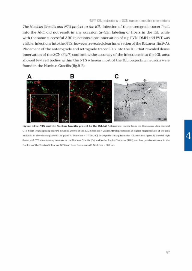
The Nucleus Gracilis and NTS project to the IGL. Injection of the anterograde tracer PhaL into the ARC did not result in any occasion (n=5)in labeling of fi bers in the IGL while with the same successful ARC injections clear innervation of e.g. PVN, DMH and PVT was visible. Injections into the NTS, however, revealed clear innervation of the IGL area (fi g.9-A). Placement of the anterograde and retrograde tracer CTB into the IGL that revealed dense innervation of the SCN (Fig.7) confi rming the accuracy of the injections into the IGL area; showed few cell bodies within the NTS whereas most of the IGL projecting neurons were found in the Nucleus Gracilis (fi g.9-B).
Figure 9.The NTS and the Nucleus Gracilis project to the IGL.(A) Anterograde tracing from the Dorsovagal Area showed
CTB fi bers (red) apposing on NPY neurons (green) of the IGL. Scale bar = 25 μm. (B) Reproduction at higher magnifi cation of the area
included in the white square of the panel A. Scale bar = 17 μm. (C) Retrograde tracing from the IGL (see also fi gure 7) showed high
density of CTB – containing neurons in the Nucleus Gracilis (Gr) and in the Raphe Obscurus (ROb), and few positive neurons in the
Nucleus of the Tractus Solitarius (NTS) and Area Postrema (AP). Scale bar = 200 μm.
181206LAYOUTversie6.indd 87 13-12-18 15:00

526835-L-bw-Buijs526835-L-bw-Buijs526835-L-bw-Buijs526835-L-bw-BuijsProcessed on: 14-12-2018Processed on: 14-12-2018Processed on: 14-12-2018Processed on: 14-12-2018 PDF page: 88PDF page: 88PDF page: 88PDF page: 88
88
Chapter 4
DiscussionThe present study demonstrates that the metabolic condition induces important changes in the NPY expression in the IGL, coinciding with similar changes in the NPY projections of the IGL to the SCN. These changes revealed that the IGL is involved in the transmission of metabolic information to the SCN. In fasted animals lesioning the IGL resulted in a significant increase of c-Fos in the SCN as compared to intact fasted animals, demonstrating the inhibitory influence of the IGL to the SCN in fasting conditions. This inhibitory input also became visible after fasting when the animal was refed because then the increase of c-Fos revealed a removal of this inhibitory input. Together these observations show that in addition to the increased inhibitory IGL input during fasting, this negative metabolic condition also results in an increase of excitatory input to the SCN via other pathways. Consequently the present observations show that at least part of the non-photic input to the SCN, arising from the IGL contains information about the metabolic condition. NPY is one of the most widely expressed neuropeptides in the brain (Adrian et al, 1983;Gehlert et al, 1987, Chromwall & Zukiwska, 2004), the vast majority of the reports relates to the NPY involvement in energy homeostasis, whereas only a minor part is focused on NPY in relation with the synchronization of the SCN or on other functions (Soscia & Harrington, 2005; Harrington et al, 2007; Kim & Harrington, 2008). The present paper brings these observations together and demonstrated that the IGL transmits metabolic information to the SCN. It is very well possible that the transmission of this signal of negative metabolic condition to the SCN facilitates arousal as suggested by (Harrington ME, 1997, Allen & Morin, 2006).After all, a stronger inhibition of neuronal activity of the SCN during the day period would in theory result in an animal that is more prone to show behavioral activity since an active SCN promotes inactivity.
The IGL as a relay for metabolic information to the Suprachiasmatic Nucleus. In 2006 Yi et. al. showed that AgRP neurons of the ARC, which co-express NPY, project to the SCN. These cells are activated by ghrelin, a hormone released by the stomach in time of fasting, demonstrating a neuronal signal to the SCN driven by endocrine input to the ARC (Yi et al, 2006). Our present results show that, it is not the ARC but the IGL which is the main source of NPY innervation in the SCN suggesting that there are two modalities by which NPY neurons may inform the SCN about the metabolic status, one mediated by the ARC possibly by AgRP as neurotransmitter rather than by NPY and another one, anatomically dominant, via the IGL and both inhibitory to the SCN under fasting conditions. Since the IGL area is characterized by a dense net of vascular capillaries with a thin endothelium in contact with neuronal elements (Moore & Card, 1994), it has been proposed that a modified blood brain barrier exists in this area (Harrington ME, 1997). Although it is well known that the permeability of the blood brain barrier is increased at very low glucose
181206LAYOUTversie6.indd 88 13-12-18 15:00

526835-L-bw-Buijs526835-L-bw-Buijs526835-L-bw-Buijs526835-L-bw-BuijsProcessed on: 14-12-2018Processed on: 14-12-2018Processed on: 14-12-2018Processed on: 14-12-2018 PDF page: 89PDF page: 89PDF page: 89PDF page: 89
89
NPY IGL projections to SCN transmit metabolic conditions
4
levels (Oztaşet al, 1985; Gulturk 2010), in literature there is no information that confirms that blood borne signals might have access to the IGL through the vascular endothelium. In addition our present results do not show any significant direct input to the IGL from the ARC or NTS, which are known to receive respectively endocrine or neural feedback from the periphery about satiety, energy supply and fat depot (Schwartz et al 2000). This warrants the question from where the IGL could receive its metabolic information. The IGL may receive metabolic information in a more indirect way via the Lateral Parabrachial Nucleus (PBN) in the midbrain (Horowitz et al, 2004) or from the Lateral Hypothalamus (LH) (Mintz et al, 2001). In addition the LH is also in reciprocal connection with the ARC, and neuropeptides expressed in these nuclei compete for the control of energy balance (Gao& Horvath, 2007). Recently, Pekala et al. showed that LH projections excite a subpopulation of orexin-sensitive IGL neurons, which include IGL NPY cells (Pekala et al, 2011). Thus, the PBN-LH-IGL axis appears to be a good candidate for the translation of the metabolic input arising from the NTS and ARC into an adaptive circadian response. The other possibility would be the direct projection from the NTS and from the nucleus Gracilis.
IGL receives projections from the Nucleus Gracilis. In addition to confirming the expected connection based on the anterograde tracing with the NTS, the retrograde tracing from the IGL showed the majority of retrogradely labeled neurons in the Nucleus Gracilis. The Nucleus Gracilis receives ascending fibers from the sensory layers of the dorsal horn that relay mainly visceral pain and inflammatory information as well as non-noxious stimuli such as the distension of abdominal viscera, temperature and baro reception (Newman PP, 1974; Simon & Schramm, 1984; Berkley &Hubscher, 1995; Al-Chaer 1996). In spite of this information the role of the Gracilis in integrating a wide range of the viscerosensory input with proprioceptive and muscular information, remains elusive (El-Chaer et al, 1997). The present study shows that neurons of the Nucleus Gracilis are projecting to the IGL, whether they indeed transmit visceral sensory information to the IGL remains to be determined. There are two studies that support the IGL sensitivity to somato sensory stimulation. First, electrical stimulation of the rat tail enhances the response of the Geniculate Complex to light input and increases serotonergic innervation (Davidowa & Albrecht, 1992). Second, binding sites for the sensory neuropeptide Calcitonin Gene Related Peptide (CGRP) are expressed in the Geniculate Complex (Skofitsch & Jacobowitz, 1985). It has been proposed that visceral information to the IGL might represent an arousal stimulus that participates to the modulation of circadian rhythms (Davidowa & Albrecht, 1992). The present results indicate that the metabolic condition might be a participating signal in this pathway. The observation of Kreier et al (2006) that the Gracilis receives sensory neuronal input from white adipose tissue suggests that possibly a shrinking energy supply might be part of the signals received by the Gracilis. The anterograde tracing in the IGL may not only be explained by the presence of a few retrogradely neurons in the NTS that project to the IGL, but also by the fact that the needle tract for the injection into the NTS passes through
181206LAYOUTversie6.indd 89 13-12-18 15:00

526835-L-bw-Buijs526835-L-bw-Buijs526835-L-bw-Buijs526835-L-bw-BuijsProcessed on: 14-12-2018Processed on: 14-12-2018Processed on: 14-12-2018Processed on: 14-12-2018 PDF page: 90PDF page: 90PDF page: 90PDF page: 90
90
Chapter 4
the Gracilis. Thus leakage of CTB into the Gracilis might also be partly responsible for the observed fibers. Hence the necessity to always demonstrate the presence of projections by the confirmation of cell bodies in the same area that gives rise to the projection.
Is food deprivation a non-photic stimulus? According to literature, a non-photic stimulus induces phase changes in SCN activity when applied during the subjective day (Mrosovsky 1988, Reebs&Mrosovsky 1989, Mead 1992, Maywood 1999, Yokota 2000, Maywood &Mrosovsky 2002) and interferes with the effects of light pulses given during the subjective night (Mrosovsky& Salmon, 1987, Ralph &Mrosovsky 1992, Mistelberger&Antle 1998). The effect of non-photic stimulation during the subjective day is mediated by NPY projection from the IGL, as suggested by a number of lesion and pharmacological studies performed over 20 years (Johnson et al, 1988; Janik & Mrosovsky, 1994; Wickland&Turek, 1994; Biello et al, 1994; Weber & Rea, 1997; Lall&Biello, 2002; Harrington &Schak, 2000) and finally demonstrated by Glass et al (2010). NPY antagonizes the retinal glutamatergic transmission, provoking a situation of conflict with light (Yannielli & Harrington 2001;Lall&Biello, 2003;Yannielli et al, 2004), suggesting an inhibitory role for NPY in the vl-SCN. According to the present set of data, together with the effect of time-caloric restriction of SCN activity (Challet et al, 1996), our results suggest that fasting provides a non-photic input to the SCN via the IGL in which the increased NPY activity may provide an increased inhibitory input to the IGL.
Re-feeding increases c-Fos in both the IGL and SCN. Consistently with a decrease in NPY-IR in the vl-SCN, we found an increase of c-Fos expression in the same area in re-fed animals in comparison to control and fasted groups. This demonstrates that in addition to fasting, refeeding is also an important signal for the SCN arising from the IGL. Consequently both fasting and feeding signals may contribute as non-photic signals the IGL message to the SCN. A significant augmentation of c-Fos activity also occurs in the IGL of the same rats, although it does not involve the NPY neurons. This suggests that another population of IGL neurons, different from NPY cells, receives information about satiety and inhibits NPY activity. Such intra-IGL inhibitory circuitry has been demonstrated by Glass et al in 2010. They showed that activation of the GABA IGL interneurons results in the suppression of NPY neuronal activity (Glass et al, 2010). The sequence of events that we described resembles the physiological changes that take place in the SCN during and after the dark-light transition, when an active well-fed animal turns into his rest phase. In fact, following light onset, the decrease of NPY levels in the SCN is accompanied by a progressive increase in c-Fos activation (Earnest et al, 1990; Schwartz et al, 1994). Thus under normal conditions these two mechanisms may support each other keeping the animal at rest. The present data show that fasting interferes with the normal NPY cycle in the SCN, by advancing and increasing the magnitude of the physiological NPY levels at ZT10. The relief from this inhibitory effect on the SCN caused
181206LAYOUTversie6.indd 90 13-12-18 15:00

526835-L-bw-Buijs526835-L-bw-Buijs526835-L-bw-Buijs526835-L-bw-BuijsProcessed on: 14-12-2018Processed on: 14-12-2018Processed on: 14-12-2018Processed on: 14-12-2018 PDF page: 91PDF page: 91PDF page: 91PDF page: 91
91
NPY IGL projections to SCN transmit metabolic conditions
4
by re-feeding triggers the activation response as indicated by the c-Fos expressing cells. Hereby it is very important to stress that an IGL lesion, which eliminates NPY containing fibers in the SCN causes a significant increase in the c-Fos activity in the vl-SCN under fasting conditions. Intact animals do not show a change in c-Fos after fasting suggesting that in addition to the inhibitory input from the IGL also an activating input from other areas is present in the SCN. This excitatory input only becomes visible after IGL lesions. This confirms that indeed NPY has an inhibitory control on the vl-SCN resulting in an activation of the SCN after refeeding when the NPYergic tone is decreased. Recently we have obtained evidence that one of the structures that in addition to the retina provides direct excitatory input to the SCN is the NTS (Buijs et al submitted), together with the present observation, this points to the possibility that non-photic input to the SCN may also be excitatory.
181206LAYOUTversie6.indd 91 13-12-18 15:00

526835-L-bw-Buijs526835-L-bw-Buijs526835-L-bw-Buijs526835-L-bw-BuijsProcessed on: 14-12-2018Processed on: 14-12-2018Processed on: 14-12-2018Processed on: 14-12-2018 PDF page: 92PDF page: 92PDF page: 92PDF page: 92
92
Chapter 4
Figure 10. Schematic proposal of the metabolic and visceral feedback from the periphery to the SCN. The Vagus nerve
(X) conveys sensory metabolic information from the viscera to the sensory nuclei of the brainstem: the NTS and Gracilis (Gr) (see e.g.
Kreier et al, 2006), which in turn transmit this information to the IGL. Here we demonstrated that food deprivation stimulates NPY
expression in the IGL, and increases the inhibitory tone of NPY and GABA in the ventro lateral regions of the SCN. Additionally, signals
of a negative metabolic status are transmitted to the SCN by (NPY/)AgRP/GABA co-expressing neurons of the ARC (Yi et al, 2006).
ConclusionThe present results provide a first insight in how the body, of which its daily physiological changes are driven by the SCN, has the capacity to signal back to its central clock. Here we show for the first time that the activity of the IGL-SCN axis is importantly changed by the metabolic condition of an animal. Additionally we see that NPY and GABA have a prominent role in transmitting this information from the IGL to the SCN. Consequently the present results demonstrate that the SCN does not depend exclusively on the ARC to obtain information about the metabolic status, but that the IGL can also directly transmit metabolic information that it receives from NTS and Gracilis (see figure 10). This integration of metabolic information within the light receiving portion of the SCN may serve, to better prepare an individual for activity in the rest phase under fasting conditions (Acosta-Galvan et al, 2011), or it may ensure a rest under fed conditions. Other unexplored possibilities are that this feedback to the SCN is also used to adapt its output to the organs of the body in order to regulate the physiology not only according to the day night cycle but also to the energy status of the body. The extreme decrease in temperature under fasting conditions that does not occur in SCN lesioned animals (Liu et al, 2002; Scheer et al, 2005) could be an example of that.
181206LAYOUTversie6.indd 92 13-12-18 15:00

526835-L-bw-Buijs526835-L-bw-Buijs526835-L-bw-Buijs526835-L-bw-BuijsProcessed on: 14-12-2018Processed on: 14-12-2018Processed on: 14-12-2018Processed on: 14-12-2018 PDF page: 93PDF page: 93PDF page: 93PDF page: 93
93
NPY IGL projections to SCN transmit metabolic conditions
4
181206LAYOUTversie6.indd 93 13-12-18 15:00

526835-L-bw-Buijs526835-L-bw-Buijs526835-L-bw-Buijs526835-L-bw-BuijsProcessed on: 14-12-2018Processed on: 14-12-2018Processed on: 14-12-2018Processed on: 14-12-2018 PDF page: 94PDF page: 94PDF page: 94PDF page: 94
94
Chapter 4
ReferencesAcosta-Galvan G, Yi CX, van der Vliet J, Jhamandas JH, Panula P, Angeles-Castellanos M, Del Carmen Basualdo M, Escobar C, Buijs RM.(2011) Interaction between hypothalamic dorsomedial nucleus and the suprachiasmatic nucleus determines intensity of food anticipatory behavior.ProcNatlAcadSci U S A. 108(14):5813-8
Adrian TE, Allen JM, Bloom SR, Ghatei MA, Rossor MN, Roberts GW, Crow TJ, Tatemoto K, Polak JM. (1983) Neuropeptide Y distribution in the human brain.Nature.306:584-6.
Al-Chaer ED, Lawand NB, Westlund KN, Willis WD. (1996) Pelvic visceral input into the nucleus gracilis is largely mediated by the postsynaptic dorsal column pathway.J Neurophysiol. 76:2675-90.
Al-Chaer ED, Westlund KN, Willis WD. (1997) Nucleus gracilis: an integrator for visceral and somatic information.J Neurophysiol. 78:521-7.
Berkley KJ, Hubscher CH. (1995) Are there separate central nervous system pathways for touch and pain?Nat Med. 1:766-73.
Biello SM, Janik D, Mrosovsky N. (1994) Neuropeptide Y and behaviorally induced phase shifts.Neuroscience. 62:273-9.
Buijs MR, Pool CW, Van Heerikhuize JJ, Sluiter AA, Van Der Sluis P, et al. (1989) Antibodies to small transmitter molecules and peptides: production and application of antibodies to dopamine, serotonin, GABA, vasopressin, vasoactive intestinal peptide, neuropeptide Y, somatostatin and substance P. Biomedical Research. 10:213–221.
Buijs RM, Kalsbeek A.(2001)Hypothalamic integration of central and peripheral clocks Nat Rev Neurosci.2:521-6.
Challet E, Pévet P, Malan A. (1996) Intergeniculate leaflet lesion and daily rhythms in food-restricted rats fed during daytime.NeurosciLett. 216:214-8.
Chronwall BM, Zukowska Z. (2004) Neuropeptide Y, ubiquitous and elusive.Peptides. 25:359-63.
Davidowa H, Albrecht D. Modulation of visually evoked responses in units of the ventral lateral geniculate nucleus of the rat by somatic stimuli.(1992) Behav Brain Res. 50:27-33.
Dibner C, Schibler U, Albrecht U. (2010) The mammalian circadian timing system: organization and coordination of central and peripheral clocks.Annu Rev Physiol. 72:517-49.
Earnest DJ, Iadarola M, Yeh HH, Olschowka JA. (1990) Photic regulation of c-fos expression in neural components governing the entrainment of circadian rhythms.Exp Neurol.109:353-61.
Gao Q, Horvath TL. (2008) Neuronal control of energy homeostasis.FEBS Lett. 582:132-41.
Gehlert DR, Chronwall BM, Schafer MP, O’Donohue TL. (1987) Localization of neuropeptide Y messenger ribonucleic acid in rat and mouse brain by in situ hybridization.Synapse. 1:25-31.
Glass JD, Guinn J, Kaur G, Francl JM.(2010) On the intrinsic regulation of neuropeptide Y release in the mammalian suprachiasmatic nucleus circadian clock.Eur J Neurosci.31:1117-26.
181206LAYOUTversie6.indd 94 13-12-18 15:00

526835-L-bw-Buijs526835-L-bw-Buijs526835-L-bw-Buijs526835-L-bw-BuijsProcessed on: 14-12-2018Processed on: 14-12-2018Processed on: 14-12-2018Processed on: 14-12-2018 PDF page: 95PDF page: 95PDF page: 95PDF page: 95
95
NPY IGL projections to SCN transmit metabolic conditions
4
Gulturk S, Demirkazik A, Kosar I, Cetin A, Dökmetas HS, Demir T. (2010) Effect of exposure to 50 Hz magnetic field with or without insulin on blood-brain barrier permeability in streptozotocin-induced diabetic rats.Bioelectromagnetics.31:262-9.
Harrington ME. (1997) The ventral lateral geniculate nucleus and the intergeniculate leaflet: interrelated structures in the visual and circadian systems.NeurosciBiobehav Rev. 21:705-27.
Harrington ME, Schak KM. (2000) Neuropeptide Y phase advances the in vitro hamster circadian clock during the subjective day with no effect on phase during the subjective night.Can J PhysiolPharmacol. 78:87-92.
Harrington M, Molyneux P, Soscia S, Prabakar C, McKinley-Brewer J, Lall G. (2007) Behavioral and neurochemical sources of variability of circadian period and phase: studies of circadian rhythms of npy-/- mice.Am J PhysiolRegulIntegr Comp Physiol. 292:1306-14.
Hastings MH, Duffield GE, Ebling FJ, Kidd A, Maywood ES, Schurov I. (1997) Non-photic signalling in the suprachiasmatic nucleus.Biol Cell. 89:495-503.
Horowitz SS, Blanchard JH, Morin LP. (2004) Intergeniculate leaflet and ventral lateral geniculate nucleus afferent connections: An anatomical substrate for functional input from the vestibulo-visuomotor system. 474(2):227-45.
Janik D, Mrosovsky N. (1994) Intergeniculate leaflet lesions and behaviorally-induced shifts of circadian rhythms.Brain Res. 651, 174-82.
Johnson RF, Smale L, Moore RY, Morin LP.(1988) Lateral geniculate lesions block circadian phase-shift responses to a benzodiazepine. ProcNatlAcadSci U S A.85:5301-4.
Jhanwar-Uniyal M, Beck B, Burlet C, Leibowitz SF. (1990) Diurnal rhythm of neuropeptide Y-like immunoreactivity in the suprachiasmatic, arcuate and paraventricular nuclei and other hypothalamic sites.Brain Res. 536:331-4.
Liu S, Chen XM, Yoda T, Nagashima K, Fukuda Y, Kanosue K. (2002) Involvement of the suprachiasmatic nucleus in body temperature modulation by food deprivation in rats.Brain Res.929(1):26-36.
Kim HJ, Harrington ME. (2008) Neuropeptide Y-deficient mice show altered circadian response to simulated natural photoperiod.Brain Res. 1246:96-100.
Kreier F, Kap YS, Mettenleiter TC, Van Heijningen C, Van D, V, Kalsbeek A, Sauerwein HP, Fliers E, Romijn JA, and Buijs RM. (2006) Tracing from fat tissue, liver and pancreas: A neuroanatomical framework for the role of the brain in type 2 diabetes. Endocrinology 147:1140-1147.
Kuroda H, Fukushima M, Nakai M, Katayama T, Murakami N. (1997) Daily wheel running activity modifies the period of free-running rhythm in rats via intergeniculate leaflet.PhysiolBehav. 61:633-7.
Lall GS, Biello SM. (2002) Attenuation of phase shifts to light by activity or neuropeptide Y: a time course study.Brain Res. 957:109-16.
181206LAYOUTversie6.indd 95 13-12-18 15:00

526835-L-bw-Buijs526835-L-bw-Buijs526835-L-bw-Buijs526835-L-bw-BuijsProcessed on: 14-12-2018Processed on: 14-12-2018Processed on: 14-12-2018Processed on: 14-12-2018 PDF page: 96PDF page: 96PDF page: 96PDF page: 96
96
Chapter 4
Lall GS, Biello SM. (2003) Attenuation of circadian light induced phase advances and delays by neuropeptide Y and a neuropeptide Y Y1/Y5 receptor agonist.Neuroscience. 119:611-8.
Maywood ES, Mrosovsky N, Field MD, Hastings MH. (1999) Rapid down-regulation of mammalian period genes during behavioral resetting of the circadian clock.ProcNatlAcadSci U S A. 96:15211-6.
Mead S, Ebling FJ, Maywood ES, Humby T, Herbert J, Hastings MH. (1992) Anonphotic stimulus causes instantaneous phase advances of the light-entrainable circadian oscillator of the Syrian hamster but does not induce the expression of c-fos in the suprachiasmatic nuclei.J Neurosci. 12:2516-22.
Mintz EM, van den Pol AN, Casano AA, Albers HE.(2001) Distribution of hypocretin-(orexin) immunoreactivity in the central nervous system of Syrian hamsters (Mesocricetusauratus). J ChemNeuroanat.21(3):225-38.
Mistlberger RE, Antle MC. (1998) Behavioral inhibition of light-induced circadian phase resetting is phase and serotonin dependent.Brain Res. 786:31-8.
Moore RY, Card JP. (1994) Intergeniculate leaflet: an anatomically and functionally distinct subdivision of the lateral geniculate complex.J Comp Neurol. 344:403-30.
Morin LP, Allen CN. (2006) The circadian visual system, 2005.Brain Res Rev. 51:1-60.
Mrosovsky N, Salmon PA. (1987) A behavioural method for accelerating re-entrainment of rhythms to new light-dark cycles.Nature. 330:372-3.
Mrosovsky N. (1998) Phase response curves for social entrainment.J Comp Physiol A. 162: 35-46.
Mrosovsky N, Edelstein K, Hastings MH, Maywood ES.(2001) Cycle of period gene expression in a diurnal mammal (Spermophilustridecemlineatus): implications for nonphotic phase shifting.J Biol Rhythms.16:471-8.
Newman PP. (1974) Visceral afferent functions of the nervous system.MonogrPhysiol Soc. 25:1-273.
Norgren R. (1976) Taste pathways to hypothalamus and amygdala.J Comp Neurol. 166:17-30.
Oztaş B, Küçük M, Sandalci U. (1985) Effect of insulin-induced hypoglycemia on blood-brain barrier permeability.Exp Neurol. 87:129-36.
Pekala D, Blasiak T, Raastad M, Lewandowski MH. (2011) The influence of orexins on the firing rate and pattern of rat intergeniculate leaflet neurons--electrophysiological and immunohistological studies.Eur J Neurosci. 34:1406-18.
Ralph MR, Mrosovsky N. (1992)Behavioral inhibition of circadian responses to light.J Biol Rhythms. 7:353-9.
Reebs SG, Mrosovsky N. (1989) Effects of induced wheel running on the circadian activity rhythms of Syrian hamsters: entrainment and phase response curve.J Biol Rhythms. 4:39-48.
Saper CB, Loewy AD. (1980) Efferent connections of the parabrachial nucleus in the rat.Brain Res.197:291-317.
Scheer FA, Pirovano C, Van Someren EJ, Buijs RM(2005) Environmental light and suprachiasmatic nucleus interact in the regulation of body temperature. 132(2):465-77.
181206LAYOUTversie6.indd 96 13-12-18 15:00

526835-L-bw-Buijs526835-L-bw-Buijs526835-L-bw-Buijs526835-L-bw-BuijsProcessed on: 14-12-2018Processed on: 14-12-2018Processed on: 14-12-2018Processed on: 14-12-2018 PDF page: 97PDF page: 97PDF page: 97PDF page: 97
97
NPY IGL projections to SCN transmit metabolic conditions
4
Schwartz WJ, Takeuchi J, Shannon W, Davis EM, Aronin N. (1994) Temporal regulation of light-induced Fos and Fos-like protein expression in the ventrolateral subdivision of the rat suprachiasmatic nucleus.Neuroscience. 58:573-83.
Simon OR, Schramm LP. (1984) The spinal course and medullary termination of myelinated renal afferents in the rat.Brain Res. 290:239-47.
Shinohara K, Tominaga K, Fukuhara C, Otori Y, Inouye SI. (1993) Processing of photic information within the intergeniculate leaflet of the lateral geniculate body: assessed by neuropeptide Y immunoreactivity in the suprachiasmatic nucleus of rats. Neuroscience. 56:813-22.
Skofitsch G, Jacobowitz DM. (1985) Autoradiographic distribution of 125I calcitonin gene-related peptide binding sites in the rat central nervous system. Peptides. 6:975-86.
Soscia SJ, Harrington ME. (2005) Neuropeptide Y does not reset the circadian clock in NPY Y2-/- mice.NeurosciLett. 373:175-8.
Weber ET, Rea MA. (1997) Neuropeptide Y blocks light-induced phase advances but not delays of the circadian activity rhythm in hamsters. NeurosciLett. 231:159-62.
Wickland C, Turek FW. (1994) Lesions of the thalamic intergeniculate leaflet block activity-induced phase shifts in the circadian activity rhythm of the golden hamster. Brain Res.660:293-300.
Wu Q, Boyle MP, Palmiter RD. (2009) Loss of GABAergic signaling by AgRP neurons to the parabrachial nucleus leads to starvation.137:1225-34.
Wu Q, Palmiter RD. (2011) GABAergic signaling by AgRP neurons prevents anorexia via a melanocortin-independent mechanism.Eur J Pharmacol. 660:21-7.
Wu Q, Clark MS, Palmiter RD.(2012) Deciphering a neuronal circuit that mediates appetite.Nature.483:594-7.
Yannielli PC, Harrington ME. (2001) The neuropeptide Y Y5 receptor mediates the blockade of “photic-like” NMDA-induced phase shifts in the golden hamster.J Neurosci. 21:5367-73.
Yannielli P, Harrington ME. (2004)Let there be “more” light: enhancement of light actions on the circadian system through non-photic pathways.ProgNeurobiol. 74:59-76.
Yannielli PC, Brewer JM, Harrington ME. (2004) Blockade of the NPY Y5 receptor potentiates circadian responses to light: complementary in vivo and in vitro studies.Eur J Neurosci. 19:891-7.
Yi CX, van der Vliet J, Dai J, Yin G, Ru L, Buijs RM. (2006) Ventromedialarcuate nucleus communicates peripheral metabolic information to the suprachiasmatic nucleus.Endocrinology. 147:283-94.
Yokota SI, Horikawa K, Akiyama M, Moriya T, Ebihara S, Komuro G, Ohta T, Shibata S. (2000) Inhibitory action of brotizolam on circadian and light-induced per1 and per2 expression in the hamster suprachiasmatic nucleus.Br J Pharmacol. 131:1739-47.
181206LAYOUTversie6.indd 97 13-12-18 15:00