utility biosensors for applications in agriculture – a revie · utility biosensors for...
TRANSCRIPT

Journal of American Science 2010;6(9)
http://www.americanscience.org [email protected] 353
Utility Biosensors for applications in Agriculture – A Review
J.S Rana*, Jyoti Jindal, Vikas Beniwal, Vinod Chhokar Department of Bio & Nanotechnology, Guru Jambheshwar University of Science & Technology, Hisar, Haryana
125001, India. [email protected]
Abstract: The quality of food is fundamentally based on biochemical composition of food. Therefore, this review summarizes recent advances in the fabrication of different types of Biosensors that have been designed for the measurement of various components in the horticultural samples. We review various biosensors ranging from electrochemical, calorimetric, optical, immunosensors to screen-printed three electrode systems. Numerous examples are provided in several key areas including glucose, fructose, malic acid, pyruvic acid, ascorbic acid, glycerol, glutamate etc. We also explore important coenzymes involved in the development of biosensors. [Journal of American Science 2010;6(9):353-375]. (ISSN: 1545-1003).
Keywords: Biosensors, Fruits, organic acids, Coenzymes, NADPH
Introduction Fruit quality monitoring is one of the major concerns within the food industry (Kriz et al., 2002). In particular, there is a growing need to develop analytical instruments which can provide quality monitoring for the entire food processing operation, including starting materials and final products (Whitaker, 1994). Biosensors are highly selective analytical instruments, due to the high selectivity of the biological recognition element employed which have been applied in an array of disciplines including medicine, industry, environmental analysis, food technology, and military (Wang, 2001). The current economics (Shobha et al., 2003) of fruit production allows the produce of one country to be shipped to consumer countries anywhere in the world. Consumers are demanding increased quality with severe economic consequences to those producers who cannot meet these demands with guaranteed consistency. Producers must therefore carefully monitor the quality of fruit through all the stages of production, storage and transport (Arif et al., 2002). The selection of a particular foodstuff by a consumer is largely based on sensory perceptions with taste, which is influenced by diverse factors including saltiness, sweetness, bitterness and acidity as perhaps the most important factors. Texture is also a key parameter and is dictated by many factors including moisture content and fat, carbohydrate and protein levels (Zczesniak, 1963). Other important sensory factors include the aroma, shape and color of the foodstuff. Therefore; rapid, portable and accurate methods for the assessment of fruit quality and physiological state are required. Due to their numerous attributes, biosensors potentially offer an accurate, fast, relatively cheap, stable, portable and user-friendly device for in situ monitoring of fruit maturity and quality.
1) Biosensor A biosensor is a compact analytical device incorporating a biological or biologically derived sensing element either associated or integrated within a physicochemical transducer (Fig.1). The usual aim of such a device is to produce either a discrete or continuous digital electronic signal that is proportional to a single analyte or a related group of analytes present in a sample (Compagnone et al., 1995).
1.1) Principle of Biosensor Analytical chemistry (Compagnone et al., 1995) plays an important role in food quality parameters because almost every sector of industry and public service relies on quality control. A food quality biosensor is a device, which can respond to some property or properties of food and transform the response(s) into a detectable signal, often an electric signal. This signal may provide direct information about the quality factor(s) to be measured or may have a known relation to the quality factor (Finn, 2003). There are various kinds of biosensors most of which work on the principle of one of the following: 1.2) Electrochemical Biosensors Electrochemical biosensors are based on monitoring electroactive species that are either produced or consumed by the action of the biological components (e.g., enzymes and cells). Transduction of the produced signal can be performed using one of several methods under two broad headings: Potentiometric Biosensors and Amperometric Biosensors (Terry et al., 2005).

Journal of American Science 2010;6(9)
http://www.americanscience.org [email protected] 354
a). b). Figure 1.a). Diagrammatic representation of a typical biosensor, b). Components of Biosensor 1.3) Potentiometric biosensors These are based on monitoring the potential of a system at a working electrode, with respect to an accurate reference electrode, under conditions of essentially zero current flow. In process, potentiometric measurements are related to the analyte activity (of a target species) in the test sample (Terry et al., 2005). Potentiometric biosensors can operate over a wide range (usually several orders of magnitude) of concentrations. The use of potentiometric biosensors for food quality analysis has not been as widely reported as for amperometric sensors. However, some of the examples where this approach has been used for food quality analysis include estimating monophenolase activity in apple juice (Dutta et al., 2001), determining the concentration of sucrose in soft drinks (Rotariu et al., 2002), measuring isocitrate concentrations in fruit juices (Kim et al., 2003), and determining urea levels in milk (Verma et al., 2003). 1.4) Amperometric Biosensors The use of amperometric biosensors in signal transduction has proved to be the most widely reported using an electrochemical approach. Both “one-shot” (disposable) sensors and on-line (multi-
measurement) devices are commercially available, monitoring a wide range of target analytes. In contrast to potentiometric devices, the principle operation of amperometric biosensors is defined by a constant potential applied between a working and a reference electrode. The applied potential results in redox reactions, causing a net current to flow. The magnitude of this current is proportional to the concentration of electro active species present in test solution and both cathodic (reducing) and anodic (oxidizing) reactions can be monitored amperometrically. Most of the amperometric biosensors described use enzymes as the biorecognition element. Typically, oxidase and dehydrogenase enzymes have been the most frequently exploited catalysts used for these biosensor formats (Terry et al., 2005). 1.5) Calorimetric Biosensors Most of the biochemical reactions are accompanied by either heat absorption or production. Sensors based on calorimetric transduction are designed to detect heat generated or consumed during a biological reaction; by using sensitive heat detection devices. Various biosensors for specific target analytes have been constructed. In the field of food quality analysis, uses of such biosensors to detect metabolites have been described (Thavarungkul et al., 1999). 1.6) Optical Biosensors These sensors are based on measuring responses to illumination or to light emission. Optical biosensors can employ a number of techniques to detect the presence of a target analyte and are based on well-founded methods including chemiluminescence, fluorescence, light absorbance, phosphoresence, photothermal techniques, surface plasmon resonance (SPR), light polarization and rotation, and total internal reflectance. For example the use of this technique have been demonstrated (Mohammed et al., 2003) to detect the presence of allergens, in particular peanuts, during food production.
1.7) Acoustic Biosensors Piezoelectric quartz crystals can be affected by a change of mass at the crystal surface; this phenomenon has been successfully exploited and used to develop acoustic biosensors. For practical applications, the surface of the crystal can be modified with recognition elements (e.g., antibodies) that can bind specifically to a target analyte. 1.8) Immunosensors

Journal of American Science 2010;6(9)
http://www.americanscience.org [email protected] 355
Immunosensors are based on exploiting the specific interaction of antibodies with antigens. Typically, immunoassays (such as the enzyme-linked immunosorbent assay technique) employ a label (e.g., enzyme, antibody, fluorescent marker) to detect the immunological reaction. The use of biosensor platforms, linked to an immunoassay format, offers a route to rapid and accurate quantitative measurements of target analytes. 2) Necessity of Food/ Fruit Quality Control Quality control is the essential part of a food industry and efficient quality assurance is becoming increasingly important. Consumers expect good quality and healthy food at a given price; with good shelf life and high safety while food inspections require good manufacturing practices, safety, labeling and compliance with the regulations. Further, food producers are increasingly asking for efficient control methods, in particular through on-line or at-line quality sensors. Their main aim is to satisfy the consumer and regulatory requirements and to improve the production feasibility, quality sorting, automation and reduction of production cost and production time subsequently (Finn, 2003).
3) Biochemical Composition of Fruits The quality of soft fruit, in terms of taste, nutrition and consumers acceptance, is fundamentally based on the biochemical composition of the fruit. In soft fruits (viz. blackcurrant and strawberry) sugar: acid ratios can be used as an important index of fruit maturity and act as a determinant of overall fruit. However, sugar: acid ratios are infrequently used due to a requirement for specific instrumentation and semi-skilled analytical scientists. Today we need a simple and low-cost alternative, which would significantly enhance both the number and extent of tests carried out. 4) Fruit Maturity, Ripening and Quality Relationships Fruit maturity at harvest is the most important factor that determines shelf life and final fruit quality. If harvested immature then fruits are more subject to shriveling and mechanical damage, and are of inferior quality when ripe, whereas overripe fruits are liable to become soft and mealy with bland flavor soon after harvest (Kader, 1999;1992). Therefore, fruits harvested either too early or too late in their season are more susceptible to post harvest physiological disorders than fruits harvested at proper maturity. Fruits can be divided into two groups: 1) Fruit that are incapable of enduring their ripening process once picked from the plant like berries, cheery, citrus fruits, grapes, lychee, pineapple,
pomegranate, and tamarillo. 2) Fruits that can be harvested mature and ripped off the plant like apple, apricot, avocado, banana, cherimoya, guava, kiwifruit, mango, nectarine, papaya, passion fruit, pear, peach, persimmon, plum, quince, sapodilla, sapote (Kader, 1999;1992). Volatile compounds are responsible for the characteristic aroma of fruits and are present in extremely small quantities (<100< g/g fresh wt.). The major volatile formed is ethylene. Scientists are trying to develop portable instruments with sensors that detect volatile production by fruits and hence detecting maturity and quality. Other strategies include the removal of a very small amount of fruit tissue and measurement of total sugar or organic acid content (Seymour et al., 1993).
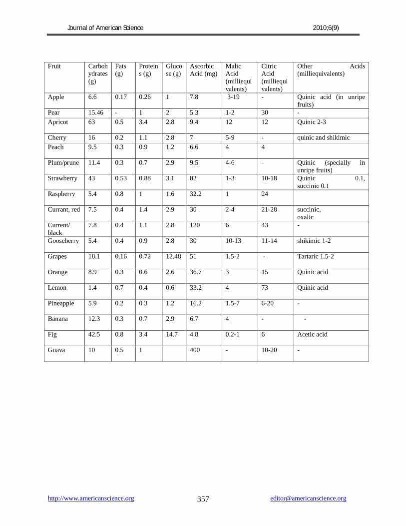
5) Major organic acids in fruits Organic acids function in growth, maturation, senescence, color, and antimicrobial activity of fruits. The low pH of fruits is due to the three most common organic acids present in fruits citric acid, malic acid, and tartaric acid. The total amount of acid in fruits varies widely, from about 0.2% in pear juice to 0.8% in limejuice. The amount and type of acid present in fruits determine the fresh taste of fruits and also affects the shelf life. 5.1) Organic Acid as an Indicator of Fruit Maturity Organic acids directly play an important role in the growth, maturation and acidity of the fruit, and also affect the shelf life of the fruit by influencing the growth of microorganisms (Cano et al., 1994). The citric, malic, oxalic, and tartaric acids ranging from 0.1 to 30 g/L were found in orange, grape, and apple juices. There is a considerable difference in the organic acid content found in various types and brands of fruit juice. For example, Minute Maid contains higher levels of oxalic and citric acids when compared to all other orange juices tested. Grape concentrate was found to have lower amount of malic acid than other grape juice, while freshly squeezed grape juice contains higher amount of tartaric acid. Brae burn apples contained the highest amount of citric acid in apples; however Granny Smith apples were the overall most acidic apples tested. Table 1 shows the biochemical composition of various fruits that can be exploited for developing a potential biosensor. 5.2) Effect of Shelf Life on the Changes in Organic Acids of Fruits The content and composition of organic acids in apples (Malus domestica Borkh, cultivars Gloster, Mutsu, Starting), and apricots (Prunus armeniaca L., cvs. Ceglédi bíborkajszi, Csongrádi kajszi, Gönci Magyar kajszi, Nagykõrösi óriás) were studied.

Journal of American Science 2010;6(9)
http://www.americanscience.org [email protected] 356
Malic, ascorbic, citric and succinic acids were found in the different extracts, depending on the type of fruits (Kovacs and Djedjro, 1994). The concentration of malic and ascorbic acids decreased with the increase in storage time and with the increasing radiation doses whereas the concentration of succinic and citric acids in general increased during storage. Calcium promotes the preservation of malic and ascorbic acids in apples. 6) Successful Examples of Organic Acid Biosensors Developed 6.1) Pyruvic Acid Onion flavour is principally directed by the perception of pungency (Abayomi et al, 2006). A disposable prototype electrochemical screen-printed (carbon-based) biosensor (C2030519D5, GEM Ltd., Gwent, UK) was constructed using pyruvate dehydrogenase immobilized on mediated Meldolas Blue electrodes and a combined Ag/AgCl reference/counter electrode, both screen-printed onto a PVC substrate to determine pungency in onions (Allium cepa L.). Electrochemical measurements were carried out using a Palm Sense potentiostat (Palm Instruments BV, The Netherlands). The biosensor developed was able to differentiate between mild and pungent bulbs with pyruvate concentrations ranging between ≈4 and 8 mM in freshly extracted juices. Electrochemical measurements were carried out at +50 mV at 21˚C. 6.2) Glucose Biosensors Most of the glucose (Fig.1) biosensors developed are based on immobilized glucose oxidase. In many cases, glucose oxidase has been associated with mediators so as to bring down the high working potential required for hydrogen peroxide breakdown (Saby et al, 1995) and (Maines et al, 1996). The β-D-glucose sensor developed was also based on glucose oxidase, at the working potential of -350 mV vs. Ag/AgCl, hydrogen peroxide was catalytically oxidized at a rhodinised carbon electrode (White et al, 1994).
A novel and simple method which do not involve enzyme or monomer modifications, for the co-immobilization of ferrocene and GOx in a poly(pyrrole) matrix for use as glucose biosensor was
developed (Foulds and Lowe, 1988). In spite of the low conductivity of the polypyrrole film formed, the biosensor’s performance was better than that of other devices reported due to redox mediation of ferrocene that lowers the working potential to 0.4 V. The characterization of the polymer prepared from an ethanolic suspension demonstrated the presence of alcohol interferes in the polymerization kinetics (Pablo et al., 2001). However, this played a beneficial role in efficient immobilization of both, the enzyme and the ferrocene, in a very thin electroactive film. This fact improved the biosensor’s time response, avoiding mass transport effects.
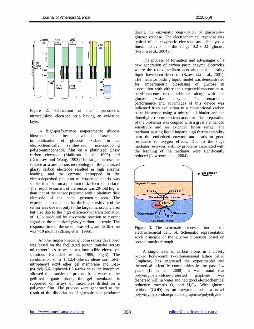
A new type of disposable amperometric biosensor was devised by screen-printing thick-film electrodes directly on a porous nitrocellulose (NC) strip (Fig.2). A glucose biosensor based on hydrogen peroxide detection was constructed by immobilizing glucose oxidase (GOx) on the NC electrode strip and by formulating a strong oxidation layer (i.e., PbO2) at the sample loading area, placed below the GOx reaction band. The screen-printed PbO2 paste serves as a sample pretreatment layer that removes interference by its strong oxidizingability. Samples applied were carried chromatographically, via the PbO2 paste, to the GOx layer, and glucose was catalyzed to liberate hydrogen peroxide, which was then detected at the electrode surface. The proposed NC/ PbO2 strip sensor is shown to be virtually insusceptible to interfering species such as acetaminophen and ascorbic and uric acids and to exhibit good performance, in terms of the sensor-to-sensor reproducibility (standard deviation, (±0.026-(±0.086 µA), the sensitivity (slope, -0.183 µA/mM), and the linearity (correlation coefficient, 0.994 in the range of 0-10 mM) (Cui et al., 2000).
Rakhi et al. (2009) constructed an amperometric biosensor, based on deposition of glucose oxidase (GOD) onto crystalline gold (Au) nanoparticle modified multiwalled carbon nanotube (MWNT) electrode. MWNTs were synthesized by catalytic chemical vapor decomposition of acetylene over rare-earth-based AB2 (DyNi2) alloy hydride catalyst. Purified MWNTs were decked with nanocrystalline Au metal clusters using a simple
chemical reduction method (Rakhi et al., 2008). The characterization of metal-decorated CNTs was done using X-ray diffraction analysis, transmission electron microscopy (TEM), high-resolution TEM, scanning electron microscopy, and energy-dispersive X-ray analysis. Amperometric biosensor fabricated by depositing GOD over Nafion-solubilized Au-
MWNT electrode retained its biocatalytic activity and obtained fast and sensitive glucose quantification. The fabricated GOD/Au-MWNT/Nafion electrode has a good glucose-biosensing potential, and it displayed a linear response up to 22 mM glucose and a detection limit of 20 μM method.
Table1: Biochemical Composition of Fruits
Journal of American Science 2010;6(9)
http://www.americanscience.org [email protected] 357
Fruit Carbohydrates (g)
Fats (g)
Proteins (g)
Glucose (g)
Ascorbic Acid (mg)
Malic Acid (milliequivalents)
Citric Acid (milliequivalents)
Other Acids (milliequivalents)
Apple 6.6 0.17 0.26 1 7.8 3-19 - Quinic acid (in unripe fruits)
Pear 15.46 - 1 2 5.3 1-2 30 -
Apricot 63
0.5 3.4 2.8 9.4 12 12 Quinic 2-3
Cherry 16 0.2 1.1 2.8 7 5-9 - quinic and shikimic
Peach 9.5
0.3 0.9 1.2 6.6 4 4
Plum/prune 11.4 0.3 0.7 2.9 9.5 4-6 - Quinic (specially in unripe fruits)
Strawberry 43 0.53 0.88 3.1 82 1-3 10-18 Quinic 0.1, succinic 0.1
Raspberry 5.4
0.8 1 1.6 32.2 1 24
Currant, red 7.5 0.4 1.4 2.9 30 2-4 21-28 succinic, oxalic
Current/ black
7.8 0.4 1.1 2.8 120 6 43 -
Gooseberry 5.4
0.4 0.9 2.8 30 10-13 11-14 shikimic 1-2
Grapes 18.1
0.16 0.72 12.48 51 1.5-2 - Tartaric 1.5-2
Orange 8.9
0.3 0.6 2.6 36.7 3 15 Quinic acid
Lemon 1.4
0.7 0.4 0.6 33.2 4 73 Quinic acid
Pineapple 5.9
0.2 0.3 1.2 16.2 1.5-7 6-20 -
Banana 12.3
0.3 0.7 2.9 6.7 4 - -
Fig 42.5
0.8 3.4 14.7 4.8 0.2-1 6 Acetic acid
Guava 10
0.5 1 400 - 10-20 -
Journal of American Science 2010;6(9)
http://www.americanscience.org [email protected] 358
Figure 2. Fabrication of the amperometric
nitrocellulose electrode strip having an oxidation
layer.
A high-performance amperometric glucose biosensor has been developed, based on immobilization of glucose oxidase in an electrochemically synthesized, nonconducting poly(o-aminophenol) film on a platinized glassy carbon electrode (Malitesta et al., 1990) and (Dempsey and Wang, 1993).The large microscopic surface area and porous morphology of the platinized glassy carbon electrode resulted in high enzyme loading, and the enzyme entrapped in the electrodeposited platinum microparticle matrix was stabler than that on a platinum disk electrode surface. The response current of the sensor was 20-fold higher than that of the sensor prepared with a platinum disk electrode of the same geometric area. The experiments concluded that the high sensitivity of the sensor was due not only to the large microscopic area but also due to the high efficiency of transformation of H2O2 produced by enzymatic reaction to current signal on the platinized glassy carbon electrode. The response time of the sensor was <4 s, and its lifetime was >10 months (Zhang et al., 1996).
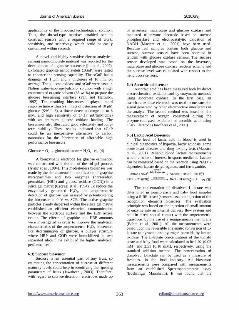
Another amperometric glucose sensor developed was based on the facilitated proton transfer across microinterfaces between two immiscible electrolyte solutions (Grandell et al., 1999, Fig.3). The combination of a 1,3:2,4-dibenzylidene sorbitol/2-nitrophenyl octyl ether gel membrane and 3-(2-pyridyl)-5,6- diphenyl-1,2,4-triazine as the ionophore allowed the transfer of protons from water to the gellified organic phase; the gel membrane is supported on arrays of microholes drilled on a polyester film. The protons were generated as the result of the dissociation of gluconic acid produced
during the enzymatic degradation of glucose-by-glucose oxidase. The electrochemical response was typical of an enzymatic electrode and displayed a linear behavior in the range 0.2-3mM glucose (Pereira et al., 2004).
The process of formation and advantages of a new generation of carbon paste enzyme electrodes where the redox mediator acts also as the pasting liquid have been described (Szuwarski et al., 2001).
The mediator pasting liquid model was demonstrated for amperometric biosensing of glucose in association with either the tertpentylferrocene or n-butylferrocene mediator/binder along with the glucose oxidase enzyme. The remarkable performance and advantages of this device was indicated from evaluation to a conventional carbon paste biosensor using a mineral oil binder and the dimethylferrocene electron acceptor. The preparation of the biosensor was coupled with a greatly enhanced sensitivity and an extended linear range. The mediator pasting liquid imparts high thermal stability onto the embedded enzyme and leads to good resistance to oxygen effects. Due to the huge mediator reservoir, stability problems associated with the leaching of the mediator were significantly reduced (Lawrence et al., 2004).
Figure 3. The schematic representation of the electrochemical cell, b). Schematic representation work principle of the glucose biosensor based on proton transfer through A single layer of carbon atoms in a closely packed honeycomb two-dimensional lattice called Graphene, has engrossed the experimental and theoretical scientific communities in the past few years (Li et al., 2008). It was found that polyvinylpyrrolidone-protected graphene was dispersed well in water and had good electrochemical reduction towards O2 and H2O2. With glucose oxidase (GOD) as an enzyme model, a novel polyvinylpyrrolidoneprotectedgraphene/polyethyleni

Journal of American Science 2010;6(9)
http://www.americanscience.org [email protected] 359
mine-functionalized ionic liquid/ GOD electrochemical biosensor was developed, that could direct electron transfer of GOD, retained its bioactivity and exhibited potential application for the fabrication of novel glucose biosensors with linear glucose response up to 14 mM (Shan et al., 2009).
Since the discovery of carbon nanotubes, CNs have been extensively used in the fabrication of electrochemical sensors and biosensors (Sotiropoulou and Chaniotakis, 2003). A catalytic electrochemical biosensor was developed using highly activated carbon nanofibers as the matrix for the immobilization of proteins and enzymes. The enzyme glucose oxidase (GOx) was immobilized on the carbon samples by adsorption at 4 °C for 24 h. The data obtained from the carbon nanofiber-based glucose biosensors presented higher sensitivity, longer lifetimes, and tremendous measurement- to-measurement reproducibility over an extended period of time. The large surface area of the nanofibers, together with their large number of active sites, provided the grounds for the adsorption of enzymes (Vamvakaki et al., 2006).
The CNT/Teflon material conveyed new potential applications for electrochemical devices by combining the advantages of CNT and “bulk” composite electrodes. Their association with the Teflon binder did not impair the electrocatalytic properties of CNT (Rao et al., 2001) and (Baughman et al., 2002). The distinct electrocatalytic activity towards hydrogen peroxide and NADH allowed accurate low-potential amperometric biosensing of glucose and ethanol, respectively, in relation with the integration of glucose oxidase and alcohol dehydrogenase/NAD+ within the three-dimensional CNT/ Teflon matrix. The accelerated electron transfer was coupled with minimization of surface fouling and surface renewability. These advantages of CNT-based composite devices were illustrated from evaluation to their graphite/Teflon counterparts. The influence of the CNT loading upon the amperometric and voltammetric data, in addition to the electrode resistance, was scrutinized. SEM images offer insights into the nature of the CNT/Teflon surface. The preparation of CNT/Teflon composites overcame a major obstacle for creating CNT-based biosensing devices and expanded the scope of CNT-based electrochemical devices (Wang and Musameh, 2003).
Biosensor has been made by immobilizing electroanalytical sensing nano-biocomposite polymer material to estimate the concentration of analyte. These materials were prepared by mixing
multiwalled carbon nanotubes (MWNTs), a Nafion cation exchanger, and glucose oxidase (GOD) in optimum concentration (Tsai et al., 2004). The MWNTs are cylindrical with a diameter range of 40-60 nm and with a length of up to several micrometers, and they provide electrical conductivity. Nafion acts as a polymer backbone to give stable and homogeneous cast thin films (Fig.4). Both MWNTs and Nafion provide negative functionalities to bind to positively charged redox enzymes such as glucose oxidase. The resulting biosensing composite material was reliable, inexpensive, and easy to use. The homogeneity of the MWNT-Nafion-GOD nanobiocomposite films was characterized by atomic force microscopy (AFM). Amperometric transducers fabricated with these materials were characterized electrochemically using cyclic voltammetry and amperometry in the presence of hydrogen peroxide and in the presence of glucose. Their linear response to hydrogen peroxide was demonstrated. The glucose biosensor sensitivity was strongly influenced by the glucose oxidase concentration within the nanobiocomposite film. The optimized glucose biosensor (2.5 mg/mL GOD) displayed a sensitivity of 330 nA/mM, a linear range of up to 2 mM, a detection limit of 4 íM, and a response time of <3 s. (Tsai et al., 2005) Figure 4. Biocomposite film modified glassy carbon electrode.
In a new approach to construct an amperometric biosensor, glucose oxidase (GOx) was immobilized on a dealuminized Y zeolite (DAY)-modified platinum electrode (Rolison, 1990). The large specific surface area of the zeolite substrate allowed high enzyme loading. The immobilized GOx in this manner was stable and could maintain its high activity for at least 3 months. The interactions between the zeolite and the enzyme were studied by means of Fourier transform infrared spectra, and the pore distribution and the surface acid property of DAY were preliminarily examined. The results showed that the hydrophilic property and the existing mesopores of DAY played an important role in the enzyme immobilization. This developed biosensor

Journal of American Science 2010;6(9)
http://www.americanscience.org [email protected] 360
exhibited good reproducibility and selectivity, owing to the uniform pore structure and unique ion-exchange property of the zeolite (Liu et al., 1997). The biosensor responded rapidly to glucose in the linear range from 2.0 × 10⁻⁶ to 3.0 × 10⁻³ M, with a detection limit of 0.5 µM. In this process enzyme, glucose oxidase, catalyzes the following reaction:
β-D glucose + O2 → δ-gluconolactone + H2O2 eq.(1)
H2O2 → H2O + O2 + 2e eq.(2)
Enzyme-based reagentless biosensors were developed using the model system of glucose dehydrogenase (GDH) and its nicotinamide adenine dinucleotide cofactor (NAD+). The biosensors were constructed using the concept of molecular imprinting. In this sensor the N1-carboxymethyl-NAD+ (Forde et al., 2005) species were covalently attached to polyamino-saccharide chains of chitosan (CHIT) and allowed to interact with GDH in an aqueous solution. The bioaffinity interactions between the NAD+ and GDH were held by cross-linking the system with the glutaric dialdehyde (GDI)-modified CHIT. Electron conductive films of suchCHIT-NAD+-GDH-GDI-CHIT macrocomplexes (MC) were prepared on glassy carbon (GC) electrodes by adding carbon nanotubes (CNT) and evaporating water. Electrochemical analysis of the GC/ CNT-MC electrodes revealed that, in contrast to the oxidase-based electrodes, they acted as oxygen-independent reagentless biosensors (Zhang et al., 2007). The application of Nafion to such biosensors predictably improved their selectivity and, unexpectedly, enhanced their sensitivity by an order of magnitude.
A new model of a composite transducer for amperometric biosensors using Solid Binding Matrix (SBM) with amphiphilic characteristics was developed based on the use of solid components instead of pasting oil (Syvorc et al., 1995). The SBM exhibited better mechanical properties, particularly compactness and plasticity, in contrast with the traditional Carbon Paste-based biosensors. SBM offered a suitable molecular environment within the transducer body, which provided efficient communication between the redox center of the biocatalyst and the mediators and efficacy of the biocatalytic process, conductivity, and especially the stability. SBM-based glucose sensor prepared was used for the determination of glucose in wine, yielding results, which were consistent with those obtained with the commercially available Glucose Enzyme Photometric Kit. The average accuracy was
6% for the whole range of analyzed concentrations (0.2-47 g/L) using the same sample dilution in a buffer (Vorc et al., 1997).
A bifunctional needle-type glucose microsensor was made that hold a pH-sensitive potentiometric tip and has a platinized surface as platform for the addressable immobilization of GOx within an electrodeposition paint and amperometric sensor. Calibration testing in the dual-detection mode underlined the functionality of the microsensor by providing the amperometric signal obtained by the oxidation of enzymatically generated H2O2 at the same time as the potentiometric detection of the pH change in front of the tip. The derived amperometric and potentiometric calibration curves displayed a significant difference in their dependence on the glucose concentration (Reddy et al., 2005). Further research on the sensor design and the method of using the dual-detection mode for interference elimination and plausibility checks of the status of glucose sensors is going on.
The glucose biosensors based on carbon nanotube (CNT) nanoelectrode ensembles (NEEs) (Tu, et al., 2003) for the selective detection of glucose have been presented. In such biosensors Glucose oxidase was covalently immobilized on CNT NEEs using carbodiimide chemistry by forming amide linkages between their amine residues and carboxylic acid groups on the CNT tips (Fig.5). The catalytic reduction of H2O2 released from the enzymatic reaction of glucose oxidase upon the glucose and oxygen on CNT NEEs directed the selective detection of glucose. The biosensor effectively performed a selective electrochemical analysis of glucose in the presence of common interferents such as uric, acetaminophen and ascorbic acids, avoiding the generation of an overlapping signal from such interferers. Such a process eliminated the need for permselective (Wang, 1991) membrane barriers or artificial electron mediators and thus simplified the sensor design and fabrication procedure.
A rapid and sensitive automated technique for glucose monitoring that might be employed during wine fermentation and processing was developed (Serban et al., 2004). The concentration of glucose in wine was directly measured by a flow injection (FI) system coupled with an automated dilutor and the “redox-versatile” modified electrode. To avoid interferences during wine analysis, different formulations of enzymatically modified carbon paste electrodes (CPE) were used and evaluated in oxidation and reduction mode. The best sensitivity

Journal of American Science 2010;6(9)
http://www.americanscience.org [email protected] 361
and selectivity for glucose monitoring in real samples using a CPE modified with glucose oxidase, horseradish peroxidase, (Rondeau et al., 1999) and ferrocene as redox mediator was obtained in cathodic mode at a fixed potential of 0 V versus Ag/AgCl. A total linear range of 0.02-50 g/L glucose was covered using this automated system and allowed the measurement of glucose in dry, medium, and sweet white or red wines without any sample pretreatment. The method developed was very rapid, simple, and reliable and does not need skilled operators and showed good correlations with the standard methods.
Figure 5. Fabrication of a glucose biosensor based on CNT nanoelectrode ensembles: (A) Electrochemical treatment of the CNT NEEs for functionalization (B) Coupling of the enzyme (GOx) to the functionalized CNT NEEs.
A highly sensitive amperometric L-glutamate biosensor based on the electrocatalytic oxidation of reduced nicotinamide adenine dinucleotide has been developed on Lauth’s Violet (known as thionine)/ multiwalled carbon nanotubes (Th-MWCNTs) composite film, which was used as a mediator and an enzyme immobilization matrix (Fig.6). The glutamate biosensor, which was fabricated by immobilizing glutamate dehydrogenase (GLDH) on the surface of Th-MWCNTs, displayed a precipitous response (ca. 3 s), a low detection limit (15.9 nM), a wide linear dynamic range (0.1 to 500 μM), and high sensitivity of 281.6 μAmM-1 cm-2, higher biological affinity, as well as good stability and reproducibility. Interferences from other biological compounds were also studied for the fabricated sensor (Rahman et al. 2009). The Th-MWCNTs system exemplified a
simple and efficient approach to the integration of GLDH and electrodes, which provided analytical access to a large group of enzymes for wide range of bioelectrochemical applications in health care fields.
eq. (3)
Figure 6. Schematic Representation of the Enzymes, Thionine, and MWCNT Immobilization Steps for the Fabrication of GLDH-Th-MWCNTs/GE Composite Electrodes
A glucose biosensor based on polymer-entrapped glucose oxidase and amperometric detection of enzymatically generated H2O2 was fabricated. The immobilization of biological recognition elements was done non-manually on the transducer surface (Nagata et al., 1995). The precipitation of a polymer film with entrapped biological recognition elements from an aqueous polymer suspension was initiated by electrochemically-induced oxidation of H2O at the electrode surface. Acidic side chains of the polymer were titrated using the locally generated H+ gradient, leading to a change in the polymer solubility and hence to the controlled deposition of a polymer film. The reproducibility of the immobilization procedure, the sensitivity (14.59 mA cm-2 M-1 at pH 7), long-term stability (up to 5000 measurements in a sequential-injection analyzer), and dependence on enzyme concentration, polymer thickness, and possibilities to fabricate multilayer sensor architectures were exploited. (Kurzawa et al., 2002, Fig.7). The similar type of material was also useful for the production of thick-film disposable sensors, where a sample drop applied on the hydrophobic surface of the electrode with immobilized oxidoreductase enzymes (e.g., glucose oxidase,

Journal of American Science 2010;6(9)
http://www.americanscience.org [email protected] 362
lactate oxidase, or L-amino acid oxidase) was used. In this biosensor, the size of the reactive layer can be modified using hydrophilic chemical additives such as PEG, and thus the rate-limiting step, the absolute level of the signal, and the response time can be altered. Accurately prepared electrodes, using a preparation protocol with no methanol and minimal concentration of hydrochloric acid, yielded excellent stability and reproducibility (Sampath and Lev, 1996).
Figure 7. Graphical representation of the palladium-modified enzyme CCE.
A highly selective amperometric glucose sensors based on rhodium-dispersed carbon paste/glucose oxidase electrodes was made (Wang et al., 1994). The dispersed rhodium particles displayed efficient and preferential electrocatalytic activity toward the liberated peroxide species and allowed cathodic detection of the glucose substrate at -0.10 V, with no interference from easily oxidizable constituents. (Wang and Chen, 1994). Such process thus eliminated the requirement for interferant-eliminating enzyme layers, permselective membrane barriers, or artificial electron mediators, hence greatly simplifying the sensor design and fabrication. The sensor selectivity was demonstrated in the presence of physiological levels of uric acid, ascorbic acid, acetaminophen, salicylic acid, galactose, tyrosine, urea, and glutathione. Attractive dynamic properties and high sensitivity were also achieved in the absence of membrane barriers. A stable response was noted over several months.
A nonenzymatic amperometric glucose sensor based on three-dimensional PtPb networks directly grown on Ti substrates using a reproducible one-step hydrothermal method was prepared. (Wang et al.,
2008). The surface morphology and bimetallic composition of the synthesized nanoporous PtPb materials were characterized by scanning electron microscopy and energy-dispersive X-ray spectrometry, respectively. Both Voltammetry and amperometric techniques were used to estimate the electrocatalytic activities of the synthesized electrodes toward nonenzymatic glucose oxidation in neutral media in the absence and in the presence of chloride ions. The manufactured nanoporous PtPb electrodes have strong and sensitive current responses to glucose. Their amperometric sensitivities increased in the order of Pt-Pb (0%) < Pt-Pb (30%) < Pt-Pb (70%) < Pt-Pb (50%). These nanoporous PtPb electrodes were also highly resistant towards poisoning by chloride ions and was capable of sensing glucose amperometrically at a very low potential, -80 mV (Ag/AgCl), where the interference from the oxidation of common interfering species such as ascorbic acid, acetamidophenol, and uric acid was effectively avoided (Wang et al., 2008).
The knowledge of the area of the measuring electrode or microcell volume was not accurately required in Potentiometric assays like that of amperometric or colorimetric assays. In potentiometric glucose assay the potential can scale linearly with concentration. Such scaling was observed when the electrode was coated with a resistive, but nevertheless electron-conducting, film where glucose was electrooxidized. The film consisted of a redox polymer film that “wires” reaction centers of covalently coimmobilized glucose oxidase to the electrode. After a potential pulse was applied to the “wired” enzyme electrode so that the electrode-bound redox centers were electrooxidized, the floating electrode potential decayed to a value that varied linearly with the concentration of glucose (Yarnitzky et al., 1998).
In this biosensor model glucose oxidase (GO) was immobilized on a nylon thread, arranged from a sheaf of numerous minor filaments which were used as biorecognition element integrated with a Clark-type oxygen sensor (Rinken et al., 2007). The immobilized enzyme was evenly distributed throughout the thread so that the activity of the enzyme could be measured in units of length. Suitable pieces of the enzyme-containing thread with a certain amount of GO could be cut for a definite biosensor or bioreactor. The enzyme concentration and substrate diffusion parameters, which together control the sensor’s working range and sensitivity, could be changed simultaneously with the change of the length of the thread. Besides glucose oxidase, experiments with other enzymes have confirmed the
Journal of American Science 2010;6(9)
http://www.americanscience.org [email protected] 363
applicability of the proposed technological solution. Thus, the thread-type matrixes enabled one to construct sensors with a required range of work, sensitivity, and selectivity, which could be easily customized within seconds.
A novel and highly sensitive electro-analytical sensing nanocomposite material was reported for the development of a glucose biosensor (Lu et al., 2007). Exfoliated graphite nanoplatelets (xGnP) were tested to enhance the sensing capability. The xGnP has a diameter of 1 μm and a thickness of 10 nm, on average. The glucose oxidase and xGnP were caste in Nafion water–isopropyl-alcohol solution with a high concentrated organic solvent (85 wt %) to prepare the glucose biosensing interface (Fan and Harrison, 1992). The resulting biosensors displayed rapid response time within 5 s, limits of detection of 10 μM glucose (S/N = 3), a linear detection range up to 6 mM, and high sensitivity of 14.17 μA/(mM·cm2) with an optimum glucose oxidase loading. The biosensors also illustrated good selectivity and long-term stability. These results indicated that xGnP could be an inexpensive alternative to carbon nanotubes for the fabrication of affordable high performance biosensors.
Glucose + O2 → gluconolactone + H2O2 eq. (4)
A bienzymatic electrode for glucose estimation was constructed with the aid of the sol-gel process (Avnir et al., 1994). This mediatorless biosensor was made by the simultaneous immobilization of graphite microparticles and two enzymes (horseradish peroxidase (HRP) and glucose oxidase (GOD)) in a silica gel matrix (Csoregi et al., 1994). To reduce the enzymically generated H2O2, the amperometric detection of glucose was assayed by potentiostating the biosensor at 0 V vs SCE. The active graphite particles evenly dispersed within the silica gel matrix established an efficient electrical communication between the electrode surface and the HRP active center. The effects of graphite and HRP amounts were investigated in order to improve the analytical characteristics of the amperometric H2O2 biosensor. For determination of glucose, a bilayer structure where HRP and GOD were immobilized in two separated silica films exhibited the higher analytical performances.
6.3) Sucrose biosensor Sucrose is an essential part of any fruit, so estimating the concentration of sucrose at different maturity levels could help in identifying the ripening parameters of fruits (Jawaheer , 2003). Therefore, with regard to sucrose detection, electrodes made up
of invertase, mutarotase and glucose oxidase and mediated tri-enzyme electrode based on sucrose phosphorylase and electrocatalytic oxidation of NADH (Maestre et al., 2001), have been used. Because real samples contain both glucose and sucrose, sucrose sensors have been operated in tandem with glucose oxidase sensors. The sucrose sensor developed was based on the invertase, mutarotase and glucose oxidase reaction scheme and the sucrose level was calculated with respect to the net glucose sensors.
6.4) Ascorbic acid sensor Ascorbic acid has been measured both by direct electrochemical oxidation and by enzymatic methods using ascorbate oxidase. In the first case, an ascorbate oxidase electrode was used to measure the signal generated by other electroactive interferents in the analyte. The second method was based on the measurement of oxygen consumed during the enzyme-catalysed oxidation of ascorbic acid using Clark Electrode (Jawaheer et al., 2003).
6.5) Lactic Acid Biosensor The level of lactic acid in blood is used in clinical diagnostics of hypoxia, lactic acidosis, some acute heart diseases and drug toxicity tests (Maestre et al., 2001). Reliable blood lactate measurements would also be of interest in sports medicine. Lactate can be measured based on the reaction using NAD+-dependent lactate dehydrogenase and ferricyanide.
The concentration of dissolved L-lactate was determined in tomato paste and baby food samples using a SIRE-based (sensors based on injection of the recognition element) biosensor. The evaluation principle was based on the injection of small amount of enzyme into an internal delivery flow system and held in direct spatial contact with the amperometric transducer by the use of a semipermeable membrane (Bohm et al., 2001). All the measurements were based upon the reversible enzymatic conversion of L-lactate to pyruvate and hydrogen peroxide by lactate oxidase. The L-lactate concentrations of the tomato paste and baby food were calculated to be 1.02 (0.02 mM) and 2.51 (0.10 mM), respectively, using the standard addition method. The concentration of dissolved L-lactate can be used as a measure of freshness in the food industry. All biosensor measurements were compared with measurements from an established Spectrophotometric assay (Boehringer Mannheim). It was found that the
Journal of American Science 2010;6(9)
http://www.americanscience.org [email protected] 364
biosensor had good correlation with the Spectrophotometric method (Kriz et al., 2002).
The biosensing of lactate is important in a variety of fields including food analysis and metabolic studies. Electrochemical lactate biosensors have been fabricated using LOx because of its accessibility and independence of external cofactors (Wei et al., 2003). The complexation of the enzyme lactate oxidase with polysaccharide chains of chitosan caused gelation of chitosan solutions. This presented a straightforward and reproducible synthetic method for the preparation of the chitosan biocatalyst (Hon, 1996). The fundamental studies discovered that the composition of the biocatalyst (CHIT-LOx) could be expressed as 1 LOx molecule per ~150 monomeric units of CHIT. The analytical tests demonstrated that the Pt/CHITLOx electrodes were extremely sensitive and stable during the electroanalysis of lactate. Therefore, they can be used for the determination of lactate at low concentrations using microdialysis probes. The CHIT-LOx biocatalyst was a promising new sensing element for the development of multilayered biosensors for analysis of undiluted lactate samples.
A novel methodology was designed for the fabrication of a NADH-dependent lactate dehydrogenase biosensor based on the integration of dehydrogenase enzyme and AuNPs with the silicate network (Jena and Raj, 2006). The enzymatically generated NADH was electro catalytically detected by the AuNPs on the silicate network. The integrated assembly has been successfully used for the amperometric biosensing of lactate and ethanol at a potential of -5 mV. The biosensor developed was very stable and highly sensitive with fast response time. The excellent performance validated the integrated assembly as an attractive sensing element for the development of other new dehydrogenase biosensors.
6.6) Phenolic Compounds Phenolic compounds are widespread in nature, and they play a significant role in living organisms. They are used in medicine and industries, including wood processing and pesticide production. Most of the phenolic derivative compounds are highly toxic, and their determination in low concentrations is the significant problem. Scientists are developing various procedures for determining phenols with biosensors (Yaropolov et al., 2005). An amperometric enzyme electrode based on laccase for determining phenolic compounds have been described (Yaropolov et al., 2005) that involves
three types of polymer materials used for enzyme immobilization on the surface of a glassy-carbon electrode: positively charged cetyl ethyl poly (ethyleneimine) (CEPEI) and negatively charged commercial Nafion and Eastman AQ 29D polymers. The detection limits of the model phenolic compounds hydroquinone and pyrocatechol in a buffer solution on laccase immobilization in a Nafion membrane were 3.5 × 10 –8 and 5.0 × 10 –8 M, respectively, at a signal-to-noise ratio of 3. Electrodes with laccase immobilized in Nafion and Eastman AQ 29D membranes exhibited the shortest response time. Incorporation of gelatin in the polymer matrices could enhance their operating stability and the stability in storage (Fahraeus and Ljungren, 1961). Gelatin avoided enzyme inactivation as a result of enzyme modification by the free-radical oxidation products of phenolic compounds. A biosensor based on crude seed hull enzyme extracts has been prepared for monitoring phenol and hydrogen peroxide. The biosensor has confirmed very promising results as a successful instrument to monitor both hydrogen peroxide and phenol (Bassi et al., 1999). It is an inexpensive biosensor that could be operated for up to 3 weeks with rapid response and stability parameters. In conditions of response to phenol detection, the developed SBP biosensor was found less sensitive than other previously reported biosensors based on purified SBP or HRP or on crude extracts of sweet potato, which have detection limits in the micromolar range for phenols. The foremost reason for this was the low activity of the enzyme extracts. Further work on the improvement of biosensor sensitivity and applications for the detection of chlorophenols and other substituted phenols are in progress.
The amperometric biosensor described glucose oxidase and polyphenol oxidase carbon paste electrodes prepared via a new strategy of carbon paste modification based on the in situ electro-polymerizaton of pyrrole monomer previously mixed within the paste (Mailley et al., 2003). Such alteration induced a better electrical percolation of the carbon structure and enhanced the enzyme entrapment within the electrode material. Therefore, attractive potentialities offered by a biocomposite electrode based on PPO for the detection of flavonols have been demonstrated to control the phenolic levels in beer samples.
6.7) Malic Acid The electrocatalytical property of single-wall carbon nanotube (SWNT) modified electrode toward NADH detection was investigated by cyclic voltammetry and amperometry techniques (Zen et al.,

Journal of American Science 2010;6(9)
http://www.americanscience.org [email protected] 365
2003). The experimental results elucidated that SWNT decreased the over voltage required for oxidation of NADH (to +300 mV vs. Ag/AgCl) and this property made them suitable for dehydrogenases based biosensors. The amperometric measurements indicated that malate dehydrogenase (MDH) could be strongly adsorbed on the surface of the SWNT-modified electrode to form an approximate monolayer film. Enzyme immobilization in Nafion membrane increased the biosensor stability and a linear calibration curve was obtained for L-malic acid concentrations between 0.2 and 1mM (Arvinte et al., 2008). The enzymatic pathway used for the determination of malic acid was catalyzed by malate dehydrogenase: Malic acid + NAD+ MDH Oxaloacetate + NADH + H+ eq. (7)
The potentiometric enzyme electrodes were fabricated using Polytyramine and the enzyme malic acid dehydrogenase that were co-deposited onto a pH-sensitive tungsten electrode (Situmorang et al., 1999). The entrapped enzyme was subsequently covalently attached to the polymer film using carbodiimide coupling. Malic acid biosensors fabricated by this method were highly reproducible with good stability, linear detection range between 0.1 and 3.5mM malic acid and the lowest concentration of malic acid detected was 0.1 mM (Situmorang et al., 2001). Since the acid produced in the enzyme reaction titrates the buffer, the relationship between malic acid concentration and electrode potential was not Nernstied but could be empirically calibrated as a logistic function. Electrodeposited polytyramine produced reproducible and stable enzyme electrodes.
Malic acid + NAD+ MDH Oxaloacetate + NADH + H+ eq. (8)
An amperometric biosensor based on malate quinone oxidoreductase (MQO) was constructed for monitoring the malolactic fermentation of wines (Bucur et al., 2006). Transducer was made from Screen-printed electrodes coupled with appropriate mediators. MQO was immobilized by physical entrapment in a photo-cross-linkable poly(vinyl alcohol) polymer (PVA-SbQ) on the surface of the working electrode. Several electrochemical mediators were also tested to lower the applied potential and minimise the matrix effects (Chaubey and Malhotra, 2002). Among them, 2,6-dichlorophenol indophenol (DPIP) and phenazine methosulfate (PMS) were chosen for further development. The working
conditions (mediator concentration, applied potential and pH) were optimized for both DPIP and PMS and the detection limits for both types of biosensors were of 5 µM malic acid. Sensitivities obtained for the linear part of the calibration curve were 0.85 and 1.7 mA/M for the biosensors based on DPIP and PMS, respectively. The principle behind the developed biosenor was as follows:
Malic acid + MQO-FAD → Oxaloacetic acid + MQO-FADH2 eq. (9)
MQO-FADH2 + Med (ox) → MQO-FAD + Med (red) eq. (10)
Med (red) → Med (ox) + 2e- eq. (11)
A new design for dehydrogenase based biosensors was developed by the coimmobilisation of a NAD(P)+-dependent dehydrogenase with salicylate hydroxylase (SHL, EC 1.14.13.1) in front of a Clark-electrode (White-Stevens and Kamin., 1972). The feasibility of the approach was tested with malic enzyme (MDH, EC 1.1.1.40) as the dehydrogenase, resulting in fabricaiton of a L-malate sensor. Some of the substantial advantages over the biosensor approaches reported earlier included effective re-oxidation of NADPH by SHL yielded an extended linear range from 0.01 to 1.2 mmol l-1 L-malate and strongly reduced NAD(P)+-requirement (B0.025 mmol l-1), while the working stability was increased to more than 30 days. The results obtained from six real samples of fruits and vegetables explained a close correlation with the standard enzymatic method (Gajovic et al., 1998). The presented scheme with SHL and the Clark-electrode could be employed together with any NAD(P)+-dependent dehydrogenase.
L-malate + NADP+ ―(MDH)→ pyruvate + CO2 + NADPH eq. (12)
NADPH + salicylic acid + O2 ―(SHL) → catechol + CO2 + NADP+ + H2O eq. (13)
A colorimetric method has been developed and optimized to measure L-malic acid in samples of fruit juices and wine (Belitz and Grosch, 1987). The method was based on oxidation of the analyte, catalyzed by malate dehydrogenase (MDH) from dry baker’s yeast, and in combination with the reduction of a tetrazolium salt (MTT: 3-(4,5-dimethylthiazol-2-yl)-2,5-diphenyltetrazolium bromide). The method

Journal of American Science 2010;6(9)
http://www.americanscience.org [email protected] 366
exhibited sensitivity in the range of 500–4000 mM of L-malic acid in the reaction cuvette, with the lower detection limit of 6.7×10⁻² g/L, the upper limit of 53.6×10⁻² g/L and a maximum standard deviation of only 2.5 % for the analyzed samples. The MDH activity from baker’s yeast was also optimized, the enzyme showed a high stability at pH=8.0–9.0 and the activity were maintained completely at temperatures up to 40 °C for 1 hour. The results show that the colorimetric method using enzymatic preparations from dry baker’s yeast was a simple and low-cost method with possibility of wide application (Peres et al., 2008).
Two amperometric biosensors for the determination of L-malic acid in food samples have been evaluated (Gajovic et al., 1997). Both biosensors made use of a Clark type O2-electrode but differ in the enzymes used (Fig.8). The first sensor was composed of 2-electrode malate dehydrogenase (decarboxylating), also known as “malic enzyme”(MDH(dec.), EC 1.1.1.40) and pyruvate oxidase (POP, EC 1.2.3.3). It covered a linear detection range from 1 µmol dm-3 to 0.9 µmol dm-3 L-malate, with a response time of 1.5 min (t90) and a relative standard deviation of 3.5%. Measurements with real samples presented good correlation with the standard enzymatic assay (difference ±7%). If the sensor was stored at room temperature, the response of the sensor was constant for 8 days. The second biosensor was based on the three enzyme sequence malate dehydrogenase (MDH, EC 1.1.1.37), oxaloacetate decarboxylase (OAC, EC 4.1.1.3) and pyruvate oxidase (POP, EC 1.2.3.3). Concentrations from 5 µmol dm-3 to 1 mmol dm-3 L-malate could be detected, within a response time of 1.5 min and with a relative standard deviation of 20% with a non-linear calibration curve. The lower detection limit for L-malate was 2 kmol dm-3 .The response was constant for 10 days when the sensor was stored at room temperature.
Figure 8. Novel L-malate biosensors. MDH: malate dehydrogenase, OAC: oxaloacetate decarboxylase, POP: pyruvate oxidase, phosphorylating, MDH (dec.): malic enzyme.
6.8) Benzoic Acid An amperometric benzoic acid-sensing inhibitor biosensor was prepared by immobilizing mushroom (Agaricus bisporus) tissue homogenate on a Clark-type oxygen electrode (Wang et al., 1996). The effects of the quantity of mushroom tissue homogenate, the quantity of gelatin and the effect of the cross-linking agent glutaraldehyde percent on the biosensor were deliberated. The most favorable concentration of phenol used as substrate was 200 µM. The biosensor responded linearly to benzoic acid in a concentration range of 25–100 µM and Standard deviation (s.d.) was found to be ±0.49 µM for 7 successive determinations at a concentration of 75 µM (Sezginturk, et al., 2005). The inhibitor biosensor based on mushroom tissue homogenate was applied for the determination of benzoic acid in fizzy lemonade, some fruits and groundwater samples. A good concord was shown when the results were compared to those obtained using AOAC method.
6.9) Fructose A superior amperometric biosensor based on a solid binding matrix (SBM) composite transducer has been used for the determination of d-fructose in various food samples (Svorc et al., 1997). The enzyme, d-fructose dehydrogenase (EC 1.1.99.11), was incorporated directly into a solid composite transducer containing both 2-hexadecanone as SBM and chemically modified graphite. The current variation caused by the presence of d-fructose was calculated amperometrically using Hexacyanoferrate(iii) as a redox mediator. The amperometric signals generated were fast, reproducible and linearly proportional to d-fructose concentrations in the range 50×10-6–10×10-3mol l-1, with a correlation coefficient of 0.999. A set of measurements at +0.20 V versus SCE for 2×10⁻³ mol l-1 D-fructose yielded a relative standard deviation for the steady-state current of 2.11%. The biosensor selectivity against anionic interferents such as L-ascorbate was enhanced by the use of chemically
Journal of American Science 2010;6(9)
http://www.americanscience.org [email protected] 367
modified graphite by a mild oxidation step. The biosensor was found stable for 6 months and the assay of D-fructose by this electrode was not affected by the presence of sugars or other interferents commonly found in food samples (Stredansky et al., 1999). 6.10) Glycerol Glycerol is the major fermentation product of Saccharomyces cerevisiae after ethanol and carbon dioxide that can indirectly contribute to the sensory characteristics of wine.
The preparation of biosensors for glycerol monitoring during fermentation was based on an enzymatic reaction catalyzed by glycerol dehydrogenase and spectrofluorometric detection (Sehovic et al., 2004). Spectrophotometric determination of glycerol in white and red wines was made using glycerol kinase and glycerol-3-phosphate oxidase with 1% accuracy. The optimized biosensors and biosensing systems were used for analysis of glycerol, glucose, and ethanol in wine with detection limit of 0.1-1 m mol L-1 for glycerol (Niculescu et al., 2003). The main advantage of biosensor with flow-through sensors was the availability of former for working, both in batch and in continuous approaches, thus making possible their use for in situ measurements. Their performance can also be automated when included in a dynamic manifold.
An amperometric biosensor for glycerol based on the glycerol dehydrogenase (GlDH) system has been designed (Lvarez-Gonzlez et al., 2000). The enzyme GlDH and its cofactor NAD+ were co-immobilized in a carbon paste electrode using an electropolymerized layer of nonconducting poly(o-phenylenediamine) (PPD) (Lobo et al., 1996). The modified electrode allowed the amperometric detection of the NADH enzymatically obtained at applied potential above 0 V (Ag/AgCl) after the partial oxidation of the immobilized NAD+. The resultant biosensor proved a fast and linear response to glycerol within the concentration range of 1.0× 10-
6 - 1.0× 10⁻⁴ M with a detection limit of 4.3 × 10⁻⁷ M. The amperometric response remained stable for at least 3 days. The biosensor was functional to the determination of glycerol in plant-extract syrup, with results in good agreement with those for the standard spectrophotometric method.
6.11) Glutamate The specifically deposited Prussian Blue denoted as “artificial peroxidase” was used as a transducer for hydrogen peroxide in the fabrication of glutamate biosensor (Karyakin et al., 2000). The
electrocatalyst used was stable, highly active, and selective to hydrogen peroxide reduction in the presence of oxygen, which allowed sensing of H2O2
around 0.0 V (Ag/AgCl). Glutamate oxidase was immobilized on the surface of the Prussian Blue-modified electrode in a Nafion layer using a nonaqueous enzymology approach (Karyakin et al., 1999). The calibration range for glutamate in flow injection system was 1× 10⁻⁷-1 × 10⁻⁴ M. The lowest concentration of glutamate detected (1× 10⁻⁷ M) and the highest sensitivity in the linear range of 0.21 A M-1 cm-2 was achieved. The influence of reductants was nearly avoided using the low potential of an indicator electrode (0.0VAg/AgCl). The striking performance characteristics of the glutamate biosensor demonstrated the advantages of Prussian Blue-based “artificial peroxidase” as transducer for hydrogen peroxide detection. A novel method of microbiosensor fabrication was based on the electrochemical polymerization of an enzyme-amphiphilic pyrroleammonium solution on the surface of a microelectrode in the absence of supporting electrolyte (Leca et al., 1994). Microbiosensors for the amperometric determination of glutamate or dopamine were made by entraping glutamate oxidase (GMO) or polyphenol oxidase (PPO) respectively in such polypyrrole films. The response of the GMO microelectrode to glutamate was based on the amperometric detection of the enzymically generated hydrogen peroxide at 0.6 V vs. SCE. The detection limit and sensitivity of this microbiosensor were 1 µM and 32 mA M-1 cm-2, respectively. The response of the PPO microelectrode to dopamine was based on the amperometric detection of the enzymically generated quinoid product at -0.2 V. The calibration range for dopamine measurement was 5 × 10⁻⁸-8 × 10⁻⁵ M and the detection limit and sensitivity were 5 × 10⁻⁸ M and 59 mA M-1 cm-2, respectively (Cosnier et al., 1997). Biosensors for glutamate (Glu) were prepared from Teflon-coated Pt wire (cylinders and disks), modified with the enzyme glutamate oxidase (GluOx) and electrosynthesized polymer PPD, poly(o-phenylenediamine. The polymer/enzyme layer was deposited in two different configurations: enzyme before polymer (GluOx/ PPD) and enzyme after polymer (PPD/GluOx). These four biosensor designs were described in terms of limit of detection, response time, Michaelis-Menten parameters for Glu (Jmax and KM(Glu)), sensitivity to Glu in the linear response region, and dependence on oxygen concentration, KM(O2). Studies presented that the two-polymer/enzyme configurations behaved correspondingly on both cylinders and disks (McMahon et al., 2006).

Journal of American Science 2010;6(9)
http://www.americanscience.org [email protected] 368
A glutamate microbiosensor based on glutamate oxidase was immobilized on the nanostructured conducting polymer layers, which were fabricated for the in vivo measurement of glutamate liberation stimulated by cocaine (Rahman et al., 2005). The nanostructured conducting polymer layers allowed the immobilization of sufficient amounts of enzyme through covalent binding, thus providing a very stable and sensitive microbiosensor for in vivo measurements (Fig.9). The interferences from ascorbate and dopamine were absolutely eradicated by co-immobilizing ascorbate oxidase and by coating the sensor surface with PEI solution. The detection limit of the glutamate micro biosensor was found to be ~0.1μM from an in vitro measurement, which was 1 order magnitude lower than a carbon fiber-based glutamate microsensor (Kulagina et al., 1999). The biosensor surface could be easily regenerated. The response time of this microbiosensor was less than 10 s; thus, it could be used to monitor the extracellular fluctuation of glutamate.
Figure 9. Representation of the fabrication of a GlOx/nano-CP/Pt microbiosensor.
The development of biosensor devices for monitoring L-glutamate (Glu) has become an exciting research area due to their important role in a range of complex matrixes, including food processing (Nakorn et al., 2003). McMahon et al, (2005) reported two first-generation glutamate (Glu) biosensors: one based on a Pt cylinder (125-µm diameter, 1-mm length) and the other on a Pt disk (125-µm diameter) with a 30 times smaller surface area. Both designs included the enzyme Glu-oxidase in a polymer (polyo- phenylenediamine) matrix deposited on the Pt surface. Unexpectedly, the smaller disk biosensor presented a combination of
elevated Glu current density and lower oxygen dependence compared with the cylinder design. A study to estimate the oxygen interference in the Glu signal showed that 90% of the disk biosensor current for 10 µM Glu remains on altering the dissolved oxygen concentration from 200 to 5 µM. These results signified the brain Glu monitoring in vivo using this design, combined with an enzyme-inactive sensor for differential elimination of electroactive interference, can now be explored without considerable influence by fluctuating tissue pO2
(McMahon and O'Neill, 2005). The oxidative deamination of Glu, catalyzed by GluOx, can be represented by the following steps:
L-glutamate + H2O + GluOx/FAD → α-ketoglutarate + NH3 + GluOx/FADH2 eq. (14)
GluOx/FADH2 + O2 → GluOx/FAD + H2O2 eq. (15) The H2O2 produced in reaction (15) was oxidized, usually amperometrically, either directly on the electrode surface at relatively high applied potentials (eq. 16)
H2O2 → O2 + 2H+ + 2e- eq. (16)
6.12) Organophosphates An amperometric biosensor based on the immobilization of organophosphorus hydrolase (OPH) onto screen printed carbon electrodes was found useful for the rapid, sensitive, and low-cost detection of organophosphate (OP) nerve agents and to monitor pesticides and insecticides in agriculture and as chemical warfare agents in military practice (Mulchandani et al., 1999). The sensor relies upon the sensitive and rapid anodic detection of the enzymatically generated p-nitrophenol product at the OPH/Nafion layer immobilized onto the thick-film electrode in the presence of the OP substrate. The amperometric signals were linearly proportional to the concentration of the hydrolyzed paraoxon and methyl parathion substrates up to 40 and 5 µM, showing detection limits of 9 × 10⁻⁸ and 7 × 10⁻⁸ M, respectively. Such detection limits were significantly lower when compared to the (2-5) × 10-6 M values reported for OPH-based potentiometric and fiber-optic devices. The high sensitivity was coupled to a faster and simplified operation, and the sensor manifests a selective response compared to analogous enzyme inhibition biosensors. The reaction scheme of OPH-catalyzed hydrolysis of organophosphate nerve agent eq. (17) followed by electrooxidation of the liberated p-nitrophenol eq. (18) is described below.

Journal of American Science 2010;6(9)
http://www.americanscience.org [email protected] 369
eq. (17)
eq.(18) 6.13) Choline Biosensor A fast-responsive and interference-free amperometric biosensor based on immobilized choline oxidase onto an electropolymerized polypyrrole film for flow injection determination of choline in milk, milk powder, and soy lecithin hydrolysates has been designed (Quinto et al., 2000). The sensor displayed an Imax value of 1.9±0.2 µA and an apparent Michaelis - Menten constant, kM, equal to 1.75±0.07 mM with the detection limits of 0.12 µM. Since even a minor deterioration of the anti-interference membrane could adversely influence the measurement accuracy, a real time examining of the biosensor selectivity has been achieved by a dual Pt electrode flow-through cell where the enzyme modified electrode was coupled to an enzyme-free electrode in a parallel configuration (Sandra et al., 2004). Finally, bracketing procedure (alternate injections of sample and standards) permitted a two-point calibration to be performed in real-time, correcting for any drift in sensor response.
Figure 10. Schematic Diagram Displaying the Enzyme and Electrode Reactions Involved in the Gluconic Acid Determination at a TTF-GADH-MPA-AuE
6.14) Gluconic Acid An integrated amperometric gluconic acid biosensor developed using a gold electrode (AuE) modified with a self-assembled monolayer (SAM) of 3-mercaptopropionic acid (MPA) on which gluconate dehydrogenase (GADH, 0.84 U) and the mediator tetrathiafulvalene (TTF, 1.5 µmol) were coimmobilized by covering the electrode surface with a dialysis membrane was described (Campuzano et al., 2003, Fig.10). The working conditions selected were Eapp = ±0.15 V and 25 ± 1 °C and the useful lifetime of one single TTF-GADH-MPA-AuE was unexpectedly long. The biosensor revealed 86% of the original sensitivity after 53 days of continuous exercise. A linear calibration plot was obtained for gluconic acid over the 6.0 × 10⁻⁷ to 2.0 × 10⁻⁵ M concentration range, with a limit of detection of 1.9 × 10⁻⁷ M. The analytical usefulness of the biosensor was calculated by determining gluconic acid in wine and must samples, and the results obtained were validated by comparison with those provided by using a commercial enzyme test kit (Campuzano et al., 2007).

Journal of American Science 2010;6(9)
http://www.americanscience.org [email protected] 370
Fig 11. Various schemes suggested for Sialic Acid determination based on enzymatic methods.
6.15) Sialic Acid The sensor was constructed by the co-immobilization of two enzymes, i.e., N-acetylneuraminic acid aldolase and pyruvate oxidase, on a polyester microporous membrane, which was then mounted on top of a platinum disk electrode ( Marzouk et al., 1997). The Sialic Acid biosensor function was based on the sequential action of the two enzymes to finally produce hydrogen peroxide, which was then detected by anodic amperometry at the platinum electrode (Fig.11). The surface of the platinum electrode was coated with an electropolymeric layer to improve the biosensor selectivity in the presence of interfering oxidizable species. Optimization of the enzyme layer composition resulted in a fast and steady current response in phosphate buffer pH 7.2 at 37 °C. The limit of detection was 10 µM, and the response was linear to 3.5 mM (r = 0.9987). The developed biosensors maintained ~85% of their initial sensitivity after 8 days and showed excellent response reproducibility (CV=2.3%). In addition, the scope of the described Sialic Acid biosensor was extended effectively to determine bound Sialic Acid (bSA) by employing soluble sialidase (Marzouk et al., 2006). 6.16) NADH It described the use in amperometric enzyme assays of a highly stable, pH insensitive flavoenzyme, reduced nicotinamide adenine dinucleotide oxidase (NADH oxidase), from the thermophilic organism Thermus aquaticus was represented (McNeil et al., 2002). The enzyme catalysed the oxidation of reduced nicotinamide adenine dinucleotide with simultaneous two-electron reduction of dioxygen to hydrogen peroxide. Moreover, the enzyme used a substituted ferrocene as an alternative mediator of electron transfer. Hydrogen peroxide was detected at +650 mV vs Ag/AgCl at a platinum electrode. The current produced by
oxidation of hydrogen peroxide was directly proportional to NADH concentration. The enzyme was used in solution to reoxidize enzymatically generated NADH and served as a basis for amperometric enzyme amplification systems for immunoassay as well as for the detection of substrate concentration for oxidoreductase enzymes. In the presence of alcohol dehydrogenase a rapid production of current occurred upon addition of ethanol over a clinically significant range. Themus aquaticus NADH oxidase emerged to be preferably suited for future exploitation in amperometric sensors for oxidoreductase substrates, contributing a number of advantages over previously reported methods.
Corresponding Author:
J.S Rana, Prof. & Chairperson, Dept. of Bio & Nanotechnology. Tel: +91-9896300256; fax: +91-1662-276240. E-mail address: [email protected]
References
1. Abayomi LA, Terry LA, White SF, Warner PJ. Development of a disposable pyruvate biosensor to determine pungency in onions (Allium cepa L.). Biosensor and. Bioelectronic. 2006;21:2176-2179.
2. Arif M, Steven J, Setford, Burton KS, Tothill IE. L-Malic acid biosensor for field-based evaluation of apple, potato and tomato horticultural produce Analyst. 2002;127:104–108.
3. Arvinte A, Rotariu L, Bala C. Amperometric low-potential detection of malic acid using single-wall carbon nanotubes based electrodes. Sensors 2008;8:1497-1507.
4. Avnir D, Braun S, Lev O, Ottolenghi M. Enzymes and other proteins entrapped in sol-gel materials. Chemistry of Materials. 1994;6:1605-1614.
5. Bassi AS, McGrath C. Carbon paste biosensor based on crude soybean seed hull extracts for phenol
Journal of American Science 2010;6(9)
http://www.americanscience.org [email protected] 371
detection. Journal Agriculure and Food Chemistry. 1999;47: 322-326.
6. Baughman RH, Zakhidov A, de Heer WA. Carbon nanotubes--the route toward applications. Science. 2002;297:787-792.
7. Belitz HD, Grosch W. Sugars. Sugar alcohols and honey. In: food chemistry, Springer-Verlag, Heidelberg, Germany. 1987.
8. Bohm S, Pijanowska D, Olthuis W, Berbveld P. A flowthrough amperometric sensor based on dialysis tubing and free enzyme reactors. Biosensor and Bioelectronic. 2001;16:391-397.
9. Bucur B, Mallat E, Gurban AM, Gocheva Y, Velasco C, Marty JL, Noguer T. Strategies to develop malic acid biosensors based on malate quinone oxidoreductase MQO. Biosensors and Bioelectronics. 2006;21:2290–2297.
10. Campuzano S, Galvez R, Pedrero M, Javier MVF, Pingarro´n JM. An integrated electrochemical fructose biosensor based on tetrathiafulvalene-modified selfassembled monolayers on gold electrodes. Analytical and Bioanalytical Chemistry. 2003;377:600-607.
11. Campuzano S, Gamella M, Serra B, Reviejo AJ, Pingarrn JM. Integrated electrochemical gluconic acid biosensor based on self-assembled monolayer-modified gold electrodes. Application to the analysis of gluconic acid in musts and wines. Journal of Agriculture and Food Chemistry. 2007;55:2109-2114.
12. Cano MP, Torija E, Marin MA, Camara M. A simple ion-exchange chromatographic determination of non-volatile organic acids in some Spanish exotic fruits. Zeitschrift für Lebensmitteluntersuchung und -Forschung A.1994;199:214-218.
13. Chaubey A, Malhotra BD. Review mediated biosensors. Biosensor Bioelectronic. 2002;17:441–456.
14. Compagnone D, McNeil CJ, Athey D, Dillio C, Guilbault GG. Biosensors. Enzyme and Microbial Technology. 1995;17: 472.
15. Cosnier S, Innocent C, Allien L, Poitry S, Tsacopoulos M. An electrochemical method for making enzyme microsensors. application to the detection of dopamine and glutamate. Analytical Chemistry. 1997;69:968-971.
16. Csoregi E. Gorton L. Marko-Varga G. Amperometric microbiosensors for detection of hydrogen peroxide and glucose based on peroxidase-modified carbon fibers. Electroanalysis. 1994;61: 925-933.
17. Cui G, Kim SJ, Choi SH, Nam H, Cha GS, Paeng KJ. A disposable amperometric sensor screen
printed on nitrocellulose strip. Analytical Chemestry. 2000;72:1925-1929.
18. Dempsey E, Wang J. Electropolymerised o-phenylenediamine film as means of immobilising lactate oxidase for a L-lactate biosensor. Talanta. 1993;40:445-451.
19. Dutta S, Padhye S, Narayanaswamy R, Persaud K.C. An optical biosensor employing tiron-immobilised polypyrrole films for estimating monophenolase activity in apple juice. Biosensor and Bioelectronic. 2001;16:287-294.
20. Fahraeus G, Ljungren U. Biochim. Substrate specificity of a purified laccase. Biochimica et Biophysica Acta. 1961; 46: 22-32.
21. Fan ZH, Harrison DJ. Permeability of glucose and other neutral species through recast perfluorosulfonated ionomer films. Analytical Chemistry. 1992;64:1304-1311.
22. Finn H. Food quality sensors. Food Group Denmark, Denmark .2003.
23. Forde J, Oakey L, Jennings L, Mulcahy P. Fundamental differences in bioaffinity of amino acid dehydrogenases for N6- and S6-linked immobilized cofactors using kinetic-based enzyme-capture strategies. Analytical Biochemistry. 2005;338:102-112.
24. Foulds NC, Lowe CR. Immobilization of glucose oxidase in ferrocene-modified pyrrole polymers. Analytical Chemestry. 1988;60:2473-2478.
25. Gajovic N, Warsinke A, Scheller FW. A bienzyme electrode for L-malate based on a novel and general design. Journal of Biotechnology. 1998;61:129–133.
26. Gajovic N, Warsinke A, Scheller FW. Comparison of two enzye sequences for a novel l-malate biosensor. Journal Chemical Technology and Biotechnology. 1997;68:31-36.
27. Grandell D, Murtoma¨ki L, Kontturi K, Sundholm GJ. Phospholipid monolayers studied by a combination of cyclic voltammetry and Langmuir techniques at the water 1,2-dichloroethane interface. Journal of Electroanalytical Chemistry. 1999;463:242-247.
28. Hon DNS. In Polysaccharides in medicinal applications. Dumitriu, S., Ed.; Dekker: New York, 1996.
29. Jawaheer S. Development of a common biosensor format for an enzyme based biosensor array to monitor fruit quality. Biosensors and Bioelectronics. 2003;18:1429-1437.
30. Jena B K, Raj CR. Electrochemical biosensor based on integrated assembly of dehydrogenase

Journal of American Science 2010;6(9)
http://www.americanscience.org [email protected] 372
enzymes and gold nanoparticles. Analytical Chemistry. 2006;78:6332-6339.
31. Kader AA. Fruit maturity, ripening and quality relationships. Acta Horticulturae 1999;485: International Symposium Effect of Pre- & Postharvest factors in Fruit Storage
32. Kader AA. Postharvest technology of horticultural crops. Second Edition, University California Division of Agriculture and Natural Resources 1992;Publication No-3311: 296.
33. Karyakin AA, Karyakina EE, Gorton L. On the mechanizm of H2O2 reduction at prussian blue modified electrodes. Electrochemical Communications . 1999;1: 78-82.
34. Karyakin AA, Karyakina, EE, Gorton L. Amperometric biosensor for glutamate using prussian blue-based “artificial peroxidase” as a transducer for hydrogen peroxide. Analytical Chemistry. 2000;72:1720-1723.
35. Kim M, Kim MJ. Isocitrate analysis using a potentiometric biosensor with immobilised enzyme in a FIA system. Food Research International. 2003;36:223-230.
36. Kovacs E, Djedjro G.A. Changes in organic acids of fruits after different treatments. ISHS Acta Horticulturae 1994;368: International Symposium on
Post harvest Treatment of Horticultural Crops.
37. Kriz K, Kraft L, Krook M, Kriz D. Amperometric determination of L-Lactate based on entrapment of lactate oxidase on a transducer surface with a semi-permeable membrane using a sire technology based biosensor. Application: Tomato paste and baby food. Journal of Agriculture and Food Chemistry. 2002;50:3419-3424.
38. Kulagina NV, Shankar L, Michael AC. Monitoring glutamate and ascorbate in the extracellular space of brain tissue with electrochemical microsensors. Analytical Chemistry. 1999;71: 5093- 5100.
39. Kurzawa C, Hengstenberg A, Schuhmann W. Immobilization method for the preparation of biosensors based on pH shift-induced deposition of biomolecule-containing polymer films. Analytical Chemistry. 2002;74:355-361.
40. Lawrence NS, Deo RP, Wang J. Biocatalytic Carbon paste sensors based on a mediator pasting liquid. Analytical Chemestry. 2004;76:3735-3739.
41. Leca B Morelis RM, Coulet PR. Influence of phospholipidic microenvironment on the performance of a polypyrrole enzyme electrode. Talanta. 1994;41:925-930.
42. Li D, Muller MB, Gilje S, Kaner RB, Wallace GG. Processable aqueous dispersions of graphene
nanosheets. Nature Nanotechnology. 2008;3:101–105.
43. Liu B, Hu R, Deng J. Characterization of Immobilization of an enzyme in a modified y zeolite matrix and its application to an amperometric glucose biosensor. Analytical Chemestry. 1997;69:2343–2348.
44. Lobo MJ, Miranda AJ, Lopez-Fonseca JM, Tunon P. Electrocatalytic detection of nicotinamide Coenzyme by poly (o-aminophenol)-and poly(o-phenylenedamine) modified carbon paste electrode. Analytical. Chemica Acta. 1996;325 :33-42.
45. Lu J, Drzal LT, Worden RM, Lee I. Simple fabrication of a highly sensitive glucose biosensor using enzymes immobilized in exfoliated graphite nanoplatelets nafion membrane. Chemistry of Materials. 2007;19:6240-6246.
46. Lvarez-Gonzlez MI, Saidman, SB, Lobo-Castan J, Miranda-Ordieres, AJ, Tun-Blanco P. Electrocatalytic detection of NADH and glycerol by NAD+-modified carbon electrodes Analytical Chemistry. 2000;72:520-527.
47. Maestre E, Katakis I, Dominguez E. Amperometric flowinjection determination of sucrose with mediated tri-enzyme electrode based on sucrose phosphorylase and electrocatalytic oxidation of NADH. Biosensors and Bioelectronics 2001;16:61-68.
48. Mailley P, Cummings EA, Mailley SC, Eggins BR, McAdams E, Cosnier S. Composite carbon paste biosensor for phenolic derivatives based on in situ electrogenerated polypyrrole binder, Analytical Chemistry. 2003;75:5422-5428.
49. Maines A, Ashworth D, Vadgama P. Enzyme electrodes for food analysis. Food Technology and Biotechnology 1996;34:31-42.
50. Malitesta C, Palmisano F, Torsi L, Zambonin P G. Glucose fast-response amperometric sensor based on glucose oxidase immobilized in an electropolymerized poly(o-phenylenediamine) film. Analytical Chemestry.1990;62:2735-2740.
51. Marzouk SAM, Ashraf SS, Khawla AAT. Prototype amperometric biosensor for sialic acid determination. Analytical Chemistry. 2006;79:1668-1674.
52. Marzouk SAM, Cosofret VV, Buck RP, Yang HH, Cascio WE, Hassan SSM. Amperometric monitoring of lactate accumulation in rabbit ischemic myocardium. Talanta. 1997;44:1527-1541.
53. McMahon CP, O'Neill RD. Polymer-enzyme composite biosensor with high glutamate sensitivity and low oxygen dependence analytical chemistry. 2005;77:1196-1199.

Journal of American Science 2010;6(9)
http://www.americanscience.org [email protected] 373
54. McMahon CP, Rocchitta G, Serra PA, Kirwan SM, Lowry JP, O'Neill RD. Control of the oxygen dependence of an implantable polymer/enzyme composite biosensor for glutamate. Analytical Chemistry. 2006;78:2352–2359.
55. McNeil CJ, Spoors JA, Cocco D, Cooper JM, Bannister JV. Thermostable reduced nicotinamide adenine dinucleotide oxidase: Application to amperometric enzyme assay. Analytical Chemistry 2002;61: 25-29.
56. Mohammed I, Mullett WM, Lai EPC, Yeung JM. Is biosensor a viable method for food allergen detection? Analytic Chimica Acta. 2003;444:97-102.
57. Mulchandani A, Mulchandani P, Chen W, Wang J, Chen L. Amperometric thick-film strip electrodes for monitoring organophosphate nerve agents based on immobilized organophosphorus hydrolase Analytical Chemistry. 1999;71:2246-2249.
58. Nagata R, Yokoyama K, Clark SA, Karube I. A glucose sensor fabricated by the screen printing technique. Biosensors and Bioelectronics. 1995;10:261-267.
59. Nakorn PN, Suphantharika M, Udomsopagit S, Surareungchai W. Poly(vinylferrocene)-poly(ethylene glycol) glutamate oxidase electrode for determination of L-glutamate in commercial soy sauces. World Journal of Microbiology and Biotechnology. 2003;19:479-85.
60. Niculescu M, Sigina S, Csoregi E. Glycerol dehydrogenase based amperometric biosensor for monitoring of glycerol in alcoholic beverages. Analytical Letters. 2003;36:1721-1737.
61. Pablo A., Fiorito, Susana I, Torresi C. Glucose amperometric biosensor based on the co-immobilization of glucose oxidase gox. Journal of Brazilian. Chemical Society. 2001;12:729-733.
62. Pereira CM, Oliveira JM, Silva, R M, Silva F. Amperometric glucose biosensor based on assisted ion transfer through gel-supported microinterfaces. Analytical Chemestry. 2004;76:5547-5551.
63. Peres MFS, Laluce C, Gattás EAL. Colorimetric enzymatic assay of L-malic acid using dehydrogenase from baker’s yeast. Food Technology and Biotechnol. 2008;46:229–233.
64. Quinto M, Losito I, Palmisano F, Zambonin C G. Needletype glucose microbiosensor based on glucose oxidase immobilized in an overoxidised polypyrrole film an in-vitro study. Fresenius Journal of Analytical Chemistry. 2000;367: 692-696.
65. Rahman MD, Aminur, Kwon N, Won, Choe ES, Shim Y. Fictionalized conducting polymer as an enzyme-immobilizing substrate: an amperometric glutamate microbiosensor for in vivo measurements. Analytical Chemistry. 2005;77:4854-4860.
66. Rahman MM, Umar A, Sawada K. High-sensitive glutamate biosensor based on NADH at lauth’s violet/multiwalled carbon nanotubes composite film on gold substrates. Journal of Physical Chemistry B. 2009;113:1511–1516.
67. Rakhi RB, Reddy ALM, Shaijumon MM, Sethupathi K, Ramaprabhu S. Electron field emitters based on multiwalled carbon nanotubes decorated with nanoscale metal clusters. Journal of Nanopartical Research. 2008;10:179-189.
68. Rakhi RB, Sethupathi K, Ramaprabhu S. A Glucose biosensor based on deposition of glucose oxidase onto crystalline gold nanoparticle modified carbon nanotube Electrode. Journal of Physical Chemistry B. 2009;113:3190-3194.
69. Rao CN, Satishkumar BC. Govindaraj A, Nath M. Nanotubes. ChemPhysChem. 2001;2:78-105.
70. Reddy KRC, Turcu F, Schulte A, Kayastha AM, Schuhmann W. Fabrication of a Potentiometric/ Amperometric Bifunctional Enzyme Microbiosensor. Analytical Chemestry. 2005;77:5063-5067.
71. Rinken T, Jrv J, Rinken A. Production of biosensors with exchangeable enzyme-containing threads. Analytical Chemistry. 2007;79:6042-6044.
72. Rolison D. Zeolite-modified electrodes and electrode-modified zeolites. Chemical Review. 1990;90: 867-878.
73. Rondeau A, Larrson N, Boujtita M, Gorton L, El Murr N. The synergetic effect of redox mediators and peroxidase in a bienzymatic biosensor for glucose assays in FIA. Analusis. 1999 ;27:649-656.
74. Rotariu L, Bala C, Magearu V. Yeast cells sucrose biosensor based on a potentiometric oxygen electrode. Analytic Chimica Acta. 2002;29:215-222.
75. Saby C, Mizutani F, Yabuki S. Glucose sensor based on carbon paste electrode incorporating polyethylene glycol/modified glucose oxidase and various mediators. Analytica Chimica Acta. 1995;304:33-39.
76. Sampath S, Lev O. Inert metal-modified, composite ceramic-carbon, amperometric biosensors: renewable, controlled reactive layer. Analytical. Chemistry. 1996;68:2015-2021.
77. Sandra P, Maurizio Q, Francesco P, Pier GZ. Determination of choline in milk, milk powder, and soy lecithin hydrolysates by flow injection analysis and amperometric detection with a choline oxidase based biosensor. Journal of Agriculture and Food Chemistry. 2004;52:4638-4642.
78. Sehovic D, Petravic V, Maric V. Glycerol determination in grape must and wine. Kemija u
industriji. 2004;53:505-516.

Journal of American Science 2010;6(9)
http://www.americanscience.org [email protected] 374
79. Serban S, Danet AF, Murr NE. Rapid and sensitive automated method for glucose monitoring in wine processing. Jourrnal of Agriculture and Food Chemistry. 2004;52:5588-5592.
80. Seymour GG, Taylor JE, Tucker GA. Biochemistry of fruit ripening. Chapman and Hall, London. 1993;121.
81. Sezginturk MK, Goktugw T, Dinckaya E. Detection of benzoic acid by an amperometric inhibitor biosensor based on mushroom tissue homogenate Food Technology and Biotechnology. 2005;43: 329–334.
82. Shan C, Yang H, Song J, Han D, Ivaska A, Niu L. Direct electrochemistry of glucose oxidase and biosensing for glucose based on graphene Analytical Chemestry. 2009;81:2378-2382.
83. Shobha J, White S.F, Rughooputh S.D.D.V, David CC. Development of a common biosensor format for an enzyme based biosensor array to monitor fruit quality. Biosensors and Bioelectronics. 2003;18: 1429-1437.
84. Situmorang M, Gooding JJ, Hibbert DB, Barnett D. Development of potentiometric biosensors using electrodeposited polytyramine as the enzyme immobilization matrix. Electroanalysis. 2001;13(18): 1469-1474.
85. Situmorang M, Gooding JJ, Hibbert DB. Immobilisation of enzyme throughout a polytyramine matrix: a versatile procedure for fabricating biosensors. Analytica Chemica Acta. 1999;394:211-223.
86. Sotiropoulou S, Chaniotakis NA. Carbon nanotube array-based biosensor. Analytical Bioanalytical Chemistry. 2003;375:103-105.
87. Stredansky M, Pizzariello A, Stredanska S, Miertu S. Determination of D-fructose in foodstuffs by an improved amperometric biosensor based on a solid binding matrix. Analytical. Communications. 1999;36:57–61.
88. Svorc JO, Miertuos S, Katrlik J, Stredansky M. Composite transducers for amperometric biosensors. the glucose sensor. Analytical Chemistry. 1997;69: 2086-2090.
89. Syvorc J, Miertus S, Stredansky M. PCT Int. Patent Application MI95A001441,1995.
90. Szuwarski N, Gueguen S, Boujtita M, Murr N E. Use of thermally simulated aging to evaluate the contribution of constituents to the activity of mediated carbon paste glucose biosensors to improve their shelf-life. Electroanalysis. 2001;13: 1237-1241.
91. Terry LA, White SF, Tigwell LJ. The Application of biosensors to fresh produce and the wider food industry. Jourrnal of Agriculture and Food Chemistry. 2005;.53: 1309-1316.
92. Thavarungkul P, Suppapitnarm P, Kanatharana, P, Mattiasson B. Batch injection analysis for the determination of sucrose in sugar cane juice using immobilized invertase and thermometric detection. Biosensor and Bioelectronic. 1999;4:19-25.
93. Tsai YC, Chen JM, Li SC, Marken F. Electroanalytical thin film electrodes based on a Nafion™ – multi-walled carbon nanotube composite. Electrochemistry Communications. 2004;6:917-922.
94. Tsai YC, Li SC, Chen JM. Cast thin film biosensor design based on a nafion backbone, a multiwalled carbon nanotube conduit, and a glucose oxidase function. Langmuir. 2005;21:3653-3658.
95. Tu Y, Lin Y, Ren ZF. Nanoelectrode arrays based on low site density aligned carbon Nanotubes Nano Letters. 2003;3 :107-109.
96. Vamvakaki V, Tsagaraki K, Chaniotakis N. Carbon nanofiber-based glucose biosensor. Analytical Chemestry. 2006;78:5538-5542.
97. Verma N, Singh M. A disposable microbial based biosensor for quality control in milk. Biosensor and Bioelectronic. 2003;18:1219- 1224.
98. Vorc J, Miertu S, Katrlk J, Stredansk M. Composite transducers for amperometric biosensors- the glucose sensor. Analytical Chemestry. 1997;69:2086-2090.
99. Wang J, Chen Q. Enzyme microelectrode array strips for glucose and lactate. Analytical. Chemistry. 1994;66:1007-1011.
100. Wang J, Kane SA, Liu J, Smyth MR, Rogers K. Mushroom tissue-based biosensor for inhibitior monitoring. Food Technology and Biotechnology. 1996;34:51–55.
101. Wang J, Liu ., Chen L, Lu F. Highly selective membrane-free, mediator-free glucose biosensor. Analytical. Chemistry. 1994;66:3600-3603.
102. Wang J, Musameh M. Carbon nanotube/ teflon composite electrochemical sensors and biosensors. Analytical Chemestry. 2003;75:2075-2079.
103. Wang J, Thomas D., Chen A. Nonenzymatic electrochemical glucose sensor based on nanoporous PtPb networks. Analytical Chemistry. 2008; 80:997-1004.
104. Wang J. Glucose biosensors: 40 years of advances and challenges. Electroanalysis. 2001;13: 983-988.
105. Wang J. Modified electrodes for electrochemical sensors. Electroanalysis. 1991;3:255.
106. Wei X, Zhang M, Gorski W. Coupling the lactate oxidase to electrodes by ionotropic gelation of biopolymer. Analytical Chemistry. 2003;75:2060-2064.

Journal of American Science 2010;6(9)
http://www.americanscience.org [email protected] 375
107. Whitaker JR. The need for biosensors in the food industry and food research. Food Science and Technology. 1994; 60:13-30.
108. White SF, Turner APF, Schmid RD, Bilitewski U, Bradley J. Investigations of platinized and rhodinized carbon electrodes for use in glucose sensors. Electroanalysis. 1994;6:625-632.
109. White-Stevens RH, Kamin H. Studies of a flavoprotein, salicylate hydroxylase. The Journal of Biological Chemistry. 1972;247:2358–2370.
110. Yarnitzky C, Caruana AM, Schmidtke DW, Heller A. Linear dependence of the potential of “wired” glucose oxidase electrodes on the concentration of glucose. Journal of Physical Chemistry B. 1998;102:10057-10061.
111. Yaropolov AI, Shleev SV, Morozova OV, Zaitseva EA, Marko-Varga G, Emneus J, Gorton L. An amperometric biosensor based on laccase immobilized in polymer matrices for determining phenolic compounds. Journal of Analytical Chemistry. 2005;60:624–628.
112. Zczesniak AS. Classification of textural characteristics. Journal of Food Science. 1963;28: 385-389.
113. Zen JM, Kumar AS, Tsai DM, Recent updates of chemically modified electrodes in analytical chemistry. Electroanalysis. 2003;1513:1073-1087.
114. Zhang M, Mullens C, Gorski W. Coimmobilization of dehydrogenases and their cofactors in electrochemical biosensors. Analytical Chemestry. 2007;79:2446-2450.
115. Zhang Z, Liu H, Deng J. a glucose biosensor based on immobilization of glucose oxidase in electropolymerized.AnalyticalChemestry.1996;68:1632-1638. 7/16/2010