use of genomics to identify bacterial undecaprenyl pyrophosphate synthetase
TRANSCRIPT

JOURNAL OF BACTERIOLOGY,0021-9193/99/$04.0010
Jan. 1999, p. 483–492 Vol. 181, No. 2
Copyright © 1999, American Society for Microbiology. All Rights Reserved.
Use of Genomics To Identify Bacterial Undecaprenyl PyrophosphateSynthetase: Cloning, Expression, and Characterization
of the Essential uppS GeneCHRISTIAN M. APFEL,* BELA TAKACS, MICHAEL FOUNTOULAKIS,
MARTIN STIEGER, AND WOLFGANG KECK
Pharmaceutical Research Preclinical Infectious Diseases, F. Hoffmann-La Roche Ltd., CH-4070 Basel, Switzerland
Received 7 August 1998/Accepted 9 November 1998
The prenyltransferase undecaprenyl pyrophosphate synthetase (di-trans,poly-cis-decaprenylcistransferase;EC 2.5.1.31) was purified from the soluble fraction of Escherichia coli by TSK-DEAE, ceramic hydroxyapatite,TSK-ether, Superdex 200, and heparin-Actigel chromatography. The protein was labeled with the photolabileanalogue of the farnesyl pyrophosphate analogue (E,E)-[1-3H]-(2-diazo-3-trifluoropropionyloxy)geranyl diphos-phate and was detected on a sodium dodecyl sulfate-polyacrylamide gel as a protein with an apparent molec-ular mass of 29 kDa. This protein band was cut out from the gel, trypsin digested, and subjected to matrix-assisted laser desorption ionization mass spectrometric analysis. Comparison of the experimental data withcomputer-simulated trypsin digest data for all E. coli proteins yielded a single match with a protein of unas-signed function (SWISS-PROT Q47675; YAES_ECOLI). Sequences with strong similarity indicative of homol-ogy to this protein were identified in 25 bacterial species, in Saccharomyces cerevisiae, and in Caenorhabditiselegans. The homologous genes (uppS) were cloned from E. coli, Haemophilus influenzae, and Streptococcus pneu-moniae, expressed in E. coli as amino-terminal His-tagged fusion proteins, and purified over a Ni21 affinitycolumn. An untagged version of the E. coli uppS gene was also cloned and expressed, and the protein purifiedin two chromatographic steps. We were able to detect Upp synthetase activity for all purified enzymes. Further,biochemical characterization revealed no differences between the recombinant untagged E. coli Upp synthetaseand the three His-tagged fusion proteins. All enzymes were absolutely Triton X-100 and MgCl2 dependent. With theuse of a regulatable gene disruption system, we demonstrated that uppS is essential for growth in S. pneumoniae R6.
Isoprenoids are among the most structurally and function-ally diverse compounds ubiquitously occurring in nature. Theenzymes for isoprenoid biosynthesis, termed prenyltransfer-ases, catalyze the head-to-tail condensation between isopente-nyl pyrophosphate (IPP), the fundamental five-carbon buildingblock in the pathway, and allylic pyrophosphate to generate avariety of prenyl pyrophosphates. These participate in the bio-synthesis of a variety of isoprenoid compounds such as terpens,steroids, dolichols, carotinoids, glycosyl carrier lipids, the sidechains of respiratory quinones and natural rubber (28).
Four biosynthetic enzymes that utilize IPP as a substrate arefound in extracts of Escherichia coli (17): an IPP isomerase (idi;SWISS-PROT Q46822 [putative assignment]) and the threeprenyltransferases farnesyl pyrophosphate synthetase (Fpp syn-thetase, ispA; EC 2.5.1.10) (18), octaprenyl pyrophosphatesynthetase (Opp synthetase, ispB) (5), and undecaprenyl py-rophosphate synthetase (Upp synthetase; EC 2.5.1.31). Fppsynthetase catalyzes the condensation of dimethylallyl pyro-phosphate (DMAPP) and IPP to yield geranyl pyrophosphate(GPP), which the enzyme uses in a second condensation reac-tion with IPP to yield the ultimate product, farnesyl pyrophos-phate (FPP). The other two prenyltransferases use FPP as thestarting molecule in further rounds of sequential condensationwith IPP. Opp synthetase generates the long-chain polyprenylpyrophosphate-like isoprenoid quinones (ubiquinone-8, mena-quinone-8, and dimethylmenaquinone-8), which have an all-trans-octaprenyl side chain (14). Upp synthetase generates un-
decaprenyl pyrophosphate (UPP, C55-PP), which contains atrans,cis-mixed isoprenoid chain (38). UPP is required as alipid carrier of glycosyl transfer in the biosynthesis of a varietyof cell wall polysaccharide components in bacteria.
Upp synthetase has been characterized after partial purifi-cation from several bacteria, including Salmonella newington(13), Bacillus subtilis (35), E. coli (10, 17), and Micrococcus lu-teus (9, 22), but it has been studied most extensively in Lacto-bacillus plantarum (2–4, 8, 20). Despite the interest in the bio-chemistry of this enzyme, bacterial genes encoding it have notbeen identified.
By bringing together classical biochemical and new genom-ics-proteomics approaches, we identified the gene encodingUpp synthetases in E. coli. Specific photolabeling of partiallypurified E. coli Upp synthetase with the FPP analogue (E,E)-[1-3H]-(2-diazo-3-trifluoropropionyloxy)geranyl diphosphate([3H]DAFTP-GDP) allowed the identification of the corre-sponding Upp synthetase-containing band in a sodium dodecylsulfate (SDS)-polyacrylamide gel of an unlabeled preparation.This unlabeled band was cut out and digested with trypsin. Themolecular masses of the peptides thus produced were deter-mined by matrix-assisted laser desorption ionization massspectrometric (MALDI-MS) analysis, and the data obtainedwere used to search a database containing the molecularmasses of peptides generated by computer-simulated trypsindigestion of all E. coli proteins. A single entry was found tomatch the experimental data (SWISS-PROT Q47675, YAE-S_ECOLI). This open reading frame and the correspondingopen reading frames from Haemophilus influenzae and Strep-tococcus pneumoniae were cloned and expressed, and all weredemonstrated to encode prenyltransferase activity.
* Corresponding author. Mailing address: F. Hoffmann-La RocheLtd., PRPI-D, Bau 69/11A, CH-4070 Basel, Switzerland. Phone: 04161688 5878. Fax: 04161 688 2377. E-mail: [email protected].
483
Dow
nloa
ded
from
http
s://j
ourn
als.
asm
.org
/jour
nal/j
b on
14
Nov
embe
r 20
21 b
y 11
1.24
0.16
.2.

MATERIALS AND METHODS
Bacterial strains, plasmids, enzymes, and chemicals. The E. coli strains usedin this study were XL2-Blue and BL21(DE3)(pLysS) (Stratagene, Basel, Swit-zerland). The strains were grown in Luria-Bertani (LB) medium (Difco Labo-ratories, Detroit, Mich.) with aeration at 37°C. The S. pneumoniae strain used,R6 (7), was routinely grown on sheep blood (3%) agar plates (liquid cultureswere propagated in Todd-Hewitt broth [Difco]) and incubated with 10% CO2 at37°C. Plasmids pET-15b, pET-16b, and pET-28b were from Novagen (Abington,England), and pJDC9 (12) was provided by R. Hakenbeck. Restriction enzymeswere from New England Biolabs (Beverly, Mass.) or Amersham PharmaciaBiotech (Dubendorf, Switzerland) and used as specified by the manufacturer. Allother chemicals were from Sigma (St. Louis, Mo.).
Purification of native Upp synthetase from E. coli. A total of 185 g (wetweight) of E. coliBL21(DE3) paste was subjected to chromatographic fraction-ation in four batches. Typically 45 g (wet weight) of E. coli paste was suspendedin 100 ml of breaking buffer (20 mM HEPES-OH [pH 8.0], 10% glycerol, 1 mMMgSO4, 150 mM NaCl, 0.1% Triton X-100). Benzonase (Merck, Darmstadt,Germany) was added to 250 U/ml to hydrolyze DNA and RNA. The proteaseinhibitors Trasylol (Bayer, Leverkusen, Germany) and ε-aminocaproic acid(Serva, Heidelberg, Germany) were added to 100 U/ml and 5 mM, respectively.Cells were broken in a precooled French pressure cell (SLM Instruments, Ur-bana, Ill.) at 1.33 3 108 Pa. Na2 EDTA was added to 5 mM (final concentration),and the lysed cell suspension was subjected to two consecutive centrifugationsteps of 20,000 3 g for 30 min and 150,000 3 g for 2 h. Solid (NH4)2SO4 wasadded to the supernatant to 30% saturation, and the suspension was stirred onice for 30 min. The precipitate formed was removed by centrifugation; thesupernatant fraction was brought to 50% ammonium sulfate saturation, and theprecipitated proteins were pelleted as described above. The 50% ammoniumsulfate pellet was dissolved in an appropriate buffer and subjected to five con-secutive chromatographic steps: TSK-DEAE 650M (TosoHaas, Stuttgart, Ger-many), ceramic hydroxyapatite (Bio-Rad, Glattbrugg, Switzerland), TSK-Ether5PW (TosoHaas), HiLoad Superdex 200 prep grade (Amersham PharmaciaBiotech), and heparin-Actigel ALD (Sterogene, Carlsbad, Calif.). Active frac-tions were pooled after each chromatographic step and dialyzed against theappropriate buffer prior to application onto the next column. Active fractionsfrom the last chromatographic step were pooled and concentrated to about 0.5ml by centrifugation at 2,000 3 g in a Millipore Ultrafree-15 device with Bi-omax-10 membrane. Concentrated proteins were subjected to SDS-polyacryl-amide gel electrophoresis (PAGE) analysis. Gels were stained with colloidalCoomassie blue (GelCode blue stain; Pierce, Rockford, Ill.) and destained withdeionized water.
Purification of recombinant Upp synthetase from an overproducing E. colistrain. E. coli paste (25 g [wet weight]), expressing wild-type (wt) E. coli Uppsynthetase (Fig. 1A), was suspended in 50 ml of breaking buffer and treated asdescribed above for the native enzyme. Proteins precipitating between 35 and50% ammonium sulfate saturation were collected by centrifugation, dissolved inbuffer A (20 mM Tris-HCl [pH 8.0], 10% glycerol, 1 mM Na2 EDTA), anddialyzed against the same buffer overnight at 4°C. The dialysate was centrifugedat 30,000 3 g for 15 min, and the supernatant was filtered through a 0.22-mm-pore-size membrane (Millex-GV; Millipore, Molsheim, France). The filtrate wasapplied onto a 50-ml Phospho-Ultrogel A6R (Biosepra, Idstein, Germany) col-umn in buffer A. The column was washed with buffer A, and bound proteins wereeluted with a 10-column-volume linear gradient of buffer B (buffer A, 1 M NaCl).Fractions containing Upp synthetase, as determined by SDS-PAGE analysis,were pooled and concentrated by dialysis against 60% saturated ammoniumsulfate. Precipitated proteins were pelleted by centrifugation and dissolved in5 ml of GF buffer (25 mM HEPES-OH [pH 8.0], 150 mM NaCl, 1 mM Na2EDTA). The solution was filtered through a 0.22-mm-pore-size membrane andsubjected to gel filtration chromatography on a HiLoad 26/60 Superdex 200 prepgrade column (Pharmacia) in GF buffer. The His-tag fusion proteins (Fig. 1A)were purified with HisTrap kit (Amersham Pharmacia Biotech) as specified bythe manufacturer.
Radiolabeling of native Upp synthetase. [3H]DAFTP-GDP, a photolabile an-alogue of the allylic pyrophosphate substrate t,t-farnesyl pyrophosphate, wasprepared as described by Liu et al. (26). Baba et al. (10) have shown that E. coliand Lactobacillus plantarum Upp synthetase can be labeled with [3H]DAFTP-GDP upon UV irradiation in the presence of IPP. Partially purified native Uppsynthetase was photolabeled with [3H]DAFTP-GDP in an attempt to isolate aradiolabeled polypeptide. The UV irradiation was performed by the method ofBaba et al. (10). Briefly, purified native enzyme was UV irradiated (15 min at 254nm) in the presence of 2.4 mM [3H]DAFTP-GDP (10 Ci/mmol) in 100 mMTris-HCl (pH 7.5)–20 mM IPP. Multiple samples of the mixture were loadedonto an SDS-polyacrylamide gel and electrophoresed, and the gel was stainedwith Coomassie blue and destained. One half of the gel was incubated in thefluorographic reagent Amplify (Amersham Pharmacia Biotech) as instructed bythe manufacturer. The gel was dried onto filter paper sheets and exposed toBiomax MR film (Kodak, Rochester, N.Y.). From the other half of the gel, thezone of each lane that corresponded to proteins of 20 to 40 kDa was cut into1-mm slices and extracted with 1 ml of Soluene-350 (Packard, Zurich, Switzer-land) at 55°C for 3 h, and radioactivity was counted after the addition of 10 mlof Hionic-Fluor (Packard) scintillation fluid.
MALDI-MS. The MALDI-MS analysis was performed as reported earlier (16).Briefly, the protein band of interest was excised from an SDS-polyacrylamide geland digested in situ. The gel piece was destained with 50% acetonitrile in 0.1 Mammonium bicarbonate and dried in a vacuum speed evaporator. The dried gelfragment was reswollen in 3 ml of 3 mM Tris-HCl (pH 8.0) containing 0.2 mg oftrypsin (Wako, Neuss, Germany) and incubated at 37°C for 12 h. Three micro-liters of 30% acetonitrile–0.1% trifluoroacetic acid was added. After sonicationfor 3 min, 1 ml of the peptide extract was applied onto 0.5 ml of air-dried matrix.The matrix solution was prepared by dissolving 15 mg of nitrocellulose (Bio-Rad,Hercules, Calif.) and 20 mg of a-cyano-4-hydroxycinnamic acid (Sigma) in 1 mlof 1:2 (vol/vol) acetone-isopropanol. The sample was analyzed on a time-of-flightmass spectrometer (PerSeptive Biosystems, Cambridge, Mass.) equipped with areflectron. An accelerating voltage of 20 kV was used. Calibration was internal tothe samples.
Assay for Upp synthetase. The standard incubation mixture for the Uppsynthetase assay contained, in a final volume of 0.1 ml, 100 mM Tris-HCl buffer(pH 7.5), 0.2 mM MgCl2, 0.05% Triton X-100, 10 mM FPP, and 10 mM [1-14C]IPP (0.5 mCi/mmol, 30,000 dpm; Amersham Pharmacia Biotech). The reactionwas initiated by addition of a suitable amount of enzyme. After incubation for30 min at 35°C, the reaction was terminated by the addition of 0.1 ml of 50%(wt/vol) trichloroacetic acid, and the mixture was kept at 100°C for 30 min tocomplete the hydrolysis of the product. The mixture was cooled and neutralizedby the addition of 0.1 ml of 5 M NaOH, followed by the addition of 0.5 ml of 2 MKCl. The products were extracted twice with 2 ml of n-hexane and backwashedwith 1 ml of water. A fixed volume of the extract was evaporated to dryness, andscintillation fluid was added for subsequent determination of the radioactivity(with modifications as specified in references 10 and 35).
Coupled assay for Upp synthetase. The assay used is a modification of themethod of Kodama et al. (21) for inorganic phosphate determination. Uppsynthetase was incubated at 35°C with 0.003 U of inorganic pyrophosphatasefrom Saccharomyces cerevisiae in a total reaction volume of 100 ml containing 100mM Tris-HCl (pH 7.5), 0.2 mM MgCl2, 10 mM IPP, 10 mM FPP, and 0.05%(wt/vol) Triton X-100. The reaction was terminated after 30 min by addition of100 ml of acidic malachite green solution (0.03% [wt/vol] malachite green–0.2%ammonium molybdate–0.05% Triton X-100 in 0.7 N KCl, filtered through What-man no. 1 paper). Increase in the optical density at 660 nm (OD660) was mea-sured spectrophotometrically, and the amount of phosphate released from IPPwas calculated from a standard curve prepared with KH2PO4.
Electrophoresis. Proteins were subjected to electrophoresis in a discontinuousslab gel system using the buffers described by Laemmli (24). An aliquot of celllysate or purified protein was diluted with at least 1/4 volume of loading buffer(0.04% bromphenol blue–1.6% SDS–27% glycerol in 50 mM Tris-HCl [pH 6.8])and was applied to an SDS–10% polyacrylamide gel. Electrophoresis was carriedout in a Bio-Rad mini-Protean II cell unit at room temperature.
General DNA techniques and transformation. Chromosomal DNA prepara-tion was performed with the Qiagen (Hilden, Germany) genomic DNA purifi-cation system. To prepare plasmid DNA, the Promega (Zurich, Switzerland)Wizard mini or maxi purification system was used. Plasmids, PCR products, andchromosomal DNA were cleaved with the appropriate restriction enzymes, li-gated, and transformed into E. coli XL1-Blue or XL2-Blue cells (6, 30). Trans-formants were selected on LB agar plates containing the appropriate antibiotic:ampicillin (100 mg/ml) for pET-13b and pET-16b, kanamycin (20 mg/ml) forpET-28a, or erythromycin (500 mg/ml) for pJDC9 and its derivatives. Transfor-mation of S. pneumoniae was performed as described by Havarstein et al. (19),with modification. Briefly, frozen aliquots of competent cells were thawed, di-luted to 1/10 with prewarmed Streptococcus medium (23), and incubated for 20min at 37°C in an atmosphere of 10% CO2. Then 1 ml of plasmid DNA (1 mg/ml)was added to 500 ml of the mixture, and incubation continued for an additional3 h. Transformants were selected on sheep blood (3%) agar plates containingerythromycin (0.5 mg/ml). Competent cells were obtained by growing strain R6 inTodd-Hewitt medium, supplemented with 5% calf serum, to an OD660 of 0.3 to0.5. The culture was diluted 1/10, 10% glycerol was added, and aliquots wereflash-frozen at 280°C.
PCR and sequencing. PCR was performed with the Expand high-fidelity DNAsystem (Boehringer, Mannheim, Germany), using the manufacturer’s recom-mended protocol, in a Perkin-Elmer (Foster City, Calif.) thermocycler. Sequenc-ing was performed by the dideoxy-chain termination method (31), using a mod-ified DNA sequencing kit (dye terminator cycle sequencing; PE AppliedBiosystems, Foster City, Calif.), and analyzed with an automated DNA sequenc-ing system (ABI Prism 310 genetic analyzer; PE Applied Biosystems). Nucleotideand amino acid sequences were analyzed with the help of the University ofWisconsin Genetics Computer Group sequence analysis package (15) and withthe program Lasergene (DNASTAR, Madison, Wis.).
Construction of the expression plasmids for uppS. uppS genes from E. coli,H. influenzae, and S. pneumoniae were expressed as His-tag fusion proteins underthe control of the T7 promoter (Fig. 1A). In all cases, full-length genes wereamplified by using the appropriate forward and reverse primers. The forwardprimer used introduced a BsaI site (59-GGTCTCTCATG-39, for E. coli andH. influenzae) or an NdeI site (59-GCCATATG-39, for S. pneumoniae) overlap-ping the potential methionine starting codon (for E. coli uppS, the GTG imme-diately upstream of the ATG) and the reverse primer a BamHI site downstreamof the stop codon. The PCR products were restricted with BsaI or NdeI and
484 APFEL ET AL. J. BACTERIOL.
Dow
nloa
ded
from
http
s://j
ourn
als.
asm
.org
/jour
nal/j
b on
14
Nov
embe
r 20
21 b
y 11
1.24
0.16
.2.
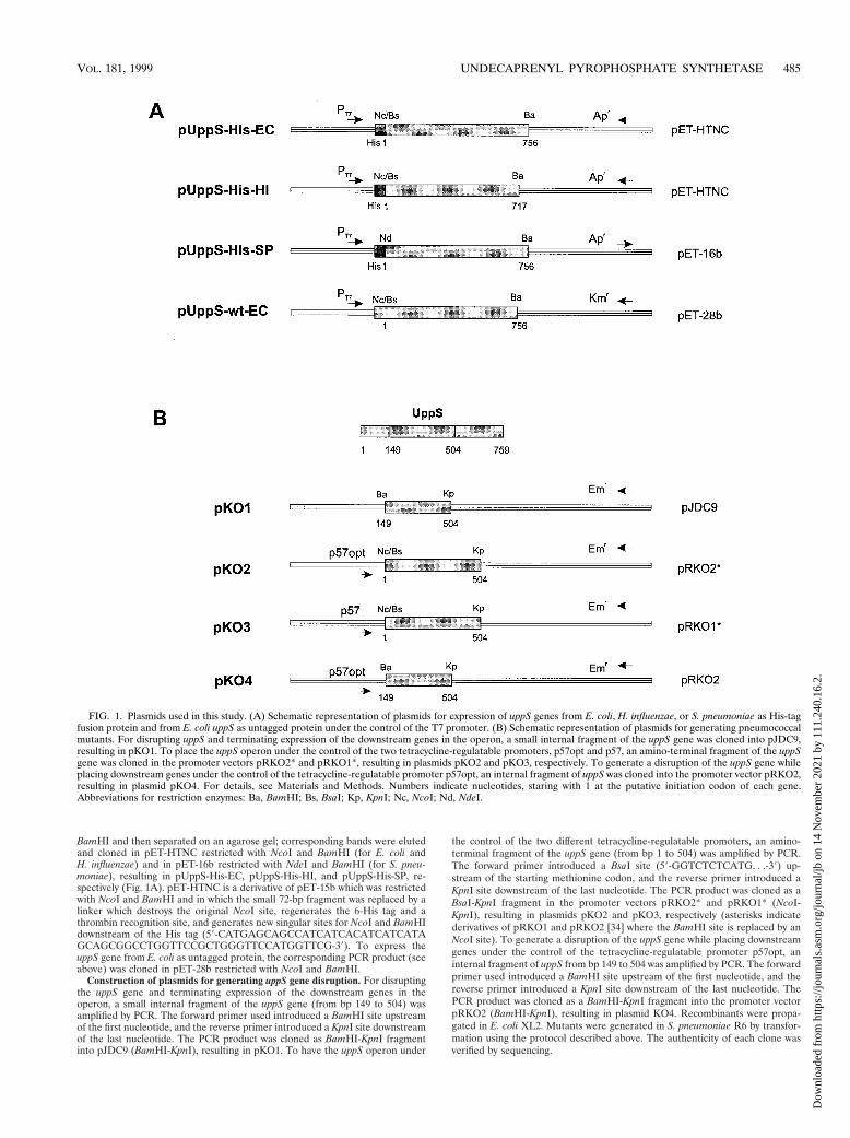
BamHI and then separated on an agarose gel; corresponding bands were elutedand cloned in pET-HTNC restricted with NcoI and BamHI (for E. coli andH. influenzae) and in pET-16b restricted with NdeI and BamHI (for S. pneu-moniae), resulting in pUppS-His-EC, pUppS-His-HI, and pUppS-His-SP, re-spectively (Fig. 1A). pET-HTNC is a derivative of pET-15b which was restrictedwith NcoI and BamHI and in which the small 72-bp fragment was replaced by alinker which destroys the original NcoI site, regenerates the 6-His tag and athrombin recognition site, and generates new singular sites for NcoI and BamHIdownstream of the His tag (59-CATGAGCAGCCATCATCACATCATCATAGCAGCGGCCTGGTTCCGCTGGGTTCCATGGTTCG-39). To express theuppS gene from E. coli as untagged protein, the corresponding PCR product (seeabove) was cloned in pET-28b restricted with NcoI and BamHI.
Construction of plasmids for generating uppS gene disruption. For disruptingthe uppS gene and terminating expression of the downstream genes in theoperon, a small internal fragment of the uppS gene (from bp 149 to 504) wasamplified by PCR. The forward primer used introduced a BamHI site upstreamof the first nucleotide, and the reverse primer introduced a KpnI site downstreamof the last nucleotide. The PCR product was cloned as BamHI-KpnI fragmentinto pJDC9 (BamHI-KpnI), resulting in pKO1. To have the uppS operon under
the control of the two different tetracycline-regulatable promoters, an amino-terminal fragment of the uppS gene (from bp 1 to 504) was amplified by PCR.The forward primer introduced a BsaI site (59-GGTCTCTCATG. . .-39) up-stream of the starting methionine codon, and the reverse primer introduced aKpnI site downstream of the last nucleotide. The PCR product was cloned as aBsaI-KpnI fragment in the promoter vectors pRKO2* and pRKO1* (NcoI-KpnI), resulting in plasmids pKO2 and pKO3, respectively (asterisks indicatederivatives of pRKO1 and pRKO2 [34] where the BamHI site is replaced by anNcoI site). To generate a disruption of the uppS gene while placing downstreamgenes under the control of the tetracycline-regulatable promoter p57opt, aninternal fragment of uppS from bp 149 to 504 was amplified by PCR. The forwardprimer used introduced a BamHI site upstream of the first nucleotide, and thereverse primer introduced a KpnI site downstream of the last nucleotide. ThePCR product was cloned as a BamHI-KpnI fragment into the promoter vectorpRKO2 (BamHI-KpnI), resulting in plasmid KO4. Recombinants were propa-gated in E. coli XL2. Mutants were generated in S. pneumoniae R6 by transfor-mation using the protocol described above. The authenticity of each clone wasverified by sequencing.
FIG. 1. Plasmids used in this study. (A) Schematic representation of plasmids for expression of uppS genes from E. coli, H. influenzae, or S. pneumoniae as His-tagfusion protein and from E. coli uppS as untagged protein under the control of the T7 promoter. (B) Schematic representation of plasmids for generating pneumococcalmutants. For disrupting uppS and terminating expression of the downstream genes in the operon, a small internal fragment of the uppS gene was cloned into pJDC9,resulting in pKO1. To place the uppS operon under the control of the two tetracycline-regulatable promoters, p57opt and p57, an amino-terminal fragment of the uppSgene was cloned in the promoter vectors pRKO2* and pRKO1*, resulting in plasmids pKO2 and pKO3, respectively. To generate a disruption of the uppS gene whileplacing downstream genes under the control of the tetracycline-regulatable promoter p57opt, an internal fragment of uppS was cloned into the promoter vector pRKO2,resulting in plasmid pKO4. For details, see Materials and Methods. Numbers indicate nucleotides, staring with 1 at the putative initiation codon of each gene.Abbreviations for restriction enzymes: Ba, BamHI; Bs, BsaI; Kp, KpnI; Nc, NcoI; Nd, NdeI.
VOL. 181, 1999 UNDECAPRENYL PYROPHOSPHATE SYNTHETASE 485
Dow
nloa
ded
from
http
s://j
ourn
als.
asm
.org
/jour
nal/j
b on
14
Nov
embe
r 20
21 b
y 11
1.24
0.16
.2.

RESULTS
Purification of native Upp synthetase. In earlier studies,different groups partially purified Upp synthetase from severalbacteria (4, 9, 10, 13, 35) by using two or three chromato-graphic steps, but bacterial genes for Upp synthetase were notidentified. As part of our strategy for isolating the gene fromE. coli, we purified the Upp synthetase by using a differentpurification scheme with an additional chromatographic puri-fication step to attain sufficient purity to identify clearly thecorresponding band by SDS-PAGE.
For the purification, we started with a total of 185 g (wet
weight) of E. coli BL21(DE3) paste collected in the logarithmicgrowth phase. The Upp synthetase of E. coli was purified asdescribed in Materials and Methods. To monitor Upp syn-thetase activity during the purification, we used the radioactiveUpp synthetase assay for the initial steps of the procedure.Later, when interfering endogenous phosphate had been re-moved, the coupled assay was used. For the first chromato-graphic step, we used a TSK-DEAE 650M column. Assayingthe fractions revealed two peaks of prenyltransferase activity.The activity in the first peak was absolutely Triton X-100 de-pendent, in contrast to the second peak; this result was inaccordance with observations published earlier (10). The assayused did not distinguish between Upp synthetase and Oppsynthetase activities, but we were able to show that recombi-nant Opp synthetase, which is not Triton X-100 dependent,coeluted from the column with the second peak of prenyltrans-ferase activity (data not shown). Therefore, the activity in thefirst peak was subjected to four additional chromatographicsteps using ceramic hydroxyapatite, TSK-Ether 5PW, HiLoadSuperdex 200, and heparin-Actigel ALD, respectively. On theHiLoad Superdex 200 column, Upp synthetase activity elutedat a molecular mass corresponding to the dimer. This is inagreement with earlier findings (10) that Upp synthetase is adimer. Despite these multiple purification steps, Upp syn-thetase was not purified to homogeneity (Fig. 2A), possiblybecause of a very low copy number of Upp synthetase per cell.
Identification of the gene encoding Upp synthetase. Becausewe could not purify the Upp synthetase to homogeneity andcould not assign the protein to a specific band on an SDS-polyacrylamide gel, it was necessary to further decrease thenumber of possible candidates. We labeled the native Uppsynthetase by cross-linking it specifically with a radiolabeledsubstrate analogue, [3H]DAFTP-GDP, and subjected the com-plex to SDS-PAGE (Fig. 2A, lane 3; Coomassie blue staining).Specificity was shown in a parallel sample in which the UVirradiation was carried out in the presence of 20 mM unlabeledFPP (Fig. 2A, lane 2). The entire gel was analyzed for radio-activity in two ways: (i) by direct autoradiography of the driedgel (Fig. 2B; exposure time, .45 days) and (ii) by cutting thelanes from a parallel gel in small 1-mm slices and counting theradioactivity in each slice (Fig. 2C). In both cases, an unam-biguous signal corresponding to a band of 29 kDa was de-tected. These results are in agreement to those of Baba et al.(10), who found, using similar methods, a protein with a size ofabout 30 kDa. However, with their purification scheme it wasnot possible to clearly assign an activity to a band on an SDS-polyacrylamide gel. The band of 29 kDa was cut out andsubjected to MALDI-MS analysis (see Materials and Meth-ods). The monoisotopic masses found were matched to thetheoretical peptide masses of the proteins of E. coli. Three
FIG. 2. SDS-PAGE of heparin column-purified Upp synthetase and radio-labeling with [3H]DAFTP-GDP. (A) Aliquots of the pool from the heparincolumn-purified Upp synthetase were radiolabeled with [3H]DAFTP-GDP andapplied to an SDS-polyacrylamide gel, which then was stained with Coomassieblue. Lanes: 1, protein molecular size markers as indicated on the left; 2, radio-labeled pool in the presence of 20 mM FPP; 3, radiolabeled pool without FPP.(B) Autoradiograph of the same gel. (C) On a duplicate gel, the region between20 and 40 kDa of lanes 2 and 3 was cut into 55 gel slices each, and theradioactivity in each slice was measured.
FIG. 3. Multiple amino acid sequence alignment of the 28 potential Upp synthetases which show homology to Upp synthetase from E. coli. Shaded areas representresidues that are identical in at least 21 of the 29 sequences (black) and similar amino acids (gray). Amino acid positions are indicated on the right. Sequences: E. coli(ECOLI; SWISS-PROT Q47675) and its homologues from H. influenzae (HAEIN; SWISS-PROT P44938), Helicobacter pylori (HELPY; SWISS-PROT P55984),S. pneumoniae (STRPN; ftp://ftp.tigr.org/pub/data/s_pneumoniae); Bacillus subtilis (BACSU; SWISS-PROT O31751), Aquifex aeolicus (AQUAE; TrEMBL O67291),Archaeoglobus fulgidus (ARCFU; TrEMBL O29049), Borrelia burgdorferi (BORBU; TrEMBL O51146), Mycobacterium tuberculosis (MYCTU; TrEMBL O53434),Mycobacterium leprae (MYCLE; SWISS-PROT P38119), Methanococcus jannaschii (METJA; SWISS-PROT Q58767), Streptomyces fradiae (STRFR; SWISS-PROTP20182), Pseudomonas aeruginosa (PSEAE; unannotated translation from EMBL entry D50811 for the cds gene), Pyrococcus horikoshii (PYRHO; TrEMBL O59258),Synechocystis (SYNY; SWISS-PROT Q55482), Chlamydia trachomatis (CHLTR; TrEMBL G3328883); Methanobacterium thermoauotrophicum (METTH; TrEMBLO26334), Neisseria gonorrhoeae (NEIGO; http://dna1.chem.ou.edu/gono.html), Saccharomyces cerevisiae (YEAST1; SWISS-PROT P35196), S. cerevisiae (YEAST2;SWISS-PROT Q03175), Caenorhabditis elegans (C.ELE; TrEMBL O18007) (amino acids 1 to 275 of 1893 amino acids), Enterococcus faecalis (ENTFA; ftp://ftp.tigr.org/pub/data/e_faecalis) (for this sequence, only the amino-terminal part is available), Corynebacterium glutamicum (CORGL; SWISS-PROT P38118), Treponema pallidum(TREPA; EMBL AE001235), Campylobacter jejuni (CAMJU; http://www.sanger.ac.uk/Projects/C_jejuni), Streptococcus pyogenes (STRPY; http://dna1.chem.ou.edu/strep.html), Brucella abortus (BRUAB; TrEMBL Q44626), Staphylococcus aureus (STAAU; (unreleased Hoffmann-La Roche data). The sequence information for thecarboxy-terminal part is available for the last six sequences only. The highly conserved regions of Upp synthetase are indicated by bars and numbered I to V. Thefollowing consensus patterns can be derived for each region: I, H-x-x-x-x-M-D-G-N-(RG)-R-(WYF)-A; II, G-H-x-x-G; III, (TS)-x-x-A-F-S-(ST)-E-N-x-x-R-x-x-x-E-V-x-x-L-M-x-L; IV, A-x-x-Y-G-G-R-x-(DE)-(LIVM)-x-x-A; V, (DE)-L-x-I-R-T-(SAG)-G-E-x-R-x-S-N-F-(ML)-(LMP)-W-Q-x-x-Y-(SAT)-E-x-x-F-x-x-x-x-W-P-(DE)-F.
486 APFEL ET AL. J. BACTERIOL.
Dow
nloa
ded
from
http
s://j
ourn
als.
asm
.org
/jour
nal/j
b on
14
Nov
embe
r 20
21 b
y 11
1.24
0.16
.2.

VOL. 181, 1999 UNDECAPRENYL PYROPHOSPHATE SYNTHETASE 487
Dow
nloa
ded
from
http
s://j
ourn
als.
asm
.org
/jour
nal/j
b on
14
Nov
embe
r 20
21 b
y 11
1.24
0.16
.2.
peptides out of 14 matched peptides derived from the proteindescribed in SWISS-PROT entry Q47675, YAES_ECOLI. Thesequence of the matching peptides covered 16% of the aminoacid sequence of the protein. The sequence coverage is anindication of confidence of protein identification. The selectedprotein showed the highest sequence coverage.
Deduced amino acid sequence and homology comparison.The deduced amino acid sequence of SWISS-PROT Q47675(YAES_ECOLI; designated here uppS [Upp synthetase]), con-tains 253 residues, which could encode a protein with a de-duced total molecular mass of 28,444 kDa. The genomic se-quence record indicates GTG as the initiation codon which liesimmediately upstream of the alternative start codon. To clarifywhether either one or both are used, we determined the N-terminal sequence of the purified Upp synthetase. We identi-fied both forms of Upp synthetase with one or two methionines(ratio of around 3:2). In the subsequent experiments (expres-sion of Upp synthetase), we took the GTG as the initiationcodon. Computer-assisted analysis and comparison of DNAsequences were performed with the BLAST program. Com-parison of the deduced Upp synthetase amino acid sequencefrom E. coli with entries in publicly available and in-housedatabases revealed a total of 28 sequences, including 25 frombacteria (7 of which were only partial sequences), 2 from S. cer-evisiae, and 1 from Caenorhabditis elegans. Alignment of theseproteins (Fig. 3) revealed strongly conserved regions where the
amino acids were identical or nearly identical in all 28 se-quences. No function has been experimentally determined forany of the proteins listed.
Cloning and overexpression in E. coli of the gene encodingUpp synthetase from different bacteria and purification ofrecombinant proteins. To verify the functionality of the Uppsynthetase, the genes from E. coli, H. influenzae, and S. pneu-moniae were cloned by PCR as N-terminal His-tag fusion pro-teins under the control of the T7 promoter (pET system) (Fig.1A). In addition to the fusion proteins, we also expressed theE. coli wt Upp synthetase to compare the activity with those ofthe His-tag fusion proteins. To generate the expression plas-mid, we cloned the DNA fragment encoding the E. coli uppSgene in pET-28b (Fig. 1A). The crude lysates of all four BL21(DE3)(pLysS) strains transformed with the corresponding ex-pression plasmids (induced by 2 mM isopropyl-b-D-thioga-lactopyranoside [IPTG] for 2 h) contained more than 1,000-fold-higher Upp synthetase activity than BL21(DE3)(pLysS)transformed with the expression vector without insert (data notshown).
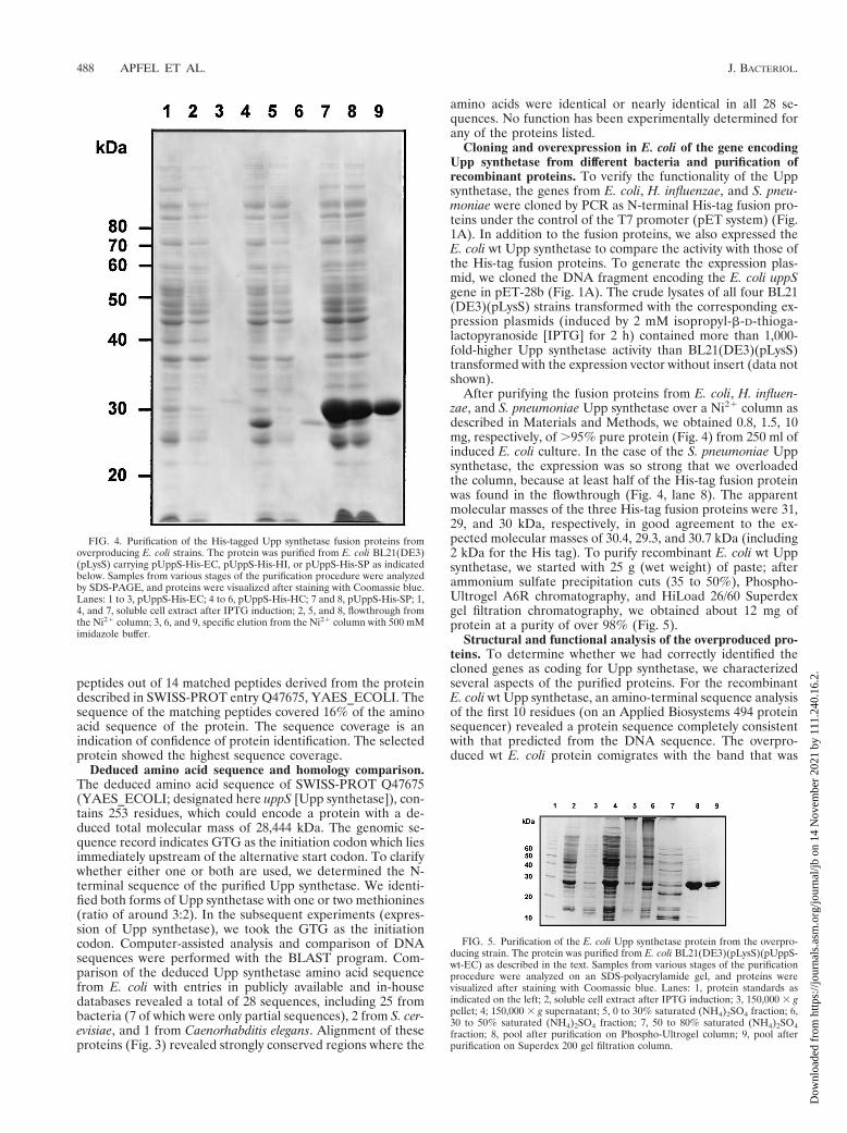
After purifying the fusion proteins from E. coli, H. influen-zae, and S. pneumoniae Upp synthetase over a Ni21 column asdescribed in Materials and Methods, we obtained 0.8, 1.5, 10mg, respectively, of .95% pure protein (Fig. 4) from 250 ml ofinduced E. coli culture. In the case of the S. pneumoniae Uppsynthetase, the expression was so strong that we overloadedthe column, because at least half of the His-tag fusion proteinwas found in the flowthrough (Fig. 4, lane 8). The apparentmolecular masses of the three His-tag fusion proteins were 31,29, and 30 kDa, respectively, in good agreement to the ex-pected molecular masses of 30.4, 29.3, and 30.7 kDa (including2 kDa for the His tag). To purify recombinant E. coli wt Uppsynthetase, we started with 25 g (wet weight) of paste; afterammonium sulfate precipitation cuts (35 to 50%), Phospho-Ultrogel A6R chromatography, and HiLoad 26/60 Superdexgel filtration chromatography, we obtained about 12 mg ofprotein at a purity of over 98% (Fig. 5).
Structural and functional analysis of the overproduced pro-teins. To determine whether we had correctly identified thecloned genes as coding for Upp synthetase, we characterizedseveral aspects of the purified proteins. For the recombinantE. coli wt Upp synthetase, an amino-terminal sequence analysisof the first 10 residues (on an Applied Biosystems 494 proteinsequencer) revealed a protein sequence completely consistentwith that predicted from the DNA sequence. The overpro-duced wt E. coli protein comigrates with the band that was
FIG. 4. Purification of the His-tagged Upp synthetase fusion proteins fromoverproducing E. coli strains. The protein was purified from E. coli BL21(DE3)(pLysS) carrying pUppS-His-EC, pUppS-His-HI, or pUppS-His-SP as indicatedbelow. Samples from various stages of the purification procedure were analyzedby SDS-PAGE, and proteins were visualized after staining with Coomassie blue.Lanes: 1 to 3, pUppS-His-EC; 4 to 6, pUppS-His-HC; 7 and 8, pUppS-His-SP; 1,4, and 7, soluble cell extract after IPTG induction; 2, 5, and 8, flowthrough fromthe Ni21 column; 3, 6, and 9, specific elution from the Ni21 column with 500 mMimidazole buffer.
FIG. 5. Purification of the E. coli Upp synthetase protein from the overpro-ducing strain. The protein was purified from E. coli BL21(DE3)(pLysS)(pUppS-wt-EC) as described in the text. Samples from various stages of the purificationprocedure were analyzed on an SDS-polyacrylamide gel, and proteins werevisualized after staining with Coomassie blue. Lanes: 1, protein standards asindicated on the left; 2, soluble cell extract after IPTG induction; 3, 150,000 3 gpellet; 4; 150,000 3 g supernatant; 5, 0 to 30% saturated (NH4)2SO4 fraction; 6,30 to 50% saturated (NH4)2SO4 fraction; 7, 50 to 80% saturated (NH4)2SO4fraction; 8, pool after purification on Phospho-Ultrogel column; 9, pool afterpurification on Superdex 200 gel filtration column.
488 APFEL ET AL. J. BACTERIOL.
Dow
nloa
ded
from
http
s://j
ourn
als.
asm
.org
/jour
nal/j
b on
14
Nov
embe
r 20
21 b
y 11
1.24
0.16
.2.

radiolabeled after purification of the native Upp synthetase(Fig. 2 and 5). In functional terms (Km values), all four proteinsbehave more or less identically in the direct and the coupledassays for Upp synthetase. They were absolutely Triton X-100and MgCl2 dependent, as exemplified in Fig. 6 for the E. coli wtUpp synthetase in the coupled assay. The optimal concentra-tion for Triton X-100 is between 0.01 and 0.1%, and that forMgCl2 is between 0.5 and 2 mM. Omission of either TritonX-100 or MgCl2 results in a residual activity of about 5 to 10%.Using reversed-phase thin-layer chromatography, we found
that the major products of the Upp synthetase reaction afterpotato acid phosphatase treatment had the same mobilityas authentic undecaprenol or decaprenol. We detected smallamounts of intermediate-length (C30 to C45) products (datanot shown).
Construction and analysis of uppS deletion mutants. Toinvestigate the function of the uppS gene, pneumococcal uppSgene mutants were obtained by site-directed insertional mu-tagenesis (29). A small internal gene fragment was cloned inplasmid pJDC9, which does not replicate in pneumococci
FIG. 6. Triton X-100 and MgCl2 dependency of purified E. coli wt Upp synthetase. The coupled assay for Upp synthetase was performed as described in Materialsand Methods. The Triton X-100 and MgCl2 concentration was varied in the indicated range. The highest activity in each experiment represents 100%. Results of arepresentative experiment are shown. Triplicate values varied from 5 to 10%.
FIG. 7. Schematic representation of the expected chromosomal uppS mutants of S. pneumoniae R6 with various gene disruption plasmids. Numbers indicate thenucleotides, starting with 1 at the initiating codon of each gene. P1 to P4 indicate the primers used in the PCR to confirm the correct integration of the plasmids.
VOL. 181, 1999 UNDECAPRENYL PYROPHOSPHATE SYNTHETASE 489
Dow
nloa
ded
from
http
s://j
ourn
als.
asm
.org
/jour
nal/j
b on
14
Nov
embe
r 20
21 b
y 11
1.24
0.16
.2.

(pKO1 [Fig. 1B]). The construct was transformed into S. pneu-moniae R6, and plasmid-encoded erythromycin resistance(Emr) was used to select for single homologous recombinationevents. However, no Emr transformants were obtained. Tofurther study the essentiality of the uppS gene for bacterialgrowth, we used a regulatable knockout system that allows theconditional expression of genes (34). The potential promoterregion in front of the streptococcal uppS gene was replaced bythe strong tetracycline-regulatable p57opt promoter (pKO2[Fig. 1B]). Transformation of S. pneumoniae R6 with this plas-mid resulted in many Emr transformants (S.p. pKO2 [Fig. 7]).Correct integration of the plasmid was confirmed by PCR anal-ysis (data not shown). No difference in growth rate was detect-able in the presence or absence of tetracycline (20 ng/ml),indicating that the basal expression from this promoter was suffi-cient to allow normal growth of the transformants. Even in theabsence of induction, the basal expression level from p57opt isrelatively high (34). Attempts to generate viable regulatableuppS knockout mutants by using the weaker and tighter pro-moter p57 (S.p. pKO3 [Fig. 7]) were successful only in thepresence of tetracycline, and we could isolate six colonies.Replating these colonies on sheep blood (3%) agar plates withand without tetracycline resulted in growth on the tetracycline-containing plates, whereas in absence of tetracycline no colo-nies were detected. In growth experiments in liquid culturesin the presence of tetracycline, S.p. pKO3 characteristicallyreached a plateau after 5 h with an OD620 of 0.5, starting at anOD620 of 0.08 to 0.1. Without tetracycline in the medium, theregulatable S.p. pKO3 knockouts grow normally for about 2 h,but then they slow down and after 4 h stop further growth atan OD620 of 0.2 to 0.3 (data not shown). The correctness ofintegration was checked by PCR. Using the primer combina-tion P1-P2 (Fig. 7), we found a band of the expected size (759bp) for S.p. R6 wt and S.p. pKO3. Using the primer combina-tion P1-P3 or P4-P2 (Fig. 7), we found a band of the expectedsize, 607 or 792 bp, respectively, only for the regulatable knock-out strains and no bands for S.p. R6 wt (data not shown).
From the S. pneumoniae sequence data, it is very likely thatthe uppS gene is the first gene in an operon. Therefore, it wasnecessary to exclude the possibility that the disruption of uppScauses a polar effect on the expression of downstream geneswhich themselves could be essential. Knowing that the tran-scription level of the constructs containing the p57opt pro-moter is sufficient for cell viability, we generated a constructwhich should disrupt the first gene, uppS, but place the down-stream genes under the control of the tetracycline-regulatablepromoter p57opt. No Emr transformants were obtained withthis plasmid (S.p. pKO4 [Fig. 7]), indicating that the first geneof the operon, uppS, is essential.
DISCUSSION
To gain further insight into the structure and function ofUpp synthetase, the only known bacterial enzyme which cata-lyzes the production of long-chain isoprenoids with cis-chainconfiguration, we attempted to identify and isolate the Uppsynthetase gene from E. coli. Our strategy was to make use ofthe recently available E. coli genomic sequence by preparing adatabase containing the masses of peptides produced by com-puter-simulated trypsin digestion of all E. coli proteins pre-dicted from the E. coli genomic sequence. The masses of pep-tides produced by experimental trypsin digestion of a purifiedprotein can be used to search the databank for proteins withsimilar profiles and so identify a candidate gene. However, thisapproach requires a sample of the protein of interest that islargely free from contaminating proteins. For this reason, it
was necessary to improve the purification scheme for the Uppsynthetase of E. coli described earlier (10), as analysis of ma-terial produced this way resulted in a list of several possiblecandidate genes (data not shown).
Subjecting 185 g (wet weight) of E. coli paste, after cellbreakage, to two differential centrifugation steps, ammoniumsulfate precipitation, and fractionation by five different chro-matographic steps yielded only approximately 50 to 150 mg ofUpp synthetase protein (judged from the band visible afterSDS-PAGE). Although this extensive purification scheme wasnot sufficient to purify the Upp synthetase to homogeneity, theremaining proteins in the mixture could be well resolved bySDS-PAGE. There are several possible explanations for thelow yield of Upp synthetase: (i) instability of the protein, whichseems to be unlikely since the recombinant protein is stable;(ii) losses during purification; and (iii) very low copy numberper cell. Assuming 10212 g (wet weight) per E. coli cell and anoptimistic recovery of 10%, there would be less than 300 to1,000 Upp synthetase molecules per cell. These numbers arecertainly within a possible range, because two steps later in thepathway of cell wall biosynthesis (after dephosphorylation ofUPP and transfer of the pentapeptide), the two membrane in-termediates in peptidoglycan metabolism, pentapeptide lipid Iand lipid II, are present in E. coli at cell copy numbers not high-er than 700 and 2,000, respectively (39). In addition, it is in-teresting that the ribosomal binding site has an extra G relativeto the AGGAG consensus and the spacing is rather long andnonoptimal (211 for GTG and 214 for the ATG), which mightcause a low level of transcription. Specific labeling of the puri-fied Upp synthetase preparation with [3H]DAFTP-GDP allowedus to positively identify the protein band on an SDS-polyacryl-amide gel that corresponds to Upp synthetase. MALDI-MSanalysis of this band’s trypsin-digested profile led to the iden-tification of a single candidate gene in E. coli (SWISS-PROTQ47675, YAES_ECOLI).
Comparison of the 28 predicted Upp synthetase sequenceswhich we identified in various databases revealed several strong-ly conserved regions (Fig. 3). We detected a single homologousprotein in all bacterial genomes for which the full sequenceinformation was available at the time with the exceptions ofMycoplasma genitalium and Mycoplasma pneumoniae, for whichno homologue was found. This is consistence with the absenceof other cell wall biosynthetic enzymes in these bacteria. Ineukaryotic genomes, two genes were identified in S. cerevisiaeand one was found in C. elegans. There are two entries inGenBank, AA086931 and W61940, that could encode N-ter-minal and C-terminal portions of a mouse UppS homolog. Inaddition, a partial sequence which showed very good homologyespecially to the strongly conserved regions at the carboxy-terminal end (boxes IV and V [see below]) was found in an in-house human database. Experiments to clone and express thefull-length human homologue and to characterize its enzymaticactivity are ongoing. It remains to be determined whether thatenzyme is identical to the dolichol synthetase in eukaryotes (1,11, 27), which synthesizes long-chain (up to C100) prenyl pyro-phosphates (also in a cis conformation).
Sequence comparison reveals at least five strongly conservedregions as well as some single conserved amino acids (outsideof the defined boxes), which very likely represent the active siteof the protein (Fig. 3). Box V is analogous to the Prosite con-sensus sequence described in Prosite document PS01066, which isannotated as “uncharacterized protein family UPF0015 signa-ture” and lists 13 sequences, all of which were also identified inour homology search.
It is interesting that in E. coli the gene immediately preced-ing uppS is dxr, a 1-deoxy-D-xylulose 5-phosphate reducto-
490 APFEL ET AL. J. BACTERIOL.
Dow
nloa
ded
from
http
s://j
ourn
als.
asm
.org
/jour
nal/j
b on
14
Nov
embe
r 20
21 b
y 11
1.24
0.16
.2.

isomerase catalyzing the formation of 2-C-methyl-D-erythritol4-phosphate in the terpenoid biosynthesis pathway (36). Thisclustering is not found in H. influenzae and S. pneumoniae.
The experiments for generating uppS gene knockouts pro-vide very strong evidence that the gene is essential for growthof S. pneumoniae. We cannot exclude the possibility that someother downstream gene(s) of the operon is essential as well.The tetracycline-regulatable knockouts illustrate the impor-tance of selecting a regulatable promoter of appropriatestrength for replacing the natural promoter of a gene andinterpreting results cautiously. If basal expression level of thepromoter is too high, no difference between induced and un-induced state is detectable. This is the most likely explanationfor the recovery of tetracycline-unresponsive transformants us-ing pKO2. Where the promoter is weaker, only in the inducedsituation is the expression level high enough for cell growth,and in the noninduced situation the cells eventually stop grow-ing. The results obtained with pKO4 and pKO3 provide verystrong evidence that the uppS gene is essential for growth ofS. pneumoniae. It remains to be shown whether this is also truefor the uppS genes of other bacteria.
Upp synthetase shows at least three differences from otherknown prenyltransferases. (i) All other synthetases, like Fppsynthetase, Opp synthetase, geranylgeranyl pyrophosphate syn-thetase, and hexaprenyl pyrophosphate synthetase, condensethe new isopentenyl unit in a trans configuration on the grow-ing chain. In contrast, Upp synthetase uses trans,trans-FPP asan initiating building block, whereas subsequent condensationsof isopentenyl units are in cis conformation. (ii) The primarystructure of Upp synthetase is completely different from thoseof the other prenyltransferases. (iii) All other known prenyl-transferases are believed to use the highly conserved aspartate-rich motif (DDxxD) for binding each pyrophosphate. This wasdemonstrated for avian Fpp synthetase by X-ray crystallogra-phy (37). Even isoprenoid cyclases (e.g., pentalenene synthase,epi-aristolochene synthase, and hopene synthase), which alsouse FPP as substrate but have little overall similarity to chainelongation prenyltransferases, use the DDxxD motif as a bind-ing site for the pyrophosphate, as demonstrated by X-ray crys-tallography (25, 33, 40). Upp synthetase is a new class of iso-prenoid synthetase because it has no such motif. We postulatethat there is another motif responsible for the binding of bothpyrophosphates. The only related and duplicated sequencemotif identified is a single aspartate (boldface) followed byan arginine (underlined) with a spacing of three amino acids(DE)GNxR and DLxLR (positions 25 to 29 and 189 to 193,respectively, in E. coli Upp synthetase) which could potentiallyserve as the pyrophosphate binding site. From a structuralpoint of view, it would be interesting to examine the three-di-mensional structure of Upp synthetase to see how naturesolved the problem of pyrophosphate binding in two differentways.
ACKNOWLEDGMENTS
We thank T. Hartung for preparing (E,E)-[1-3H]-(2-diazo-3-trifluo-ropropionyloxy)geranyl diphosphate, Marie-Francoise Takacs for help-ing in purification of the proteins, and Elizabeth Gillick and RichardMoon for critically reading the manuscript. We also acknowledgeOlivier Partouche, Bernard Rutten, and Christian Lacoste (all from F.Hoffmann-La Roche Ltd., Basel, Switzerland) for expert technicalassistance.
We acknowledge the Streptococcal Genome Sequencing Project,funded by USPHS/NIH grant AI38406, and B. A. Roe, S. P. Linn, L.Song, X. Yuan, S. Clifton, M. McShan, and J. Ferretti; the GonococcalGenome Sequencing Project, funded by USPHS/NIH grant AI38399,and B. A. Roe, S. P. Lin, L. Song, X. Yuan, S. Clifton, Tom Ducey, LisaLewis, and D. W. Dyer; and the C. jejuni Sequencing Group at the
Sanger Center and the E. faecalis early releases from The Institute forGenomic Research.
ADDENDUM
During the review process, a paper describing the Microco-cus luteus UppS protein and gene was published (32).
REFERENCES1. Adair, W. L., N. Cafmeyer, and R. K. Keller. 1984. Solubilization and char-
acterization of the long chain prenyltransferases involved in dolichyl phos-phate biosynthesis. J. Biol. Chem. 259:4441–4446.
2. Allen, C. M., M. V. Keenan, and J. Sack. 1976. Lactobacillus plantarumundecaprenyl pyrophosphate synthetase: purification and reaction require-ments. Arch. Biochem. Biophys. 175:236–248.
3. Allen, C. M., and J. D. Muth. 1977. Lipid activation of undecaprenyl pyro-phosphate synthetase from Lactobacillus plantarum. Biochemistry 16:2908–2915.
4. Allen, C. M. 1985. Purification and characterization of undecaprenyl-pyro-phosphate synthetase. Methods Enzymol. 110:281–299.
5. Asai, K., S. Fujisaki, Y. Nishimura, T. Nishino, K. Okada, T. Nakagawa, M.Kawamukai, and H. Matsuda. 1994. The identification of E. coli ispB (cel)gene encoding the octaprenyl diphosphate synthase. Biochem. Biophys. Res.Commun. 202:340–345.
6. Ausubel, F. M., R. Brent, R. E. Kingston, D. D. Moore, J. G. Seidman, J. A.Smith, and K. Struhl (ed.). 1997. Current protocols in molecular biology.Wiley Interscience, New York, N.Y.
7. Avery, O. T., C. M. MacLeod, and M. McCarty. 1944. Studies on the chem-ical nature of the substance inducing transformation of pneumococcal types.J. Exp. Med. 79:137–158.
8. Baba, T., and C. M. Allen. 1978. Substrate specificity of undecaprenyl pyro-phosphate synthetase from Lactobacillus plantarum. Biochemistry 17:5598–5604.
9. Baba, T., and C. M. Allen. 1980. Prenyl transferases from Micrococcus luteus:characterization of undecaprenyl pyrophosphate synthetase. Arch. Biochem.Biophys. 200:474–484.
10. Baba, T., J. Muth, and C. M. Allen. 1985. Photoaffinity labeling of undeca-prenyl pyrophosphate synthetase with a farnesyl pyrophosphate analogue.J. Biol. Chem. 260:10467–10473.
11. Bukhtiyarov, Y. E., Y. A. Shabalin, and I. S. Kulaev. 1993. Solubilization andcharacterization of dehydrodolichyl diphosphate synthase from the yeastSaccharomyces carlbergensis. J. Biochem. 113:721–728.
12. Chen, J. D., and D. A. Morrison. 1988. Construction and properties of a newinsertion vector, pJDC9, that is protected by transcriptional terminators anduseful for cloning of DNA of Streptococcus pneumoniae. Gene 64:155–164.
13. Christenson, J. G., S. K. Gross, and P. W. Robbins. 1969. Enzymatic syn-thesis of the antigen carrier lipid. J. Biol. Chem. 244:5436–5439.
14. Collins, M. D., and D. Jones. 1981. Distribution of isoprenoid quinonestructural types in bacteria and their taxonomic implementations. Microbiol.Rev. 45:316–354.
15. Devereux, J., P. Haeberli, and O. Smithies. 1984. A comprehensive set ofsequence analysis programs for the VAX. Nucleic Acids Res. 12:387–395.
16. Fountoulakis, M., and H. Langen. 1997. Identification of proteins by matrix-assisted laser desorption ionization-mass spectrometry following in-gel di-gestion in low-salt, nonvolatile buffer and simplified peptide recovery. Anal.Biochem. 250:153–156.
17. Fujisaki, S., T. Nishino, and H. Katsuki. 1986. Isoprenoid synthesis inEscherichia coli. Separation and partial purification of four enzymes involvedin the synthesis. J. Biochem. 99:1327–1337.
18. Fujisaki, S., H. Hara, Y. Nishimura, K. Horiuchi, and T. Nishino. 1990.Cloning and nucleotide sequence of the ispA gene responsible for farnesyldiphosphate synthase activity in E. coli. J. Biochem. 108:995–1000.
19. Havarstein, L. S., G. Coomaraswamy, and D. A. Morrison. 1995. An un-modified heptadecapeptide pheromone induces competence for genetictransformation in Streptococcus pneumoniae. Proc. Natl. Acad. Sci. USA 92:11140–11144.
20. Keenan, M. V., and C. M. Allen. 1974. Characterization of undecaprenylpyrophosphate synthetase from Lactobacillus plantarum. Arch. Biochem.Biophys. 161:375–383.
21. Kodama, T., K. Fukui, and K. Kometani. 1986. The initial phosphate burstin ATP hydrolysis by myosin and subfragment-1 as studied by a modifiedmalachite green method for determination of inorganic phosphate. J. Bio-chem. 99:1465–1472.
22. Kurokawa, T., K. Ogura, and S. Seto. 1971. Formation of polyprenyl phos-phates by a cell-free enzyme of Micrococcus lysodeikticus. Biochem. Biophys.Res. Commun. 45:251–257.
23. Lacks, S. 1966. Integration efficiency and genetic recombination in pneumo-coccal transformation. Genetics 53:207–235.
24. Laemmli, U. K. 1970. Cleavage of structural proteins during the assembly ofthe head of bacteriophage T4. Nature 227:680–685.
25. Lesburg, C. A., G. Zhai, D. E. Cane, and D. W. Christianson. 1997. Crystal
VOL. 181, 1999 UNDECAPRENYL PYROPHOSPHATE SYNTHETASE 491
Dow
nloa
ded
from
http
s://j
ourn
als.
asm
.org
/jour
nal/j
b on
14
Nov
embe
r 20
21 b
y 11
1.24
0.16
.2.

structure of pentalenene synthase: mechanistic insights on terpenoid cycliza-tion reactions in biology. Science 277:1820–1824.
26. Liu, J., R. D. Stipanovic, and C. R. Benedict. 1996. Synthesis of a tritiumlabeled photolabile analogue of farnesyl-diphosphate: (E,E)-[1-3H]-(2-diazo-3-trifluoropropionyloxy)geranyl diphosphate. J. Labelled Compd. Ra-diopharm. 38:139–148.
27. Matsuoka, S., Sagami, H., Kurisaki, A., and K. Ogura. 1991. Variable prod-uct specificity of microsomal dehydrodolichyl diphosphate synthase from ratliver. J. Biol. Chem. 266:3464–3668.
28. Ogura, K., T. Koyama, and H. Sagami. 1997. Polyprenyl diphosphate syn-thases. Subcell. Biochem. 28:57–87.
29. Pozzi, G., M. R. Oggioni, R. Manganelli, and P. Plevani. 1991. Geneticmanipulation of streptococci by chromosomal integration of recombinantDNA, p. 59–61. In G. M. Dunny, P. P. Cleary, and L. L. McKay (ed.),Genetics and molecular biology of streptococci, lactococci, and enterococci.Washington, D.C.
30. Sambrook, R., E. Fritsch, and T. Maniatis. 1989. Molecular cloning: alaboratory manual, 2nd ed. Cold Spring Harbor Laboratory, Cold SpringHarbor, N.Y.
31. Sanger, F., S. Nicklen, and A. R. Coulson. 1977. DNA sequencing withchain-terminating inhibitors. Proc. Natl. Acad. Sci. USA 71:5463–5467.
32. Shimizu, N., T. Koyama, and K. Ogura. 1998. Molecular cloning, expression,and purification of undecaprenyl diphosphate synthase: no sequence simi-larity between E- and Z-prenyl diphosphate synthases. J. Biol. Chem. 273:19476–19481.
33. Starks, C. M., K. Back, J. Chappell, and J. P. Noel. 1997. Structural basis forcyclic terpene biosynthesis by tobacco 5-epi-aristolochene synthase. Science277:1815–1820.
34. Stieger, M., B. Wohlgensinger, M. Kamber, R. Lutz, and W. Keck. Integra-tional plasmids for the tetracycline regulated expression of genes in Strepto-coccus pneumoniae. Gene, in press.
35. Takahashi, I., and K. Ogura. 1982. Prenyltransferase of Bacillus subtilis:undecaprenyl pyrophosphate synthetase and geranylgeranyl pyrophosphatesynthetase. J. Biochem. 92:1527–1537.
36. Takahashi, S., T. Kuzuyama, H. Watanabe, and H. Seto. 1998. A 1-deoxy-D-xylulose 5-phosphate reductoisomerase catalyzing the formation of 2-C-methyl-D-erythritol 4-phosphate in an alternative nonmevalonate pathwayfor terpenoid biosynthesis. Proc. Natl. Acad. Sci. USA 95:9879–9884.
37. Tarshis, L. C., P. J. Proteau, B. A. Kellogg, J. C. Sacchettini, and C. D.Poulter. 1996. Regulation of product chain length by isoprenyl diphosphatesynthases. Proc. Natl. Acad. Sci. USA 93:15018–150223.
38. Umbreit, J. N., and J. L. Strominger. 1972. Isolation of the lipid intermediatein peptidoglycan biosynthesis from Escherichia coli. J. Bacteriol. 122:1306–1309.
39. van Heijenoort, Y., M. Gomez, M. Derrien, J. Ayala, and J. van Heijenoort.1992. Membrane intermediates in the peptidoglycan metabolism of Esche-richia coli: possible roles of PBP 1b and PBP 3. J. Bacteriol. 174:3549–3557.
40. Wendt, K. U., K. Poralla, and G. E. Schulz. 1997. Structure and function ofa squalene cyclase. Science 277:1811–1815.
492 APFEL ET AL. J. BACTERIOL.
Dow
nloa
ded
from
http
s://j
ourn
als.
asm
.org
/jour
nal/j
b on
14
Nov
embe
r 20
21 b
y 11
1.24
0.16
.2.