use of foliar and stem anatomical characters in the
TRANSCRIPT
Available online at http://ajol.info/index.php/ijbcs
Int. J. Biol. Chem. Sci. 8(5): 2232-2243, October 2014
ISSN 1997-342X (Online), ISSN 1991-8631 (Print)
© 2014 International Formulae Group. All rights reserved. DOI : http://dx.doi.org/10.4314/ijbcs.v8i5.26
Original Paper http://indexmedicus.afro.who.int
Use of foliar and stem anatomical characters in the identification of Ludwigia
(Linn.) species in Nigeria
A. E. FOLORUNSO*, K. F. ADELALU and Matthew OZIEGBE
Department of Botany, Faculty of Science, Obafemi Awolowo University, Ile-Ife, Nigeria. *Corresponding author; E-mail: [email protected]; Tel.: +234 8035068602
ABSTRACT
Foliar and stem anatomical characters that could be used for the identification of nine species of Ludwigia L. viz., Ludwigia abyssinica A. Rich., L. adscendens subsp. diffusa (Forssk)., L. decurrens variety A Walter., L. decurrens variety B Walter., L. erecta (Linn.) Hara, L. hyssopifolia (G.Don) Exell., L. leptocarpa (Nutt.), L. octovalvis variety brevisepala (Jacq.) Raven and L. octovalvis variety linearis (Jacq.) Raven in Nigeria are reported. This is envisaged to facilitate the management of these Ludwigia species; most importantly the invasive species and also to reduce the fluctuations in their taxonomic classifications. Fresh leaves and matured stem of these nine (9) species of Ludwigia from South-western Nigeria were subjected to anatomical study under Light Microscope. The occurrence of hypoamphistomatic stomata and several other stomata types such as anomocytic, staurocytic, anisocytic, cyclocytic and laterocytic in these advanced species of Ludwigia could be responsible for their invasiveness in the environments. Similarly, the mean vessel diameter in Ludwigia species studied represents invasive character; the species with higher mean vessel diameter are likely to be more invasive in their habits. Baseline information on the stem anatomy of the Ludwigia species has been provided; in addition, diagnostic characters for easy identification and classification of the taxa are also reported. The leaf and stem anatomical characters in the Ludwigia species are constant despite their environmental plasticity. © 2014 International Formulae Group. All rights reserved.
Keywords: Ludwigia, epidermal, stomata, morphology, diagnostic, invasive. INTRODUCTION
Reports from literature tag Ludwigia as an invasive species, they are known to be invasive in most parts of the world, serving as a threat to native plant species and blocking navigational channels (Oziegbe and Faluyi, 2011). An introduced species becomes a ‘transformer invader’ when its development produces serious ecological and economic problems (Richardson et al., 2000). The main consequence on ecosystems is the decline of
native species, or even the extinction of rare and vulnerable taxa (Burke and Grime, 1996). A set of advantageous biological traits characterizes most invasive plants: competitive life strategy (Pysek et al., 2012), large ecological tolerance, vegetative growth, fast growth, longevity, high seed production, good germination, plasticity of breeding systems, allelopathy, photosynthesis in C4 and polyploidy (Verlaque et al., 2002). All these assets are rarely found together on a given

A. E. FOLORUNSO et al. / Int. J. Biol. Chem. Sci. 2232-2243, 2014
2233
plant species, but the presence of one or two among them can sometimes be sufficient (Muller, 2002). However, the alien Ludwigia taxa possess most of these characters, which might explain their expansion in Nigeria and their classification among the 200 most aggressive world plant invaders (Oziegbe et al., 2010). Six month old seeds of Ludwigia species are more likely to contribute to plant populations than the freshly shed seeds germination media investigated. Also, the production of high number of seeds, enable them to compete and adapt in different ecological conditions (Oziegbe et al., 2011).
The management of these invasive species requires their precise identification, but this appears to be extremely complex (Dandelot et al., 2005). Dutartre et al. (2004) reported that Ludwigia species are phenotypically plastic, such that their growth forms vary under different environmental conditions, which often complicates species identification and has led to a number of fluctuations in their taxonomic classifications. Moreover, the anatomical characteristics of Ludwigia are sparingly mentioned in the general account of Metcalfe and Chalk (1979) and earlier reports on the foliar epidermal morphology and stem anatomy of the genus elsewhere in West Africa are lacking.
In this study, foliar and stem anatomical characters of Ludwigia species were studied to report taxonomic and diagnostic characters which could probably be used for their identification; characters that may be responsible for their invasiveness will also be reported.
MATERIALS AND METHODS Collection of specimen
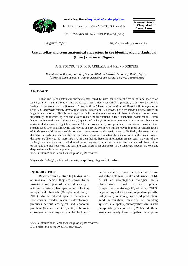
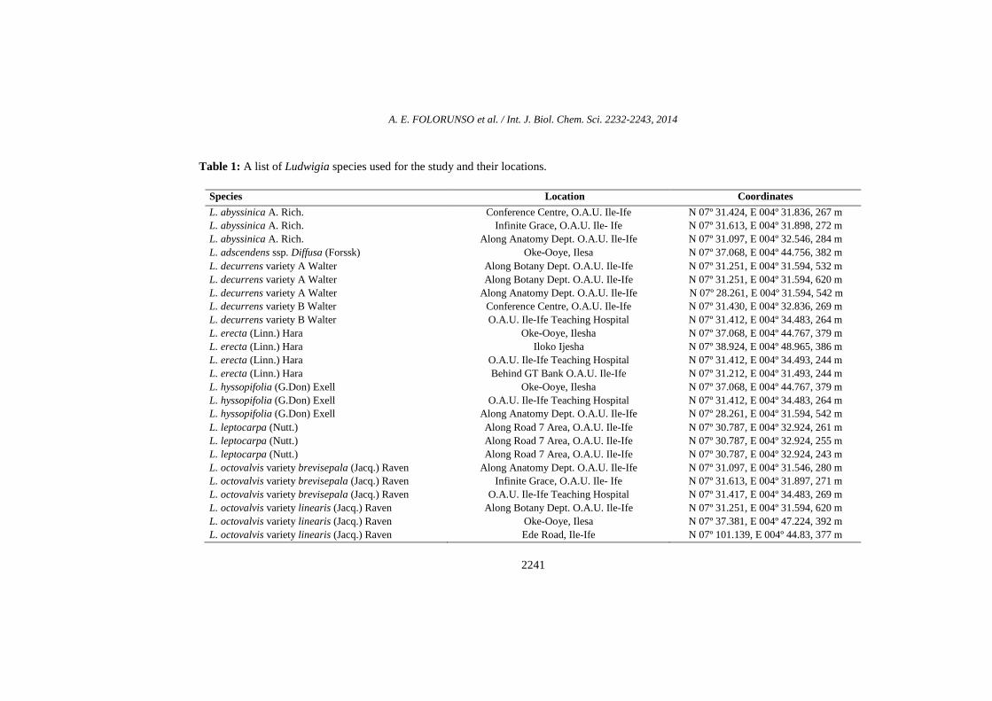
Fresh leaves of nine (9) species of Ludwigia common to Nigeria in the family Onagraceae were collected from the South-western Nigeria (Table 1). Figure 1 shows the morphological habits of the Ludwigia species studied. The altitudes and geographical coordinates of the localities were taken using a GPS device. Species identification and
confirmation was done at the Herbarium of Obafemi Awolowo University Campus (IFE) and Forestry Research Institute of Nigeria Herbarium (FHI), Ibadan.
Foliar anatomical study
Portions of the leaves were taken from the median part (midway between the tip and the base) from ten accessions of each species. These were put into Jeffrey’s maceration mixture (10% chromic acid and concentrated hydrochloric acid) and kept in oven at 60 ºC for about 15 minutes. Each sample was then washed thoroughly in 5 changes of water. The adaxial and abaxial epidermis was separated by means of dissecting needle and forceps. The epidermal surfaces were stained in 1% safranin O for about five minutes, washed with 4 changes of water to remove excess stain and then temporary mounts were made in 25% glycerol. Stomatal index was calculated according to Dilcher (1974).
Stem anatomical study
For the stem anatomical study, fine sections (8 µm - 15 µm thick) of the transverse sections, tangential longitudinal sections, and radial longitudinal section, were done using a Reichert microtome. The sections for the stem were stained with 1% aqueous solution of safranin “O” and counter stained with 1% aqueous solution of alcian blue and mounted in 25% glycerine on clean glass slide for microscopic studies. Quantitative characters of the stem were measured with the aid of an ocular micrometer inserted in the eyepiece of the microscope. These measurements were later multiplied by the ocular constant with respect to the power under which they were taken and photomicrographs of the slides were taken with Leitz camera mounted on Dialux Research Microscope. RESULTS Ludwigia abyssinica A. Rich.
The abaxial epidermal cells are irregular with sinuous to undulating anticlinal

A. E. FOLORUNSO et al. / Int. J. Biol. Chem. Sci. 2232-2243, 2014
2234
wall and the periclinal wall is smooth (Figure 2). The epidermal cells are 22.5 µm – 47.5 µm long and 10.0 µm – 25.0 µm broad. Stomata being anomocytic are elliptical in shape, size ranges between 17.5 µm – 27.5 µm long and 12.5 µm – 17.5 µm broad, the stomata index is 28.9% (Figure 2). Trichomes are simple and non glandular. Styloids are present. On the adaxial surface, the epidermal cells are irregular with straight to undulating anticlinal wall and the periclinal wall is smooth. The epidermal cell size ranges from 25.0 µm – 60.0 µm long and 15 µm – 27.5 µm broad. Anisocytic and staurocytic stomata are present, they are elliptical in shape and their size ranges between 20.0 µm – 25.0 µm long and 12.5 µm – 27.5 µm in broad, the stomata index is 6.8% (Figure 2). Trichomes are simple and non glandular.
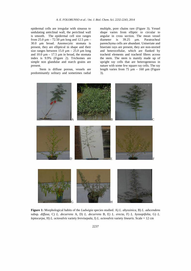
The stem is diffuse porous, vessels are predominantly solitary and sometimes in radial multiple, pore chains are present (Figure 3). Vessel shape varies from elliptic to circular to angular in cross section and the mean vessel diameter is 67.5 µm. Starch grains are present within the paratracheal parenchyma cells. The rays are upright, largely uniseriate, heterocellular and non – storied. Ray length varies from 58.0 µm – 198.0 µm (Figure 3).
L. adscendens subsp. diffusa (Forssk) The abaxial epidermal cells are
irregular with sinuous to undulating anticlinal wall and the periclinal wall is smooth (Figure 2). The epidermal cells are 25.0 µm – 52.5 µm long and 10.0 µm – 25.0 µm broad. Stomata types are anomocytic and staurocytic, they are elliptical in shape, size ranges between 17.5 µm – 25.0 µm long and 10.0 µm – 15.0 µm broad, the stomata index is 9.9% (Figure 2). Trichomes are simple non glandular. Raphides are present. On the adaxial surface, the epidermal cells are irregular with sinuous to undulating anticlinal wall and the periclinal wall is smooth. The epidermal cell size ranges from 25.0 µm – 52.5 µm long and 10.0 µm – 25.0 µm broad. The stomata is anomocytic, they are elliptical in shape and
their size ranges between 20.0 µm – 22.5 µm long and 12.5 µm – 15.0 µm in broad, the stomata index is 11.0% (Figure 2). Trichomes are simple and non glandular, prismatic crystals are present.
The stem is diffuse porous, vessels are majorly solitary and sometimes radial multiple, pore chains present (Figure 3). Vessel shape varies from elliptic to circular in cross section; the mean vessel diameter is 58.0 µm. The paratracheal parenchyma cells within the pith are occupied with starch grains. The stem shows uniseriate rays with adjourning vessel elements. The rays are upright, non – storied and heterocellular in nature. The ray length varies from 37.0 µm – 84.0 µm. There are tracheid fibres and square cells across the stem (Figure 3). L. decurrens variety A Walter
The abaxial epidermal cells are irregular with straight to undulating anticlinal wall, the periclinal wall is smooth (Figure 2). The epidermal cells are 27.5 µm – 57.5 µm long and 7.5 µm – 30.0 µm broad. Stomata being anomocytic are elliptical in shape, size ranges between 12.5 µm – 22.5 µm long and 7.5 µm – 15.0 µm broad, the stomata index is 29.1% (Figure 2). Scale trichome is present. On the adaxial surface, the epidermal cells are irregular with wavy to undulating anticlinal wall, the periclinal wall is smooth. The epidermal cell size ranges from 32.5 µm – 62.5 µm long and 12.5 µm – 25.0 µm broad. Anisocytic, laterocytic and staurocytic stomata are present, they are elliptical in shape and their size ranges between 12.5 µm – 22.5 µm long and 7.5 µm – 15.0 µm in broad, the stomata index is 13.4% (Figure 2). Prismatic crystals and styloids are present.
Stem is diffuse porous, solitary vessels are predominant with few pore chains across the section, vessel pores vary from circular to elliptic in shape (Figure 3). The mean vessel diameter is 92.8 µm. Parenchyma cells are paratracheal and polygonal. Raphides and medullary bundles are present within the parenchyma cells in pith. The rays are upright, largely uniseriate, heterocellular and non –

A. E. FOLORUNSO et al. / Int. J. Biol. Chem. Sci. 2232-2243, 2014
2235
storied. The ray length varies from 32.0 µm – 110.0 µm (Figure 3). There are tracheid fibres across the stem.
L. decurrens variety B Walter The abaxial epidermal cells are
irregular with sinuous to undulating anticlinal wall, the periclinal wall is smooth (Figure 2). The epidermal cells are 30.0 µm – 65.0 µm long and 12.5 µm – 30.0 µm broad. Stomata being anomocytic, are elliptical in shape, size ranges between 15.0 µm – 25.0 µm long and 10.0 µm – 15.0 µm broad, the stomata index is 23.8% (Figure 2). Starch grains are present. On the adaxial surface, the epidermal cells are irregular with wavy to undulating anticlinal wall, the periclinal wall is smooth. The epidermal cell size ranges from 25.0 µm – 60.0 µm long and 10.0 µm – 42.5 µm broad. Anomocytic stomata is present, they are elliptical in shape and their size ranges between 12.5 µm – 25.0 µm long and 10.0 µm – 15.0 µm in broad, the stomata index is 10.7% (Figure 2). Starch grains are present with scale trichome.
Stem is diffuse porous, vessels are predominantly solitary and sometimes cluster, pore chains are present (Figure 3). Vessel shape varies from elliptic to circular to angular in cross section; the mean vessel diameter is 81.0 µm. Paratracheal parenchyma cells are abundant. Starch grains occupied the pith and medullary bundles are present within the pith. The stem shows both uniseriate rays and biseriate rays with adjourning tracheid elements. The rays are non – storied, the cells of the rays are homocellular in uniseriate rays and heterocellular in biseriate rays. The ray length varies from 37.0 µm – 98.0 µm (Figure 3). There are tracheid fibres across the stem. The stem is majorly made up of upright ray cells that are heterogeneous in nature with some few square ray cells. There are also tracheids fibres across the section.
L. erecta (Linn.) Hara The abaxial epidermal cells are
irregular with sinuous to undulating anticlinal wall, the periclinal wall is smooth (Figure 2). The epidermal cells are 32.5 µm – 70.0 µm
long and 12.5 µm – 25.0 µm broad. Stomata being staurocytic, are elliptical in shape, size ranges between 12.5 µm – 30 µm long and 7.5 µm – 15 µm broad, the stomata index is 27.9% (Figure 2). Trichomes are simple non glandular. Raphides are present. On the adaxial surface, the epidermal cells are irregular with sinuous to undulating anticlinal wall, the periclinal wall is smooth. The epidermal cell size ranges from 25.0 µm – 70.0 µm long and 10 µm – 30.0 µm broad. Anomocytic stomata is present, they are elliptical in shape and their size ranges between 15.0 µm – 25.0 µm long and 10.0 µm – 15.0 µm in broad, the stomata index is 17.1% (Figure 2). Raphides and starch grains are present.
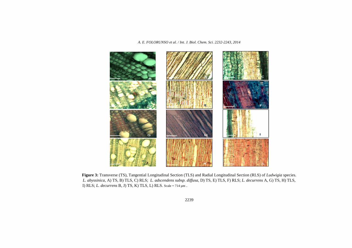
Stem is diffuse porous, and made up of both solitary and multiple vessels, their shapes vary from circular to elliptic and sometime angular across the section (Figure 3). The mean vessel diameter is 85.5 µm. Paratracheal parenchyma cells are abundant. The stem shows both uniseriate rays and biseriate rays with adjourning tracheid elements. The rays are upright, non – storied, homocellular in uniseriate rays and heterocellular in biseriate rays. The ray length varies from 33.0 µm – 125.0 µm (Figure 3). There are tracheid fibres across the stem.
L. hyssopifolia (G.Don) Exell The abaxial epidermal cells are
irregular with sinuous to undulating anticlinal wall, the periclinal wall is smooth (Figure 2). The epidermal cells are 25.5 µm – 55.0 µm long and 10.0 µm – 45.0 µm broad. Stomata being anomocytic, are elliptical in shape, size ranges between 12.5 µm – 25.0 µm long and 10.0 µm – 15 µm broad, the stomata index is 31.8%. Styloids are present. On the adaxial surface, the epidermal cells are irregular with sinuous to undulating anticlinal wall, the periclinal wall is smooth (Figure 2). The epidermal cell size ranges from 25.0 µm – 70.0 µm long and 10 µm – 22.5 µm broad. Anomocytic stomata is present, they are elliptical in shape and their size ranges between 15.0 µm – 22.5 µm long and 10.0 µm

A. E. FOLORUNSO et al. / Int. J. Biol. Chem. Sci. 2232-2243, 2014
2236
– 15.0 µm in broad, the stomata index is 16.1% (Figure 2). Trichomes are simple non glandular.
Stem is diffuse porous, and made up of solitary and sometimes radial multiple vessels, pore chains present (Figure 3). Vessel shape varies from elliptic to circular in cross section; the mean vessel diameter is 58.25 µm. Paratracheal parenchyma cells are abundant and those within the pith are occupied with some raphides, monohydric crystal, druses and starch grains. The non-storied rays which are uniseriate and biseriate are flanked by tracheid elements. These rays are heterocellular and upright with adjourning tracheid vessel elements and tracheid fibres. The ray length varies from 17 µm - 57 µm (Figure 3).
L. leptocarpa (Nutt.) The abaxial epidermal cells are
irregular with sinuous to undulating anticlinal wall, the periclinal wall is smooth (Figure 2). The epidermal cells are 20.0 µm – 42.5 µm long and 10.0 µm – 37.5 µm broad. Stomata being anisocytic and staurocytic, are elliptical in shape, size ranges between 17.5 µm – 25.0 µm long and 12.5 µm – 20.0 µm broad, the stomata index is 18.6% (Figure 2). Trichomes are simple non glandular. On the adaxial surface, the epidermal cells are irregular with straight to sinuous to undulating anticlinal wall, the periclinal wall is smooth. The epidermal cell size ranges from 20.0 µm – 47.50 µm long and 10.0 µm – 25.0 µm broad. Anomocytic stomata is present, they are elliptical in shape and their size ranges between 17.5 µm – 25.0 µm long and 12.5 µm – 15.0 µm in broad, the stomata index is 1.8% (Figure 2). Both non glandular unicellular and non glandular multicellular trichomes are present and numerous.
Stem is diffuse porous, vessels are predominantly solitary and sometimes cluster, and pore chains are rare (Figure 3). On the epidermis are multicellular non glandular trichomes. Vessel shape varies from elliptic to circular in cross section. The mean vessel diameter is 125.75 µm. The rays are upright, heterocellular, uniseriate and multiseriate with
adjourning tracheal fibres. The rays are non – storied with varying cell sizes and the ray length varies from 49 µm - 215 µm (Figure 3). L. octovalvis variety brevisepala (Jacq.) Raven
The abaxial epidermal cells are irregular vary from straight to sinuous to undulating anticlinal wall, the periclinal wall is striated (Figure 2). The epidermal cells are 25.0 µm – 67.5 µm long and 10.0 µm – 32.5 µm broad. Stomata being anomocytic and cyclocytic, are elliptical in shape, size ranges between 17.5 µm – 50.0 µm long and 7.5 µm – 52.5 µm broad, the stomata index is 23.4% (Figure 2). Trichomes are simple non glandular. Raphides are present and starch grains are present. On the adaxial surface, the epidermal cells are irregular with straight to undulating anticlinal wall, the periclinal wall is smooth. The epidermal cell size ranges from 22.5 µm – 57.50 µm long and 12.5 µm – 52.5 µm broad. Anomocytic and anisocytic stomata are present, they are elliptical in shape and their size ranges between 15.0 µm – 27.5 µm long and 7.5 µm – 17.5 µm in broad, the stomata index is 14.8% (Figure 2). Trichomes are simple non glandular and starch grains are present.
Stem is diffuse porous. On the section are many pore chains and sometime radial multiple vessels, stem is made up of solitary vessels which are not circular but vary from elliptic to angular in shape (Figure 3). The mean vessel diameter is 131.5 µm. There are uniseriate rays across the section. On the epidermis are multicellular non glandular trichomes. The rays are uniseriate, non-storied and homocellular with adjourning tracheid elements, upright ray cells that are homogeneous in nature, ray length varies from 46 µm - 123 µm (Figure 3).
L. octovalvis variety linearis (Jacq.) Raven The abaxial epidermal cells are
irregular varies from sinuous to undulating anticlinal wall, the periclinal wall is striated (Figure 2). The epidermal cells are 25.0 µm – 77.5 µm long and 12.5 µm – 30.0 µm broad. Stomata being staurocytic, are elliptical in shape, size ranges between 15.0 µm – 25.0 µm long and 10.0 µm – 15.0 µm broad, the stomata index is 25.6% (Figure 2). Trichomes are simple non glandular. Raphides and starch grains are present. On the adaxial surface, the
A. E. FOLORUNSO et al. / Int. J. Biol. Chem. Sci. 2232-2243, 2014
2237
epidermal cells are irregular with sinuous to undulating anticlinal wall, the periclinal wall is smooth. The epidermal cell size ranges from 25.0 µm – 72.50 µm long and 12.5 µm – 30.0 µm broad. Anomocytic stomata is present, they are elliptical in shape and their size ranges between 15.0 µm – 25.0 µm long and 10.0 µm – 17.5 µm in broad, the stomata index is 9.9% (Figure 2). Trichomes are simple non glandular and starch grains are present.
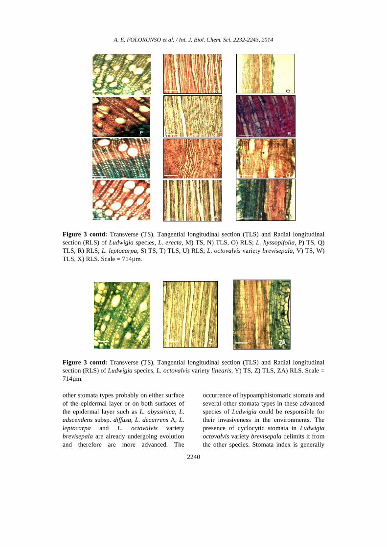
Stem is diffuse porous, vessels are predominantly solitary and sometimes radial
multiple, pore chains rare (Figure 3). Vessel shape varies from elliptic to circular to angular in cross section. The mean vessel diameter is 39.25 µm. Paratracheal parenchyma cells are abundant. Uniseriate and biseriate rays are present, they are non-storied and heterocellular, which are flanked by tracheid elements and tracheid fibres across the stem. The stem is mainly made up of upright ray cells that are heterogeneous in nature with some few square ray cells. The ray length varies from 75 µm – 160 µm (Figure 3).
A B C
D E F
G H I Figure 1: Morphological habits of the Ludwigia species studied. A) L. abyssinica, B) L. adscendens subsp. diffusa, C) L. decurrens A, D) L. decurrens B, E) L. erecta, F) L. hyssopifolia, G) L. leptocarpa, H) L. octovalvis variety brevisepala, I) L. octovalvis variety linearis. Scale = 12 cm

A. E. FOLORUNSO et al. / Int. J. Biol. Chem. Sci. 2232-2243, 2014
2238
Figure 2: Abaxial and Adaxial epidermal surfaces of Ludwigia species. L. abyssinica, A) abaxial, B) adaxial; L. adscendens subsp. diffusa, C) abaxial, D) adaxial; L. decurrens A, E) abaxial, F) adaxial; L. decurrens B, G) abaxial, H) adaxial; L. erecta, I) abaxial, J) adaxial; L. hyssopifolia, K) abaxial, L) adaxial; L. leptocarpa, M) abaxial N) adaxial; L. octovalvis variety brevisepala, O) abaxial, P) adaxial; L. octovalvis variety linearis, Q) abaxial, R) adaxial. Scale = 714µm. DISCUSSION The taxonomic values of epidermal features are well documented in literatures, where they have been used to delimit a number of plant taxa (Jayeola et al., 2001; Ogundipe, 2004; Arzani et al., 2013; Folorunso and Awosode, 2013). All the species of Ludwigia studied are hypoamphistomatic and in addition, five stomata types were observed across the species on both the abaxial and adaxial surfaces which are: anomocytic, staurocytic, anisocytic, cyclocytic and laterocytic. This is contrary to the report of Metcalfe and Chalk (1979) and Kadiri and Olowokudejo (2010), which says that the
stomata distribution is hypostomatic and that paracytic stomata is the only stomata type present in the family. Anomocytic stomata are common on the adaxial and abaxial surfaces of L. decurrens variety B, L. erecta, L. hyssopifolia and L. octovalvis variety linearis. Metcalfe and Chalk, 1979 had earlier reported anomocytic stomata otherwise known as ranunculaceous as the most primitive stomata type. It may therefore be inferred that the species with solely anomocytic stomata on both surface such as L. decurrens variety B, L. erecta, L. hyssopifolia and L. octovalvis variety linearis are more primitive while the species with
A. E. FOLORUNSO et al. / Int. J. Biol. Chem. Sci. 2232-2243, 2014
2239
Figure 3: Transverse (TS), Tangential Longitudinal Section (TLS) and Radial Longitudinal Section (RLS) of Ludwigia species. L. abyssinica, A) TS, B) TLS, C) RLS; L. adscendens subsp. diffusa, D) TS, E) TLS, F) RLS; L. decurrens A, G) TS, H) TLS, I) RLS; L. decurrens B, J) TS, K) TLS, L) RLS. Scale = 714 µm .
A. E. FOLORUNSO et al. / Int. J. Biol. Chem. Sci. 2232-2243, 2014
2240
Figure 3 contd: Transverse (TS), Tangential longitudinal section (TLS) and Radial longitudinal section (RLS) of Ludwigia species, L. erecta, M) TS, N) TLS, O) RLS; L. hyssopifolia, P) TS, Q) TLS, R) RLS; L. leptocarpa, S) TS, T) TLS, U) RLS; L. octovalvis variety brevisepala, V) TS, W) TLS, X) RLS. Scale = 714µm.
Figure 3 contd: Transverse (TS), Tangential longitudinal section (TLS) and Radial longitudinal section (RLS) of Ludwigia species, L. octovalvis variety linearis, Y) TS, Z) TLS, ZA) RLS. Scale = 714µm. other stomata types probably on either surface of the epidermal layer or on both surfaces of the epidermal layer such as L. abyssinica, L. adscendens subsp. diffusa, L. decurrens A, L. leptocarpa and L. octovalvis variety brevisepala are already undergoing evolution and therefore are more advanced. The
occurrence of hypoamphistomatic stomata and several other stomata types in these advanced species of Ludwigia could be responsible for their invasiveness in the environments. The presence of cyclocytic stomata in Ludwigia octovalvis variety brevisepala delimits it from the other species. Stomata index is generally
A. E. FOLORUNSO et al. / Int. J. Biol. Chem. Sci. 2232-2243, 2014
2241
Table 1: A list of Ludwigia species used for the study and their locations.
Species Location Coordinates
L. abyssinica A. Rich. L. abyssinica A. Rich. L. abyssinica A. Rich.
Conference Centre, O.A.U. Ile-Ife Infinite Grace, O.A.U. Ile- Ife
Along Anatomy Dept. O.A.U. Ile-Ife
N 07º 31.424, E 004º 31.836, 267 m N 07º 31.613, E 004º 31.898, 272 m N 07º 31.097, E 004º 32.546, 284 m
L. adscendens ssp. Diffusa (Forssk) Oke-Ooye, Ilesa N 07º 37.068, E 004º 44.756, 382 m L. decurrens variety A Walter L. decurrens variety A Walter L. decurrens variety A Walter
Along Botany Dept. O.A.U. Ile-Ife Along Botany Dept. O.A.U. Ile-Ife
Along Anatomy Dept. O.A.U. Ile-Ife
N 07º 31.251, E 004º 31.594, 532 m N 07º 31.251, E 004º 31.594, 620 m N 07º 28.261, E 004º 31.594, 542 m
L. decurrens variety B Walter L. decurrens variety B Walter
Conference Centre, O.A.U. Ile-Ife O.A.U. Ile-Ife Teaching Hospital
N 07º 31.430, E 004º 32.836, 269 m N 07º 31.412, E 004º 34.483, 264 m
L. erecta (Linn.) Hara L. erecta (Linn.) Hara L. erecta (Linn.) Hara L. erecta (Linn.) Hara
Oke-Ooye, Ilesha Iloko Ijesha
O.A.U. Ile-Ife Teaching Hospital Behind GT Bank O.A.U. Ile-Ife
N 07º 37.068, E 004º 44.767, 379 m N 07º 38.924, E 004º 48.965, 386 m N 07º 31.412, E 004º 34.493, 244 m N 07º 31.212, E 004º 31.493, 244 m
L. hyssopifolia (G.Don) Exell L. hyssopifolia (G.Don) Exell L. hyssopifolia (G.Don) Exell
Oke-Ooye, Ilesha O.A.U. Ile-Ife Teaching Hospital
Along Anatomy Dept. O.A.U. Ile-Ife
N 07º 37.068, E 004º 44.767, 379 m N 07º 31.412, E 004º 34.483, 264 m N 07º 28.261, E 004º 31.594, 542 m
L. leptocarpa (Nutt.) L. leptocarpa (Nutt.) L. leptocarpa (Nutt.)
Along Road 7 Area, O.A.U. Ile-Ife Along Road 7 Area, O.A.U. Ile-Ife Along Road 7 Area, O.A.U. Ile-Ife
N 07º 30.787, E 004º 32.924, 261 m N 07º 30.787, E 004º 32.924, 255 m N 07º 30.787, E 004º 32.924, 243 m
L. octovalvis variety brevisepala (Jacq.) Raven L. octovalvis variety brevisepala (Jacq.) Raven L. octovalvis variety brevisepala (Jacq.) Raven
Along Anatomy Dept. O.A.U. Ile-Ife Infinite Grace, O.A.U. Ile- Ife
O.A.U. Ile-Ife Teaching Hospital
N 07º 31.097, E 004º 31.546, 280 m N 07º 31.613, E 004º 31.897, 271 m N 07º 31.417, E 004º 34.483, 269 m
L. octovalvis variety linearis (Jacq.) Raven L. octovalvis variety linearis (Jacq.) Raven L. octovalvis variety linearis (Jacq.) Raven
Along Botany Dept. O.A.U. Ile-Ife Oke-Ooye, Ilesa Ede Road, Ile-Ife
N 07º 31.251, E 004º 31.594, 620 m N 07º 37.381, E 004º 47.224, 392 m N 07º 101.139, E 004º 44.83, 377 m
A. E. FOLORUNSO et al. / Int. J. Biol. Chem. Sci. 2232-2243, 2014
2242
higher on the abaxial surfaces of all the species than on the adaxial surfaces of all the species except in L. adscendens subsp. diffusa, this is the only floating species with inflated roots produced from the nodes. On the abaxial surface, L. hyssopifolia has the highest stomata index and L. adscendens subsp. diffusa has the lowest stomata index. However, on the adaxial surface, L. erecta has the highest stomata index while L. leptocarpa has the lowest. The presence of styloids in L. abyssinica, L. decurrens A and L. hyssopifolia is significant, these are the species with highest stomata index. The presence of striated periclinal wall on the abaxial surface of both Ludwigia octovalvis variety brevisepala and Ludwigia octovalvis variety linearis delimit them from the other species with smooth periclinal wall.
Jayeola (2000) reported that the occurrence and distribution of parenchyma cells within the wood, apart from their presence as boundary parenchyma may also serve to distinguish two groups of species. All the species of Ludwigia studied have paratracheal parenchyma cells. Trend from heterogenous to homogenous has been noted as a reliable tool to infer specialization; a primitive position for heterogenous rays and an advanced position for homogenous wood rays have been suggested (Outer and Veenandal, 1997; Folorunso and Awosika, 2013). The rays are largely uniseriate and heterocellular in all the species of Ludwigia except in Ludwigia octovalvis variety brevisepala, L. decurrens B and L. erecta. Multiseriate ray is found only in L. leptocarpa while biseriate rays are observed in L. decurrens B, L. erecta, L. hyssopifolia and Ludwigia octovalvis variety linearis, the remaining species have only uniseriate rays. L. leptocarpa recorded the longest ray length while the shortest ray length was recorded in L. hyssopifolia. The mean vessel diameter is highest in Ludwigia octovalvis variety brevisepala and lowest in Ludwigia octovalvis variety linearis. Several quantitative and qualitative characters delimit L. decurrens A from L. decurrens B as observed in this study: L. decurrens A has the highest stomata index and mean vessel diameter. Mean vessel diameter in Ludwigia species studied represents an invasive character; the species
with higher mean vessel diameter are likely to be more invasive in their habits. The presence of laterocytic and staurocytic stomata, styloids and raphides in L. decurrens A is diagnostic while in L. decurrens B they are absent. Biseriate heterocellular rays are only present in L. decurrens B. However, these L. decurrens A and L. decurrens B are separated from the other species by having medullary bundles which is absent in the remaining Ludwigia species studied.
The study of the leaf epidermal surfaces of Ludwigia revealed a number of important micromorphological characters and these characters exhibit interesting variations that are of significance for identification. These findings support previous studies in some other genera where leaf and stem anatomical characters have been used as a veritable taxonomic tool (Adedeji and Illoh, 2004; Saheed and Illoh, 2010; Akçin and Binzet, 2010; Folorunso and Awosode, 2013).
Conclusion This study has provided baseline
information on the stem anatomy of the Ludwigia species studied. Additional informations on the foliar anatomy of the species are similarly reported. Diagnostic characters for the delimitation of Ludwigia species have been reported, this is envisaged to facilitate identification and classification of the taxa, and characters that could be invasive are also reported. Despite their environmental plasticity, their leaf and stem anatomical characters are constant. REFERENCES Adedeji O, Illoh HC. 2004. Comparative
foliar anatomy of 10 species in the genus Hibiscus Linn. in Nigeria. New Botanists, 31: 147–180.
Akçin OE, Binzet R. 2010. The micromorphological and anatomical properties of Onosma angustissimum Hausskn. & Bornm. and O. cassium Boiss. (Boraginaceae). Bangladesh Journal of Plant Taxonomy, 17(1): 1-8.
Arzani K, Ghasemi M, Yadollahi A, Hokmabadi, H. 2013. Study of foliar epidermal anatomy of four Pistachio rootstock under water stress. IDESIA (Chile), 31(1): Enero–Abril.
A. E. FOLORUNSO et al. / Int. J. Biol. Chem. Sci. 2232-2243, 2014
2243
Burke MJW, Grime JP. 1996. An experimental study of plant community invasibility. Ecology, 77: 776–790.
Dandelot S, Verlaque R, Dutartre A, Cazaubon A. 2005. Ecological, dynamic, and taxonomic problems due to Ludwigia (Onagraceae) in France. Hydrobiologia, 551: 131-136.
Dilcher DL. 1974. Approaches to the identification of angiosperm leaf remains. Botanical Review, 40(1): 1–157.
Dutartre A, Dandelot S, Haury J, Lambert E, Le Goff P, Menozzi M-J. 2004. Les jussies: characterisation des relations entre sites, populations et activites humaines. In: Implications pour la gestion. Rapport intermediaire programme INVABIO, Cemagref, Bordeaux, p. 44.
Folorunso AE, Awosika OS. 2013. Use of wood characters in the identification of Nigerian species of Bignoniaceae. Nigerian Journal of Botany, 26(2): 307-324.
Folorunso AE, Awosode OD. 2013. Comparative anatomy of invasive and non – invasive species in the family Asteraceae in Nigeria. International Journal of Biological and Chemical Sciences, 7(5): 1804–1819.
Jayeola AA. 2000. Comparative ecological anatomy of two aggressive weeds in Nigeria: Tithonia diversifolia (Hemsl.) A. Gray and Chromolaena odorata (R.H. King and Schrad. Ex D C) H. Robinson. Bulletin of Science Association of Nigerian, 23: 11-16.
Jayeola AA, Thorpe JR, Adenegan TA. 2001. Macromorphological and micromorphological studies of the West Africa Rhizophora L. Feddes Repertorium, 112(5–6): 349–356.
Kadiri AB, Olowokudejo JD. 2010. Systematic significant of foliar epidermal morphology in the West African species of Ludwigia (Onagraceae). Phytologia Balcanica, 16(1): 57-64.
Metcalfe CR, Chalk L. 1979. Anatomy of the Dicotyledons. (2nd edn, Vol. 1). Clarendon Press: Oxford.
Muller S. 2002. Les espèces végétales invasives en France: Bilan des
connaissances et propositions d’actions. Revue d’Ecologie-Terre Vie, 7: 53–69.
Ogundipe OT. 2004. Foliar micromorphology of the species of Blighia konig (Sapindaceae) in West Africa. Nigeria Journal of Botany, 17: 53–61.
Outer RW, Veenendaal WLH. 1997. Wood Anatomy of Uncarina leandrii H. Humb. (Pedaliaceae) and its Relation to Bignoniaceae. I.A.W.A. Bulletin, 4(1): 1983.
Oziegbe M, Julius OF, Oluwaranti A. 2010. Effect of seed age and soil texture on the germination of some Ludwigia species (Onagraceae) in Nigeria. Acta Botanica Croatica, 69(2): 249–257.
Oziegbe M, Faluyi JO. 2011. Reproductive biology of Ludwigia leptocarpa and L. adscendens subsp. Diffusa in Ile-Ife, Nigeria. Turkish Journal of Botany, 36: 167–173.
Oziegbe M, Faluyi JO, Azeez SO. 2011. Comparative vegetative and fruit characteristics of seven Ludwigia Linn. species in Nigeria. Nigerian Journal of Botany, 24(2): 219–230.
Pysek P, Jarosik V, Hulme PE, Pergl J, Hejda M. 2012. A global assessment of invasive plants impacts on resident species, communities and ecosystems: the interaction ofimpact measures, invading species’ traits and environment. Global Change Biology, 18(5): 1725-1737
Richardson DM, Pysek P, Rejmanek MG, Barbour FD, West CJ. 2000. Naturalization and invasion of alien plants: concepts and definition. Diversity and Distribution, 6: 93-107
Saheed SA, Illoh HC. 2010. A taxonomic study of some species in Cassinae (Leguminosae) using leaf epidermal characters. Notulae Botanica Horti Agrobotanici Cluj, 38(1): 21 – 27.
Verlaque R, Aboucaya A, Fridlender A. 2002. Les xe´nophytes envahissants en France : Ecologie, types biologiques et polyploïdie. Botanica Helvetica, 112: 121–136.