use of exercise testing in the evaluation of ... · cardiopulmonary exercise tests in high-risk...
TRANSCRIPT

Use of exercise testing in the evaluationof interventional efficacy: an official ERSstatementLuis Puente-Maestu1,2,3, Paolo Palange4, Richard Casaburi5,Pierantonio Laveneziana6,7, François Maltais8, J. Alberto Neder9,10,Denis E. O’Donnell11, Paolo Onorati4,12, Janos Porszasz5, Roberto Rabinovich13,Harry B. Rossiter5,14, Sally Singh15, Thierry Troosters16,17 and Susan Ward18
Affiliations: 1Servicio de Neumología del Hospital Universitario Gregorio Marañón, Madrid, Spain. 2Instituto deInvestigación Sanitaria Gregorio Marañón, Madrid, Spain. 3Facultad de Medicina de la UniversidadComplutense de Madrid, Madrid, Spain. 4Dipartimento di Sanità Pubblica e Malattie Infettive, SapienzaUniversità di Roma, Rome, Italy. 5Rehabilitation Clinical Trials Center, Division of Pulmonary and Critical CarePhysiology and Medicine, Los Angeles Biomedical Research Institute at Harbor-UCLA Medical Center,Torrance, CA, USA. 6UMRS1158 Neurophysiologie Respiratoire Expérimentale et Clinique, SorbonneUniversités, UPMC Université Paris 06, INSERM, Paris, France. 7Service des Explorations Fonctionnelles de laRespiration, de l’Exercice et de la Dyspnée, Assistance Publique-Hôpitaux de Paris (AP-HP), GroupeHospitalier Pitié-Salpêtrière Charles Foix, Paris, France. 8Institut Universitaire Cardiologie et de Pneumologiede Québec, Université Laval, Québec, QC, Canada. 9Laboratory of Clinical Exercise Physiology, Division ofRespiratory and Critical Care Medicine, Queen’s University and Kingston General Hospital, Kingston, ON,Canada. 10Division of Respirology, Clinical Exercise Physiology Unit, Federal University of Sao Paulo, SaoPaulo, Brazil. 11Respiratory Investigation Unit, Division of Respiratory and Critical Care Medicine, Queen’sUniversity and Kingston General Hospital, Kingston, ON, Canada. 12Ospedale Civile di Alghero, ASL1-Sassari,Alghero (SS), Italy. 13ELEGI Colt Laboratory, Centre for Inflammation Research, The Queen`s MedicalResearch Institute, University of Edinburgh, Edinburgh, UK. 14Faculty of Biological Sciences, University ofLeeds, Leeds, UK. 15Centre for Exercise and Rehabilitation Science, Glenfield Hospital, University Hospitals ofLeciester NHS Trust, Leciester, UK. 16Dept of Rehabilitation Sciences, KU Leuven, Leuven, Belgium.17Respiratory Rehabilitation Division, University Hospital Gasthuisberg, Leuven, Belgium. 18Human Bio-Energetics Research Centre, Crickhowell, UK.
Correspondence: Luis Puente-Maestu, Hospital General Universitario Gregorio Marañón, Servicio deNeumología, c/ Doctor Ezquerdo 46, 28007 Madrid, Spain. E-mail: [email protected]
ABSTRACT This document reviews 1) the measurement properties of commonly used exercise tests inpatients with chronic respiratory diseases and 2) published studies on their utilty and/or evaluationobtained from MEDLINE and Cochrane Library searches between 1990 and March 2015.
Exercise tests are reliable and consistently responsive to rehabilitative and pharmacological interventions.Thresholds for clinically important changes in performance are available for several tests. In pulmonaryarterial hypertension, the 6-min walk test (6MWT), peak oxygen uptake and ventilation/carbon dioxideoutput indices appear to be the variables most responsive to vasodilators. While bronchodilators do notalways show clinically relevant effects in chronic obstructive pulmonary disease, high-intensity constantwork-rate (endurance) tests (CWRET) are considerably more responsive than incremental exercise tests and6MWTs. High-intensity CWRETs need to be standardised to reduce interindividual variability. Additionalphysiological information and responsiveness can be obtained from isotime measurements, particularly ofinspiratory capacity and dyspnoea. Less evidence is available for the endurance shuttle walk test. Althoughthe incremental shuttle walk test and 6MWT are reliable and less expensive than cardiopulmonary exercisetesting, two repetitions are needed at baseline. All exercise tests are safe when recommended precautions arefollowed, with evidence suggesting that no test is safer than others.
@ERSpublicationsA review of exercise testing to evaluate interventions aimed to improve exercise tolerance inrespiratory patients http://ow.ly/U37mQ
Copyright ©ERS 2016
This article has supplementary material available from erj.ersjournals.com
Received: May 12 2015 | Accepted after revision: Sept 14 2015 | First published online: Jan 21 2016
Conflict of interest: Disclosures can be found alongside the online version of this article at erj.ersjournals.com
Eur Respir J 2016; 47: 429–460 | DOI: 10.1183/13993003.00745-2015 429
TASK FORCE REPORTERS STATEMENT
IntroductionFrom an evidence-based perspective, performance during standardised exercise tests (both laboratory andfield tests) with associated pathophysiological responses are of considerable importance in themultidimensional evaluation of most respiratory diseases [1, 2]. Exercise testing is fundamental for theaccurate quantification of cardiorespiratory fitness and for identifying mechanisms underlying exerciseintolerance, particularly with regard to activities having a significant aerobic-energetic requirement [3, 4].However, none of the primary test formats could reasonably be regarded as stressing purely “aerobic”mechanisms; their symptom-limited character also confers varying degrees of “anaerobiosis”. Severalindices of physiological response relate independently to major clinical outcomes such as survival andhospital admissions, thus allowing considerable improvement in prognostic stratification [1, 5–12].
Aware of its importance, regulatory agencies such as the United States Food and Drug Administration(FDA) and European Medicines Agency/Committee for Medicinal Products for Human Use have issued(in draft or final form) guidelines for the pharmaceutical industry recognising exercise testing as anefficacy end-point for interventions in chronic obstructive pulmonary disease (COPD) and pulmonaryarterial hypertension (PAH) [13–15]. Direct assessment of the effects of interventions on exerciseperformance is also likely to be relevant for other chronic respiratory conditions.
There is now a substantial body of evidence relating to the value of different exercise indices in assessing theeffects of therapeutic interventions in respiratory diseases. In 2012, the scientific committee of the EuropeanRespiratory Society (ERS) approved the constitution of a task force whose goal was to reviewcomprehensively the value and limitations of different exercise indices as outcomes for therapeuticinterventions, based upon current scientific evidence. This document summarises the work of the task force.
The primary target audience comprises clinicians and researchers who use exercise testing in theevaluation of interventions. The task force agreed that only indices with demonstrable impact onintervention-related improvement in exercise tolerance would be addressed. Aspects such as qualitycontrol, laboratory requisites and safety measures are not included, as these are well covered elsewhere.Some tests, such as the sit-to-stand test and the gait-speed test are not addressed, since they dependsubstantially more on muscle strength, equilibrium and gait balance than on the mechanisms of oxygenand carbon dioxide transport to which interventions such as endurance training, bronchodilators andpulmonary vasoactive substances are directed. Other tests, such as the step test and the stair-climbing test,are mentioned only in the online supplementary material, since it was considered that there wasinsufficient published information at the time of writing to support their inclusion.
MethodsStudies that report the evaluation or use of the incremental (or ramp) exercise test (IET), the constantwork-rate exercise test (CWRET), the 6-min walk test (6MWT), the incremental shuttle walk test (ISWT)and the endurance shuttle walk test (ESWT) in adults and children with chronic respiratory diseases werereviewed, without restrictions on study design. MEDLINE and the Cochrane Library were searched from1990 to December 21, 2014. Selected references considered of great relevance were included up to March2015. Reference lists of all primary studies and review articles were examined for additional citations. Onlystudies written in English, or for which an English translation was available, were consulted. Studies wereincluded that referred (singly or in combination) to reported validity (i.e. the extent to which a test orvariable is related to the function of a physiological system or to patient-meaningful variables such assymptoms or physical activity), precision or reproducibility, prognostic information (i.e. relationship withthe natural history of the disease), discrimination (i.e. whether a variable can differentiate the severity ofthe disease as conventionally measured), clinical meaningful difference and test response to interventions.Reviewers excluded studies that did not meet the inclusion criteria based on title or abstract. Studies thatmet the inclusion criteria were retrieved in full text to determine whether they were suitable for inclusion.The articles included by the primary author of each section were approved by a second reviewer withexpertise in the field. In case of discrepancies, differences were resolved by consensus.
Laboratory-based exercise testsLaboratory-based tests are conducted on either a cycle ergometer or a motorised treadmill. Broad arrays ofphysiological responses are measured, most commonly on a breath-by-breath basis, throughout the test, atthe limit of tolerance and in recovery [3, 4].
Incremental work-rate testsThe IET permits the evaluation of both submaximal and peak exercise responses, providing several indicesrelevant to the evaluation of patients with respiratory diseases [3, 4, 16]. This allows the identification ofunderlying mechanisms of exercise intolerance [3, 17, 18]. Use of the IET can also rule out certain medicalconditions that pose a risk for exercise interventions, thus increasing their safety [3, 18].
430 DOI: 10.1183/13993003.00745-2015
ERS STATEMENT | L. PUENTE-MAESTU ET AL.

ProcedureAs the procedure is well standardised and is automated (i.e. computer-driven cycle ergometer ortreadmill), there is little interoperator variability [3, 18]. The cycle ergometer has more advocates than thetreadmill because it is less expensive, occupies little space, is less prone to movement artefacts so making iteasier to take additional measurements, requires relatively little patient practice and (unlike the treadmill)the external power output is accurately known [3, 18]. Conversely, walking on the treadmill may be morefamiliar to the patient [3, 18]. Physiological responses to cycle ergometer and treadmill tests differ, as canthe physiological mechanisms limiting exercise tolerance. For example, in COPD patients, cycle ergometryresults in a greater likelihood of exercise intolerance resulting from leg fatigue than from dyspnoea [19](online supplementary material). However, arterial desaturation occurs more frequently with treadmillwalking than with cycling in COPD patients [20]. Imposing linear incremental work-rate (WR) profiles fortreadmill exercise can be problematic, because most speed/grade increments incorporated into clinicalexercise testing do not result in linear increases in WR [21]. A useful recent development is a protocol thatgenerates a linear WR profile through continuous incrementing of both speed and grade [21]. Continuousmonitoring of arterial oxygen saturation (SpO2) and heart rate, with verbal encouragement during the test,are recommended [3] (table 1).
It is conventional practice in healthy individuals to select the rate at which WR is incremented (ΔWR/Δt)for an IET such that the tolerable limit (tLIM) is reached within ∼10 min [3, 18]; i.e. ΔWR/Δt does notaffect peak oxygen uptake (V′O2peak) [22] (online supplementary material). However, no recommendationshave been developed for respiratory disease populations. Some data suggest that shorter test durations(e.g. ∼5–9 min) may be as suitable for COPD [23]. A ΔWR/Δt of 5–10 W·min−1 may be used in moresevere patients to ensure a sufficient test duration [3, 18]. However, as oxygen uptake lags WR throughoutthe IET, peak WR (WRpeak) will be higher the more rapidly WR is incremented [22] (onlinesupplementary material).
Most modern breath-by-breath cardiopulmonary exercise testing systems provide a variety of possibilitiesregarding data averaging (suitably designed mixing chamber systems can provide adequate temporalresolution for incremental testing). Differences in data averaging (i.e. number of breaths, or time intervalssuch as 10, 20 or 30 s) can have profound effects on some variables (table 1). Averaging of data over 20–30-s intervals has been recommended [3], but 10-s averages have also been used in some trials [24–26].Therefore, data averaging needs to be standardised and maintained constant within any given trial.
SafetyAdverse events are rare during properly supervised tests (table 1). In the largest study looking at 5060cardiopulmonary exercise tests in high-risk cardiovascular patients, including 196 PAH patients, theadverse event rate was 0.16% with no fatalities [27]. In a study of PAH in adults, there were no events in242 tests [28]. Cardiopulmonary exercise testing appears also to be safe in the paediatric population [29].In patients without known cardiac problems, the complication rate is even lower, with a rate of death of2–5 per 100000 tests [3]. Personnel conducting tests should be qualified to detect potentiallylife-threatening signals and follow safety recommendations [3, 18] (table 1).
Peak oxygen uptakeV′O2peak represents the highest oxygen uptake (V′O2) achieved in the IET at the subject’s limit of tolerance;i.e. it is a symptom-limited measure. With good subject effort, V′O2peak is closely reflective of the subject’s“maximum” V′O2, the gold-standard index of aerobic capacity [3, 18].
ValidityV′O2peak is a useful outcome (tables 1 and 2) because normal reference values have been better establishedthan for other exercise variables [3, 18] and because, combined with other response variables obtained inthe IET, characteristic pathophysiological profiles of the underlying causes of impairment can be discerned[3, 18, 30]. Sufficient aerobic capacity is required to adequately perform daily living activities [31] andV′O2peak demonstrates a good correlation (r=0.54) with daily living activity [32].
With appropriate quality control measures (i.e. verification of calibration gases, training of personnel fromthe different laboratories on the procedures, and serial biological controls), V′O2peak is consistent amongdifferent laboratories in multisite COPD clinical trials [24, 33]. Inaccurate results from less-experienced sites[34] was considered the cause of the failure of V′O2peak to show an effect with the approved dose (100 mg) ofsitaxsentan (an endothelin-A receptor antagonist) in the Strategies to Increase Confidence, Independenceand Energy (STRIDE)-1 study [35]. Therefore, to use V′O2peak as an outcome in clinical trials, quality controlmeasures should be taken, peak criteria standardised and expertise validated at all sites.
DOI: 10.1183/13993003.00745-2015 431
ERS STATEMENT | L. PUENTE-MAESTU ET AL.

PrecisionV′O2peak is remarkably repeatable, with no learning effect and coefficients of variation ranging from 3% to9% in respiratory patients [3, 28, 36] (tables 1 and 2). Therefore, there is no need to perform more thanone baseline IET in clinical trials when maximal effort criteria are standardised.
Prognostic informationV′O2peak is an excellent general predictor of survival for most chronic respiratory diseases [5, 6, 9–12, 37, 38].In one study, V′O2peak (but not 6-min walking distance (6MWD)) was able to predict clinical stability inidiopathic PAH [39]. However, to date there is little information on the impact on survival of interventionsyielding improvements in V′O2peak in respiratory patients.
DiscriminationV′O2peak can gauge severity in PAH patients [40]. In COPD, V′O2peak can stratify severity by survival [5]and spirometric Global Initiative for Chronic Obstructive Lung Disease (GOLD) stages [41]. In interstitialpulmonary fibrosis (IPF), arterial oxygen tension at V′O2peak can also help to gauge severity [2].
TABLE 1 Characteristics of the tests reviewed
Mainvariables
Physiological/perceptualvariables
Referencevalues
Safety Facilities Multicentretrials
experience
Prognosticinformation
Remarks
IET V′O2peak,WRpeak, θL
and V′E–V′CO2
indices
V′O2, V′E, WR,HR, θL, V′E–V′CO2
indices, SpO2,dyspnoea,
leg fatigue, IC,flow–volumeand bloodanalysis
Available forV′O2peak,WRpeak,
HRpeak andV′E–V′CO2
indices
2–5/100000 severeaccidents;
monitoring andCPR available or at
handrecommended
Cycle ortreadmill, a
room, metabolicsystem, cardiacmonitoring andpulse oximeter
+++ Survival Relatively expensive;additional
measurementseasier on cycle
CWRET tLIM, isotimeIC and
perceptions
Isotime V′O2, V′E,WR, HR, SpO2,dyspnoea,leg fatigue,flow–volumeand blood
analysis, IC andothers
Good; monitoringand CPR available
or at handrecommended#
Cycle ortreadmill, a
room, metabolicsystem orspirometer,cardiac
monitoring andpulse oximeter
+++ Relatively expensive;can be performedwithout metabolicsystem; additionalmeasurementseasier on cycle
ISWT Distance anddyspnoea
HR, SpO2,dyspnoea andleg fatigue
Available Not measured;monitoring and
CPR available or athand
recommended#
10-m corridorand pulseoximeter
++ Survivalre-admission(in COPD)
Audio signal undercopyright; walk tests
induce moredesaturation
ESWT Time ordistance,dyspnoea
HR, SpO2,dyspnoea, leg
fatigue
Good; monitoringand CPR available
or at handrecommended#
10-m corridorand pulseoximeter
++ Audio signal undercopyright; walkingtests induce more
desaturation
6MWT Distance anddyspnoea
HR, SpO2,dyspnoea andleg fatigue
Available Good; not as muchexperience as withlaboratory tests;monitoring andCPR available or
at handrecommended#
30-m corridorand pulseoximeter
+++ Survival;hospitalisation
andexacerbations (in
COPD)
Dependent onencouragement andtrack length andlayout; walk tests
induce moredesaturation
IET: incremental exercise test; CWRET: constant work-rate exercise test; ISWT: incremental shuttle walk test; ESWT: endurance shuttle walk test;6MWT: 6-min walk test; V′O2peak: peak oxygen uptake; WRpeak: peak work-rate (or peak power); θL: lactate threshold; V′E–V′CO2: ventilation–CO2
output indices; V′O2: oxygen uptake; V′E: minute ventilation; WR: work-rate; HR: heart rate; SpO2: arterial oxygen saturation measured by pulseoximetry; IC: inspiratory capacity; HRpeak: peak heart rate; CPR: cardiopulmonary resuscitation; tLIM: time to the limit of tolerance, typically forconstant work-rate tests; COPD: chronic obstructive pulmonary disease. #: CWRT and ESWT are usually performed after an IET or a ISWT,respectively, in which the safety of exercising is assessed; ¶: additional physiological information if a portable (expensive) metabolic system is used.
432 DOI: 10.1183/13993003.00745-2015
ERS STATEMENT | L. PUENTE-MAESTU ET AL.

Clinically meaningful differenceThere is very little information about what constitutes a minimal clinically important difference (MCID) inV′O2peak (table 2). In the National Emphysema Treatment Trial (NETT), 4±1 W was consideredthe symptoms-anchored MCID for severe COPD patients [42], translating into a V′O2peak change of∼0.04±0.01 L·min−1.
In patients with PAH, V′O2peak has been suggested as a goal of therapy, with <10 mL·min−1·kg−1
indicating poor prognosis and the need to escalate treatment and >15 mL·min−1·kg−1 indicating betterprognosis [43].
Evaluative aspectsRegarding nonpharmacological interventions, several studies including GOLD stages 2–4 COPD patientsof widely ranging age (some aged >75 years) [44] have shown significant, yet modest, increases in V′O2peak
after lower-limb endurance muscle training [25, 44–47]. In general, endurance training increases V′O2peak
in PAH [48, 49] IPF [50], cystic fibrosis [51, 52] and asthma [53]. Reported changes following pulmonaryrehabilitation in COPD patients are in the range 0.1–0.5 L·min−1 or ∼10–40% of baseline, with a meanimprovement of ∼11% [54]. Rehabilitation in PAH patients increases V′O2peak by 1–1.5 mL·min−1·kg−1
[48, 49], and a similar responsiveness is observed in IPF patients [50]. Lung transplant surgery [55, 56]and successful lung volume reduction surgery [57] also consistently result in a greater post-interventionV′O2peak (table 3). V′O2peak has been also shown to be responsive to oxygen therapy both in patients withCOPD and those with cystic fibrosis who desaturate during exercise [58, 59].
With regard to pharmacological interventions, V′O2peak can improve after short- [60] and long-actinginhaled bronchodilator therapy [60–63] in COPD patients, although the magnitude of this response tendsto be modest (0.04–0.18 L·min−1) [60] and it is not always evident [60, 64]. V′O2peak has been able todetect the long-term (i.e. 3–12 months) effects of several approved pharmacological therapies for PAH.The effects seen in idiopathic PAH are of the order of 1.5–2 mL·min−1·kg−1 or ∼9–14% [65, 66]. In onePAH study, the effect observed was less deterioration than in the control group (−7% versus −16%,respectively) [67]; however, this is not the case in all studies [35]. V′O2peak has been shown to significantlyimprove after oxygen therapy in IPF [68] and COPD [69].
TABLE 2 Measurement properties of the tests reviewed
Main variables Practice testneeded
Standardisation MCID Reproducibility
IET V′O2peak, WRpeak andV′E–V′CO2 indices
No Adjusting WR incrementsto the patient capacity
Criteria for “good effort”
NoSuggested targets in PAH:
V′O2 >10 L·min−1·kg−1
V′E–V′CO2 slope <45
V′O2peak 5–10%WRpeak 5–10%
V′E–V′CO2 indices ±2.3both within and
between individualsCWRET tLIM, isotime IC and
isotime dyspnoeaNo Intensity of baseline tests
to attain tLIM between 180and 480 s recommended
to reduce variabilityPedalling rate
Criteria to terminatethe test
105 s or 33%Bronchodilator trials suggest thatclinical outcomes improve with
increases >60 s
tLIM: 5–10% withinindividuals
23.7% betweenindividuals
ISWT Time/distance,dysponea and SpO2
Yes Audio signal 47 m NA
ESWT Time/distance,dyspnoea and SpO2
Yes Audio signalOptimum intensity/duration
of baseline test yetto be defined
Time 56–71 sDistance 72–81 m
Time −7±72 sDistance −7±113 m
both within individuals
6MWT Distance/SpO2 anddyspnoea
Yes Track lengthEncouragement
25–33 mIn PAH, MCID relates to symptom
amelioration,but not to survival
Distance 5–8% whenperformed twicewithin individuals
MCID: minimal clinically important difference; IET: incremental exercise test; CWRET: constant work-rate exercise test; ISWT: incrementalshuttle walk test; ESWT: endurance shuttle walk test; 6MWT: standardised 6-min walk test; V′O2peak: peak oxygen uptake; WRpeak: peakwork-rate (or peak power); V′E–V′CO2: ventilation–carbon dioxide output indices; WR: work-rate; PAH: pulmonary arterial hypertension;V′O2: oxygen uptake; tLIM: tme to the limit of tolerance, typically for constant work-rate tests; IC: inspiratory capacity; SpO2: arterial blood oxygensaturation measured by pulse oximetry; NA: not applicable.
DOI: 10.1183/13993003.00745-2015 433
ERS STATEMENT | L. PUENTE-MAESTU ET AL.

Peak work-rateWRpeak is the highest WR achieved in an IET at the subject’s limit of tolerance. It has the advantage overV′O2peak that it can be determined without a metabolic measurement system. However, a disadvantage isthat it is dependent on the rate of WR increase [22] (online supplementary material).
PrecisionCoefficients of variation for WRpeak in adult respiratory patients range from 3.5% to 13.8%, averaging 7.5%[3]. In children with cystic fibrosis, WRpeak appears to be slightly less reproducible, with a coefficient ofvariation ∼10% [70] (table 2).
Prognostic informationThere is little direct information about the predictiveness of WRpeak, since most of the studies evaluatingthe IET have focused on V′O2peak. WRpeak in children with cystic fibrosis [71] and in adults with PAH ispredictive of survival [12] (table 1).
DiscriminationWRpeak differs among COPD patients according to spirometric GOLD stage, but with significant overlap [41].
TABLE 3 Responsiveness of the tests to rehabilitation
Variable COPD PAH ILD Cystic fibrosis/bronchiectasis
IET V′O2peak Mean ↑ of 11% Mean ↑ of 1–1.5 mL·min−1·kg−1
Mean ↑ of1.2 mL·min−1·kg−1
Limited evidencesuggests that it is
responsiveWRpeak Mean ↑ of 6.8 W No Limited evidence
suggests that it isresponsive
V′E–V′CO2 indices Yes
CWRET tLIM Yes; several studies;usually large effects
Most studiesshow ↑ > MCID (105 s)Limited comparative
evidence suggests that it ismore responsivethan other tests
Limited evidencesuggests that it is
responsive
Limited evidencesuggests that it is
responsive
Limited evidencesuggests that it is
responsive
Isotime IC Yes Limited comparativeevidence suggests thatit is more responsive
than other testsIsotimedyspnoea
Yes
ISWT Time or distance YesMean ↑ of 38 m
No available information Limited evidencesuggests that it is
responsive
No availableinformation
ESWT Time or distance YesSeveral studies
report ↑ of 100–400 s
No available information Limited evidencesuggests that it is
responsive
Limited evidencesuggests that it is
responsive
6MWT Distance Mean ↑ of 44 m Improvementsof 50–80 m
YesMean ↑ of 39 m
COPD: chronic obstructive pulmonary disease; PAH: pulmonary arterial hypertension; ILD: interstitial lung disease; IET: incremental exercise test;CWRET: constant work-rate exercise test; ISWT: incremental shuttle walk test; ESWT: endurance shuttle walk test; 6MWT: 6-min walk test; V′O2peak:peak oxygen uptake; ↑: significant improvement; WRpeak: peak work-rate (or peak power); V′E–V′CO2: ventilation–carbon dioxide output indices; tLIM::time to the limit of tolerance, typically for constant work-rate tests; MCID: minimal clinically important difference; IC: inspiratory capacity.
434 DOI: 10.1183/13993003.00745-2015
ERS STATEMENT | L. PUENTE-MAESTU ET AL.

Clinically meaningful differenceFrom the NETT data, anchor- and distribution-based analysis suggested 4±1 W as the MCID for severeCOPD patients [42] (table 2).
Evaluative aspectsWRpeak is at least as responsive as V′O2peak to rehabilitative interventions in studies including COPDpatients ranging widely in age and severity [25, 44–47, 72]. In a meta-analysis of 16 studies of pulmonaryrehabilitation, the mean (95% CI) pooled effect was 6.8 (1.9–11.6) W increase in WRpeak [73]. Anothersystematic review comparing continuous with interval training reported increases (95% CI) in WRpeak of11 (9–13) W and 10 (8–11) W, respectively [74]. WRpeak increases after rehabilitation in PAH patients by15–25 W [48, 49] (table 3). WRpeak was also increased after lung volume reduction surgery [42, 57].Oxygen therapy has shown average increases of ⩾10 W in patients with COPD, IPF and cystic fibrosiswho desaturate during exercise [58, 59, 75].
WRpeak was an outcome in a few studies with both short-acting (salbutamol and ipratropium) andlong-acting (formoterol, salmeterol and tiotropium) bronchodilators in COPD patients. Most, but not allof these studies reported significant, yet small (i.e. 3–10 W) improvements in WRpeak [60, 64] (table 4).
Ventilation–carbon dioxide output indicesProcedureThere is some controversy in the literature about how best to estimate the slope of the ventilation–carbondioxide output (V′E–V′CO2) relationship (online supplemenary material). We recommend that the slopeestimation is confined to the demonstrably linear region of the V′E–V′CO2 relationship, i.e. excluding thecurvilinear region beyond the point at which arterial (PaCO2) and end-tidal carbon dioxide tension arereduced by ventilatory compensation for the exercise-associated metabolic acidosis. Because V′E–V′CO2
minimum (i.e. the lowest value attained on the IET, typically at the respiratory compensation point) andV′E–V′CO2 at the lactate threshold (θL) (online supplemenary material) are so similar (r=0.99; with limitsof agreement ∼±1) and θL may be difficult to discern in some respiratory patients, V′E–V′CO2 minimummay be a more reliable measurement to use [76].
ValidityBoth V′E–V′CO2 slope and V′E–V′CO2 minimum are typically elevated in most respiratory and cardiacdiseases, reflective of an increased dead space and/or decreased PaCO2 set-point [3, 40, 43, 65, 66] (onlinesupplemenary material). In PAH patients, V′E–V′CO2 indices or their changes with interventions correlate
TABLE 4 Responsiveness of the different tests to bronchodilators for chronic obstructive pulmonary disease (COPD),vasodilators for pulmonary arterial hypertension (PAH) or pirfenidone for idiopathic pulmonary fibrosis (IPF)
Variable COPD PAH IPF
IET V′O2peak Modest and inconsistent ↑; few studies 1.5–2 mL·min−1·kg−1 or↑ 9–14%
V′E–V′CO2 indices Anecdotal evidence ↑ 3–6 units/10–30%
CWRET tLIM 14/26 (54%) and 3/11 (27%) of reviewedstudies on long- and short-acting bronchodilators,
respectively, showed ↑ >MCID (105 s)Limited comparative evidence suggest that it is
more responsive than other testsIsotime IC and dyspnoea Most studies show ↑ >0.2 L and are associated
with improvements in dyspnoeaintensity of ⩾1 unit on a Borg scale
ISWT Time/distance Inconsistent ↑; few studies
ESWT Time/distance Inconsistent ↑; few studies No ↑; few studies
6MWT Distance Most studies report increases <MCID (33 m) Pooled mean effect 38 m ↑ 24 m with pirfenidone
IET: incremental exercise test; CWRET: constant work-rate exercise test; ISWT: incremental shuttle walk test; ESWT: endurance shuttle walktest; 6MWT: 6 min walk test; V′O2peak: peak oxygen uptake; ↑: significant improvement; V′E–V′CO2: ventilation–carbon dioxide output indices; tLIM::time to the limit of tolerance, typically for constant work-rate tests; MCID: minimal clinically important difference; IC: inspiratory capacity.
DOI: 10.1183/13993003.00745-2015 435
ERS STATEMENT | L. PUENTE-MAESTU ET AL.

significantly with pulmonary artery pressure and with increased pulmonary vascular resistance (PVR) orchanges in PVR [65, 77, 78], and their profiles can detect right-to-left shunt (e.g. foramen ovale) [79]. InCOPD, V′E–V′CO2 slope correlates with the degree of emphysema (r=0.77) as measured by computedtomography [80]. Reference values for V′E–V′CO2 minimum have been established [76].
PrecisionTest–retest reproducibility is similar for V′E–V′CO2 slope and V′E–V′CO2 minimum in healthy adults(i.e. 95% CI ±2.3) [81], although better reproducibility has been reported for V′E–V′CO2 minimum andV′E–V′CO2 at θL than for V′E–V′CO2 slope [76]. V′E–V′CO2 slope is repeatable in PAH and cardiac patients(coefficient of variation ∼5%, range 1–11%) [28, 82] (table 2).
Prognostic informationV′E–V′CO2 slope is predictive of survival in idiopathic PAH [77, 83], cystic fibrosis [71] and IPF [37].However, changes in V′E–V′CO2 indices after interventions are not as predictive of survival and clinicalevents as V′O2peak in idiopathic PAH [39, 77] (table 1).
DiscriminationV′E–V′CO2 indices can detect patients with PAH and left-to-right shunt [79]. V′E–V′CO2 at θL increases inproportion to disease severity in PAH patients [40, 84]. In a small sample of moderate and severe COPDpatients, V′E–V′CO2 at θL discriminated those with PAH from those without [85]. A high V′E–V′CO2 slopealso identified PAH in patients with IPF [86].
Clinically meaningful differenceNo MCID has been established for any of the V′E–V′CO2 indices. In PAH, values for V′E–V′CO2 at θL <45have been proposed as a target for treatment [43].
EvaluationV′E–V′CO2 slope did not change significantly after exercise training in PAH patients in two studies [48, 49](table 3). V′E–V′CO2 slope was reduced significantly after surgical (thromboendarterectomy and lung transplant)intervention and the decreases were strongly related to the reduction in PVR post-surgery [87, 88]. V′E–V′CO2
indices respond to pharmacological interventions: with phosphodiesterase-5 inhibitors [89], endothelin receptorantagonists [67] and prostanoids [90–92] in idiopathic PAH; with bosentan and sildenafil in adult patients withEisenmenger syndrome [93]; and with beraprost in patients with thromboembolic pulmonary hypertension[94]. Responses were generally between 3 and 6 units or 10–20%. However, there was no effect of nitric oxideinhalation [95]. A study of ghrelin in underweight COPD patients showed an average reduction in V′E–V′CO2
slope of ∼4 units [96]. In cystic fibrosis, oxygen therapy reduced the minimum V′E–V′CO2 [58].
High-intensity constant work-rate exercise testsThese tests are widely used to assess changes in exercise tolerance following interventions and theassociated responses of key physiological and perceptual variables. The high-intensity CWRET has grownin popularity, particularly in COPD, because it can characterise exercise tolerance in a single exercise bout.
ProcedureAs for the IET, CWRETs are typically performed on the treadmill [25, 97] or cycle ergometer [26, 62, 98–104],although alternative modes of exercise better suited to the patient’s limitations can be used. As for the IET,CWRET implementation is automated and it is therefore less operator-dependent if the criteria for testtermination are standardised (table 1). An IET must first be completed (ideally on a separate day or at leastallowing a sufficient rest period) for an appropriate WR for the CWRET to be estimated [105]. The moststraightforward approach is to assign this WR based on a fixed percentage of IET WRpeak, although thisapproach has some limitations [106, 107] (online supplementary material).
Continuous monitoring of SpO2 and ECG, with verbal encouragement throughout, are recommended [3].Concomitant gas exchange measurement increases the value of the test by providing insight into putativemediators of the exercise limitation. With cycle ergometer tests, it is important to define a priori thecriteria to determine intolerance, e.g. the maximum duration for which the patient may pedal below aminimum pedalling frequency despite encouragement. Typical criteria for termination include ⩽10 ssustained below a lower-bound target frequency despite verbal encouragement (typically 60 rpm, butbounds of 50–70 rpm for COPD patients are acceptable). The point at which the patient is unable toregain the target frequency despite encouragement defines tLIM.
We recommend limiting the target duration for the pre-intervention CWRET to between 180 s and 480 s.There are some important physiological, statistical and practical reasons necessitating this relatively narrowrange [26, 102, 105, 107] (online supplementary material). Exercise durations within this range are typicallylimited by the integrated functioning of cardiopulmonary and neuromuscular systems, rather than boredom or
436 DOI: 10.1183/13993003.00745-2015
ERS STATEMENT | L. PUENTE-MAESTU ET AL.

discomfort [3, 4, 18]. Physiologically, the relationship between WR and tLIM is not linear; therefore interpretingthe magnitude of intervention-related tLIM change should be better made from a common baseline duration.Statistically, the variability of pre-intervention tLIM among subjects in published randomised controlled trials istypically high (coefficient of variation 20–60%, median 42%) (figures 1–3). The minimisation of interindividual
GIMENO-SANTOS, 2014 [252]BLANCO, 2013 [253]
LAVIOLETTE, 2008 [119]VAN ’T HUL, 2006 [254]
PUENTE-MAESTU, 2006 [115]EMTNER, 2003 [129]
ONG, 2004 [255]PUENTE-MAESTU, 2003 [111]
HAWKINS, 2002 [256]O’DONNELL, 1998 [257]
CAMBACH, 1997 [258]LOUVARIS, 2014 [259]VOGIATZIS, 2013 [260]LOUVARIS, 2012 [261]
LAVENEZIANA, 2011 [262]SCORSONE, 2010 [263]
CHIAPPA, 2009 [264]BUTCHER, 2009 [265]
EVES, 2006 [266]PALANGE, 2004 [130]LOUVARIS, 2014 [259]PORSZASZ, 2013 [267]VOGIATZIS, 2013 [260]SIQUEIRA, 2010 [268]HERAUD, 2008 [269]
EVES, 2006 [266]HAWKINS, 2002 [256]
O’DONNELL, 2001 [270]OLIVEIRA, 2010 [271]
CARRASCOSA, 2010 [272]BORGHI-SILVA, 2008 [273]
VAN ’T HUL, 2004 [274]HAWKINS, 2002 [256]
HERNANDEZ, 2001 [275]BIANCHI, 1998 [276]
DOLMAGE, 1997 [277]
7310
16829282437211920
13010121711301211101210151211251019
10521201645198
1510
393245404336424027415946424740504552473846324247524727374039423927263229
807080757075807070757575757580807580608075807575606070757575757570808065
First author, year [ref.] Subjects n FEV1 L WR W
0 100Improvement in tLIM s
200 300 400 500 600 700
GIMENO-SANTOS, 2014 [252]BLANCO, 2013 [253]
LAVIOLETTE, 2008 [119]VAN ’T HUL, 2006 [254]
PUENTE-MAESTU, 2006 [115]EMTNER, 2003 [129]
ONG, 2004 [255]PUENTE-MAESTU, 2003 [111]
HAWKINS, 2002 [256]O’DONNELL, 1998 [257]
CAMBACH, 1997 [258]LOUVARIS, 2014 [259]VOGIATZIS, 2013 [260]LOUVARIS, 2012 [261]
LAVENEZIANA, 2011 [262]SCORSONE, 2010 [263]
CHIAPPA, 2009 [264]BUTCHER, 2009 [265]
EVES, 2006 [266]PALANGE, 2004 [130]LOUVARIS, 2014 [259]PORSZASZ, 2013 [267]VOGIATZIS, 2013 [260]SIQUEIRA, 2010 [268]HERAUD, 2008 [269]
EVES, 2006 [266]HAWKINS, 2002 [256]
O’DONNELL, 2001 [270]OLIVEIRA, 2010 [271]
CARRASCOSA, 2010 [272]BORGHI-SILVA, 2008 [273]
VAN ’T HUL, 2004 [274]HAWKINS, 2002 [256]
HERNANDEZ, 2001 [275]BIANCHI, 1998 [276]
DOLMAGE, 1997 [277]
First author, year [ref.]b)
a)
0 25
Improvement in tLIM %
50 75 100 125 150 175 200
Rehabilitation
Heliox
Oxygen
NIV
Suggested mean MCID threshold#
FIGURE 1 a) Absolute and b) relative changes in time to the limit of tolerance (tLIM) in constant work-rate exercise test studies using a cycleergometer with nonpharmacological interventions in chronic obstructive pulmonary disease patients. FEV1: forced expiratory volume in 1 s;WR: work-rate; NIV: noninvasive ventilation; MCID: minimum clinically important difference. #: suggested mean MCID thresholds a) 105 s (lowerlimit of 95% CI 60 s); b) 33% (lower limit of 95% CI 22%) [102].
DOI: 10.1183/13993003.00745-2015 437
ERS STATEMENT | L. PUENTE-MAESTU ET AL.

variability [105, 107] can therefore economise the sample size needed to detect an effect. Practically, longexercise tests are undesirable for both the patient and the testing facility. Furthermore, elimination of longbaseline tLIM values will reduce the occurrence of very long post-intervention tests (most frequently seen aftermuscle training), requiring premature termination by the investigators and therefore invalidating interpretationof the magnitude of intervention-related tLIM change using parametric statistics [26, 105, 107].
Typically, CWRET WRs are selected to be 75–80% of IET WRpeak. Pooled data from one retrospective[108] and two prospective [33, 109] studies (total n=2608) suggest that 75–80% WRpeak results in thetarget tLIM being achieved in ∼57% of COPD patients (∼25% <180 s and ∼18% >480 s) (figures 4 and 5).For patients not achieving a baseline tLIM within 180–480 s, a practical strategy is to adjust the WR tobring tLIM within the desired range (i.e. ±5 W), and repeating the test [102]. Measurements of thecurvature constant of the power–duration relationship in COPD patients [26, 105, 110] support thisapproach, and suggest that 5-W adjustments (up or down) are sufficient to reset an initial tLIM between∼120 and ∼840 s to within the recommended range in an additional 30% of the patients [33, 108, 109].Additionally, 5-W adjustments are within the technical capacity of most cycle ergometers.
SafetyThere have been no reports of adverse events while performing CWRETs. These tests are usuallyperformed after an IET, which effectively serves as a prescreening for adverse reactions to exercise. Whilethere is no specific information about the threshold at which arterial desaturation becomes hazardous, ithas been recommended that the test should be terminated if SpO2 falls below 80% [3, 17, 18].
ALIVERTI, 2005 [278]
OGA, 2004 [279]
OGA, 2003 [280]
O’DONNELL, 2009 [159]
PEPIN, 2005 [174]
OGA, 2004 [279]
SAEY, 2003 [281]
OGA, 2003 [280]
OGA, 2000 [247]
SCUARCIALUPI, 2014 [282]
BERTON, 2010 [117]
LAVENEZIANA, 2009 [283]
18
37
67
16
17
37
18
67
38
30
12
12
40
47
44
90
56
47
38
44
41
46
38
57
80
80
80
85
80
80
80
80
80
75
75
75
First author, year [ref.]a) Subjects n FEV1 L WR W
0 100
T
200 300Improvement in tLIM s
400 500 600 700
ALIVERTI, 2005 [278]
OGA, 2004 [279]
OGA, 2003 [280]
O’DONNELL, 2009 [159]
PEPIN, 2005 [174]
OGA, 2004 [279]
SAEY, 2003 [281]
OGA, 2003 [280]
OGA, 2000 [247]
SCUARCIALUPI, 2014 [282]
BERTON, 2010 [117]
LAVENEZIANA, 2009 [283]
First author, year [ref.]b)
0Improvement in tLIM %
175150125100755025 200
SABA
SAMA
SABA+SAMA
Suggested mean MCID threshold#
Suggested lower limit of 95% CI MCID threshold
FIGURE 2 a) Absolute and b) percentage changes in time to the limit of tolerance (tLIM) in constant work-rateexercise test studies using a cycle ergometer in relation to placebo with short-acting β2-adrenoceptoragonists (SABA), short-acting antimuscarinics (SAMA) and their combination in chronic obstructive pulmonarydisease patients. Descriptive data for individual studies are shown only once. FEV1: forced expiratory volumein 1 s; WR: work-rate; MCID: minimum clinically important difference. #: suggested mean MCID thresholdsa) 105 s, b) 33%; ¶: suggested lower limit of 95% CI MCID thresholds a) 60 s, b) 22% [102].
438 DOI: 10.1183/13993003.00745-2015
ERS STATEMENT | L. PUENTE-MAESTU ET AL.

Tolerance timeTolerance time for a CWRET is the duration from the WR imposition to the point of task failure. Due tothe curvature of the WR–tLIM relationship, tLIM (conventionally expressed in seconds or minutes) is aparticularly sensitive index of interventional change in several respiratory diseases.
CANTO, 2012 [284]VAN DER VAART, 2011 [285]
O’DONNELL, 2011 [101]ZHANG, 2010 [286]ZHANG, 2010 [287]WORTH, 2010 [288]NEDER, 2007 [289]
O’DONNELL, 2006 [100]O’DONNELL, 2004 [62]
MAN, 2004 [290]GUENNETTE, 2013 [291]
MAGNUSSEN, 2012 [292]GUENNETTE, 2011 [293]
WORTH, 2010 [286]O’DONNELL, 2006 [100]
BEEH, 2014 [118]COOPER, 2013 [294]
YOSHIIMURA, 2012 [295]BEEH, 2012 [296]
MALTAIS, 2011 [104]VAN DER VAART, 2011 [285]
O’DONNELL, 2006 [100]CASABURI, 2005 [297]
MALTAIS, 2005 [103]O’DONNELL, 2004 [62]
BEEH, 2014 [118]MAGNUSSEN, 2012 [292]
CANTO, 2012 [284]BERTON, 2010 [117]
3825902020
11121
108231615
30917--
11251911217
108181
-1847
26118784-
33
39416156553839424231874638––
564456405743–
4032434256–
47
80507585857580757580857575––
759075757575–
7580757575–
75
First author, year [ref.]a) Subjects n FEV1 L WR W
0 100
Improvement in tLIM s
200 300 400 500 600 700
CANTO, 2012 [284]VAN DER VAART, 2011 [285]
ZHANG, 2010 [286]ZHANG, 2010 [287]WORTH, 2010 [288]NEDER, 2007 [289]
O’DONNELL, 2006 [100]O’DONNELL, 2004 [62]
MAN, 2004 [290]GUENNETTE, 2013 [291]
MAGNUSSEN, 2012 [292]GUENNETTE, 2011 [293]
WORTH, 2010 [288]O’DONNELL, 2006 [100]
BEEH, 2014 [118]COOPER, 2013 [294]
YOSHIIMURA, 2012 [295]BEEH, 2012 [296]
MALTAIS, 2011 [104]VAN DER VAART, 2011 [285]
O’DONNELL, 2006 [100]CASABURI, 2005 [297]
MALTAIS, 2005 [103]O’DONNELL, 2004 [62]
BEEH, 2014 [118]MAGNUSSEN, 2012 [292]
CANTO, 2012 [284]BERTON, 2010 [117]
First author, year [ref.]b)
0 25
Improvement in tLIM %
50 75 100 125 150 175 200
LABA
LABA + ICS
LAMA
LAMA + LABA
Suggested mean MCID threshold#
Suggested lower limit of 95% CI MCID threshold
FIGURE 3 a) Absolute and b) percentage changes in time to the limit of tolerance (tLIM) in constant work-rate exercise test studies using a cycleergometer in relation to placebo in studies with long-acting β2-adrenoceptor agonists (LABA), long-acting antimuscarinics (LAMA), inhaledcorticosteroids (ICS) and their combination in chronic obstructive pulmonary disease patients. Descriptive data for individual studies are shownonly once. FEV1: forced expiratory volume in 1 s; WR: work-rate; MCID: minimum clinically important difference. #: suggested mean MCIDthresholds a) 105 s, b) 33%; ¶: suggested lower limit of 95% CI MCID thresholds a) 60 s, b) 22% [102].
DOI: 10.1183/13993003.00745-2015 439
ERS STATEMENT | L. PUENTE-MAESTU ET AL.
ValidityIn COPD patients the increase in tLIM following exercise training is likely to reflect improvements in theaerobic capacity of the trained muscles [111, 112] and a delayed onset of metabolic acidosis [98, 111, 113],as well as slowed increases in operating lung volume and breathlessness [114, 115]. Interventions designedto improve ventilatory function (i.e. bronchodilators) also lead to an improved tLIM, probably because ofthe latter mechanism [116], although respiratory-muscle unloading may also improve oxygen supply to thelower-limb locomotor muscles [117].
Interventions able to improve tLIM are associated with increased health-related quality of life (HRQoL)[25, 101, 102] and physical activity levels [118]. tLIM has been shown to perform consistently well as a
0Exercise endurance time min
0
10
20
30
40
Fre
qu
en
cy
n
50
60
70 “Desirable”
tLIM rangea)
5 10 15 20 25 30
Endurance time s
0–
30
31
–6
06
1–
90
91
–1
20
12
1–
15
01
51
–1
80
18
1–
21
02
11
–2
40
24
1–
27
02
71
–3
00
30
1–
33
03
31
–3
60
36
1–
39
03
91
–4
20
42
1–
45
04
51
–4
80
48
1–
51
05
11
–5
40
54
1–
57
05
71
–6
00
60
1–
63
06
31
–6
60
66
1–
69
06
91
–7
20
72
1–
75
07
51
–7
80
78
1–
81
08
11
–8
40
84
1–
87
08
71
–9
00
90
1–
93
09
31
–9
60
96
1–
99
09
91
–1
02
01
02
1–
10
50
10
51
–1
08
01
08
1–
11
10
11
11
–1
14
01
14
1–
11
70
11
71
–1
20
0
0
2
4
6
8
Pa
tie
nts
n 10
12
14
“Desirable”
tLIM rangeb)
16
18
FIGURE 4 Endurance time (time to the limit of tolerance (tLIM)) variability in response to cycle ergometerconstant work-rate exercise test performed at a) 75% and b) 80% of peak work-rate in moderate-to-severechronic obstructive pulmonary disease patients. a) n=463. Reproduced from [33] with permission from thepublisher. b) n=92. Reproduced with permission [109].
440 DOI: 10.1183/13993003.00745-2015
ERS STATEMENT | L. PUENTE-MAESTU ET AL.

sensitive outcome variable among different laboratories in multicentre clinical trials, provided thatquality-control procedures are implemented [33] (table 1).
PrecisionRepeatability for tLIM was addressed in a multicentre trial of 463 COPD patients who performed twoCWRETs 5 days apart [33]. There was a small but significant (p<0.001) increase of 34 s (coefficient ofvariation ∼24%) in tLIM in the second CWRET, suggesting a small ordering effect. Nonetheless, theintraclass correlation coefficient (ICC) was 0.84 (95% CI 0.81–0.87) [33] (table 2). A smaller (∼12 s)nonsignificant ordering effect and similar ICC were also found in two studies of 60 [106] and 25 [102]COPD patients, in which CWRET was repeated on the same day or the next day, respectively, in singlelaboratories with experience in CWRET [102, 106]. The low mean difference and high ICC for repeated
0
60
12
0
18
0
24
0
30
0
36
0
42
0
48
0
54
0
60
0
66
0
72
0
78
0
tLIM s
0
28
75
228
249
189
131
75
53
2920 22 16
8 8 7 7 5 2 2
354
6
8
10
12
14
16
18
20
22
24
26
Pa
tie
nts
%
Desired tLIMa)
84
0
90
0
96
0
10
20
10
80
11
40
≥1
20
0
0
60
12
0
18
0
24
0
30
0
36
0
42
0
48
0
54
0
60
0
66
0
72
0
78
0
tLIM s
0
2 10
86
213205
141
83
3828
21
715
4 3 2 3 2 2 2 1 1
16
4
6
8
10
12
14
16
18
20
22
24
26
Pa
tie
nts
%
Desired tLIM
b)
84
0
90
0
96
0
10
20
10
80
11
40
≥1
20
0
FIGURE 5 Endurance time (time to the limit of tolerance (tLIM)) variability in response to cycle ergometerconstant work-rate exercise tests performed at 75% of peak work-rate in a large sample (n=2053) of chronicobstructive pulmonary disease patients. a) Males; b) females. Reproduced and modified from [108] withpermission from the publisher.
DOI: 10.1183/13993003.00745-2015 441
ERS STATEMENT | L. PUENTE-MAESTU ET AL.

constant work-rate exercise tests suggests good to very good adherence for tests performed under identicalconditions.
Prognostic informationTo date, there are no studies establishing potential relationships between increase in tLIM and survival,healthcare costs or exacerbation rates (table 1).
DiscriminationSince one goal of the test design is to standardise tLIM and reduce intersubject variability, by design theCWRET tLIM is not discriminative. tLIM is designed to assess the efficacy of interventions.
Clinically meaningful differenceThere is limited information on MCID for tLIM after interventions. In COPD, 100-s (95% CI 60–140 s) or33% (95% CI 18–48%) change from baseline using cycle ergometry related well with positivepatient-reported outcomes after pulmonary rehabilitation [102, 119]. The use of MCID as a percentageappears to be less dependent on baseline tLIM when 75% and 85% of WRpeak were compared [102] (table 2).However, data from bronchodilator studies suggest that improvements in lung function that seem to beclinically important are often associated with increases of tLIM <100 s (figures 2 and 3).
EvaluationIn COPD, tLIM is responsive to rehabilitative interventions, as well as to interventions aimed at unloadingthe respiratory system, such as breathing heliox, oxygen therapy, noninvasive ventilatory support (figure 1)and lung volume reduction surgery [120]. Most nonpharmacological interventions in COPD produceclinically important improvements in tLIM (i.e. a 100-s or 33% improvement was reached in 30 (91%) outof 33 and 27 (82%) out of 33 investigations, respectively) (figure 1). tLIM is also responsive torehabilitation in idiopathic PAH [121, 122] and IPF [123, 124]. In one observational study of 53 IPFpatients, the effect size for tLIM after rehabilitation was larger than for V′O2peak, WRpeak, 6MWD andISWT [123] (table 3). Oxygen therapy significantly increases tLIM by an average of 162 (95% CI 118–207)s in patients with COPD who desaturate during exercise [59]. Although there are very few studies on theeffects of oxygen therapy on tLIM, in two studies a substantial improvement in tLIM was found [125].Improvements in tLIM with oxygen therapy have also been reported in cystic fibrosis [58].
High-intensity CWRETs have been used to evaluate responses to short- and long-acting bronchodilators inCOPD (figures 2 and 3, table 4). Only 14 (54%) out of 26 studies of long-acting bronchodilators and three(27%) out of 11 studies of short-acting bronchodilators showed average increases in tLIM >100 s. Thisproportion decreased to eight (31%) out of 26 for long-acting bronchodilators if the criterion of 33%improvement was used. An effect >60 s (the one-tailed lower confidence limit of the MCID calculatedfrom [102]) was observed in 22 (84%) out of 26 of the studies presented in figures 2 and 3. An effect>22% (the one-tailed lower confidence limit of MCID calculated from [102]) was seen in 20 (77%) out of26 of the studies (figures 2 and 3).
“Isotime” responsesIsotime responses are measurements of variables made at specific time points, typically during a CWRET.The analysis of responses at a standardised time pre- and post-intervention has proven valuable in thephysiological interpretation of tLIM changes. Importantly, unlike tLIM, isotime responses areeffort-independent. They include V′O2, V′CO2, V′E, inspiratory capacity, breathing pattern, dyspnoea, legeffort, muscle fatigue, cardiac output, heart rate and arterial lactate concentration ([La−]a).
Isotime responses other than inspiratory capacity and dyspnoeaValidityThe physiological meaning of isotime measurements depends on the variable and intervention, but ingeneral terms, isotime reduction in V′O2, V′CO2, V′E, leg effort, muscle fatigue and [La−]a are consideredmarkers of increased aerobic and decreased anaerobic energy transfer in the working muscles [98, 111,113, 126]. A decrease in isotime V′E may also be the consequence of reduced dead space volume to tidalvolume ratio, because the metabolic and acid–base ventilatory demands of the task have been reduced orbecause the patient adopts a more efficient breathing pattern [45, 101, 114, 115, 118, 127].
PrecisionIn a sample of 463 COPD patients in a multicentre trial, within-subject coefficient of variation for isotimeV′O2 was 8.2% and for isotime V′E was 7.4% [33].
442 DOI: 10.1183/13993003.00745-2015
ERS STATEMENT | L. PUENTE-MAESTU ET AL.

Prognostic informationNo specific information has been obtained on the ability of isotime measurements to predict clinicaloutcomes.
DiscriminationNo information is available on the ability of isotime measurements to stratify patients with respiratorydiseases.
Clinically meaningful differenceMCID is not established for isotime measurements.
EvaluativeIsotime V′O2, V′CO2 and V′E, as well as cardiac output and [La−]a and other variables are responsive to anumber of interventions in COPD [25, 62, 98, 99, 120, 128–131] and in some other conditions such ascystic fibrosis [132] and PAH [122, 133]. Isotime comparisons are also responsive outcomes ofinterventions directed to train arm muscles [134–136].
Inspiratory capacityIn contrast to healthy individuals [137], in patients with expiratory airflow limitation, end-expiratory lungvolume increases during exercise as expiratory time becomes reduced with increasing breathing frequency,a phenomenon called dynamic hyperinflation [137].
Inspiratory capacity is used increasingly as an outcome measure in clinical trials to test the efficacy ofbronchodilators and other interventions [62, 99, 101, 114, 115, 118, 120, 129, 130].
ProcedureSubjects are required to take a deep inspiration, after normal expiration, at predetermined intervals duringexercise (typically every 2 min). Dynamic hyperinflation can be also evaluated as the difference betweeninspiratory capacity at rest and during exercise [33, 138, 139].
ValidityPeak negative oesophageal pressure during exercise inspiratory capacity manoeuvres is similar to that atrest, despite changes in inspiratory capacity [140]. This suggests that changes in inspiratory capacityduring exercise are due to dynamic hyperinflation and not to reduced respiratory muscle capacity.
While exaggerated dyspnoea and exercise intolerance are multifactorial in COPD, dynamic hyperinflationis believed to be an important contributor to each [141]. Inspiratory capacity may be more sensitive thanother lung function variables to changes in expiratory airflow limitation [62, 99, 142, 143]. Dynamichyperinflation correlates with carbon dioxide retention and hypoxaemia during exercise [144, 145] anddaily living activity in COPD patients [146].
It has been shown that inspiratory capacity can be reliably determined at isotime and peak exercise inmulticentre clinical trials [33, 101, 118, 127].
PrecisionReported within-subject coefficient of variation is 12–20% for exercise inspiratory capacity, but precision isless for change in inspiratory capacity (58–88%) [33, 138, 147]. In one study, variability was found to belarger for manoeuvres performed at the beginning and close to the end of exercise [138].
Prognostic informationDynamic hyperinflation during exercise predicts mortality [148].
DiscriminationOn average, there is a tendency to greater dynamic hyperinflation with COPD severity [149]. However,there was a significant overlap among groups [149].
Clinically meaningful differenceChanges in inspiratory capacity >0.14 L (or 4.5% predicted) are beyond the 95% confidence interval [150]and have been consistently associated with significantly increased tLIM in moderate-to-severe COPDpatients (figure 6).
DOI: 10.1183/13993003.00745-2015 443
ERS STATEMENT | L. PUENTE-MAESTU ET AL.

EvaluationInspiratory capacity is responsive to change during exercise following numerous pharmacological (figure 6)and nonpharmacological interventions, including lung volume reduction and pulmonary rehabilitation [114,115, 120], with effects in the range of 0.2–0.3 L or ∼15% pred [139].
DyspnoeaDyspnoea is a characteristic symptom of most respiratory diseases. Two different approaches are used tomeasure dyspnoea: ratings based on daily living activities and ratings during specific exercise tasks. Theinformation obtained by these two methods is different [151, 152]. As the task force focused on exercisetesting, the latter are discussed here.
ProcedureDyspnoea is measured using either the 10-point Borg scale (CR-10) [153] or a 100-mm visual analogue scale(VAS) [154, 155]. The CR-10 scale is derived from the original Borg 6–20-point perceived exertion scale(corresponding to a heart rate range of 60–200 bpm) [155, 156], modified to an open-ended 10-point scaleincluding written indicators of severity to anchor specific numbers on the scale [153]. This was updated in2010 with categories similar to the original 1982 scale, but having 19 points [155, 156]. In addition, a“modified CR-10 Borg scale”, which is not open-ended, is frequently used [155, 156]. Readers should be awarethat all of them are described (and frequently misquoted) as Borg or CR-10 scales in the literature [155].
Before exercise testing, subjects must be familiarised with the CR-10 or VAS, preferably by means of writteninstructions [154, 155]. Dyspnoea should be measured at rest when the patient is ready to exercise, at least every2 min during the test and at end-exercise [154, 155]. Symptom ratings should precede inspiratory capacitymanoeuvres by at least five breaths to avoid interference [147]. End-exercise dyspnoea is quite variable amongsubjects [110, 152, 157] and frequently patients rate it similarly after interventions [26, 62, 99, 101, 118, 127,131]; reflecting that exercise tolerance may have increased, exercise is often terminated at a similar intensity ofdyspnoea. Thus, a more robust approach for comparing the effect of interventions on dyspnoea during IETs orCWRETs is to compare dyspnoea at isotime, or standardised for V′O2 [158] or V′E [158, 159]. Another methodis comparing “dyspnoea slopes”, i.e. the rating of dyspnoea as a function of time or WR during an IET [158].
ValidityThe majority of respiratory patients experience dyspnoea during exercise [66, 99, 157, 160]. CR-10 or VASdyspnoea ratings are subjective and therefore of most value when change within an individual is assessed.However, they are also used for intersubject comparisons [154, 155].
PrecisionDyspnoea ratings are reproducible in the short- and long-term [33, 154, 155, 161]. ICC for isotime andpeak CR-10 are 0.79 and 0.81, respectively [33].
Prognostic informationTo our knowledge, neither isotime nor end-exercise dyspnoea scores have been specifically studied aspredictive variables.
O’DONNELL, 2006 [100]
WORTH, 2010 [288]
O’DONNELL, 2011 [101]
O’DONNELL, 2004 [99]
MALTAIS, 2005 [103]
MALTAIS, 2011 [104]
O’DONNELL, 2004 [62]
MAN, 2004 [290]
NEDER, 2007 [289]
BEEH, 2011 [127]
O’DONNELL, 2006 [291]
First author, year [ref.]
*
*
*
*
*
*
*
*
*
*
*
*
*
*
*
*
*
*
*
*
*
*
*
0 30 60
Endurance time s
90
MCID# MCID MCIDMCID
120 150 180 0.0 0.1 0.2 0.3 0.4
IC at isotime L
0.0 –0.5 –1.0 –1.5 –2.0
c)b)a)
Dyspnoea at isotime Borg CR-10
End-exercise
FIGURE 6 Relationships between the changes in a) endurance time (time to the limit of tolerance (tLIM)); b) dyspnoea at isotime; and c) isotimeinspiratory capacity (IC) after the administration of bronchodilators. Mean minimal clinically important difference (MCID) thresholds of a) 105 s;b) 1 point; and c) 0.2 L. #: lower limit of 95% CI 60 s.*: p<0.05.
444 DOI: 10.1183/13993003.00745-2015
ERS STATEMENT | L. PUENTE-MAESTU ET AL.

DiscriminationDyspnoea ratings are highly variable for the same effort among subjects with similar spirometricimpairment [33, 154, 155, 157, 161] and, probably because of that, no significant correlation between peakexercise dyspnoea and spirometric severity has been detected in COPD patients [162].
Minimal important differenceUsing a distribution-based approach, differences of two points in the CR-10 and 10–20 mm for the VAShave been suggested as clinically relevant thresholds [154, 155]. However, some propose that since theCR-10 scale is not strictly linear (yet is designed with ratio properties), smaller changes of the order of 1.0can be considered relevant for less intensive interventions (i.e. acute response to oxygen therapy orbronchodilator therapy) [163] (figure 6).
EvaluationIsotime dyspnoea, as measured by the CR-10 and VAS scales, is sufficiently sensitive to evaluatetherapeutic interventions in COPD patients [45, 62, 99, 101, 118, 120, 127, 129, 131]. However, thatimprovements in patient-reported dyspnoea with bronchodilator therapy during CWRETs are variable isprobably due to measurement variability in this outcome as well as the modest numbers of patients inseveral of these studies [156]. Oxygen therapy significantly reduces dyspnoea (CR-10) by a pooled mean of−1.15 (95% CI −1.65–−0.65) in COPD patients who desaturated during exercise [128]; and a statisticallysignificant, but clinically very small, effect in mildly or nonhypoxaemic COPD patients who are dyspnoeicat rest, i.e. −0.37 (95% CI −0.50–−0.24) [164]. In patients with ILD, also dyspnoeic at rest, the effects ofambulatory oxygen therapy on dyspnoea (CR-10) in hypoxaemic [165] and nonhypoxaemic subjectsparallel those described for COPD [166].
Field testsThese simple exercise tests require less technical equipment than laboratory-based tests; therefore, they arecheaper, but at the expense of obtaining less physiological information. However, with recently availableportable (although costly) equipment, it is now possible to perform field tests with full cardiopulmonaryphysiological monitoring. The most common field tests are the 6MWT, the ISWT and ESWT. Theirmetabolic (i.e. V′O2) profiles differ considerably (figure 7), highlighting the differences between them.
SafetyField tests are generally safe. A specific report of complications associated with premature termination ofthe 6MWT indicated a 6% occurrence, with oxygen desaturation being the most common (∼5%), followedby “symptoms” (∼1%) and chest pain and tachycardia (<1%) [167]. Both 6MWT and ISWT are used inPAH patients with no severe problems being reported [66]. Nonetheless, the available evidence shows thatin both COPD and ILD patients, 6MWT typically elicits V′O2 and heart rate responses as high as 85–90%of the cycle ergometer IET peak, and not infrequently, even greater values are achieved [168, 169]. Thisalso seems to be the case in most PAH patients [170]. For ISWT, V′O2 and heart rate responses approachpeak cycle ergometer IET values [168, 171] (figure 7). Therefore, there is no rationale for considering riskmitigation in field testing compared to maximal laboratory exercise tests. As such, the degree ofmonitoring typically undertaken for an IET (i.e. ECG monitoring (direct or telemetric) or pulse oximetry)would be expected to enhance safety in field tests (table 1).
FIGURE 7 Oxygen uptake (V′O2)profile in three different exercisetests in eight chronic obstructivepulmonary disease patients. a)6-min walk test; b) incrementalshuttle walk test (ISWT); and c)incremental cycle ergometerexercise test (IET). Although the IETand ISWT involve different exercisemodalities and active musclemasses, a similar peak V′O2 isattained. Reproduced and modifiedfrom [168] with permission fromthe publisher.
00
0.4
0.8
1.2
1.6
2.0
a)
b) c)
1 2 3 4 5 6 7 8 9 10
Time min
V´O
2 L
·min
–1
DOI: 10.1183/13993003.00745-2015 445
ERS STATEMENT | L. PUENTE-MAESTU ET AL.

The ESWT is usually performed after an ISWT, and so is likely to be safer than the ISWT becausecontraindications to exercise testing may have been identified in the prior test.
Reasons to stop the test, and contraindications to exercise, are identical to the IET recommendations [3,18]. While there is no information about the risks of desaturation, it is recommended that tests beterminated if SpO2 falls to <80% [172].
Shuttle walk testsIncremental shuttle walk testThe ISWT (table 1) is an externally paced incremental walking test [173]. The ISWT has been shown tobe feasible in a wide variety of populations, including patients with COPD [174], ILD [175] and inchildren with cystic fibrosis [176].
ProcedureSubjects are required to walk around two markers 9 m apart (10 m course). Single audio cues (beeps)signal the time at which the subject is expected to turn at the marker. Walking speed is increased eachminute. The ISWT has 12 levels (walking speeds) and therefore lasts a maximum of 12 min. Noencouragement is given during the test: the only verbal cues provided refer to an impending increase inwalking speed [173]. ISWT performance is usually defined as the distance achieved. It is recommended tomeasure SpO2 and heart rate [172, 177] (table 1).
ValidityAvailable data suggest that ISWT distance correlates well (r=0.66–0.88) with measured IET V′O2peak in avariety of respiratory conditions [172]. Furthermore, the metabolic response profile is comparable to IETvalues [168] (figure 7). ISWT distance has been shown to be correlated significantly with daily livingactivity (r=0.17–0.58) [32, 172], with quadriceps muscle strength (r=0.47) and with forced expiratoryvolume in 1 s (FEV1) and age in severe COPD, but not with muscle mass or body mass index [178].ISWT distance accounted for 55% of the variability of St George’s Respiratory Questionnaire total scoreand 53% of Short-Form (SF)-36 [179]. The ISWT has been used in a multicentre clinical trial [180].
PrecisionOn average, there is a small (20–25 m), but statistically significant, increase between the first two ISWTsperformed on the same day or on different days [172, 181]. To ensure reproducible results whenevaluating responses to interventions, the recommendation is that two ISWTs be performed at baselineand the greatest distance recorded.
Prognostic informationThe ISWT is a significant predictor of survival and readmission in COPD patients [182, 183] (table 2).
DiscriminationThere is no information about whether ISWT is discriminative in respiratory diseases. ISWT distancedemonstrates a mild correlation with FEV1 [178]. In one study of COPD patients, walking <170 m on anIWST was associated with greater mortality [183].
Clinically meaningful differenceThe MCID value for the ISWT was set at 47 m [180, 184], with additional benefits reported at 79 m [184](table 2).
EvaluationIn COPD the ISWT is usually sensitive to rehabilitative interventions (figure 8). In a recent meta-analysis ofnine trials, a mean improvement of 38 m (95% CI 22–51 m) was found [73]. In COPD patients recoveringfrom a stay in the intensive care unit, ISWT was found to improve by a mean of 64 m after rehabilitation [185].
ISWT detected small improvements after oxygen therapy in COPD patients with exercise desaturation[186, 187] (table 3).
ISWT is sensitive to both short- and long-acting anticholinergic and β2-agonist bronchodilators in COPDin most [172, 177, 188–190], but not all [191] studies. The mechanisms of improvement have not beenstudied extensively, but are probably related to improvements in lung function [189, 190]. In non-cysticfibrosis bronchiectasis patients, treatment of exacerbations with 14 days of intravenous antibioticssignificantly improved ISWT performance [192] (table 4).
446 DOI: 10.1183/13993003.00745-2015
ERS STATEMENT | L. PUENTE-MAESTU ET AL.
Endurance shuttle walk testThe ESWT [193] is derived from the ISWT, in much the same way as the laboratory CWRET derives fromthe IET. Like the ISWT, it is externally paced and its intensity is tailored to the exercise tolerance of theindividual patient (table 1).
ProcedureThe ESWT uses the same course and auditory signal method as the ISWT; however, a constant walkingcadence is maintained throughout the test. The ESWT starts with a 100-s “warm-up” at a slow pace [193],followed by the “exercise” phase at the prescribed speed (typically 80% of peak ISWT) calculated from aprevious ISWT result [193]. Results are expressed in seconds or in metres.
As the considerations of the velocity–duration curve for the ESWT are essentially identical to those of theCWRET power–duration curve, interindividual variability of test duration is expected to be high, unlessthe duration is purposely standardised. Thus, coefficient of variations have been reported to range between60% and 120% of the corresponding means [174, 194–198]. While no recommendation on how to adjusttest duration has yet been proposed, it is reasonable to assume that the range of baseline test durationsused for the CWRET is desirable (i.e. 180–480 s) (table 1).
ValidityThere is little information on the mechanisms determining baseline ESWT duration. It is likely that thephysiological determinants are analogous to those of CWRET (online supplementary material), althoughsome evidence suggests that ventilatory limitation may be more prominent in walking than in cycleergometry. In a small group of moderate COPD patients, dyspnoea was reported more frequently duringthe ESWT than for cycle ergometry CWRET (70% versus 41%, respectively) [174]. In COPD patients,ESWT duration moderately correlates with FEV1 (r=0.44, p<0.001), but not with measures of muscle massor strength [178]. In one small study in older individuals, ESWT was found to be limited by a ceilingeffect and could not be performed successfully in approximately half the patients [199]. The test has beenused in multicentre clinical trials [198, 200].
PrecisionIn the original study defining the ESWT, a significant ordering effect between first and second ESWTattempts was observed (an increase of 59 s or 47 m). However, subsequent reports showed that differencesbetween tests performed on different days were lower (e.g. 15 s) and nonsignificant [194, 201, 202]. In arecent multicentre clinical trial involving 255 moderate-to-severe COPD patients, the mean±SD differencesbetween the first two ESWT performances (−7±72 s and −7±113 m for endurance time and distance,respectively) were not statistically significant [194] (table 2).
Prognostic informationThere is no information about changes in ESWT having prognostic value for variables such as survival,exacerbations or healthcare burden.
REVITT, 2013 [298]JONES, 2014 [299]DODD, 2012 [300]
SEYMOUR, 2010 [301]LEUNG, 2010 [302]MOORE, 2009 [303]SINGH, 2008 [184]
LIU, 2008 [304]DEACON, 2008 [305]SEWELL, 2005 [306]
MAN, 2004 [307]GRIFFITHS, 2000 [308]
GARROD, 2000 [186]
First author, year [ref.] MCID
7766778888888
Hospital outpatientsHospital outpatientsHomeHospital outpatientsHospital outpatientsHospital outpatientsHospital outpatientsHospital outpatientsHospital outpatientsHospital outpatientsHospital outpatientsHospital outpatientsHome-based
37218045
200010016045426036
23926148
Duration
weeks
Type
Programme
Patients
n
0 10
ISWT change m
20 30 40 50 60 70 80 90
FIGURE 8 Change in incremental shuttle walk test (ISWT) distance in a selection of studies using ISWT as anoutcome following rehabilitation. MCID: minimal clinically important difference (47 m).
DOI: 10.1183/13993003.00745-2015 447
ERS STATEMENT | L. PUENTE-MAESTU ET AL.

DiscriminationSince one goal of the ESWT design is to standardise the initial duration to reduce its intersubjectvariability, it is anticipated that ESWT will not provide additional discriminative information to the ISWTneeded to set the test speed.
Clinical meaningful differenceA few studies have addressed the MCID for change in ESWT in response to interventions in COPD.Employing an anchor-based approach, it was not possible find a valid estimation of the MCID for ESWTfollowing pulmonary rehabilitation [174]. For pharmacotherapies (salmeterol and ipratropium), an MCIDof change in ESWT duration of 65 s was found (95% CI 45–85 s), equivalent to 95 m (95% CI 60–115 m).These ESWT changes represented 13–15% of baseline values. These figures were confirmed by the sameauthors in a multicentre trial including 255 patients in which changes of 56–61 s and 70–82 m in ESWTwere perceived by the participants as relevant [194] (table 2).
EvaluationESWT is responsive to pulmonary rehabilitation in patients with COPD (the pooled mean of studies infigure 9 is ∼360 s), bronchiectasis [203] and IPF [175] (table 3). ESWT is also responsive to oxygentherapy in patients with COPD [204] and IPF [175] who desaturate during exercise.
Experience with ESWT to evaluate response to bronchodilators is more limited than with CWRET(table 4). Significant changes are seen with short- and long-acting anticholinergics or salmeterol in COPDpatients, with improvements between 70 s and 164 s [174, 195–198]. In contrast, the MCID for ESWT wasnot reached with ipratropium in one study of patients with mild COPD [205] and in the post hoc integrateddata analysis of two studies with the fixed combination umeclidinium/vilanterol (55/22 μg) [198].
In idiopathic PAH patients, ESWT distance was not increased by sildenafil, but 6MWD did increase [206].
6-min walk testThe 6MWT is the result of the evolution of a previous test aimed to assess functional capacity bymeasuring distance walked in a controlled length of time. A 6-min duration was found to be the bestcompromise between variability and length, while remaining discriminative [207].
ProcedureThe 6MWT measures the distance that an individual can walk on an indoor 30-m flat corridor for a6-min period [172]. Tracks <15 m have been shown to reduce 6MWD [208]. Due to a distinctfamiliarisation effect [172, 177, 209, 210], a minimum of two tests should be performed, with at least15 min of intervening rest, with the greatest distance in the two tests being recorded [172, 177]. 6MWD isexpressed in metres or feet [172, 177]. Other variables such as minimum SpO2, peak heart rate anddyspnoea and fatigue ratings can be measured [172, 177].
ALBORES, 2013 [309]RINGBAEK, 2013 [310]
LINNEBERG, 2012 [311]ALTENBURG, 2012 [312]MCKEOUGH, 2011 [202]
VEST, 2011 [313]VEST, 2011 [313]
LIDDELL, 2010 [314]LIDDELL, 2010 [314]
LEUNG, 2010 [302]LEUNG, 2010 [302]EVANS, 2010 [315]
WATERHOUSE, 2010 [316]WATERHOUSE, 2010 [316]
RINGBAEK, 2008 [317]EATON, 2006 [318]REVILL, 1999 [193]
First author, year [ref.]
787664668877
8–12777
12
Hospital outpatientsHospital outpatientsHospital outpatientsHospital outpatientsCommunity-based rehabilitationHospital outpatientsHospital outpatientsHospital outpatientsHospital outpatientsHospital outpatientsHospital outpatientsHospital outpatientsOutpatientsHospital outpatientsHospital outpatientsHospital outpatientsHome-based
Walk exercise trainingCycle exercise trainingTwice-weekly sessionsOnce-weekly session
Primary careSecondary care
Duration
weeks Type Notes
0 100
ESWT change s
200 300 400 500 600 700 800
FIGURE 9 Improvement in endurance shuttle walk test (ESWT) performance (time in seconds) in chronic obstructive pulmonary disease patientsfollowing pulmonary rehabilitation.
448 DOI: 10.1183/13993003.00745-2015
ERS STATEMENT | L. PUENTE-MAESTU ET AL.

ValidityIn several studies of COPD, PAH and ILD patients, 6MWD correlates with IET V′O2peak and WRpeak [170,211–215]. In the largest study to date (2906 COPD patients), the correlation between 6MWD and WRpeak
was 0.67 [214]. 6MWD correlates (r=0.40–0.65) with FEV1 in COPD [213, 215–217]. In IPF, 6MWD doesnot appear to show significant correlation with either forced vital capacity or transfer factor of the lung forcarbon monoxide [211, 218, 219] (table 1). In PAH, 6MWD correlates with resting cardiac output andPVR [212], New York Heart Association Functional Class [212] and HRQoL [220]. Similarly, 6MWDchange following PAH treatment correlated with changes in cardiac index, PVR [221] and HRQoL [220].However, it has proven very difficult to demonstrate any prognostic power of 6MWD change with therapy.In a meta-analysis of 22 PAH treatment studies, neither the change in haemodynamics nor 6MWDpredicted subsequent clinical events [221]. In a pooled analysis of 10 PAH treatment studies submitted tothe FDA (2404 patients), despite a significant improvement in 6MWD, this accounted for only 22.1% ofthe treatment effect; the authors concluded that it was, at best, a modest surrogate end-point for clinicalevents [222]. 6MWD also correlates with physical activity (r=0.40–0.85) [223–225] and with HRQoL anddaily living dyspnoea (r=0.25–0.50) [213, 216, 219]. The 6MWT is a more sensitive test for identifyingexercise-induced desaturation than the cycle ergometer IET [226]. It has been widely used in multicentrestudies of respiratory patients [42, 172, 177, 219, 227, 228], and is the only test that has been used so farin IPF clinical trials [219, 228]. However, the 6MWT has a ceiling effect as the linear relationship betweenIET V′O2peak and 6MWD is lost in less impaired patients [229, 230].
PrecisionWhen the 6MWT is performed twice, 6MWD is reproducible, with ICC >0.70, with a coefficient ofvariation range of 5–8% for COPD, ILD and cystic fibrosis [172, 177] (tables 1 and 2).
Prognostic informationOf any testing format, the 6MWT has the most comprehensive prognostic information available in mostchronic respiratory diseases [172, 177] (table 1). Several of the variables measured, such as 6MWD [172,177], desaturation in IPF [231, 232] and impaired heart rate recovery in IPF [231] and PAH [233] have beenfound to be predictive of survival. In COPD, 6MWD is also related to hospital admissions [234] (table 1).
DiscriminationIn a large COPD cohort, 6MWD was related to the severity of obstruction, although there wasconsiderable overlap between GOLD obstruction stages [227]. In addition, 6MWD can stratify the risk ofmortality in COPD [235] and is part of the commonly recommended BODE (body mass index, airflowobstruction, dysponea, exercise capacity) index [1].
Clinically meaningful differenceTwo recent documents issued by the ERS and American Thoracic Society concluded that the MCID liesbetween 25 and 33 m, independent of disease [172, 177] (table 2). Given the poor performance of 6MWDchange in predicting clinical outcomes in PAH [43, 221, 222], the use of MCID may be of limited significance,and values of 6MWD associated with better prognosis (380 m as suggested in one study [236], or 440 m assuggested by the REVEAL (Registry to Evaluate Early and Long-term Pulmonary Arterial HypertensionDisease Management) registry [237]) can be better targets for assessing the effectiveness of treatment [43].This might also be the case for other respiratory diseases if exercise is used as a surrogate of survival.
EvaluationNonpharmacological interventionsAccording to a meta-analysis of rehabilitative interventions in COPD (including 38 trials), the mean effectof rehabilitation on 6MWD is 44 (95% CI 33–55) m comparing treatment and control groups [72, 238].
The 6MWT has been used frequently to assess the effects of pulmonary rehabilitation. According to ameta-analysis of rehabilitative interventions in COPD (including 38 trials), the mean effect of rehabilitationon 6MWD is 44 m (95% CI 33–55 m) comparing treatment and control groups [73]. In a review ofrehabilitation after a COPD exacerbation (including six trials), the mean effect was 78 m (95% CI 12–143 m)comparing treatment and control groups [185]. Nonetheless, the limited comparative information availablesuggests that 6MWT is less responsive than cycle ergometer CWRET tLIM in assessing the effects ofpulmonary rehabilitation in COPD [119]. Responses greater than the MCID were observed in 57% of theparticipants with CWRET tLIM, but only in 27% with 6MWD [119]. Other interventions in COPD for which6MWD has been shown to be responsive are inspiratory muscle training [239], lung volume reductionsurgery [240] and oxygen therapy [241], with improvements of 50–95 m in distance walked.
DOI: 10.1183/13993003.00745-2015 449
ERS STATEMENT | L. PUENTE-MAESTU ET AL.

A meta-analysis of two trials comparing exercise training with control groups in IPF showed statisticallysignificant improvement of 6MWD of 39 m (95% CI 15–62 m) [242], and a recent systematic review ofrehabilitation in ILD reported a mean overall improvement of 44 m (95% CI 26–63 m) and 36 m (95% CI16–55 m) in patients with IPF [50]. 6MWD was also shown to improve after rehabilitation in idiopathic andthromboembolic PAH patients [121, 243, 244]. In these studies, control groups usually deteriorated, resultingin differences between the rehabilitation and control groups of 80–110 m (table 3). In COPD, oxygentherapy improves 6MWD by 12–59 m in hypoxaemic patients. This difference is larger (17–109 m) when thecontrol is walking with compressed air rather than breathing ambient air, because of the weight handicap[172]. In one retrospective ILD study, the 29 patients not on oxygen therapy prior to testing walked a meandistance of 81.2 m further using optimal ambulatory oxygen (p=0.01), while the 41 patients already ondomiciliary oxygen walked a mean distance of 16.9 m (p=0.02) [245]. In another retrospective study of 52patients with ILD who desaturated on exercise, oxygen therapy increased 6MWD (∼30 m, p=0.01) [165].
Pharmacological interventionsThe 6MWT has a low responsiveness for evaluating bronchodilator interventions in COPD: most studiesreport changes in 6MWD with bronchodilation that are below the MCID value. These range from 20to 42 m with short-acting β2-agonists [60, 246], from 6 to 39 m with short-acting anticholinergics[60, 247], from 21 to 54 m with long-acting β2-agonists [239, 248, 249] and ∼10 m with long-actinganticholinergics [250].
In a systematic review of 26 randomised clinical trials (n=3519), pooled mean increase in 6WMD forprostacyclin analogues (nine trials) was 35 m (95% CI 17–53 m), for endothelin receptor antagonists (eighttrials) 46 m (95% CI 38–54 m) and for phosphodiesterase-5 inhibitors (six trials) 34 m (95% CI 25–43 m)[251]. Pooled 6WMD increase for all three drug types (23 trials) was 38 m (95% CI 30–47 m) [251]. Inone small study, 6MWD was found to be more responsive than ESWT distance or CWRET tLIM in PAHpatients taking sildenafil [206].
The pooled analysis of the studies PIPF-004 and PIPF-006 showed that after 72 weeks of treatment withpirfenidone there was a statistically significant difference in 6MWD between treatment and control groupsof 24 m (p<0.001), just below the MCID [228].
ConclusionIn respiratory patients, exercise testing is useful in the clinical and research setting to assess the effects ofinterventions. It also allows appraisal of the degree and mechanisms of impairment, and it is a strongindependent prognostic factor. Several methods for evaluating exercise capacity are available. The severityand cause of exercise intolerance are best assessed by conducting standardised laboratory exercise testingin which detailed physiological measurements are made while patients perform cycle ergometry ortreadmill walking. Protocols can be either constant (“endurance”) or incremental. Simpler tests are alsoused, although the physiological information gathered is more limited: the 6MWT is relatively simple andhas been used extensively; the ISWT and ESWT are better standardised and have been also used in severalclinical trials. In COPD, endurance tests (i.e. CWRET and ESWT) are more responsive to interventions,both pharmacological and nonpharmacological, than incremental tests (i.e. IET and ISWT) and the6MWT. The cycle ergometer CWRET has been used more extensively than the ESWT. It has theadvantage that 1) the WR is precisely quantified; and 2) several physiological variables can be easilymeasured (e.g. inspiratory capacity and isotime responses), which allows elucidation and targeting ofputative mechanisms of improvement or deterioration and which are responsive to interventions.Careful standardisation is necessary to reduce interindividual variability and economise sample size inclinical trials.
While a majority of bronchodilator studies show a relevant effect in enhancing the exercise capacity ofCOPD patients, a clinically relevant effect is not always found. Some of the inconsistency may beexplained by differences in the mode and duration of action of bronchodilators; however, considerablevariations may be due to inherent differences in study design or patients studied. In particular, the bestmethod of assessing exercise tolerance requires further investigation before those bronchodilators whichconsistently improve exercise endurance can be fully appreciated.
The application of exercise testing to PAH has been dominated by the 6MWT, which has been used assurrogate measurement to obtain registration for most medications now used in that condition; however,the accumulated evidence suggests that change in 6MWD has little or no prognostic power and targetedabsolute values are potentially better surrogates of changes in the natural history of the disease. BothV′O2peak and indices of the V′E–V′CO2 relationship are also useful targets for treatments. Limited evidencesuggests that V′O2peak may be a better predictor of the time to clinical deterioration.
450 DOI: 10.1183/13993003.00745-2015
ERS STATEMENT | L. PUENTE-MAESTU ET AL.

Less information about exercise testing in other chronic respiratory conditions is available, but being astrong predictor of survival and a source of information to understand the mechanisms of exerciseintolerance, exercise testing appears as relevant for these as it is for COPD and PAH.
Exercise testing is generally safe. The available evidence does not favour any particular format as safer, andstandard precautions must be adopted in all of them.
AcknowledgementsThanks to Thomy Tonia (Institute of Social and Preventive Medicine, University of Bern, Bern, Switzerland) for hermethodological advice, Valérie Rebeaud (Scientific Activities Department, European Respiratory Society, Lausanne,Switzerland) for her logistic assistance and Vasileios Andrianopoulos (Department of Research and Education, CIRO+,Centre of Expertise for Chronic Organ Failure, Eindhoven, the Netherlands) for his kind assistance with figure 5.
References1 Celli BR, Cote CG, Marin JM, et al. The body-mass index, airflow obstruction, dyspnea, and exercise capacity
index in chronic obstructive pulmonary disease. N Engl J Med 2004; 350: 1005–1012.2 King TE Jr, Tooze JA, Schwarz MI, et al. Predicting survival in idiopathic pulmonary fibrosis: scoring system and
survival model. Am J Respir Crit Care Med 2001; 164: 1171–1181.3 ATS/ACCP Statement on cardiopulmonary exercise testing. Am J Respir Crit Care Med 2003; 167: 211–277.4 Palange P, Ward SA, Carlsen KH, et al. Recommendations on the use of exercise testing in clinical practice.
Eur Respir J 2007; 29: 185–209.5 Cote CG, Pinto-Plata VM, Marin JM, et al. The modified BODE index: validation with mortality in COPD. Eur
Respir J 2008; 32: 1269–1274.6 Fell CD, Liu LX, Motika C, et al. The prognostic value of cardiopulmonary exercise testing in idiopathic
pulmonary fibrosis. Am J Respir Crit Care Med 2009; 179: 402–407.7 Lama VN, Flaherty KR, Toews GB, et al. Prognostic value of desaturation during a 6-minute walk test in
idiopathic interstitial pneumonia. Am J Respir Crit Care Med 2003; 168: 1084–1090.8 Marin JM, Carrizo SJ, Casanova C, et al. Prediction of risk of COPD exacerbations by the BODE index.
Respir Med 2009; 103: 373–378.9 Myers J, Prakash M, Froelicher V, et al. Exercise capacity and mortality among men referred for exercise testing.
N Engl J Med 2002; 346: 793–801.10 Oga T, Nishimura K, Tsukino M, et al. Analysis of the factors related to mortality in chronic obstructive
pulmonary disease: role of exercise capacity and health status. Am J Respir Crit Care Med 2003; 167: 544–549.11 Tojo N, Ichioka M, Chida M, et al. Pulmonary exercise testing predicts prognosis in patients with chronic
obstructive pulmonary disease. Intern Med 2005; 44: 20–25.12 Wensel R, Francis DP, Meyer FJ, et al. Incremental prognostic value of cardiopulmonary exercise testing and
resting haemodynamics in pulmonary arterial hypertension. Int J Cardiol 2013; 167: 1193–1198.13 Draft Guidance for Industry. Chronic Obstructive Pulmonary Disease: Developing Drugs for Treatment.
Rockville, US Department of Health and Human Services Food and Drug Administration Center for DrugEvaluation and Research (CDER), 2007.
14 CHMP Guideline on the Clinical Investigations of Medicinal Products for the Treatment of Pulmonary ArterialHypertension. London, EMEA/CHMP, 2008.
15 Guideline on Clinical Investigation of Medicinal Products in the Treatment of Chronic Obstructive PulmonaryDisease (COPD). London, EMEA/CHMP, 2012.
16 Guazzi M, Adams V, Conraads V, et al. EACPR/AHA Scientific Statement. Clinical recommendations forcardiopulmonary exercise testing data assessment in specific patient populations. Circulation 2012; 126:2261–2274.
17 Palange P, Carlone S, Forte S, et al. Cardiopulmonary exercise testing in the evaluation of patients withventilatory vs circulatory causes of reduced exercise tolerance. Chest 1994; 105: 1122–1126.
18 Wasserman K, Hansen JE, Sue DY, et al. Principles of Exercise Testing and Interpretation: IncludingPathophysiology and Clinical Applications. 5th Edn. Philadelphia, Lippincott Williams & Wilkins, 2011.
19 Palange P, Forte S, Onorati P, et al. Ventilatory and metabolic adaptations to walking and cycling in patients withCOPD. J Appl Physiol 2000; 88: 1715–1720.
20 Mahler DA, Gifford AH, Waterman LA, et al. Mechanism of greater oxygen desaturation during walkingcompared with cycling in patients with COPD. Chest 2011; 140: 351–358.
21 Porszasz J, Casaburi R, Somfay A, et al. A treadmill ramp protocol using simultaneous changes in speed andgrade. Med Sci Sports Exerc 2003; 35: 1596–1603.
22 Davis JA, Whipp BJ, Lamarra N, et al. Effect of ramp slope on determination of aerobic parameters from theramp exercise test. Med Sci Sports Exerc 1982; 14: 339–343.
23 Benzo RP, Paramesh S, Patel SA, et al. Optimal protocol selection for cardiopulmonary exercise testing in severeCOPD. Chest 2007; 132: 1500–1505.
24 O’Donnell DE, Maltais F, Porszasz J, et al. The continuum of physiological impairment during treadmill walkingin patients with mild-to-moderate COPD: patient characterization phase of a randomized clinical trial. PLoS One2014; 9: e96574.
25 Puente-Maestu L, Sánz ML, Sánz P, et al. Comparison of effects of supervised versus self-monitored trainingprogrammes in patients with chronic obstructive pulmonary disease. Eur Respir J 2000; 15: 517–525.
26 Puente-Maestu L, SantaCruz A, Vargas T, et al. Effects of training on the tolerance to high-intensity exercise inpatients with severe COPD. Respiration 2003; 70: 367–370.
27 Skalski J, Allison TG, Miller TD. The safety of cardiopulmonary exercise testing in a population with high-riskcardiovascular diseases. Circulation 2012; 126: 2465–2472.
28 Hansen JE, Sun XG, Yasunobu Y, et al. Reproducibility of cardiopulmonary exercise measurements in patientswith pulmonary arterial hypertension. Chest 2004; 126: 816–824.
DOI: 10.1183/13993003.00745-2015 451
ERS STATEMENT | L. PUENTE-MAESTU ET AL.

29 Smith G, Reyes JT, Russell JL, et al. Safety of maximal cardiopulmonary exercise testing in pediatric patients withpulmonary hypertension. Chest 2009; 135: 1209–1214.
30 Balady GJ, Arena R, Sietsema K, et al. Clinician’s guide to cardiopulmonary exercise testing in adults: a scientificstatement from the American Heart Association. Circulation 2010; 122: 191–225.
31 Velloso M, Stella SG, Cendon S, et al. Metabolic and ventilatory parameters of four activities of daily livingaccomplished with arms in COPD patients. Chest 2003; 123: 1047–1053.
32 Zwerink M, van der Palen J, van der Valk P, et al. Relationship between daily physical activity and exercisecapacity in patients with COPD. Respir Med 2013; 107: 242–248.
33 O’Donnell DE, Travers J, Webb KA, et al. Reliability of ventilatory parameters during cycle ergometry inmulticentre trials in COPD. Eur Respir J 2009; 34: 866–874.
34 Oudiz RJ, Barst RJ, Hansen JE, et al. Cardiopulmonary exercise testing and six-minute walk correlations inpulmonary arterial hypertension. Am J Cardiol 2006; 97: 123–126.
35 Barst RJ, Langleben D, Frost A, et al. Sitaxsentan therapy for pulmonary arterial hypertension. Am J Respir CritCare Med 2004; 169: 441–447.
36 McKone EF, Barry SC, FitzGerald MX, et al. Reproducibility of maximal exercise ergometer testing in patientswith cystic fibrosis. Chest 1999; 116: 363–368.
37 Miki K, Maekura R, Hiraga T, et al. Impairments and prognostic factors for survival in patients with idiopathicpulmonary fibrosis. Respir Med 2003; 97: 482–490.
38 Pianosi P, Leblanc J, Almudevar A. Peak oxygen uptake and mortality in children with cystic fibrosis. Thorax2005; 60: 50–54.
39 Deboeck G, Scoditti C, Huez S, et al. Exercise testing to predict outcome in idiopathic versus associatedpulmonary arterial hypertension. Eur Respir J 2012; 40: 1410–1419.
40 Sun XG, Hansen JE, Oudiz RJ, et al. Exercise pathophysiology in patients with primary pulmonary hypertension.Circulation 2001; 104: 429–435.
41 Thirapatarapong W, Armstrong HF, Thomashow BM, et al. Differences in gas exchange between severities ofchronic obstructive pulmonary disease. Respir Physiol Neurobiol 2013; 186: 81–86.
42 Puhan MA, Chandra D, Mosenifar Z, et al. The minimal important difference of exercise tests in severe COPD.Eur Respir J 2011; 37: 784–790.
43 McLaughlin VV, Gaine SP, Howard LS, et al. Treatment goals of pulmonary hypertension. J Am Coll Cardiol2013; 62: Suppl. 25, D73–D81.
44 Corhay JL, Nguyen D, Duysinx B, et al. Should we exclude elderly patients with chronic obstructive pulmonarydisease from a long-time ambulatory pulmonary rehabilitation programme? J Rehabil Med 2012; 44: 466–472.
45 Casaburi R, Porszasz J, Burns MR, et al. Physiologic benefits of exercise training in rehabilitation of patients withsevere chronic obstructive pulmonary disease. Am J Respir Crit Care Med 1997; 155: 1541–1551.
46 Maltais F, LeBlanc P, Jobin J, et al. Intensity of training and physiologic adaptation in patients with chronicobstructive pulmonary disease. Am J Respir Crit Care Med 1997; 155: 555–561.
47 Vogiatzis I, Williamson AF, Miles J, et al. Physiological response to moderate exercise workloads in a pulmonaryrehabilitation program in patients with varying degrees of airflow obstruction. Chest 1999; 116: 1200–1207.
48 Chan L, Chin LM, Kennedy M, et al. Benefits of intensive treadmill exercise training on cardiorespiratoryfunction and quality of life in patients with pulmonary hypertension. Chest 2012; 143: 333–343.
49 Grünig E, Lichtblau M, Ehlken N, et al. Safety and efficacy of exercise training in various forms of pulmonaryhypertension. Eur Respir J 2012; 40: 84–92.
50 Dowman L, Hill CJ, Holland AE. Pulmonary rehabilitation for interstitial lung disease. Cochrane Database SystRev 2014; 10: CD006322.
51 Hulzebos HJ, Snieder H, van der Et J, et al. High-intensity interval training in an adolescent with cystic fibrosis:a physiological perspective. Physiother Theory Pract 2011; 27: 231–237.
52 Santan Sosa E, Groeneveld IF, Gonzalez-Saiz L, et al. Intrahospital weight and aerobic training in children withcystic fibrosis: a randomized controlled trial. Med Sci Sports Exerc 2012; 44: 2–11.
53 Ram FS, Robinson SM, Black PN. Effects of physical training in asthma: a systematic review. Br J Sports Med2000; 34: 162–167.
54 Troosters T, Casaburi R, Gosselink R, et al. Pulmonary rehabilitation in chronic obstructive pulmonary disease.Am J Respir Crit Care Med 2005; 172: 19–38.
55 Orens JB, Becker FS, Lynch JP 3rd, et al. Cardiopulmonary exercise testing following allogeneic lungtransplantation for different underlying disease states. Chest 1995; 107: 144–149.
56 Reinsma GD, ten Hacken NH, Grevink RG, et al. Limiting factors of exercise performance 1 year after lungtransplantation. J Heart Lung Transplant 2006; 25: 1310–1316.
57 Dolmage TE, Waddell TK, Maltais F, et al. The influence of lung volume reduction surgery on exercise inpatients with COPD. Eur Respir J 2004; 23: 269–274.
58 Elphick HE, Mallory G. Oxygen therapy for cystic fibrosis. Cochrane Database Syst Rev 2013; 7: CD003884.59 Bradley JM, Lasserson T, Elborn S, et al. A systematic review of randomized controlled trials examining the
short-term benefit of ambulatory oxygen in COPD. Chest 2007; 131: 278–285.60 Liesker JJ, Wijkstra PJ, Ten Hacken NH, et al. A systematic review of the effects of bronchodilators on exercise
capacity in patients with COPD. Chest 2002; 121: 597–608.61 Akkoca Yildiz O, Onen ZP, Demir G, et al. Is there any difference between effects of ipratropium bromide and
formoterol on exercise capacity in moderate COPD patients? Tuberk Toraks 2006; 54: 105–113.62 O’Donnell DE, Voduc N, Fitzpatrick M, et al. Effect of salmeterol on the ventilatory response to exercise in
chronic obstructive pulmonary disease. Eur Respir J 2004; 24: 86–94.63 Yoshimura K, Maekura R, Hiraga T, et al. Effects of tiotropium on sympathetic activation during exercise in
stable chronic obstructive pulmonary disease patients. Int J Chron Obstruct Pulmon Dis 2012; 7: 109–117.64 Aguilaniu B. Impact of bronchodilator therapy on exercise tolerance in COPD. Int J Chron Obstruct Pulmon Dis
2010; 5: 57–71.65 Arena R, Lavie CJ, Milani RV, et al. Cardiopulmonary exercise testing in patients with pulmonary arterial
hypertension: an evidence-based review. J Heart Lung Transplant 2010; 29: 159–173.66 Johnson MK, Thompson S. The role of exercise testing in the modern management of pulmonary arterial
hypertension. Diseases 2014; 2: 120–147.
452 DOI: 10.1183/13993003.00745-2015
ERS STATEMENT | L. PUENTE-MAESTU ET AL.

67 Barst RJ, McGoon M, McLaughlin V, et al. Beraprost therapy for pulmonary arterial hypertension. J Am CollCardiol 2003; 41: 2119–2125.
68 Harris-Eze AO, Sridhar G, Clemens RE, et al. Oxygen improves maximal exercise performance in interstitial lungdisease. Am J Respir Crit Care Med 1994; 150: 1616–1622.
69 Maltais F, Simon M, Jobin J, et al. Effects of oxygen on lower limb blood flow and O2 uptake during exercise inCOPD. Med Sci Sports Exerc 2001; 33: 916–922.
70 Kent L, O’Neill B, Davison G, et al. Cycle ergometer tests in children with cystic fibrosis: reliability andfeasibility. Pediatr Pulmonol 2012; 47: 1226–1234.
71 Moorcroft AJ, Dodd ME, Webb AK. Exercise testing and prognosis in adult cystic fibrosis. Thorax 1997; 52:291–293.
72 Lacasse Y, Goldstein R, Lasserson TJ, et al. Pulmonary rehabilitation for chronic obstructive pulmonary disease.Cochrane Database Syst Rev 2006; 4: CD003793.
73 McCarthy B, Casey D, Devane D, et al. Pulmonary rehabilitation for chronic obstructive pulmonary disease.Cochrane Database Syst Rev 2015; 2: CD003793.
74 Beauchamp MK, Nonoyama M, Goldstein RS, et al. Interval versus continuous training in individuals withchronic obstructive pulmonary disease – a systematic review. Thorax 2010; 65: 157–164.
75 Pouwels-Fry S, Pouwels S, Fournier C, et al. Effects of oxygen on exercise-induced increase of pulmonary arterialpressure in idiopathic pulmonary fibrosis. Sarcoidosis Vasc Diffuse Lung Dis 2008; 25: 133–139.
76 Sun XG, Hansen JE, Garatachea N, et al. Ventilatory efficiency during exercise in healthy subjects. Am J RespirCrit Care Med 2002; 166: 1443–1448.
77 Groepenhoff H, Vonk-Noordegraaf A, van de Veerdonk MC, et al. Prognostic relevance of changes in exercisetest variables in pulmonary arterial hypertension. PLoS One 2013; 8: e72013.
78 Reybrouck T, Mertens L, Schulze-Neick I, et al. Ventilatory inefficiency for carbon dioxide during exercise inpatients with pulmonary hypertension. Clin Physiol 1998; 18: 337–344.
79 Sun XG, Hansen JE, Oudiz RJ, et al. Gas exchange detection of exercise-induced right-to-left shunt in patientswith primary pulmonary hypertension. Circulation 2002; 105: 54–60.
80 Paoletti P, De Filippis F, Fraioli F, et al. Cardiopulmonary exercise testing (CPET) in pulmonary emphysema.Respir Physiol Neurobiol 2011; 179: 167–173.
81 Davis JA, Sorrentino KM, Ninness EM, et al. Test-retest reliability for two indices of ventilatory efficiencymeasured during cardiopulmonary exercise testing in healthy men and women. Clin Physiol Funct Imaging 2006;26: 191–196.
82 Keteyian SJ, Brawner CA, Ehrman JK, et al. Reproducibility of peak oxygen uptake and other cardiopulmonaryexercise parameters: implications for clinical trials and clinical practice. Chest 2010; 138: 950–955.
83 Schwade J, Blomqvist CG, Shapiro W. A comparison of the response to arm and leg work in patients withischemic heart disease. Am Heart J 1977; 94: 203–208.
84 Yasunobu Y, Oudiz RJ, Sun XG, et al. End-tidal PCO2 abnormality and exercise limitation in patients withprimary pulmonary hypertension. Chest 2005; 127: 1637–1646.
85 Holverda S, Bogaard HJ, Groepenhoff H, et al. Cardiopulmonary exercise test characteristics in patients withchronic obstructive pulmonary disease and associated pulmonary hypertension. Respiration 2008; 76: 160–167.
86 Gläser S, Obst A, Koch B, et al. Pulmonary hypertension in patients with idiopathic pulmonary fibrosis – thepredictive value of exercise capacity and gas exchange efficiency. PLoS One 2013; 8: e65643.
87 Iwase T, Nagaya N, Ando M, et al. Acute and chronic effects of surgical thromboendarterectomy on exercisecapacity and ventilatory efficiency in patients with chronic thromboembolic pulmonary hypertension. Heart2001; 86: 188–192.
88 Theodore J, Robin ED, Morris AJ, et al. Augmented ventilatory response to exercise in pulmonary hypertension.Chest 1986; 89: 39–44.
89 Oudiz RJ, Roveran G, Hansen JE, et al. Effect of sildenafil on ventilatory efficiency and exercise tolerance inpulmonary hypertension. Eur J Heart Fail 2007; 9: 917–921.
90 Wensel R, Opitz CF, Ewert R, et al. Effects of iloprost inhalation on exercise capacity and ventilatory efficiency inpatients with primary pulmonary hypertension. Circulation 2000; 101: 2388–2392.
91 Wax D, Garofano R, Barst RJ. Effects of long-term infusion of prostacyclin on exercise performance in patientswith primary pulmonary hypertension. Chest 1999; 116: 914–920.
92 Ting H, Sun XG, Chuang ML, et al. A noninvasive assessment of pulmonary perfusion abnormality in patientswith primary pulmonary hypertension. Chest 2001; 119: 824–832.
93 Yang-Ting S, Aboulhosn J, Sun XG, et al. Effects of pulmonary vasodilator therapy on ventilatory efficiencyduring exercise in adults with Eisenmenger syndrome. Congenit Heart Dis 2011; 6: 139–146.
94 Nagaya N, Shimizu Y, Satoh T, et al. Oral beraprost sodium improves exercise capacity and ventilatory efficiencyin patients with primary or thromboembolic pulmonary hypertension. Heart 2002; 87: 340–345.
95 Riley MS, Pórszász J, Engelen MP, et al. Responses to constant work rate bicycle ergometry exercise in primarypulmonary hypertension: the effect of inhaled nitric oxide. J Am Coll Cardiol 2000; 36: 547–556.
96 Miki K, Maekura R, Nagaya N, et al. Effects of ghrelin treatment on exercise capacity in underweight COPDpatients: a substudy of a multicenter, randomized, double-blind, placebo-controlled trial of ghrelin treatment.BMC Pulm Med 2013; 13: 37.
97 Cooper CB, Abrazado M, Legg D, et al. Development and implementation of treadmill exercise testing protocolsin COPD. Int J Chron Obstruct Pulmon Dis 2010; 5: 375–385.
98 Casaburi R, Patessio A, Ioli F, et al. Reductions in exercise lactic acidosis and ventilation as a result of exercisetraining in patients with obstructive lung disease. Am Rev Respir Dis 1991; 143: 9–18.
99 O’Donnell DE, Flüge T, Gerken F, et al. Effects of tiotropium on lung hyperinflation, dyspnoea and exercisetolerance in COPD. Eur Respir J 2004; 23: 832–840.
100 O’Donnell DE, Sciurba F, Celli B, et al. Effect of fluticasone propionate/salmeterol on lung hyperinflation andexercise endurance in COPD. Chest 2006; 130: 647–656.
101 O’Donnell DE, Casaburi R, Vincken W, et al. Effect of indacaterol on exercise endurance and lung hyperinflationin COPD. Respir Med 2011; 105: 1030–1036.
102 Puente-Maestu L, Villar F, de Miguel J, et al. Clinical relevance of constant power exercise duration changes inCOPD. Eur Respir J 2009; 34: 340–345.
DOI: 10.1183/13993003.00745-2015 453
ERS STATEMENT | L. PUENTE-MAESTU ET AL.

103 Maltais F, Hamilton A, Marciniuk D, et al. Improvements in symptom-limited exercise performance over 8 hwith once-daily tiotropium in patients with COPD. Chest 2005; 128: 1168–1178.
104 Maltais F, Celli B, Casaburi R, et al. Aclidinium bromide improves exercise endurance and lung hyperinflation inpatients with moderate to severe COPD. Respir Med 2011; 105: 580–587.
105 van der Vaart H, Murgatroyd SR, Rossiter HB, et al. Selecting constant work rates for endurance testing inCOPD: the role of the power-duration relationship. COPD 2014; 11: 267–276.
106 van ’t Hul AJ, Gosselink R, Kwakkel G. Constant-load cycle endurance performance: test-retest reliability andvalidity in patients with COPD. J Cardiopulm Rehabil 2003; 23: 143–150.
107 Whipp BJ, Ward SA. Quantifying intervention-related improvements in exercise tolerance. Eur Respir J 2009; 33:1254–1260.
108 Andrianopoulos V, Wagers SS, Groenen MT, et al. Characteristics and determinants of endurance cycleergometry and six-minute walk distance in patients with COPD. BMC Pulm Med 2014; 14: 97.
109 Vivodtzev I, Gagnon P, Pepin V, et al. Physiological correlates of endurance time variability duringconstant-workrate cycling exercise in patients with COPD. PLoS One 2011; 6: e17007.
110 Neder JA, Jones PW, Nery LE, et al. Determinants of the exercise endurance capacity in patients with chronicobstructive pulmonary disease. The power-duration relationship. Am J Respir Crit Care Med 2000; 162: 497–504.
111 Puente-Maestu L, Tena T, Trascasa C, et al. Training improves muscle oxidative capacity and oxygenationrecovery kinetics in patients with chronic obstructive pulmonary disease. Eur J Appl Physiol 2003; 88: 580–587.
112 Rossiter HB. Exercise: kinetic considerations for gas exchange. Compr Physiol 2011; 1: 203–244.113 Maltais F, LeBlanc P, Simard C, et al. Skeletal muscle adaptation to endurance training in patients with chronic
obstructive pulmonary disease. Am J Respir Crit Care Med 1996; 154: 442–447.114 Porszasz J, Emtner M, Goto S, et al. Exercise training decreases ventilatory requirements and exercise-induced
hyperinflation at submaximal intensities in patients with COPD. Chest 2005; 128: 2025–2034.115 Puente-Maestu L, Abad YM, Pedraza F, et al. A controlled trial of the effects of leg training on breathing pattern
and dynamic hyperinflation in severe COPD. Lung 2006; 184: 159–167.116 O’Donnell DE. Hyperinflation, dyspnea, and exercise intolerance in chronic obstructive pulmonary disease.
Proc Am Thorac Soc 2006; 3: 180–184.117 Berton DC, Barbosa PB, Takara LS, et al. Bronchodilators accelerate the dynamics of muscle O2 delivery and
utilisation during exercise in COPD. Thorax 2010; 65: 588–593.118 Beeh KM, Watz H, Puente-Maestu L, et al. Aclidinium improves exercise endurance, dyspnea, lung
hyperinflation, and physical activity in patients with COPD: a randomized, placebo-controlled, crossover trial.BMC Pulm Med 2014; 14: 209.
119 Laviolette L, Bourbeau J, Bernard S, et al. Assessing the impact of pulmonary rehabilitation on functional statusin COPD. Thorax 2008; 63: 115–121.
120 Hopkinson NS, Toma TP, Hansell DM, et al. Effect of bronchoscopic lung volume reduction on dynamichyperinflation and exercise in emphysema. Am J Respir Crit Care Med 2005; 171: 453–460.
121 de Man FS, Handoko ML, Groepenhoff H, et al. Effects of exercise training in patients with idiopathicpulmonary arterial hypertension. Eur Respir J 2009; 34: 669–675.
122 Mainguy V, Maltais F, Saey D, et al. Effects of a rehabilitation program on skeletal muscle function in idiopathicpulmonary arterial hypertension. J Cardiopulm Rehabil Prev 2010; 30: 319–323.
123 Arizono S, Taniguchi H, Sakamoto K, et al. Endurance time is the most responsive exercise measurement inidiopathic pulmonary fibrosis. Respir Care 2014; 59: 1108–1115.
124 Jackson RM, Gómez-Marín OW, Ramos CF, et al. Exercise limitation in IPF patients: a randomized trial ofpulmonary rehabilitation. Lung 2014; 192: 367–376.
125 Holland AE. Exercise limitation in interstitial lung disease – mechanisms, significance and therapeutic options.Chron Respir Dis 2010; 7: 101–111.
126 Borghi-Silva A, Oliveira CC, Carrascosa C, et al. Respiratory muscle unloading improves leg muscle oxygenationduring exercise in patients with COPD. Thorax 2008; 63: 910–915.
127 Beeh KM, Wagner F, Khindri S, et al. Effect of indacaterol on dynamic lung hyperinflation and breathlessness inhyperinflated patients with COPD. COPD 2011; 8: 340–345.
128 Bradley JM, Lasserson T, Elborn S, et al. A systematic review of randomized controlled trials examining theshort-term benefit of ambulatory oxygen in COPD. Chest 2007; 131: 278–285.
129 Emtner M, Porszasz J, Burns M, et al. Benefits of supplemental oxygen in exercise training in nonhypoxemicchronic obstructive pulmonary disease patients. Am J Respir Crit Care Med 2003; 168: 1034–1042.
130 Palange P, Valli G, Onorati P, et al. Effect of heliox on lung dynamic hyperinflation, dyspnea, and exerciseendurance capacity in COPD patients. J Appl Physiol 2004; 97: 1637–1642.
131 Puente-Maestu L, Sánz ML, Sánz P, et al. Effects of two types of training on pulmonary and cardiac responses tomoderate exercise in patients with COPD. Eur Respir J 2000; 15: 1026–1032.
132 Henke KG, Regnis JA, Bye PT. Benefits of continuous positive airway pressure during exercise in cystic fibrosisand relationship to disease severity. Am Rev Respir Dis 1993; 148: 1272–1276.
133 Lee WT, Brown A, Peacock AJ, et al. Use of non-invasive haemodynamic measurements to detect treatmentresponse in precapillary pulmonary hypertension. Thorax 2011; 66: 810–814.
134 Epstein SK, Celli BR, Martinez FJ, et al. Arm training reduces the V′O2 and V′E cost of unsupported arm exerciseand elevation in chronic obstructive pulmonary disease. J Cardiopulm Rehabil 1997; 17: 171–177.
135 Menadue C, Alison JA, Piper AJ, et al. Non-invasive ventilation during arm exercise and ground walking inpatients with chronic hypercapnic respiratory failure. Respirology 2009; 14: 251–259.
136 Romagnoli I, Scano G, Binazzi B, et al. Effects of unsupported arm training on arm exercise-related perception inCOPD patients. Respir Physiol Neurobiol 2013; 186: 95–102.
137 Younes M. Determinants of thoracic excursions. In: Whipp BJ, Wasserman K, eds. Pulmonary Physiology andPathophysiology of Exercise. New York, Dekker, 1991; pp. 1–65.
138 Dolmage TE, Goldstein RS. Repeatability of inspiratory capacity during incremental exercise in patients withsevere COPD. Chest 2002; 121: 708–714.
139 Guenette JA, Chin RC, Cory JM, et al. Inspiratory capacity during exercise: measurement, analysis, andinterpretation. Pulm Med 2013; 2013: 956081.
454 DOI: 10.1183/13993003.00745-2015
ERS STATEMENT | L. PUENTE-MAESTU ET AL.
140 Yan S, Kaminski D, Sliwinski P. Reliability of inspiratory capacity for estimating end-expiratory lung volumechanges during exercise in patients with chronic obstructive pulmonary disease. Am J Respir Crit Care Med 1997;156: 55–59.
141 O’Donnell DE, Laveneziana P. Dyspnea and activity limitation in COPD: mechanical factors. COPD 2007; 4:225–236.
142 Newton MF, O’Donnell DE, Forkert L. Response of lung volumes to inhaled salbutamol in a large population ofpatients with severe hyperinflation. Chest 2002; 121: 1042–1050.
143 O’Donnell DE, Forkert L, Webb KA. Evaluation of bronchodilator responses in patients with "irreversible"emphysema. Eur Respir J 2001; 18: 914–920.
144 Díaz O, Villafranca C, Ghezzo H, et al. Breathing pattern and gas exchange at peak exercise in COPD patientswith and without tidal flow limitation at rest. Eur Respir J 2001; 17: 1120–1127.
145 Puente-Maestu L, García de Pedro J, Martínez-Abad Y, et al. Dyspnea, ventilatory pattern, and changes indynamic hyperinflation related to the intensity of constant work rate exercise in COPD. Chest 2005; 128:651–656.
146 Garcia-Rio F, Lores V, Mediano O, et al. Daily physical activity in patients with chronic obstructive pulmonarydisease is mainly associated with dynamic hyperinflation. Am J Respir Crit Care Med 2009; 180: 506–512.
147 O’Donnell DE, Lam M, Webb KA. Measurement of symptoms, lung hyperinflation, and endurance duringexercise in chronic obstructive pulmonary disease. Am J Respir Crit Care Med 1998; 158: 1557–1565.
148 Ozgür ES, Nayci SA, Özge C, et al. An integrated index combined by dynamic hyperinflation and exercisecapacity in the prediction of morbidity and mortality in COPD. Respir Care 2012; 57: 1452–1459.
149 Vogiatzis I, Stratakos G, Athanasopoulos D, et al. Chest wall volume regulation during exercise in COPD patientswith GOLD stages II to IV. Eur Respir J 2008; 32: 42–52.
150 O’Donnell DE, Revill SM, Webb KA. Dynamic hyperinflation and exercise intolerance in chronic obstructivepulmonary disease. Am J Respir Crit Care Med 2001; 164: 770–777.
151 de Jong W, van der Schans CP, Mannes GP, et al. Relationship between dyspnoea, pulmonary function andexercise capacity in patients with cystic fibrosis. Respir Med 1997; 91: 41–46.
152 Hajiro T, Nishimura K, Tsukino M, et al. Analysis of clinical methods used to evaluate dyspnea in patients withchronic obstructive pulmonary disease. Am J Respir Crit Care Med 1998; 158: 1185–1189.
153 Borg GA. Psychophysical bases of perceived exertion. Med Sci Sports Exerc 1982; 14: 377–381.154 Cazzola M, MacNee W, Martinez FJ, et al. Outcomes for COPD pharmacological trials: from lung function to
biomarkers. Eur Respir J 2008; 31: 416–469.155 Hareendran A, Leidy NK, Monz BU, et al. Proposing a standardized method for evaluating patient report of the
intensity of dyspnea during exercise testing in COPD. Int J Chron Obstruct Pulmon Dis 2012; 7: 345–355.156 Mahler DA, O’Donnell DE. Recent advances in dyspnea. Chest 2015; 147: 232–241.157 Hamilton AL, Killian KJ, Summers E, et al. Symptom intensity and subjective limitation to exercise in patients
with cardiorespiratory disorders. Chest 1996; 110: 1255–1263.158 Franco MJ, Olmstead EM, Tosteson AN, et al. Comparison of dyspnea ratings during submaximal constant work
exercise with incremental testing. Med Sci Sports Exerc 1998; 30: 479–482.159 O’Donnell DE, Laveneziana P, Ora J, et al. Evaluation of acute bronchodilator reversibility in patients with
symptoms of GOLD stage I COPD. Thorax 2009; 64: 216–223.160 Swigris JJ, Streiner DL, Brown KK, et al. Assessing exertional dyspnea in patients with idiopathic pulmonary
fibrosis. Respir Med 2014; 108: 181–188.161 Wilson RC, Jones PW. Long-term reproducibility of Borg scale estimates of breathlessness during exercise.
Clin Sci (Lond) 1991; 80: 309–312.162 Oga T, Tsukino M, Hajiro T, et al. Analysis of longitudinal changes in dyspnea of patients with chronic
obstructive pulmonary disease: an observational study. Respir Res 2012; 13: 85.163 Ries AL. Minimally clinically important difference for the UCSD Shortness of Breath Questionnaire, Borg Scale,
and Visual Analog Scale. COPD 2005; 2: 105–110.164 Uronis HE, Ekström MP, Currow DC, et al. Oxygen for relief of dyspnoea in people with chronic obstructive
pulmonary disease who would not qualify for home oxygen: a systematic review and meta-analysis. Thorax 2015;70: 492–494.
165 Visca D, Montgomery A, de Lauretis A, et al. Ambulatory oxygen in interstitial lung disease. Eur Respir J 2011;38: 987–990.
166 Nishiyama O, Miyajima H, Fukai Y, et al. Effect of ambulatory oxygen on exertional dyspnea in IPF patientswithout resting hypoxemia. Respir Med 2013; 107: 1241–1246.
167 Jenkins S, Čečins N. Six-minute walk test: observed adverse events and oxygen desaturation in a large cohort ofpatients with chronic lung disease. Intern Med J 2011; 41: 416–422.
168 Casas A, Vilaro J, Rabinovich R, et al. Encouraged 6-min walking test indicates maximum sustainable exercise inCOPD patients. Chest 2005; 128: 55–61.
169 Holland AE, Dowman L, Fiore J Jr, et al. Cardiorespiratory responses to 6-minute walk test in interstitial lungdisease: not always a submaximal test. BMC Pulm Med 2014; 14: 136.
170 Deboeck G, Niset G, Vachiery JL, et al. Physiological response to the six-minute walk test in pulmonary arterialhypertension. Eur Respir J 2005; 26: 667–672.
171 Hill K, Dolmage TE, Woon L, et al. Comparing peak and submaximal cardiorespiratory responses during fieldwalking tests with incremental cycle ergometry in COPD. Respirology 2012; 17: 278–284.
172 Singh SJ, Puhan MA, Andrianopoulos V, et al. An official systematic review of the European Respiratory Society/American Thoracic Society: measurement properties of field walking tests in chronic respiratory disease.Eur Respir J 2014; 44: 1447–1478.
173 Singh SJ, Morgan MD, Scott S, et al. Development of a shuttle walking test of disability in patients with chronicairways obstruction. Thorax 1992; 47: 1019–1024.
174 Pepin V, Saey D, Whittom F, et al. Walking versus cycling: sensitivity to bronchodilation in chronic obstructivepulmonary disease. Am J Respir Crit Care Med 2005; 172: 1517–1522.
175 Johnson-Warrington V, Williams J, Bankart J, et al. Pulmonary rehabilitation and interstitial lung disease: aidingthe referral decision. J Cardiopulm Rehabil Prev 2013; 33: 189–195.
DOI: 10.1183/13993003.00745-2015 455
ERS STATEMENT | L. PUENTE-MAESTU ET AL.

176 Coelho CC, Aquino Eda S, de Almeida DC, et al. Comparative analysis and reproducibility of the modifiedshuttle walk test in normal children and in children with cystic fibrosis. J Bras Pneumol 2007; 33: 168–174.
177 Holland AE, Spruit MA, Troosters T, et al. An official European Respiratory Society/American Thoracic Societytechnical standard: field walking tests in chronic respiratory disease. Eur Respir J 2014; 44: 1428–1446.
178 Steiner MC, Singh SJ, Morgan MD. The contribution of peripheral muscle function to shuttle walkingperformance in patients with chronic obstructive pulmonary disease. J Cardiopulm Rehabil 2005; 25: 43–49.
179 Dowson LJ, Newall C, Guest PJ, et al. Exercise capacity predicts health status in α1-antitrypsin deficiency. Am JRespir Crit Care Med 2001; 163: 936–941.
180 Dodd JW, Hogg L, Nolan J, et al. The COPD assessment test (CAT): response to pulmonary rehabilitation.A multicentre, prospective study. Thorax 2011; 66: 425–429.
181 Dyer F, Marriner P, Cheema K, et al. Is a practice incremental shuttle walk test really necessary? Chron RespirDis 2011; 8: 201–205.
182 Emtner MI, Arnardottir HR, Hallin R, et al. Walking distance is a predictor of exacerbations in patients withchronic obstructive pulmonary disease. Respir Med 2007; 101: 1037–1040.
183 Ringbaek T, Martinez G, Brøndum E, et al. Shuttle walking test as predictor of survival in chronic obstructivepulmonary disease patients enrolled in a rehabilitation program. J Cardiopulm Rehabil Prev 2010; 30: 409–414.
184 Singh SJ, Jones PW, Evans R, et al. Minimum clinically important improvement for the incremental shuttlewalking test. Thorax 2008; 63: 775–777.
185 Puhan MA, Gimeno-Santos E, Scharplatz M, et al. Pulmonary rehabilitation following exacerbations of chronicobstructive pulmonary disease. Cochrane Database Syst Rev 2011; 10: CD005305.
186 Garrod R, Paul EA, Wedzicha JA. Supplemental oxygen during pulmonary rehabilitation in patients with COPDwith exercise hypoxaemia. Thorax 2000; 55: 539–543.
187 Sandland CJ, Morgan MD, Singh SJ. Detecting oxygen desaturation in patients with COPD: incremental versusendurance shuttle walking. Respir Med 2008; 102: 1148–1152.
188 Deschênes D, Pepin V, Saey D, et al. Locus of symptom limitation and exercise response to bronchodilation inchronic obstructive pulmonary disease. J Cardiopulm Rehabil Prev 2008; 28: 208–214.
189 Dyer CA, Singh SJ, Stockley RA, et al. The incremental shuttle walking test in elderly people with chronic airflowlimitation. Thorax 2002; 57: 34–38.
190 Verkindre C, Bart F, Aguilaniu B, et al. The effect of tiotropium on hyperinflation and exercise capacity inchronic obstructive pulmonary disease. Respiration 2006; 73: 420–427.
191 Aalbers R, Ayres J, Backer V, et al. Formoterol in patients with chronic obstructive pulmonary disease:a randomized, controlled, 3-month trial. Eur Respir J 2002; 19: 936–943.
192 Murray MP, Turnbull K, Macquarrie S, et al. Assessing response to treatment of exacerbations of bronchiectasisin adults. Eur Respir J 2009; 33: 312–318.
193 Revill SM, Morgan MD, Singh SJ, et al. The endurance shuttle walk: a new field test for the assessment ofendurance capacity in chronic obstructive pulmonary disease. Thorax 1999; 54: 213–222.
194 Borel B, Pepin V, Mahler DA, et al. Prospective validation of the endurance shuttle walking test in the context ofbronchodilation in COPD. Eur Respir J 2014; 44: 1166–1146.
195 Bédard ME, Brouillard C, Pepin V, et al. Tiotropium improves walking endurance in COPD. Eur Respir J 2012;39: 265–271.
196 Brouillard C, Pepin V, Milot J, et al. Endurance shuttle walking test: responsiveness to salmeterol in COPD.Eur Respir J 2008; 31: 579–584.
197 Pepin V, Brodeur J, Lacasse Y, et al. Six-minute walking versus shuttle walking: responsiveness to bronchodilationin chronic obstructive pulmonary disease. Thorax 2007; 62: 291–298.
198 Maltais F, Singh S, Donald AC, et al. Effects of a combination of umeclidinium/vilanterol on exercise endurancein patients with chronic obstructive pulmonary disease: two randomized, double-blind clinical trials. Ther AdvRespir Dis 2014; 8: 169–181.
199 Witham MD, Sugden JA, Sumukadas D, et al. A comparison of the Endurance Shuttle Walk test and the SixMinute Walk test for assessment of exercise capacity in older people. Aging Clin Exp Res 2012; 24: 176–180.
200 Maltais F, Mahler DA, Pepin V, et al. Effect of fluticasone propionate/salmeterol plus tiotropium versustiotropium on walking endurance in COPD. Eur Respir J 2013; 42: 539–541.
201 Revill SM, Williams J, Sewell L, et al. Within-day repeatability of the endurance shuttle walk test. Physiotherapy2009; 95: 140–143.
202 McKeough ZJ, Leung RW, Alison JA. Shuttle walk tests as outcome measures: are two incremental shuttle walktests and two endurance shuttle walk tests necessary? Am J Phys Med Rehabil 2011; 90: 35–39.
203 Mandal P, Sidhu MK, Kope L, et al. A pilot study of pulmonary rehabilitation and chest physiotherapy versuschest physiotherapy alone in bronchiectasis. Respir Med 2012; 106: 1647–1654.
204 Revill SM, Singh SJ, Morgan MD. Randomized controlled trial of ambulatory oxygen and an ambulatoryventilator on endurance exercise in COPD. Respir Med 2000; 94: 778–783.
205 Gagnon P, Saey D, Provencher S, et al. Walking exercise response to bronchodilation in mild COPD:a randomized trial. Respir Med 2012; 106: 1695–1705.
206 Mainguy V, Malenfant S, Neyron AS, et al. Repeatability and responsiveness of exercise tests in pulmonaryarterial hypertension. Eur Respir J 2013; 42: 425–434.
207 Butland RJ, Pang J, Gross ER, et al. Two-, six-, and 12-minute walking tests in respiratory disease. Br Med J (ClinRes Ed) 1982; 284: 1607–1608.
208 Beekman E, Mesters I, Hendriks EJ, et al. Course length of 30 metres versus 10 metres has a significant influenceon six-minute walk distance in patients with COPD: an experimental crossover study. J Physiother 2013; 59:169–176.
209 Eiser N, Willsher D, Doré CJ. Reliability, repeatability and sensitivity to change of externally and self-pacedwalking tests in COPD patients. Respir Med 2003; 97: 407–414.
210 Hernandes NA, Wouters EF, Meijer K, et al. Reproducibility of 6-minute walking test in patients with COPD.Eur Respir J 2011; 38: 261–267.
211 Eaton T, Young P, Milne D, et al. Six-minute walk, maximal exercise tests: reproducibility in fibrotic interstitialpneumonia. Am J Respir Crit Care Med 2005; 171: 1150–1157.
456 DOI: 10.1183/13993003.00745-2015
ERS STATEMENT | L. PUENTE-MAESTU ET AL.

212 Miyamoto S, Nagaya N, Satoh T, et al. Clinical correlates and prognostic significance of six-minute walk test inpatients with primary pulmonary hypertension. Comparison with cardiopulmonary exercise testing. Am J RespirCrit Care Med 2000; 161: 487–492.
213 Oga T, Nishimura K, Tsukino M, et al. Relationship between different indices of exercise capacity and clinicalmeasures in patients with chronic obstructive pulmonary disease. Heart Lung 2002; 31: 374–381.
214 Sillen MJ, Vercoulen JH, van ’t Hul AJ, et al. Inaccuracy of estimating peak work rate from six-minute walkdistance in patients with COPD. COPD 2012; 9: 281–288.
215 Wijkstra PJ, TenVergert EM, van der Mark TW, et al. Relation of lung function, maximal inspiratory pressure,dyspnoea, and quality of life with exercise capacity in patients with chronic obstructive pulmonary disease.Thorax 1994; 49: 468–472.
216 Brown CD, Benditt JO, Sciurba FC, et al. Exercise testing in severe emphysema: association with quality of lifeand lung function. COPD 2008; 5: 117–124.
217 Rejeski WJ, Foley KO, Woodard CM, et al. Evaluating and understanding performance testing in COPD patients.J Cardiopulm Rehabil 2000; 20: 79–88.
218 Doyle TJ, Washko GR, Fernandez IE, et al. Interstitial lung abnormalities and reduced exercise capacity. Am JRespir Crit Care Med 2012; 185: 756–762.
219 du Bois RM, Weycker D, Albera C, et al. Six-minute-walk test in idiopathic pulmonary fibrosis: test validationand minimal clinically important difference. Am J Respir Crit Care Med 2011; 183: 1231–1237.
220 Chua R, Keogh AM, Byth K, et al. Comparison and validation of three measures of quality of life in patientswith pulmonary hypertension. Intern Med J 2006; 36: 705–710.
221 Savarese G, Musella F, D’Amore C, et al. Haemodynamics, exercise capacity and clinical events in pulmonaryarterial hypertension. Eur Respir J 2013; 42: 414–424.
222 Gabler NB, French B, Strom BL, et al. Validation of 6-minute walk distance as a surrogate end point inpulmonary arterial hypertension trials. Circulation 2012; 126: 349–356.
223 Hill K, Dolmage TE, Woon L, et al. Defining the relationship between average daily energy expenditure andfield-based walking tests and aerobic reserve in COPD. Chest 2012; 141: 406–412.
224 Mainguy V, Provencher S, Maltais F, et al. Assessment of daily life physical activities in pulmonary arterialhypertension. PLoS One 2011; 6: e27993.
225 Pitta F, Troosters T, Spruit MA, et al. Characteristics of physical activities in daily life in chronic obstructivepulmonary disease. Am J Respir Crit Care Med 2005; 171: 972–977.
226 Poulain M, Durand F, Palomba B, et al. 6-minute walk testing is more sensitive than maximal incremental cycletesting for detecting oxygen desaturation in patients with COPD. Chest 2003; 123: 1401–1407.
227 Agusti A, Calverley PM, Celli B, et al. Characterisation of COPD heterogeneity in the ECLIPSE cohort.Respir Res 2010; 11: 122.
228 Noble PW, Albera C, Bradford WZ, et al. Pirfenidone in patients with idiopathic pulmonary fibrosis(CAPACITY): two randomised trials. Lancet 2011; 377: 1760–1769.
229 Frost AE, Langleben D, Oudiz R, et al. The 6-min walk test (6MW) as an efficacy endpoint in pulmonaryarterial hypertension clinical trials: demonstration of a ceiling effect. Vascul Pharmacol 2005; 43: 36–39.
230 Lipkin DP, Scriven AJ, Crake T, et al. Six minute walking test for assessing exercise capacity in chronic heartfailure. Br Med J (Clin Res Ed) 1986; 292: 653–655.
231 Holland AE, Hill CJ, Glaspole I, et al. Impaired chronotropic response to 6-min walk test and reduced survivalin interstitial lung disease. Respir Med 2013; 107: 1066–1072.
232 Lettieri CJ, Nathan SD, Browning RF, et al. The distance-saturation product predicts mortality in idiopathicpulmonary fibrosis. Respir Med 2006; 100: 1734–1741.
233 Minai OA, Gudavalli R, Mummadi S, et al. Heart rate recovery predicts clinical worsening in patients withpulmonary arterial hypertension. Am J Respir Crit Care Med 2012; 185: 400–408.
234 Spruit MA, Polkey MI, Celli B, et al. Predicting outcomes from 6-minute walk distance in chronic obstructivepulmonary disease. J Am Med Dir Assoc 2012; 13: 291–297.
235 Cote CG, Pinto-Plata V, Kasprzyk K, et al. The 6-min walk distance, peak oxygen uptake, and mortality inCOPD. Chest 2007; 132: 1778–1785.
236 Sitbon O, Humbert M, Nunes H, et al. Long-term intravenous epoprostenol infusion in primary pulmonaryhypertension: prognostic factors and survival. J Am Coll Cardiol 2002; 40: 780–788.
237 Benza RL, Miller DP, Gomberg-Maitland M, et al. Predicting survival in pulmonary arterial hypertension:insights from the Registry to Evaluate Early and Long-Term Pulmonary Arterial Hypertension DiseaseManagement (REVEAL). Circulation 2010; 122: 164–172.
238 Lacasse Y, Wong E, Guyatt GH, et al. Meta-analysis of respiratory rehabilitation in chronic obstructivepulmonary disease. Lancet 1996; 348: 1115–1119.
239 Weiner P, Magadle R, Berar-Yanay N, et al. The cumulative effect of long-acting bronchodilators, exercise, andinspiratory muscle training on the perception of dyspnea in patients with advanced COPD. Chest 2000; 118:672–678.
240 Criner GJ, Cordova FC, Furukawa S, et al. Prospective randomized trial comparing bilateral lung volumereduction surgery to pulmonary rehabilitation in severe chronic obstructive pulmonary disease. Am J Respir CritCare Med 1999; 160: 2018–2027.
241 Leach RM, Davidson AC, Chinn S, et al. Portable liquid oxygen and exercise ability in severe respiratorydisability. Thorax 1992; 47: 781–789.
242 Holland A, Hill C. Physical training for interstitial lung disease. Cochrane Database Syst Rev 2008; 4: CD006322.243 Fox BD, Kassirer M, Weiss I, et al. Ambulatory rehabilitation improves exercise capacity in patients with
pulmonary hypertension. J Card Fail 2011; 17: 196–200.244 Mereles D, Ehlken N, Kreuscher S, et al. Exercise and respiratory training improve exercise capacity and quality
of life in patients with severe chronic pulmonary hypertension. Circulation 2006; 114: 1482–1489.245 Frank RC, Hicks S, Duck AM, et al. Ambulatory oxygen in idiopathic pulmonary fibrosis: of what benefit?
Eur Respir J 2012; 40: 269–270.
DOI: 10.1183/13993003.00745-2015 457
ERS STATEMENT | L. PUENTE-MAESTU ET AL.

246 Satake M, Takahashi H, Sugawara K, et al. Inhibitory effect of procaterol on exercise dynamic lung hyperinflationduring the 6-min walk test in stable patients with chronic obstructive pulmonary disease. Arzneimittelforschung2011; 61: 8–13.
247 Oga T, Nishimura K, Tsukino M, et al. The effects of oxitropium bromide on exercise performance in patientswith stable chronic obstructive pulmonary disease. A comparison of three different exercise tests. Am J RespirCrit Care Med 2000; 161: 1897–1901.
248 Boyd G, Morice AH, Pounsford JC, et al. An evaluation of salmeterol in the treatment of chronic obstructivepulmonary disease (COPD). Eur Respir J 1997; 10: 815–821.
249 Cazzola M, Biscione GL, Pasqua F, et al. Use of 6-min and 12-min walking test for assessing the efficacy offormoterol in COPD. Respir Med 2008; 102: 1425–1430.
250 Okudan N, Gök M, Gökbel H, et al. Single dose of tiotropium improves the 6-minute walk distance in chronicobstructive pulmonary disease. Lung 2006; 184: 201–204.
251 Macchia A, Marchioli R, Tognoni G, et al. Systematic review of trials using vasodilators in pulmonary arterialhypertension: why a new approach is needed. Am Heart J 2010; 159: 245–257.
252 Gimeno-Santos E, Rodriguez DA, Barberan-Garcia A, et al. Endurance exercise training improves heart raterecovery in patients with COPD. COPD 2014; 11: 190–196.
253 Blanco I, Santos S, Gea J, et al. Sildenafil to improve respiratory rehabilitation outcomes in COPD: a controlledtrial. Eur Respir J 2013; 42: 982–992.
254 van ’t Hul A, Gosselink R, Hollander P, et al. Training with inspiratory pressure support in patients with severeCOPD. Eur Respir J 2006; 27: 65–72.
255 Ong KC, Chong WF, Soh C, et al. Comparison of different exercise tests in assessing outcomes of pulmonaryrehabilitation. Respir Care 2004; 49: 1498–1503.
256 Hawkins P, Johnson LC, Nikoletou D, et al. Proportional assist ventilation as an aid to exercise training in severechronic obstructive pulmonary disease. Thorax 2002; 57: 853–859.
257 O’Donnell DE, McGuire M, Samis L, et al. General exercise training improves ventilatory and peripheral musclestrength and endurance in chronic airflow limitation. Am J Respir Crit Care Med 1998; 157: 1489–1497.
258 Cambach W, Chadwick-Straver RV, Wagenaar RC, et al. The effects of a community-based pulmonaryrehabilitation programme on exercise tolerance and quality of life: a randomized controlled trial. Eur Respir J1997; 10: 104–113.
259 Louvaris Z, Vogiatzis I, Aliverti A, et al. Blood flow does not redistribute from respiratory to leg muscles duringexercise breathing heliox or oxygen in COPD. J Appl Physiol 2014; 117: 267–276.
260 Vogiatzis I, Louvaris Z, Habazettl H, et al. Cerebral cortex oxygen delivery and exercise limitation in patientswith COPD. Eur Respir J 2013; 41: 295–301.
261 Louvaris Z, Zakynthinos S, Aliverti A, et al. Heliox increases quadriceps muscle oxygen delivery during exercisein COPD patients with and without dynamic hyperinflation. J Appl Physiol 2012; 113: 1012–1023.
262 Laveneziana P, Valli G, Onorati P, et al. Effect of heliox on heart rate kinetics and dynamic hyperinflation duringhigh-intensity exercise in COPD. Eur J Appl Physiol 2011; 111: 225–234.
263 Scorsone D, Bartolini S, Saporiti R, et al. Does a low-density gas mixture or oxygen supplementation improveexercise training in COPD? Chest 2010; 138: 1133–1139.
264 Chiappa GR, Queiroga F Jr, Meda E, et al. Heliox improves oxygen delivery and utilization during dynamicexercise in patients with chronic obstructive pulmonary disease. Am J Respir Crit Care Med 2009; 179:1004–1010.
265 Butcher SJ, Lagerquist O, Marciniuk DD, et al. Relationship between ventilatory constraint and muscle fatigueduring exercise in COPD. Eur Respir J 2009; 33: 763–770.
266 Eves ND, Petersen SR, Haykowsky MJ, et al. Helium-hyperoxia, exercise, and respiratory mechanics in chronicobstructive pulmonary disease. Am J Respir Crit Care Med 2006; 174: 763–771.
267 Porszasz J, Cao R, Morishige R, et al. Physiologic effects of an ambulatory ventilation system in chronicobstructive pulmonary disease. Am J Respir Crit Care Med 2013; 188: 334–342.
268 Siqueira AC, Borghi-Silva A, Bravo DM, et al. Effects of hyperoxia on the dynamics of skeletal muscle oxygenationat the onset of heavy-intensity exercise in patients with COPD. Respir Physiol Neurobiol 2010; 172: 8–14.
269 Heraud N, Prefaut C, Durand F, et al. Does correction of exercise-induced desaturation by O2 always improveexercise tolerance in COPD? A preliminary study. Respir Med 2008; 102: 1276–1286.
270 O’Donnell DE, D’Arsigny C, Webb KA. Effects of hyperoxia on ventilatory limitation during exercise inadvanced chronic obstructive pulmonary disease. Am J Respir Crit Care Med 2001; 163: 892–898.
271 Oliveira CC, Carrascosa CR, Borghi-Silva A, et al. Influence of respiratory pressure support on hemodynamicsand exercise tolerance in patients with COPD. Eur J Appl Physiol 2010; 109: 681–689.
272 Carrascosa CR, Oliveira CC, Borghi-Silva A, et al. Haemodynamic effects of proportional assist ventilationduring high-intensity exercise in patients with chronic obstructive pulmonary disease. Respirology 2010; 15:1185–1191.
273 Borghi-Silva A, Carrascosa C, Oliveira CC, et al. Effects of respiratory muscle unloading on leg muscleoxygenation and blood volume during high-intensity exercise in chronic heart failure. Am J Physiol Heart CircPhysiol 2008; 294: H2465–H2472.
274 van ’t Hul A, Gosselink R, Hollander P, et al. Acute effects of inspiratory pressure support during exercise inpatients with COPD. Eur Respir J 2004; 23: 34–40.
275 Hernandez P, Maltais F, Gursahaney A, et al. Proportional assist ventilation may improve exercise performancein severe chronic obstructive pulmonary disease. J Cardiopulm Rehabil 2001; 21: 135–142.
276 Bianchi L, Foglio K, Pagani M, et al. Effects of proportional assist ventilation on exercise tolerance in COPDpatients with chronic hypercapnia. Eur Respir J 1998; 11: 422–427.
277 Dolmage TE, Goldstein RS. Proportional assist ventilation and exercise tolerance in subjects with COPD. Chest1997; 111: 948–954.
278 Aliverti A, Rodger K, Dellacà RL, et al. Effect of salbutamol on lung function and chest wall volumes at rest andduring exercise in COPD. Thorax 2005; 60: 916–924.
279 Oga T, Nishimura K, Tsukino M, et al. Exercise responses during endurance testing at different intensities inpatients with COPD. Respir Med 2004; 98: 515–521.
458 DOI: 10.1183/13993003.00745-2015
ERS STATEMENT | L. PUENTE-MAESTU ET AL.
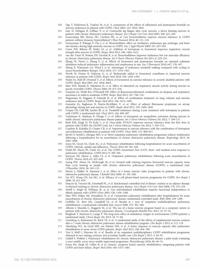
280 Oga T, Nishimura K, Tsukino M, et al. A comparison of the effects of salbutamol and ipratropium bromide onexercise endurance in patients with COPD. Chest 2003; 123: 1810–1816.
281 Saey D, Debigare R, LeBlanc P, et al. Contractile leg fatigue after cycle exercise: a factor limiting exercise inpatients with chronic obstructive pulmonary disease. Am J Respir Crit Care Med 2003; 168: 425–430.
282 Scuarcialupi ME, Berton DC, Cordoni PK, et al. Can bronchodilators improve exercise tolerance in COPDpatients without dynamic hyperinflation? J Bras Pneumol 2014; 40: 111–118.
283 Laveneziana P, Palange P, Ora J, et al. Bronchodilator effect on ventilatory, pulmonary gas exchange, and heartrate kinetics during high-intensity exercise in COPD. Eur J Appl Physiol 2009; 107: 633–643.
284 Canto ND, Ribeiro JP, Neder JA, et al. Addition of tiotropium to formoterol improves inspiratory musclestrength after exercise in COPD. Respir Med 2012; 106: 1404–1412.
285 van der Vaart H, Postma DS, Grevink R, et al. Bronchodilation improves endurance but not muscular efficiencyin chronic obstructive pulmonary disease. Int J Chron Obstruct Pulmon Dis 2011; 6: 229–235.
286 Zhang W, Fievez L, Zhang F, et al. Effects of formoterol and ipratropium bromide on repeated cadmiuminhalation-induced pulmonary inflammation and emphysema in rats. Eur J Pharmacol 2010; 647: 178–187.
287 Zhang X, Waterman LA, Ward J, et al. Advantages of endurance treadmill walking compared with cycling toassess bronchodilator therapy. Chest 2010; 137: 1354–1361.
288 Worth H, Förster K, Eriksson G, et al. Budesonide added to formoterol contributes to improved exercisetolerance in patients with COPD. Respir Med 2010; 104: 1450–1459.
289 Neder JA, Fuld JP, Overend T, et al. Effects of formoterol on exercise tolerance in severely disabled patients withCOPD. Respir Med 2007; 101: 2056–2064.
290 Man WD, Mustfa N, Nikoletou D, et al. Effect of salmeterol on respiratory muscle activity during exercise inpoorly reversible COPD. Thorax 2004; 59: 471–476.
291 Guenette JA, Webb KA, O’Donnell DE. Effect of fluticasone/salmeterol combination on dyspnea and respiratorymechanics in mild-to-moderate COPD. Respir Med 2013; 107: 708–716.
292 Magnussen H, Paggiaro P, Schmidt H, et al. Effect of combination treatment on lung volumes and exerciseendurance time in COPD. Respir Med 2012; 106: 1413–1420.
293 Guenette JA, Raghavan N, Harris-McAllister V, et al. Effect of adjunct fluticasone propionate on airwayphysiology during rest and exercise in COPD. Respir Med 2011; 105: 1836–1845.
294 Cooper CB, Celli BR, Jardim JR, et al. Treadmill endurance during 2-year treatment with tiotropium in patientswith COPD: a randomized trial. Chest 2013; 144: 490–497.
295 Yoshimura K, Maekura R, Hiraga T, et al. Effects of tiotropium on sympathetic activation during exercise instable chronic obstructive pulmonary disease patients. Int J Chron Obstruct Pulmon Dis 2012; 7: 109–117.
296 Beeh KM, Singh D, Di Scala L, et al. Once-daily NVA237 improves exercise tolerance from the first dose inpatients with COPD: the GLOW3 trial. Int J Chron Obstruct Pulmon Dis 2012; 7: 503–513.
297 Casaburi R, Kukafka D, Cooper CB, et al. Improvement in exercise tolerance with the combination of tiotropiumand pulmonary rehabilitation in patients with COPD. Chest 2005; 127: 809–817.
298 Revitt O, Sewell L, Morgan MD, et al. Short outpatient pulmonary rehabilitation programme reduces readmissionfollowing a hospitalization for an exacerbation of chronic obstructive pulmonary disease. Respirology 2013; 18:1063–1068.
299 Jones SE, Green SA, Clark AL, et al. Pulmonary rehabilitation following hospitalisation for acute exacerbation ofCOPD: referrals, uptake and adherence. Thorax 2014; 69: 181–182.
300 Dodd JW, Marns PL, Clark AL, et al. The COPD Assessment Test (CAT): short- and medium-term response topulmonary rehabilitation. COPD 2012; 9: 390–394.
301 Seymour JM, Moore L, Jolley CJ, et al. Outpatient pulmonary rehabilitation following acute exacerbations ofCOPD. Thorax 2010; 65: 423–428.
302 Leung RW, Alison JA, McKeough ZJ, et al. Ground walk training improves functional exercise capacity morethan cycle training in people with chronic obstructive pulmonary disease (COPD): a randomised trial.J Physiother 2010; 56: 105–112.
303 Moore J, Fiddler H, Seymour J, et al. Effect of a home exercise video programme in patients with chronicobstructive pulmonary disease. J Rehabil Med 2009; 41: 195–200.
304 Liu WT, Wang CH, Lin HC, et al. Efficacy of a cell phone-based exercise programme for COPD. Eur Respir J2008; 32: 651–659.
305 Deacon SJ, Vincent EE, Greenhaff PL, et al. Randomized controlled trial of dietary creatine as an adjunct therapyto physical training in chronic obstructive pulmonary disease. Am J Respir Crit Care Med 2008; 178: 233–239.
306 Sewell L, Singh SJ, Williams JE, et al. Can individualized rehabilitation improve functional independence inelderly patients with COPD? Chest 2005; 128: 1194–1200.
307 Man WD, Polkey MI, Donaldson N, et al. Community pulmonary rehabilitation after hospitalisation for acuteexacerbations of chronic obstructive pulmonary disease: randomised controlled study. BMJ 2004; 329: 1209.
308 Griffiths TL, Burr ML, Campbell IA, et al. Results at 1 year of outpatient multidisciplinary pulmonaryrehabilitation: a randomised controlled trial. Lancet 2000; 355: 362–368.
309 Albores J, Marolda C, Haggerty M, et al. The use of a home exercise program based on a computer system inpatients with chronic obstructive pulmonary disease. J Cardiopulm Rehabil Prev 2013; 33: 47–52.
310 Ringbaek T, Martinez G, Lange P. The long-term effect of ambulatory oxygen in normoxaemic COPD patients: arandomised study. Chron Respir Dis 2013; 10: 77–84.
311 Linneberg A, Rasmussen M, Buch TF, et al. A randomised study of the effects of supplemental exercise sessionsafter a 7-week chronic obstructive pulmonary disease rehabilitation program. Clin Respir J 2012; 6: 112–119.
312 Altenburg WA, de Greef MH, ten Hacken NH, et al. A better response in exercise capacity after pulmonaryrehabilitation in more severe COPD patients. Respir Med 2012; 106: 694–700.
313 Vest S, Moll L, Petersen M, et al. Results of an outpatient multidisciplinary COPD rehabilitation programmeobtained in two settings: primary and secondary health care. Clin Respir J 2011; 5: 84–91.
314 Liddell F, Webber J. Pulmonary rehabilitation for chronic obstructive pulmonary disease: a pilot study evaluatinga once-weekly versus twice-weekly supervised programme. Physiotherapy 2010; 96: 68–74.
315 Evans RA, Singh SJ, Collier R, et al. Generic, symptom based, exercise rehabilitation; integrating patients withCOPD and heart failure. Respir Med 2010; 104: 1473–1481.
DOI: 10.1183/13993003.00745-2015 459
ERS STATEMENT | L. PUENTE-MAESTU ET AL.

316 Waterhouse JC, Walters SJ, Oluboyede Y, et al. A randomised 2 × 2 trial of community versus hospitalpulmonary rehabilitation, followed by telephone or conventional follow-up. Health Technol Assess 2010; 14:1–140.
317 Ringbaek T, Brøndum E, Martinez G, et al. Rehabilitation in COPD: the long-term effect of a supervised 7-weekprogram succeeded by a self-monitored walking program. Chron Respir Dis 2008; 5: 75–80.
318 Eaton T, Young P, Nicol K, et al. The endurance shuttle walking test: a responsive measure in pulmonaryrehabilitation for COPD patients. Chron Respir Dis 2006; 3: 3–9.
460 DOI: 10.1183/13993003.00745-2015
ERS STATEMENT | L. PUENTE-MAESTU ET AL.