upper gastrointestinal tract … · the zenker di-verticulum is posterior to the pharyngoesophageal...
TRANSCRIPT

4139
case series
Rev. Colomb. Radiol. 2015; 26(1): 4139-44
Palabras clave (DeCS)
DivertículoTracto gastrointestinal
superior Tomografía computarizada
multidetector
1Doctor, radiologist, CediMed. Medellín, Colombia.
2Doctor, resident of Radiolo-gy, CES University, Medellín, Colombia.
3 Doctor, resident of Radiology, Pontificia Bolivariana Universi-ty. Medellín, Colombia.
4Student of Medicine, CES University, Medellín, Colombia.
UPPER GASTROINTESTINAL TRACT DIVERTICULUM AND PSEUDODIVERTICULOSIS: MULTIDETECTOR COMPUTED TOMOGRAPHY (MDCT) FINDINGS: CASE SERIESDivertículos y pseudodivertículos del tracto digestivo superior: Hallazgos por Tomografía Computarizada Multidetector (TCMD): Serie de casos
Alejandro Zuluaga S.1
Jorge Ochoa G. 2
Sebastián Bustamante Z.3
Carolina Gutiérrez M.3
Nicolás Zuluaga M.4
Summary
Diverticulum and pseudodiverticula of the gastrointestinal tract are incidentally found in different diagnostic studies performed in clinical practice. This article presents a detailed review of the diverticular disease of the upper gastrointestinal tract and its clinical, epidemiological aspects are discussed, as well as different imaging findings are discussed. A special emphasis is made on its appearance through Multidetector Computed Tomography (MDCT).
ResumenEn diferentes estudios diagnósticos realizados en la práctica clínica encontramos de manera incidental divertículos o pseudodivertículos del tracto gastrointestinal. Este artículo presenta una revisión detallada de la patología diverticular del tracto digestivo superior y se analizan sus aspectos clínicos, epidemiológicos y los diferentes hallazgos por imagen, haciendo hincapié en su apariencia por tomografía computarizada multidetector (TCMD).
The pathology of the superior gastrointestinal tract is usually evaluated through endoscopic studies and barium fluoroscopic studies. However, it is common that diverticula or pseudodiverticula are incidentally found in the esophagus, the stomach or the duodenum in Multidetector Computed Tomography (MDCT) exams. It is important to correctly diagnose these
entities, as well as be familiar with its most frequent manifestations on MDT and its possible complications. It is also essential not to mistake these diverticular alterations with pathologies of organs which are next to the digestive tract, as it frequently occurs in con-genital gastric diverticula, which may simulate nodular lesions or cysts of the left suprarenal gland due to its posterior location.
Key words (MeSH)
DiverticulumUpper gastrointestinal
tract Multidetector computed
tomography
4140 Upper Gastrointestinal Tract Diverticulum and Pseudodiverticulosis: Multidetector Computed Tomography (MDCT) Findings: Case Series. Zuluaga A., Ochoa J., Bustamante S., Gutiérrez C., Zuluaga N.
case series
Intramural pseudodiverticulosis of the esophagus
This is an uncommon benign condition, in which several and small pseudodiverticula (2-5 mm) are identified. These have a narrow neck, inside of the esophageal wall, and are usually distributed diffusely in the esophagus. It occurs in all age groups, but it is most common between the sixth and seventh decade of life, with predominance in the male sex.
Associations with other conditions are known, such as: esopha-geal stenosis (90% of patients), esophageal candidiasis, esophagitis, esophageal motility disorders and carcinomas.
The pseudodiverticula of the esophagus are dilated excretory ducts of the profound submucous glands, which are obstructed through des-quamated epithelium, inflammatory-infectious detritus or submucous fibrosis.
This condition usually manifests itself with acute or chronic dys-phagia. In studies with barium, several small diverticula are observed, which are filled with the contrast medium and may be distributed diffusely or in groups.
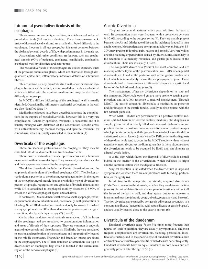
In MDCT, a diffuse thickening of the esophageal wall is usually identified. Occasionally, millimeter-sized aerial collections in the wall are also identified (case 1).
Mediastinis episodes have been described amongst the complica-tions in the rupture of pseudodiverticula, however this is a very rare complication. Generally speaking, treatment is successful and it is usually managed with dilatation of the concomitant stenotic areas, with anti-inflammatory medical therapy and specific treatment for candidiasis, which is usually associated to the condition (1).
Diverticula of the esophagus These are saccular protrusions of the esophagus. They may be
classified into drive diverticula and traction diverticula. These drive diverticula are made up of mucous and submucous
membranes without muscular layer. They are usually round or saccular and their appearance is round in the esophagogram.
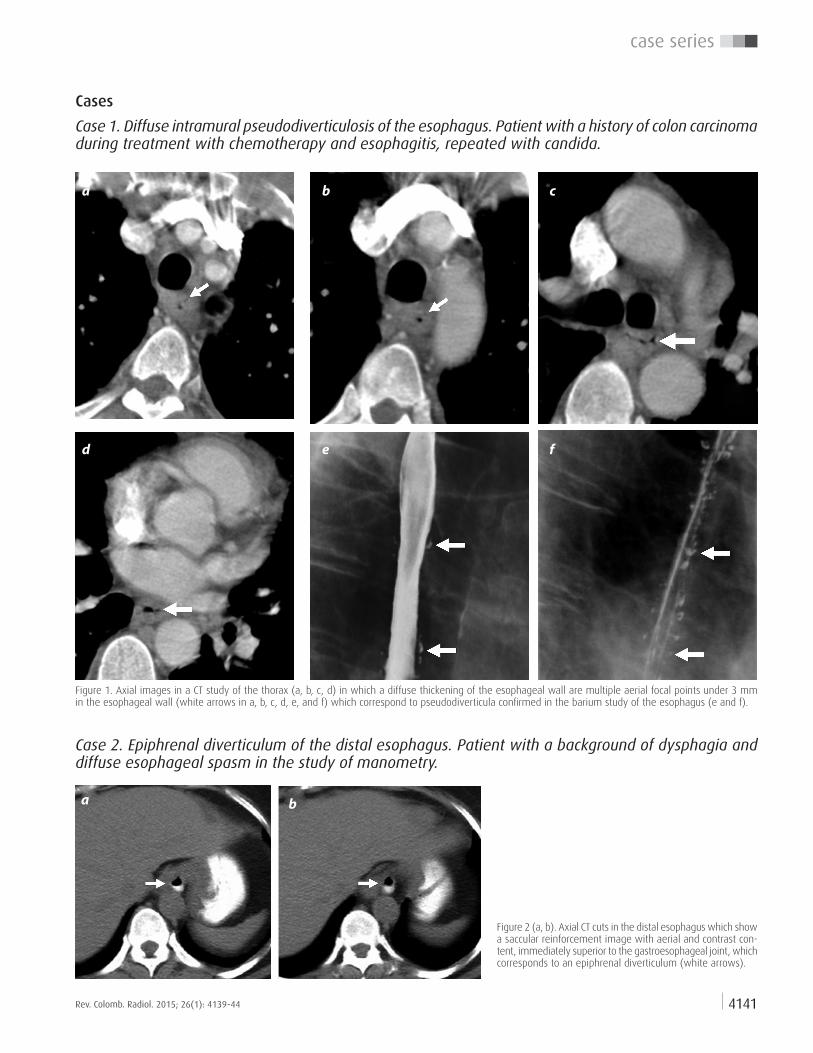
The drive diverticula include the Zenker diverticulum and the epiphrenic diverticulum of the distal esophagus (DE). The Zenker di-verticulum is posterior to the pharyngoesophageal union in the region of the cricopharyngeal muscle (patients with this type of diverticulum present dysphagia, regurgitation and episodes of bronchial inhalation), while DE is associated to esophageal motility disorders (75-90% of cases) as a diffuse esophageal spasm and achalasia.
If not treated, DE could manifest themselves with dysphagia, reflux, or pneumonia due to inhalation and, occasionally, with perforation or bleeding. Small DE do not require treatment, only follow-up. DE which is very symptomatic or DE with moderate or large size require surgical correction, ideally with laparoscopy (2) (case 2).
On the other hand, traction diverticula are made up of all the layers of the esophagus and are associated to granulomatous inflammation and areas of fibrosis of the esophagus. They are common in endemic areas of tuberculosis and histoplasmosis. Similarly, they are associated to erosion and perforation of the esophagus and are preferably located in the middle esophagus. Triangular and irregular images are found in the esophagogram. The Killian-Jamieson diverticulum is a type of diverticulum or esophageal bag which is located in the anterolateral region of the cervical esophagus (3).
Gastric DiverticulaThey are saccular dilatations which protrude from the gastric
wall. Its presentation is not very frequent, with a prevalence between 0.1-2.6%, according to the autopsy series (4). They are mainly present between the 5th and 6th decade of life and its incidence is equal in men and in women. Most patients are asymptomatic; however, between 18-30% may present abdominal pain, nausea and emesis. Very rarely does one find bleeding or perforation caused by diverticulitis, secondary to the retention of alimentary remnants, and gastric juice inside of the diverticulum. Their size is usually 1-3 cm.
The congenital diverticula (“true”) are most common and are made up of three layers of the gastrointestinal wall. 70% of congenital diverticula are found in the posterior wall of the gastric fundus, at a level which is immediately below the esophagogastric joint. These diverticula tend to have a relevant differential diagnosis: a cystic focal lesion of the left adrenal gland (case 3).
The management of gastric diverticula depends on its size and the symptoms. Diverticula over 4 cm are more prone to causing com-plications and have low response to medical management. Through MDCT, the gastric congenital diverticula is manifested as posterior nodular images in the gastric fundus, usually in close contact with the left adrenal gland (5).
When MDCT studies are performed with a positive contrast me-dium (diluted barium or iodized contrast medium), the diagnosis is simple, given that it is usually filled with contrast medium in supine position due to its posterior location (reinforcement contrast images which present continuity with the gastric lumen) which eases the differ-entiation of adrenal lesions (cases 4 and 5). Difficulties in the diagnosis of these diverticula tend to occur in the MDCT studies with or without negative or neutral contrast medium, given that in these circumstances the diverticulum tends to be occupied by liquid and can simulate an adrenal cystic lesion.
A useful sign which favors the diagnosis of diverticula is a small bubble in the interior of the diverticulum, which indicates its origin and the communication with the digestive tract (5).
Surgical resection is indicated when the diverticulum is large and symptomatic, or when there are complications with bleeding, perfora-tion, or malignity (6).
In addition to the congenital diverticula, acquired diverticula (“false”) are present in the stomach, whether they are drive or traction (case 6). Acquired drive diverticula are pseudodiverticula without all the layers of the gastric wall, and they appear due to an increase in intraluminal pressure (chronic cough, obesity, pregnancy, and bezoars). Traction diverticula are caused by perigastric adherences secondary to a concomitant disease (pancreatitis, acid-peptic disease or gastric bypass), and are usually located close to the gastric antrum (6).
Diverticula of the duodenumDuodenal diverticula (case 7) are five times more frequent than
jejunal or ileal; in addition, they are usually asymptomatic. The most frequent complications are diverticulitis, bleeding, perforation, intes-tinal obstruction, and in the specific case of duodenal diverticula, bile obstruction or obstructive pancreatitis, which does not occur frequently. Duodenal diverticula have an equal incidence in both sexes and are generally present after the age of 50 (7).
4141Rev. Colomb. Radiol. 2015; 26(1): 4139-44
case series
Figure 1. Axial images in a CT study of the thorax (a, b, c, d) in which a diffuse thickening of the esophageal wall are multiple aerial focal points under 3 mm in the esophageal wall (white arrows in a, b, c, d, e, and f) which correspond to pseudodiverticula confirmed in the barium study of the esophagus (e and f).
Figure 2 (a, b). Axial CT cuts in the distal esophagus which show a saccular reinforcement image with aerial and contrast con-tent, immediately superior to the gastroesophageal joint, which corresponds to an epiphrenal diverticulum (white arrows).
Cases
Case 1. Diffuse intramural pseudodiverticulosis of the esophagus. Patient with a history of colon carcinoma during treatment with chemotherapy and esophagitis, repeated with candida.
Case 2. Epiphrenal diverticulum of the distal esophagus. Patient with a background of dysphagia and diffuse esophageal spasm in the study of manometry.
a
d
b
e
c
f
a b

4142 Upper Gastrointestinal Tract Diverticulum and Pseudodiverticulosis: Multidetector Computed Tomography (MDCT) Findings: Case Series. Zuluaga A., Ochoa J., Bustamante S., Gutiérrez C., Zuluaga N.
case series
Figure 3 (a). Simple CT of the abdomen for the study of ureteral lithiasis (uroCT). A ho-mogenous nodular lesion with well-defined edges was identified as an incidental finding, with soft tissue density, located in the left suprarenal topography, posterior to the gastric fundus (white arrow). It is difficult to differentiate between an adrenal lesion and a diverticulum of the gastric fundus with the findings in this image.Figure 3 (b). The diagnosis of the diverticulum of the gastric fundus is eased in this image immediately under image (a) due to a small bubble of the diverticulum (arrow head).
Figure 4 (a, b). Posterior congenital diverticulum in the typical gastric fundus, due to the CT of the abdomen with positive oral contrast medium. The diverticulum is full of positive contrast medium and with a bubble sign (white arrow). (c) In a barium study of the superior digestive tract, the diverticulum is manifested as a well-defined contrast reinforcement medium, posterior in the gastric fundus (black arrow). (d, e). This image is typical of a congenital diverticulum, which can be visualized endoscopically.
Case 3. Congenital gastric diverticulum of the fundus
Case 4. Congenital gastric diverticulum of the fundus
a b
a b
c d e

4143Rev. Colomb. Radiol. 2015; 26(1): 4139-44
case series
Case 5. Aspect of different congenital gastric diverticula of the fundus by CT
Figure 5 (a, b, c). CT of three patients with posterior congenital gastric diverticula in the fundus; there is a total or partial filling with oral contrast medium and the diverticula in a and b have a bubble in the interior (white arrows).
a b c
a b
c d e
Case 6. False gastric diverticula in a patient with several phytobezoars and acute obstruction in the gastroenteroanastomosis site
Patient with a background of old gastric surgery due to previous benign pylhoric obstruction, with a scar origin by peptic ulcer. In the previous operation (uncommon, figure 6 g), without performing a partial gastrectomy and preserving the scar pylhoric region with ste-nosis (white arrow in the 6 g figure), a jejunum-section was performed in order to lead to the stomach. However, the proximal jejunal loop
anastomosed to the gastric body (“afferent loop”, clear green loop in figure 6 g, which drains the duodenal content and jejunal proximal to the stomach, figure 6 e) and the jejunal loop distal to the distal gastric antrum (“efferent loop” red loop in figure 6g, which evacuates the gastric content). The patient attends medical consultation due to an acute obstructive condition of the superior digestive tract (figure 6 d).
Figure 6 (a, b). Endoscopic projections of the stomach. Multiple pseudodiverticular images in the gastric wall can be seen (false diverticula) (white arrows in a), which represent a chronic impression in the mucous membrane due to several gastric phytobezoars (black arrow in b).
Figure 6 (c, d, e). Axial multicut CT images (c, d), and oblique coronal multiplanar reconstruction (3), in which multiple foreign bodies are identified in the stomach and in the second portion of the duodenum, with well-defined shapes, oval and with different sizes, with a phenomenon of internal vacuum and dense edge, which corresponds to several phytobezoars (arrows). Some phytobezoars indent the gastric mucous membrane (white arrow head in d), without causing external reinforcement areas of the gastric wall due to CT. One of the phytobezoars is found in the origin of the efferent loop, causing gastric obstruction and secondarily, obstruction of the afferent loop and the duodenum.

4144 Upper Gastrointestinal Tract Diverticulum and Pseudodiverticulosis: Multidetector Computed Tomography (MDCT) Findings: Case Series. Zuluaga A., Ochoa J., Bustamante S., Gutiérrez C., Zuluaga N.
case series
Figure 7 (a, b). Axial CT cuts of a patient who presents a diverticulum in the second portion of the duodenum, with aerial content and alimentary remnants (white arrows); the diverticulum is not associated to complications.Figure 7 (c). Barium study of the upper digestive tract in a different patient, with acquired diverticula located in the second and third portion of the duodenum, which do not have signs of complication.
Case 7. Diverticula of the duodenum.
ConclusionsMultidetector computerized tomography (MDCT) has become an
essential and complementary diagnostic tool for the study of diverticular and pseudodiverticular pathology of the upper digestive tract.
Esophageal candidiasis, esophagitis, esophageal motility disorders and esophageal carcinomas must be taken into account as pathologies which cause pseudodiverticula in the esophagus.
Congenital or true diverticula of the stomach become a relevant diagnosis, when the nodular lesions of the left adrenal gland are a significant differential.
Patients with a background of pancreatitis, acidic-peptic disease or gastric bypass may develop traction diverticula in the stomach, which are caused by perigastric adherences and are usually located close to the gastric antrum.
References1. Koyama S, Watanabe M, Iijima T. et al. Esophageal intramural pseudodiverticulosis
(diffuse type). J Gastroenterol. 2002;37:644-64.2. Zaninotto G, Portale G, Costantini M, et al. Therapeutic strategies for epiphrenic
diverticula: systematic review. World J Surg. 2011;35:1447-53.
3. Ba-Ssalamah A, Zacherl J, Noebauer-Huhmann IM, et al. Dedicated multi-detector CT of the esophagus: spectrum of diseases. Abdom Imaging. 2009;34:3-18.
4. Meeroff M, Gollan JRM, Meeroff JC. Gastric diverticulum. Am J Gastroenterol. 1967;47:189-203.
5. Schwartz AN, Goiney RC, Graney DO. Gastric diverticulum simulating an adrenal mass: CT appearance and embryogenesis. Am J Roentgenol. 1986;146:553-4.
6. Marano L, Reda G, Porfidia R, et al. Large symptomatic gastric diverticula: Two case reports and a brief review of literature. World J Gastroenterol. 2013;19:6114-7.
7. Ferreia-Aparicio FE, Gutiérrez-Vega R, Gálvez-Molna Y, et al. Diverticular disease of the small bowel. Case Rep Gastroenterol. 2012;6:668-76.
Corresponding AuthorAlejandro Zuluaga S. CediMedCalle 7 # 39-197, piso 3Medellín, [email protected]
Received for evaluation: September 23, 2014Accepted for publication: February 11, 2015
g
ba c
f
Figure 6(f). Surgical intervention image during the extraction of phytobezoars. A phytobezoar can be found in this procedure, impacted in the origin of the efferent loop (black arrow in d and g). In addition, a base chronic partial stenosis can be observed. It is believed that the patient developed phytobezoars due to partial chronic stenosis of the origin of the efferent loop, with delayed gastric emptying which favors the formation of bezoars. The yellow-greenish color of phytobezoars is influen-ced by the chronic drainage of the duodenal (bile) content to the stomach.