update in acute kidney injury - cmeinfo.com
TRANSCRIPT

UPDATE IN ACUTE KIDNEY INJURY
Emily Robinson, MD, MPHInstructor in Medicine, HMS
Renal Division, BWHOctober 1, 2021
Disclosures
■ Nothing to disclose

Case: Etiology of obstruction
■ 35 yo male with a congenital solitary kidney
■ Baseline Cr 1.2mg/dl
■ Presents to ER in severe pain
■ Ultrasound shows hydronephrosis
■ Labs show a creatinine increase to 2.7mg/dl
Based on his age alone, what is the most likely etiology of the obstruction?
■ A. Kidney stone
■ B. Prostatic obstruction
■ C. Retroperitoneal neoplastic disease
■ D. Anatomic abnormality

Based on his age alone, what is the most likely etiology of the obstruction?
■ A. Kidney stone
■ B. Prostatic obstruction
■ C. Retroperitoneal neoplastic disease
■ D. Anatomic abnormality
Most Common Causes of Obstruction by Age
■ Children
– Anatomic abnormalities
■ Young Adults
– Kidney stones
■ Older Adults
– Prostatic obstruction
– Retroperitoneal or pelvic neoplasms
– Kidney stones

Case: The “Negative” Urinalysis
■ 65 yo female presents to her PCP feeling “unwell” for 3 weeks with poor PO intake
■ Labs checked and Cr 3.7
■ She is referred to the ER and admitted for AKI, started on IV fluids
■ Amongst other workup, a urinalysis is performed and the dipstick is reported as “negative” with no blood, protein, leukocytes, or nitrites
Which of the following is NOT in your differential given this urinalysis?
■ A. Myeloma cast nephropathy
■ B. Dehydration due to poor PO intake
■ C. Tumor lysis syndrome from a new lymphoma
■ D. Rhabdomyolysis

Which of the following is NOT in your differential given this urinalysis?
■ A. Myeloma cast nephropathy
■ B. Dehydration due to poor PO intake
■ C. Tumor lysis syndrome from a new lymphoma
■ D. Rhabdomyolysis
Bland urinalysis
■ No RBCs, protein, or WBCs
■ Often pre-renal or post-renal
■ DO NOT consider glomerulonephritis or nephrotic syndrome if urine is bland
■ Other considerations
– Myeloma cast nephropathy
– Vascular ischemia without infarction
– Tumor lysis
– Acute phosphate nephropathy
– Hypercalcemia

Urinalysis: Proteinuria
■ “Proteinuria” on a dipstick is really albuminuria
■ Proteinuria DOES NOT rule out myeloma/light chain disease
■ Proteinuria on a dipstick is completely dependent on the urine concentration so need to quantify if positive
■ Traditionally “microalbuminuria”, or <300mg/g albumin, was below the level the dipstick could detect, but that is no longer the case with better dipsticks and also depends on concentration
Urinalysis: Hematuria
■ Hematuria without RBCs on sediment- think myoglobin/rhabdomyolysis!
■ Can indicate a glomerulonephritis
■ Some nephrotic syndromes have hematuria
■ Foley placement will cause hematuria
Urinalysis: White Blood Cells
■ AIN
■ UTI/pyelonephritis
■ Sterile pyuria
■ Can be seen with GNs as well
■ Can be seen with “dirty” urines, especially in women
Case: Use of the Protein:creatinine ratio
■ 65 yo male with diabetes and microalbuminuria (last malb/cr55mg/g) for foot amputation
■ Develops AKI post-op, Cr from 0.9 to 3.3 over 3 days
■ Pr/Cr checked as part of workup of AKI, ratio found to be ~4g
■ 24h urine performed, found to have 1.2g/24 hours
■ Why the discrepancy?

Why the discrepancy?
■ A. We are measuring different types of proteins in the two assay
■ B. The ratio is inaccurate while serum creatinine is rising
■ C. In diabetics we should be using malb/cr, not pr/cr
■ D. 24 hour collection was undercollected
Why the discrepancy?
■ A. We are measuring different types of proteins in the two assay
■ B. The ratio is inaccurate while serum creatinine is rising
■ C. In diabetics we should be using malb/cr, not pr/cr
■ D. 24 hour collection was undercollected

Protein:Creatinine Ratio in AKI
■ Pr/Cr takes into account BOTH protein and creatinine
■ AKI is NOT a steady state during the creatinine rise
■ Thus, denominator of ratio is low and overestimates 24h excretion
Protein:Creatinine Ratio
■ Works at the population level since the average daily creatinine excretion is 1g/day
■ More limited at the patient level but good for tracking, although both Pr and Cr excretion variable during a day
■ Very limited in AKI as creatinine excretion is not in a steady state

Case: Use of the fractional excretion of sodium (FeNa)
■ 45 yo male alcoholic “found down”
■ Appeared short of breath, got a PE protocol CT scan which was negative
■ Admitted to hospital
■ Baseline Cr 0.6
■ Day 2 Cr 1.8
■ Eating poorly but drinking water
■ UOP 1200cc/day
■ FeNa on Day 2 was 0.6%
■ Aggressive fluids given for presumed prerenal azotemia
■ Day 3 Cr 2.3, renal fellow looks at urine and sees muddy brown casts
Which of the following is NOT a cause for the low FeNa?
■ A. He is dehydrated as well
■ B. Poor solute intake with good UOP decreases the sodium concentration
■ C. Contrast causes afferent arteriolar vasoconstriction
■ D. Serum creatinine level is fluid in AKI

Which of the following is NOT a cause for the low FeNa?
■ A. He is dehydrated as well
■ B. Poor solute intake with good UOP decreases the sodium concentration
■ C. Contrast causes afferent arteriolar vasoconstriction
■ D. Serum creatinine level is fluid in AKI
FeNa
■ (Una x SCr)/(SNa x UCr) x 100
■ If <1% then prerenal
■ Limitations
– ONLY applies to patients with low GFR and oliguria– if GFR normal and/or urine output normal, sodium excretion depends on dietary sodium intake
– Serum creatinine level is fluid in AKI
– Diuretic therapy can increase sodium even if prerenal
– Other causes of AKI can induce a low FeNa
Other causes of AKI with low FeNa
■ ATN with some underlying ischemia or poor perfusion such as sepsis
■ Contrast nephropathy
■ Myoglobinuria
■ Any cause of AKI where tubular function is preserved despite decreased glomerular function
Fractional excretion of urea (FeUrea)
■ (UUN x SCr)/(SUN x PCr) x 100
■ If <35% suggestive of prerenal
■ Less sensitive than FeNa in patients NOT on diuretics
■ More sensitive than FeNa in patients on diuretics
■ Similar limitations to the FeNa

Case: Urine Electrolytes
■ 23 yo female admitted for abdominal pain, thought to be irritable bowel syndrome in past
■ Creatinine up from 0.7 baseline to 1.1 at admission
■ Noted to have K 3.2 and bicarb 34
■ Urine lytes show:
– Urine sodium: 70mmol/L
– Urine chloride: <assay
■ After much questioning after seeing urine lytes, admits to surreptitious vomiting
Why are the urine sodium and chloride values so disparate?
■ A. She ingested sodium bicarbonate tabs
■ B. She also surreptitiously took lasix, raising the urine sodium
■ C. Sodium is being excreted with the excess bicarbonate
■ D. She has Bartter’s syndrome
Why are the urine sodium and chloride values so disparate?
■ A. She ingested sodium bicarbonate tabs
■ B. She also surreptitiously took lasix, raising the urine sodium
■ C. Sodium is being excreted with the excess bicarbonate
■ D. She has Bartter’s syndrome
Urine Electrolytes: Sodium
■ Low urine sodium suggests a sodium-avid state
■ Not considered as accurate as a FeNa as does not account for rate of water reabsorption and has same other limitations as the FeNa
■ Lower limit of assay range differs at different hospitals!

Urine Electrolytes: Chloride
■ Reabsorbed with sodium, usually low when sodium is low
■ Helpful in volume depletion with alkalosis
– Bicarbonate is excreted as NaHCO3, thus urine sodium may not be less than assay
– However, urine chloride will still be low indicating volume depletion
– Usually the case if alkalosis is more severe than the volume depletion or else NaHCO3 will be reabsorbed proximally as well
Case: Urine Sediment
■ 85 yo male with dementia and baseline Cr 0.8 is admitted with confusion and diarrhea
■ Cr at admit is 2.4
■ Given IV fluids and creatinine goes down to 0.8 over 3 days
■ You look back at the urine microscopy report from the day of admission

Which of the following is a likely finding?
■ A. Muddy brown casts
■ B. Hyaline casts
■ C. Waxy casts
■ D. RBC casts
Which of the following is a likely finding?
■ A. Muddy brown casts
■ B. Hyaline casts
■ C. Waxy casts
■ D. RBC casts

Urine Sediment: Casts
■ Hyaline casts often seen with poor perfusion
■ Waxy casts are seen with CKD
■ Granular casts are very non-specific- ATN, poor perfusion, CKD
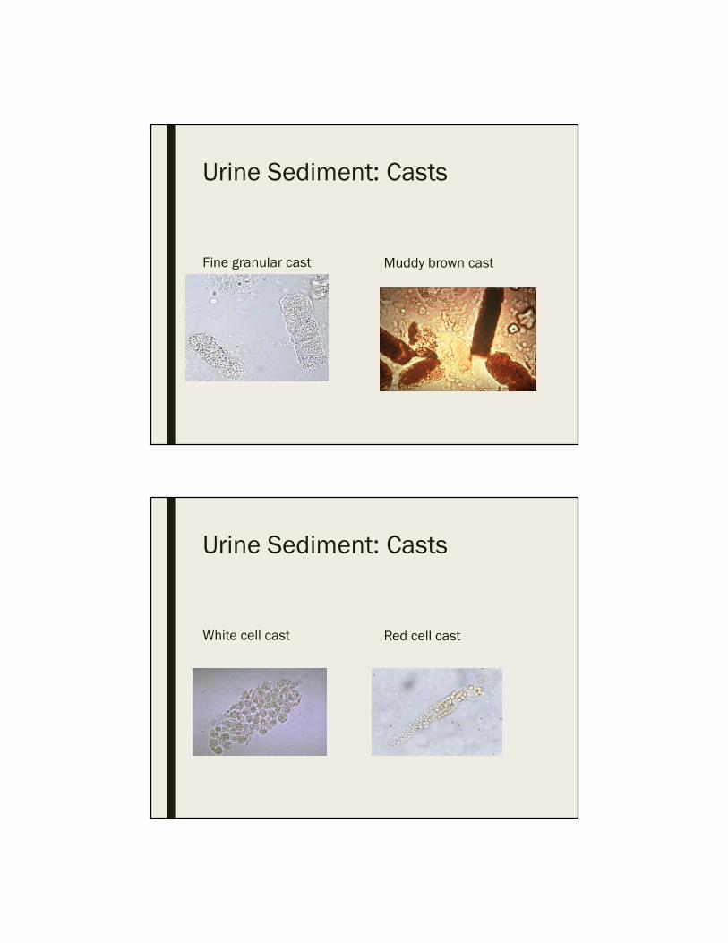
■ WBC casts usually AIN or pyelonephritis but can be GN as well
■ RBC casts usually GN
Urine Sediment: Casts
Hyaline cast Waxy cast
Urine Sediment: Casts
Fine granular cast Muddy brown cast
Urine Sediment: Casts
White cell cast Red cell cast

Case: Glomerulonephritis and the urinalysis
■ 74 yo female with hx of GERD, recent history of sinusitis, is found to have Cr 1.7mg/dl and sent to the ER for workup
■ In days prior to the presentation was also eating and drinking less due to feeling unwell
■ BP 149/66, HR 91, O2 sat 96%
■ Exam otherwise unremarkable, no edema
■ Creatinine in ER 1.9mg/dl
■ ANCA level done due to the sinus and pulmonary complaints, found to be positive
Which of the following sets of urine results should prompt concern for a glomerulonephritis?
■ A. Blood 2+, leukocyte esterase 2+, nitrite positive, protein 1+
■ B. Blood negative, leukocyte esterase negative, protein 3+
■ C. Blood 3+, leukocyte esterase trace, protein 3+, nitrite negative
■ D. All negative results
Urine findings in glomerulonephritis
■ If don’t see blood AND protein, VERY unlikely to be a glomerulonephritis
■ Urine sediment sometimes reported as having RBC casts, but many times no RBC casts so does not rule out
■ Can see leukocytes in a glomerulonephritis, does not rule out
■ UTIs can have blood and protein but GNs should not have positive nitrites
■ If AKI and urine sediment shows blood and protein, renal consult ASAP is necessary!

Symptoms that should prompt thinking of glomerulonephritis
■ Any pulmonary or sinus symptoms and/or hemoptysis consider ANCA disease and anti-glomerular basement membrane disease (i.e. Goodpasture’s syndrome), IgA can present this way as well
■ Other GNs can present with an array of symptoms, usually some overlap with rheumatologic symptoms and/or infections
Case: AIN
■ 66 yo male on multiple medications presents to his PCP with a rash and fever
■ Found on labs to have Cr 2.5, send to ER for admission and workup
■ Also noted to have a WBC differential with 7% eosinophilia
■ ER physician presumes AIN

Which of the following would be the LEAST likely cause of this presentation?
■ A. Ibuprofen
■ B. Furosemide
■ C. TMP/SMX
■ D. Omeprazole
Which of the following would be the LEAST likely cause of this presentation?
■ A. Ibuprofen
■ B. Furosemide
■ C. TMP/SMX
■ D. Omeprazole
Proton Pump Inhibitors and AIN
■ Do NOT present as classical AIN
■ Often late-onset after at least 4-6 months of the medication
■ Rarely present with rash, fever, or eosinophilia
■ May resolve with stopping the medication but will often require steroids
Urine Eosinophils
■ Used to test for AIN but sensitivity and specificity are poor
■ Sensitivity and specificity averages for >1% eosinophils on Hansel stain
– Sensitivity- 63-91%
– Specificity- 85-93%
■ Other conditions with eosinophils
– Transplant rejection
– Pyelonephritis/cystitis/prostatitis
– RPGN
– Atheroembolic disease

Plasma Eosinophils
■ Seen in 20-35% of cases of acute AIN in various studies
■ Classic triad of rash, fever, and eosinophilia only in ~10% of cases
Case: Imaging in AKI
■ 67 yo female presents to the ER with hypertensive emergency
■ She has not seen an MD in 10 years and has never had kidney function tested
■ Cr 3.7mg/dl

Which imaging modality can help you determine if this is acute or chronic?■ A. Nuclear scan
■ B. Xray
■ C. Renal ultrasound
■ D. MRI
Which imaging modality can help you determine if this is acute or chronic?■ A. Nuclear scan
■ B. Xray
■ C. Renal ultrasound
■ D. MRI
Renal Ultrasound in AKI
■ Should order only if you think it will help with making a diagnosis
■ R/o hydronephrosis
■ Infiltrative disease if one or both kidneys are very large
■ Cortical thinning and/or increased echogenicity
– Suggest underlying chronic disease
■ Disparate kidney sizes
– Atrophic kidney
– Unilateral RAS
Case: Contrast Nephropathy
■ 57 yo female with diabetes and Cr 1.4mg/dl is admitted for SOB and pleuritic chest pain.
■ You are worried about a PE and want to do a PE CT but are understandably concerned about her renal function

Which of the following is currently recommended for renal prophylaxis?
■ A. Isotonic Saline
■ B. Isotonic Bicarbonate
■ C. N-Acetylcysteine
■ D. Saline with furosemide to prevent volume overload
Which of the following is currently recommended for renal prophylaxis?
■ A. Isotonic Saline
■ B. Isotonic Bicarbonate
■ C. N-Acetylcysteine
■ D. Saline with furosemide to prevent volume overload

Contrast Nephropathy
■ Higher risk: diabetes, myeloma, prior renal insufficiency, proteinuria
■ Increase in Cr within 24-48 hours, peak 3-5 days, baseline in 6-10 days
■ Usually non-oliguric
■ Can see muddy brown casts
■ FeNa often <1% with high specific gravity of urine
Prevention of contrast nephropathy
■ This is highly debatable and many studies
■ Isotonic saline may be better than 1/2NS
■ Isotonic bicarb data is equivocal
■ Saline WITH furosemide appears to cause a higher risk of CIN
■ Data regarding N-acetylcysteine (NAC) equivocal despite many meta-analyses, most recent analyses appear to NOT show a benefit
■ Currently the most consistent recommendation is to give saline if no contraindications given it is cheapest with the least side effects, some studies do show benefit

PRESERVE Trial
Weisbord SD et al. N Engl J Med 2018;378:603-614
PRESERVE Trial
Weisbord SD et al. N Engl J Med 2018;378:603-614

But what about AMACING? Is there benefit to saline?
Nijssen EC et al, Lancet, 2017; 389. 1312-1322.
Why not follow AMACING and avoid prophylaxis?
■ Randomized, controlled trial of “high risk” patients set up as non-inferiority trial of saline versus placebo
■ However, group thought not to be high risk enough:
– GFR 30-59ml/min/1.73m2 but only 35% were <45ml/min/1.73m2
– 48% intraarterial contrast which is higher risk, only 6 in each group had AKI and none required dialysis
– Only 2.7% overall had AKI which is lower than general risk in high risk group in most studies
Case: NSAIDs and AKI
■ 47 yo female with DM 1 and albuminuria but normal creatinine
■ Prescribed ibuprofen 600mg QID for a rotator cuff injury
■ 2 weeks later notices significant leg swelling and goes to PCP
■ Cr 5.4mg/dl and UA with 3+ protein
Which of the following is NOT an affect of NSAIDs on the kidney?
■ A. Decrease in afferent vasodilation to decrease GFR
■ B. Interstitial nephritis
■ C.Minimal change disease
■ D. Membranous nephropathy
■ E. Focal and segmental glomerulosclerosis

Which of the following is NOT an affect of NSAIDs on the kidney?
■ A. Decrease in afferent vasodilation to decrease GFR
■ B. Interstitial nephritis
■ C.Minimal change disease
■ D. Membranous nephropathy
■ E. Focal and segmental glomerulosclerosis
NSAIDs
■ Inhibits prostaglandin-mediated afferent vasodilation
■ Causes an ischemic state by itself, or decreases threshold to other injury
■ Other causes of AKI with NSAIDs:
– Acute interstitial nephritis
– Minimal change disease
– Membranous nephropathy

Case: Diuretics and AKI
■ 82 yo male admitted with baseline Cr 1.3mg/dl admitted with SOB
■ PE CT done and shows PE
■ 2 days later creatinine starts to trend upwards but UOP is still good
■ You follow him and 2 days after that creatinine is 4.5mg/dl and UOP is 10cc/h
Which of the following is a true statement regarding diuretics and AKI?
■ A. If he gets diuretics on day 4 and responds, this predicts better recovery
■ B. He should not get diuretics on day 4 as this will further dehydrate the kidneys and do harm
■ C. He should get diuretics on day 2 to prevent worsening AKI
■ D. Diuretics should never be given in AKI
Are Diuretics Useful?
■ Diuretics should NOT be given prophylactically to prevent AKI
– 126 patients given dopamine, saline, or furosemide for 48 hours around cardiac surgery
– Patients given furosemide had more AKI
■ Diuretic responsiveness to oliguria does predict better recovery
– Shorter duration of oliguria and better UOP but may just be lest severe ATN
Lassner et al, JASN, 2000. 11(1): 97

Case: COVID-19 and AKI
■ 67 yo African-American male with HTN and DM, admitted to the hospital with shortness of breath and diagnosed with COVID
■ Admitted to ICU and intubated
■ Found to have progressive AKI with Cr up to 5.0 and oliguria
■ Initiated on HD
Which of the following is NOT a likely cause of the COVID-19-associated AKI?
■ A. ATN due to hypotension
■ B. Direct toxic effect of the virus
■ C. Collapsing glomerulopathy
■ D. Acute interstitial nephritis
■ E. Thrombotic microangiopathy
Which of the following is NOT a likely cause of the COVID-19-associated AKI?
■ A. ATN due to hypotension
■ B. Direct toxic effect of the virus
■ C. Collapsing glomerulopathy
■ D. Acute interstitial nephritis
■ E. Thrombotic microangiopathy
COVID-19 and AKI
■ Studies showing anywhere for 3-37% of hospitalized patients with COVID-19 develop AKI
■ Almost all have ATN on biopsy
■ Unclear if due to hemodynamics, cytokine release, or direct viral cytotoxicity
■ Some cases of COVID vaccine and glomerular disease development
■ Multiple case reports of African-Americans with AKI and biopsy showing collapsing glomerulopathy
– Potentially in the setting of an APOL1 mutation

Disclosures
■ Nothing to disclose
Thank you!