unraveling the structure of viral replication complexes at
TRANSCRIPT

ORIGINAL RESEARCH ARTICLEpublished: 31 January 2013
doi: 10.3389/fpls.2013.00006
Unraveling the structure of viral replication complexes atsuper-resolutionOlga Linnik 1, Johannes Liesche2, JensTilsner 3,4 and Karl J. Oparka1*1 Institute of Molecular Plant Sciences, University of Edinburgh, Edinburgh, UK2 Faculty of Life Sciences, University of Copenhagen, Frederiksberg C, Denmark3 Biomedical Sciences Research Complex, University of St Andrews, Fife, UK4 Cell and Molecular Sciences, The James Hutton Institute, Dundee, UK
Edited by:Jean-François Laliberté, InstitutNational de la Recherche Scientifique,Canada
Reviewed by:Richard Nelson, Samuel RobertsNoble Foundation, Inc., USATetsuro Okuno, Kyoto University,Japan
*Correspondence:Karl J. Oparka, Institute of MolecularPlant Sciences, University ofEdinburgh, King’s Buildings, MayfieldRoad, Edinburgh EH9 3JR, UK.e-mail: [email protected]
During infection, many RNA viruses produce characteristic inclusion bodies that containboth viral and host components. These structures were first described over a centuryago and originally termed “X-bodies,” as their function was not immediately appreciated.Whilst some inclusion bodies may represent cytopathic by-products of viral protein over-accumulation, X-bodies have emerged as virus “factories,” quasi-organelles that coordinatediverse viral infection processes such as replication, protein expression, evasion of hostdefenses, virion assembly, and intercellular transport. Accordingly, they are now generallyreferred to as viral replication complexes (VRCs).We previously used confocal fluorescencemicroscopy to unravel the complex structure of X-bodies produced by Potato virus X (PVX).Here we used 3D-structured illumination (3D-SIM) super-resolution microscopy to map thePVX X-body at a finer scale. We identify a previously unrecognized membrane structureinduced by the PVX “triple gene block” (TGB) proteins, providing new insights into thecomplex interplay between virus and host within the X-body.
Keywords: PVX, viral replication complex, 3D-SIM, super-resolution,TGB proteins, endoplasmic reticulum, Golgi
INTRODUCTIONVIRAL REPLICATION COMPLEXESIn the process of host invasion, many plant viruses induce the for-mation of characteristic inclusion bodies that were initially termed“X-bodies” due to their unclear role (Goldstein, 1924). Variouslyreferred to as amorphous inclusions, amorphous bodies, amoe-boid bodies, vacuolate bodies, or viroplasms, such inclusion bodieswere described in early studies by Goldstein (1926), Sheffield(1939, 1949). Inclusion bodies have been valuable in the diag-nosis of plant virus diseases (Martelli and Russo, 1977; Edwardsonand Christie, 1978), and many detailed studies of their structurewere conducted using electron microscopy (Esau, 1967; Shallaand Shepard, 1972; Christie and Edwardson, 1977). Although theobservation of inclusion bodies during infection provided someinsight into their role, their detailed structure and function was amystery until the arrival of molecular tools.
Plant viruses predominantly have positive sense, single-stranded RNA genomes ((+)ssRNA; Hull, 2002). (+)ssRNAviruses replicate on the cytoplasmic surfaces of modified host cellmembranes, and many viral inclusion bodies have been revealedto be “virus factories,” i.e., replication sites (Miller and Krijnse-Locker, 2008; den Boon et al., 2010; Laliberté and Sanfaçon, 2010).Accordingly, these viral structures are now mostly referred to asviral replication complexes or VRCs (Asurmendi et al., 2004).
Viral RNA (vRNA)-dependent RNA polymerases (“replicases”)are usually active as oligomeric arrays (Lyle et al., 2002; Kopeket al., 2007; Spagnolo et al., 2010), and the host membranes theyoccupy serve as scaffolds to assemble these complexes (Nishikioriet al., 2006). However, the functions of VRCs are more complex
than simply functioning to anchor replicase proteins to mem-branes. In addition to vRNA and proteins, they often incorporatehost components including rearranged host membranes (Schaadet al., 1997; Carette et al., 2000; Dunoyer et al., 2002; Ritzenthaleret al., 2002; Zamyatnin et al., 2002; Turner et al., 2004) that forma sheltered environment for the viral genome (Miller and Krijnse-Locker, 2008; den Boon et al., 2010; Laliberté and Sanfaçon, 2010).Besides being the primary centers of viral replication, VRCs mayalso facilitate viral access to essential host resources such as ribo-somes, enzymes, and nucleotides. In animal RNA viruses, viralpackaging may be closely linked to viral egress via the secretorypathway and budding from the plasma membrane (den Boonet al., 2010). Similarly in plant viruses, VRCs could be sites ofassembly of movement-competent ribonucleoprotein complexes(RNPs) for intercellular transport via plasmodesmata (Schoelzet al., 2011; Tilsner and Oparka, 2012). With such a complex vari-ety of processes coordinated in close proximity within VRCs, adetailed knowledge of the spatial organization of host and viralfactors is crucial to understanding the functions of VRCs. Renewedultrastructural investigations, using electron tomography, haveyielded high-resolution “maps” of the VRCs of Flock house virus(FHV) and SARS corona virus (Kopek et al., 2007; Knoops et al.,2008). However, similar studies are lacking for plant viruses. Inthe case of FHV, combination of tomographic and biochemicaldata enabled estimations of the numbers of replicase moleculesand (−)RNA replication templates in the membrane invagina-tions that harbor the replication machinery (Kopek et al., 2007).However, electron microscopy is limited in its ability to localizespecific macromolecules within VRCs. This is more easily done
www.frontiersin.org January 2013 | Volume 4 | Article 6 | 1
Linnik et al. Viral replication complex at super-resolution
using fluorescence microscopy coupled to fluorescently labeledantibodies or fluorescent protein fusions.
Until recently, confocal laser scanning microscopy providedthe highest resolution possible in fluorescence microscopy, withmaximum resolutions of ∼200 nm in the focal plane (x-y) and∼500 nm along the focal axis (z ; Huang et al., 2009). Such ideal res-olution is rarely achieved in heterogenous, living specimens, andfor practical purposes confocal microscopy has approximately 50-to 100-fold lower resolution than electron microscopy, resultingin an inability to use confocal microscopy for structural mapping.
In recent years, various “super-resolution” microscopy(nanoscopy) approaches have been developed that overcomethe diffraction barrier that limits conventional light microscopy,enabling fluorescence imaging at resolutions smaller than thewavelength of the emitted light (Huang et al., 2009; Schermellehet al., 2010). Hence, these technologies are ideally suited to gainnew insights into the structure-function relationships of VRCs(Horsington et al., 2012; Malkusch et al., 2012; Pereira et al.,2012). In practical terms, however, not all approaches are equallywell suited to plants. In particular, the cell wall limits penetrationof antibodies into plant cells. Therefore, the use of a geneticallyencoded fluorescent reporter fused with a protein of interest thatis transcribed within the cell provides a better approach for intra-cellular studies. Additionally, the autofluorescence backgroundcreated by chloroplasts and cell walls is particularly problem-atic for approaches that require single-molecule imaging such asphotoactivation localization microscopy (PALM) and stochasticoptical reconstruction microscopy (STORM; Tilsner and Flors,unpublished).
By contrast, three-dimensional structured illuminationmicroscopy (3D-SIM) is a widefield imaging approach that isamenable to most specimens suitable for confocal microscopy. In3D-SIM, a diffraction grating is superimposed upon the sample,and rotated during image acquisition. Sub-diffraction informa-tion is contained in the shifting diffraction patterns, and canbe extracted by mathematical transformation, permitting imagedeconvolution with a resolution of ∼100 nm in x-y and 200 nmin z (Gustafsson et al., 2008; Huang et al., 2009). This consti-tutes an approximate two-fold increase in resolution over confocalmicroscopy, but in practical terms provides a significant increasein biological detail (Fitzgibbon et al., 2010; Phillips et al., 2012). Wehave previously used 3D-SIM to obtain super-resolution images ofphloem sieve elements, including the localization of a viral move-ment protein to plasmodesmata (Fitzgibbon et al., 2010). To makethe phloem accessible to 3D-SIM, we partially digested cell wallmaterial and separated the cells of the tissue. Here, we employed3D-SIM to analyze the X-body of a model virus, Potato virusX (PVX), and to demonstrate the suitability of the technique toimaging three-dimensional structures in leaf epidermal cells. Thisapproach also should be suitable to a multitude of plant cell biol-ogy studies, including those conducted in the absence of virusinfection.
THE POTATO VIRUS X-BODYPotato virus X is a (+)ssRNA virus important for agriculture(Adams et al., 2004). It serves as a model virus for analysisof RNA silencing and virus movement, as a vector for proteinoverexpression and knockdown and as a virus-induced gene
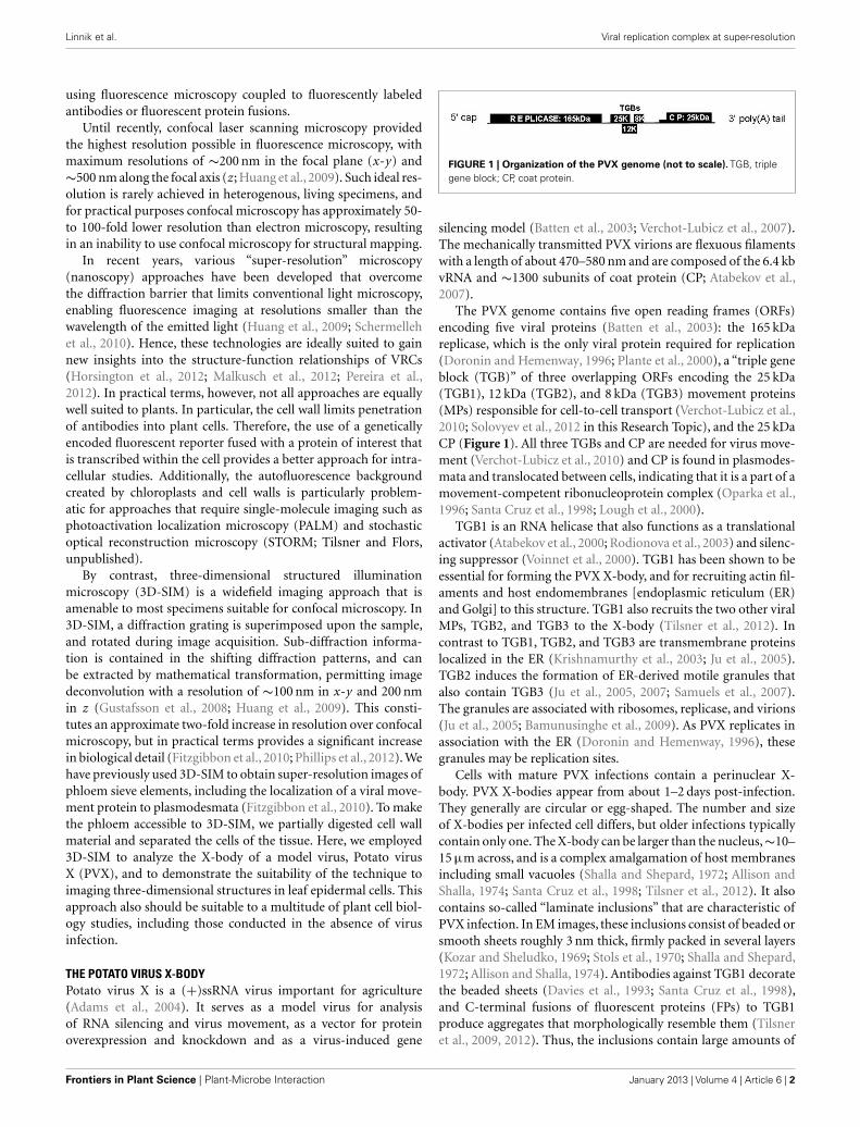
FIGURE 1 | Organization of the PVX genome (not to scale). TGB, triplegene block; CP, coat protein.
silencing model (Batten et al., 2003; Verchot-Lubicz et al., 2007).The mechanically transmitted PVX virions are flexuous filamentswith a length of about 470–580 nm and are composed of the 6.4 kbvRNA and ∼1300 subunits of coat protein (CP; Atabekov et al.,2007).
The PVX genome contains five open reading frames (ORFs)encoding five viral proteins (Batten et al., 2003): the 165 kDareplicase, which is the only viral protein required for replication(Doronin and Hemenway, 1996; Plante et al., 2000), a “triple geneblock (TGB)” of three overlapping ORFs encoding the 25 kDa(TGB1), 12 kDa (TGB2), and 8 kDa (TGB3) movement proteins(MPs) responsible for cell-to-cell transport (Verchot-Lubicz et al.,2010; Solovyev et al., 2012 in this Research Topic), and the 25 kDaCP (Figure 1). All three TGBs and CP are needed for virus move-ment (Verchot-Lubicz et al., 2010) and CP is found in plasmodes-mata and translocated between cells, indicating that it is a part of amovement-competent ribonucleoprotein complex (Oparka et al.,1996; Santa Cruz et al., 1998; Lough et al., 2000).
TGB1 is an RNA helicase that also functions as a translationalactivator (Atabekov et al., 2000; Rodionova et al., 2003) and silenc-ing suppressor (Voinnet et al., 2000). TGB1 has been shown to beessential for forming the PVX X-body, and for recruiting actin fil-aments and host endomembranes [endoplasmic reticulum (ER)and Golgi] to this structure. TGB1 also recruits the two other viralMPs, TGB2, and TGB3 to the X-body (Tilsner et al., 2012). Incontrast to TGB1, TGB2, and TGB3 are transmembrane proteinslocalized in the ER (Krishnamurthy et al., 2003; Ju et al., 2005).TGB2 induces the formation of ER-derived motile granules thatalso contain TGB3 (Ju et al., 2005, 2007; Samuels et al., 2007).The granules are associated with ribosomes, replicase, and virions(Ju et al., 2005; Bamunusinghe et al., 2009). As PVX replicates inassociation with the ER (Doronin and Hemenway, 1996), thesegranules may be replication sites.
Cells with mature PVX infections contain a perinuclear X-body. PVX X-bodies appear from about 1–2 days post-infection.They generally are circular or egg-shaped. The number and sizeof X-bodies per infected cell differs, but older infections typicallycontain only one. The X-body can be larger than the nucleus,∼10–15 µm across, and is a complex amalgamation of host membranesincluding small vacuoles (Shalla and Shepard, 1972; Allison andShalla, 1974; Santa Cruz et al., 1998; Tilsner et al., 2012). It alsocontains so-called “laminate inclusions” that are characteristic ofPVX infection. In EM images, these inclusions consist of beaded orsmooth sheets roughly 3 nm thick, firmly packed in several layers(Kozar and Sheludko, 1969; Stols et al., 1970; Shalla and Shepard,1972; Allison and Shalla, 1974). Antibodies against TGB1 decoratethe beaded sheets (Davies et al., 1993; Santa Cruz et al., 1998),and C-terminal fusions of fluorescent proteins (FPs) to TGB1produce aggregates that morphologically resemble them (Tilsneret al., 2009, 2012). Thus, the inclusions contain large amounts of
Frontiers in Plant Science | Plant-Microbe Interaction January 2013 | Volume 4 | Article 6 | 2
Linnik et al. Viral replication complex at super-resolution
TGB1, but it is not clear if they consist entirely of the TGB1 pro-tein. It was proposed that the beaded sheets could be active sitesof viral protein synthesis (Kozar and Sheludko, 1969; Shalla andShepard, 1972). The smooth sheets had virus particles betweenthe layers of the sheets (Shalla and Shepard, 1972), whereas thebeaded sheets did not (Stols et al., 1970; Shalla and Shepard, 1972).Whilst the beaded sheets superficially resemble ribosome-studdedER membranes, no lipids were found to be present in them, buttreatment with potassium permanganate destroyed them, indicat-ing that they are proteinaceous. The beads, found on both surfacesof the sheets, are too small to be ribosomes (Shalla and Shepard,1972). Surprisingly, more recent work on TGB1 does not referto these early data on TGB1 beaded sheets. Fluorescent fusions ofTGB2 and TGB3 also localized to the X-body (Samuels et al., 2007;Tilsner et al., 2012). Lastly, encapsidated PVX virions surround theX-body and when the CP is fused to GFP, virions appear as flu-orescent cages around the inclusions (Oparka et al., 1996; SantaCruz et al., 1998; Tilsner et al., 2012).
Recently, we undertook a detailed structural and functionalanalysis of the PVX X-body and its biogenesis (Tilsner et al., 2012).The X-body is formed by gradual accumulation of the ER-derived,TGB2/3-containing granules around the TGB1 beaded sheets.Non-encapsidated vRNA, visualized with a fluorescent reporterconstruct in vivo, localizes to whorls that tightly encircle the TGB1inclusions. The presence of “naked” RNA inside the X-body, andencapsidated virions at its periphery, along with the associationof TGB2/3 granules with replicase, strongly suggested that the X-body is indeed a replication site, i.e., a VRC. In the absence ofTGB1, no X-body is formed. Without an X-body, PVX still accu-mulates, but fewer virion aggregates are observed, indicating thatthe X-body may play a role in efficient virus encapsidation (Tilsneret al., 2012). In uninfected cells, ectopically expressed TGB1 canrecruit TGB2 and TGB3 into a “pseudo-VRC,” which has a similarstructure to the X-body.
In order to analyze the reorganized membrane structures ofthe PVX X-body at higher resolution, we turned to 3D-SIMmicroscopy. Here, we present results utilizing this technology toreveal new details of membrane organization within the PVX VRCand we demonstrate the applicability of 3D-SIM to general studiesof plant subcellular structures.
MATERIALS AND METHODSFLUORESCENT REPORTER AND VIRUS CONSTRUCTSBombardment vectors for expression of TGB1-mCherry, GFP-TGB2, and TGB3-GFP, and binary vectors for agroinfiltration ofTGB1-TagRFP, GFP-TGB2, TGB3-GFP, and unfused TGB2 andTGB3, as well as a binary vector for expression of a completePVX genome with an endogenous TGB1-mCherry fusion werepreviously described (Ju et al., 2005; Tilsner et al., 2009, 2012).PVX.GFP-CP and PVX.mCherry-CP constructs were previouslydescribed (Santa Cruz et al., 1996; Tilsner et al., 2009). In somecases, a 35S promoter-driven PVX.GFP-CP bombardment con-struct (Christophe Lacomme, unpublished) was used for infec-tions. A transgenic Nicotiana benthamiana line expressing ER-GFP(Haseloff et al., 1997), and a transgenic Nicotiana tabacum lineexpressing Golgi (ST)-GFP (Boevink et al., 1998), were describedpreviously.
EXPRESSION IN PLANTSInfectious PVX RNA was obtained by T7 in vitro transcrip-tion from plasmid constructs containing PVX.GFP-CP andPVX.mCherry-CP modified cDNA copies, as described in SantaCruz et al. (1996). Combinations of agrobacteria carrying binaryexpression constructs were infiltrated into N. benthamiana leavesat an OD600 of 0.15 or 0.25 each, as described previously (Tilsneret al., 2012). Microprojectile bombardments were carried out witha custom built gene gun according to the description in Gaba andGal-On (2006).
IMAGING AND IMAGE PROCESSINGConfocal microscopy was performed as described in Tilsner et al.(2009, 2012). For super-resolution imaging, lower epidermal peelswere prepared using a pair of fine forceps to peel carefully butquickly an epidermal peel from the lower epidermis of N. ben-thamiana or N. tabacum plants. Along the length of the peels,thickness varied from a few cells to a single cell layer. Immediatelyafter peeling, the epidermal peels were fixed by floating them ina fixative solution for 30–45 min at room temperature (for detailssee Fitzgibbon et al., 2010). The epidermal peels were assembledon a cover slip, not on a glass slide, in order to have the peel asclose as possible to the cover slip. Finally, the peels were mountedin Citifluor AF1 antifade medium (Agar Scientific), pressing gentlyto remove residual Citifluor from under the cover slip. The sam-ples were sealed with nail varnish, and viewed through a coverslip for 3D-SIM imaging with an OMX version 2 microscope(Applied Precision) as described in (Fitzgibbon et al., 2010). GFPwas excited at 488 nm and TagRFP and mCherry were excited at594 nm. Image processing was done as described in Fitzgibbonet al. (2010). Figures were assembled with Adobe Photoshop andImageJ software. TGB2 and TGB3 membrane hoops and Golgidimensions were measured using softWoRx (Applied Precision)software. Mean outer and inner diameters of the membrane hoopswere compared by one-way ANOVA followed by Least SignificantDifference and Duncan’s Multiple Range Tests using SPSS software(IBM).
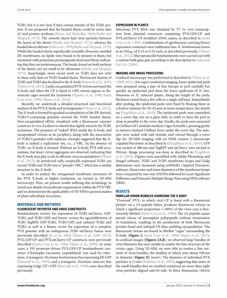
RESULTSFIBRILLAR VIRION BUNDLES SURROUND THE X-BODY“Overcoat” PVX, in which viral CP is fused with a fluorescentprotein via a 2A peptide linker, produces fluorescent virions inwhich a significant proportion (∼80%) of the virus coat is fluo-rescently labeled (Santa Cruz et al., 1996). The 2A peptide causespartial release of incomplete polypeptide without terminationof translation, resulting in the production of both fluorescentprotein-fused and unfused CP, thus enabling encapsidation. Thefluorescent virions are found in fibrillar “cages” surrounding theX-body (Figure 2; Santa Cruz et al., 1998; Tilsner et al., 2012).In confocal images (Figures 2A,B), we observed large bundles ofvirus filaments but were unable to resolve the fine structure of thevirion cages. Using 3D-SIM, we were able to resolve a fine net-work of virus bundles, the smallest of which were about 100 nmin diameter (Figure 2C insert). The diameter of individual PVXparticles is 13 nm (Atabekov et al., 2007), suggesting that some ofthe small bundles that we resolved contained no more than eightvirus particles aligned side-by-side. In three dimensions (Movie
www.frontiersin.org January 2013 | Volume 4 | Article 6 | 3
Linnik et al. Viral replication complex at super-resolution
FIGURE 2 | PVX virion “cages” encasing the X-body. (A) Live-cellconfocal overview of PVX.GFP-CP-infected cells with two perinuclear(n: nucleus) X-bodies. (B) Higher magnification confocal image ofa virion cage surrounding the X-body from a fixed sample.
(C) High-resolution 3D-SIM image. The insert shows an enlargementof the area in the rectangle in which individual virion filaments areresolved to <100 nm diameter. Bars (A): 50 µm; (B,C): 10 µm; [insertin (C)]: 500 nm.
S1 in Supplementary Materials), the viral cages formed a complexinterconnected network of virions that surrounded host and viralstructures at its center.
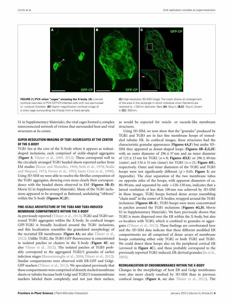
SUPER-RESOLUTION IMAGING OF TGB1 AGGREGATES AT THE CENTEROF THE X-BODYTGB1 lies at the core of the X-body where it appears as walnut-shaped inclusions, each comprised of sickle-shaped aggregates(Figure 3; Tilsner et al., 2009, 2012). These correspond well tothe circularly arranged TGB1 beaded sheets reported earlier fromEM studies (Kozar and Sheludko, 1969; Stols et al., 1970; Shallaand Shepard, 1972; Davies et al., 1993; Santa Cruz et al., 1998).Using 3D-SIM we were able to resolve the fibrillar composition ofthe TGB1 aggregates, showing even more clearly their correspon-dence with the beaded sheets observed in EM (Figures 3B–D;Movie S2 in Supplementary Materials). Many of the TGB1 inclu-sions appeared to be arranged as flattened, undulating “ribbons”within the X-body (Figures 3C,D).
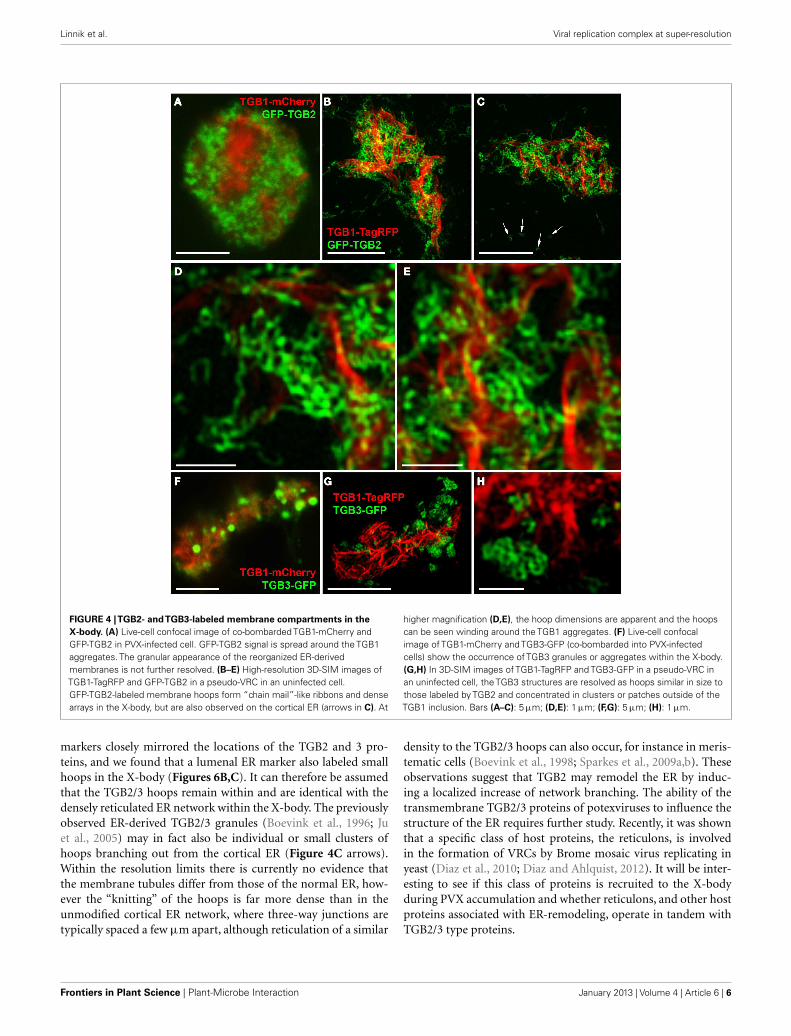
FINE-SCALE ARCHITECTURE OF THE TGB2 AND TGB3-INDUCEDMEMBRANE COMPARTMENTS WITHIN THE X-BODYAs previously reported (Tilsner et al., 2012), TGB2 and TGB3 sur-round TGB1 aggregates within the X-body. In confocal imagesGFP-TGB2 is broadly localized around the TGB1 inclusions,and this localization resembles the granulated morphology ofthe recruited ER membranes (Figure 4A; see also Tilsner et al.,2012). Unlike TGB2, the TGB3-GFP fluorescence is concentratedin isolated patches or clusters in the X-body (Figure 4F; seealso Tilsner et al., 2012). The isolated patches of TGB3 prob-ably correspond to the aggregated TGB2/3 granules of earlierinfection stages (Bamunusinghe et al., 2009; Tilsner et al., 2012).Similar compartments were observed with ER-GFP and Golgi-GFP markers (Tilsner et al., 2012). We speculated previously thatthese compartments were comprised of densely stacked membranesheets or tubules because both Golgi and TGB2/3 transmembranemarkers labeled them completely, and not just their surface,
as would be expected for vesicle- or vacuole-like membranestructures.
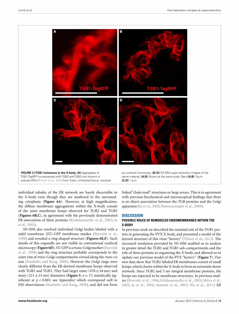
Using 3D-SIM, we now show that the “granules” produced byTGB2 and TGB3 are in fact fine membrane hoops of remod-eled tubular ER. In confocal images, these structures had thecharacteristic granular appearance (Figures 4A,F) but under 3D-SIM they appeared as donut-shaped loops (Figures 4B–E,G,H)with an outer diameter of 296± 37 nm and an inner diameterof 123± 15 nm for TGB2 (n= 8; Figures 4D,E) or 296± 49 nm(outer) and 134± 31 nm (inner) for TGB3 (n= 21; Figure 4H),respectively. Outer and inner diameters of the TGB2 and TGB3hoops were not significantly different (p > 0.05, Figure 5; seeAppendix). The clear separation of the two membrane tubeson opposite sides of the hoops, with apparent diameters of ca.80–90 nm, and separated by only ∼120–130 nm, indicates that alateral resolution of less than 100 nm was achieved by 3D-SIMin these images. TGB2 hoops formed dense arrays resembling“chain mail” in the center of X-bodies, wrapped around the TGB1inclusions (Figures 4B–E). TGB3 hoops were more concentratedin patches around the TGB1 inclusions (Figures 4G,H; MovieS3 in Supplementary Materials). We have previously shown thatTGB2 is more dispersed over the ER within the X-body, but alsoco-localizes with TGB3, which is confined to granules or aggre-gates (Tilsner et al., 2012). These findings are corroborated hereand the 3D-SIM data indicate that these different modified ERcompartments are all comprised of dense arrays of membranehoops containing either only TGB2 or both TGB2 and TGB3.We could detect these hoops also on the peripheral cortical ER(arrowed in Figure 4C), and these probably correspond to thepreviously reported TGB2-induced, ER-derived granules (Ju et al.,2005).
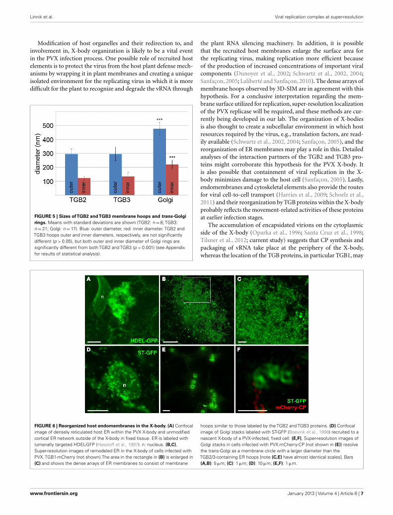
REORGANIZATION OF ENDOMEMBRANES WITHIN THE X-BODYChanges in the morphology of host ER and Golgi membraneswere also more clearly resolved by 3D-SIM than in previousconfocal images (Figure 6, see also Tilsner et al., 2012). The
Frontiers in Plant Science | Plant-Microbe Interaction January 2013 | Volume 4 | Article 6 | 4
Linnik et al. Viral replication complex at super-resolution
FIGURE 3 |TGB1 inclusions in the X-body. (A) Aggregates ofTGB1-TagRFP co-expressed with TGB2 and TGB3 (not shown) inpseudo-VRCs (Tilsner et al., 2012) from fixed, uninfected tissue, resolved
by confocal microscopy. (B–D) 3D-SIM super-resolution images of thesame material. (A,B) Shown at the same scale. Bars (A,B): 5 µm;(C,D): 1 µm.
individual tubules of the ER network are barely discernible inthe X-body even though they are unaltered in the surround-ing cytoplasm (Figure 6A). However, at high magnification,the diffuse membrane aggregations within the X-body consistof the same membrane hoops observed for TGB2 and TGB3(Figures 6B,C), in agreement with the previously demonstratedER-association of these proteins (Krishnamurthy et al., 2003; Juet al., 2005).
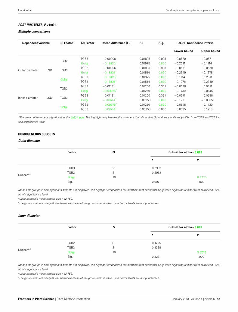
3D-SIM also resolved individual Golgi bodies labeled with asialyl transferase (ST)-GFP membrane marker (Boevink et al.,1998) and revealed a ring-shaped structure (Figures 6E,F). Suchdetails of this organelle are not visible in conventional confocalmicroscopy (Figure 6D). ST-GFP is a trans-Golgi marker (Boevinket al., 1998) and the ring structure probably corresponds to theouter rim of trans-Golgi compartments viewed along the trans-cisaxis (Staehelin and Kang, 2008). However the Golgi rings wereclearly different from the ER-derived membrane hoops observedwith TGB2 and TGB3. They had larger outer (478± 44 nm) andinner (221± 31 nm) diameters (Figure 5; n= 17; statistically sig-nificant at p < 0.001; see Appendix) which correspond well toEM observations (Staehelin and Kang, 2008), and did not form
linked “chain mail” structures or large arrays. This is in agreementwith previous biochemical and microscopical findings that thereis no direct association between the TGB proteins and the Golgiapparatus (Ju et al., 2005; Bamunusinghe et al., 2009).
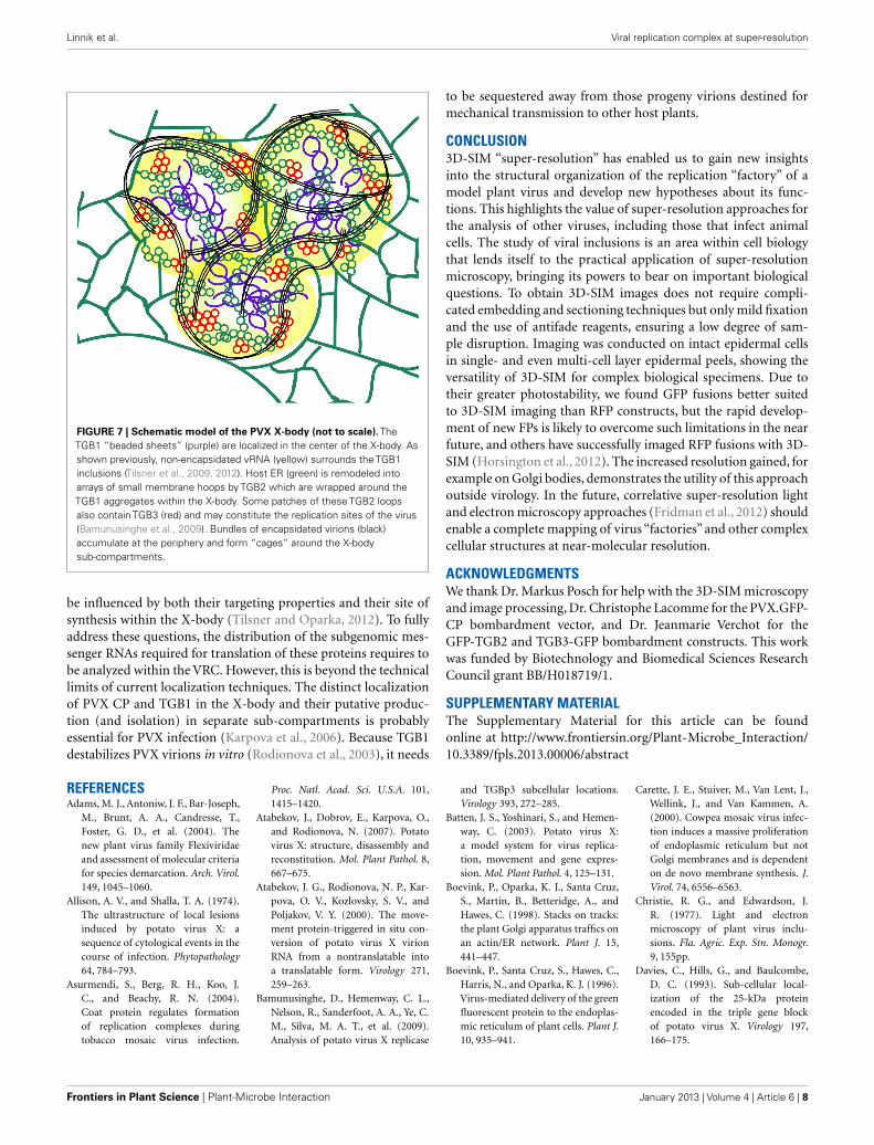
DISCUSSIONPOSSIBLE ROLES OF REMODELED ENDOMEMBRANES WITHIN THEX-BODYIn previous work we described the essential role of the TGB1 pro-tein in generating the PVX X-body, and presented a model of thelayered structure of this virus “factory” (Tilsner et al., 2012). Theincreased resolution provided by 3D-SIM enabled us to analyzein greater detail the TGB2 and TGB3 sub-compartments and therole of these proteins in organizing the X-body, and allowed us toupdate our previous model of the PVX “factory” (Figure 7). Ournew data show that TGB2-labeled ER membranes consist of smallhoops, which cluster within the X-body to form an extremely densenetwork. Since TGB2 and 3 are integral membrane proteins, thehoops are expected to be membrane structures. In previous stud-ies (Boevink et al., 1996; Krishnamurthy et al., 2003; Mitra et al.,2003; Ju et al., 2005; Samuels et al., 2007; Wu et al., 2011) ER
www.frontiersin.org January 2013 | Volume 4 | Article 6 | 5
Linnik et al. Viral replication complex at super-resolution
FIGURE 4 |TGB2- andTGB3-labeled membrane compartments in theX-body. (A) Live-cell confocal image of co-bombarded TGB1-mCherry andGFP-TGB2 in PVX-infected cell. GFP-TGB2 signal is spread around the TGB1aggregates. The granular appearance of the reorganized ER-derivedmembranes is not further resolved. (B–E) High-resolution 3D-SIM images ofTGB1-TagRFP and GFP-TGB2 in a pseudo-VRC in an uninfected cell.GFP-TGB2-labeled membrane hoops form “chain mail”-like ribbons and densearrays in the X-body, but are also observed on the cortical ER (arrows in C). At
higher magnification (D,E), the hoop dimensions are apparent and the hoopscan be seen winding around the TGB1 aggregates. (F) Live-cell confocalimage of TGB1-mCherry and TGB3-GFP (co-bombarded into PVX-infectedcells) show the occurrence of TGB3 granules or aggregates within the X-body.(G,H) In 3D-SIM images of TGB1-TagRFP and TGB3-GFP in a pseudo-VRC inan uninfected cell, the TGB3 structures are resolved as hoops similar in size tothose labeled by TGB2 and concentrated in clusters or patches outside of theTGB1 inclusion. Bars (A–C): 5 µm; (D,E): 1 µm; (F,G): 5 µm; (H): 1 µm.
markers closely mirrored the locations of the TGB2 and 3 pro-teins, and we found that a lumenal ER marker also labeled smallhoops in the X-body (Figures 6B,C). It can therefore be assumedthat the TGB2/3 hoops remain within and are identical with thedensely reticulated ER network within the X-body. The previouslyobserved ER-derived TGB2/3 granules (Boevink et al., 1996; Juet al., 2005) may in fact also be individual or small clusters ofhoops branching out from the cortical ER (Figure 4C arrows).Within the resolution limits there is currently no evidence thatthe membrane tubules differ from those of the normal ER, how-ever the “knitting” of the hoops is far more dense than in theunmodified cortical ER network, where three-way junctions aretypically spaced a few µm apart, although reticulation of a similar
density to the TGB2/3 hoops can also occur, for instance in meris-tematic cells (Boevink et al., 1998; Sparkes et al., 2009a,b). Theseobservations suggest that TGB2 may remodel the ER by induc-ing a localized increase of network branching. The ability of thetransmembrane TGB2/3 proteins of potexviruses to influence thestructure of the ER requires further study. Recently, it was shownthat a specific class of host proteins, the reticulons, is involvedin the formation of VRCs by Brome mosaic virus replicating inyeast (Diaz et al., 2010; Diaz and Ahlquist, 2012). It will be inter-esting to see if this class of proteins is recruited to the X-bodyduring PVX accumulation and whether reticulons, and other hostproteins associated with ER-remodeling, operate in tandem withTGB2/3 type proteins.
Frontiers in Plant Science | Plant-Microbe Interaction January 2013 | Volume 4 | Article 6 | 6
Linnik et al. Viral replication complex at super-resolution
Modification of host organelles and their redirection to, andinvolvement in, X-body organization is likely to be a vital eventin the PVX infection process. One possible role of recruited hostelements is to protect the virus from the host plant defense mech-anisms by wrapping it in plant membranes and creating a uniqueisolated environment for the replicating virus in which it is moredifficult for the plant to recognize and degrade the vRNA through
FIGURE 5 | Sizes ofTGB2 andTGB3 membrane hoops and trans-Golgirings. Means with standard deviations are shown (TGB2: n=8; TGB3:n=21; Golgi: n=17). Blue: outer diameter, red: inner diameter. TGB2 andTGB3 hoops outer and inner diameters, respectively, are not significantlydifferent (p > 0.05), but both outer and inner diameter of Golgi rings aresignificantly different from both TGB2 and TGB3 (p < 0.001) (see Appendixfor results of statistical analysis).
the plant RNA silencing machinery. In addition, it is possiblethat the recruited host membranes enlarge the surface area forthe replicating virus, making replication more efficient becauseof the production of increased concentrations of important viralcomponents (Dunoyer et al., 2002; Schwartz et al., 2002, 2004;Sanfaçon, 2005; Laliberté and Sanfaçon, 2010). The dense arrays ofmembrane hoops observed by 3D-SIM are in agreement with thishypothesis. For a conclusive interpretation regarding the mem-brane surface utilized for replication, super-resolution localizationof the PVX replicase will be required, and these methods are cur-rently being developed in our lab. The organization of X-bodiesis also thought to create a subcellular environment in which hostresources required by the virus, e.g., translation factors, are read-ily available (Schwartz et al., 2002, 2004; Sanfaçon, 2005), and thereorganization of ER membranes may play a role in this. Detailedanalyses of the interaction partners of the TGB2 and TGB3 pro-teins might corroborate this hypothesis for the PVX X-body. Itis also possible that containment of viral replication in the X-body minimizes damage to the host cell (Sanfaçon, 2005). Lastly,endomembranes and cytoskeletal elements also provide the routesfor viral cell-to-cell transport (Harries et al., 2009; Schoelz et al.,2011) and their reorganization by TGB proteins within the X-bodyprobably reflects the movement-related activities of these proteinsat earlier infection stages.
The accumulation of encapsidated virions on the cytoplasmicside of the X-body (Oparka et al., 1996; Santa Cruz et al., 1998;Tilsner et al., 2012; current study) suggests that CP synthesis andpackaging of vRNA take place at the periphery of the X-body,whereas the location of the TGB proteins, in particular TGB1, may
FIGURE 6 | Reorganized host endomembranes in the X-body. (A) Confocalimage of densely reticulated host ER within the PVX X-body and unmodifiedcortical ER network outside of the X-body in fixed tissue. ER is labeled withlumenally targeted HDEL-GFP (Haseloff et al., 1997). n: nucleus. (B,C),Super-resolution images of remodeled ER in the X-body of cells infected withPVX. TGB1-mCherry (not shown).The area in the rectangle in (B) is enlarged in(C) and shows the dense arrays of ER membranes to consist of membrane
hoops similar to those labeled by the TGB2 and TGB3 proteins. (D) Confocalimage of Golgi stacks labeled with ST-GFP (Boevink et al., 1998) recruited to anascent X-body of a PVX-infected, fixed cell. (E,F), Super-resolution images ofGolgi stacks in cells infected with PVX.mCherry-CP [not shown in (E)] resolvethe trans-Golgi as a membrane circle with a larger diameter than theTGB2/3-containing ER hoops [note (C,E) have almost identical scales]. Bars(A,B): 5 µm; (C): 1 µm; (D): 10 µm; (E,F): 1 µm.
www.frontiersin.org January 2013 | Volume 4 | Article 6 | 7
Linnik et al. Viral replication complex at super-resolution
FIGURE 7 | Schematic model of the PVX X-body (not to scale). TheTGB1 “beaded sheets” (purple) are localized in the center of the X-body. Asshown previously, non-encapsidated vRNA (yellow) surrounds the TGB1inclusions (Tilsner et al., 2009, 2012). Host ER (green) is remodeled intoarrays of small membrane hoops by TGB2 which are wrapped around theTGB1 aggregates within the X-body. Some patches of these TGB2 loopsalso contain TGB3 (red) and may constitute the replication sites of the virus(Bamunusinghe et al., 2009). Bundles of encapsidated virions (black)accumulate at the periphery and form “cages” around the X-bodysub-compartments.
be influenced by both their targeting properties and their site ofsynthesis within the X-body (Tilsner and Oparka, 2012). To fullyaddress these questions, the distribution of the subgenomic mes-senger RNAs required for translation of these proteins requires tobe analyzed within the VRC. However, this is beyond the technicallimits of current localization techniques. The distinct localizationof PVX CP and TGB1 in the X-body and their putative produc-tion (and isolation) in separate sub-compartments is probablyessential for PVX infection (Karpova et al., 2006). Because TGB1destabilizes PVX virions in vitro (Rodionova et al., 2003), it needs
to be sequestered away from those progeny virions destined formechanical transmission to other host plants.
CONCLUSION3D-SIM “super-resolution” has enabled us to gain new insightsinto the structural organization of the replication “factory” of amodel plant virus and develop new hypotheses about its func-tions. This highlights the value of super-resolution approaches forthe analysis of other viruses, including those that infect animalcells. The study of viral inclusions is an area within cell biologythat lends itself to the practical application of super-resolutionmicroscopy, bringing its powers to bear on important biologicalquestions. To obtain 3D-SIM images does not require compli-cated embedding and sectioning techniques but only mild fixationand the use of antifade reagents, ensuring a low degree of sam-ple disruption. Imaging was conducted on intact epidermal cellsin single- and even multi-cell layer epidermal peels, showing theversatility of 3D-SIM for complex biological specimens. Due totheir greater photostability, we found GFP fusions better suitedto 3D-SIM imaging than RFP constructs, but the rapid develop-ment of new FPs is likely to overcome such limitations in the nearfuture, and others have successfully imaged RFP fusions with 3D-SIM (Horsington et al., 2012). The increased resolution gained, forexample on Golgi bodies, demonstrates the utility of this approachoutside virology. In the future, correlative super-resolution lightand electron microscopy approaches (Fridman et al., 2012) shouldenable a complete mapping of virus “factories” and other complexcellular structures at near-molecular resolution.
ACKNOWLEDGMENTSWe thank Dr. Markus Posch for help with the 3D-SIM microscopyand image processing, Dr. Christophe Lacomme for the PVX.GFP-CP bombardment vector, and Dr. Jeanmarie Verchot for theGFP-TGB2 and TGB3-GFP bombardment constructs. This workwas funded by Biotechnology and Biomedical Sciences ResearchCouncil grant BB/H018719/1.
SUPPLEMENTARY MATERIALThe Supplementary Material for this article can be foundonline at http://www.frontiersin.org/Plant-Microbe_Interaction/10.3389/fpls.2013.00006/abstract
REFERENCESAdams, M. J., Antoniw, J. F., Bar-Joseph,
M., Brunt, A. A., Candresse, T.,Foster, G. D., et al. (2004). Thenew plant virus family Flexiviridaeand assessment of molecular criteriafor species demarcation. Arch. Virol.149, 1045–1060.
Allison, A. V., and Shalla, T. A. (1974).The ultrastructure of local lesionsinduced by potato virus X: asequence of cytological events in thecourse of infection. Phytopathology64, 784–793.
Asurmendi, S., Berg, R. H., Koo, J.C., and Beachy, R. N. (2004).Coat protein regulates formationof replication complexes duringtobacco mosaic virus infection.
Proc. Natl. Acad. Sci. U.S.A. 101,1415–1420.
Atabekov, J., Dobrov, E., Karpova, O.,and Rodionova, N. (2007). Potatovirus X: structure, disassembly andreconstitution. Mol. Plant Pathol. 8,667–675.
Atabekov, J. G., Rodionova, N. P., Kar-pova, O. V., Kozlovsky, S. V., andPoljakov, V. Y. (2000). The move-ment protein-triggered in situ con-version of potato virus X virionRNA from a nontranslatable intoa translatable form. Virology 271,259–263.
Bamunusinghe, D., Hemenway, C. L.,Nelson, R., Sanderfoot, A. A., Ye, C.M., Silva, M. A. T., et al. (2009).Analysis of potato virus X replicase
and TGBp3 subcellular locations.Virology 393, 272–285.
Batten, J. S., Yoshinari, S., and Hemen-way, C. (2003). Potato virus X:a model system for virus replica-tion, movement and gene expres-sion. Mol. Plant Pathol. 4, 125–131.
Boevink, P., Oparka, K. J., Santa Cruz,S., Martin, B., Betteridge, A., andHawes, C. (1998). Stacks on tracks:the plant Golgi apparatus traffics onan actin/ER network. Plant J. 15,441–447.
Boevink, P., Santa Cruz, S., Hawes, C.,Harris, N., and Oparka, K. J. (1996).Virus-mediated delivery of the greenfluorescent protein to the endoplas-mic reticulum of plant cells. Plant J.10, 935–941.
Carette, J. E., Stuiver, M., Van Lent, J.,Wellink, J., and Van Kammen, A.(2000). Cowpea mosaic virus infec-tion induces a massive proliferationof endoplasmic reticulum but notGolgi membranes and is dependenton de novo membrane synthesis. J.Virol. 74, 6556–6563.
Christie, R. G., and Edwardson, J.R. (1977). Light and electronmicroscopy of plant virus inclu-sions. Fla. Agric. Exp. Stn. Monogr.9, 155pp.
Davies, C., Hills, G., and Baulcombe,D. C. (1993). Sub-cellular local-ization of the 25-kDa proteinencoded in the triple gene blockof potato virus X. Virology 197,166–175.
Frontiers in Plant Science | Plant-Microbe Interaction January 2013 | Volume 4 | Article 6 | 8

Linnik et al. Viral replication complex at super-resolution
den Boon, J. A., Diaz, A., and Ahlquist,P. (2010). Cytoplasmic viral replica-tion complexes. Cell Host Microbe 8,77–85.
Diaz, A., and Ahlquist, P. (2012). Role ofhost reticulon proteins in rearrang-ing membranes for positive-strandRNA virus replication. Curr. Opin.Microbiol. 15, 519–524.
Diaz, A., Wang, X., and Ahlquist,P. (2010). Membrane-shaping hostreticulon proteins play crucial rolesin viral RNA replication com-partment formation and function.Proc. Natl. Acad. Sci. U.S.A. 107,16291–16296.
Doronin, S. V., and Hemenway, C.(1996). Synthesis of potato virusX RNAs by membrane-containingextracts. J. Virol. 70, 4795–4799.
Dunoyer, P., Ritzenthaler, C., Hem-mer, O., Michler, P., and Fritsch,C. (2002). Intracellular localizationof the peanut clump virus replica-tion complex in tobacco BY-2 pro-toplasts containing green fluorescentprotein-labeled endoplasmic reticu-lum or Golgi apparatus. J. Virol. 76,865–874.
Edwardson, J. R., and Christie, R. G.(1978). Use of virus-induced inclu-sions in classification and diagnosis.Annu. Rev. Phytopathol. 16, 31–55.
Esau, K. (1967). Anatomy of plant virusinfections. Annu. Rev. Phytopathol. 5,45–76.
Fitzgibbon, J., Bell, K., King, E., andOparka, K. (2010). Super-resolutionimaging of Plasmodesmata usingthree-dimensional structured illu-mination microscopy. Plant Physiol.153, 1453–1463.
Fridman, K., Mader, A., Zwerger, M.,Elia, N., and Medalia, O. (2012).Advances in tomography: prob-ing the molecular architecture ofcells. Nat. Rev. Mol. Cell Biol. 13,736–742.
Gaba, V., and Gal-On, A. (2006).Inoculation of plants using bom-bardment. Curr. Protoc. Microbiol.16B.3.1–16B.3.14.
Goldstein, B. (1924). Cytological studyof living cells of tobacco plantsaffected with mosaic disease. Bull.Torrey Botan. Club 51, 261–272.
Goldstein, B. (1926). A cytological studyof the leaves and growing points ofhealthy and mosaic diseased tobaccoplants. Bull. Torrey Botan. Club 53,499–600.
Gustafsson,M. G. L.,Shao,L.,Carlton,P.M., Wang, C. J. R., Golubovskaya, I.N., Cande,W. Z., et al. (2008). Three-dimensional resolution doubling inwide-field fluorescence microscopyby structured illumination. Biophys.J. 94, 4957–4970.
Harries, P. A., Park, J. W., Sasaki, N.,Ballard, K. D., Maule, A. J., and Nel-son, R. S. (2009). Differing require-ments for actin and myosin by plantviruses for sustained intercellularmovement. Proc. Natl. Acad. Sci.U.S.A. 106, 17594–17599.
Haseloff, J., Siemering, K. R., Prasher, D.C., and Hodge, S. (1997). Removalof a cryptic intron and subcellu-lar localization of green fluorescentprotein are required to mark trans-genic Arabidopsis plants brightly.Proc. Natl. Acad. Sci. U.S.A. 94,2122–2127.
Horsington, J., Turnbull, L.,Whitchurch, C. B., and New-some, T. P. (2012). Sub-viralimaging of vaccinia virus usingsuper-resolution microscopy. J.Virol. Methods 186, 132–136.
Huang, B., Bates, M., and Zhuang,X. (2009). Super-resolution fluo-rescence microscopy. Annu. Rev.Biochem. 78, 993–1016.
Hull, R. (2002). Matthews’ Plant Virol-ogy, 4th Edn. San Diego, CA: Acade-mic Press.
Ju, H. J., Brown, J. E., Ye, C. M., andVerchot-Lubicz, J. (2007). Mutationsin the central domain of potato virusX TGBp2 eliminate granular vesiclesand virus cell-to-cell trafficking. J.Virol. 81, 1899–1911.
Ju, H.-J., Samuels, T. D., Wang, Y.-S.,Blancaflor, E., Payton, M., Mitra,R., et al. (2005). The potato virusX TGBp2 movement protein asso-ciates with endoplasmic reticulum-derived vesicles during virus infec-tion. Plant Physiol. 138, 1877–1895.
Karpova, O. V., Zayakina, O. V.,Arkhipenko, M. V., Sheval, E. V.,Kiselyova, O. I., Poljakov, V. Y.,et al. (2006). Potato virus XRNA-mediated assembly of single-tailed ternary ‘coat protein-RNA-movement protein’ complexes. J.Gen. Virol. 87, 2731–2740.
Knoops, K., Kikkert, M., Worm, S. H.E., Zevenhoven-Dobbe, J. C., vander Meer, Y., Koster, A. J., et al.(2008). SARS-Coronavirus replica-tion is supported by a reticulovesic-ular network of modified endoplas-mic reticulum. PLoS Biol. 6:e226.doi:10.1371/journal.pbio.0060226
Kopek, B. G., Perkins, G., Miller, D.J., Ellisman, M. H., and Ahlquist,P. (2007). Three-dimensional analy-sis of a viral rna replicationcomplex reveals a virus-inducedmini-organelle. PLoS Biol. 5:e220.doi:10.1371/journal.pbio.0050220
Kozar, F. E., and Sheludko, Y. M. (1969).Ultrastructure of potato and Daturastramonium plant cells infected withpotato virus X. Virology 38, 220–229.
Krishnamurthy, K., Heppler, M., Mitra,R., Blancaflor, E., Payton, M., Nelson,R. S., et al. (2003). The Potato virusX TGBp3 protein associates withthe ER network for virus cell-to-cellmovement. Virology 309, 135–151.
Laliberté, J. F., and Sanfaçon, H. (2010).Cellular remodeling during plantvirus infection. Annu. Rev. Phy-topathol. 48, 69–91.
Lough,T. J.,Netzler,N. E.,Emerson,S. J.,Sutherland, P., Carr, F., Beck, D. L., etal. (2000). Cell-to-cell movement ofpotexviruses: evidence for a ribonu-cleoprotein complex involving thecoat protein and first triple geneblock protein. Mol. Plant MicrobeInteract. 13, 962–974.
Lyle, J. M., Bullitt, E., Bienz, K., andKirkegaard, K. (2002). Visualizationand functional analysis of RNA-dependent RNA polymerase lattices.Science 296, 2218–2222.
Malkusch, S., Muranyi, W., Müller,W., Kräusslich, H.-G., and Heile-mann, M. (2012). Single-moleculecoordinate-based analysis of themorphology of HIV-1 assembly siteswith near-molecular spatial reso-lution. Histochem. Cell Biol. 139,173–179.
Martelli, G. P., and Russo, M. (1977).Plant virus inclusion bodies. Adv.Virus Res. 21, 175–266.
Miller, S., and Krijnse-Locker, J. (2008).Modification of intracellular mem-brane structures for virus repli-cation. Nat. Rev. Microbiol. 6,363–374.
Mitra, R., Krishnamurthy, K., Blan-caflor, E., Payton, M., Nelson, R. S.,and Verchot-Lubicz, J. (2003). ThePotato virus X TGBp2 protein asso-ciation with the endoplasmic reticu-lum plays a role in but is not suffi-cient for viral cell-to-cell movement.Virology 312, 35–48.
Nishikiori, M., Dohi, K., Mori, M.,Meshi, T., Naito, S., and Ishikawa, M.(2006). Membrane-bound tomatomosaic virus replication proteinsparticipate in RNA synthesis, and areassociated with host proteins in apattern distinct from those that arenot membrane bound. J. Virol. 80,8459–8468.
Oparka, K. J., Roberts, A. G., Roberts,I. M., Prior, D. A. M., and SantaCruz, S. (1996). Viral coat proteinis targeted to, but does not gate,plasmodesmata during cell-to-cellmovement of potato virus X. PlantJ. 10, 805–813.
Pereira, C. F., Rossy, J., Owen, D. M.,Mak, J., and Gaus, K. (2012). HIVtaken by STORM: super-resolutionfluorescence microscopy of a viralinfection. Virol. J. 9, 84.
Phillips, D., Nibau, C., Wnetrzak,J., and Jenkins, G. (2012). Highresolution analysis of meioticchromosome structure andbehaviour in barley (Hordeumvulgare L.). PLoS ONE 7:e39539.doi:10.1371/journal.pone.0039539
Plante, C. A., Kim, K. H., Pillai-Nair,N., Osman, T. A. M., Buck, K. W.,and Hemenway, C. L. (2000). Sol-uble, template-dependent extractsfrom Nicotiana benthamiana plantsinfected with potato virus X tran-scribe both plus- and minus-strand RNA templates. Virology 275,444–451.
Ritzenthaler, C., Laporte, C., Gaire, F.,Dunoyer, P., Schmitt, C., Duval, S.,et al. (2002). Grapevine fanleaf virusreplication occurs on endoplasmicreticulum-derived membranes. J.Virol. 76, 8808–8819.
Rodionova, N. P., Karpova, O. V.,Kozlovsky, S. V., Zayakina, O. V.,Arkhipenko, M. V., and Atabekov,J. G. (2003). Linear remodeling ofhelical virus by movement proteinbinding. J. Mol. Biol. 333, 565–572.
Samuels, T. D., Ju, H.-J., Ye, C.-M.,Motes, C. M., Blancaflor, E. B., andVerchot-Lubicz, J. (2007). Subcellu-lar targeting and interactions amongthe potato virus X TGB proteins.Virology 367, 375–389.
Sanfaçon, H. (2005). Replication ofpositive-strand RNA viruses inplants: contact points between plantand virus components. Can. J. Bot.83, 1529–1549.
Santa Cruz, S., Chapman, S., Roberts,A. G., Roberts, I. M., Prior, D. A.M., and Oparka, K. J. (1996). Assem-bly and movement of a plant viruscarrying a green fluorescent proteinovercoat. Proc. Natl. Acad. Sci. U.S.A.93, 6286–6290.
Santa Cruz, S., Roberts, A. G., Prior, D.A. M., Chapman, S., and Oparka, K.J. (1998). Cell-to-cell and phloem-mediated transport of potato virusX: the role of virions. Plant Cell 10,495–510.
Schaad,M. C., Jensen,P. E., and Carring-ton, J. C. (1997). Formation of plantRNA virus replication complexes onmembranes: role of an endoplas-mic reticulum-targeted viral pro-tein. EMBO J. 16, 4049–4059.
Schermelleh, L., Heintzmann, R., andLeonhardt, H. (2010). A guideto super-resolution fluorescencemicroscopy. J. Cell Biol. 190,165–175.
Schoelz, J. E., Harries, P. A., and Nel-son, R. S. (2011). Intracellular trans-port of plant viruses: finding thedoor out of the cell. Mol. Plant 4,813–831.
www.frontiersin.org January 2013 | Volume 4 | Article 6 | 9

Linnik et al. Viral replication complex at super-resolution
Schwartz, M., Chen, J., Janda, M.,Sullivan, M., Den Boon, J., andAhlquist, P. (2002). A positive-strand RNA virus replication com-plex parallels form and functionof retrovirus capsids. Mol. Cell 9,505–514.
Schwartz, M., Chen, J., Lee, W.-M.,Janda, M., and Ahlquist, P. (2004).Alternate, virus-induced membranerearrangements support positive-strand RNA virus genome replica-tion. Proc. Natl. Acad. Sci. U.S.A. 101,11263–11268.
Shalla, T. A., and Shepard, J. F. (1972).The structure and antigenic analy-sis of amorphous inclusion bodiesinduced by potato virus X. Virology49, 654–667.
Sheffield, F. M. L. (1939). Micrurgi-cal studies on virus-infected plants.Proc. R. Soc. Lond. B Biol. Sci. 126,529–538.
Sheffield, F. M. L. (1949). The virus inthe plant cell. Exptl. Cell Res. Suppl.1, 178–182.
Solovyev, A. G., Kalinina, N. O.,and Morozov, S. Y. (2012). Recentadvances in research of plant virusmovement mediated by triple geneblock. Front. Plant Sci. 3:276.doi:10.3389/fpls.2012.00276
Spagnolo, J. F., Rossignol, E., Bullitt,E., and Kirkegaard, K. (2010).Enzymatic and nonenzymaticfunctions of viral RNA-dependent
RNA polymerases within oligomericarrays. RNA 16, 382–393.
Sparkes, I. A., Frigerio, L., Tolley, N.,and Hawes, C. (2009a). The plantendoplasmic reticulum: a cell-wideweb. Biochem. J. 423, 145–155.
Sparkes, I. A., Runions, J., Hawes, C.,and Griffing, L. (2009b). Movementand remodeling of the endoplas-mic reticulum in nondividing cellsof tobacco leaves. Plant Cell 21,3937–3949.
Staehelin, L. A., and Kang, B.-H. (2008).Nanoscale architecture of endoplas-mic reticulum export sites and ofGolgi membranes as determined byelectron tomography. Plant Physiol.147, 1454–1468.
Stols, A. L. H., Hill-van der Meulen, G.W., and Toen, M. K. I. (1970). Elec-tron microscopy of Nicotiana gluti-nosa leaf cells infected with potatovirus X. Virology 40, 168–170.
Tilsner, J., Linnik, O., Christensen,N. M., Bell, K., Roberts, I. M.,Lacomme, C., et al. (2009). Live-cellimaging of viral RNA genomes usinga pumilio-based reporter. Plant J. 57,758–770.
Tilsner, J., Linnik, O.,Wright, K. M., Bell,K., Roberts,A. G., Lacomme, C., et al.(2012). The TGB1 movement pro-tein of potato virus X re-organisesactin and endomembranes into the‘X-body’ a viral replication factory.Plant Physiol. 158, 1359–1370.
Tilsner, J., and Oparka, K. J. (2012).Missing links? – The connectionbetween replication and movementof plant RNA viruses. Curr. Opin.Virol. 2, 699–705.
Turner, K. A., Sit, T. L., Callaway, A.S., Allen, N. S., and Lommel, S. A.(2004). Red clover necrotic mosaicvirus replication proteins accumu-late at the endoplasmic reticulum.Virology 320, 276–290.
Verchot-Lubicz, J., Torrance, L.,Solovyev, A. G., Morozov, S. Y.,Jackson, A. O., and Gilmer, D.(2010). Varied movement strate-gies employed by triple geneblock-encoding viruses. Mol. PlantMicrobe Interact. 23, 1231–1247.
Verchot-Lubicz, J., Ye, C. M., andBamunusinghe, D. (2007). Mol-ecular biology of potexviruses:recent advances. J. Gen. Virol. 88,1643–1655.
Voinnet, O., Lederer, C., and Baulcombe,D. C. (2000). A viral movement pro-tein prevents spread of the genesilencing signal in Nicotiana ben-thamiana. Cell 103, 157–167.
Wu, C.-H., Lee, S.-C., and Wang, C.-W. (2011). Viral protein targetingto the cortical endoplasmic reticu-lum is required for cell-cell spread-ing in plants. J. Cell Biol. 193,521–535.
Zamyatnin, A. A., Solovyev, A. G.,Sablina, A. A., Agranovsky, A. A.,
Katul, L., Vetten, H. J., et al. (2002).Dual-colour imaging of membraneprotein targeting directed by poasemilatent virus movement pro-tein TGBp3 in plant and mam-malian cells. J. Gen. Virol. 83,651–662.
Conflict of Interest Statement: Theauthors declare that the research wasconducted in the absence of any com-mercial or financial relationships thatcould be construed as a potential con-flict of interest.
Received: 15 November 2012; accepted:09 January 2013; published online: 31January 2013.Citation: Linnik O, Liesche J, Tilsner Jand Oparka KJ (2013) Unraveling thestructure of viral replication complexesat super-resolution. Front. Plant Sci. 4:6.doi: 10.3389/fpls.2013.00006This article was submitted to Frontiers inPlant-Microbe Interaction, a specialty ofFrontiers in Plant Science.Copyright © 2013 Linnik, Liesche, Tilsnerand Oparka. This is an open-access arti-cle distributed under the terms of theCreative Commons Attribution License,which permits use, distribution andreproduction in other forums, providedthe original authors and source are cred-ited and subject to any copyright noticesconcerning any third-party graphics etc.
Frontiers in Plant Science | Plant-Microbe Interaction January 2013 | Volume 4 | Article 6 | 10

Linnik et al. Viral replication complex at super-resolution
APPENDIX
DESCRIPTIVES
N Mean (µm) SD SE 95% Confidence interval for mean Minimum Maximum
Lower bound Upper bound
OUTER DIAMETER
TGB2 8 0.2963 0.03662 0.01295 0.2656 0.3269 0.25 0.34
TGB3 21 0.2962 0.04944 0.01079 0.2737 0.3187 0.20 0.37
Golgi 16 0.4775 0.04405 0.01101 0.4540 0.5010 0.41 0.56
Total 45 0.3607 0.09843 0.01467 0.3311 0.3902 0.20 0.56
INNER DIAMETER
TGB2 8 0.1225 0.01488 0.00526 0.1101 0.1349 0.11 0.14
TGB3 21 0.1338 0.03106 0.00678 0.1197 0.1479 0.10 0.20
Golgi 16 0.2213 0.03074 0.00769 0.2049 0.2376 0.17 0.28
Total 45 0.1629 0.05229 0.00780 0.1472 0.1786 0.10 0.28
TEST OF HOMOGENEITY OF VARIANCES
Levene statistic df1 df2 Sig.
Outer diameter 0.198 2 42 0.821
Inner diameter 1.420 2 42 0.253
ANOVA
Sum of squares df Mean square F Sig.
OUTER DIAMETER
Between groups 0.339 2 0.169 81.444 0.000
Within groups 0.087 42 0.002
Total 0.426 44
INNER DIAMETER
Between groups 0.085 2 0.043 51.153 0.000
Within groups 0.035 42 0.001
Total 0.120 44
www.frontiersin.org January 2013 | Volume 4 | Article 6 | 11
Linnik et al. Viral replication complex at super-resolution
POST HOC TESTS, P < 0.001.
Multiple comparisons
Dependent Variable (I) Factor (J) Factor Mean difference (I-J) SE Sig. 99.9% Confidence interval
Lower bound Upper bound
Outer diameter LSD
TGB2TGB3 0.00006 0.01895 0.998 −0.0670 0.0671
Golgi −0.18125* 0.01975 0.000 −0.2511 −0.1114
TGB3TGB2 −0.00006 0.01895 0.998 −0.0671 0.0670
Golgi −0.18131* 0.01514 0.000 −0.2349 −0.1278
GolgiTGB2 0.18125* 0.01975 0.000 0.1114 0.2511
TGB3 0.18131* 0.01514 0.000 0.1278 0.2349
Inner diameter LSD
TGB2TGB3 −0.01131 0.01200 0.351 −0.0538 0.0311
Golgi −0.09875* 0.01250 0.000 −0.1430 −0.0545
TGB3TGB2 0.01131 0.01200 0.351 −0.0311 0.0538
Golgi −0.08744* 0.00958 0.000 −0.1213 −0.0535
GolgiTGB2 0.09875* 0.01250 0.000 0.0545 0.1430
TGB3 0.08744* 0.00958 0.000 0.0535 0.1213
*The mean difference is significant at the 0.001 level. The highlight emphasizes the numbers that show that Golgi does significantly differ from TGB2 and TGB3 at
this significance level.
HOMOGENEOUS SUBSETS
Outer diameter
Factor N Subset for alpha = 0.001
1 2
Duncana,b
TGB3 21 0.2962
TGB2 8 0.2963
Golgi 16 0.4775
Sig. 0.997 1.000
Means for groups in homogeneous subsets are displayed. The highlight emphasizes the numbers that show that Golgi does significantly differ from TGB2 and TGB3
at this significance level.aUses harmonic mean sample size = 12.759.bThe group sizes are unequal. The harmonic mean of the group sizes is used. Type I error levels are not guaranteed.
Inner diameter
Factor N Subset for alpha = 0.001
1 2
Duncana,b
TGB2 8 0.1225
TGB3 21 0.1338
Golgi 16 0.2213
Sig. 0.328 1.000
Means for groups in homogeneous subsets are displayed. The highlight emphasizes the numbers that show that Golgi does significantly differ from TGB2 and TGB3
at this significance level.aUses harmonic mean sample size = 12.759.bThe group sizes are unequal. The harmonic mean of the group sizes is used. Type I error levels are not guaranteed.
Frontiers in Plant Science | Plant-Microbe Interaction January 2013 | Volume 4 | Article 6 | 12
Linnik et al. Viral replication complex at super-resolution
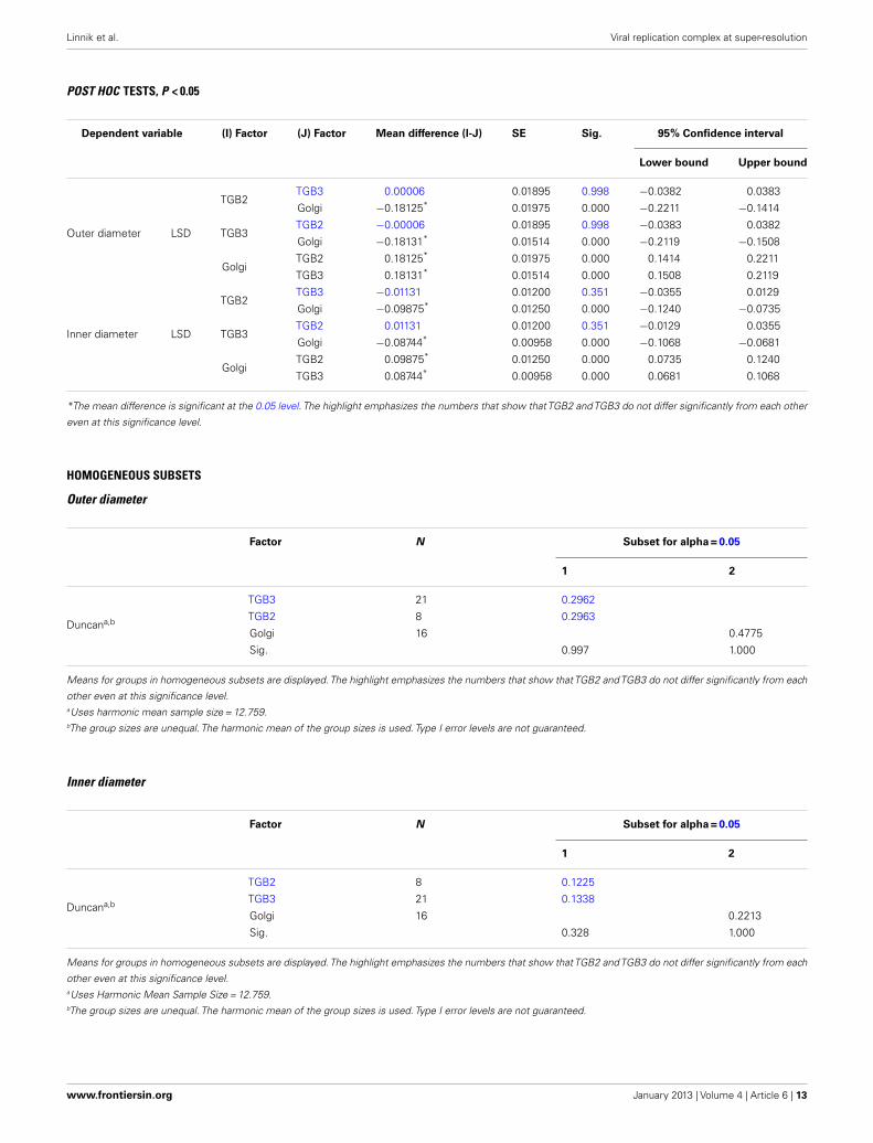
POST HOC TESTS, P < 0.05
Dependent variable (I) Factor (J) Factor Mean difference (I-J) SE Sig. 95% Confidence interval
Lower bound Upper bound
Outer diameter LSD
TGB2TGB3 0.00006 0.01895 0.998 −0.0382 0.0383
Golgi −0.18125* 0.01975 0.000 −0.2211 −0.1414
TGB3TGB2 −0.00006 0.01895 0.998 −0.0383 0.0382
Golgi −0.18131* 0.01514 0.000 −0.2119 −0.1508
GolgiTGB2 0.18125* 0.01975 0.000 0.1414 0.2211
TGB3 0.18131* 0.01514 0.000 0.1508 0.2119
Inner diameter LSD
TGB2TGB3 −0.01131 0.01200 0.351 −0.0355 0.0129
Golgi −0.09875* 0.01250 0.000 −0.1240 −0.0735
TGB3TGB2 0.01131 0.01200 0.351 −0.0129 0.0355
Golgi −0.08744* 0.00958 0.000 −0.1068 −0.0681
GolgiTGB2 0.09875* 0.01250 0.000 0.0735 0.1240
TGB3 0.08744* 0.00958 0.000 0.0681 0.1068
*The mean difference is significant at the 0.05 level. The highlight emphasizes the numbers that show thatTGB2 andTGB3 do not differ significantly from each other
even at this significance level.
HOMOGENEOUS SUBSETS
Outer diameter
Factor N Subset for alpha = 0.05
1 2
Duncana,b
TGB3 21 0.2962
TGB2 8 0.2963
Golgi 16 0.4775
Sig. 0.997 1.000
Means for groups in homogeneous subsets are displayed.The highlight emphasizes the numbers that show thatTGB2 andTGB3 do not differ significantly from each
other even at this significance level.aUses harmonic mean sample size = 12.759.bThe group sizes are unequal. The harmonic mean of the group sizes is used. Type I error levels are not guaranteed.
Inner diameter
Factor N Subset for alpha = 0.05
1 2
Duncana,b
TGB2 8 0.1225
TGB3 21 0.1338
Golgi 16 0.2213
Sig. 0.328 1.000
Means for groups in homogeneous subsets are displayed.The highlight emphasizes the numbers that show thatTGB2 andTGB3 do not differ significantly from each
other even at this significance level.aUses Harmonic Mean Sample Size = 12.759.bThe group sizes are unequal. The harmonic mean of the group sizes is used. Type I error levels are not guaranteed.
www.frontiersin.org January 2013 | Volume 4 | Article 6 | 13