university of groningen impaired organ perfusion morariu
TRANSCRIPT

University of Groningen
Impaired Organ PerfusionMorariu, Aurora
IMPORTANT NOTE: You are advised to consult the publisher's version (publisher's PDF) if you wish to cite fromit. Please check the document version below.
Document VersionPublisher's PDF, also known as Version of record
Publication date:2005
Link to publication in University of Groningen/UMCG research database
Citation for published version (APA):Morariu, A. (2005). Impaired Organ Perfusion: Assessment of Early Diagnosis and InterventionalStrategies. s.n.
CopyrightOther than for strictly personal use, it is not permitted to download or to forward/distribute the text or part of it without the consent of theauthor(s) and/or copyright holder(s), unless the work is under an open content license (like Creative Commons).
The publication may also be distributed here under the terms of Article 25fa of the Dutch Copyright Act, indicated by the “Taverne” license.More information can be found on the University of Groningen website: https://www.rug.nl/library/open-access/self-archiving-pure/taverne-amendment.
Take-down policyIf you believe that this document breaches copyright please contact us providing details, and we will remove access to the work immediatelyand investigate your claim.
Downloaded from the University of Groningen/UMCG research database (Pure): http://www.rug.nl/research/portal. For technical reasons thenumber of authors shown on this cover page is limited to 10 maximum.
Download date: 03-02-2022
Chapter 5Organ Perfusion During Cardiopulmonary Bypass: Blood Rheology andEndothelial (Dys)function.
Acute Isovolemic Hemodilution Triggers Pro–Inflammatoryand Pro–Coagulatory Endothelial Activation in Vital Organs:
Role of Erythrocytes Aggregation
AM Morariu1, MHJ Maathuis2, SA Asgeirsdottir3, HG Leuvenink2, PW Boonstra4,W van Oeveren1, RJ Ploeg2, G Molema3, G Rakhorst1
1Department of Biomedical Engineering, University Medical Center Groningen, The Nether-
lands.2Department of Surgical Research Laboratory, University Medical Center Groningen, The
Netherlands.3Department of Medical Biology, University Medical Center Groningen, The Netherlands.4Department of Cardiovascular Surgery, University Medical Center Groningen, The Nether-
lands.
Submitted to Cardiovascular Research.
87
Blood Rheology and Endothelial (Dys)function
Abstract
The essential role of erythrocytes as oxygen carriers is historically well established,however their function to aggregate with consequences on homeostasis is under debate.The pathogenic potential of low erythrocyte aggregation might have implications forpatients undergoing on–pump cardiopulmonary bypass who are severely hemodiluteddue to preoperative isovolemic hemodilution (IHD), circuit priming, and large fluidinfusions peri–operatively. Considering the vascular endothelium sensitivity to vari-ations in blood rheology, we hypothesize that low erythrocyte aggregation will beresponsible for activation of vascular endothelium during acute IHD. To verify thistheory, we induced acute IHD (30ml/kg exchange–transfusion with colloid–solutions)in an “aggregating species” (pigs, n=15), and investigated the hypoxic oxidative stress(plasma Malondialdehyde, ex–vivo oxygen radicals production in heart, lung, kidney,liver, ileum tissue biopsies), erythrocyte aggregation (LORCA), and endothelial acti-vation (Real Time Quantitative RT–PCR to analyze von Willebrand Factor (vWF),E– and P–Selectins, endothelial nitric oxide synthase gene–expression in tissue biop-sies). The production of superoxide and hydroxyl radicals, measured as H2O2 gener-ation, was similar at all times in sham–operated and hemodiluted animals, proving amaintained oxygen delivery to tissues. Acute IHD was followed by a dramatic dropin erythrocyte aggregation and immediate pro–thrombotic (significant vWF mRNAup–regulation in heart, lungs, kidney, liver, ileum) and pro–inflammatory (significantE– and P–Selectins mRNA up–regulation in lungs and ileum) endothelial activation.Low erythrocyte aggregation was statistically significantly correlated with increasedmRNA–expression of vWF (heart, liver, ileum) and P–Selectin (lungs, ileum andheart). These results suggest that low erythrocyte aggregation can actively triggerendothelium–dependent thrombogenic and pro–inflammatory response during acuteisovolemic hemodilution.
88
Chapter 5
5.1 Introduction
The essential role of erythrocytes as oxygen carriers is historically well established,however, their function to aggregate with consequences on homeostasis is under de-bate. The aggregation property of red blood cells (RBC) is mainly considered to bepathophysiologic, since aggregation is elevated in many disease states such as diabetesmellitus1 and hypertension2.Current understandings of blood rheology suggest complex mechanisms related to redblood cell hyper-aggregation. RBC hyper-aggregation is the main cause of increasedblood viscosity under low shear conditions3. Increased aggregation is expected toaugment the energy cost for breakdown of aggregates as blood approaches the mi-crocirculation4. Enhanced RBC aggregation tends to promote axial accumulationof RBC in blood vessels, resulting in a less–viscous, plasma–rich region near vesselwalls5. Decreased local viscosity of the marginal layers in blood vessels might be asso-ciated with decreased pressure gradients and hence lower wall–shear stresses for somevessels, thereby affecting vascular control mechanisms that are modulated by shearstress. Studies investigating the response of flow adapted endothelial cells, either invivo or in vitro, demonstrated that positive or negative variation in shear stress at thevascular wall leads within minutes to membrane depolarization, increased intracellularCa2+, nitric oxide and reactive oxygen species generation6. In addition to synthesisand release on demand, several stored compounds are secreted during mechanical en-dothelial cell stimulation, in a Ca2+ dependent way. Elevation in intracellular Ca2+
triggers release of several vasoactive factors and factors involved in hemostasis andthrombolysis: nitric oxide (NO), prostacyclins, von Willebrand factor, tissue factor,tissue plasminogen activator, adhesion molecules and chemoattractant proteins7. Inthis respect, increased RBC aggregation was reported to result in diminished nitricoxide–dependent vascular control and decreased endothelial NO synthase expression8.
To date and rather remarkable, the scientific approach to unravel this issue has com-pletely ignored the pathogenic potential of low erythrocyte aggregation states. Someauthors have suggested that normal levels of aggregation may serve homeostasis, hav-ing functional significance for normal physiology, as red cell aggregation is normallypresent in humans and other “athletic” species9,10. This hypothesis, however, hasnever been investigated before, and also, never been placed in a clinical relevant con-text.
The pathogenicity of low erythrocyte aggregation could have major implications forhemodiluted patients. This situation routinely occurs in cardiac patients undergoingon–pump cardiopulmonary bypass who are severely hemodiluted due to therapeuticpreoperative isovolemic hemodilution, priming of the extracorporeal circuit and largefluid infusions peri–operatively. Excessive hemodilution prevails also during sustainedfluid resuscitation in traumatic–hemorrhagic shock patients. In addition to the con-
89
Blood Rheology and Endothelial (Dys)function
sequences of hypoxic stress, the implications of low erythrocyte aggregation duringacute hemodilution might prove to be essential for a full understanding of microcircu-lation impairment and deteriorated tissue perfusion in these patients. Considering thesensitivity of the vascular endothelium to variations in blood rheology, we hypoth-esized that low erythrocyte aggregation will be responsible for activating vascularendothelium during acute isovolemic hemodilution.In this study we addresses the pathophysiology of acute isovolemic hemodilution ina clinical relevant animal model, studying hypoxic oxidative stress, red blood cellaggregation, and subsequent vascular endothelial activation.
5.2 Methods
This study was set up as a comparative, controlled, pseudo–double blind animal study,including a total of 15 adult pigs (60–80 kg). The experiments were in accordancewith institutional and legislator regulations and approved by the local Committee forAnimal Experiments. Two colloid solutions commonly used in clinical practice asplasma expanders were taken to induce acute isovolemic hemodilution (IHD). Thisexperimental design was also based on our previous studies showing different effects ofdifferent molecular weight of hydroxyethyl starches (HES) on human red blood cells,with a pro–aggregatory effect increasing with the molecular weight of the colloid11,12.The animals were randomized in three groups:
• group 1 (n=6) 30 ml/kg isovolemic exchange transfusion with HAES–sterile 3%(HES 200/0.5, median molecular weight 200 kD, supplemented with Ringer’slactate to a final concentration of 3%).
• group 2 (n=6): 30ml/kg isovolemic exchange transfusion with Voluven 3% (6%HES 130/0.4, median molecular weight 130 kD, supplemented with Ringer’slactate to a final concentration of 3%).
• group 3 (n=3): control group sham–operated animals.
Anaesthesia was induced with ketamine (i.m. 10 mg/kg) and diazepam (i.m. 1 mg/kg).Before intubation, the ventilation was performed using a mixture of O2 and isoflurane4%. After tracheal intubation, ventilation was performed with isoflurane 1.5–2%.Isovolemic hemodilution was induced after cannulation of the jugular vein and carotidartery, by infusing HES at the arterial site, and a simultaneous withdrawal of anequal volume of blood. The drops in Hematocrit (Hct) and Hemoglobin (Hb) weremonitored throughout the experiment, and adjusted to a constant value of 40% of theinitial value (Fig. 5.1). No inotropic support was included in the protocol.After 3 hours of maintaining the isovolemic hemodilution, tissue biopsies were ob-tained from the small intestine (ileum, luminal site), a randomly selected kidney
90

Chapter 5
Figure 5.1: Hematocrit (%) variation dur-ing three hours of acute IHD, infused witheither 3% HES 130/0.4 solution or 3% HES200/0.5 solution. The controls are rep-resented by sham–operated animals. Thevalues are represented as mean (symbols)and standard error of the mean (bars).
(cortex), liver, lung, and heart. The biopsies were snap frozen in liquid nitrogen andstored at –80◦ C for real time RT–PCR measurements and histological assessments.Blood samples were collected at three time points: baseline (5 min after placement ofthe cannulae), post–infusion (5 min after induction of isovolemic hemodilution), andat the end of the experiment (3 hours of isovolemic hemodilution).
Test of red blood cell aggregation
The RBC aggregation measurements were performed on fresh arterial blood samples,using a Laser–assisted Optical Rotation Cell Analyzer (LORCA R&R Mechatronics,Hoorn, The Netherlands), and quantified as Aggregation Index (AI). This methodclosely mimics the in vivo blood flow conditions by applying a large range (0 to500 s−1) variations in shear rate and measuring the response in erythrocyte aggrega-tion as indicated by the variation in the backscattered intensity from the blood layer13.In short, for the determination of red cell aggregation, the blood was brought under ashear rate of 500 s−1, after which the shear was stopped. The backscattered intensityfrom the blood layer was measured during 120 s after shear stop. The intensity dropsbecause of red blood cell aggregation14.
Viscosity measurements of plasma samples were performed with an automated dy-
91

Blood Rheology and Endothelial (Dys)function
namic shear rheometer with cone–plate geometry (AR1000 Rheometer, TA Instru-ments). During measurements the temperature was set at 37◦ C and the shear rateof operation at 100 s−1.
Test of hypoxic oxidative stress
Plasma Malondialdehyde (MDA) – enzymatic detection, according to the method de-scribed by Esterbauer and Cheeseman15.
H2O2 production in bioptic tissues: a fluorophore–nitroxide (Molecular Probes, Eu-gene, OR, USA) was used to image ex–vivo superoxide and hydroxyl radicals gener-ated by cells16. The reaction of fluorophore–nitroxide with superoxide results in a lossof electron spin resonance signal intensity concurrent with an increase in fluorescenceemission. The fluorophore–nitroxide also reacts with methyl radicals generated bythe reaction of hydroxyl radicals with DMSO17.Biopsies from tissue of approximately 2 mm3 and dry weight of 2–5 mg were incubatedfor 10 minutes in a microtiterplate in 50 µl of 0.1M Tris–HCl buffer (pH 8.0) containing2.5mM pyruvate and 5 mM succinate to stimulate mitochondrial activity18. Then50 µl Tris–buffer containing 2 µM fluorescamine and DMSO (final concentration 2.5%)was added. The reaction was started after the addition of 5 µl FeII-EDTA (final Feconcentration 2 µM) in Tris buffer. In this way, both superoxide and hydroxyl radicalswere converted and measured as H2O2
19. The biopsies were incubated in this mixturefor 10 min at room temperature on a plate shaker. After removal of the biopsies thefluorescence was measured in a multilabel counter (Victor2, EG&G Wallac, Turku,Finland) by using 390 nm excitation and 510 nm emission filters. Standard curveswere obtained by adding known amounts of H2O2 to the assay medium.During incubation hemoglobin was released from the biopsies, resulting in quenchingof the fluorescence signal. Thus, a separate standard curve was prepared includingstepwise diluted hemoglobin ranging from 0.1 to 1.2 g/L. The linear relationship be-tween hemoglobin concentrations and fluorescence signal was used to correct for thehemoglobin signal quenching. Hemoglobin concentration in the supernatant of the in-cubated biopsies was measured by the method of Harboe20. Finally, measured H2O2
concentration was corrected for the dry weight of the biopsy.
Diaminobenzidine (DAB) staining – the production of H2O2 by cells in paraformaldehyde–fixed sections of ileum mucosa was histochemically demonstrated by incubating themfor 30 min with 25 mg DAB/50 ml Tris/HCL pH 7.6, at 60◦ C. Catalase (150µg/ml,1400 U/ml) inhibited the reaction, indicating that H2O2 was required to produce thechromogenic DAB staining.
Endothelial activation: Real–Time Quantitative Taqman RT–PCR on von Wille-brand factor, E–Selectin, P–Selectin, and endothelial nitric oxide synthase (eNOS)
92

Chapter 5
gene expression in heart, lung, kidney, liver and intestinal tissue biopsies.Total RNA was extracted using RNeasy Mini Kits (Qiagen, Venlo, The Netherlands),as recommended by the supplier. Total RNA was treated with 2U of DNase I (RNase–Free DNase, Qiagen, Venlo, The Netherlands) in a volume of 15µl to remove con-taminating DNA (15 min at 37◦ C). First–strand cDNA synthesis: the mix of RNA(1 µg), 0.25 µg random hexamer primers and 2 ng of dNTPs (Promega, Leiden, TheNetherlands) was heated for 5 min at 65◦ C and incubated on ice for at least 1 min,subsequently. The master mix [200 U SuperScript III (Invitrogen, Breda, The Nether-lands) with 4µl of 5× first–strand buffer, 1 µl 0.1 M dithiothreitol, and 40 U RNase-OUT ribonuclease inhibitor (Invitrogen)] was added to the samples in a total volumeof 20 µl; finally a reverse transcriptase program was performed (5 min at 25◦ C, 60 minat 50◦ C, 15 min at 70◦ C, ∼ at 4◦ C).Quantitative PCR amplifications were performed on an ABI Prism 7900HT SequenceDetection System (Applera Nederland, Nieuwekerk a/d IJssel, The Netherlands).Primers and probes for von Willebrand factor, E– and P–Selectin, eNOS, CD31 (en-dothelial marker) and GAPDH (house keeping gene) were developed commercially(Custom TaqMan R© Assays, Applied Biosystems–Applera Nederland BV, Nieuwekerka/d IJssel, The Netherlands). The mRNA coordinates for the exon–exon boundarieswere determined by aligning the human genomic sequences with pig mRNA sequences(Spidey alignment program, http://www.ncbi.nlm.nih.gov). As a precaution toprevent amplification of genomic DNA, primer/probe sequences were chosen suchthat they span exon junctions or lie in distant exons separated by long introns. ThePCR step contained 1 µl of the appropriate RT reaction, 10 µl of TaqMan universalPCR master mix (Applied Biosystems), 200 nM primers, and 100 nM TaqMan probein a final volume of 20 µl. The PCR cycling conditions were 2 min at 50◦ C, 10 minat 95◦ C, and 40 two–step cycles of 15 s at 95◦ C and 60 s at 60◦ C. All samples wereassayed in triplicate.Relative quantification of the mRNA levels was done by subtracting the GAPDH CT
(threshold cycle) from the investigated gene CT value (∆CT = CT gene – CT GAPDH).Results were normalized with the average value of the respective gene in control sham–operated animals, arbitrarily set to 1. Results were finally expressed as2−∆CT gene / 2−∆CT CD31 which represents an index of the relative amount of mRNAexpressed in each tissue, corrected for the number of endothelial cells presented ineach biopsy.
Plasma concentrations of endothelial vWF were investigated by means of ELISA(Coamatic von Willebrand Factor kit, Nodia BV, Amsterdam, The Netherlands).
Statistical Analysis
The statistical analysis was performed using SPSS (Statistical Package for the So-cial Sciences). Before analysis, the data was tested for distribution according to
93

Blood Rheology and Endothelial (Dys)function
Kolmogorov–Smirnov goodness of fit test. The variations over the study period wereinvestigated using repeated measures ANOVA. To investigate differences betweengroups, continuous variables where compared by means of parametric (Student TTest) or nonparametric tests (Mann–Whitney). Correlation between non–parametricvariables was performed with Sperman’s correlation test. Results are presented asmean±SEM (unless stated otherwise). Statistical significance was accepted at p<0.05.
5.3 Results
Immediately post–infusion the hematocrit (Hct, Fig. 5.1) reached 39.9±1.9% of base-line values in HES 130/0.4 hemodiluted animals and 41.3±2.2% of baseline values inHES 200/0.5 hemodiluted animals, but recovered by the end of the 3 experimentalhours to 65.5±5.9% and 57.9±4.3% of baseline values, respectively. The extraordi-nary compensating capacity of the circulating number of erythrocytes was probablyachieved by way of mobilizing spleen–trapped erythrocytes. This observation wassupported by a smaller size and pale color of spleens in hemodiluted animals, ascompared with those of sham–operated animals. To exclude the possibility of hemo-concentration due to loss of infused fluid through urine or extravascular extravasa-tions, we performed plasma viscosity measurements. The baseline plasma viscositylevels (1.7±0.05mPa.s) dropped in the hemodiluted animals immediately after infu-sion (1.39±0.06mPa.s HES 130/0.4; 1.4±0.07mPa.s HES 200/0.5) and remained lowuntil the end of the experiment (1.35±0.13mPa.s and 1.44±0.06mPa.s, respectively)proving a comparable level of plasma dilution during the entire experiment.
Baseline Post–infusion 3 hours IHD
MAP (mmHg)controls 71.8±13.1 64.3±6.3 53.5±2.7
HES 130/0.4 71.0±16.6 46.1±4.8?? 48.3±5.5HES 200/0.5 67.3±9.0 52.6±17.4 49.5±5.5
HR (beats/min)controls 100±4 99±1 106±9
HES 130/0.4 90±10 108±13 138±11??
HES 200/0.5 91±9 106±10 124±22
Arterial PO2 (mmHg)controls 70.1±2.8 70.7±3.1 69.1±1.6
HES 130/0.4 63.9±6.5 70.1±6.8 70.4±5.3HES 200/0.5 62.4±7.1 68.4±7.8 69.6±7.1
Table 5.1: Mean arterial pressure (MAP), heart rate (HR) and arterial partial oxygenpressure (PO2) during three experimental hours of isovolemic hemodilution
94

Chapter 5
Hemodynamics
Mean arterial pressure (MAP) and heart rate (HR) were monitored throughout the ex-periment (Table 5.1). MAP decreased gradually and significantly (Wilks Sig.<0.001)in all animals during the experiment. Immediately post–infusion, MAP was signif-icantly lower in the HES 130/0.4 group (Mann–Whitney p=0.024) than the controlgroup; after 3 hours of hemodilution no significant differences were seen anymore.The heart rate increased gradually, with a stronger rise registered in hemodilutedanimals (Wilks Sig. p=0.009). At the end of experiment, the HES 130/0.4 group hadsignificantly higher heart rates than the control group (Mann–Whitney p=0.024).
RBC Aggregation
RBC Aggregation (Fig. 5.2) decreased significantly after induction of hemodilution(Wilks Sig.=0.002), with an overall lower aggregation index (AI) in the experimentalanimals as compared with sham–operated animals (between subjects effect sig.=0.001).In HES 130/0.4 group, AI dropped to 39.2±4.8% of baseline values and maintainedlow during the experiment with values of 37.05±3.3% of baseline values at the endof experiment. In HES 200/0.5, AI declined post–infusion to 49.3±5.9% of baselinevalues and maintained low with 47.7±6.5% of baseline values at the end of experi-ment. Although the AI tended to be higher in the HES 200/0.5 group than in theHES 130/0.4 group, the differences were not significant at any time point.
Figure 5.2: RBC aggregation during threehours of acute IHD, infused with either 3%HES 130/0.4 solution or 3% HES 200/0.5solution. The controls are represented bysham–operated animals. The boundary ofthe box closest to zero indicates the 25th
percentile, the line within the box marksthe median of 6 measurements, and theboundary of the box farthest from zeroindicates the 75th percentile. Whiskersabove and below the box indicate the 90th
and 10th percentiles. Symbol ◦ representsthe outliers.
95
Blood Rheology and Endothelial (Dys)function
Hypoxic oxidative stress
Arterial PO2 increased moderately but not significantly after hemodilution (Table5.1), expressing either an improvement in pulmonary gas exchange or a decreaseddiffusional oxygen exit.
Plasma Malondialdehyde (MDA), (Fig. 5.3a) dropped significantly right after infu-sion, due to the dilution effect. The relative increase in plasma MDA during the 3experimental hours was comparable in the sham–operated animals (0.34±0.09µmol),HES 130/0.4 infused animals (0.30±0.16µmol) and HES 200/0.5 infused animals(0.40±0.18µmol).
Oxygen radicals production (Fig. 5.3b). All animals showed a significantly higherH2O2 production in abdominal organs (ileum, kidney, liver) than in heart and lungtissues. Oxygen radicals production was comparable in all animals with no significantdifference at any time point between hemodiluted and sham–operated animals.
A DAB staining of H2O2 producing cells in the ileum was performed, as the ileumseemed to be one of the organs exposed to oxidative stress. Fig. 5.3(c,d) shows asimilar villi morphology, comparable staining intensity and distribution of H2O2 pro-ducing cells in both hemodiluted (Fig. 5.3c) and sham–operated animals (Fig. 5.3d).
Vascular endothelial activation
Von Willebrand Factor (vWF) mRNA (Fig. 5.4a) was significantly up–regulated inHES 130/0.4 hemodiluted animals when compared with sham–operated animals inall organs studied (Mann–Whitney: ileum, kidney, lung and heart p=0.024, liverp=0.048). The same outcome was found in HES 200/0.5, with the exception of thelungs, where differences did not reach significance (Mann–Whitney: ileum, kidney,and heart p=0.024, liver p=0.048, lung p=0.095). vWF mRNA responses did notdiffer between HES 130/0.4 and HES 200/0.5 treated animals.
Plasma vWF systemic release (Fig. 5.4b) translates the information found at mRNAlevel. Indeed, the relative increase in vWF plasma concentrations during three ex-perimental hours in HES 130/0.4 group (30.32±4.6%) and in HES 200/0.5 group(27.9±1.3%) were significantly higher than the control values (0.1±0.01%) in sham–operated animals (Mann–Whitney p=0.024 for both comparisons).
E–Selectin mRNA (Fig. 5.5a) was significantly up–regulated in the ileum and lungs ofHES 130/0.4 hemodiluted animals, as compared with the sham–operated ones (Mann–Whitney p=0.048, and p=0.024, respectively). HES 200/0.5 hemodilution resulted insignificantly up–regulated E–Selectin mRNA in the ileum (Mann–Whitney p=0.024)
96
Chapter 5
when compared with control levels.
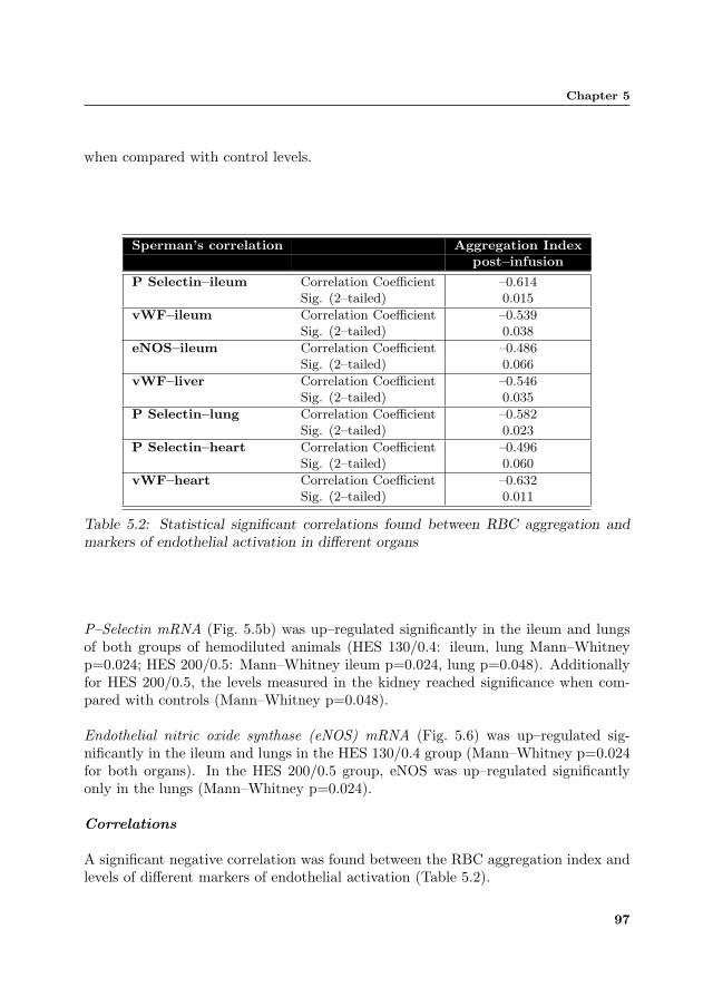
Sperman’s correlation Aggregation Indexpost–infusion
P Selectin–ileum Correlation Coefficient –0.614Sig. (2–tailed) 0.015
vWF–ileum Correlation Coefficient –0.539Sig. (2–tailed) 0.038
eNOS–ileum Correlation Coefficient –0.486Sig. (2–tailed) 0.066
vWF–liver Correlation Coefficient –0.546Sig. (2–tailed) 0.035
P Selectin–lung Correlation Coefficient –0.582Sig. (2–tailed) 0.023
P Selectin–heart Correlation Coefficient –0.496Sig. (2–tailed) 0.060
vWF–heart Correlation Coefficient –0.632Sig. (2–tailed) 0.011
Table 5.2: Statistical significant correlations found between RBC aggregation andmarkers of endothelial activation in different organs
P–Selectin mRNA (Fig. 5.5b) was up–regulated significantly in the ileum and lungsof both groups of hemodiluted animals (HES 130/0.4: ileum, lung Mann–Whitneyp=0.024; HES 200/0.5: Mann–Whitney ileum p=0.024, lung p=0.048). Additionallyfor HES 200/0.5, the levels measured in the kidney reached significance when com-pared with controls (Mann–Whitney p=0.048).
Endothelial nitric oxide synthase (eNOS) mRNA (Fig. 5.6) was up–regulated sig-nificantly in the ileum and lungs in the HES 130/0.4 group (Mann–Whitney p=0.024for both organs). In the HES 200/0.5 group, eNOS was up–regulated significantlyonly in the lungs (Mann–Whitney p=0.024).
Correlations
A significant negative correlation was found between the RBC aggregation index andlevels of different markers of endothelial activation (Table 5.2).
97

Blood Rheology and Endothelial (Dys)function
Figure 5.3: Hypoxic oxidative stress during 3 hours of acute IHD (a) Plasma Malon-dialdehyde (MDA): The values are represented as mean (symbols) and standard errorof the mean (bars); (b) Hydrogen peroxide (H2O2) production in heart, lung, kidney,liver, ileum tissue biopsies. Box plots graph data represent statistical values (see leg-end Fig. 5.2). (c) Diaminobenzidine (DAB) staining of H2O2–producing cells (browncoloration) in paraformaldehyde–fixed sections of ileum mucosa of hemodiluted and(d) sham–operated animals.
98

Chapter 5
Figure 5.4: von Willebrand factor (vWF) (a) vWF relative gene expression of in theheart, lung, kidney, liver, ileum tissue biopsies after 3 h IHD. HES 130/0.4 Mann-Whitney: ileum, kidney, lung and heart p=0.024, liver p=0.048. HES 200/0.5 Mann–Whitney: ileum, kidney, and heart p=0.024, liver p=0.048, lung p=0.095. (b) vWFplasma concentrations.
5.4 Discussion
Using our experimental model of acute isovolemic hemodilution we documented an im-mediate pro–thrombotic and pro–inflammatory endothelial activation in heart, lung,kidney, liver, and ileum, accompanied by a dramatic drop in erythrocyte aggregation.Erythrocyte aggregability correlated significantly with markers of endothelial activa-tion suggesting a causality effect.
The dynamic rheological properties of blood are defined mainly by the coordinatedself–organization of RBCs advancing in the arterio–venular direction21. RBC hyper-aggregation is nowadays a generally recognized pathogenic factor, mainly due to clini-cal observation of increased RBC aggregation during disorders associated with macroand/or microvascular impairment, e.g. hypertension, diabetes mellitus, and chronicvenous insufficiency1,2,22. Hypo-aggregation of RBCs has been never described in apathologic context. Given the strong conditioning effect of RBC aggregation on bloodrheology, and thus on mechanic endothelial activation, we hypothesized low RBC ag-
99

Blood Rheology and Endothelial (Dys)function
Figure 5.5: E–Selectin (a) and P–Selectin (b) relative gene expression of in the heart,lung, kidney, liver, ileum tissue biopsies after 3 h IHD. E–Selectin: Mann–WhitneyHES 130/0.4: ileum p=0.048, lungs p=0.024; HES 200/0.5: ileum p=0.024. P–Selectin: Mann–Whitney HES 130/0.4: ileum, lung p=0.024; HES 200/0.5: ileump=0.024, lung p=0.048, kidney p=0.048.
Figure 5.6: Endothelial nitric oxide synthase(eNOS) relative gene expression in the heart,lung, kidney, liver, ileum tissue biopsies after3 h IHD. HES 130/0.4 Mann–Whitney: ileum,lungs p=0.024. HES 200/0.5 Mann–Whitneylungs p=0.024.
100

Chapter 5
gregation to be a pathogenic co–factor in endothelial activation during acute isov-olemic hemodilution. To verify this hypothesis, we induced acute isovolemic hemod-ilution in an “aggregating species”, the pig23, and investigated simultaneously thehypoxic oxidative stress, red blood cell aggregation, and gene regulation of von Wille-brand factor, E– and P–Selectin, and eNOS, as markers of endothelial activation.Hemodilution, by reducing the number of circulating RBCs is expected to decreasethe oxygen–carrying capacity of blood and oxygen delivery to the tissue. However,during moderate levels of hemodilution, reduction of the systemic hematocrit up to50% is compensated with an increased blood flow velocity and decreased diffusionaloxygen exit from arterioles, resulting in augmented or maintained oxygen delivery totissue24. In addition, reduction of systemic Hct during intentional hemodilution isnot mirrored at the microcirculatory level, with capillary Hct sustained near controllevels25, thus maintaining tissue oxygenation.In an experimental animal study, Deem en al.26 showed that acute normovolemichemodilution in healthy rabbits resulted in improved gas–exchange efficiency, asshown by higher arterial PO2, lower alveolar–arterial PO2 difference, and increasedexpired NO. They postulated that the improvement in oxygenation appeared to berelated to increased uniformity of pulmonary blood flow, and/or an increase in con-centration of the vaso– and bronchodilator substance NO. Our data support thisassumption and consistently show an up–regulation of eNOS in the lung tissue duringacute hemodilution.In our approach to detect changes in tissue oxygenation, we tested ex–vivo the mi-tochondrial (dys)function in the vital organs (heart, lung, kidney, liver, ileum) ofhemodiluted animals, reflected by the production of reactive oxygen species whenoxidizing pyruvate and succinate. Thus, we aimed at detecting mitochondria thatwere pre–exposed to hypoxia during hemodilution. The production of superoxide andhydroxyl radicals, measured as H2O2 generation, was similar at all time points insham–operated and hemodiluted animals, which indicates that a similar hypoxic ox-idative stress was present, and oxygen delivery to the tissue during hemodilution wasmaintained. However, different organs seemed to have different exposure to hypoxia,with a more profound mitochondrial dysfunction in abdominal organs (ileum, kidney,liver) versus a preserved function of mitochondria in the myocardium and lung tissue.The results found in tissue biopsies were mirrored by the plasma MDA determina-tions, that showed similar relative increase in systemic lipid peroxidation productsduring three experimental hours when hemodiluted animals and sham–operated an-imals were compared. These results suggest that, at least in this animal model, theperioperative stress and the anesthetic management are more important triggers ofoxidative stress in abdominal organs, than hemodilution per–se.Because hypoxic stress seems to be negligible in this model of acute isovolemic hemod-ilution, we suggest that the effects observed in endothelial activation were mainly dueto the drop in RBC aggregation.
101

Blood Rheology and Endothelial (Dys)function
Erythrocyte aggregation and endothelium–dependent pro–thrombotic activation
First reliable observations on the involvement of red blood cell in the process ofclot formation were made by Turitto et al.27 who showed that under flow conditionsplatelet adhesion and thrombus formation increase as hematocrit values increase from10% to 70%. They hypothesized that red cells may have a significant influence onhemostasis and thrombosis and the nature of this effect is apparently related to theflow conditions. More recently, it was demonstrated that erythrocytes markedly in-crease platelet eicosanoid formation, promote release of intracellular platelet granulecomponents, and induce recruitment of additional platelets from the microenviron-ment into the forming thrombus28,29.The data presented in this study suggest a new pathway for erythrocyte involvementin clot formation: due to their function to aggregate, erythrocyte could modulateendothelial activation with von Willebrand factor release, with a subsequent pro–thrombotic effect. von Willebrand factor, which is stored in the endothelial Weibel–Palade storage granules, has unique biomechanical properties and a critical biologicalrole as an adhesive protein. It mediates the adhesion of platelets to an injured vascu-lar wall by binding on platelet surface and to collagen in the subendothelium. vWFis one of the most potent activators of platelets. Activation of platelets causes themto release additional vWF from their α–storage granules. Increased levels of plasmavon Willebrand factor contribute directly to thrombosis, impeding the normal flow ofcirculating blood30. In our experiment, acute isovolemic hemodilution was followedby a dramatic drop in red blood cell aggregation, which resulted in immediate pro–thrombotic endothelial activation as shown by a systemic increase in plasma vWFlevels. Analysis of vWF mRNA expression levels in different vital organs showed aconcomitant up–regulation in heart, lungs, kidney, liver, and small intestine. In ad-dition, low red blood cell aggregation states were significantly associated with highvWF mRNA expression in heart, liver and ileum suggesting maybe a causality effect.An understanding of how disturbed blood flow might lead to disease is now emerging.Transferring this knowledge to a clinical relevant situation, as the one of the patientsundergoing on–pump cardiopulmonary bypass, we hypothesize that lower incidenceof thrombotic events could be achieved by avoiding excessive peri–operative hemodi-lution.
Low erythrocyte aggregation and endothelium–dependent pro–inflammatory response
The presence of a high RBC aggregation proved already its relevance in diagnosingthe patients’ inflammatory status, using clinical observations of positive correlationsbetween enhanced RBC aggregation and high plasma levels of C–reactive protein andfibrinogen31. There is also evidence that RBC hyper-aggregation enhances the ten-
102

Chapter 5
dencies of leukocytes to adhere to the postcapillary endothelium, a process recognizedas essential in inflammation. Pearson et al.32 reported that increased RBC aggrega-tion was associated with increased adhesion of white blood cells to the endothelium,possibly because of an enhanced probability of contact between leukocytes and thepostcapillary venular wall.In this study we discovered that RBC hypo-aggregation, documented in our modelof acute isovolemic hemodilution, was statistically significant correlated with up–regulation of endothelial adhesion molecules, E– and P–Selectins, especially in lungsand small intestine. E– and P–Selectins belong to the Selectin family of adhesionmolecules and both have been reported to increase in circulation or at lesion sitesof several diseases reflecting endothelial activation. Selectins play important roles inthe inflammatory responses by facilitating leukocyte rolling and leukocytes activa-tion33,34.Translation of these data in clinical terms suggests that acute hemodilution may leadto inflammatory stress of pulmonary capillaries. Subsequent diffusion limitation maybe expected. Similar, an increased inflammatory response in the small intestine asso-ciated with acute hemodilution, might contribute to a loss in barrier function of theintestinal mucosa with subsequent translocation of endotoxins and/or bacteria.
Conclusions
The data presented in this study show that acute isovolemic hemodilution definitelytriggers endothelial activation. Since the effects of hypoxic oxidative stress seem to benegligible in this model, red blood cell hypo-aggregation could be considered as a newpathophysiologic mechanism which could be held responsible for pro–inflammatoryand pro–coagulatory endothelial activation. We hypothesize that a reduced inflamma-tory response and a lower incidence of thrombotic events will be achieved by avoidingexcessive peri–operative hemodilution during on–pump cardiopulmonary bypass.
Acknowledgements: We acknowledge the entire technician team of the Exper-imental Animal Facility and professionals of Surgical Research Laboratory, UMCG,for their time, physical and intellectual effort invested in the present study. We thankHaemoScan (Groningen, The Netherlands) and Nodia BV (Amsterdam, The Nether-lands) for their support.
103

Bibliography
References
1. Martinez M, Vaya A, Server R, Gilsanz A, Aznar J. Alterations in erythrocyte aggre-gability in diabetics: the influence of plasmatic fibrinogen and phospholipids of the redblood cell membrane. Clin. Hemorheol. Microcirc. 1998;18:253–8.
2. Cicco G, Pirrelli A. Red blood cell (RBC) deformability, RBC aggregability and tissueoxygenation in hypertension. Clin. Hemorheol. Microcirc. 1999;21:169–77.
3. Baskurt OK and Meiselman HJ. Cellular determinants of low shear blood viscosity.Biorheology. 1997;34:235–247.
4. Vicaut E. Opposite effects of red blood cell aggregation on resistance to blood flow. J.Cardiovasc. Surg. (Torino). 1995;36:361–368.
5. Cokelet GR and Goldsmith HL. Decreased hydrodynamic resistance in the two-phaseflow of blood through small vertical tubes at low flow rates. Circ. Res. 1991;68:1–17.
6. Fisher AB, Chien S, Barakat AI, Nerem RM. Endothelial cellular response to alteredshear stress. Am. J. Physiol. Lung Cell Mol. Physiol. 2001;281:L529–33.
7. Nilius B, Droogmans G. Ion Channels and their functional role in vascular endothelium.Physiological Reviews. 2001;4:1415–9.
8. Baskurt OK, Yalcin O, Ozdem S, Armstrong JK, and Meiselman HJ. Modulation of en-dothelial nitric oxide synthase expression by red blood cell aggregation. Am. J. Physiol.Heart Circ. Physiol. 2004;286:H222–H228.
9. Popel AS, Johnson PC, Kameneva MV, Wild MA. Capacity for red blood cell aggrega-tion is higher in athletic mammalian species than in sedentary species. J. Appl. Physiol.1994;77:1790–4.
10. Bishop JJ, Nance PR, Popel AS, Intaglietta M, Johnson PC. Effect of erythrocyte aggre-gation on velocity profiles in venules. Am. J. Physiol. Heart Circ. Physiol. 2001;280:222–36.
11. Morariu AM, Vd Plaats A, V Oeveren W, ’T Hart NA, Leuvenink HG, Graaff R, PloegRJ, Rakhorst G. Hyperaggregating effect of hydroxyethyl starch components and Uni-versity of Wisconsin solution on human red blood cells: a risk of impaired graft perfusionin organ procurement? Transplantation. 2003;76:37–43.
12. Morariu AM, Gu YJ, Huet RC, Siemons WA, Rakhorst G, Oeveren WV. Red blood cellaggregation during cardiopulmonary bypass: a pathogenic cofactor in endothelial cellactivation? Eur. J. Cardiothorac. Surg. 2004;26:939–46.
13. Hardeman MR, Goedhart PT, Lettinga KP. Laser-assisted optical rotational cell anal-yser (L.O.R.C.A.); I. A new instrument for measurement of various structural hemorhe-ological parameters. Clinical Hemorheology. 1994;14:605–618.
104

Chapter 5
14. Graaff R, Gu YJ, Boonstra PW, van Oeveren W, Rakhorst G. Analysis of red bloodcell aggregation in cardio-pulmonary bypass (CPB) surgery. Int. J. Artif. Organs.2004;27:488–94.
15. Esterbauer H, Cheeseman KH Determination of aldehydic lipid peroxidation products:malonaldehyde and 4-hydroxynonenal. Methods Enzymol. 1990;186:407–21.
16. Pou S., Huang Y. I., Bhan A., Bhadti V. S., Hosmane R. S., Wu S. Y., Cao G. L. andRosen G. M. A Fluorophore-Containing Nitroxide as a Probe to Detect Superoxide andHydroxyl Radical Generated by Stimulated Neutrophils. Anal. Biochem. 1993;212:85–90.
17. Li B, Gutierrez PL, Blough NV. Trace determination of hydroxyl radical in biologicalsystems. Anal. Chem. 1997;69:4295–302.
18. Chen Q, Vazquez EJ, Moghaddas S, Hoppel CL. Production of reactive oxygen speciesby mitochondria: central role of complex III. J. Biol. Chem. 2003;278:36027–31.
19. Li B, Gutierrez PL, Blough NV. Trace determination of hydroxyl radical using fluores-cence detection. Methods Immunol. 1999;300:202–216.
20. Harboe A. Method for determination of hemoglobin in plasma by near-ultraviolet spec-trophotometry. Scand. Clin. Lab. Invest. 1959;11:66–70.
21. Mchedlishvili G. Disturbed blood flow structuring as critical factor of hemorheologicaldisorders in microcirculation. Clin. Hemorheol. Microcirc. 1998;19:315–25.
22. Krieger E, van Der Loo B, Amann-Vesti BR, Rousson V, Koppensteiner R. C-reactiveprotein and red cell aggregation correlate with late venous function after acute deepvenous thrombosis. J. Vasc. Surg. 2004;40:644–9.
23. Windberger U, Bartholovitsch A, Plasenzotti R, Korak KJ, Heinze G. Whole bloodviscosity, plasma viscosity and erythrocyte aggregation in nine mammalian species: ref-erence values and comparison of data. Exp. Physiol. 2003;88:431-40.
24. Tsai AG, Friesenecker B, McCarthy M, Sakai H, Intaglietta M. Plasma viscosity regu-lates capillary perfusion during extreme hemodilution in hamster skinfold model. Am.J. Physiol. 1998;275:H2170–80.
25. Lipowsky HH, and Firrell JC. Microvascular hemodynamics during systemic hemodilu-tion and hemoconcentration. Am. J. Physiol. Heart Circ. Physiol. 1986;250:H908–H922.
26. Deem S, Hedges RG, McKinney S, Polissar NL, Alberts MK, Swenson ER. Mechanismsof improvement in pulmonary gas exchange during isovolemic hemodilution. J. Appl.Physiol. 1999;87:132–41.
27. Turitto VT, Weiss HJ. Red blood cells: their dual role in thrombus formation. Science.1980;207:541–543.
105

Bibliography
28. Valles J, Santos MT, Aznar J, Marcus AJ, Martinez-Sales V, Portoles M, BroekmanMJ, Safier LB. Erythrocytes metabolically enhance collagen-induced platelet respon-siveness via increased thromboxane production, ADP release, and recruitment. Blood.1991;78:154–162.
29. Santos MT, Valles J, Aznar J, Marcus AJ, Broekman MJ, Safier LB. Prothrombotic ef-fects of erythrocytes on platelet reactivity: reduction by aspirin. Circulation. 1997;95:63–68.
30. Ruggeri ZM. Role of von Willebrand factor in platelet thrombus formation. Ann. Med.2000;32:2–9.
31. Ami RB, Barshtein G, Zeltser D, Goldberg Y, Shapira I, Roth A, Keren G, MillerH, Prochorov V, Eldor A, Berliner S, Yedgar S. Parameters of red blood cell aggre-gation as correlates of the inflammatory state. Am. J. Physiol. Heart Circ. Physiol.2001;280:H1982–8.
32. Pearson MJ, Lipowsky HH. Influence of erythrocyte aggregation on leukocyte margina-tion in postcapillary venules of rat mesentery. Am. J. Physiol. Heart Circ. Physiol.2000;279:H1460–71.
33. Ley K, Allietta M, Bullard DC, Morgan S. Importance of E-selectin for firm leukocyteadhesion in vivo. Circ. Res. 1998;83:287–94.
34. Kunkel EJ, Chomas JE, Ley K. Role of primary and secondary capture for leukocyte
accumulation in vivo. Circ. Res. 1998;82:30–8.
106