university of groningen dental implants in maxillofacial
TRANSCRIPT

University of Groningen
Dental implants in maxillofacial prosthodonticsKorfage, Anke
DOI:10.1016/j.bjoms.2014.05.01310.1016/j.ijom.2013.04.00310.1002/hed.24053IMPORTANT NOTE: You are advised to consult the publisher's version (publisher's PDF) if you wish to cite fromit. Please check the document version below.
Document VersionPublisher's PDF, also known as Version of record
Publication date:2015
Link to publication in University of Groningen/UMCG research database
Citation for published version (APA):Korfage, A. (2015). Dental implants in maxillofacial prosthodontics: An asset in head and neck cancer andSjögren's syndrome patients. University of Groningen. https://doi.org/10.1016/j.bjoms.2014.05.013,https://doi.org/10.1016/j.ijom.2013.04.003, https://doi.org/10.1002/hed.24053
CopyrightOther than for strictly personal use, it is not permitted to download or to forward/distribute the text or part of it without the consent of theauthor(s) and/or copyright holder(s), unless the work is under an open content license (like Creative Commons).
The publication may also be distributed here under the terms of Article 25fa of the Dutch Copyright Act, indicated by the “Taverne” license.More information can be found on the University of Groningen website: https://www.rug.nl/library/open-access/self-archiving-pure/taverne-amendment.
Take-down policyIf you believe that this document breaches copyright please contact us providing details, and we will remove access to the work immediatelyand investigate your claim.
Downloaded from the University of Groningen/UMCG research database (Pure): http://www.rug.nl/research/portal. For technical reasons thenumber of authors shown on this cover page is limited to 10 maximum.
Download date: 11-03-2022

90
Chapter 4Recommendations for implant-retained nasal prostheses after ablative tumour surgery: minimal surgical aftercare, high implant survival and satisfied patients
This chapter is an edited version of: Anke Korfage, Gerry M. Raghoebar, Willem D. Noorda, Boudewijn E. Plaat, Arjan Vissink, Anita Visser. Recom-mendations for implant-retained nasal prostheses after ablative tumour surgery: minimal surgical aftercare, high implant survival and satisfied patients. Accepted for publication in Head and Neck.

92 93
Introduction Nasal defects can occur as a result of ablative oncologic surgery, trauma and congenital disorders, with ablative oncologic surgery being the most common1,2. Congenital absent noses are extremely rare2. For emotional and cosmetic reasons, nasal defects can be very distressing to patients. These defects can impair the patients’ social life 3.
Currently, nasal defects are reconstructed with surgical techniques (e.g., forehead flap)4-6, prosthetic techniques3,7,8, or a combination of these two. Surgical reconstruction is difficult to perform and its outcome has not been described in large patient numbers. Furthermore, treatment of a local tumour recurrence may necessitate removal of the surgical reconstruction. An advantage of rehabilitation with nasal prostheses above surgical reconstruction is that the defect resulting from ablative tumour surgery can be observed in total, allowing for thorough oncological inspections. Furthermore, nasal prostheses match a natural cosmetic situation3. Therefore, total rhinectomy defects resulting from tumour surgery are preferably rehabilitated with nasal prostheses. Retention of nasal prostheses include fixation on glasses and gluing to the skin with silicone-based adhesives7. None of these fixation methods are optimal because they limit the patients’ activities. Especially in warm climates or in a moist environment such as the nasal cavity, the skin glue can dissolve or fail to attach to the skin. In addition, it is difficult to correctly position the prosthesis with skin adhesives. Adhesives can cause skin irritation, allergic reactions, and discolouration and deterioration of the edges of the silicone prostheses.In 1979 the use of extra oral endosseous implants for retention of craniofacial prostheses was introduced by Brånemark et al.9 Since then, endosseous implants have acquired an important position in the prosthetic rehabilitation of patients with craniofacial defects, both in irradiated and non-irradiated patients3,9-12. Advantages of fixating craniofacial prostheses (especially nasal prostheses) on endosseous implants include easier maintenance of these prostheses (no glue remnants), easier mounting of prostheses in the right position and improved retention compared to adhesive prostheses. Therefore patients’ satisfaction with implant-retained craniofacial prostheses is higher compared with adhesive prostheses9,13,14.In the literature overall implant survival of implants used for implant-retained nasal prostheses varies largely between 50%-100% with a median survival of 85.5% for non-irradiated patients and 80.0% for irradiated patients1,15-26. However, treatment protocols of inserting implants for implant-retained nasal prostheses in these studies were all different. Amongst others, there is no consensus with regard to implant location, type and length of implants, treatment of irradiated and non-irradiated patients and dentate patients. Furthermore, aftercare and patients’ satisfaction are hardly discussed. The number of patients is usually low and follow-up periods vary. Currently, only a few studies report on the results of nasal implants with a long follow-up19,21,24. Therefore, the aim of this study was to assess aftercare, clinical outcome of the implants
Abstract BackgroundNasal defects resulting from tumour resection are preferably rehabilitated with implant-retained nasal prostheses. Aftercare, clinical outcome of the implants and patients’ satisfaction with implant-retained nasal prostheses were assessed.
MethodsTwenty-eight consecutive patients needing total rhinectomy due to tumour resection between 1998 and 2013 were treated according to a standardized protocol with two implants in the nasal floor. Surgical and prosthetic aftercare was scored using patient records. Finally in 2014 skin reaction, peri-implant bone loss and patients’ satisfaction were assessed in all 13 still living patients.
ResultsIn total 56 implants were inserted (median follow-up 35.1 months, IQR 8.9-63.3). Implant survival was 96.4%, was independent of radiotherapy. Peri-implant skin was healthy and patients’ satisfaction high. Longevity of the prostheses was limited.
ConclusionsRehabilitation of nasal defects resulting from total rhinectomy with implant-retained nasal prostheses according to our protocol resulted in high patient satisfaction and favourable treatment outcome.

94 95
7 or 10 mm, Nobel Biocare, Gothenburg, Sweden) were inserted according to a 2-stage procedure. Before implant insertion, the prominent bony lip of the piriform aperture was trimmed and the anterior part of the nasal septum and the inferior turbinates were removed. Next, the two implants were inserted via the nasal floor into the maxillary bone at an angle of 60˚ with the horizontal transversal plane (Figure 1). The implants were covered with a split skin graft.
Figure 2. Superstructure with bar combined with magnet on two implants inserted in nasal floor in a 73-years old man after total rhinectomy: angulation of the implants (a), and inside of the prosthesis (b)
Figure 3. Implant-retained nasal prosthesis in situ in an 84-years old woman after total rhinectomy
and patients’ satisfaction in a relatively large group of patients rehabilitated with an implant-retained nasal prosthesis after total rhinectomy due to tumour ablation. All patients were treated according to a standardized protocol with two endosseous implants in the nasal floor. Both dentate and edentulous patients were included in this study.
Materials and MethodsPatients and implantsAll consecutive patients (n=28) treated between 1998 and 2013 with implant-retained nasal prostheses after total rhinectomy due to tumour resection in the Department of Oral and Maxillofacial Surgery and the Department of Otorhinolaryngology/ Head and Neck Surgery of the University Medical Center Groningen (UMCG) were included in this analysis. All patients underwent both tumour surgery and implant insertion in the UMCG. Treatment planning and implant insertion were carried out by one experienced oral and maxillofacial surgeon within the setting of a multidisciplinary team specialized in the treatment and rehabilitation of patients with extra oral defects. This multidisciplinary team was composed of maxillofacial surgeons, maxillofacial prosthodontists, ear, nose and throat surgeons and plastic surgeons.
Figure 1. Planning of implant angulations (approximately 60°) with the horizontal plane (a) and position in nasal floor (b) in a dentate patient
Treatment protocolPreoperative available bone height was measured on lateral radiographs in edentulous patients or computed tomographic (CT) scans or conebeam CTs (CBCTs) in dentate patients. From 2010, implant planning in dentate patients was fully digitalized from 2010 as described in detail by Van der Meer et al.27
In all 28 patients, two implants (Brånemark dental implants with diameter 3.75 mm, length

96 97
Patients’ satisfaction In all living patients in 2014, patients’ satisfaction with the implant-retained nasal prosthesis was scored. Patients’ satisfaction was expressed on a 10-point rating scale (1–10); ‘‘1’’ being completely dissatisfied and ‘‘10’’ being completely satisfied as was also done in the study of Schoen et al9.
Statistical analysisThe data were analysed using non-parametric tests. A Mann-Whitney U test was used to assess differences between irradiated and non-irradiated patients and a Wilcoxon signed-rank test was used to assess differences in bone level in time (IBM SPSS Statistics 22). In all tests a p-value < 0.05 was considered statistically significant.
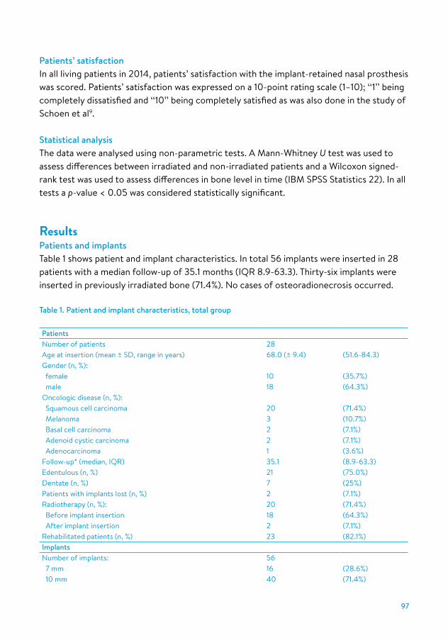
ResultsPatients and implantsTable 1 shows patient and implant characteristics. In total 56 implants were inserted in 28 patients with a median follow-up of 35.1 months (IQR 8.9-63.3). Thirty-six implants were inserted in previously irradiated bone (71.4%). No cases of osteoradionecrosis occurred.
Table 1. Patient and implant characteristics, total group
PatientsNumber of patients 28Age at insertion (mean ± SD, range in years) 68.0 (± 9.4) (51.6-84.3)Gender (n, %): female 10 (35.7%) male 18 (64.3%)Oncologic disease (n, %): Squamous cell carcinoma 20 (71.4%) Melanoma 3 (10.7%) Basal cell carcinoma 2 (7.1%) Adenoid cystic carcinoma 2 (7.1%) Adenocarcinoma 1 (3.6%)Follow-up* (median, IQR) 35.1 (8.9-63.3)Edentulous (n, %) 21 (75.0%)Dentate (n, %) 7 (25%)Patients with implants lost (n, %) 2 (7.1%)Radiotherapy (n, %): 20 (71.4%) Before implant insertion 18 (64.3%) After implant insertion 2 (7.1%)Rehabilitated patients (n, %) 23 (82.1%)Implants Number of implants: 56 7 mm 16 (28.6%) 10 mm 40 (71.4%)
To ensure adequate osseointegration, a healing time of at least three months was considered before uncovering. In cases where postoperative radiotherapy was performed, the osseointegration period was increased with three months9. Patients could wear an adhesive nasal prosthesis in the meantime. During the second stage, i.e. the procedure for uncovering the implants and abutment connection, the implants were uncovered under local anaesthesia. The skin around the implants was, when applicable, thinned subcutaneously to prevent pocket formation and inflammation around the implants. Thereafter, the abutments were connected on the implants. To keep the soft peri-implant tissues in place, gauze soaked in ointment was wrapped around the abutments. After one week, the sutures were removed and new gauze was wrapped around the abutments. Three weeks after abutment connection, the maxillofacial prosthodontist started fabricating the superstructures and nasal prostheses. The implant-retained nasal prostheses were made of intrinsically pigmented silicone elastomers. Retention was achieved with a bar-clip retention system (Haderclips or Friatec clips (former Friadent, now Dentsply IH GmbH, Mannheim, Germany). Since 2011 the bars were milled from titanium (with a macro-dolder clip, Cendres+Métaux, Biel-Bienne, Switserland) and combined with a Steco magnet (Steco-System-Technik, Hamburg, Germany) (Figure 2 and 3).
Surgical and prosthetic aftercareSurgical and prosthetic aftercare from implant insertion to last available follow-up was retrospectively scored in all patients by assessing patient records according to Visser et al.1 Surgical aftercare included subcutaneous tissue reduction, split skin grafts and the need for ointment application in case of peri-implant skin infections. Prosthetic aftercare was scored as need for clip repairs, fabrication of new prostheses, repair of superstructure, fabrication of new superstructure, consultation for activation or repair of clips, hygiene instructions, and tightening of loose abutments or superstructure.
Clinical and radiographic assessmentsAll living patients were recalled for a final clinical assessment in 2014 to score skin reaction and peri-implant bone loss. Skin reactions were scored according to the skin reaction scale of Tolman and Taylor20 as: (0), no irritation, (1) slight redness, (2) tissue redness and moist but no granulation tissue present, (3) tissue redness and moist with granulation tissue present, or (4) active infection present requiring removal of abutment.Rotational radiographs (orthopantomograms) were made at the time of abutment connection surgery and during the last follow-up to evaluate the implant-surrounding bone height. Peri-implant bone loss was classified according to Geertman et al.28: (0), no apparent bone loss, (1) reduction of bone level not exceeding one-third of the length of the implant, (2) reduction of bone level exceeding one-third of the length of the implant but not exceeding one-half of the length of the implant, (3) reduction of bone level exceeding one-half of the length of the implant, (4) total reduction of bone along the implant.
96 97
Patients’ satisfaction In all living patients in 2014, patients’ satisfaction with the implant-retained nasal prosthesis was scored. Patients’ satisfaction was expressed on a 10-point rating scale (1–10); ‘‘1’’ being completely dissatisfied and ‘‘10’’ being completely satisfied as was also done in the study of Schoen et al9.
Statistical analysisThe data were analysed using non-parametric tests. A Mann-Whitney U test was used to assess differences between irradiated and non-irradiated patients and a Wilcoxon signed-rank test was used to assess differences in bone level in time (IBM SPSS Statistics 22). In all tests a p-value < 0.05 was considered statistically significant.
ResultsPatients and implantsTable 1 shows patient and implant characteristics. In total 56 implants were inserted in 28 patients with a median follow-up of 35.1 months (IQR 8.9-63.3). Thirty-six implants were inserted in previously irradiated bone (71.4%). No cases of osteoradionecrosis occurred.
Table 1. Patient and implant characteristics, total group
PatientsNumber of patients 28Age at insertion (mean ± SD, range in years) 68.0 (± 9.4) (51.6-84.3)Gender (n, %): female 10 (35.7%) male 18 (64.3%)Oncologic disease (n, %): Squamous cell carcinoma 20 (71.4%) Melanoma 3 (10.7%) Basal cell carcinoma 2 (7.1%) Adenoid cystic carcinoma 2 (7.1%) Adenocarcinoma 1 (3.6%)Follow-up* (median, IQR) 35.1 (8.9-63.3)Edentulous (n, %) 21 (75.0%)Dentate (n, %) 7 (25%)Patients with implants lost (n, %) 2 (7.1%)Radiotherapy (n, %): 20 (71.4%) Before implant insertion 18 (64.3%) After implant insertion 2 (7.1%)Rehabilitated patients (n, %) 23 (82.1%)Implants Number of implants: 56 7 mm 16 (28.6%) 10 mm 40 (71.4%)
To ensure adequate osseointegration, a healing time of at least three months was considered before uncovering. In cases where postoperative radiotherapy was performed, the osseointegration period was increased with three months9. Patients could wear an adhesive nasal prosthesis in the meantime. During the second stage, i.e. the procedure for uncovering the implants and abutment connection, the implants were uncovered under local anaesthesia. The skin around the implants was, when applicable, thinned subcutaneously to prevent pocket formation and inflammation around the implants. Thereafter, the abutments were connected on the implants. To keep the soft peri-implant tissues in place, gauze soaked in ointment was wrapped around the abutments. After one week, the sutures were removed and new gauze was wrapped around the abutments. Three weeks after abutment connection, the maxillofacial prosthodontist started fabricating the superstructures and nasal prostheses. The implant-retained nasal prostheses were made of intrinsically pigmented silicone elastomers. Retention was achieved with a bar-clip retention system (Haderclips or Friatec clips (former Friadent, now Dentsply IH GmbH, Mannheim, Germany). Since 2011 the bars were milled from titanium (with a macro-dolder clip, Cendres+Métaux, Biel-Bienne, Switserland) and combined with a Steco magnet (Steco-System-Technik, Hamburg, Germany) (Figure 2 and 3).
Surgical and prosthetic aftercareSurgical and prosthetic aftercare from implant insertion to last available follow-up was retrospectively scored in all patients by assessing patient records according to Visser et al.1 Surgical aftercare included subcutaneous tissue reduction, split skin grafts and the need for ointment application in case of peri-implant skin infections. Prosthetic aftercare was scored as need for clip repairs, fabrication of new prostheses, repair of superstructure, fabrication of new superstructure, consultation for activation or repair of clips, hygiene instructions, and tightening of loose abutments or superstructure.
Clinical and radiographic assessmentsAll living patients were recalled for a final clinical assessment in 2014 to score skin reaction and peri-implant bone loss. Skin reactions were scored according to the skin reaction scale of Tolman and Taylor20 as: (0), no irritation, (1) slight redness, (2) tissue redness and moist but no granulation tissue present, (3) tissue redness and moist with granulation tissue present, or (4) active infection present requiring removal of abutment.Rotational radiographs (orthopantomograms) were made at the time of abutment connection surgery and during the last follow-up to evaluate the implant-surrounding bone height. Peri-implant bone loss was classified according to Geertman et al.28: (0), no apparent bone loss, (1) reduction of bone level not exceeding one-third of the length of the implant, (2) reduction of bone level exceeding one-third of the length of the implant but not exceeding one-half of the length of the implant, (3) reduction of bone level exceeding one-half of the length of the implant, (4) total reduction of bone along the implant.

98 99
With regard to prosthetic aftercare in 23 patients with an implant-retained prosthesis, 65.2% of the patients were in need for (repeated) hygiene instructions and 30.4% of the patients needed (repeated) repair of clips (Table 2).Median time between implant insertion and implant-retained nasal prosthesis placement in 23 patients was 6.8 months (IQR 5.5-8.9). In 7 patients only one implant-retained prosthesis was made because the patient deceased before a new prosthesis could be made (n=3) or because the first prosthesis was still acceptable at time of last follow-up (n=4). In total, 47 replacing prostheses were made in the other 16 patients with a median life span of these prostheses of 11.6 months (IQR 6.8-15.2) (Figure 4). Main reason for prosthesis replacement was discolouration (Table 2).
Clinical and radiographic assessmentsThirteen patients were available for clinical measurements in 2014 (Table 3).
Table 3. Implant characteristics of the 13 patients that could be recalled during last follow-up in 2014
PatientsAge at insertion (mean ± SD, range in years) 65.8 ± 9.5 (51.6-83.8)Gender (n, %): female 5 male 8Oncologic disease (n, %): Squamous cell carcinoma 8 (61.5%) Melanoma 2 (15.4%) Basal cell carcinoma 1 (7.7%) Adenoid cystic carcinoma 1 (7.7%) Adenocarcinoma 1 (7.7%)Follow-up (median, IQR) 39.7 (13.0-62.2)Edentulous (n, %) 6 (46.2%)Dentate (n, %) 7 (53.8%)Patients with implants lost (n, %) 0 (0%)Radiotherapy (n, %): 10 (76.9%) Before implant insertion 9 (69.2%) After implant insertion 1 (7.7%)Rehabilitated patients (n, %) 13 (100%)Implants Number of implants: 26 7 mm 8 (30.8%) 10 mm 18 (69.2%)Lost implants (n, %) 0 (0%)Insertion during ablative tumour surgery (n, %) 10 (38.5%)Implants used for prostheses (n, %) 26 (100%)
In total 11 patients had died (5 due to tumour-related disease, 6 non-tumour-related), three patients had been lost for follow-up and one patient had residual tumour, without involvement of the peri-implant skin, delaying prosthetic rehabilitation. Median follow-up of
Lost implants (n, %) 2 (3.6%)Insertion during ablative tumour surgery (n, %) 42 (75.0%)Implants used for prostheses (n, %) 46 (82.1%)
* Follow-up is defined as time between implant insertion and last follow-up or time between implant insertion and patient deceased.
Two implants failed after 13 and 53 months (1x 10 mm, 1x 7 mm, respectively) in two patients, one irradiated and one non-irradiated, resulting in an overall implant survival rate of 96.4%. In one patient the lost implant was successfully replaced by a new implant. In the other patient general anaesthesia in case of implant re-insertion was a high risk procedure due to comorbidity. This patient functioned well with a magnet-retained nasal prosthesis on the remaining implant. In total 10 implants (in 5 patients) were not used for prosthetic rehabilitation, due to death of 4 patients (3 tumour-related, 1 non-tumour-related) before the rehabilitation could start and due to residual tumour, delaying prosthetic rehabilitation (1 patient).
Surgical and prosthetic aftercareWith regard to surgical aftercare, subcutaneous tissue reduction was performed in 2 out of 28 patients (Table 2). No other surgical interventions were needed.
Table 2. Surgical and prosthetic aftercare given and main reasons for replacing implant-retained nasal prostheses in all patients until last follow-up
Surgical aftercare (n patients, %) 28 Thinning skin 2 (7.1%) Application ointment 0 (0%) Skin graft 0 (0%)Prosthetic aftercare (n patients, %): 23 Hygiene instruction 15 (65.2%) Repair clips 7 (30.4%) Retightening of loose abutments/suprastructure 6 (26.1%) Activating clips 5 (21.7%) Fabrication new bar 1 (4.3%)Number of replaced prostheses (n, %): 47 Reasons: Discoloration 35 (74.4%) Fit 7 (14.9%) Attachment problems of acrylic carrier to silicone 2 (4.3%) Rupture of silicone 2 (4.3%) Fractured clip carrier 1 (2.1%)
Surgical and prosthetic aftercareWith regard to surgical aftercare, subcutaneous tissue reduction was performed in 2 out of 28 patients (Table 2). No other surgical interventions were needed.

100 101
Patients’ satisfaction Patients’ satisfaction with the implant-retained nasal prosthesis was high (median of 8.0, IQR 8.0-9.0).
DiscussionInsertion of two endosseous implants in the nasal floor to support nasal prostheses according to our protocol is accompanied by a high implant survival rate, hardly any surgical aftercare, good peri-implant skin health, negligible peri-implant bone loss and high patient’s satisfaction. The average life span of silicone nasal prostheses is limited, however, mainly due to discoloration. In literature, several factors that might influence survival of implants used for implant-retained nasal prostheses are mentioned. E.g., a lower implant survival rate was reported in the glabella region compared to the nasal floor18,19,22, but comparable survival rates between these regions were reported too21,24. Therefore, it is suggested to use intraoral implants with a length of at least 7 mm since these implants showed a higher survival rate compared with the shorter, craniofacial type implants22,24. Also, the timing of implant insertion in the nasal floor, either during the ablative tumour surgery or at a second stage, has been reported to affect the implant survival rate. Dings et al. 25 reported an improved success rate for implants inserted during the ablative surgery, thus before radiotherapy, while others reported that implants placed in irradiated bone are accompanied by a lower survival rate18,19,22. In most of the above mentioned studies, patient and implant numbers are limited. In the present study, reporting the results of a rather large patient group treated according to a standardized protocol of inserting intraoral implants with a length of 7 or 10 mm in the nasal floor, very promising results are reported when applying this protocol. Implant survival (96.4%) was amongst the highest reported, both in irradiated and non-irradiated patients, and this survival rate was also irrespective whether the implants were inserted during ablative surgery or thereafter. Since inserting implants during ablative surgery saves a considerable amount of time for patients in being rehabilitated with an implant-retained prosthesis and our favourable results, we recommend this approach9. Planning and insertion of implants in the nasal floor is a complicated procedure when the patient has natural teeth in the anterior portion of the maxilla because of the risk of damaging the roots of the natural teeth during the surgical procedure for inserting the implants. When digitally planning the implants according to the technique of Van der Meer et al.27, the implants can be safely inserted in the nasal floor of dentate subjects. The median life-span of approximately 1 year for silicone nasal prostheses as observed in our study is comparable to the lifespan reported in the (limited) literature on this subject1,14,29. In all studies reported thus far, discolouration is the main reason for replacing the prosthesis. However, the issue of frequent remakes is clinically less relevant as a remake of an implant-retained nasal prosthesis is relatively easy and fast with the use of a mould.
these 13 patients from implant insertion until final assessment was 39.7 months (IQR 13.0-62.2). Peri-implant tissues around the implants were healthy in most patients (Table 4).
Figure 4. Kaplan-Meijer of survival of implant-retained prostheses
00
20
40
60
80
100
20 40 60
Months
Pros
thes
is su
rviv
al (%
)
Table 4. Results of clinical and radiographic assessments of 13 patients during last follow-up in 2014
Skin reaction around implants (26 implants, 100%) 0 16 (61.5%) 1 2 (7.7%) 2 7 (26.9%) 3 1 (3.8%) 4 0 (0%)Peri-implant bone loss (0-4) Median time in months between implant insertion- first radiograph (IQR) 3.0 (2.4-4.5) Median time in months between implant insertion- radiograph final assessment (IQR) 37.3 (8.5-71.0) Median score bone loss first radiograph (IQR) 0.13 (0.00-0.56) Median score bone loss final radiograph (IQR) 0.38 (0.19-0.81)
No difference was seen in skin reaction between irradiated and non-irradiated patients (Mann-Whitney U test p=0.161). Median time between first radiograph and radiograph during final assessment was 37.3 months (IQR 8.5-71.0). No significant difference in bone level was observed between first and final radiograph (Wilcoxon signed-rank test p=0.084, Table 4). There was no difference in bone level at first and final radiographs between irradiated and non-irradiated patients (Mann-Whitney U test p=0.60 and p=0.60, respectively, Table 4). The level of peri-implant bone was comparable to the peri-implant bone level in patients that deceased before 2014, but of whom sequential radiographs after implant insertion were available (n=5; data not shown).
102 103
References
1. Visser A, Raghoebar GM, van Oort RP, Vissink A. Fate of implant-retained craniofacial prostheses: life span and aftercare. Int J Oral Maxillofac Implants 2008;23:89-98.
2. Olsen OE, Gjelland K, Reigstad H, Rosendahl K. Congenital absence of the nose: a case report and literature review. Pediatr Radiol 2001;31:225-232.
3. Ariani N, Visser A, van Oort RP, et al. Current state of craniofacial prosthetic rehabilitation. Int J Prosthodont 2013;26:57-67.
4. Clauser L, Curioni C. Total facial rehabilitation: the evolving concept of reconstructive surgery. J Cranio-fac Surg 1997;8:194-200
5. Van der Lei B. Tissue expansion of a forehead flap for nasal reconstruction. Br J Plast Surg 1997;50:217-218.
6. Henry EL, Hart RD, Mark Taylor S, et al. Total nasal reconstruction: use of a radial forearm free flap, tita-nium mesh, and a paramedian forehead flap. J Otolar-yngol Head Neck Surg 2010;39:697-702.
7. Van Oort RP, Reintsema H, van Dijk G, Raghoebar GM, Roodenburg JL. Indications for extra-oral im-plantology. J Invest Surg 1994;7:275-281.
8. Branemark PI, Albrektsson T. Titanium implants permanently penetrating human skin. Scand J Plast Reconstr Surg 1982;16:17-21.
9. Schoen PJ, Raghoebar GM, van Oort RP, et al. Treatment outcome of bone-anchored cra-niofacial prostheses after tumor surgery. Cancer 2001;92:3045-3050.
10. Abu-Serriah MM, McGowan DA, Moos KF, Bagg J. Extra-oral craniofacial endosseous implants and radio-therapy. Int J Oral Maxillofac Surg 2003;32:585-592.
11. Abu-Serriah MM, McGowan DA, Moos KF, Bagg J. Extra-oral endosseous craniofacial implants: current status and future developments. Int J Oral Maxillofac Surg 2003;32:452-458.
12. Granstrom G. Craniofacial osseointegration. Oral Dis 2007;13:261-269.
13. Chang TL, Garrett N, Roumanas E, Beumer J 3rd. Treatment satisfaction with facial prostheses. J Pros-thet Dent 2005;94:275-280.
14. Hooper SM, Westcott T, Evans PL, Bocca AP, Jagger DC. Implant-supported facial prostheses provided by a maxillofacial unit in a U.K. regional hos-pital: longevity and patient opinions. J Prosthodont 2005;14:32-38.
15. Parel SM, Branemark PI, Tjellstrom A, Gion G. Osseointegration in maxillofacial prosthetics. Part II: Extraoral applications. J Prosthet Dent 1986;55:600-606.
16. Lundgren S, Moy PK, Beumer J 3rd, Lewis S. Surgical considerations for endosseous implants in the craniofacial region: a 3-year report. Int J Oral Maxil-lofac Surg 1993;22:272-277.
17. Granström G, Bergström K, Tjellström A, Brane-mark P-I. A detailed analysis of titanium implants lost in irradiated tissues. Int J Oral Maxillofac Implants 1994;9:653-662.
18. Roumanas E, Nishimura RD, Beumer J 3rd, Moy PK, Weinlander M, Lorant J. Craniofacial defects and osseointegrated implants: six-year follow-up report on the success rates of craniofacial implants at UCLA. Int J Oral Maxillofac Implants 1994;9:579-585.
19. Nishimura RD, Roumanas E, Moy PK, Sugai T. Nasal defects and osseointegrated implants: UCLA experience. J Prosthet Dent 1996;76:597-602.
Discolouration might be related to ingrowth of skin flora30 and pigments in the silicone31. Further research on this subject is needed. It also has to be mentioned that we did not observe any clip replacements since we started using the macro-dolder clip system in 2011.No peri-implant skin reactions were seen in the majority of the implants, and when present they are usually mild, which is in agreement with the literature that severe soft tissue reactions around implants used for implant-retained nasal prostheses are rare1,20,23,24,26. Possibly the easy access to implants in the nasal floor, facilitating peri-implant hygiene, and thin(ned) skin around the implants contribute to this. Peri-implant bone loss was negligible and independent of irradiation. These favourable results need to be interpreted with caution, however. In the present study rotational panoramic radiographs were used for evaluation of peri-implant bone loss. Although rotational panoramic radiographs are widely used in the evaluation of bone around intraoral implants; they lack sharpness, distort images and superimpose bony structures of the spine and reproducibility is difficult to achieve32. For implants in the nasal floor, no validated radiographic evaluation is available to evaluate peri-implant bone loss. The score for bone loss used in this study28 can be seen as a rough estimation of the bone level, suitable for comparison of relatively large differences. In this study, no large differences in bone loss between first and last radiograph and between irradiated and non-irradiated patients were seen. Patients’ satisfaction was very high, as confirmed by several studies regarding patients with implant-retained craniofacial prostheses9,13,14. Whether satisfaction of patients supplied with an implant-retained nasal prosthesis differs from satisfaction of surgically reconstructed patients is unknown.In conclusion, insertion of two intraoral implants in the nasal floor according to our protocol provides a predictable and reliable treatment option for prosthodontic rehabilitation of patients after rhinectomy, given the high implant survival rate, healthy condition of the peri-implant tissues and high patients’ satisfaction. Conflict of interestNone declared.

104 105
20. Tolman DE, Taylor PF. Bone-anchored craniofa-cial prosthesis study. Int J Oral Maxillofac Implants 1996;11:159-168.
21. Flood TR, Russell K. Reconstruction of nasal de-fects with implant-retained nasal prostheses. Br J Oral Maxillofac Surg 1998;36:341-345.
22. Roumanas ED, Freymiller EG, Chang TL, Aghaloo T, Beumer J,3rd. Implant-retained prostheses for fa-cial defects: an up to 14-year follow-up report on the survival rates of implants at UCLA. Int J Prosthodont 2002;15:325-332.
23. Karayazgan-Saracoglu B, Zulfikar H, Atay A, Gu-nay Y. Treatment outcome of extraoral implants in the craniofacial region. J Craniofac Surg 2010;21:751-758.
24. Ethunandan M, Downie I, Flood T. Implant-retained nasal prosthesis for reconstruction of large rhinectomy defects: the Salisbury experience. Int J Oral Maxillofac Surg 2010;39:343-349.
25. Dings JP, Maal TJ, Muradin MS, et al. Extra-oral implants: insertion per- or post-ablation? Oral Oncol 2011;47:1074-1078.
26. Curi MM, Oliveira MF, Molina G, et al. Extraoral implants in the rehabilitation of craniofacial defects: implant and prosthesis survival rates and peri-implant soft tissue evaluation. J Oral Maxillofac Surg 2012;70:1551-1557.
27. Van der Meer WJ, Vissink A, Raghoebar GM, Visser A. Digitally designed surgical guides for placing extraoral implants in the mastoid area. Int J Oral Max-illofac Implants 2012;27:703-707.
28. Geertman ME, Boerrigter EM, Van Waas MA, van Oort RP. Clinical aspects of a multicenter clinical trial of implant-retained mandibular overdentures in patients with severely resorbed mandibles. J Prosthet Dent 1996;75:194-204.
29. Karakoca S, Aydin C, Yilmaz H, Bal BT. Retro-spective study of treatment outcomes with implant-retained extraoral prostheses: survival rates and pros-thetic complications. J Prosthet Dent 2010;103:118-126.
30. Ariani N, Vissink A, van Oort RP, Kusdhany L, Djais A, Rahardjo TB, van der Mei HC, Krom BP. Microbial biofilms on facial prostheses. Biofouling 2012;28:583-91.
31. Dos Santos DM, Goiato MC, Sinhoreti MA, Fernandes AU, Ribeiro PdoP, Dekon SF. Color stabil-ity of polymers for facial prosthesis. J Craniofac Surg 2010;21:54-58.
32. Meijer HJ, Steen WH, Bosman F. Standardized radiographs of the alveolar crest around implants in the mandible. J Prosthet Dent 1992;68:318-321.

106
Chapter 5Dental implants: a good treatment option in patients with Sjögren’s syndrome
This chapter is an edited version of: Anke Korfage, Gerry M. Raghoebar, Suzanne Arends, Petra M. Meiners, Anita Visser, Frans G.M. Kroese, Hendrika Bootsma, Arjan Vissink. Dental implants: a good treatment option in patients with Sjögren’s syndrome. (submitted)