university of groningen auditory mechanics of the frog
TRANSCRIPT
University of Groningen
Auditory mechanics of the frog basilar papillaSchoffelen, Richard Leonard Maria
IMPORTANT NOTE: You are advised to consult the publisher's version (publisher's PDF) if you wish to cite fromit. Please check the document version below.
Document VersionPublisher's PDF, also known as Version of record
Publication date:2009
Link to publication in University of Groningen/UMCG research database
Citation for published version (APA):Schoffelen, R. L. M. (2009). Auditory mechanics of the frog basilar papilla. s.n.
CopyrightOther than for strictly personal use, it is not permitted to download or to forward/distribute the text or part of it without the consent of theauthor(s) and/or copyright holder(s), unless the work is under an open content license (like Creative Commons).
The publication may also be distributed here under the terms of Article 25fa of the Dutch Copyright Act, indicated by the “Taverne” license.More information can be found on the University of Groningen website: https://www.rug.nl/library/open-access/self-archiving-pure/taverne-amendment.
Take-down policyIf you believe that this document breaches copyright please contact us providing details, and we will remove access to the work immediatelyand investigate your claim.
Downloaded from the University of Groningen/UMCG research database (Pure): http://www.rug.nl/research/portal. For technical reasons thenumber of authors shown on this cover page is limited to 10 maximum.
Download date: 16-01-2022

CHAPTER 2
Mechanics of the exceptionalanuran ear
R.L.M. SchoffelenJ.M. Segenhout
P. van Dijk
Published as: J Comp Physiol A 194(5), 417 - 428, 2008.
6 CHAPTER 2
Abstract:The anuran ear is frequently used for studying fundamental properties of vertebrate auditorysystems. This is due to its unique anatomical features, most prominently the lack of a basi-lar membrane and the presence of two dedicated acoustic end organs, the basilar papilla andthe amphibian papilla. Our current anatomical and functional knowledge implies that threedistinct regions can be identified within these two organs. The basilar papilla functions asa single auditory filter. The low-frequency portion of the amphibian papilla is an electri-cally tuned, tonotopically organized auditory end organ. The high-frequency portion of theamphibian papilla is mechanically tuned and tonotopically organized, and it emits sponta-neous otoacoustic emissions. This high-frequency portion of the amphibian papilla shows aremarkable, functional resemblance to the mammalian cochlea.
2.1 IntroductionThe anatomy and physiology of the amphibian ear show both remarkable resemblances andstriking differences when compared to the mammalian auditory system. The differences be-tween the human and the amphibian auditory system are too significant to warrant directgeneralizations of results from the animal model to the human situation. However, studyinghearing across species helps to understand the relation between the structure and function ofthe auditory organs (Fay and Popper 1999). Thus, we hope and expect that the knowledgegained about the amphibian auditory system fits into our understanding of auditory systemsin general.
Over the course of history a number of diverse amphibian species developed. Currentlyonly three orders remain: anurans, urodeles, and caecilians. Their evolutionary relationship,as well as the evolutionary path of the individual orders, is still under debate. However,they are generally grouped into a single subclass Lissamphibia of the class Amphibia (Wever1985).
The ancestral lineage of amphibians separated from the mammalian lineage approxi-mately 350 million years ago, in the paleozoic era. Many of the important developments inthe auditory systems emerged after the ancestral paths separated (Manley and Clack 2003).This implies that shared features, like the tympanic middle ear, developed independently indifferent vertebrate lineages.
The anurans -frogs and toads- form the most diverse order of amphibians. The livingspecies are classified into two suborders, Archaeobatrachia and Neobatrachia (Wever 1985).Both within and between these suborders there is a large variation in the anatomy and phys-iology of auditory systems. The most thoroughly studied species belong to family Ranidae,as is reflected in the work referenced in this paper.
The hearing organs of anurans are often falsely assumed to be more primitive than thoseof mammals, crocodiles, and birds. The relatively simple structure and functioning of theamphibian ear offer an excellent possibility to study hearing mechanisms (e.g. Ronken 1990;Meenderink 2005). On the other hand, the sensitivity of the frog inner ear, which appearsto be able to detect (sub)angstrøm oscillations (Lewis et al. 1985), shows that the frog ear

MECHANICS OF THE EXCEPTIONAL ANURAN EAR 7
functions as a sophisticated sensor.While the ears of most vertebrate species contain one dedicated acoustic end organ, the
frog ear has two, the amphibian papilla and the basilar papilla1. Like in other vertebrates,these organs contain hair cells for the transduction of mechanical waves into electrical (neu-ral) signals. In mammals, birds and lizards, the hair cells are set on a basilar membrane.The frog inner ear lacks such a flexible substrate for its sensory cells. The hair bundles ofthe frog’s auditory organs are covered by a tectorial membrane, as they are in all terrestrialvertebrates except for some lizards species (Manley 2006).
In mammals the mechanical tuning of the basilar membrane is the primary basis for fre-quency selectivity. In absence of the basilar membrane, the frog’s auditory organs must relysolely on the tectorial membrane and the hair cells themselves for frequency selectivity.
Recently, Simmons et al. (2007) and Lewis and Narins (1999) published reviews of thefrog’s ear anatomy and physiology. In the current paper, we focus on the mechanics of the in-ner ear, specifically on the mechanics of the tectorial membrane. Only one publication existson direct mechanical/acoustical measurements of structures in the frog inner ear (Purgue andNarins 2000a). Therefore, many of our inferences will result from indirect manifestationsof inner ear mechanics, as observed in anatomical, electro-physiological and otoacoustic-emission studies. Nevertheless, these studies provide a consistent view of the mechanics ofthe anuran inner ear.
2.2 Anatomy2.2.1 Middle earThe ears of most terrestrial vertebrates can be divided into three principal parts: the outerear, the middle ear and the inner ear. In mammals, the outer ear consists of a pinna and anear canal, which terminates at the tympanic membrane. In most frog species the outer ear isabsent2, and the tympanic membrane is found in a bony ring, the tympanic annulus, in theside of the skull.
The tympanic membrane defines the distal boundary of the middle-ear cavity. This air-filled cavity is spanned by the ossicular chain, which serves to transfer vibrations of thetympanic membrane to the oval window of the inner ear. In the frog, the ossicular chainconsists of two structures, the extra-columella and the columella (Jørgensen and Kanneworff1998; Mason and Narins 2002a). The cartilaginous extra-columella is loosely connected tothe center of the tympanic membrane. Medially, it flexibly connects to the partially ossifiedcolumella. The columella widens to form a footplate at its medial end, where it attaches tothe oval window of the inner ear. Acoustic stimuli primarily enter the inner ear through theoval window.
The middle-ear’s primary function is to compensate for the impedance mismatch betweenthe air and the fluid-filled inner ear. There are two contributions to this compensation (Jaslowet al. 1988; Werner 2003). The first contribution results from the small area of the ovalwindow relative to the area of the tympanic membrane. This causes a concentration of theexternal force exerted on the tympanic membrane. The second contribution involves a leveraction of the columella footplate. The footplate attaches to the otic capsule along its ventral
1See section 2.2 for an explanation of the anatomical terms used.2Some species, like Amolops tormotus (Feng et al. 2006), have a cavity in front of the tympanic membrane
which is considered to be an ear canal and thus an outer ear.
8 CHAPTER 2
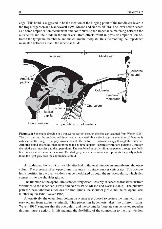
edge. This bond is suggested to be the location of the hinging point of the middle ear lever inthe frog (Jørgensen and Kanneworff 1998; Mason and Narins 2002b). The lever action servesas a force amplification mechanism and contributes to the impedance matching between theoutside air and the fluids in the inner ear. Both effects result in pressure amplification be-tween the tympanic membrane and the columella footplate, thus overcoming the impedancemismatch between air and the inner-ear fluids.
Middle earInner ear
Amphibian
papilla
Basilar
papilla
Sacculus
m. opercularis m. columellarisRound window
Columella
Operculum
Tympanic
membrane
Airborne
sound
Figure 2.1: Schematic drawing of a transverse section through the frog ear (adapted from Wever 1985).The division into the middle, and inner ear is indicated above the image; a selection of features isindicated in the image. The grey arrows indicate the paths of vibrational energy through the inner ear.Airborne sound enters the inner ear through the columellar path; substrate vibrations putatively throughthe middle-ear muscles and the operculum. The combined acoustic vibration passes through the fluid-filled inner ear to the round window. The dark grey areas in the inner ear represents the perilymphaticfluid, the light grey area the endolympatic fluid.
An additional bony disk is flexibly attached to the oval window in amphibians: the oper-culum. The presence of an operculum in anurans is unique among vertebrates. The opercu-lum’s position in the oval window can be modulated through the m. opercularis, which alsoconnects it to the shoulder girdle.
The function of the operculum is not entirely clear. Possibly, it serves to transfer substratevibrations to the inner ear (Lewis and Narins 1999; Mason and Narins 2002b). The putativepath for these vibrations includes the front limbs, the shoulder girdle and the m. opercularis(Hetherington 1988; Wever 1985).
Alternatively, the operculum-columella system is proposed to protect the inner ear’s sen-sory organs from excessive stimuli. This protection hypothesis takes two different forms.Wever (1985) suggests that the operculum and the columella footplate can be locked togetherthrough muscle action. In this manner, the flexibility of the connection to the oval window

MECHANICS OF THE EXCEPTIONAL ANURAN EAR 9
decreases and the input impedance increases, which in turn decreases the input-signal ampli-tude of the pressure wave in the inner ear. It has also been suggested that the action of the m.opercularis could uncouple the operculum and the footplate (Mason and Narins 2002b). Thiswould allow the operculum to move out of phase with the footplate. The out-of-phase motioncould absorb part of the inner ear fluid displacement caused by the motion of the footplate.Effectively this creates an energetic by-pass and decreases the amplitude in the inner ear.
A tympanic middle ear as described above is considered to be the typical situation (Jaslowet al. 1988), which can be found in the family of Ranidae. However, a wide range of vari-ations in middle ear structures is found across species. In some species a bony disk occu-pies the tympanic annulus rather than a membrane, e.g. Xenopus leavis (Wever 1985), andthere are a number of ‘earless’ frogs. The tympanic membrane and the tympanic annulusare absent in these species. A functioning inner ear and a partial middle ear usually exist,although the middle ear cavity may be filled with connective tissue (e.g. Telmatobius exsul,Jaslow et al. 1988), or not exist at all (e.g. species in the Bombina family, Hetherington andLindquist 1999; Wever 1985). Remarkably, some of these ‘earless’ frogs have a mating calland exhibit neurophysiological responses (Bombina bombina, Walkowiak 1988; Atelopus,Lindquist et al. 1998) at typically auditory frequencies, which implies they have another pathfor the transfer of airborne sound to the inner ear (Jaslow et al. 1988), for example throughthe lungs (Narins et al. 1988; Lindquist et al. 1998; Hetherington and Lindquist 1999).
2.2.2 Inner earThe inner ear in the frog has two membranous windows: the oval window and the roundwindow. As mentioned above, acoustic energy primarily enters the inner ear through theoval window. The round window is the main release point of this energy (Purgue and Narins2000a). A similar lay-out can be found in other terrestrial vertebrates. However, the roundwindow of the frog does not open into the middle ear as it does in mammals. Rather, it canbe found in the top of the mouth cavity, under a lining of muscle tissue.
Within the inner ear there are two intertwined membranous compartments: the perilym-phatic and the endolymphatic labyrinths (see figure 2.1). The perilymphatic labyrinth con-nects to both the oval window and the round window. Starting at the oval window and goingmedially, it passes through a narrow foramen, and widens into the otic cavity, forming theperiotic cistern. Continuing medially, it narrows again into the periotic canal. This canalconnects the periotic cistern with the perilymphatic space at the round window (Purgue andNarins 2000b).
Between the lateral perilymphatic cistern and the round window, part of the endolym-phatic space can be found. The endolymphatic space also includes the semi-circular canalslocated dorsally from the otic system. It contains the sensory organs of hearing and balance.In the frog inner ear, there are eight sensory epithelia (Lewis and Narins 1999; Lewis et al.1985), located as follows:
• three cristae in the semi-circular canals, which are sensitive to rotational accelerationof the head,
and one each in:
• the utricule, which detects linear acceleration,
• the lagena, which detects both linear acceleration and non-acoustic vibrations (Castonet al. 1977),
10 CHAPTER 2
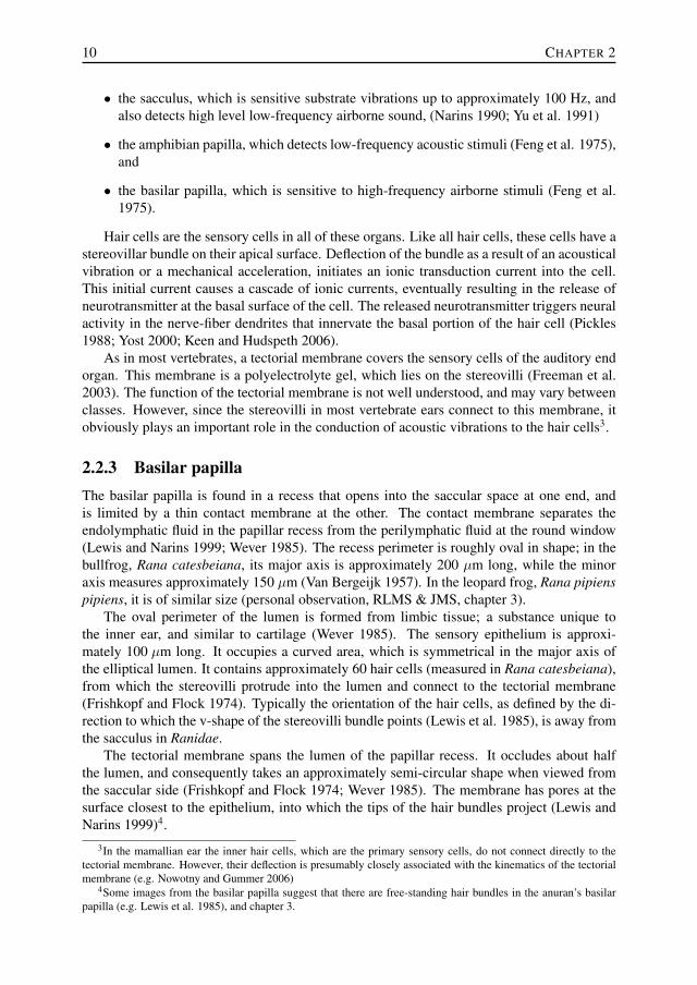
• the sacculus, which is sensitive substrate vibrations up to approximately 100 Hz, andalso detects high level low-frequency airborne sound, (Narins 1990; Yu et al. 1991)
• the amphibian papilla, which detects low-frequency acoustic stimuli (Feng et al. 1975),and
• the basilar papilla, which is sensitive to high-frequency airborne stimuli (Feng et al.1975).
Hair cells are the sensory cells in all of these organs. Like all hair cells, these cells have astereovillar bundle on their apical surface. Deflection of the bundle as a result of an acousticalvibration or a mechanical acceleration, initiates an ionic transduction current into the cell.This initial current causes a cascade of ionic currents, eventually resulting in the release ofneurotransmitter at the basal surface of the cell. The released neurotransmitter triggers neuralactivity in the nerve-fiber dendrites that innervate the basal portion of the hair cell (Pickles1988; Yost 2000; Keen and Hudspeth 2006).
As in most vertebrates, a tectorial membrane covers the sensory cells of the auditory endorgan. This membrane is a polyelectrolyte gel, which lies on the stereovilli (Freeman et al.2003). The function of the tectorial membrane is not well understood, and may vary betweenclasses. However, since the stereovilli in most vertebrate ears connect to this membrane, itobviously plays an important role in the conduction of acoustic vibrations to the hair cells3.
2.2.3 Basilar papillaThe basilar papilla is found in a recess that opens into the saccular space at one end, andis limited by a thin contact membrane at the other. The contact membrane separates theendolymphatic fluid in the papillar recess from the perilymphatic fluid at the round window(Lewis and Narins 1999; Wever 1985). The recess perimeter is roughly oval in shape; in thebullfrog, Rana catesbeiana, its major axis is approximately 200 µm long, while the minoraxis measures approximately 150 µm (Van Bergeijk 1957). In the leopard frog, Rana pipienspipiens, it is of similar size (personal observation, RLMS & JMS, chapter 3).
The oval perimeter of the lumen is formed from limbic tissue; a substance unique tothe inner ear, and similar to cartilage (Wever 1985). The sensory epithelium is approxi-mately 100 µm long. It occupies a curved area, which is symmetrical in the major axis ofthe elliptical lumen. It contains approximately 60 hair cells (measured in Rana catesbeiana),from which the stereovilli protrude into the lumen and connect to the tectorial membrane(Frishkopf and Flock 1974). Typically the orientation of the hair cells, as defined by the di-rection to which the v-shape of the stereovilli bundle points (Lewis et al. 1985), is away fromthe sacculus in Ranidae.
The tectorial membrane spans the lumen of the papillar recess. It occludes about halfthe lumen, and consequently takes an approximately semi-circular shape when viewed fromthe saccular side (Frishkopf and Flock 1974; Wever 1985). The membrane has pores at thesurface closest to the epithelium, into which the tips of the hair bundles project (Lewis andNarins 1999)4.
3In the mamallian ear the inner hair cells, which are the primary sensory cells, do not connect directly to thetectorial membrane. However, their deflection is presumably closely associated with the kinematics of the tectorialmembrane (e.g. Nowotny and Gummer 2006)
4Some images from the basilar papilla suggest that there are free-standing hair bundles in the anuran’s basilarpapilla (e.g. Lewis et al. 1985), and chapter 3.

MECHANICS OF THE EXCEPTIONAL ANURAN EAR 11
a
rostral
lateral
AP twig of
VIII-nervecontact
membrane
tectorial
curtain
to round
windowb
240
140
300
200
240
375
500
550
800
940
475
TM,
triangular
patch
TM,
S-shaped
extension
to sacculus
Figure 2.2: Schematic drawing of the amphibian papilla of the bullfrog, Rana catesbeiana, (adaptedfrom Lewis et al. 1982, rotated to match orientation of fig. 2.1). Labels: TM - tectorial membrane,AP - amphibian papilla. a: General overview of the AP; the dashed outline indicates the location of thesensory epithelium, b: hair cell orientation in the sensory epithelium; dashed line indicates the positionof the tectorial curtain. The numbers along the perimeter indicate the characteristic frequency of theauditory nerve fibers connecting to that site (in Hz).
2.2.4 Amphibian papillaThe amphibian papilla can be found in a recess, that extends medially from the saccular spaceand, in frogs with derived ears, bends caudally to end at a contact membrane. Like the basilarpapilla’s contact membrane, the membrane separates the endolymphatic fluid in the papillarecess from the perilymphatic fluid at the round window.
The sensory epithelium is set on the dorsal surface of this recess (Lewis and Narins 1999).The epithelium itself has a complex shape; it consists of a triangular patch at the rostralend, and an s-shaped caudal extension towards the contact membrane (see fig. 2.2). Theexact shape and length of the caudal extension varies across species, with the most elaborateextensions occurring in species of the family of Ranidae (Lewis 1984), while some specieslack the s-shaped extension alltogether (Lewis 1981).
In the epithelium, the hair cell orientation follows a complicated pattern (see fig. 2.2b).In the rostral patch the cells are orientated towards the sacculus. On the rostral half of thes-shaped extension, they are oriented along the s-shape. However, on the caudal half, theorientation rotates 90◦ to become perpendicular to the s-shape (Lewis 1981).
An elaborate tectorial membrane is found on the hair bundles. A bulky structure coversthe rostral patch, while the membrane gets thinner along the caudal extension (Lewis et al.1982). A tectorial curtain spans the papilla recess approximately halfway between the saccu-lus and the contact membrane (Shofner and Feng 1983; Wever 1985). The curtain, also calledthe sensing membrane (Yano et al. 1990), spans the entire cross section of the lumen. A smallslit in the tectorial curtain may function as a shunt for static fluid-pressure differences (Lewis
12 CHAPTER 2
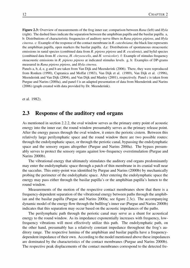
Figure 2.3: Overview of measurements of the frog inner ear; comparison between Rana (left) and Hyla(right). The dashed lines indicate the separation between the amphibian papilla and the basilar papilla. a,b: Distributions of characteristic frequencies of auditory nerve fibers in Rana pipiens pipiens, and Hylacinerea. c: Example of the response of the contact membrane in R. catesbeiana; the black line representsthe amphibian papilla, open markers the basilar papilla. d,e: Distributions of spontaneous otoacousticemissions in ranid species (combined data from R. pipiens pipiens and R. esculenta), and hylid species(combined data from H. cinerea, H. chrysoscelis, and H. versicolor). f: Example of stimulus frequencyotoacoustic emissions in R. pipiens pipiens at indicated stimulus levels. g, h: Examples of DP-gramsmeasured in Rana pipiens pipiens, and Hyla cinerea.Panels a, b, d, e, g and h are taken from Van Dijk and Meenderink (2006). There, they were reproducedfrom Ronken (1990), Capranica and Moffat (1983), Van Dijk et al. (1989), Van Dijk et al. (1996),Meenderink and Van Dijk (2004), and Van Dijk and Manley (2001), respectively. Panel c is taken fromPurgue and Narins (2000a), and panel f is an adapted presentation of data from Meenderink and Narins(2006) (graph created with data provided by Dr. Meenderink).
et al. 1982).
2.3 Response of the auditory end organsAs mentioned in section 2.2.2, the oval window serves as the primary entry point of acousticenergy into the inner ear; the round window presumably serves as the primary release point.After the energy passes through the oval window, it enters the periotic cistern. Between thisrelatively large perilymphatic space and the round window there are two possible routes:through the endolymphatic space, or through the periotic canal, bypassing the endolymphaticspace and the sensory organs altogether (Purgue and Narins 2000a). The bypass presum-ably serves to protect the sensory organs against low-frequency overstimulation (Purgue andNarins 2000b).
The vibrational energy that ultimately stimulates the auditory end organs predominantlymay enter the endolymphatic space through a patch of thin membrane in its cranial wall nearthe sacculus. This entry-point was identified by Purgue and Narins (2000b) by mechanicallyprobing the perimeter of the endolyphatic space. After entering the endolymphatic space theenergy may pass either through the basilar papilla’s or the amphibian papilla’s lumen to theround window.
Measurements of the motion of the respective contact membranes show that there is afrequency-dependent separation of the vibrational energy between paths through the amphib-ian and the basilar papilla (Purgue and Narins 2000a; see figure 2.3c). The accompanyingdynamic model of the energy flow through the bullfrog’s inner ear (Purgue and Narins 2000b)indicates that this separation may occur based on the acoustic impedances of the paths.
The perilymphatic path through the periotic canal may serve as a shunt for acousticalenergy to the round window. As its impedance exponentially increases with frequency, low-frequency vibrations will most effectively utilize this path. The endolymphatic path, onthe other hand, presumably has a relatively constant impedance throughout the frog’s au-ditory range. The respective lumina of the amphibian and basilar papilla have a frequency-dependent impedance of their own. According to the model mentioned above these impedancesare dominated by the characteristics of the contact membranes (Purgue and Narins 2000b).The respective peak displacements of the contact membranes correspond to the detected fre-

MECHANICS OF THE EXCEPTIONAL ANURAN EAR 13
0 1000 2000 3000 40000
4
8
12
16
20
24
28
Num
ber
of fibers
0 1000 2000 3000 40000
4
8
12
16
20
24
Characteristic frequency
Num
ber
of fibers
0 1000 2000 3000 4000-15
-10
-5
0
5
10
15
20
25
Primary frequency f1 (Hz)DP
OA
E level (d
B S
PL)
0 1000 2000 3000 4000-15
-10
-5
0
5
10
15
20
25
Primary frequency f1 (Hz)DP
OA
E level (d
B S
PL)
0 1000 2000 3000 40000
2
4
6
8
10
12
14
SOAE frequency (Hz)
Num
ber
of S
OA
E p
eaks
0 1000 2000 3000 40000
4
8
12
16
20
SOAE frequency (Hz)
Num
ber
of S
OA
E p
eaks
60
4000
50
0 1000 2000 30000
10
20
30
4080dB
68dB
62dB
SFOAE frequency (Hz)
SF
OA
E a
mplit
ude (
dB
SP
L)
1000 2000 30000
0
-20
-10
Rela
tive a
mplit
ude (
dB
)
Frequency (Hz)
20
10
Rana Hyla
AP BP BPAP
30
a
g
f
ed
c
b
h
Characteristic frequency

14 CHAPTER 2
102
103
100
120
1000 4000100
10dB
!f
Frequency (Hz)
Thre
shold
(dB
SP
L)
2.0
1.2
1.5
1.2
80
60
20
40
Figure 2.4: Tuning curves measured in the auditory nerve in R. catesbeiana (unpublished measure-ments by JMS & PvD, 1992; various specimens). The numbers in italics indicate Q10dB values(Q10dB = CF
∆f ).
quencies in the associated organs (Purgue and Narins 2000a).
2.3.1 Basilar papillaThe basilar papilla’s tectorial membrane is presumably driven by a vibrating pressure gradientbetween the the sacculus and the basilar papilla’s contact membrane. No reports have beenpublished on direct measurements of the mechanical response of the tectorial membrane,or on the basilar papilla’s hair-bundle mechanics. However, the hair-cell orientation in thebasilar papilla implies that the tectorial membrane’s primary mode of motion is to and fromthe sacculus.
Auditory nerve-fiber recordings from the frog basilar papilla show a frequency-selectiveresponse (see figure 2.4 for examples of tuning curves). The range of characteristic fre-quencies in nerve fibers from the basilar papilla is species dependent. In the leopard frog,they are approximately between 1200 and 2500 Hz (Ronken 1991); in the bullfrog they areslightly lower, between 1000 and 1500 Hz (Shofner and Feng 1981; Ronken 1991). In theHyla-family the characteristic frequencies appear to be significantly higher; in Hyla cinerea,the green treefrog, they range from 2.8 to 3.9 kHz (Ehret and Capranica 1980; Capranica andMoffat 1983), and in Hyla regilla roughly from 2 to 3 kHz (Stiebler and Narins 1990; Ronken1991). Where studied in other species, the characteristic frequencies of the basilar papilla’snerve fibers fall roughly within the bounds defined by the bullfrog at the low end and thegreen treefrog at the high end (Scaphiopus couchi: approximately 1-1.5 kHz, Capranica andMoffat 1975,Ronken 1991; Eleutherodactylus coqui: approx. 2-4 kHz, Narins and Capranica1980, Narins and Capranica 1976, Ronken 1991; Physalaemus pustulosus group: around 2.2kHz, Wilczynski et al. 2001).
In each individual frog the tuning curves of the auditory nerve fibers appear to have a
MECHANICS OF THE EXCEPTIONAL ANURAN EAR 15
CF (kHz)
CF (kHz)
0 0.5 1.0 1.5 2.0 2.5
Q10dB
0
1
2
3
4
Q10dB
0.2 0.5 1 2 5 10 20
2
4
6
8
10
12CAT
LEOPARD FROG
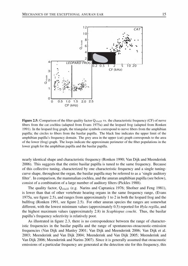
Figure 2.5: Comparison of the filter quality factor Q10dB vs. the characteristic frequency (CF) of nervefibers from the cat cochlea (adapted from Evans 1975a) and the leopard frog (adapted from Ronken1991). In the leopard frog graph, the triangular symbols correspond to nerve fibers from the amphibianpapilla; the circles to fibers from the basilar papilla. The black line indicates the upper limit of theamphibian papilla’s frequency domain. The grey area in the upper (cat) graph corresponds to the areaof the lower (frog) graph. The loops indicate the approximate perimeter of the fiber populations in thelower graph for the amphibian papilla and the basilar papilla.
nearly identical shape and characteristic frequency (Ronken 1990; Van Dijk and Meenderink2006). This suggests that the entire basilar papilla is tuned to the same frequency. Becauseof this collective tuning, characterized by one characteristic frequency and a single tuning-curve shape, throughout the organ, the basilar papilla may be referred to as a ‘single auditoryfilter’. In comparison, the mammalian cochlea, and the anuran amphibian papilla (see below),consist of a combination of a large number of auditory filters (Pickles 1988).
The quality factor, Q10dB (e.g. Narins and Capranica 1976; Shofner and Feng 1981),is lower than that of other vertebrate hearing organs in the same frequency range, (Evans1975a, see figure 2.5), and ranges from approximately 1 to 2 in both the leopard frog and thebullfrog (Ronken 1991, see figure 2.5). For other anuran species the ranges are somewhatdifferent, with the lowest minimum values (approximately 0.5) reported for Hyla regilla, andthe highest maximum values (approximately 2.8) in Scaphiopus couchi. Thus, the basilarpapilla’s frequency selectivity is relatively poor.
As illustrated in figure 2.3, there is no correspondence between the range of character-istic frequencies in the basilar papilla and the range of spontaneous-otoacoustic-emissionfrequencies (Van Dijk and Manley 2001; Van Dijk and Meenderink 2006; Van Dijk et al.2003; Meenderink and Van Dijk 2004; Meenderink and Van Dijk 2005; Meenderink andVan Dijk 2006; Meenderink and Narins 2007). Since it is generally assumed that otoacousticemissions of a particular frequency are generated at the detection site for this frequency, this

16 CHAPTER 2
suggests that the basilar papilla does not generate spontaneous emissions. However, it doesemit distortion-product otoacoustic emissions (Van Dijk and Manley 2001), and stimulus-frequency otoacoustic emissions (Palmer and Wilson 1982; Meenderink and Narins 2006).The peak amplitudes of the distortion-product otoacoustic emissions match the characteris-tic frequencies of the auditory nerve fibers innervating the basilar papilla (Meenderink et al.2005b).
The amplitude and phase characteristics of the distortion product otoacoustic emissionscan be qualitatively modeled by assuming the basilar papilla to be a single passive non-linearauditory filter (Meenderink et al. 2005b). Thus, nerve-fiber recordings, otoacoustic-emissionmeasurements and a model based on these measurements show that the basilar papilla func-tions as a single frequency band auditory receptor. This frequency band is relatively broad,and the center frequency may depend on species and individual animals.
The hypothesis that considers the basilar papilla as a single resonator was originally putforward by Van Bergeijk (1957). He investigated the mechanical response of the tectorialmembrane in a scale model consisting of a thin rubber tectorium spanning a lumen in a stiffwall. A number of different vibration modes existed in this model. Although Van Bergeijk’smodel is vastly oversimplified, the basic idea that the mechanical tuning of the tectorial mem-brane may be the basis of the basilar papilla’s frequency selectivity is still viable.
2.3.2 Amphibian papillaAs in the basilar papilla, the tectorial membrane in the amphibian papilla is presumably drivenby a vibrating pressure difference between the sacculus and the round window. Due to themore elaborate tectorial membrane and the more complex pattern of hair cell orientations,the motion of the membrane may be expected to be more complex than that of the basilarpapilla’s tectorial membrane. The tectorial curtain is in the sound path through the papilla,and presumably plays a role in conveying vibrations to the tectorial membrane and the hairbundles.
Electro-physiological recordings from and subsequent dye-filling of single fibers of theauditory nerve show that the amphibian papilla has a tonotopic organization (Lewis et al.1982). The fibers innervating the triangular patch have low characteristic frequencies, downto approximately 100 Hz. The frequencies increase gradually along the caudal extension. Inthe bullfrog, the upper frequency is about 1000 Hz; an overview of the tonotopic organizationis given in figure 2.2b5.
The frequency selectivity of the amphibian papilla’s nerve fibers is similar to that of mam-malian auditory nerve fibers with the same characteristic frequency. This is in contrast to thesignificantly poorer frequency selectivity in the basilar papilla’s nerve fibers (Ronken 1990;Evans 1975a, see also figure 2.5).
In the low-frequency, rostral part of the papilla, the hair cells are electrically tuned (Pitch-ford and Ashmore 1987; Smotherman and Narins 1999a). This tuning stems from the elec-trical properties of the cell membrane’s ion channels. The hair-cell tuning characteristicsparallel the tonotopy of the single nerve-fiber recordings. Therefore, frequency selectivity in
5In some species of the Hyla-family the upper frequency in the amphibian papilla is markedly higher than in thebullfrog (Ronken 1991), up to approximately 2.8 kHz in H. cinerea (Ehret and Capranica 1980). However, even inthese species the vast majority of the recorded fibers from the amphibian papilla have best frequencies below 1250Hz.

MECHANICS OF THE EXCEPTIONAL ANURAN EAR 17
the rostral part of the amphibian papilla appears to be primarily determined by the electricalcharacteristics of the hair cells.
However, there is a fundamental discrepancy between the tuning characteristics of thehair cells and the auditory nerve fibers. While hair cells exhibit a second-order resonance(Pitchford and Ashmore 1987) auditory neurons display a higher-order filter characteristic(Lewis 1984). Nevertheless, due to the parallels in the tonotopic organization, the assumptionthat the frequency selectivity is determined by the electrical tuning seems viable for the rostralpart of the amphibian papilla. The higher-order responses in the neural signal may result fromcoupling between hair cells, which may be mechanical, for instance through the tectorialmembrane.
Neurons innervating the rostral portion of the amphibian papilla display non-linear two-tone suppression similar to that in other vertebrates (Capranica and Moffat 1980; Benedixet al. 1994). Another manifestation of non-linear behavior can be found in the responseto noise: second-order Wiener kernels of low-frequency neurons show off-diagonal compo-nents, which are an indication of non-linearity (Van Dijk et al. 1994; Van Dijk et al. 1997a).The spectro-temporal receptive fields constructed from these Wiener kernels exhibit suppres-sive side bands besides the main characteristic-frequency band of the fiber (Lewis and VanDijk 2004).
Hair cells caudal to the tectorial curtain do not display electrical resonance (Smothermanand Narins 2000). Therefore, the tuning of this high-frequency, caudal region of the papillamust result from the mechanical properties of the tectorial membrane and the hair cells.
Based on the hair cell orientation, displayed in figure 2.2b, the tectorial membrane mo-tion in the amphibian papilla is expected to be far more complex than in the basilar papilla.Assuming that the hair bundles are orientated in such a way that they are maximally deflectedby the connected tectorial membrane, the rostral patch of the membrane should be movingto and from the sacculus, if the appropriate stimuli are presented. The rostral part of the s-shaped extension is moving along its major axis, whereas the extension caudal to the tectorialcurtain should be moving in a transverse direction.
The amphibian papilla appears to be the only source of spontaneous otoacoustic emissionsin the frog inner ear (Van Dijk et al. 1989; Van Dijk et al. 1996; Long et al. 1996; Van Dijkand Manley 2001; figure 2.3d-e). The frequency distribution of these emissions correspondsto the range of best frequencies of the neurons projecting to the portion of the amphibianpapilla caudal to the tectorial curtain. It is generally assumed that an otoacoustic emissionof a specific frequency is generated at the location in the inner ear where that frequency isdetected. Under this assumption, the presence of spontaneous otoacoustic emissions indicatesthat the caudal portion of the amphibian papilla exhibits spontaneous activity. Presumably,this activity is related to active amplification of input signals in this area.
The caudal region of the amphibian papilla is also involved in the generation of distortion-product otoacoustic emissions (Van Dijk and Manley 2001; Meenderink and Van Dijk 2004),and stimulus-frequency otoacoustic emissions (Meenderink and Narins 2006). The distortion-product otoacoustic emissions from the amphibian papilla are more vulnerable to metabolicinjuries than those from the basilar papilla (Van Dijk et al. 2003). Also, both the spontaneous(Van Dijk et al. 1996) and distortion-product (Meenderink and Van Dijk 2006) otoacousticemissions display a clear dependence on body temperature. These results combine to indicatethat the s-shaped extension of the amphibian papilla caudal to the tectorial curtain functionsas an active hearing organ.
18 CHAPTER 2
2.4 DiscussionOur aim in this review is to outline what is known about the mechanical response properties ofthe amphibian and basilar papilla. Only one published report exists of the direct mechanicalmeasurements of structures associated with these papillae (Purgue and Narins 2000a). Themeasurements show that the response of the contact membrane is frequency dependent foreach papilla. The movement of the contact membrane may be assumed to reflect the fluidmotion within the respective papilla. The contact membrane of the amphibian papilla shows amaximum response when the ear is stimulated with relatively low acoustic frequencies, whilethe basilar papilla’s contact membrane exhibits a maximum response to higher frequencies.
The amphibian and the basilar papilla are the only hearing organs found in terrestrialvertebrates in which the hair cells are not on a flexible basilar membrane. Instead the hair cellsare embedded in a relatively stiff cartilaginous support structure. Any frequency-selectiveresponse, therefore, most likely originates from the mechanical or electrical properties of thehair cells, or the mechanical properties of the tectorial membrane, or a combination of thesefactors. Since there are no direct mechanical measurements of either the hair cells in thepapillae or the tectorial membranes, we cannot come to any definite conclusions regardingtheir properties. However, the available morphological and functional data allow for somehypotheses.
The most conspicuous functional characteristic of the amphibian papilla is its tonotopicorganization (Lewis et al. 1982). Rostral to the tectorial curtain the hair-cell orientationis essentially parallel to the tonotopic axis. In this low-frequency region of the amphibianpapilla the tectorial membrane apparently moves in a rostro-caudal direction. In contrast, thehair-bundle orientation suggests that the tectorial-membrane motion is perpendicular to thetonotopic axis in the high-frequency, caudal region of the papilla. The tectorial membrane’scaudal end, therefore, appears to vibrate in a markedly different direction than its rostral end.
In the low-frequency region of the amphibian papilla the hair cells display electrical tun-ing. The tuning properties of the hair cells parallel the tonotopic organization as measuredfrom the afferent nerve fibers (Pitchford and Ashmore 1987). This strongly suggests that thetuning characteristics of the nerve fibers are primarily determined by the electrical hair-cellresonances. The auditory nerve-fiber recordings reflect the presence of high-order filtering(Lewis 1984), whereas hair cells essentially function as second-order resonances. It is, there-fore, likely that coupling between the hair cells shapes the frequency responses in the nervefibers. Such coupling may be mechanical, e.g. by the tectorial membrane, or electrical, or acombination of mechanical and electrical.
Hair cells in the high-frequency, caudal region do not display any electrical resonance(Smotherman and Narins 1999a). This implies that the frequency selectivity must be basedon mechanical tuning, probably by the tectorial membrane. The caudal region of the amphib-ian papilla shares some notable characteristics with the mammalian cochlea (see also Lewis1981):
1. the papilla is elongated, and it exhibits a tonotopic gradient along the long axis;
2. the orientation of the hair cells is perpendicular to the tonotopic axis, indicating that thehair cells are stimulated most efficiently by a deflection perpendicular to the tonotopicaxis;
3. frequency selectivity –very probably– relies on mechanical tuning;

MECHANICS OF THE EXCEPTIONAL ANURAN EAR 19
4. frequency selectivity is similar, with Q10dB-values ranging from 0.8 to 2.2; and
5. both spontaneous and distortion-product otoacoustic emissions are generated. Theseemissions are physiologically vulnerable.
The presence of spontaneous otoacoustic emissions shows that at least part of the amphibianpapilla’s caudal extension functions as an active hearing organ. In this respect it is similar tothe mammalian cochlea and other vertebrate hearing organs (Lewis and Narins 1999). Oneactive mechanism in the mammalian cochlea is the prestin-mediated active somatic lengthchanges in the outer hair cells (Brownell et al. 1985; Yost 2000; Zheng et al. 2000; Liber-man et al. 2002; Dallos 2003). However, this mechanism is probably exclusively presentin mammalian outer hair cells. Active hair bundle movements have been reported as an al-ternative active mechanism in anuran saccular hair cells (Martin and Hudspeth 1999; Martinet al. 2003; Bozovic and Hudspeth 2003); this mechanism may be present in the auditoryorgans as well. Although the fundamental active mechanism may differ between species, thefunctional result seems to be very similar across vertebrates: high auditory sensitivity andgood frequency selectivity (Manley 2000).
The basilar papilla seems to function in a much simpler manner. Both neural recordingsand otoacoustic emission measurements suggest that it functions as a single auditory filter.Since the hair cells in the basilar papilla are unlikely to be electrically tuned, its frequencyselectivity most likely results from mechanical tuning, probably via the tectorial membrane.
The basilar papilla is remarkable in that no spontaneous otoacoustic emissions have beenrecorded in its frequency range. The absence of such emissions can either be caused by thefact that they are not generated within the papilla, or by the fact that the transmission of suchemissions to the tympanic membrane is inhibited. However, distortion-product otoacousticemissions can be recorded in this range (e.g. Van Dijk and Manley 2001). This implies thatthe outward transmission is not inhibited, and therefore that spontaneous emissions are mostlikely not generated within the basilar papilla.
Furthermore, the amplitude of the basilar papilla’s distortion-product otoacoustic emis-sions depends less on temperature than that of the amphibian papilla’s (Meenderink andVan Dijk 2006). Also, emissions from the basilar papilla are less sensitive to the disrup-tion of oxygen supply (Van Dijk et al. 2003). Apparently, emissions from the basilar papillaare relatively independent of the metabolic rate, and therefore, it has been suggested that thebasilar papilla is not an active hearing organ (Vassilakis et al. 2004; Van Dijk and Meenderink2006).
In conclusion, the frog inner ear takes an exceptional place among the hearing organs ofterrestrial vertebrates. It includes two auditory end organs, which both lack the basilar mem-brane present in every other terrestrial vertebrate species. Instead the hair cells are embeddedin a relatively stiff structure. They are stimulated by the motion of the tectorial membrane.Although the basilar and amphibian papilla are similar in this respect, they appear to functionby different mechanisms. In fact, even within the amphibian papilla two distinctly differ-ent functional regions can be identified. The low-frequency portion, rostral to the tectorialcurtain, contains hair cells that exhibit electrical tuning. The hair cells are most sensitive todeflection along the tonotopic axis, thus this is presumably the tectorial membrane’s directionof vibration. By contrast, the region caudal to the tectorial curtain shows more similarities to,for example, the mammalian cochlea: the hair cell orientation is perpendicular to the tono-topic axis, and the presence of spontaneous otoacoustic emissions suggests that it functions
20 CHAPTER 2
as an active hearing organ. Finally, the basilar papilla is yet different: it appears to functionas a single passive auditory filter. Thus the frog inner ear includes two auditory end organswith three functional regions.
AcknowledgementsWe would like to thank Dr. J.E.C. Wiersinga-Post for her comments on an earlier version ofthe manuscript.
This study was supported by the Heinsius Houbolt Foundation and the Netherlands Or-ganisation for Scientific Research, and is part of the research program of our department:Communication through Hearing and Speech.
Previously unpublished data described in this paper were obtained in experiments con-ducted in compliance with the "Principles of animal care", publication No. 86-23, revised1985 of the National Institute of Health, and with the current legislation, at the time of theexperiments, of the country in which they were conducted (The Netherlands).