universitÀ degli studi di milano dipartimento di … · tesi di dottorato di: sara negrini...
TRANSCRIPT
UNIVERSITÀ DEGLI STUDI DI MILANO
Facoltà di Medicina e Chirurgia
Scuola di Dottorato in Scienze biochimiche
Dipartimento di Chimica, Biochimica e Biotecnologie per la Medicina
CORSO DI DOTTORATO DI RICERCA IN BIOCHIMICA CICLO XXVI
BIO/10
Tesi di dottorato di ricerca
THE TRANSCRIPTION FACTOR REST HAS A KEY ROLE IN THE CONTROL OF
PROLIFERATION AND NEURITE OUTGROWTH IN NEURAL PC12 CELLS.
Tesi di dottorato di:
Sara NEGRINI
Matr.Nr. R09299
Tutor: Chiar.mo Prof. Jacopo MELDOLESI
Coordinatore: Chiar.mo Prof. Sandro SONNINO
Anno Accademico 2012/2013

UNIVERSITÀ DEGLI STUDI DI MILANO
Facoltà di Medicina e Chirurgia
Scuola di Dottorato in Scienze biochimiche
Dipartimento di Chimica, Biochimica e Biotecnologie per la Medicina
CORSO DI DOTTORATO DI RICERCA IN BIOCHIMICA CICLO XXVI
BIO/10
Tesi di dottorato di ricerca
THE TRANSCRIPTION FACTOR REST HAS A KEY ROLE IN THE CONTROL OF
PROLIFERATION AND NEURITE OUTGROWTH IN NEURAL PC12 CELLS.
Tesi di dottorato di:
Sara NEGRINI
Matr.Nr. R09299
Tutor: Chiar.mo Prof. Jacopo MELDOLESI
……………………………
Coordinatore: Chiar.mo Prof. Sandro SONNINO
……………………………
Anno Accademico 2012/2013

Contents
I
CONTENTS
Abbreviations used III
I. INTRODUCTION
I.1. Molecular mechanisms governing neurosecretion competence 1
I.1.1. REST/NRSF regulation of neurogenesis 1
I.1.2. The role of REST in cell function: from the molecular to the
cellular studies 2
I.2. The PC12 cell model 3
I.2.1. Neurosecretion is a REST-dependent process 3
I.2.2. PC12-27 as a high REST neural cell model 3
I.2.3. Two new processes governed by REST: proliferation and
NGF signaling/neurite outgrowth
6
II. RESULTS
III. Differential proliferation of PC12-27 with respect wtPC12 8
III.1. Proliferation pf PC12-27 cells is faster than that of wtPC12 8
III.2. The faster proliferation rate of high-REST PC12-27 cells
reflects downregulation of TSC2 and increased β-catenin
co-transcriptional activity 11
III.3. REST, TSC2 and β-catenin, inter-connected in a feed-
forward loop, control PC12 cell proliferation
16
IV. Neurite outgrowth 20
IV.1. NGF receptor expression and signaling 21
IV.2. The NGF signaling cascade 23
IV.2.a. TrkA auto-phosphorylation 24
IV.2.b. Phosphorylation of ERK and Akt 25
IV.2.c. mTORC1 and mTORC2 29
IV.2.d. PI3K-dependent pathways 31
IV.3. Transient transfection of p75NTR
in PC12-27 increases
mTORC2 activity 31
IV.4. Generation and characterization of PC12-27/p75NTR
stable
clones 32
IV.5. NGF signaling in wtPC12 and PC12-27 cells: a new role of
p75NTR
34

Contents
II
IV.6. mTORC1 and mTORC2 in PC12 cells: role of p75NTR
37
IV.7. About the phenotype of PC12-27/p75NTR
cells 39
IV.8. Stable silencing of p75NTR
by miRNA in PC12 cells
40
V. DISCUSSION
42
V.1. REST, TSC2 and β-catenin govern proliferation working as
a signaling/effector loop 42
V.2. The regulation of neurite outgrowth was still largely
unknown 44
V.3. A new role for p75NTR
44
V.4. Conclusion 45
VI. MATERIALS AND METHODS
VI.1. Cell cultures 46
VI.2.1. Stable and transient transfections 46
VI.2.2. Expression plasmids 47
VI.2.3. mRNA isolation and Real-Time PCR 47
VI.3.1. Protein Assays; Western Blotting 47
VI.3.2. Immunofluorescence and bright field micorscopy 48
VI.3.3. Antibodies and chemicals 49
VI.4. Materials 49
VI.5. Statistical analyses 50
References 51

Abbreviations
III
ABBREVIATIONS USED
4E-BP1 Factor 4E binding protein 1
Akt Protein kinase B (PKB)
cAMP Cyclic adenosine monophospate
cDNA Complementary DNA
CFSE 5-(6)-carboxyfluorescein
succinimidylester
Cm Clamp capacitance
Dapi 4’6-diamidino-2-phenilindole-
dihydrochloride
DBD Dominant negative construct
DMEM Dulbecco’s modified eagle’s medium
ECL Enhanced Chemiluminescence
Endo-IWR 1 Wnt pathway inhibitor
ERK Mapk1: mitogen-activated protein kinase
1
FACS Fluorescence activated cell sorter
FCIII Fetal clone III
FCS Fetal clone serum
FCS Forward scatter
FITC Fluorescein isothiocyanate
GAPDH Glyceraldehyde-3-phosphate
dehydrogenase
GFP Green fluorescent protein
GSK3β Glycogen synthase kinase-3 beta
H2B Histone IIb
hr hours
HRP Horse radish peroxidase
IF Immunofluorescence
IRS1 Insulin receptor substrate 1
kDa kilodaltons
LB Lysis buffer
Map2 Microtubule-associated protein 2
MFI Means fluorescent intensity
min Minute
miRNA microRNA
mM millimolar
NGF Nerve growth factor protein
NR Nerve growth factor and rapamycin
together
NRSF Neuron-restricted silencer factor
NW Nerve growth factor and wortmannin
together
p75NTR
Low-affinity nerve growth factor
receptor (also called p75 neurotrophin
receptor)

Abbreviations
IV
pAb Polyclonal antibody
PAGE Polyacrylamide gel electrophoresis
PBS Phosphate-buffered saline
PCR Polymerase chain reaction
pF Picofarad
PKC Protein kinase C
R Rapamycin
REST Repressor Element-1 silencing
transcription factor
RT Room temperature
RT-PCR Real time-PCR
s.e.m.
S6 Ribosomal protein
SDS Sodium dodecyl sulfate
shRNA Short hairpin RNA
TBS Tris buffered saline
TCF-LEF T-cell factor and lymphoid enhancing
factor
TRITC Tetramethyl-rhodamine isothiocyanate
TrkA Neurotrophic tyrosine kinase, receptor,
type 1 (also called tropomyosin-related
kinase A)
TSC1 Tuberous sclerosis 1
TSC2 Tuberous sclerosis 2
WB Western blotting
WT Wild type
μm
micromolar

Introduction
1
I. INTRODUCTION
I.1. Molecular mechanisms governing neurosecretion competence
I.1.1. REST/NRSF regulation of neurogenesis
REST (also known as NRSF), a transcription repressor identified
simultaneously by two laboratories in 1995 (Chong et al., 1995; Schoenherr and
Anderson, 1995), was first recognized as the controller of a few neural cell-specific
genes and then proposed to play the role of the master gene in the neuron
differentiation program (Ballas and Mandel, 2005). The level of REST is high in stem
cells and in non-neural cells, and this prevents the expression of many genes specific
of neural cells. These genes, together with additional genes also repressed by REST,
include in their promoter, or in other regulatory areas, one or more sequences known
as RE-1, specific for the REST binding.
The REST complexes repress neurogenesis by targeting and blocking the
transcription of their substrates (from Coulson et al., 2005)

Introduction
2
Bound REST works as a scaffold at two sites located near its N and the C termini,
permitting the binding of co-factors and of numerous enzymes. Almost two thousand
genes, potential targets of REST, might thus be repressed in their transcription (Bruce
et al., 2004; Otto et al., 2007).
During differentiation of neural precursors, the level of the repressor drops due
primarily to the increase of its proteasomic turnover, and this changes the state of the
REST-dependent genes, making possible the expression of hundreds of them. (See
the detailed Figure-Abstract here above).
I.1.2. The role of REST in cell function: from the molecular to the cellular studies.
Extensive studies carried out since the discovery of REST revealed a number of
interesting properties of the neuron differentiation and of the mechanisms involved in
the latter process (reviews: Ballas and Mandel 2005; Ooi and Wood, 2007;
Majumder, 2006). Concomitantly, REST was shown to play a critical role in the
growth of tumors, not only neural, but also non-neural (reviews: Majumder, 2006;
Tomasoni, Negrini et al., 2013). These studies, however, were mostly focused on
molecular processes such as gene expression and its control. No major interest was
paid to the role of REST in cell biology, in particular in the integrated processes that
sustain the functioning of neural cells. Indeed, only one such process, i.e.
neurosecretion, was investigated in detail. In 2006 the group of Neal Buckley (Bruce
et al., 2006) reported that the phenotype of the neurosecretory cell PC12, a
pheochromocytoma cell similar to chromaffin cells (Greene and Tischler, 1976), was
profoundly affected by the transfection of REST constructs, with down regulation of
141 genes including a few coding for proteins of the secretory process, such as the
two chromogranin cargo proteins and one SNARE, SNAP25. The direct control of
REST on these events was confirmed by the decrease, in the cells stably transfected
with a repressor, of the dense-core vesicles, typical of catecholamine-secreting cells,
and by the rescue of some repressed genes in these stably transfected cells re-
transfected with a dominant negative construct of the repressor.

Introduction
3
I.2. The PC12 cell model
I.2.1 Neurosecretion is a REST-dependent process.
PC12 are peculiar in many respects (Greene and Tischler, 1976). Years ago
their well known heterogeneity was shown to include clones that miss spontaneously
the dense-core vesicles (Corradi et al., 1996; Pance et al., 1999). Pance et al. (2006),
by studying one such defective clones, were able to find properties analogous to the
PC12 transfected with the REST constructs of Bruce et al. (2006). However they
failed to observe any rescue of the neurosecretory phenotype upon expression in the
defective cells of dominant-negative REST constructs. The subsequent studies of
D’Alessandro et al. (2008), carried out by the comparison of a wild-type clone of
PC12 (referred to from here-on as wtPC12) with the defective PC12-27 clone initially
characterized by Corradi et al., demonstrated that numerous neurosecretion genes are
repressed by REST; that transfection of REST in the wtPC12 induced the repression
of these genes accompanied by a decrease of the dense-core vesicles to 20% of their
usual size; that the dominant negative construct of REST, when accompanied by the
inhibitor of histone deacetylase, trichostatin A, did induce the rescue not only of
single neurosecretion proteins but of whole dense-core vesicles (DCVs) that were
discharged by exocytosis upon increase of the cytosolic Ca2+
concentration. In
conclusion, therefore, although some aspects of the process were still unclear, the
cellular process neurosecretion was shown conclusively to be governed by REST.
I.2.2 PC12-27 as a high REST neural cell model
In the course of a study on the heterogeneity of the PC12 cell line, a clone was
isolated (named PC12-27: Clementi et al., 1992) that, although retaining various
aspects of the neuroendocrine phenotype, is incompetent for regulated neurosecretion.
Morphological, biochemical and molecular investigations of this clone (Corradi et al.,
1996; Borgonovo et al., 1998) provided evidence of its lack not only of both types of
secretory vesicles, the clear vesicles and the DCVs (as shown in Figure 1), but also of
components of the neurosecretory machinery, e.g. the SNARE proteins (syntaxin 1,
SNAP 25 and VAMP2), the granins (chromogranin B-CgB- and secretogranin 2-
SgII), and the Ca2+
-sensor (synaptotagmin 1), together with the catecholamine uptake
Introduction
4
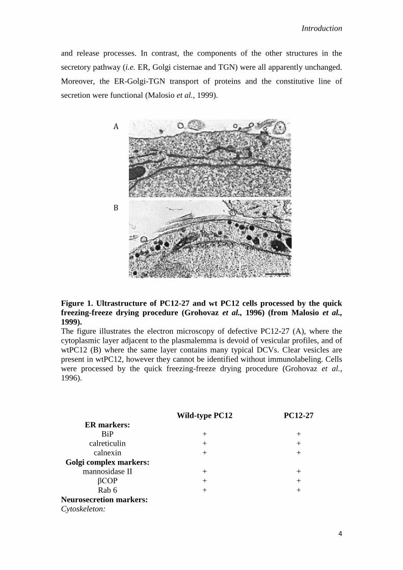
and release processes. In contrast, the components of the other structures in the
secretory pathway (i.e. ER, Golgi cisternae and TGN) were all apparently unchanged.
Moreover, the ER-Golgi-TGN transport of proteins and the constitutive line of
secretion were functional (Malosio et al., 1999).
Figure 1. Ultrastructure of PC12-27 and wt PC12 cells processed by the quick
freezing-freeze drying procedure (Grohovaz et al., 1996) (from Malosio et al.,
1999). The figure illustrates the electron microscopy of defective PC12-27 (A), where the
cytoplasmic layer adjacent to the plasmalemma is devoid of vesicular profiles, and of
wtPC12 (B) where the same layer contains many typical DCVs. Clear vesicles are
present in wtPC12, however they cannot be identified without immunolabeling. Cells
were processed by the quick freezing-freeze drying procedure (Grohovaz et al.,
1996).
Wild-type PC12 PC12-27
ER markers:
BiP + +
calreticulin + +
calnexin + +
Golgi complex markers:
mannosidase II + +
βCOP + +
Rab 6 + +
Neurosecretion markers:
Cytoskeleton:
A
B

Introduction
5
neurofilament H subunit + +
N-kinesin + +
synapsin I + +
Signaling:
α-latrotoxin receptor + +
N-type Ca2+
channel + +
XLαs G protein + +
tyrosine hydroxylase + +
DCGs and cklear vesicle
markers:
DβH + -
vAChT + -
chromogranin B + -
secretogranin II + -
synaptophysin + -
VAMP2 + -
synaptotagmin I + -
t-SNAREs:
syntaxin 1A + -
SNAP25 + -
SNARE regulators:
rbSec1/munc18 + -
rab3A + -
Membrane recycling:
AP180 + -
AP2 + +
dynamin + +/-
synaptojanin + +
amphyphysin + +
Table 1. Expression of markers of the ER, Golgi, DCGs, clear vesicles and of
various properties of neurosecretory cells in the wtPC12 and PC12-27 clones
(from Malosio et al., 1999)
The REST dependence of neurosecretion, a typical process of neural cells, was
not surprising. The project of my work in the laboratory was to investigate whether
the REST dependence was limited to that process or concerned also other functions of
PC12 cells. For these studies the laboratory did use the same wtPC12/PC12-27 clone
model employed by D’Alessandro et al. in 2008. Our approach consisted in the
comparative analysis of a typical neural cell clone, with the usual very low level of
REST, and of another PC12 clone, with a REST level 50-80 fold higher. As already
mentioned, these clones had been characterized in detail in previous studies (Clementi

Introduction
6
et al., 1992; Corradi et al., 1996; Borgonovo et al., 1998; Malosio et al., 1999;
D’Alessandro et al., 2008). Therefore they were a very favorable model for our
investigation.
I.2.3. Two new processes governed by REST: proliferation and NGF signaling/
neurite outgrowth.
My study was focused in sequence on two functions, proliferation and neurite
outgrowth. Mature neurons do not proliferate, however the other neural cells do
during their whole life. Thus, low REST, which is shared by all neural cells, does not
seem to prevent the process. The question investigated was whether REST has
anything to do with proliferation and, in case, what are the mechanisms of its effects.
Neurite outgrowth is in contrast a typical property of neural cells, taking place in
response to appropriate growth factors. Beginning with the first paper dealing with the
isolation of PC12 (Greene and Tischler, 1976), hundreds of papers have investigated
the neurite outgrowth induced by long-term exposure to NGF. Among these papers
was the paper by Leoni et al. (1999) demonstrating that PC12-27 cells lack this
property. For quite some time the defective neurite outgrowth of PC12-27 cells was
attributed to the lack of the tyrosine kinase receptor of NGF, TrkA. A detailed
investigation of the problem, however, had never been done.
The results reported in this thesis bring new evidence in both the already
mentioned areas, proliferation and NGF signaling. PC12-27 cells were found to grow
much faster than the wtPC12, and this property was shown to depend on REST, due
however to a mechanism different from the usual transcription repression, i.e. a
posttranscriptional decrease of the GAP protein TSC2. The latter decrease is shown to
operate not via the conventional TSC2 signaling way, i.e. via the decreased inhibition
of Rheb and, as a consequence, via the increased activity of mTORC1. Rather, the
decreased TSC2 induced by high REST is shown to operate via a decreased turnover
of -catenin, with increased transcriptional activity of the latter. TrkA, on the other
hand, was found to be expressed at the same level in both the wtPC12 and the PC12-
27 cells. Part of the signaling triggered by the neurotrophin, i.e. that mediated by the
ERKs 1 and 2, was the same in the two clones. Missing in the PC12-27 cells was in
Introduction
7
contrast the other NGF receptor, p75NTR
, whose gene was found to be a typical target
of REST endowed with two RE-1 sequences in its promoter. This result was
unexpected because the role of p75NTR
has been considered for long time to consist
primarily in the increased affinity of TrkA for its ligand, NGF. Our results
demonstrate that, in contrast, p75NTR
is needed for a critical step of TrkA signaling,
i.e. the activation of the PI3K-Akt cascade. Moreover, such activation is shown to be
necessary for the neurotrophin to induce neurite outgrowth to occur.
In conclusion, the two projects of our research have demonstrated the critical
role of REST in two cellular processes, proliferation and neurite outgrowth, in which
the dependence on the repressor was unknown. In addition, they have revealed the
mechanisms by which these effects of REST take place, bringing new information in
two fields of great importance, the multiplicity of the TSC2-dependent cascades and
the signaling at NGF receptors, important in general for all neural cells.
Results
8
II. RESULTS
In this thesis we will report the results of two research lines both concerning the
role of REST in important cellular function, proliferation (section III) and neurite
outgrowth (section IV) of neural cells. In both cases our studies have identified the
molecular mechanisms involved.
III. Differential proliferation of PC12-27 with respect wtPC12.
To investigate the role of REST in proliferation, and subsequently in neurite
outgrowth, we took advantage of two PC12 clones extensively characterized in our
laboratory: wtPC12 and PC12-27 (Malosio et al., 1999; Grundschober et al., 2002).
Whereas the first expresses the very low levels of REST typical of mature neural
cells, the second spontaneously expresses levels of REST 50 fold higher
(D’Alessandro et al., 2008). The low and high REST levels account for full or
defective competence for neurosecretion, respectively (D’Alessandro et al., 2008).
III.1. Proliferation of PC12-27 cells is faster than that of wtPC12.
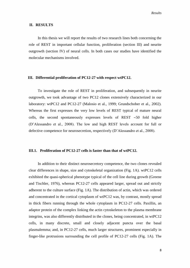
In addition to their distinct neurosecretory competence, the two clones revealed
clear differences in shape, size and cytoskeletal organization (Fig. 1A). wtPC12 cells
exhibited the quasi-spherical phenotype typical of the cell line during growth (Greene
and Tischler, 1976), whereas PC12-27 cells appeared larger, spread out and strictly
adherent to the culture surface (Fig. 1A). The distribution of actin, which was ordered
and concentrated in the cortical cytoplasm of wtPC12 was, by contrast, mostly spread
in thick fibers running through the whole cytoplasm in PC12-27 cells. Paxillin, an
adaptor protein of the complex linking the actin cytoskeleton to the plasma membrane
integrins, was also differently distributed in the clones, being concentrated, in wtPC12
cells, in many discrete, small and closely adjacent puncta over the basal
plasmalemma; and, in PC12-27 cells, much larger structures, prominent especially in
finger-like protrusions surrounding the cell profile of PC12-27 cells (Fig. 1A). The
Results
9
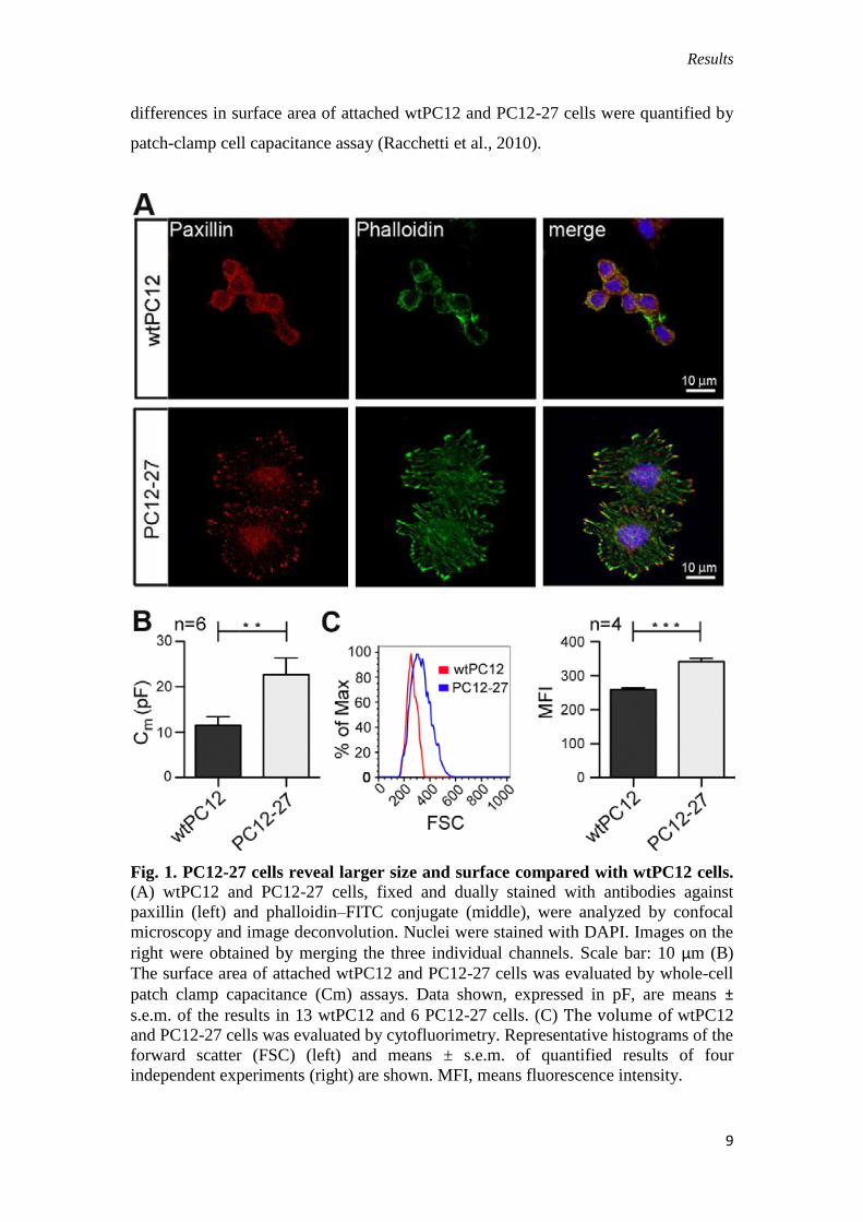
differences in surface area of attached wtPC12 and PC12-27 cells were quantified by
patch-clamp cell capacitance assay (Racchetti et al., 2010).
Fig. 1. PC12-27 cells reveal larger size and surface compared with wtPC12 cells.
(A) wtPC12 and PC12-27 cells, fixed and dually stained with antibodies against
paxillin (left) and phalloidin–FITC conjugate (middle), were analyzed by confocal
microscopy and image deconvolution. Nuclei were stained with DAPI. Images on the
right were obtained by merging the three individual channels. Scale bar: 10 μm (B)
The surface area of attached wtPC12 and PC12-27 cells was evaluated by whole-cell
patch clamp capacitance (Cm) assays. Data shown, expressed in pF, are means ±
s.e.m. of the results in 13 wtPC12 and 6 PC12-27 cells. (C) The volume of wtPC12 and PC12-27 cells was evaluated by cytofluorimetry. Representative histograms of the
forward scatter (FSC) (left) and means ± s.e.m. of quantified results of four
independent experiments (right) are shown. MFI, means fluorescence intensity.
Results
10
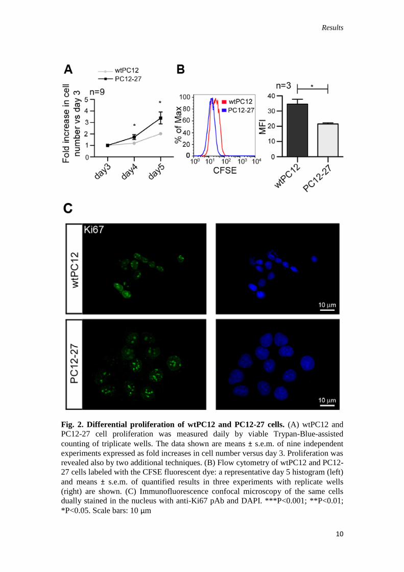
Fig. 2. Differential proliferation of wtPC12 and PC12-27 cells. (A) wtPC12 and
PC12-27 cell proliferation was measured daily by viable Trypan-Blue-assisted
counting of triplicate wells. The data shown are means ± s.e.m. of nine independent
experiments expressed as fold increases in cell number versus day 3. Proliferation was
revealed also by two additional techniques. (B) Flow cytometry of wtPC12 and PC12-
27 cells labeled with the CFSE fluorescent dye: a representative day 5 histogram (left)
and means ± s.e.m. of quantified results in three experiments with replicate wells
(right) are shown. (C) Immunofluorescence confocal microscopy of the same cells
dually stained in the nucleus with anti-Ki67 pAb and DAPI. ***P<0.001; **P<0.01;
*P<0.05. Scale bars: 10 μm
Results
11
When compared with wtPC12, PC12-27 cells showed almost double
capacitance values (22.7±3.7 versus 11.4±2.0 pF, corresponding to 1634 versus 892
mm2) (Fig. 1B). Likewise, when the cells were analyzed by flow cytometry, PC12-27
cells reproducibly showed a significantly larger forward scatter (FSC), which is
proportional to cell size (Fig. 1C).
We also noticed that PC12-27 cells reproducibly reached confluence faster than
wtPC12 cells. To investigate the possibility that high levels of REST confer a
proliferative advantage, we analyzed the two clones by daily, viable Trypan-Blue-
assisted counting and established their single cell division rates by the 5-(6)-
carboxyfluorescein succinimidylester (CFSE) dilution assay. Compared with wtPC12
cells we found that, starting by day 4 after seeding, PC12-27 cells accumulated to
higher numbers (Fig. 1D), and this was due to a faster rate of their division on a per
cell basis (Fig. 1D,E).
To exclude the possibility that PC12 cells entering senescence might contribute
to the observed differences, expression of Ki67, a indicator of active proliferation,
was investigated. Fig. 1F shows that Ki67 immunolabeling was intense and followed
the distribution of chromatin in the nuclei of both wtPC12 and PC12-27 cells,
characterized by small and large areas, respectively. This result supports the notion
that the growth advantage of PC12-27 over PC12 cells (depicted in Fig. 2A) is caused
by increased proliferation of the former, rather than by premature senescence of the
latter. Thus, low and high REST-expressing PC12 cells show differences not only in
neurosecretion (D’Alessandro et al., 2008), but also in cell size, shape and
proliferation rate.
III.2. The faster proliferation rate of high-REST PC12-27 cells reflects
downregulation of TSC2 and increased β-catenin co-transcriptional activity
The general phenotype of PC12-27 cells, combined with their faster rate of
proliferation, was reminiscent of the phenotype previously reported for HeLa cells
defective of rictor (Sarbassov et al., 2004). Rictor is a member of the mammalian
target of rapamycin protein kinase complex 2 (mTORC2). Knockdown of rictor
results in a defect of mTORC2 accompanied by overstimulation of mTORC1 with

Results
12
ensuing inhibition and activation, respectively, of the signaling cascades governed by
the two mTOR complexes (Sarbassov et al., 2004). To investigate whether mTORCs
have a role in wtPC12 and PC12-27 cells, we assayed the phosphorylation of target
proteins downstream of the two complexes. S6 and 4E-BP1, commonly used as
readouts of mTORC1 activity, were phosphorylated in both wtPC12 and PC12-27
cells, however, to a higher extent in the latter. By contrast, phosphorylation of Akt at
S473, a readout of mTORC2 activity, and of the Akt substrate, glycogen synthase
kinase 3 (GSK3) at S9, was lower in the high-REST PC12-27 cells when compared
with the low-REST wtPC12 cells (data not shown).
Fig. 3. In high-REST PC12-27 cells, reduced TSC2 levels correlate with -catenin
nuclear accumulation. (A) Lysates and (B) nuclear and cytosolic fractions of
wtPC12 and PC12-27 cells were analyzed by SDS-PAGE and western blotting. -
tubulin and the histone H2b were used for normalization. Representative western
blots, with molecular size markers (indicated here and in the following figures in
kDa), are shown to the left; means ± s.e.m. of the results of three independent
experiments quantified by densitometry, on the right. a.u., arbitrary units.

Results
13
Thus, in PC12-27 cells, the signaling of both mTORC1 and mTORC2 appears
to be deregulated. To establish whether the proliferative advantage of PC12-27 cells
was dependent on the increased activity of mTORC1, we investigated the effect of the
inhibitory drug rapamycin. In spite of the marked inhibition of the mTORC1
phosphorylation, the proliferation of high-REST PC12-27 cells was largely insensitive
to the drug (data not shown). Because of the well-known inhibitory action of
rapamycin on mTORC1, and in spite of the caveats associated with the use of
pharmacological tools, these results strongly suggest that the faster proliferation of
PC12-27 cells depends only to a minor extent on the kinase. Because of this,
mTORC1 was not investigated any further. We therefore turned our attention to the
regulatory steps upstream of mTORC1. A main controller of the latter kinase is the
tuberous sclerosis (TSC) complex, which is composed of two proteins, TSC1 and
TSC2. The complex, by its binding to the small GTPase Rheb, inhibits mTORC1.
Concomitantly, the TSC complex promotes mTORC2 signaling (Huang et al., 2008;
Huang and Manning, 2009; Laplante and Sabatini, 2009). The TSC1–TSC2 complex
can have an impact on cell proliferation through its positive control of the turnover of
--catenin (Mak et al., 2003; Jozwiak and Wlodarski, 2006; Barnes et al., 2010). We
thus investigated the expression of the TSC complex and -catenin in wtPC12 and
PC12-27 cells. Although TSC1 levels did not differ to a significant extent (data not
shown), TSC2 levels were consistently and significantly lower (–45%) in PC12-27
compared to wtPC12 cells (Fig. 3A).
This was due to posttranscriptional event(s), because Tsc2 mRNA levels were
similar in wtPC12 and PC12-27 cells (data not shown). In line with reduced TSC2
levels, expression of -catenin was higher in PC12-27 cells when compared with
wtPC12 cells, with significantly higher distribution to the nucleus (Fig. 3B). This
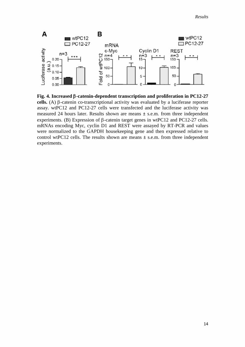
correlated with a higher -catenin-dependent transcriptional activity (revealed by a
luciferase reporter assay, Fig. 4A) and with the higher expression of known -
catenin–TCF-LEF target genes Myc, Rest (Willert et al., 2002; Nishihara et al., 2003)
and (to a lower extent) also Ccnd1 (Fig. 4B).
Results
14
Fig. 4. Increased -catenin-dependent transcription and proliferation in PC12-27
cells. (A) -catenin co-transcriptional activity was evaluated by a luciferase reporter
assay. wtPC12 and PC12-27 cells were transfected and the luciferase activity was
measured 24 hours later. Results shown are means ± s.e.m. from three independent
experiments. (B) Expression of -catenin target genes in wtPC12 and PC12-27 cells.
mRNAs encoding Myc, cyclin D1 and REST were assayed by RT-PCR and values
were normalized to the GAPDH housekeeping gene and then expressed relative to
control wtPC12 cells. The results shown are means ± s.e.m. from three independent
experiments.

Results
15
Fig. 5. The increased proliferation of PC12-27 cells depends on -catenin. (A) -
catenin co-transcriptional activity evaluated by the luciferase reporter assay: effects of
quercetin (a blocker of β-catenin-dependent transcription, 100 μM) and endo-IWR1
(that favors -catenin degradation, 10 μM) administered from the fourth and fifth day
of culture. Nt, untreated cells. Results shown are means ± s.e.m. from three
independent experiments. (B) Expression of Myc and Rest genes in PC12-27 cells,
effects of quercetin and endo-IWR1, conditions and processing and presentation of
the results as in 4B. Results shown are means ± s.e.m. from three independent
experiments. (C) wtPC12 and PC12-27 cell proliferation measured daily by viable
Trypan-Blue-assisted counting of triplicate wells as in Fig. 2A. Treatment with
quercetin and endo-IWR1 as in A. Results shown are means ± s.e.m. from three
independent experiments. Significance shown between PC12-27 cells without and
with drug. ***P<0.001; **P<0.01; *P<0.05.
Results
16
To investigate whether -catenin transcription activity was indeed responsible
for the higher target gene expression and the proliferation advantage of PC12-27 cells,
we adopted a pharmacological approach using two drugs known to operate by
different mechanisms. We took advantage of quercetin, a blocker of the -catenin–
TCF-LEF transcription (Park et al., 2005) and of endo-IWR1, which favors -catenin
degradation (Chen et al., 2009). In PC12-27 cells, both drugs inhibited to a significant
extent the -catenin-dependent reporter gene expression (Fig. 5A) and the expression
of Myc and Rest (Fig. 5B), whereas in wtPC12, these effects were smaller and non-
significant (not shown). Moreover, the two drugs abrogated the proliferation
advantage of high-REST PC12-27 cells (Fig. 5C). Taken together, results obtained by
the comparison of wtPC12 and PC12-27 cells link REST levels to TSC2 levels and to
-catenin nuclear activity, which is critical for cell proliferation.
III.3. REST, TSC2 and -catenin, inter-connected in a feed-forward loop,
control PC12 cell proliferation
The results reported so far (Figs 1-5), which reveal differences in structure,
signaling and proliferation between the two PC12 clones, the low-REST wtPC12 and
the high-REST PC12-27, suggested that REST, TSC2 and -catenin might be
interconnected in a signaling loop controlling proliferation of PC12 cells. In view of
the considerable differences in gene expression existing between the two clones
(Grundschober et al., 2002), however, the possibility of the results to be correlative,
rather than conclusive, could not be excluded. To directly prove the link of REST,
TSC2 and -catenin we carried out gene complementation and down-regulation
experiments in wtPC12 cells.
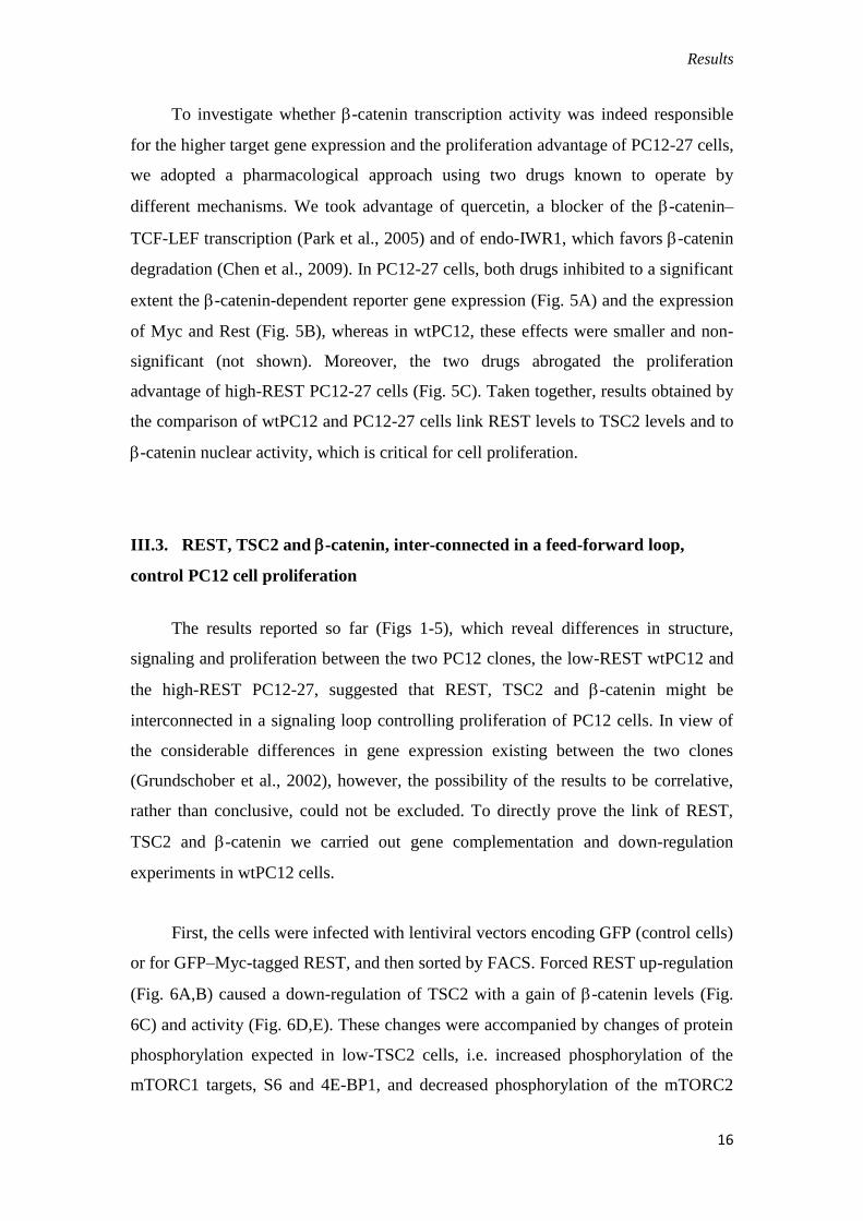
First, the cells were infected with lentiviral vectors encoding GFP (control cells)
or for GFP–Myc-tagged REST, and then sorted by FACS. Forced REST up-regulation
(Fig. 6A,B) caused a down-regulation of TSC2 with a gain of -catenin levels (Fig.
6C) and activity (Fig. 6D,E). These changes were accompanied by changes of protein
phosphorylation expected in low-TSC2 cells, i.e. increased phosphorylation of the
mTORC1 targets, S6 and 4E-BP1, and decreased phosphorylation of the mTORC2
Results
17
target Akt and of GSK3 (data not shown). REST-infected PC12 cells also revealed a
proliferation advantage when compared with control-infected cells (Fig. 6F).
Fig. 6. Expression of REST, TSC2 and -catenin, and of their targets. (A)
Expression of Rest mRNA. (B) Levels of REST in the two infected cell populations,
representative western blots also showing -tubulin used for normalization (left); and
means ± s.e.m. of the results of three independent experiments quantified by
densitometry (right). (C) TSC2 and -catenin in the two infected populations,
presentation as in A. (D) -catenin co-transcriptional activity evaluated by the
luciferase reporter assay in the two infected cell populations. Conditions and
Results
18
processing and presentation of the results (from three independent experiments) as in
Fig. 4A. (E) Expression of -catenin target genes. Conditions, processing and
presentation of the data (from three independent experiments) as in Fig. 4B. (F)
Proliferation of the two infected cell populations measured daily by viable Trypan-
Blue-assisted counting of triplicate wells; means ± s.e.m. from three independent
experiments as in Fig. 1D. ***P<0.001; **P<0.01; *P<0.05.
Journal of
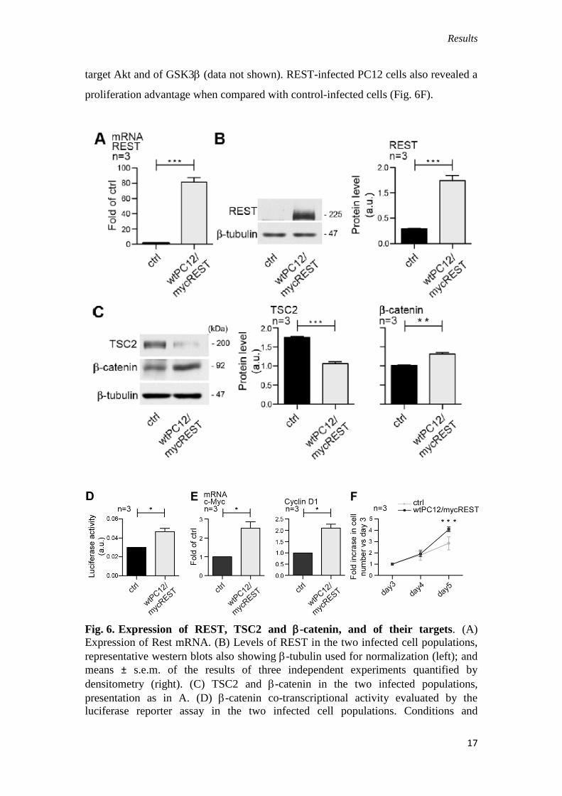
Fig. 7. TSC2 regulates -catenin levels and cell proliferation in wtPC12. wtPC12
cells were stably transfected with the control construct (ctrl) or the shRNA TSC2
construct (shTSC2). (A) Representative western blots of cells infected with the two
constructs showing the levels of TSC2, -catenin and REST together with H2b and -
tubulin used for normalization (left); and means ± s.e.m. of the results of six
independent experiments quantified by densitometry are shown on the right. (B)
wtPC12, PC12-27, control and shTSC2 cells proliferation measured daily by viable,
Trypan-Blue-assisted counts of triplicate wells; data are means ± s.e.m. from three
independent experiments as in Fig. 1. Significance in A is shown between control and
shTSC2-transfected cell populations.
We next investigated the effects of TSC2 knockdown by shRNA and of over-
expression of a constitutively active form of -catenin. TSC2 down-regulation
correlated with increased accumulation of REST and -catenin that proved to be
Results
19
transcriptionally active (Fig. 7A). In addition, cells are accumulated to greater
numbers compared with control wtPC12 and similarly to PC12-27 cells (Fig. 7B).
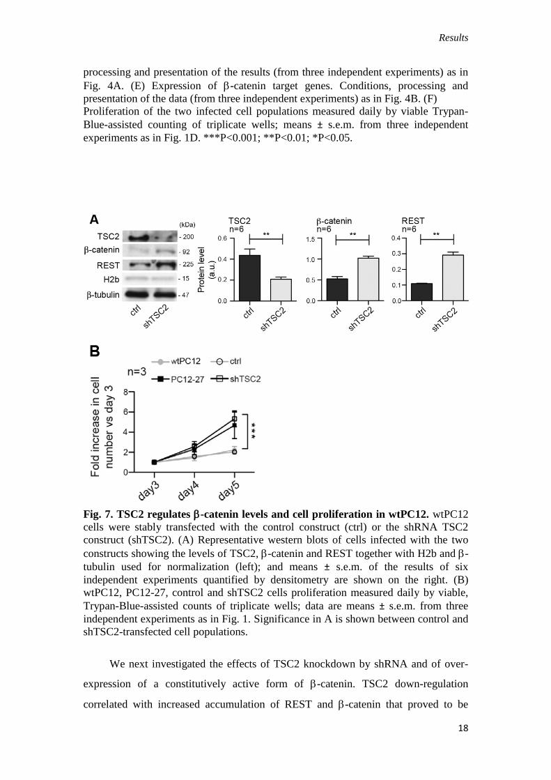
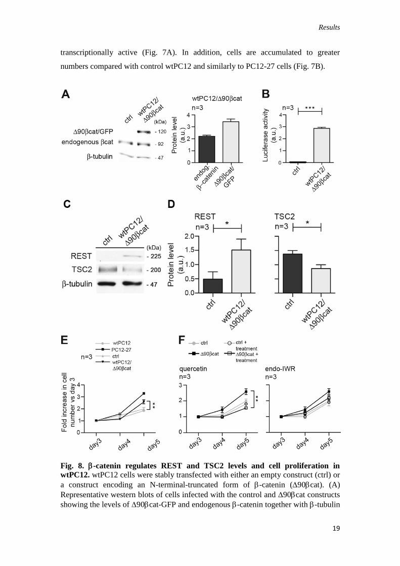
Fig. 8. -catenin regulates REST and TSC2 levels and cell proliferation in
wtPC12. wtPC12 cells were stably transfected with either an empty construct (ctrl) or
a construct encoding an N-terminal-truncated form of -catenin (90cat). (A)
Representative western blots of cells infected with the control and 90cat constructs
showing the levels of 90cat-GFP and endogenous -catenin together with -tubulin

Results
20
used for normalization (left); and comparison of the levels (means ± s.e.m.) of the two
forms of -catenin, results of three independent experiments quantified by
densitometry (right). (B) -catenin co-transcriptional activity evaluated by the
luciferase reporter assay in the control- and 90cat-transfected cells. Conditions and
processing/presentation of the results (from three independent experiments) as in Fig.
4A. (C) Representative western blots of cells infected with the control and the
90cat constructs showing the levels of REST and TSC2 together with -tubulin
used for normalization (left); the means s.e.m. of the results of three independent
experiments about the REST and TSC2 levels quantified by densitometry are shown
on the right. (D) Control and 90cat-transfected cell proliferation measured daily by
viable Trypan-Blue-assisted counts of triplicate wells. Comparison with non-
transfected wtPC12 and PC12-27 (left). The results are means s.e.m. from three
independent experiments. Significance shown is between control and 90cat. (E)
Effects of quercetin (middle) and endo-IWR (right) employed as in Fig. 5, in the
control and 90cat populations. The results are means s.e.m. from three
independents. Significance shown is between 90cat and 90cat + drug cell
populations. ***P<0.001; **P<0.01; *P<0.05.
In PC12 cells stably transfected with a -catenin construct, the expression of
Δ90Cat was reflected by gained -catenin co-transcriptional activity (Fig. 8A, B)
and the levels of the REST protein were increased, whereas TSC2 levels were
decreased (Fig. 8C). Moreover, the cells also revealed a proliferation advantage that
was similar to that of PC12-27 cells when compared with parental and control cells
(Fig. 8D). Interestingly, this advantage was largely abrogated by quercetin (Fig. 8E,
left panel), but not by endo-IWR (Fig. 8E, right panel). Thus, forcing up-regulation of
REST, down-regulation of TSC2 or increased transcriptional activity of -catenin
promote reciprocal changes in their relative expression levels, and have an impact on
wtPC12 cell proliferation. These results, which recapitulate in wtPC12 the properties
of PC12-27 cells, directly link REST, TSC2 and -catenin in a feed-forward loop
favoring PC12 cell proliferation.
IV. Neurite outgrowth
A key property of PC12 cells, based on which the cells have been often
employed as a neuron cell model, is their response to NGF with the outgrowth of
neurites (Greene and Tischler, 1976). This property is however absent in the high
REST PC12-27 cells. The mechanism by which the high REST of PC12-27 prevents

Results
21
the outgrowth response had never been thoroughly investigated. In this section,
labeled IV, we will report our findings obtained about the mechanism by which high
REST represses this process.
IV. 1. NGF receptor expression and signaling
The first task of this study was the analysis of the expression and functioning of
the NGF receptors, TrkA and p75NTR
, compared in two PC12 clones, the wtPC12 and
the high REST PC12-27. In contrast to previous hypotheses (Leoni et al., 1999;
Schulte et al., 2010), the two clones, analyzed under resting conditions (10% serum in
the medium), were found to be close both in terms of mRNA and protein of the TrkA
receptor. In contrast p75NTR
, which is prominent in wtPC12, was inappreciable in
PC12-27 cells at both the mRNA and the protein level (Fig. 9A, B). These results
could be due to a direct repression of p75NTR
expression by the high REST of PC12-
27 cells. In fact the gene of the receptor includes in its promoter two RE-1 (Tab. 1),
the DNA sequence specific of REST binding (Wu and Xie, 2006). In contrast, no RE-
1 sequence is present in the promoter of TrkA (Bruce et al., 2004). TrkA and p75NTR
were immunolabeled both before and after detergent permeabilization of the cells, to
reveal their surface and total complement, respectively.
Fig. 9. Expression of TrkA and p75NTR
. The mRNA and protein of the two receptors
were revealed in the two clones by RT-PCR (A) and western blotting (B). Notice the
lack of p75NTR
in the PC12-27 clone. Here, and in the following figures, the number of
gels analyzed quantitatively is given by the numbers over the panels; the numbers
flanking the gels are the MDa of the immunolabeled proteins, given only in the figure
showing the protein for the first time. The significance of the results, given as means
of 6 experiments s.e.m., is calculated with respect to the sample labeled 0 in each
cell population. *P<0.05; **P<0.01;***P<0.001.

Results
22
Tab.1 The two RE-1s (the DNA sequences specific of REST binding) present in
the promoter of p75NTR
gene
http://broad.mit.edu/~xhx/projects/NRSE/
TTCAGCACTGGAGACTGAGGCC
CCCAGACCCTAGGAGAGAGGCT
Fig. 10. Surface
immunolabeling of the two
receptors in the wtPC12 and
PC12-27 cells (A). The
fractions of the total receptors
distributed to the surface, given
as percentages, are shown (B).
Scale bars: 5 μm (left), and 10
μm (right).
Results
23
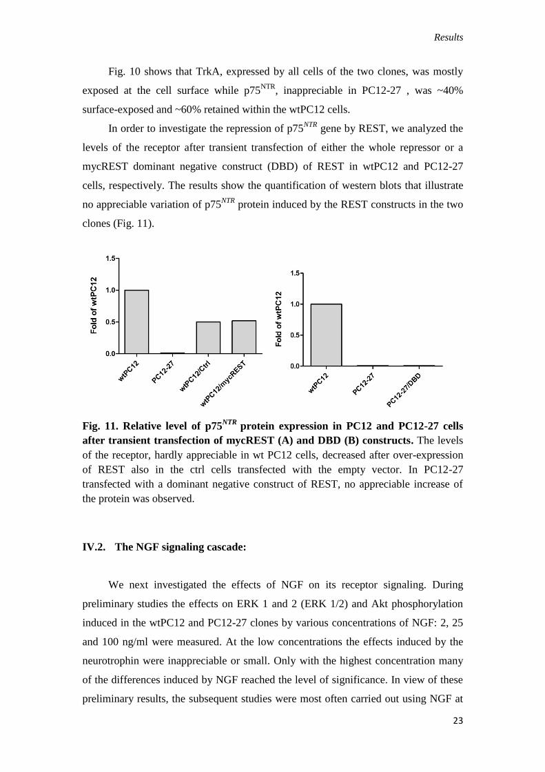
Fig. 10 shows that TrkA, expressed by all cells of the two clones, was mostly
exposed at the cell surface while p75NTR
, inappreciable in PC12-27 , was ~40%
surface-exposed and ~60% retained within the wtPC12 cells.
In order to investigate the repression of p75NTR
gene by REST, we analyzed the
levels of the receptor after transient transfection of either the whole repressor or a
mycREST dominant negative construct (DBD) of REST in wtPC12 and PC12-27
cells, respectively. The results show the quantification of western blots that illustrate
no appreciable variation of p75NTR
protein induced by the REST constructs in the two
clones (Fig. 11).
Fig. 11. Relative level of p75NTR
protein expression in PC12 and PC12-27 cells
after transient transfection of mycREST (A) and DBD (B) constructs. The levels
of the receptor, hardly appreciable in wt PC12 cells, decreased after over-expression
of REST also in the ctrl cells transfected with the empty vector. In PC12-27
transfected with a dominant negative construct of REST, no appreciable increase of
the protein was observed.
IV.2. The NGF signaling cascade:
We next investigated the effects of NGF on its receptor signaling. During
preliminary studies the effects on ERK 1 and 2 (ERK 1/2) and Akt phosphorylation
induced in the wtPC12 and PC12-27 clones by various concentrations of NGF: 2, 25
and 100 ng/ml were measured. At the low concentrations the effects induced by the
neurotrophin were inappreciable or small. Only with the highest concentration many
of the differences induced by NGF reached the level of significance. In view of these
preliminary results, the subsequent studies were most often carried out using NGF at

Results
24
100 ng/ml, a concentration widely employed in recent studies of the literature (see,
among others, Koch et al., 2008; Miranda et al., 2001; Pincheira et al., 2009; Wang et
al., 2013).
IV.2.a. TrkA auto-phosphorylation
In a first series of phosphorylation studies the wtPC12 and PC12-27 cells were
incubated in low (1%) serum medium for 24 hr before treatments, and then analyzed
in the same medium. The time-course of the TrkA phosphorylation at various tyrosine
residues during the first 20 min of NGF treatment is illustrated in Fig. 12.
Fig. 12. NGF-induced TrkA
auto-phosphorylation
responses in wtPC12 and
PC12-27 cells. The time-
course of the auto-
phosphorylations induced by
NGF (100 ng/ml) at three
sites of the TrkA receptor,
Y751 is shown in the top,
Y490 in the middle and
Y670, Y674 and Y675,
analyzed together, in the
bottom (A). The time-course
data of western blot are
illustrated in quantitative
terms in the panel below (B)
Results
25
Figure 12 shows that in the wtPC12, the Y751 site was rapidly phosphorylated,
reaching the highest level at 3 min and then declining to the resting level. In PC12-27
cells the Y751 phosphorylation, evident at rest, failed to increase significantly during
the stimulation. The phosphorylation of the Y490 site was well appreciable only in the
wt cells, with the highest values at 5–10 min, whereas the phosphorylation of the
Y670, Y674 and Y675, three sites of limited importance for TrkA signaling (Biarc et
al., 2013) that were investigated together, was similar in the two clones, with only
limited changes induced by NGF stimulation (Fig. 12).
Considering togheter the data of Figs 9-12 we conclude that the level of TrkA is
similar in the two, low and high REST PC12 clones. In contrast, the NGF-induced
auto-phosphorylation of the receptor, especially that of the Y490 site, is defective in
PC12-27 cells. This might be due to the lack of cooperation of TrkA with the other
NGF receptor, p75NTR
, which lacks in the PC12-27 cells.
IV.2.b. Phosphorylation of ERK and Akt.
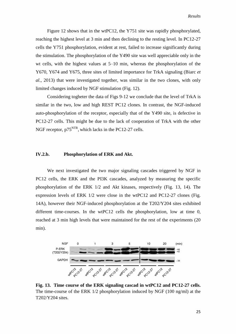
We next investigated the two major signaling cascades triggered by NGF in
PC12 cells, the ERK and the PI3K cascades, analyzed by measuring the specific
phosphorylation of the ERK 1/2 and Akt kinases, respectively (Fig. 13, 14). The
expression levels of ERK 1/2 were close in the wtPC12 and PC12-27 clones (Fig.
14A), however their NGF-induced phosphorylation at the T202/Y204 sites exhibited
different time-courses. In the wtPC12 cells the phosphorylation, low at time 0,
reached at 3 min high levels that were maintained for the rest of the experiments (20
min).
Fig. 13. Time course of the ERK signaling cascad in wtPC12 and PC12-27 cells.
The time-course of the ERK 1/2 phosphorylation induced by NGF (100 ng/ml) at the
T202/Y204 sites.
Results
26
In PC12-27 cells, the resting level was higher than that of wtPC12. The NGF-induced
increase occurred, however it was delayed, reaching top levels similar to those of
stimulated wtPC12 only after 10 min and thereafter (Fig. 13).
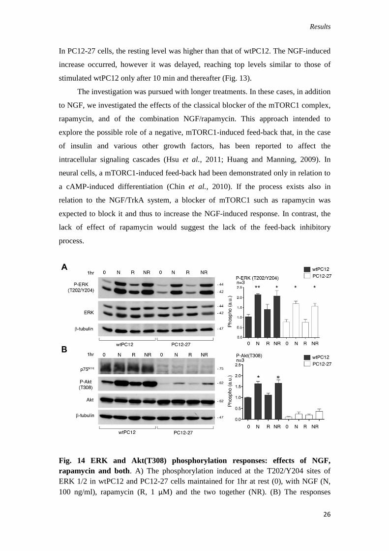
The investigation was pursued with longer treatments. In these cases, in addition
to NGF, we investigated the effects of the classical blocker of the mTORC1 complex,
rapamycin, and of the combination NGF/rapamycin. This approach intended to
explore the possible role of a negative, mTORC1-induced feed-back that, in the case
of insulin and various other growth factors, has been reported to affect the
intracellular signaling cascades (Hsu et al., 2011; Huang and Manning, 2009). In
neural cells, a mTORC1-induced feed-back had been demonstrated only in relation to
a cAMP-induced differentiation (Chin et al., 2010). If the process exists also in
relation to the NGF/TrkA system, a blocker of mTORC1 such as rapamycin was
expected to block it and thus to increase the NGF-induced response. In contrast, the
lack of effect of rapamycin would suggest the lack of the feed-back inhibitory
process.
Fig. 14 ERK and Akt(T308) phosphorylation responses: effects of NGF,
rapamycin and both. A) The phosphorylation induced at the T202/Y204 sites of
ERK 1/2 in wtPC12 and PC12-27 cells maintained for 1hr at rest (0), with NGF (N,
100 ng/ml), rapamycin (R, 1 μM) and the two together (NR). (B) The responses
Results
27
induced by the same treatments at the P-Akt(T308). In each cell sample the levels of
the ERK (B), Akt and p75NTR
(C) proteins did not change during the experiments. The
data on P-ERK 1/2(T202/Y204) and P-Akt(T308) are also shown in quantized terms
on the right of panels B and C. The numbers flanking the gels are the MDa of the
immunolabeled proteins. The statistical analysis and the significance of the
differences are shown by the asterisks as specified in the legend for Fig. 10.
The ERK results obtained by 1 hr treatments with NGF, rapamycin and
NGF/rapamycin are shown in Fig. 14A. Upon NGF treatment, the levels of ERK 1/2
were unchanged whereas those of their phosphorylation were almost doubled in
wtPC12 and significantly increased, although to a moderately lower extent, in PC12-
27 cells. In contrast, both the resting and the NGF-induced levels of ERK 1/2,
phosphorylation appeared unchanged in both clones upon treatment with rapamycin
(Fig. 14A).
Also the levels of Akt were similar in the wtPC12 and PC12-27 cells, with no
changes induced by the various treatments with NGF and rapamycin investigated
(Fig. 14B). As far as the phosphorylations, that of Akt(T308), indicative of the PI3K
cascade, increased slowly in the wtPC12 cells, whereas in the PC12-27 cells it
remained apparently unchanged during the first 20 min of NGF treatment (data not
shown). One hr (Fig. 14B) or longer (up to 48 hr, data not shown) treatments of
wtPC12 cells with NGF induced significant increases of the Akt(T308)
phosphorylation. Rapamycin alone, administered for 1 or 24 hr, modified neither the
basal nor the NGF-induced Akt(T308) phosphorylation of wtPC12 (Fig. 14B and data
not shown).

Results
28
Fig. 15 Phosphorylation of two Akt targets, TSC2(S939) and GSK3β(S9),
induced by NGF and rapamycin in wtPC12 and PC12-27 cells. The changes in
phosphorylation of TSC2(S939) and GSK3b(S9) induced in the two cell clones by 1
hr treatment with NGF (N, 100 ng/ml), rapamycin (R, 1 μM) or the two together. The
numbers flanking the gels are the MDa of the immunolabeled proteins. Compared to
the results of P-Akt(T308), shown in Fig. 14B, the changes of the two targets induced
by rapamycin are larger, especially in the PC12-27 cells that are unresponsive to
NGF.
In the PC12-27 cells the basal phosphorylation of Akt(T308) was much lower
than that of wtPC12. NGF and, to a lower extent, also rapamycin (administered alone
or together for 1 (Fig. 14B) or 24 hr (data not shown) did apparently induce some
increases of P-Akt(T308) which however remained statistically non significant (Fig.
14B).
Two targets of Akt, TSC2(S939) and GSK3β(S9), exhibited phosphorylation
patterns different from those of Akt(T308). Specifically, the increases in the wtPC12
cells were smaller, while those induced by rapamycin were larger than those of
Akt(T308) (Fig. 14). In the PC12-27 cells, the resting phosphorylation of TSC2(S939)
and GSK3β(S9) was low. In these cells no significant increase was induced by NGF.
In contrast, rapamycin induced significant increases (Fig. 15). Taken together with the
data of Fig. 14B, the data of Fig. 15 suggest that some mTORC1-induced feed-back
Results
29
inhibition of the PI3K cascade may operate in PC12-27. In wtPC12 cells, however, no
sign of the feed-back was appreciable.
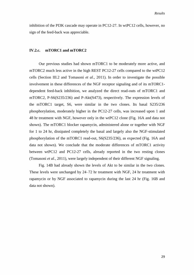
IV.2.c. mTORC1 and mTORC2
Our previous studies had shown mTORC1 to be moderately more active, and
mTORC2 much less active in the high REST PC12-27 cells compared to the wtPC12
cells (Section III.2 and Tomasoni et al., 2011). In order to investigate the possible
involvement in these differences of the NGF receptor signaling and of its mTORC1-
dependent feed-back inhibition, we analyzed the direct read-outs of mTORC1 and
mTORC2, P-S6(S235/236) and P-Akt(S473), respectively. The expression levels of
the mTORC1 target, S6, were similar in the two clones. Its basal S235/236
phosphorylation, moderately higher in the PC12-27 cells, was increased upon 1 and
48 hr treatment with NGF, however only in the wtPC12 clone (Fig. 16A and data not
shown). The mTORC1 blocker rapamycin, administered alone or together with NGF
for 1 to 24 hr, dissipated completely the basal and largely also the NGF-stimulated
phosphorylation of the mTORC1 read-out, S6(S235/236), as expected (Fig. 16A and
data not shown). We conclude that the moderate differences of mTORC1 activity
between wtPC12 and PC12-27 cells, already reported in the two resting clones
(Tomasoni et al., 2011), were largely independent of their different NGF signaling.
Fig. 14B had already shown the levels of Akt to be similar in the two clones.
These levels were unchanged by 24–72 hr treatment with NGF, 24 hr treatment with
rapamycin or by NGF associated to rapamycin during the last 24 hr (Fig. 16B and
data not shown).
Results
30
Fig. 16 Read-outs of mTORC1 (P-S6(S235/236)) and mTORC2 (P-
Akt(S473)) in wtPC12 and PC12-27 cells. (A,B) wtPC12 and PC12-27 cells were
treated for 48 hr with no stimulant (0), with NGF (N, 100 ng/ml), with rapamycin in
the last 24 hr (R, 0.1 μM) and with the two together (NR). The quantization of the
data is on the right panels. The levels of the S6 and Akt proteins were not changed by
the treatments. The numbers flanking the gels are the MDa of the immunolabeled
proteins. Statistical analysis and significance of the differences is given as specified in
the legend for Fig. 1.
In terms of phosphorylation, the results with the direct mTORC2 read-out,
Akt(S473) were quite different in wtPC12 and PC12-27 cells. In the wtPC12, 1 to 24
hr treatment with NGF induced increases of 15–20-fold (Fig. 16B and data not
shown). Treatment of wtPC12 with rapamycin induced increases similar to those
induced by NGF. The two increases, however, were not additive when the cells were
exposed to the combined NGF/rapamycin treatment (Fig. 16B and data not shown). In
the PC12-27 clone, the resting phosphorylation of Akt(S473), distinctly lower than
that of wtPC12, was not changed significantly by NGF. In contrast rapamycin did
increase the P-Akt(S473) by over 5-fold. The combination with NGF did not change
the increased P-Akt(S473) induced by rapamycin (Fig. 16B and data not shown).

Results
31
IV.2.d. PI3K dependence of the pathways
In parallel experiments, the key role of the PI3K cascade in the control of
mTORC2 was confirmed by the use of the PI3K inhibitor wortmannin. In both the
wtPC12 and PC12-27 clones treated with NGF for 48 hr, treatment with the drug (0.3
mM) for the last 10 min attenuated considerably the P-TSC2(S939) and P-GSK3β(S9)
and eliminates completely the P-Akt(S473) phosphorylation (Fig. 17).
Fig. 17. mTORC2 read-outs were analyzed after wortmannin treatment in
wtPC12 and PC12-27 cells. The phosphorylation of Akt(S473) induced by NGF (N,
100 ng/ml, 60 min) in wtPC12 cells was completely dissipated by the addition of
wortmannin (NW, 0.3 mM) during the last 10 min.
Summing up, the results show that p75NTR
has a role in the control of mTORC2
activity, revealed by the direct read-out Akt(S473). The findings with PC12-27 cells
strengthens, at the mTORC2 level, the non-significant rapamycin results of the PI3K
cascade dependent phosphorylation of Akt(T308) illustrated in Fig. 14B. In these cells
the low mTORC2 activity, unaffected by NGF, appears to be affected by the
mTORC1-dependent feed-back inhibition process inhibited by rapamycin.
IV.3. Transient transfection of p75NTR
in PC12-27 increases mTORC2 activity
In order to confirm the involvement of p75NTR
in mTORC2 activation and in the
differentiation process, defective PC12-27 cells were transiently transfected with the
full length receptor cDNA. Transient transfection resulted in great increases of the P-

Results
32
Akt(T308) and P-GSK3β phosphorylations, the read-outs of the mTORC2 activity,
whereas P-ERK was unchanged (Fig. 18).
Fig. 18. Transient transfection of p75NTR
in defective PC12 cells induces
mTORC2 activation. Panel shows that transient transfection of full length receptor
leads-out the phosphorylation of Akt(T308) and GSK3β(S9), whereas there is no
difference in ERK activation.
IV.4. Generation and characterization of PC12-27/p75NTR
stable clones
PC12-27 cells were stably transfected with a construct of p75NTR
human full
length cDNA and the screening of the clones for p75NTR
expression was performed by
western blot analysis. Figure 19 shows the western blot detection of positive clones,
however their level of expression is variable. Taking into consideration the level of
the receptor in PC12 wt, we did choose one of these sub-clones, sub-clone 8, that
from here-on is named PC12-27/p75NTR
.
Fig. 19. Expression of p75
NTR in the stably transfected PC12-27 sub-clones. As
demonstrate, sub-clone 8 expresses level of p75NTR
comparable at those of PC12 wt,
whereas sub-clones 1,2,3,5 and 10 over-express the receptor, and sub-clones 1,4 and 9
express only low levels.

Results
33
Fig. 20. Expression of p75NTR
and time-course of TrkA auto-phosphorylation at
the Y751 and Y490 sites in PC12-27 cells transfected with the vector, empty
(PC12-27/Ctrl) or including the full length p75NTR
(PC12-27/p75NTR
). (A) The
western blot of p75NTR
in wtPC12, PC12-27/Ctrl and PC12-27/p75NTR
cells.
Quantization of the data, documenting the similar levels of the receptor in the wtPC12
and PC12-27/p75NTR
cells, is on the right. (B) Surface immunolocalization of p75NTR
in the PC12-27/Ctrl and PC12-27p75NTR
cells. The quantization of the results is on the
right. Scale bar: 10 μm. (C,D) The time-course of the TrkA auto-phosphorylation at
the Y751 and Y490 sites induced by NGF (100 ng/ml) in the two transfected sub-
clones, PC12-27/Ctrl and PC12-27/p75NTR
. The quantization of these data is shown
Results
34
on the right. Statistical analysis and significance of the differences is given as
specified in the legend of Fig. 10.
IV.5. NGF signaling in wtPC12 and PC12-27 cells: role of p75NTR
The sub-clone, PC12-27/p75NTR
exhibited a surface distribution of p75NTR
similar to those observed in the wtPC12 (compare Fig. 20A,B to Fig. 10A,B). To
exclude possible artifacts due to hyper/hypo-expression or altered distribution of the
receptor, the sub-clone was selected for subsequent studies, using as control a PC12-
27 sub-clone transfected with the empty vector (PC12-27/Ctrl).
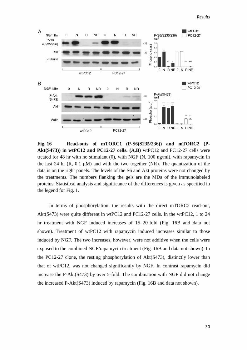
Fig. 21. Stable expression of p75NTR
and time-course of TrkA auto-
phosphorylation at the Y751 and Y490 sites in PC12-27 cells transfected with the
vector, empty (PC12-27/Ctrl) or including the full length p75NTR
(PC12-
27/p75NTR
). A is a time course of the NGF responses; B and C the responses to NGF ,
rapamycin and the two together, with the quantitation of the results below the Western
Results
35
blots; D the same in the PC12-27/p75NTR
, however in the presence of the TrkA
inhibitor, Calbiochem 648450.
During the first 20 min treatment of PC12-27/p75NTR
cells with NGF, the Y751
phosphorylation of TrkA increased markedly (Fig. 20C), similar to the wtPC12 cells,
whereas that PC12-27/Ctrl cells resembled that of the non-transfected PC12-27 cells,
i.e. it did not change significantly. At the Y490 site the differences of phosphorylation
observed between the cells transfected with and without p75NTR
were even larger. In
the PC12-27/Ctrl cells this phosphorylation remained almost inappreciable, as in the
non-transfected PC12-27 cells, whereas in the PC12-27/p75NTR
cells it increased
significantly and rapidly upon NGF addition, reaching a maximum at 5–10 min, as in
the wtPC12 (Fig. 20D). The study of the two cascades, of ERK and PI3K, confirmed
the marked changes of the NGF signaling induced in PC12-27 cells by the expression
of p75NTR
. In the case of ERK the phosphorylation of ERK 1/2(T202 and Y204),
induced by 1–20 min treatment with NGF, exhibited a faster rate in the PC12-
27/p75NTR
cells compared to the PC12-27/Ctrl cells (Fig. 21A), similar to the faster
rate of the wtPC12 compared to the PC12-27 cells shown in Fig. 13.
Also the responses induced in the PC12-27/Ctrl and PC12-27/p75NTR
cells by 1
hr treatment with NGF, alone or with rapamycin, resembled the responses induced by
the same treatments in the PC12-27 and wtPC12 cells, respectively. With rapamycin
alone the changes were small and non significant in both transfected PC12-27 cell
sub-clones (Fig. 21B). In contrast, in the case of P-Akt(T308), the increases in PC12-
27/p75NTR
cells induced by NGF were larger than in PC12-27/Ctrl (Fig. 21C). Also
with the two Akt targets, TSC2(S939) and GSK3β(S9), the phosphorylations induced
by NGF and also by rapamycin in the PC12-27/p75NTR
were distinctly larger than
those in the PC12/Ctrl cells (Fig. 22).
Results
36
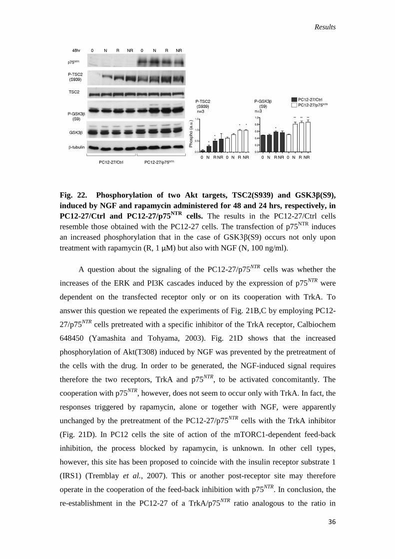
Fig. 22. Phosphorylation of two Akt targets, TSC2(S939) and GSK3β(S9),
induced by NGF and rapamycin administered for 48 and 24 hrs, respectively, in
PC12-27/Ctrl and PC12-27/p75NTR
cells. The results in the PC12-27/Ctrl cells
resemble those obtained with the PC12-27 cells. The transfection of p75NTR
induces
an increased phosphorylation that in the case of GSK3β(S9) occurs not only upon
treatment with rapamycin (R, 1 μM) but also with NGF (N, 100 ng/ml).
A question about the signaling of the PC12-27/p75NTR
cells was whether the
increases of the ERK and PI3K cascades induced by the expression of p75NTR
were
dependent on the transfected receptor only or on its cooperation with TrkA. To
answer this question we repeated the experiments of Fig. 21B,C by employing PC12-
27/p75NTR
cells pretreated with a specific inhibitor of the TrkA receptor, Calbiochem
648450 (Yamashita and Tohyama, 2003). Fig. 21D shows that the increased
phosphorylation of Akt(T308) induced by NGF was prevented by the pretreatment of
the cells with the drug. In order to be generated, the NGF-induced signal requires
therefore the two receptors, TrkA and p75NTR
, to be activated concomitantly. The
cooperation with p75NTR
, however, does not seem to occur only with TrkA. In fact, the
responses triggered by rapamycin, alone or together with NGF, were apparently
unchanged by the pretreatment of the PC12-27/p75NTR
cells with the TrkA inhibitor
(Fig. 21D). In PC12 cells the site of action of the mTORC1-dependent feed-back
inhibition, the process blocked by rapamycin, is unknown. In other cell types,
however, this site has been proposed to coincide with the insulin receptor substrate 1
(IRS1) (Tremblay et al., 2007). This or another post-receptor site may therefore
operate in the cooperation of the feed-back inhibition with p75NTR
. In conclusion, the
re-establishment in the PC12-27 of a TrkA/p75NTR
ratio analogous to the ratio in
Results
37
wtPC12 was found to rescue the NGF signaling from a partially inactive to a fully
active state.
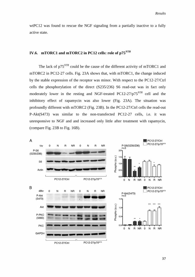
IV.6. mTORC1 and mTORC2 in PC12 cells: role of p75NTR
The lack of p75NTR
could be the cause of the different activity of mTORC1 and
mTORC2 in PC12-27 cells. Fig. 23A shows that, with mTORC1, the change induced
by the stable expression of the receptor was minor. With respect to the PC12-27/Ctrl
cells the phosphorylation of the direct (S235/236) S6 read-out was in fact only
moderately lower in the resting and NGF-treated PC12-27/p75NTR
cell and the
inhibitory effect of rapamycin was also lower (Fig. 23A). The situation was
profoundly different with mTORC2 (Fig. 23B). In the PC12-27/Ctrl cells the read-out
P-Akt(S473) was similar to the non-transfected PC12-27 cells, i.e. it was
unresponsive to NGF and and increased only little after treatment with rapamycin,
(compare Fig. 23B to Fig. 16B).
Results
38
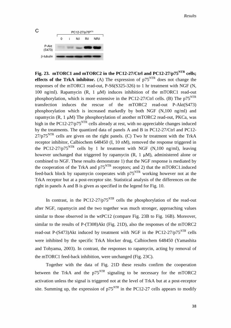
Fig. 23. mTORC1 and mTORC2 in the PC12-27/Ctrl and PC12-27/p75NTR
cells;
effects of the TrkA inhibitor. (A) The expression of p75NTR
does not change the
responses of the mTORC1 read-out, P-S6(S325-326) to 1 hr treatment with NGF (N,
100 ng/ml). Rapamycin (R, 1 μM) induces inhibition of the mTORC1 read-out
phosphorylation, which is more extensive in the PC12-27/Ctrl cells. (B) The p75NTR
transfection induces the rescue of the mTORC2 read-out P-Akt(S473)
phosphorylation which is increased markedly by both NGF (N,100 ng/ml) and
rapamycin (R, 1 μM) The phosphorylation of another mTORC2 read-out, PKCa, was
high in the PC12-27/p75NTR
cells already at rest, with no appreciable changes induced
by the treatments. The quantized data of panels A and B in PC12-27/Ctrl and PC12-
27/p75NTR
cells are given on the right panels. (C) Two hr treatment with the TrkA
receptor inhibitor, Calbiochem 648450 (I, 10 nM), removed the response triggered in
the PC12-27/p75NTR
cells by 1 hr treatment with NGF (N,100 ng/ml), leaving
however unchanged that triggered by rapamycin (R, 1 μM), administered alone or
combined to NGF. These results demonstrate 1) that the NGF response is mediated by
the cooperation of the TrkA and p75NTR
receptors; and 2) that the mTORC1.induced
feed-back block by rapamycin cooperates with p75NTR
working however not at the
TrkA receptor but at a post-receptor site. Statistical analysis of the differences on the
right in panels A and B is given as specified in the legend for Fig. 10.
In contrast, in the PC12-27/p75NTR
cells the phosphorylation of the read-out
after NGF, rapamycin and the two together was much stronger, approaching values
similar to those observed in the wtPC12 (compare Fig. 23B to Fig. 16B). Moreover,
similar to the results of P-(T308)Akt (Fig. 21D), also the responses of the mTORC2
read-out P-(S473)Akt induced by treatment with NGF in the PC12-27/p75NTR
cells
were inhibited by the specific TrkA blocker drug, Calbiochem 648450 (Yamashita
and Tohyama, 2003). In contrast, the responses to rapamycin, acting by removal of
the mTORC1 feed-back inhibition, were unchanged (Fig. 23C).
Together with the data of Fig. 21D these results confirm the cooperation
between the TrkA and the p75NTR
signaling to be necessary for the mTORC2
activation unless the signal is triggered not at the level of TrkA but at a post-receptor
site. Summing up, the expression of p75NTR
in the PC12-27 cells appears to modify
Results
39
both the signaling of NGF and the activity of mTORC2, bringing them to levels
approaching those of wtPC12.
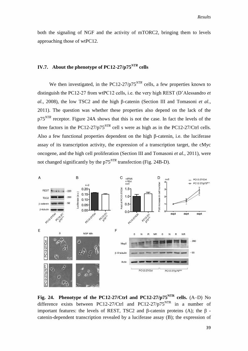
IV.7. About the phenotype of PC12-27/p75NTR
cells
We then investigated, in the PC12-27/p75NTR
cells, a few properties known to
distinguish the PC12-27 from wtPC12 cells, i.e. the very high REST (D’Alessandro et
al., 2008), the low TSC2 and the high β-catenin (Section III and Tomasoni et al.,
2011). The question was whether these properties also depend on the lack of the
p75NTR
receptor. Figure 24A shows that this is not the case. In fact the levels of the
three factors in the PC12-27/p75NTR
cell s were as high as in the PC12-27/Ctrl cells.
Also a few functional properties dependent on the high β-catenin, i.e. the luciferase
assay of its transcription activity, the expression of a transcription target, the cMyc
oncogene, and the high cell proliferation (Section III and Tomasoni et al., 2011), were
not changed significantly by the p75NTR
transfection (Fig. 24B-D).
Fig. 24. Phenotype of the PC12-27/Ctrl and PC12-27/p75NTR
cells. (A–D) No
difference exists between PC12-27/Ctrl and PC12-27/p75NTR
in a number of
important features: the levels of REST, TSC2 and β-catenin proteins (A); the β -
catenin-dependent transcription revealed by a luciferase assay (B); the expression of
Results
40
the β-catenin-target gene, cMyc (C); the rate of cell proliferation (D). (E) Phase
contrast images of the PC12-27/Ctrl and PC12-27/p75NTR
before and after a 48 hr
treatment with NGF (100ng/ml). Scale bar: 20 μm. (F) The expression of two
neuronal markers, Map2 and β -III tubulin, in PC12-27/Ctrl and PC12-27/p75NTR
cells
incubated for 48 hr with no treatment, with NGF (N, 100 ng/ml), rapamycin (R, 0.1
μM during the last 24 hr) and the two together.
Finally, we investigated whether, and to what extent, the expression of p75NTR
modifies two aspects of the phenotype sensitive to NGF that are greatly defective in
PC12-27 cells, the outgrowth of neurites and the expression of neuron-type markers.
Fig. 24E compares the morphology of the PC12-27/Ctrl and PC12-27/p75NTR
cells, at
rest and upon 48 hr treatment with NGF. The flat structure of PC12-27/Ctrl cells,
similar to that of the non-transfected PC12-27 cells (Section III and Tomasoni et al.,
2011) was hardly affected by the 48 hr treatment with NGF. In the PC12-27/p75NTR
cells, on the other hand, the shape was not changed much, however the NGF-induced
neurite outgrowth response was evident in terms of both number of neurites sprouted
per cell and average neurite length (Fig.24E). In the resting PC12-27/Ctrl cells the
levels of the two neuronal markers investigated, Map2 and β-III tubulin, were low.
Treatment for 48 hr with NGF, 24 hr with rapamycin or the two together induced only
small or no increases. In PC12-27/p75NTR
cells, the resting levels of the two markers
were higher, however the increases induced by NGF and rapamycin were small and
non significant (Fig.24F).
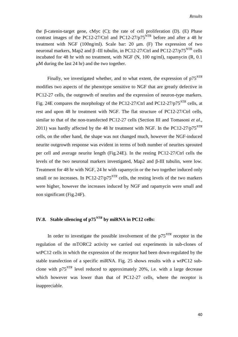
IV.8. Stable silencing of p75NTR
by miRNA in PC12 cells:
In order to investigate the possible involvement of the p75NTR
receptor in the
regulation of the mTORC2 activity we carried out experiments in sub-clones of
wtPC12 cells in which the expression of the receptor had been down-regulated by the
stable transfection of a specific miRNA. Fig. 25 shows results with a wtPC12 sub-
clone with p75NTR
level reduced to approximately 20%, i.e. with a large decrease
which however was lower than that of PC12-27 cells, where the receptor is
inappreciable.
Results
41
Fig. 25. Phenotype of the PC12-27/Ctrl and PC12-27/p75NTR
cells. In the sub-
clone illustrated here, in which the level of the receptor was decreased of about 80%,
the decrease of the mTORC2 read-out P-Akt(S473) was decreased of about 35%.
These changes were apparently ineffective on the PI3K cascade since the P-
Akt(T308) and P-GSK3β(S9) were unchanged, while the level of the neuronal marker
MAP2 was significantly decreased. The general phenotype of the wtPC12 cells was
apparently unaffected by the miRNA expression. Scale bar: 20 μm.
The miRNA transfection did not modify the general phenotype of the wtPC12
cells, that remained largely spherical, different from the flat shape of the PC12-27
cells (Tomasoni et al., 2011). Likewise, the P-Akt(T308) and the P-GSK3β(S9) were
unchanged. In contrast, the Akt(S473) phosphorylation was decreased of about 35%,
suggesting the mTORC2 activity to be reduced (Fig. 25).

Discussion
42
V. DISCUSSION
The data reported in this thesis about the role of REST in PC12 cells are quite
numerous and, in many respects, unexpected. The possibility that high REST induces
proliferation of neurosecretory cells had been reported especially in relation to neural
fast-growing and aggressive tumors (reviews: Coulson, 2005; Majumder, 2006;
Negrini et al, 2013). In these cases, however, a precise mechanism of the REST
stimulation had never been identified. The condition of PC12 cells is quite different
from that of the above tumors. The cell line originates from a pheochromocytoma, a
differentiated, slow-growing, non-aggressive tumor. Therefore the findings we did
obtain by comparative investigation of the two PC12 clones, characterized one by the
typical low level of REST and the other by a high level of the transcription repressor,
had apparently little to do with those on tumors reported previously. Mechanistically,
our findings appeared reasonable when we found the increase of REST to be
accompanied by a decrease of TSC2, a GAP protein known to govern negatively the
activity mTORC1. The mechanism by which the TSC2 decrease is induced by high
REST is unclear. Certainly the effect is not due to the typical function of REST, the
transcription repression, because the level of the TSC2 mRNA was not decreased in
PC12-27 cells. At this stage we can only hypothesize that the decrease of the TSC2
protein is due to its increased turnover induced by REST indirectly, via the activation
of an ubiquitinase system that remains to be identified.
V.1. REST, TSC2 and β-catenin govern proliferation working as a
signaling/effector loop.
However, the increased activity of mTORC1, was shown to account for only a
minor fraction of the fast proliferation of the high REST PC12-27 cells. The major
contribution to the proliferation was mediated in fact by the increase of -catenin,
dependent of the decrease of its fast metabolism and as a consequence, of its
localization to the nucleus. A negative control of the TSC complex on the -catenin
metabolism had been reported (Jozwiak and Wlodarski 2006; Barnes et al., 2010),
however this control pathway, at variance with the TSC complex-mTORC1 pathway,
Discussion
43
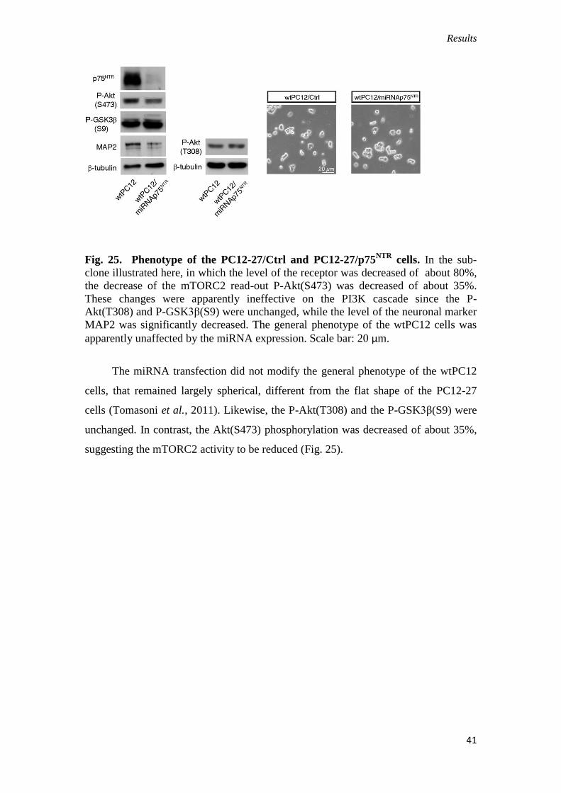
had failed to become popular. In this case the REST-TSC2 was found to be
particularly efficient because -catenin, together with its classical co-transcription
targets, including cMyc and cyclin D1, was found to increase the transcription of
REST. Thus REST, TSC2 and -catenin appear to control the proliferation of PC12
cells working as a loop (Fig. 23). Evidently each of these factors has multiple
functions, many of which previously known. Our work has now identified a new,
important function, operative in neural cells, in which the three factors operate
coordinately. Additional aspects of the data we have presented in the section III of this
thesis can be found in the paper we have published about this work (see Tomasoni,
Negrini et al., 2011). The model in Figure 26, taken from that paper, illustrates the
REST control of proliferation as emerged from our studies.
Fig. 26. A model of the feed-forward loop signaling paradigm governing
PC12 cell proliferation and neurosecretion. The figure summarizes the results
illustrated in Figs 1−7, emphasizing the coordinated regulation of the two cell
functions. Changes of one (or more) of the interconnected factors, REST, TSC2 and
β-catenin (β-cat), impact the whole signaling loop and affect both proliferation and
neurosecretion as shown by the variable thickness of the connections. Other
processes, not yet investigated, might be impacted as well. In low-REST cells (left)
repression of TSC2 is weak, the TSC1−TSC2 complex reinforces the proteasome
degradation of β-catenin, transcription of the β-catenin−TCF-LEF target genes,
including the oncogenes and REST, is low. As a consequence proliferation of cells
(dependent on β-catenin) is low whereas neurosecretion (repressed only marginally by
low levels of REST) is high. In high-REST cells (right), repression of TSC2 is strong,
with ensuing increase of β-catenin and of the β-catenin−TCF-LEF oncogene and
REST transcription. As a consequence, proliferation is high whereas neurosecretion is
repressed to a considerable extent by the high levels of REST (See the detailed Figure-
Abstract here above).

Discussion
44
V.2. The regulation of neurite outgrowth was still largely unknown.
The section IV of this thesis deals with another key issue in our neural cell
investigation, i.e. the mechanism that govern the outgrowth of neurites. This property
is general among neural cell lines. In most of them, however, the response visible
upon stimulation with various factors, although present is quite small. In contrast, in
the case of wtPC12 the outgrowth induced by long-term treatment with NGF is
extensive, similar to the one occurring in the initial phase of in vitro differentiation of
neurons. The difference between wtPC12 and neurons appears in the subsequent
phase, because neurons convert one of the neurites into a typical axon, whereas
wtPC12 don’t. Thus wtPC12 are a model of the initial phase of neural cell
differentiation, that was employed in thousands of studies published in the last 30
years. The high REST PC12-27 cells differ from their wt counterparts because their
NGF-induced neurite outgrowth is almost non-existent. The comparative investigation
of the two PC12 clones was therefore offering the possibility to clarify whether REST
is involved in the control of the outgrowth and what is the mechanism of this control.
V.3. A new role for p75NTR
.
The initial hypothesis put forth to explain the lack of outgrowth in the PC12-27
cells (and in another clone that is now known to be of high REST as well,
D’Alessandro et al., 2008) was the lack in the defective cells of the tyrosine kinase
receptor of NGF, TrkA (Leoni et al., 1999). This hypothesis appeared reasonable
because TrkA is believed to govern almost completely the responses induced by the
factor, with the second receptor, p75NTR
, playing only minor roles: the increase of the
TrkA affinity for their ligand or a number of other cooperative activities. When
however we did investigate the expression of the NGF receptors in the high REST
PC12-27 cells we were surprised to find that TrkA was unchanged, whereas p75NTR
was almost non-existent. This finding, on the one hand, was reasonable because, from
the data of the literature, the p75NTR
gene was shown to include two RE-1 sequences
in its promoter; and because, on the other hand, the response to NGF of PC12-27 cells
was not absent but specifically missing of one pathway, the PI3K-Akt pathway, and

Discussion
45
not that of ERK1/2. The reinforcement by P75NTR
of the TrkA-induced activation of
the PI3K-Akt cascade had been envisaged (Roux et al., 2001). However its
consequences, in particular the activation of mTORC2 and the key role of the two
receptor cooperation in neurite outgrowth have never been reported. Our experimental
approach permitted us to investigate comparatively the activation of the cascade
pathway signaling not only in wtPC12 and PC12-27, but also in the latter stably
transfected with either an empty vector or p75NTR
at levels analogous to those of the
wtPC12. Taken together the results revealed without any doubt that TrkA activation in
unable to trigger the PI3K-Akt cascade unless its works cooperatively with p75NTR
;
that the lack of this activation precludes the activation of mTORC2 (which depends
on PI3K), and thus of the cascade of events governed by the latter kinase; and that one
of the PI3K-dependent processes was just the neurite outgrowth. Clearly the PC12-27
subclone transfected with p75NTR was not at all identical to wtPC12. The subclone,
in fact, maintained its high level of REST and, among other things, showed to be
more sensitive to the feed-back inhibition on the receptor signaling induced by the
activation of the mTORC1 complex. Nevertheless the data we have obtained provided
new information about the coordinate functioning of the two NGF receptors and about
the intracellular cascades triggered by their activation. A partial presentation of the
data of our Section IV, illustrating the events induced by NGF in cells expressing only
TrkA or TrkA together to p75NTR
is shown in Fig. 24.
V.4. Conclusion.
In conclusion, our two subsequent investigation of two important chapters of
cell biology, proliferation and neurite outgrowth in PC12 cells, have revealed new
aspects of the role of REST in those neural cells. REST is not only a controller of a
few properties specific of neural cells such as neurosecretion, but is involved in
multiple functions and properties, not only proliferation and neurite outgrowth but
probably many others, that remain to be investigated. A critical aspect for the future
studies will be the choice of the cellular models that can influence significantly the
effects of REST, making the various REST-sensitive genes available or not to the
repression of the transcription factor.
Materials and Methods
46
VI. MATERIALS AND METHODS
VI.1. Cell cultures.
The rat pheocromocytoma wtPC12 and PC12-27 clones, and the various
subclones, were grown in Dulbecco’s Modified Eagle’s medium (Lonza, Basel,
Switzerland) supplemented with either 1% (starvation) or 10% horse serum
(Euroclone) together with 5% fetal clone serum III (FCIII) (Hyclone Laboratories,
Logan, UT). Starvation and complete medium were supplemented also with 2mM
ultraglutamine and 100 U/ml penicillin and streptomycin (Lonza). All cells were
grown at 37C, in a 5% C02 humidified atmosphere.
VI.2.1. Stable and transient transfections.
For stable transfection of p75NTR
, PC12-27 clone cells at about 40%
confluence in 10 cm diameter dishes were treated with 10μl of lipofectamine 2000
TM reagent (Invitrogen) dissolved in 5ml of medium without antibiotics
supplemented with 0.3 μg of plenty-OE-sRFP plasmid including the full length
human p75NTR
together with 0.3 μg of pcDNA3.1Hygro(+) vector carrying the
hygromicin resistance gene (Invitrogen). The controls received the vectors only.
After 5 hour treatments the above medium was exchanged with the complete
medium, and 48 hours after transfection the cells were split in 10 cm dishes at 1:8
ratio for selection in complete medium supplemented with 500 μg/ml of hygromicin
B (Invitrogen). Screening of the subclones for the expression of p75NTR
construct
was performed by western blotting. The wtPC12/miRNA-p75NTR
clones were
produced by co-transfection of cells in 10 cm diameter dish with 9 μg of
pRRLsinPPT plasmid including the p75NTR
miRNA, to downregulate the receptor,
and 1 μg of pcDNA3.1Hygro(+), using the same procedure employed for the
overexpression. The hygromicin B concentration used for the selection of these
subclones was 200 μg/ml.
Transient transfections of the PC12-27 clone with mycREST, DBD-REST and
p75NTR
constructs were performed using cells at about 40% confluence in 10 cm
diameter dishes containing 5ml of medium without antibiotics supplemented with
10 μg of the plasmids and 10μl of lipofectamine 2000 TM
. The controls received the
vectors only. After 5 hours the media were exchanged with the complete medium,

Materials and Methods
47
and 48 hours after transfection the cells were used for protein extraction. The
transfections were confirmed by RT-PCR and immunoblotting.
VI.2.2. Expression plasmids.
The plenty-OE-sRFP vector including the full length human p75NTR
cDNA,
and the pRRLsinPPT vector with the p75NTR
miRNA were both generous gifts of
P.A.Barker (Montreal Neurological Institute, McGill University, Montreal, Quebec,
Canada). The dominant negative construct of REST, corresponding to the sequence
encoding the DNA-binding domain (amino acid residues 234-437) fused to a Myc
epitope and inserted into the pAdTrack-CMV vector, was the generous gift of E.
Cattaneo (Department of Pharmacological Sciences and Centre of Excellence on
Neurodegenerative Diseases, University of Milan, Italy). The pcDNA3.1/Hygro(+)
vector, used in the controls and for the co-transfections, was from Invitrogen.
VI.2.3. mRNA isolation and Real-Time PCR
Total cellular RNA was extracted from cultured cells according to the RNeasy
Mini Kit protocol (QIAGEN, Valencia, CA, USA), and its concentration was
determined by spectophotometry. Total RNA (1-2 μg) was used to generate cDNA
templates for RT-PCR, using the random hexamers provided with the RevertAid
First Strand cDNA Synthesis (from Thermo Scientific) kit. RT-PCR was performed
on a LightCycler® 480 System (FastStart DNA Master SYBR Green I of Roche
Apll. Sci.) according to a standard protocol, using 50 ng cDNA as template. Values
are expressed either as fold of wt or as PC12-27/control (Ctrl) cell ratio.
Primer sequences used for PCR amplification of human p75NTR
gene were the
following: forward p75NTR
: GTGGCCAGAATAACATTCCTATCGC; reverse
p75NTR
: GAAGGCAAGTTTTGGGGTGATGGTGC
Primers were used at the final concentration of 500 nM. Values were
normalized to the concentration of calmodulin mRNA.
VI.3.1. Protein Assays; Western Blotting
In order to generate the total cell extracts, the cells of the various clones and
subclones were washed in PBS and solubilised on ice in lysis buffer containing 1%
Triton X-100, 50 mM Tris-HCl, pH 7.5, 250 mM NaCl, 5 mM EDTA, together with

Materials and Methods
48
phosphatase inhibitor (Roche) and protease inhibitor (Sigma) cocktails. The
mixtures were incubated for 15 min at 4C and then centrifuged at 14,000 rpm for
15 min. The pellets were discarded and the supernatants recovered. To investigate
the time-course of TrkA phosphorylation the incubated cells were transferred
rapidly to ice and then suspended in ice-cold lysis buffer containing 10 mM Tris-
HCl pH 7.8, 150 mM NaCl, 1 mM EDTA, 1% (v/v) Nonidet P40, 1% (w/v) sodium
deoxycholate, with protease and phosphatase inhibitors. The lysates were cleared by
centrifugation at 16.000 g for 20 min at 4°C, and the supernatants were analyzed
(Takahashi et al., 2011).
Protein concentrations were determined by the BCA assay. Appropriate
amounts (most often 30 μg) were separated by SDS-PAGE and then transferred to
nitrocellulose filters (Whatman) by overnight incubation at 250 mA. Filters were
first blocked for 1 h at room temperature (RT) with 5% non-fat dry milk in Tris
Buffered Saline (TBST) containing 200 mM NaCl, 50mMTris pH7.4, with 0.5%
Tween20; then incubated for 2h at room temperature or, alternatively, overnight at
4C, with the primary antibody diluted in TBST-1% BSA-0.2% sodium azide;
washed 5 times for 10 min in TBST, and then incubated for 1 h at room temperature
with the appropriate peroxidase-conjugated secondary antibody (1g/ml) in 5%
non-fat dry milk in TBST. Upon further 5, 10 min washings in TBST, they were
finally developed with either the ECL chemiluminescence detection system
(Amersham Pharmacia) or the Femto Signal (Pierce). Western blot bands were
quantified by the ImageJ program (rsb.info.nih.gov/ij), normalized to markers that
do not change their concentration during the experiment (β-tubulin, actin or
GAPDH) that had been immunolabeled in parallel. Data are expressed as arbitrary
units (a.u.).
VI.3.2. Immunofluorescence and bright field microscopy
Cell monolayers plated on coverslips were fixed for 10 min at room
temperature in 4% formaldehyde dissolved in PBS, quenched with 0.1 M glycine,
then permeabilized for 20 minutes a room temperature in PBS containing 0.2%
Triton X-100 and 1% BSA, and washed. They were then incubated with primary
antibodies in dilution buffer (DB, containing PBS and BSA 1%) for 1 h at room

Materials and Methods
49
temperature. Then, they were extensively washed in PBS, exposed to fluorescence-
conjugated secondary antibodies in DB for 1 h at RT, washed again and finally
mounted in gelvatol. In some cases nuclei were stained with DAPI. The cells were
analysed in a Perkin-Elmer Ultraview ERS confocal and Delta Vision microscopes.
Image deconvolution was performed in a wide-field microscope of the Delta Vision
system.
VI.3.3. Antibodies and chemicals:
The anti-p75NTR
extracellular domain (REX) rat polyclonal antibody (pAb)
was a gift of L. Reichardt (Departments of Physiology, University of California,
San Francisco). The other antibodies were from the following commercial sources:
anti-P-Akt(S473), anti-P-Akt(T308), anti-GSK3β, anti-PGSK3β(S9), anti-TSC2,
anti-PKCα(S660), and anti-S6 monoclonal antibodies(mAbs); anti-TrkA, anti-P-
TrkA(Y490), anti-ERK 1/2 and anti-P-ERK 1/2(T202/Y204), anti-P-TSC2(S939),
anti-Akt and anti-P-S6(S235/236) rabbit pAbs, Cell Signaling; anti-P-TrkA(Y751)
and anti-P-TrkA(Y670/Y674/Y675) rabbit pAb: Invitrogen; anti-TrkA C20
extracellular domain rabbit pAb, anti-actin goat pAb: Santa Cruz; anti-p75NTR
rabbit pAb, Promega; anti-REST rabbit pAb: Upstate; anti-βtubulin mAb and anti-
actin rabbit pAb: Sigma; anti--catenin mAb: BD Transduction; anti-Map2 mAb,
Millipore; anti--III tubulin mAb, Covance; FITC-conjugated and TRITC-
conjugated goat anti-rabbit pAbs, and goat anti-mouse IgG subclasses: Southern
Biotech.; horseradish peroxidase-conjugated goat anti-mouse and anti-rabbit pAbs:
Bio-Rad. NGF was from Alomone; the BCA Protein Assay Kit from Pierce;
rapamycin and the TrkA inhibitor 648450 from Calbiochem; the fluorescent DNA
binding probe DAPI, wortmannin and other chemicals, from Sigma–Aldrich.
VI.4. Materials
The ECL western blotting detection reagents and chemiluminescence films
were from Amersham Pharmacia; the BCA protein protein assay kit and
SuperSignal West Femto Maximum Sensitivity Substrate were from Pierce. The
SDS-PAGE apparatus was from Hoefer. The Maxi Kit for EndoFree plasmid DNA
purification and the RNeasy Mini Kit for total RNA isolation were from QIAGEN.
The Mini Kit for plasmid DNA isolation and purification was from Promega.

Materials and Methods
50
Lipofectamine 2000 TM
for plasmid DNA transfection, the ThermoScript TM
and the
RevertAid First Strand cDNA Synthesis were from Thermo Scientific. Competent
cell TOP10 was from Invitrogen; the 4’,6-diamidino-2-phenilindole-dihydrochloride
(DAPI) from Sigma-Aldrich.
VI.5. Statistical analyses
The significance of the data was assessed using the two-tailed unpaired t-test
and the Anova test, making reference to unstimulated samples of both controls and
variously stimulated cell preparations. Data shown are means ± s.e. The number of
experiments is specified in the Figures or Figure legends. P<0.05 is considered
significantly different. In the Figures, *** means P<0.001; **P<0.01; *P<0.05.
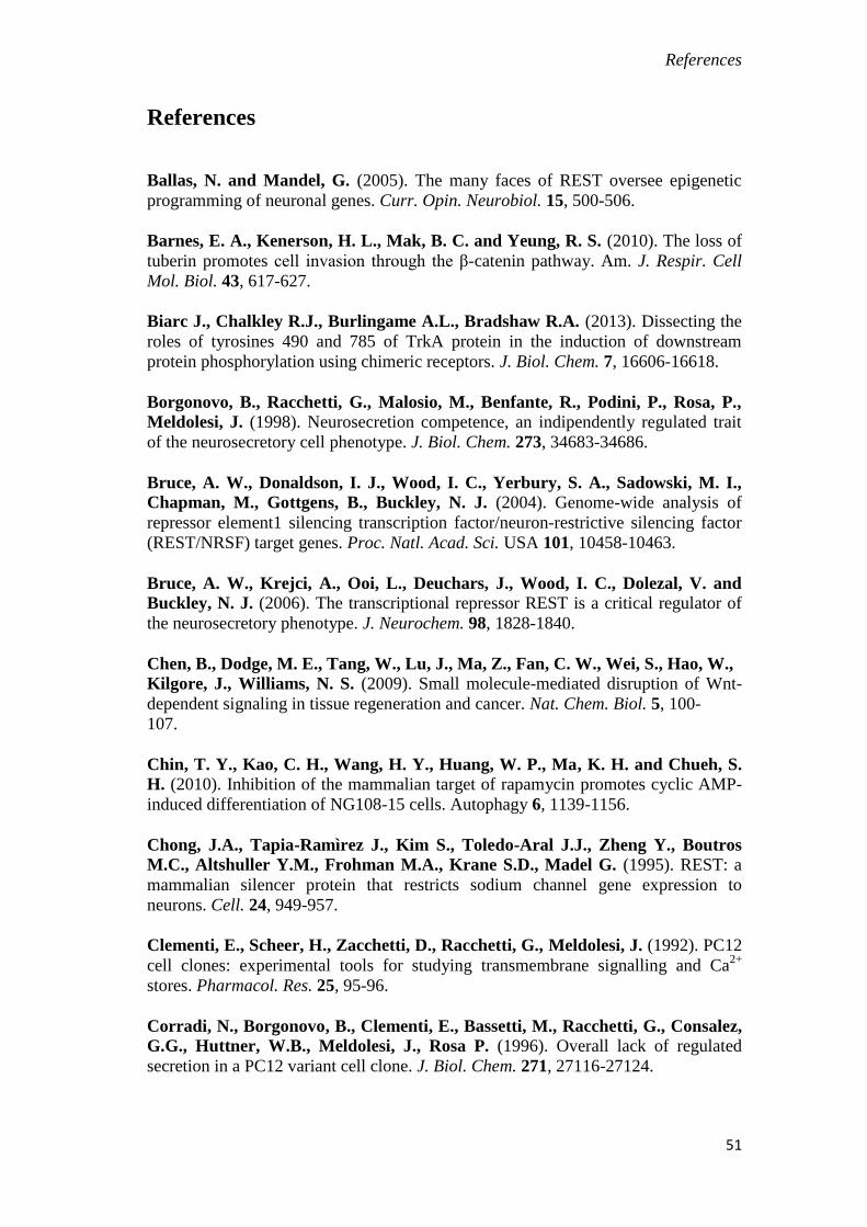
References
51
References
Ballas, N. and Mandel, G. (2005). The many faces of REST oversee epigenetic
programming of neuronal genes. Curr. Opin. Neurobiol. 15, 500-506.
Barnes, E. A., Kenerson, H. L., Mak, B. C. and Yeung, R. S. (2010). The loss of
tuberin promotes cell invasion through the β-catenin pathway. Am. J. Respir. Cell
Mol. Biol. 43, 617-627.
Biarc J., Chalkley R.J., Burlingame A.L., Bradshaw R.A. (2013). Dissecting the
roles of tyrosines 490 and 785 of TrkA protein in the induction of downstream
protein phosphorylation using chimeric receptors. J. Biol. Chem. 7, 16606-16618.
Borgonovo, B., Racchetti, G., Malosio, M., Benfante, R., Podini, P., Rosa, P.,
Meldolesi, J. (1998). Neurosecretion competence, an indipendently regulated trait
of the neurosecretory cell phenotype. J. Biol. Chem. 273, 34683-34686.
Bruce, A. W., Donaldson, I. J., Wood, I. C., Yerbury, S. A., Sadowski, M. I.,
Chapman, M., Gottgens, B., Buckley, N. J. (2004). Genome-wide analysis of
repressor element1 silencing transcription factor/neuron-restrictive silencing factor
(REST/NRSF) target genes. Proc. Natl. Acad. Sci. USA 101, 10458-10463.
Bruce, A. W., Krejci, A., Ooi, L., Deuchars, J., Wood, I. C., Dolezal, V. and
Buckley, N. J. (2006). The transcriptional repressor REST is a critical regulator of
the neurosecretory phenotype. J. Neurochem. 98, 1828-1840.
Chen, B., Dodge, M. E., Tang, W., Lu, J., Ma, Z., Fan, C. W., Wei, S., Hao, W.,
Kilgore, J., Williams, N. S. (2009). Small molecule-mediated disruption of Wnt-
dependent signaling in tissue regeneration and cancer. Nat. Chem. Biol. 5, 100-
107.
Chin, T. Y., Kao, C. H., Wang, H. Y., Huang, W. P., Ma, K. H. and Chueh, S.
H. (2010). Inhibition of the mammalian target of rapamycin promotes cyclic AMP-
induced differentiation of NG108-15 cells. Autophagy 6, 1139-1156.
Chong, J.A., Tapia-Ramìrez J., Kim S., Toledo-Aral J.J., Zheng Y., Boutros
M.C., Altshuller Y.M., Frohman M.A., Krane S.D., Madel G. (1995). REST: a
mammalian silencer protein that restricts sodium channel gene expression to
neurons. Cell. 24, 949-957.
Clementi, E., Scheer, H., Zacchetti, D., Racchetti, G., Meldolesi, J. (1992). PC12
cell clones: experimental tools for studying transmembrane signalling and Ca2+
stores. Pharmacol. Res. 25, 95-96.
Corradi, N., Borgonovo, B., Clementi, E., Bassetti, M., Racchetti, G., Consalez,
G.G., Huttner, W.B., Meldolesi, J., Rosa P. (1996). Overall lack of regulated
secretion in a PC12 variant cell clone. J. Biol. Chem. 271, 27116-27124.

References
52
Coulson, J.M. (2005). Transcriptional regulation: cancer, neurons and the REST.
Curr. Biol. 6, 665-668.
D’Alessandro, R., Klajn, A., Stucchi, L., Podini, P., Malosio, M. L. and
Meldolesi, J. (2008). Expression of the neurosecretory process in PC12 cells is
governed by REST. J. Neurochem. 105, 1369-1383.
Greene, L. A. and Tischler, A. S. (1976). Establishment of a noradrenergic clonal
line of rat adrenal pheochromocytoma cells which respond to nerve growth factor.
Proc.Natl. Acad. Sci. USA 73, 2424-2428.
Grohovaz, F., Bossi, M., Pezzati, R., Meldolesi, J., Tarelli, F.T. (1996). High
resolution ultrastructural mapping of total calcium: electron spectroscopic
imaging/electron energy loss spectroscopy analysis of a physically/chemically
processed nerve-muscle preparation. Proc. Natl. Acad. Sci. USA 93, 4799-4803.
Grundschober, C., Malosio, M. L., Astolfi, L., Giordano, T., Nef, P. and
Meldolesi, J. (2002). Neurosecretion competence. A comprehensive gene
expression program identified in PC12 cells. J. Biol. Chem. 277, 36715-36724.
Hsu, P. P., Kang, S. A., Rameseder, J., Zhang, Y., Ottina, K. A., Lim, D.,
Peterson, T. R., Choi, Y., Gray, N. S., Yaffe, M. B. et al. (2011). The mTOR-
regulated phosphoproteome reveals a mechanism of mTORC1-mediated inhibition
of growth factor signaling. Science 332, 1317-1322.
Huang, J. and Manning, B. D. (2009). A complex interplay between Akt, TSC2
and the two mTOR complexes. Biochem. Soc. Trans. 37, 217-222.
Huang, J., Dibble, C. C., Matsuzaki, M. and Manning, B. D. (2008). The TSC1-
TSC2 complex is required for proper activation of mTOR complex 2. Mol. Cell.
Biol. 28, 4104-4115.
Jozwiak, J. and Wlodarski, P. (2006). Hamartin and tuberin modulate gene
transcription via β-catenin. J. Neurooncol. 79, 229-234.
Koch, A., Scherr, M., Breyer, B., Mancini, A., Kardinal, C., Battmer, K., Eder,
M. and Tamura, T. (2008). Inhibition of Abl tyrosine kinase enhances nerve
growth factor-mediated signaling in Bcr-Abl transformed cells via the alteration of
signaling complex and the receptor turnover. Oncogene 27, 4678-4689.
Laplante, M. and Sabatini, D. M. (2009). mTOR signaling at a glance. J. Cell Sci.
122, 3589-3594.
Leoni, C., Menegon, A., Benfenati, F., Toniolo, D., Pennuto, M. and Valtorta, F. (1999). Neurite extension occurs in the absence of regulated exocytosis in PC12
subclones. Mol. Biol. Cell 10, 2919-2931.
Majumder, S. (2006). REST in good times and bad: roles in tumor suppressor and
oncogenic activities. Cell Cycle 5, 1929-1935.
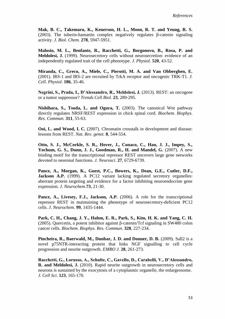
References
53
Mak, B. C., Takemaru, K., Kenerson, H. L., Moon, R. T. and Yeung, R. S. (2003). The tuberin-hamartin complex negatively regulates β-catenin signaling
activity. J. Biol. Chem. 278, 5947-5951.
Malosio, M. L., Benfante, R., Racchetti, G., Borgonovo, B., Rosa, P. and
Meldolesi, J. (1999). Neurosecretory cells without neurosecretion: evidence of an
independently regulated trait of the cell phenotype. J. Physiol. 520, 43-52.
Miranda, C., Greco, A., Miele, C., Pierotti, M. A. and Van Obberghen, E. (2001). IRS-1 and IRS-2 are recruited by TrkA receptor and oncogenic TRK-T1. J.
Cell. Physiol. 186, 35-46.
Negrini, S., Prada, I., D’Alessandro, R., Meldolesi, J. (2013). REST: an oncogene
or a tumor suppressor? Trends Cell Biol. 23, 289-295.
Nishihara, S., Tsuda, L. and Ogura, T. (2003). The canonical Wnt pathway
directly regulates NRSF/REST expression in chick spinal cord. Biochem. Biophys.
Res. Commun. 311, 55-63.
Ooi, L. and Wood, I. C. (2007). Chromatin crosstalk in development and disease:
lessons from REST. Nat. Rev. genet. 8, 544-554.
Otto, S. J., McCorkle, S. R., Hover, J., Conaco, C., Han, J. J., Impey, S.,
Yochum, G. S., Dunn, J. J., Goodman, R., H. and Mandel, G. (2007). A new
binding motif for the transcriptional repressor REST uncovers large gene networks
devoted to neuronal functions. J. Neurosci. 27, 6729-6739.
Pance, A., Morgan, K., Guest, P.C., Bowers, K., Dean, G.E., Cutler, D.F.,
Jackson A.P. (1999). A PC12 variant lacking regulated secretory organelles:
aberrant protein targeting and evidence for a factor inhibiting neuroendocrine gene
expression. J. Neurochem.73, 21-30.
Pance, A., Livesey, F.J., Jackson, A.P. (2006). A role for the transcriptional
repressor REST in maintaining the phenotype of neurosecretory-deficient PC12
cells. J. Neurochem. 99, 1435-1444.
Park, C. H., Chang, J. Y., Hahm, E. R., Park, S., Kim, H. K. and Yang, C. H. (2005). Quercetin, a potent inhibitor against β-catenin/Tcf signaling in SW480 colon
cancer cells. Biochem. Biophys. Res. Commun. 328, 227-234.
Pincheira, R., Baerwald, M., Dunbar, J. D. and Donner, D. B. (2009). Sall2 is a
novel p75NTR-interacting protein that links NGF signalling to cell cycle
progression and neurite outgrowth. EMBO J. 28, 261-273.
Racchetti, G., Lorusso, A., Schulte, C., Gavello, D., Carabelli, V., D’Alessandro,
R. and Meldolesi, J. (2010). Rapid neurite outgrowth in neurosecretory cells and
neurons is sustained by the exocytosis of a cytoplasmic organelle, the enlargeosome.
J. Cell Sci. 123, 165-170.

References
54
Roux, P.P., Bhakar, A.L., Kennedy, T.E., Barker, P.A. (2001). The p75
neurotrophin receptor activates Akt (protein kinase B) through a
phosphatidylinositol 3-kinase-dependent pathway. J. Biol. Chem. 276, 23097-23104.
Sarbassov, D. D., Ali, S. M., Kim, D. H., Guertin, D. A., Latek, R. R.,
Erdjument-Bromage, H., Tempst, P. and Sabatini, D. M. (2004). Rictor, a novel
binding partner of mTOR, defines a rapamycin-insensitive and raptor-independent
pathway that regulates the cytoskeleton. Curr. Biol. 14, 1296-1302.
Schoenherr, C.J. and Anderson D.J. (1995). The neuron-restrictive silencer factor
(NRSF): a coordinate repressor of multiple neuron-specific genes. Science 267,
1360-1363.
Schulte, C., Racchetti, G., D’Alessandro, R. and Meldolesi, J. (2010). A new
form of neurite outgrowth sustained by the exocytosis of enlargeosomes expressed
under the control of REST. Traffic 11, 1304-1314.
Tomasoni, R.*, Negrini, S.*, Fiordaliso, S., Klajn, A., Tkatch, T., Mondino, A.,
Meldolesi, J., D’Alessandro, R. (2011). A signaling loop of REST, TSC2 and β-
catenin governs proliferation and function of PC12 neural cells. JCS 124, 3174-
3186.
Tremblay, F., Brulé, S., Hee Um, S., Li, Y., Masuda, K., Roden, M., Sun, X. J.,
Krebs, M., Polakiewicz, R. D., Thomas, G. et al. (2007). Identification of IRS-1
Ser-1101 as a target of S6K1 in nutrient- and obesity-induced insulin resistance.
Proc. Natl. Acad. Sci. USA 104, 14056-14061.
Wang, H., Duan, X., Ren, Y., Liu, Y., Huang, M., Liu, P., Wang, R., Gao, G.,
Zhou, L., Feng, Z. et al. (2013). FoxO3a negatively regulates nerve growth factor-
induced neuronal differentiation through inhibiting the expression of neurochondrin
in PC12 cells. Mol. Neurobiol. 47, 24-36.
Willert, J., Epping, M., Pollack, J. R., Brown, P. O. and Nusse, R. (2002). A
transcriptional response to Wnt protein in human embryonic carcinoma cells. BMC
Dev. Biol. 2, 8.
Wu, J. and Xie, X. (2006). Comparative sequence analysis reveals an intricate
network among REST, CREB and miRNA inmediating neuronal gene expression.
Genome Biol. 7, R85.
Yamashita, T. and Tohyama, M. (2003). The p75 receptor acts as a displacement
factor that releases Rho from Rho-GDI. Nat. Neurosci. 6, 461-467.
*these authors have contributed equally to the work