universidade paulista simultaneous multiple ectoparasitic ... · futura drª lidiana flora vidôto...
TRANSCRIPT

UNIVERSIDADE PAULISTA
Simultaneous multiple ectoparasitic infection in
cultured Goldfish (Carassius auratus) associated with
environmental conditions and management - Multiple
parasitism in goldfish
Dissertação apresentada ao Programa de Pós-Graduação em Patologia Ambiental e Experimental da Universidade Paulista – UNIP para a obtenção do título de Mestre em Patologia Ambiental e Experimental.
CARLA RENATA SERANTONI MOYSES
SÃO PAULO
2014

UNIVERSIDADE PAULISTA
Simultaneous multiple ectoparasitic infection in
cultured Goldfish (Carassius auratus) associated with
environmental conditions and management - Multiple
parasitism in goldfish
Dissertação apresentada ao Programa de Pós-Graduação em Patologia Ambiental e Experimental da Universidade Paulista – UNIP para a obtenção do título de Mestre em Patologia Ambiental e Experimental. Orientadora: Prof. (ª): Dr.(ª) Maria Anete Lallo
CARLA RENATA SERANTONI MOYSES
SÃO PAULO
2014

Moyses, Carla Renata Serantoni. Simultaneous multiple ectoparasitic infection in cultured goldfish (Carassius auratus) associated with environmental conditions and management: multiple parasitism in goldfish / Carla Renata Serantoni Moyses. - 2014. 30 f. : il. color.
Dissertação de Mestrado apresentado ao Programa de Pós-Graduação em Medicina Veterinária da Universidade Paulista, São Paulo, 2014. Área de concentração: Patologia Ambiental e Experimental. Orientadora: Prof. Dra. Maria Anete Lallo.
1. Carassius auratus. 2. Ciliates parasites. 3. Fish ectoparasites. 4. Goldfish. 5. Protozoan. I. Lallo, Maria Anete (orientadora). II. Título.

CARLA RENATA SERANTONI MOYSES
Simultaneous multiple ectoparasitic infection in
cultured Goldfish (Carassius auratus) associated with
environmental conditions and management - Multiple
parasitism in goldfish
Dissertação apresentada ao Programa de Pós-Graduação em Patologia Ambiental e Experimental da Universidade Paulista – UNIP para a obtenção do título de Mestre em Patologia Ambiental e Experimental.
Aprovado em:
BANCA EXAMINADORA
_________________________________
Orientadora: Prof.(ª): Dr.(ª) Maria Anete Lallo
Universidade Paulista – UNIP
_________________________________
Prof. Dr. José Guilherme Xavier
Universidade Paulista – UNIP
__________________________________
Prof.(ª) Dr.(ª) Diva Denelle Spadacci Morena
Instituto Butantan – IBU

DEDICATÓRIA
Dedico este trabalho à minha família:
Ao meu pai, Enéas que me ensinou a aproveitar a vida e os momentos dentro
da Universidade, o meu eterno professor;
À minha melhor amiga e conselheira, Dolores, pela sua dedicação
incondicional, minha alma gêmea, não existem palavras que descrevem meu amor e
minha gratidão por ter aceitado ser minha mãe;
À minha irmã, Claudia por tudo o que representa pra mim, uma das mulheres
mais inteligentes que já conheci ou irei conhecer, você é tudo que almejo ser um dia;
Ao meu irmão, André você é a prova viva e meu melhor exemplo de força de
vontade e dedicação;
Ao Thiago por valorizar tudo quanto sou no mínimo que faço! Amo você.

AGRADECIMENTOS
A Deus por me amparar nos momentos difíceis, me dar força interior para
superar as dificuldades, mostrar os caminhos nas horas incertas e me suprir em
todas as minhas necessidades.
À minha orientadora e Profª Drª Maria Anete Lallo, por acreditar em mim, me
mostrar o caminho da ciência, fazer parte da minha vida nos momentos bons e ruins,
por ser exemplo de profissional e de mulher a qual sempre fará parte da minha vida.
Nesse mundo, repleto de pessoas ruins, você me faz acreditar que os bons são a
maioria. Só tenho a agradecer aos seus ensinamentos (pessoais e acadêmicos),
orientações, palavras de incentivo, puxões de orelha, paciência e dedicação.
À minha família, a qual amo muito, pelo carinho, paciência e incentivo.
À Profª Drª Diva Denelle Spadacci Morena por sua ajuda na Microscopia
Eletrônica de Transmissão, e ao Profº Drº José Guilherme Xavier por sua ajuda e
total contribuição para a leitura das lâminas, por acreditarem no futuro deste projeto
e contribuírem para o meu crescimento profissional e por serem exemplos a serem
seguidos. Vossa participação foi fundamental para a realização deste trabalho.
As minhas eternas Professoras, Drº Raquel Machado Cavalca Coutinho e
futura Drª Lidiana Flora Vidôto da Costa, se estou aqui terminando essa fase da
minha vida, é porque vocês acreditaram em mim e me incentivaram a iniciá-la.
A todos os envolvidos nesse projeto, muito obrigada.

SUMÁRIO
INTRODUCTION ......................................................................................................... 8
MATERIALS AND METHODS .................................................................................... 9
RESULTS.................................................................................................................. 11
DISCUSSION ............................................................................................................ 14
CONCLUSION .......................................................................................................... 20
REFERENCES .......................................................................................................... 21
6
Simultaneous multiple ectoparasitic infection in cultured Goldfish (Carassius
auratus) associated with environmental conditions and management
Multiple parasitism in goldfish
Carla Renata Serantoni Moyses1, Diva Denelle Spadacci-Morena2, José Guilherme
Xavier1, Maria Anete Lallo1*
1 Environmental and Experimental Pathology Post-Graduation, Paulista University
(UNIP), 2 Butantan Institute, Laboratory of Pathophysiology.
*Corresponding author: Mailing address: Universidade Paulista, Rua Dr. Bacelar
1212, 4º. andar, CEP: 04026002. São Paulo, SP, Brasil. Phone-Fax: 55.
11.5586.4093, Mobile phone: 55. 11.9986.9607. E-mail: [email protected] or
Carla Renata Serantoni Moyses E-mail: [email protected]
Diva Denelle Spadacci-Morena E-mail: [email protected]
José Guilherme Xavier E-mail: [email protected]

7
Abstract
Parasitic infections of fish are common and often debilitating the host, but multiple
and simultaneous parasitism is rarely found. Its occurrence is usually associated with
environmental and management issues. In this study, we describe the prevalence of
multiple and concurrent parasitic infections in goldfish (Carassius auratus) raised in
fish farms. Fish with skin damage (nodules and ulceration) were subjected to
necropsy and examinations with fresh scraped of skin and gills, histopathological
examination with HE and Giemsa stain, ultrastructural study by transmission and
scanning electron microscopy. We identified in the skin the multiple parasitic infection
by Gyrodactylidae, Epystilis sp., Vorticella sp., Trichodina sp., Ichthyophthirius
multifilis, Tetrahymena sp. and Ichthyobodo necatrix associated with epithelial cell
hyperplasia and epidermal sloughing. Although no gross lesions were observed in
the gills, we identify a large number of parasites (Gyrodactylidae, Piscinoodinium sp.,
Ichthyophthirius multifilis, Vorticella sp. and Trichodina sp.) and microscopic lesions
such as epithelial hypertrophy and hyperplasia, fusion of secondary lamellae,
leukocyte infiltration, epithelial cells detachment, resulting in respiratory distress. In
conclusion, multiple and simultaneous parasitism was very prevalent in goldfish with
skin lesions, usually associated with ciliates and flagellate protozoan and it was
associated with adverse environmental conditions and inadequate management.
Keywords: Carassius auratus, ciliates parasites, fish ectoparasites, goldfish,
protozoan

8
INTRODUCTION
Aquarium fish trade is a very important sector in all over the world (1). The
culture of ornamental fish is very popular in the pet market of Brazil, however, the fish
production by farmers is carried on intensive livestock farms with rudimentary
handling. Fish in aquaculture farms are often subjected to acute or chronic stressors
such as handling, transportation, sorting, temperature changes, high rearing density
and poor water quality, which affect it physiology. Thus, the growth, behavior, welfare
and reproduction can be strongly affected by stressors. As a result, the presence of
the disease is common cause high mortality and severe economic losses (2).
In fish cultured populations, parasites often cause serious outbreaks of
disease, especially when the presence of dense populations of fish kept in particular
environmental conditions may favors certain parasites species so that the parasite
population increases to a very high level (3). Among fish parasites, protozoa are the
most dangerous group that probably causes more diseases in fish cultures than any
other ones (2). Ciliates, particularly sessilines such as Apiosoma, Scopulata,
Ambiphrya and Epistylis are obligate parasites, which utilize gills and skin merely as
a substrate for attachment, causing massive destruction. Even moderate infection of
these parasites on small fish may prove a fatal disease, since the infection may
cause the fish to stop feeding (4).
The aquatic parasites usually classified in six groups include Protozoa,
Platyhelminthes, Nematode, Acanthocephala, Annelida and Arthropoda. Protozoans
can be ectoparasites or endoparasites depending on their species and are the most
common parasites encountered in cultured fish (4,5). With some exceptions (e.g.
Ichthyophthirius multifiliis, Chilodonella and Ichthyobodo) the external protozoans are

9
not obligate fish parasites, but may occur on a variety of surfaces including logs and
plants or even as parasites on other fish ectoparasites (4).
Goldfish (Carassius auratus) was one of the first fish species to be
domesticated and is the most commonly kept aquarium fish. It is closely related to
the less colorful carp, C. aerates, which is native to Eastern Asia and was
domesticated in China more than a thousand years ago. The freshwater sub- tropical
goldfish inhabits rivers, lakes, ponds and ditches with stagnant or slow-flowing water
(6). The fish pathology and parasitology is undeveloped in Brazil, despite its
abundant fish populations and the huge pet market of aquaculture. Data on parasite
and diseases caused by then in goldfish are not available in Brazil and besides, the
multiple concurrent parasitism and its consequences have not been reported in the
world. So the purpose of this study was to describe the multiple-parasitism in farmed
goldfish (Carassius auratus) associated with inadequate management conditions.
MATERIALS AND METHODS
Fish. Goldfish (Carassius auratus) was obtained from commercial aquaculture farms
in São Paulo state, Brazil, where several fish species were growing together only
separated by age. Farmers studied had high mortality of goldfish especially in winter.
The farms evaluated in this study showed three negative factors in relation to the
environment conditions: high population density in the tanks; abundant diet
predisposes to the high content of ammonia, and slightly alkaline pH, was conducted
an analysis of certain physical and chemical parameters of water of 20 tanks where
these animals are 120 to 150 days before being sold. Among them are: pH,
ammonia, nitrite and nitrate. The animals are exchanged the tanks every 30 days.
The fish are removed with the emptying of the tanks, is placed cal with chicken

10
manure or quail, both without specified quantity. The food is a mixture of powdered
feed with protein levels to 48%, granulated feed and 1 liter of oil. The mixture is
stored in a drum and is offered to the animals three times a day, without quantity.
Thirty fish with whitish hyperplastic lesions or ulcerative skin lesions were objects of
research by portraying the main problem of the fish farmers. Fish were collected in
the winter season when the temperature ranges from 10 to 20 ºC. The animals were
taken to the laboratory where they remained housed in tanks with a continuous flow
water system at 22 ± 1ºC in the aquatic facility of the Paulista University and fed ad
libitum for 24 h. Prior to necropsy, fish were anaesthetized by immersion in a solution
of tricaine methane sulfonate 150 mg/L (MS222 Sigma) until stop breathing. Animals
in the Aquatic Facility were maintained according to the guidelines of the Brazilian
National Council for the Control of Animal Experimentation (CONCEA) and all
procedures were approved by the ethics committee of the Paulista University.
Collection and processing of specimens for light microscopy. After the
collection of biometric data of each fish, skin area with damage was scraped to
collecting material and fresh observation under a light microscope. Parasites were
identified using morphological parameters (7). The body, fins, mouth, eyes and inner
cappings were examined for the location of possible parasites. Subsequently, the
gills, skin and other organs were removed and fixed in Bouin's solution for 8 hours
and kept in 70% alcohol. These materials were embedded in paraffin for histological
analysis and histological sections were stained routinely with Hematoxylin-Eosin (HE)
and Giemsa.
Electron and Scanning Microscopy. For electron microscopy (TEM), small pieces
of gills and skin lesions were fixed in 2% glutaraldehyde in 0.2 M cacodylate buffer
(pH 7.2) at 4ºC for 10 h, and post-fixed in 1% OsO4 buffered for 2 h and then in 5%
11
uranyl acetate overnight at 4 ºC. The fragments were dehydrated in an ascending
ethanol series with propylene oxide and embedded in Spurr resin. Blocs were cut
and semi-thin sections were stained with toluidine blue and photographed under a
light microscope. Ultrathin sections were double stained with aqueous uranyl acetate
and lead citrate and then observed under a ZEISS EM 109 TEM operated at 80 kV.
Fragments of skin and gills were fixed and dehydrated as described for TEM and
then processed in drying apparatus, subsequently mounted on copper stubs and gold
coated. The fragments had been examined using JEOL JSM-65 MCA scanning
electron microscope (SEM).
RESULTS
The intensive rearing, due to its high rate of settlement and artificial feeding,
generates a large amount of waste, which may be in solid in suspension or dissolved
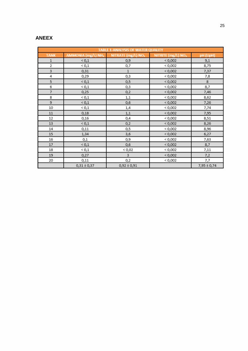
(feces, unconsumed ration, flake, mucus, agents therapeutic)(8). The physical and
chemical parameters of the water changes, so you must monitor it constantly and
wastes removed as that may pose risks to the creation. Among the species dissolved
in water from the metabolic activity of fish and degradation of proteins of
unconsumed feed, the ammoniacal nitrogen, found in the form of NH3 (ammonia) and
NH4+ (ion ammonium)(9). The first is highly toxic; your concentration depends on the
pH and the temperature of the water. Concentrations above 0.6 mg L-1 are lethal for
ornamental fish. The limit recommended by the EIFAC (European Inland Fisheries
Advisory Commission) is 0.025 mg L-1(10). The result of the analysis of water for
ammonia had an average of 0.31 mg/L with a standard deviation of ±0.37. For nitrite
all tanks were with the same concentration of <0.002 being insignificant. For nitrate,
obtained an average of 0.92 mg/L with a standard deviation of ±0.91, even if it's not
12
toxic nitrate is a quality parameter for finalizing the nitrogen cycle, being a source of
food for algae and plants. For the average pH was 7.95 with standard deviation of
0.74. The pH levels can vary widely, from 6.5 to 8.5 point literatures. Levels slightly
above or below may not take the animals to death, but are more debilitated and
lethargic. Although there is a wide variation, ideally the pH stayed between 7.5 and
8.5 for nitrifying bacteria (nitrossomas) which could play its role.(8)
Lesions. All animals examined had elevated skin lesions and thickening of the
dermis covered with mucus or with cotton wool spots appearance and necrotic
tissue. In some cases the ulcerated lesions were hemorrhagic appearance. Skin
lesions were located in the dorsal and lateral fish, sometimes with fins. The gills
showed no pronounced macroscopic lesions, sometimes a slight swelling and mucus
was observed. Some fish died by manipulating before anesthesia.
The fresh examination performed by scraping the skin lesions or gills and
allowed the observation of various parasites (Fig.1). We identified in the skin
Gyrodactylidae, Epystilis sp., Vorticella sp., Trichodina sp., Ichthyophthirius multifilis,
Tetrahymena sp. and Ichthyobodo necatrix. In gills, we found Gyrodactylidae,
Piscinoodinium sp., Ichthyophthirius multifilis, Vorticella sp. and Trichodina sp.
By histological examination of the gills we observed that the epithelium
showed hyperplasia in different degrees and detachment of epithelial cells. There
were some cases where the hyperplasia was more severe, resulting in the fusion of
some secondary lamellae (Fig.2). Frequently, alterations such as blood congestion,
hypertrophy of epithelial cells and lamellar disorganization were also observed. Some
examples of more severe lesions found in the gill were lamellar aneurysms and
hemorrhages with rupture of the lamellar epithelium. Inflammation of the epithelium
13
of primary and secondary lamellae was also observed, characterized by the
presence of large numbers of granulocytes (Fig.3). Epithelium associated protozoan
dome shaped structures with slightly concave, consistent with protozoa of the genus
Trichodina sp. (Fig.4) aboral adhesive disc were observed or in the presence of
Gyrodactylidae or Ichthyophthirius multifilis (Fig.2). In some cases protozoan
Ichthyophthirius sp. was seen blocking a blood vessel, resulting in localized
hyperemia and congestion, evident by vascular distension.
Significant epithelial cell hyperplasia and hypertrophy with eventual exhaustion
of mucous cells and epidermal sloughing was seen (Fig.3). Because the enzymes
released by some parasites, there was extensive tissue damage with areas of
necrosis and impairment of muscles under the skin of the affected regions (Fig.3).
The parasite lies within an interstitial tissue space, which contains cellular debris and
proteinaceous tissue fluid. A large number of invading parasites were seen in the
skin (Fig.3). The epithelium immediately surrounding the parasite was hyperplastic,
the cells were degenerating, appear hydropic and necrotic with pyknotic nuclei.
Inflammatory infiltrate was characterized by the presence of lymphocytes,
macrophages and neutrophils in the dermis.
Ciliates
Sessile peritrichs. The peritrichious ciliates Epystilis sp. and Vorticella sp. were
present in large numbers in the skin lesions fact that it is extremely unusual and most
often occurs in fish predisposed by debilitating environmental or infectious factors, or
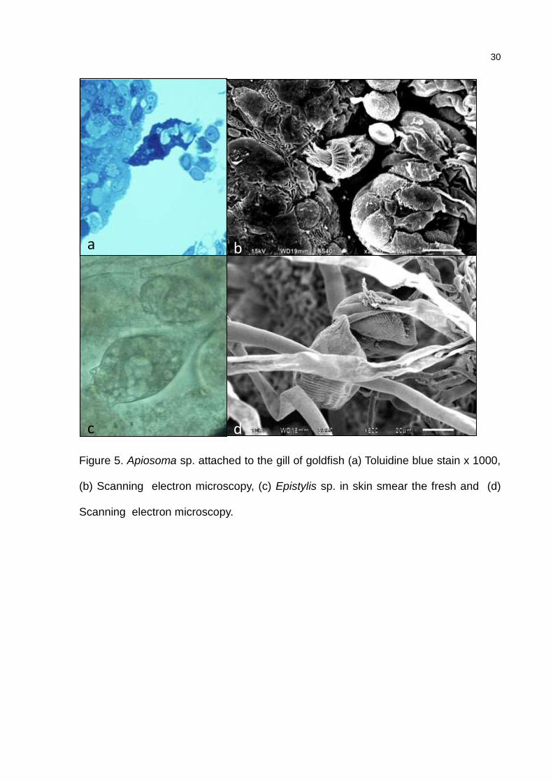
both in this case. Epistylis is colonial ciliates with bell-shaped or conical body (Fig.1,
5). A non-contractile stalk bears several or many zooids. Vorticella sp. is a sessile
peritrich was characterized by its inverted bell-shaped body and occurred solitarily on

14
a retractile stalk (Fig.1). Apiosoma sp. is a solitary ciliates with a scopula that is
circular directly attached to the substract (Fig.5).
Mobile peritrichs. Trichodina sp. appear as saucer-shaped, hemispheric, dumbbell-
shaped, and sac-like or flattened cylindrical organisms in cross-section, or as round
discs in oral or aboral view (Fig.1, 4).
Hymenostomatan ciliates. Ichthyophthirius multifilis is a large in size (about 0.1-1.0
mm) contains a horse-shaped macronucleus (Fig.1, 2) was often seen in the gills
associated with lesions. Tetrahymena sp. showed pyriform shape or ovoid with a
prominent oval macronucleus visible in H-E stained sections.
Flagellates. Piscinoodinum sp. trophonts was attached to gill with ovoid, pyriform or
sac-like appearance, with a single eccentric nucleus but without visible flagella. In
smears of fresh gills, trophonts showed chloroplasts (Fig.4 c-d). Ichthyobodo necatrix
was only observed in the fresh smears of skin and gill lesions. The free-swimming
stage was oval to kidney-shaped with size ranging to 10-15 m in length (Fig.1).
DISCUSSION
Disease in fishes is closely linked to environmental stress. In cultured fish
populations, parasites often cause serious outbreaks of disease (8). The presence of
dense population of fish, as was observed in the farms analyzed in this study, kept in
particular environmental conditions may favors certain parasite species so that the
parasite population increases to a very high level. Furthermore, the abundant supply
diet increases the level of ammonia in water and basified to pH and these factors
favor the development of parasites in fish observed in this study. It should also be
pointed out as a stressor form of management for the sale of animal specimens.
15
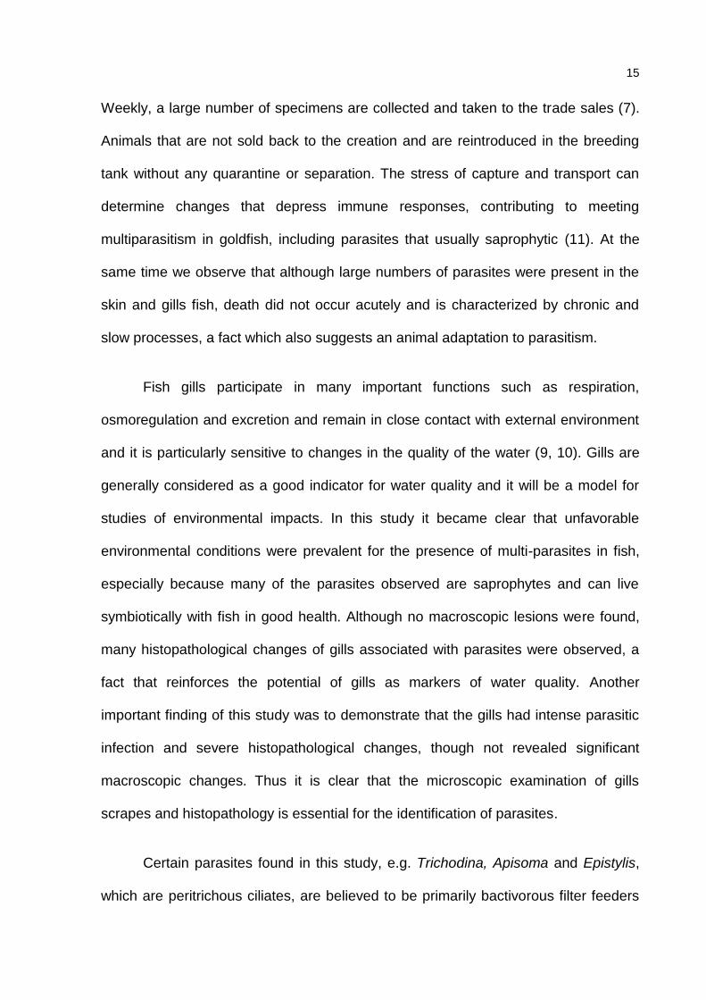
Weekly, a large number of specimens are collected and taken to the trade sales (7).
Animals that are not sold back to the creation and are reintroduced in the breeding
tank without any quarantine or separation. The stress of capture and transport can
determine changes that depress immune responses, contributing to meeting
multiparasitism in goldfish, including parasites that usually saprophytic (11). At the
same time we observe that although large numbers of parasites were present in the
skin and gills fish, death did not occur acutely and is characterized by chronic and
slow processes, a fact which also suggests an animal adaptation to parasitism.
Fish gills participate in many important functions such as respiration,
osmoregulation and excretion and remain in close contact with external environment
and it is particularly sensitive to changes in the quality of the water (9, 10). Gills are
generally considered as a good indicator for water quality and it will be a model for
studies of environmental impacts. In this study it became clear that unfavorable
environmental conditions were prevalent for the presence of multi-parasites in fish,
especially because many of the parasites observed are saprophytes and can live
symbiotically with fish in good health. Although no macroscopic lesions were found,
many histopathological changes of gills associated with parasites were observed, a
fact that reinforces the potential of gills as markers of water quality. Another
important finding of this study was to demonstrate that the gills had intense parasitic
infection and severe histopathological changes, though not revealed significant
macroscopic changes. Thus it is clear that the microscopic examination of gills
scrapes and histopathology is essential for the identification of parasites.
Certain parasites found in this study, e.g. Trichodina, Apisoma and Epistylis,
which are peritrichous ciliates, are believed to be primarily bactivorous filter feeders

16
relying on bacteria present in water or mucous for nutrition. Presumably, their mode
of feeding is innocuous to the host. Trichodina adhesive discs attach to the epithelial
surface thereby presumably impairing host cell respiration, energy production, and
cell survival. In a heavily infected fish, their adherence and suction on the epithelium
may cause enough damage to produce the clinical signs of anorexia, lethargy and
weight loss (15) in accordance with the signals by nodes found in this study with
massive infection.
Epistylis sp. and the related species Vorticella sp. are sessile and stalked
ciliated protozoans generally found attached to vegetation or crustaceans (16).
Epistylis sp. is sessile, colonial ectocommensal ciliate attacking the surface of fish
skin and gills (7) and Vorticella sp. have inverted bell-shaped body and occurred
solitarily on a retractile stalk. Vorticella sp. was reported for the first time from gills of
Carassius auratus in Brazil in the present study. They frequently affect goldfish and
many species of bottom-dwelling freshwater fish. Epistylis sp. first appeared on the
tips of both dorsal and pectoral fin spines. Then, Epistylis colonies spread down the
spines and eventually covered much of the anterior region of the body. Cutaneous
lesions observed in fish studied had exactly this location, so Epistylis sp. was major
pathogens in this study. This ciliophora is potentially harmful if it is in large number
especially to gill tissue where gas exchange may be impeded by the large numbers
of parasites physically covering gill, the infected fish show lethargy and death, which
reduces their productivity (17).
Were summarized 72 species and one subspecies of Apiosoma (18), which
may be considered ectocomensal or ectoparasite. To date, there are few studies that
demonstrate the pathogenic effects of Apiosoma in goldfish or other fish species. We

17
observed the presence of Apiosoma together with other parasites in the gills and as a
result of parasitism simultaneously several pathological changes were described.
This genus belongs to the peritrichious sessile ciliates survive alone and adhere to
the gills by scopula (19).
Ich is one of the most common diseases of freshwater fish and virtually all
freshwater fish are susceptible to infection and up to 100% mortality may occur (15).
Stress plays a major role in Ich epidemia. In current study, all fish examined had
Ichthyophthirius multifilis in the examination of the skin and gills, although typical
whitish nodules distributed throughout the body have not been observed, many
parasitic specimens were seen in skin lesions and histopathological lesions of the
gills, increased mucus production and gill filaments hyperplasia which is in
agreement to results previously observed (20). The epithelial cell erosion and
ulceration that has been resulted from the entrance and exit of the parasite damage
determine the host's skin that favors the multiplication of the parasites (12). The
mechanical action of the trophont of Ichthyophthirius multifilis is responsible for the
tissue damage. In the gills, there is a more marked epithelial hyperplasia and the
trophonts tend to migrate towards larger blood vessels.
Tetrahymena sp. is a saprozoic ciliate protozoan that feeds on organic matter
and bacteria in natural habitats (21). Tetrahymena sp. is causative agents of
tetrahymenosis or “tet disease”, also known as guppy killer disease in tropical
aquarium fish, which causes severe economic losses in commercial fish farms
worldwide. Ornamental fish species reported to be infected with Tetrahymena sp.
include zebrafish (Danio rerio), angelfish (Pterophyllum scalare), neon tetra
(Paracheirodon innesi) and others (21, 122). Susceptibility to this parasite increases

18
in fish that are wounded and/or weakened by stress conditions, such as high
ammonia level, high organic load, extreme water temperature, non-optimal shipment
conditions or a disease (6). In our study, Tetrahymena sp. was found only in part of
the animals associated with skin lesions and probably environmental conditions
identified predisposed to infection by the parasite.
The bodonid flagellates Ichthyobodo necator (formerly Costia necatrix) is
significant cause of morbidity and mortality in aquaculture fishes. This parasite is
often restricted to protected areas on fish gills, pectoral and pelvic fins, and on areas
adjacent to the dorsal fin (23,24). Nevertheless, Ichthyobodo induces significant
epithelial cell hyperplasia and hypertrophy with eventual exhaustion of mucous cells,
and epidermal sloughing (3,223,224). The location and type of injury were similar to
those described by us in goldfish. The large number of parasites Ichthyobodo necator
was observed in skin lesions, but mostly they were seen in scrapings from the gills or
skin lesions, contributing to the number of lesions that were observed.
Piscinoodinum sp. pathogenicity is high (7). In heavily infected fish, symptoms
are signs of discomfort, a golden, velvety hue on the body surface, spreading
opercula, folding of fins and eventually emaciation. There may be petechiae in the
skin and even slight inflammation. The gill lamellae may be fused, and epithelial
hyperplasia may involve entire gills filaments. Eventually, there is epithelial cell
degeneration and necrosis. The presence of Piscinoodimun and Ichthyobodo
certainly contributed to the presence of lesions on gills and skin, since the both
flagellates possess great potential pathogenic.
Gyrodactylidae are the most common and prevalent ectoparasites which can
produce severe parasitic disease in aquaculture (25). Morbidity and mortality caused

19
by excessive parasite loads of dactylogyrids are common in cultured fishes and have
also occurred in wild fishes (25, 26). Fish appear to co-exist with their specific
monogeneans, in natural habitats as well as in culture conditions, even when
infestations are intense. We observed high amount of Gyrodactylidae lesions of pel
and gill of fish studied in exams fresh and light microscopy. A few monogeneans,
notoriously gyrodactylids, are, however, pathogenic to their host fish, usually to
younger fish and in intensive culture conditions (25, 26). Histopathological changes in
the gills are hardly detectable in most instances even in relatively intense infections.
The fish infected with Dactylogyrus showed clinical symptoms including the lethargy,
unilateral swimming and erosion on gill filament and scale loss (26), such changes
have been described by the owner of the farm. Gill filament fusion, secondary
filament hyperplasia and aneurism were reported in fishes which were infected by
Dactylogyrus sp. (27, 28), which is in agreement with results obtained from this study.

20
CONCLUSION
Multiple and simultaneous parasitism was very prevalent in goldfish with skin
lesions, usually associated with ciliates and flagellate protozoan. We attribute this
parasitism to the adverse environmental conditions and inadequate management.
Acknowledgement
Magna Aparecida Mautauro Soares for making the material for light
microscopy and Michelle Sanchez Freitas Correia for performing the scanning
electron microscopy.

21
REFERENCES
1. De Silva SS, Turchini GM. 2008. Towards Understanding the Impacts of the Pet
Food Industry on World Fish and Seafood Supplies. J. Agric. Environ. Ethics.
21:459-467.
2. Ashley PJ. 2007. Fish welfare: current issues in aquaculture. Appl. Anim. Behav.
Sci. 104:199-235.
3. Roberts HE, Brian Palmeiro B, Scott Weber E. 2009. Bacterial and Parasitic
Diseases of Pet Fish. Vet. Clin. Exot. Anim. 12: 609–638.
4. Woo PTK, Bruno DW. (eds.). 2011. Fish Diseases and DisordersViral, Bacterial
and Fungal Infections. Second Edition, vol. 3. CABI, Wallingford, OX.
5. Purivirojkul W, Boonsoong B. 2012. A new species of Tetraphyllidean
(Onchobothriidae) cestode from the brown-nbanded mabbooshark Chiloscyllium
punctatum (Elasmobranchii: Hemiscyllidae). J. Parasitol. 98:1216-1219.
6. Sharon G, Pimenta Leibowitz M, Kumar Chettri J, Isakov N, Zilberg D. 2014.
Comparative study of infection with Tetrahymena of different ornamental fish
species. J. Comp. Path. 150:316-324.
7. Lom J, Dykova L (ed). 1992. Protozoan Parasites of Fishes - Developments in
Aquaculture and Fisheries Science, 26. Elsevier Sci., Amsterdam.
8. REN, J. S.; Stenton-Dozey, J.; Plew, D. R.; Fang, J.; Gall, M. An ecosystem model
for optimising production in integrated multitrophic aquaculture systems. Ecol.
Model. Vol. 246, p. 34-36, 2012.
9. BOYD, C. E. Water quality in warmwater fish ponds. Agricultural Experiment
Station: Alabama, 1984.

22
10. TIMMONS, M. B.; Ebeling, J. M.; Wheaton, F. W.; Summerfelt, S. T.; Vinci, B. J.
Sistemas de Recirculación para La Acuicultura. Fundación Chile: Santiago, 2002.
11. Barber I. 2007. Parasites, behaviour and welfare in fish. App. An. Beh. Sci.
104:251–264.
12. Mazon AF, Pinheiro GHD, Fernandes MN. 2002. Hematological and physiological
changes induced by short-term exposure to copper in the freshwater fish,
Prochilodus scrofa. Braz. J. Biol. 62:621-631.
13. Camargo MMP, Martinez CBR. 2007. Histopathology of gills, kidney and liver of a
Neotropical fish caged in an urban stream. Neotrop. Ichtyol. 5:327-336.
14. Hoffman GL. 1970. Parasites of North American freshwater fishes. Univ.
California Press, Los Angeles, CA.
15. Noga EJ. 1996. Fish disease: diagnosis and treatment. St. Mosby, St. Louis, MO.
16. Saglam N, Sarieyyupoglu M. 2002 A study on Tetrahymena pyriformis
(Holotrichous) and Epistylis sp. (Peritrichous) found on freshwater leech,
Nephelopsis obscura. Pak. J. Biol. Sci. 5:497-498.
17. Paperna I. 1991. Diseases caused by parasites in the aquaculture of warm water
fish. An. Rev. Fish Dis, 1:155-194.
18. Li M, Wang J, Zhu D, Gu Z, Zhang J, Gong X. 2008. Study of Apiosoma piscicola
(Blanchard 1885) occurring on fry of freshwater fishes in Hongze, China with
consideration of the genus Apiosoma. Parasitol. Res. 102: 931–937.

23
19. Reda ESA. 2011. A Review of some ecto- and endo-protozoan parasites
infecting Sarotherodon galilaeus and Tilapia zillii from Damietta branch of River
Nile, Egypt. J. Amer. Sci. 7:362-373.
20. D6ickerson HW. 2012. Ichthyophthirius mutifiliis. In Woo PTK, Buchmann K. (ed)
Fish Parasites – Pathobiology and protection Cabi, Oxfordshire, OX.
21. Ponpornpisit A, Endo M, Murata H. 2000. Experimental infections of a ciliate
Tetrahymena pyriformis on ornamental fishes. Fish. Sc. 66:1026-1031.
22. Astrofsky KM, Schech JM, Sheppard BJ, Obenschain CA, Chin AM, Kacergis
MC, Laver ER, Bartholomew JL, Fox JG. 2002. High mortality due to
Tetrahymena sp. infection in laboratory maintained zebrafish (Brachydanio rerio).
Comp. Med. 52:363-367.
23. Reavill D, Roberts HE. 2007. Diagnostic cytology of fish. Vet. Clin. Exot. Anim.
10:207–34.
24. Buchmann K, Sigh J, Nielsen CV, Dalgaard M. 2001. Host responses against the
fish parasitizing ciliate Ichthyophthirius multifiliis. Vet. Parasitol. 100:105–116.
25. Thilakaratne ID, Rajapaksha G, Hewakopara A, Rajapakse RP, Faizal AC. 2003.
Parasitic infections in freshwater ornamental fish in Sri Lanka. Dis. Aquat. Organ.
54:157–62.
26. Baker DG, Kent ML, Fournie JL. 2007. Parasites of fishes. p. 69–116. In: Baker
DG. (ed). Flynn’s parasites of laboratory animals. 2 ed. Blackwell, Hoboken, NJ.
27. Scott M. 1985. Dynamics of challenge infections of Gyrodactylus bullatarudis
Turnbull (Monogenea) on guppies, Poecilia reticulata (Peters). J. Fish Dis. 8:495-
503.

24
28. Shinn AP, Hansen H, Olstad K, Bachmann L, Bakke TA. 2004. The use of
morphometric characters to discriminate specimens of laboratory reared and wild
populations of Gyrodactylus salaris and G. thymalli (Monogenea). Folia. Parasitol.
51:239–252.
25
ANEEX

26
Figure legends
Figure 1. Parasites found on the skin or gills in fresh examination in goldfish (a) Skin
lesion; (b) Gyrodactylidae; (c) Epystilis sp.; (d) Vorticella sp.; (e) Ichthyophthirius
multifilis; (f) Trichodina sp.; (g) Ichthyobodo necatrix. (400x)
a
f e
b c d
g

27
Figure 2. There is an extensive proliferation of branchial epithelium around the
invading parasites and total lamellar fusion associated with space-occupying
intralamellar monogenean Gyrodactylidae (a - Giemsa x 100, b - Giemsa x 400, c -
Giemsa x 400) and trophonts of Ichthyophthirius multifilis are observed in secondary
lamellae and blood vessel (arrow) (d - HE x 100, e - Giemsa x 400, f - Giemsa x 100).
d e f
c b a

28
Figure 3. Inflammation of the epithelium of primary and secondary lamellae
characterized by the presence of large numbers of granulocytes (a – Toluidine blue
stain x 100, b - Toluidine blue stain x 400). Epithelial cell hyperplasia and hypertrophy
and epidermal sloughing was seen (a – HE stain x 100). A large number of parasites
in skin lesion surrounded by epithelial and inflammatory cells (d - Toluidine blue stain
x 100).
a b
c d

29
Figure 4. Trichodina sp. found in the space between the secondary lamellae ofthe
gills of goldfish. (a) Toluidine blue stain x 1000; (b) Transmission electron microscopy
of Trichodina sp. in gill; (c) Piscinoodinium sp. in fresh specimens; (d) Scanning
electron microscopy of trophont of Piscinoodinium sp. attached to the gill of goldfish
a b
c d
30
Figure 5. Apiosoma sp. attached to the gill of goldfish (a) Toluidine blue stain x 1000,
(b) Scanning electron microscopy, (c) Epistylis sp. in skin smear the fresh and (d)
Scanning electron microscopy.
a
d c
b a
c