universidad de granada -...
TRANSCRIPT

UNIVERSIDAD DE GRANADA
FACULTAD DE MEDICINA
DEPARTAMENTO DE FARMACOLOGÍA
E INSTITUTO DE NEUROCIENCIAS
DIFFERENTIAL MODULATION BY SIGMA-1 RECEPTORS OF
µ-OPIOID-INDUCED ANTINOCICEPTION AND SIDE EFFECTS:
ROLE OF PERIPHERAL SIGMA-1 RECEPTORS
TESIS DOCTORAL PRESENTADA POR
Cristina Sánchez Fernández
Licenciada en Biología, para optar al grado de:
DOCTOR POR LA UNIVERSIDAD DE GRANADA
(Con la mención de Doctor Internacional)
Habiendo obtenido la Suficiencia Investigadora dentro del Programa de Doctorado
“Biomedicina” en el Área de Conocimiento de Farmacología
Granada, 2006

Editor: Editorial de la Universidad de GranadaAutor: Cristina Sánchez FernándezD.L.: GR 1985-2014ISBN: 978-84-9083-185-4


COMPROMISO DE RESPETO DE LOS DERECHOS DEL AUTOR
El doctorando Cristina Sánchez Fernández y los directores de la tesis Enrique José Cobos
del Moral, José Manuel Entrena Fernández y José Manuel Baeyens Cabrera.
Garantizamos, al firmar esta tesis doctoral, que el trabajo ha sido realizado por el
doctorando bajo la dirección de los directores de la tesis y hasta donde nuestro
conocimiento alcanza, en la realización del trabajo, se han respetado los derechos de otros
autores a ser citados, cuando se han utilizado sus resultados o publicaciones.
Granada 9 de Junio de 2014
Director/es de la Tesis Doctorando
Enrique José Cobos del Moral Cristina Sánchez Fernández
José Manuel Entrena Fernández
José Manuel Baeyens Cabrera


D. ENRIQUE JOSÉ COBOS DEL MORAL, INVESTIGADOR
CONTRATADO DEL DEPARTAMENTO DE FARMACOLOGÍA E
INVESTIGADOR DEL INSTITUTO DE NEUROCIENCIAS DE LA
UNIVERSIDAD DE GRANADA
CERTIFICA:
Que el trabajo de investigación titulado, “DIFFERENTIAL
MODULATION BY SIGMA-1 RECEPTORS OF µ-OPIOID-
INDUCED ANTINOCICEPTION AND SIDE EFFECTS: ROLE OF
PERIPHERAL SIGMA-1 RECEPTORS.” ha sido realizado por Dña.
Cristina Sánchez Fernández para optar al grado de Doctor por la
Universidad de Granada, en el Instituto de Neurociencias y el
Departamento de Farmacología de la Universidad de Granada, bajo mi
dirección.
Y para que conste donde proceda se firma este certificado en Granada a
9 de Junio de 2014
Fdo. Enrique José Cobos del Moral
Fdo. Cristina Sánchez Fernández


D. JOSÉ MANUEL ENTRENA FERNÁNDEZ, RESPONSABLE DE LA
UNIDAD DE ANÁLISIS DE COMPORTAMIENTO ANIMAL DEL
CENTRO DE INSTRUMENTACIÓN CIENTÍFICA (SITUADA EN EL
CENTRO DE INVESTIGACIÓN BIOMÉDICA).
CERTIFICA:
Que el trabajo de investigación titulado, “DIFFERENTIAL
MODULATION BY SIGMA-1 RECEPTORS OF µ-OPIOID-
INDUCED ANTINOCICEPTION AND SIDE EFFECTS: ROLE OF
PERIPHERAL SIGMA-1 RECEPTORS.” ha sido realizado por Dña.
Cristina Sánchez Fernández para optar al grado de Doctor por la
Universidad de Granada, en el Instituto de Neurociencias y el
Departamento de Farmacología de la Universidad de Granada, bajo mi
dirección.
Y para que conste donde proceda se firma este certificado en Granada a
9 de Junio de 2014
Fdo. José Manuel Entrena Fernández
Fdo. Cristina Sánchez Fernández


D. JOSÉ MANUEL BAEYENS CABRERA, CATEDRÁTICO DE
FARMACOLOGÍA DE LA UNIVERSIDAD DE GRANADA Y MIEMBRO
DEL INSTITUTO DE NEUROCIENCIAS DE GRANADA
CERTIFICA:
Que el trabajo de investigación titulado, “DIFFERENTIAL
MODULATION BY SIGMA-1 RECEPTORS OF µ-OPIOID-
INDUCED ANTINOCICEPTION AND SIDE EFFECTS: ROLE OF
PERIPHERAL SIGMA-1 RECEPTORS.” ha sido realizado por Dña.
Cristina Sánchez Fernández para optar al grado de Doctor por la
Universidad de Granada, en el Instituto de Neurociencias y el
Departamento de Farmacología de la Universidad de Granada, bajo mi
dirección.
Y para que conste donde proceda se firma este certificado en Granada a
9 de Junio de 2014
Fdo. José Manuel Baeyens Cabrera
Fdo. Cristina Sánchez Fernández


La realización de esta tesis ha sido posible gracias a una beca
predoctoral de Formación de Profesorado Universitario
(FPU) del Ministerio de Educación y Ciencia, y a la
financiación de nuestro grupo de investigación por la Junta
de Andalucía (grupo CTS-109), por fondos FEDER, por el
CDTI (proyecto Genius Pharma), Ministerio de Educación y
Ciencia (proyecto SAF2006-06122), Ministerio de Ciencia e
Innovación (proyecto SAF2010-15343) y Laboratorios Esteve.


A mis padres, por apoyarme en cada
una de las decisiones que he tomado a lo
largo de mi vida.
A Enrique


“La ciencia es el fundamento de todo
progreso, que mejora la vida humana y
alivia el sufrimiento”
“Science is at the base of all the
progress that lightens the burden of life and
lessens its suffering”
Irène Joliot-Curie


IIINNNDDDEEEXXX


Index Doctoral Thesis
RREESSUUMMEENN
1. ANTECEDENTES, HIPÓTESIS Y OBJETIVOS .............................................. 1
1.1. Antecedentes ............................................................................................... 1
1.2. Hipótesis y objetivos ................................................................................... 4
2. MÉTODOS .............................................................................................................. 7
2.1. Animales de experimentación ................................................................... 7
2.2. Fármacos y administración de fármacos ................................................. 7
2.3. Ensayos de nocicepción frente a un estímulo mecánico romo
(test de presión de la pata) ......................................................................... 8
2.3.1. Descripción general del procedimiento de evaluación de la
respuesta dolorosa ................................................................................... 8
2.3.2. Comparación de la respuesta dolorosa en ratones salvajes y
knockout σ1.............................................................................................. 8
2.3.3. Evaluación del efecto de la inhibición del receptor σ1 en la
antinocicepción opioide ........................................................................... 9
2.4. Determinación de los efectos de la inhibición del receptor σ1
en efectos no analgésicos de los opioides: hiperlocomoción e
inhibición del tránsito intestinal ............................................................... 10
2.4.1. Evaluación de la hiperlocomoción inducida por morfina ......................... 10
2.4.2. Evaluación de la inhibición del tránsito intestinal inducida por
opioides ................................................................................................... 10
2.5. Estudio de la expresión del receptor σl en diversas áreas del
sistema nervioso mediante “western blotting” ........................................ 10
2.6. Ensayos de fijación de radioligando ......................................................... 11
3. RESULTADOS Y DISCUSIÓN ............................................................................ 12
3.1. Comparación de la respuesta dolorosa frente a un estímulo
mecánico romo en ratones salvajes y knockout σ1 .................................. 13
3.2. Comparación del efecto de la administración sistémica de
agonistas opioides µ de acción central en ratones salvajes y
knockout σ1 ................................................................................................ 13
Doctoral Thesis Index
3.3. Efecto de la administración de antagonistas selectivos σ1, y de
su asociación con la administración sistémica de agonistas
opioides µ de acción central en el dolor nociceptivo producido
por un estímulo mecánico en ratones salvajes ........................................ 14
3.4. Efectos de la inhibición del receptor σ1 en la antinocicepción
inducida por la loperamida ....................................................................... 16
3.5. Contribución de los receptores opioides periféricos a la
antinocicepción inducida por la administración sistémica
(subcutánea) de agonistas opioides µ en condiciones normales
y de inhibición del receptor σ1 .................................................................. 17
3.6. Efectos de la inhibición local (intraplantar) del receptor σ1 en
la antinocicepción mecánica inducida por la administración
sistémica de agonistas opioides µ .............................................................. 19
3.7. Efecto de la inhibición del receptor σ1 en los efectos
antinociceptivos de la administración local (i.pl.) de morfina ............... 20
3.8. Expresión del receptor σ1 a nivel del sistema nervioso central
y periférico .................................................................................................. 21
3.9. Modulación de efectos adversos opioides (hiperlocomoción e
inhibición del tránsito intestinal) por la inhibición del
receptor σ1 .................................................................................................. 22
3.10. Ensayos de saturación de [3H]DAMGO en membranas de
médula espinal, cerebro y piel plantar de la pata trasera en
ratones salvajes y KO-σ1 ........................................................................... 23
3.11. Afinidad de los ligandos σ1 por el sitio de unión de
[3H]DAMGO, y de los fármacos opioides por el sitio de unión
de [3H](+)-pentazocina .............................................................................. 24
4. CONCLUSIONES .................................................................................................. 25
4.1. Conclusiones específicas ............................................................................ 25
4.2. Conclusión general ..................................................................................... 27
IINNTTRROODDUUCCTTIIOONN
1. THE SOMATOSENSORY SYSTEM .................................................................. 29
1.1. Primary afferent fibers .............................................................................. 29
1.1.1. Aβ-fibers ................................................................................................... 30
1.1.2. C- and Aδ-fibers ....................................................................................... 32

Index Doctoral Thesis
1.1.2.1. Sensitivity of C- and Aδ-fibers to different types of sensory
stimulation............................................................................................. 33
1.1.2.1.1. Subtypes of C-fibers ............................................................................ 35
1.1.2.1.2. Subtypes of Aδ-fibers.......................................................................... 37
1.1.3. Dorsal root ganglia (DRGs): classification of DRG neurons by
size and molecular markers ..................................................................... 38
1.2. Spinal Cord ................................................................................................. 42
1.2.1. Laminae organization of the spinal cord and primary afferent
fibers received in the dorsal horn ............................................................ 43
1.2.2. Second order neurons in the spinal cord ................................................... 44
1.3. Ascending sensory pathways .................................................................... 45
1.3.1. Ascending pathways through the dorsal columns ..................................... 45
1.3.2. Anterolateral system ................................................................................. 47
1.3.2.1. Neospinothalamic tract (nSTT) .............................................................. 47
1.3.2.2. Paleospinothalamic tract (pSTT) ........................................................... 49
1.3.2.2.1. Spinoreticular tract .............................................................................. 50
1.3.2.2.2. Spinomesencephalic tract .................................................................... 50
1.3.2.2.3. Spinohypothalamic tract...................................................................... 51
1.4. The somatosensory cortex ......................................................................... 51
1.5. Pain modulation: the gate control theory and the descending
control systems ........................................................................................... 53
1.5.1. The gate control theory ............................................................................. 53
1.5.2. Descending modulatory influences from the brainstem............................ 55
2. µ-OPIOID DRUGS: MECHANISM OF ACTION, THERAPEUTIC
USE AND SIDE EFFECTS .................................................................................. 59
2.1. Opioid System: historical overview .......................................................... 59
2.2. Endogenous opioid peptides ...................................................................... 60
2.3. Opioid drugs: classification by intrinsic efficacy .................................... 61
2.4. The µ-opioid receptor and its molecular mechanisms of
signaling ...................................................................................................... 63
2.4.1. µ-opioid receptor splice variants and the differences in the
signaling induced by µ agonists .............................................................. 64

Doctoral Thesis Index
2.5. Therapeutic use of µ-opioid drugs ............................................................ 67
2.5.1. Opioid analgesia ....................................................................................... 67
2.5.1.1. Modulation of pain pathways by µ-opioid drugs ................................... 67
2.5.1.2. Clinical use as analgesics ...................................................................... 69
2.5.2. Antitussive properties of opioid drugs ...................................................... 71
2.5.3. Antidiarrheal ............................................................................................. 72
2.6. Opioid side effects ...................................................................................... 73
2.7. Tolerance and dependence .............................................................. 77
3. SIGMA-1 (1) RECEPTORS AND PAIN: FROM CENTER TO
PERIPHERY .......................................................................................................... 81
3.1. Discovery of σ receptors ............................................................................ 81
3.2. Cloning and structural characteristics of σ1 receptor ............................ 82
3.3. Pharmacological profile of σ1 receptors ................................................... 84
3.4. Anatomical and subcellular distribution of σ1 receptors ....................... 85
3.5. σ1 receptor as a calcium-sensing and ligand-operated
chaperone .................................................................................................... 86
3.5.1. σ1 receptors as modulators of the activity of receptors and
channels through protein-protein interaction: modulation of
opioid and NMDA receptors ................................................................... 88
3.6. Evidence for the role of σ1 receptors on animal models of pain ............ 91
3.6.1. Modulation of opioid effects by σ1 receptors ........................................... 91
3.6.1.1. Modulation by σ1 receptors of opioid-induced thermal
antinociception ..................................................................................... 92
3.6.1.1.1. Modulation by σ1 receptors of µ-opioid-induced thermal
antinociception ................................................................................... 92
3.6.1.1.2. Modulation by σ1 receptors of κ- and δ-opioid-induced
thermal antinociception ...................................................................... 99
3.6.1.2. Modulation by σ1 receptors of µ-opioid-induced mechanical
antinociception ..................................................................................... 103
3.6.1.3. Modulation by σ1 receptors of non-analgesic (adverse) effects
of opioids .............................................................................................. 106
3.6.2. Pain modulation by σ1 receptors in the absence of opioid drugs .............. 107

Index Doctoral Thesis
3.6.2.1. σ1 receptors and pain induced by chemical irritants: formalin
and capsaicin ........................................................................................ 108
3.6.2.2. σ1 receptors and neuropathic pain ......................................................... 111
3.6.2.3. σ1 receptors and inflammatory pain ....................................................... 115
3.7. Conclusions and final remarks ................................................................. 117
RRAATTIIOONNAALLEE,, HHYYPPOOTTHHEESSIISS AANNDD GGOOAALLSS
1.1. Rationale ..................................................................................................... 119
1.2. Hypothesis and goals .................................................................................. 122
PPUUBBLLIISSHHEEDD PPAAPPEERRSS
1. POTENTIATION OF MORPHINE-INDUCED MECHANICAL
ANTINOCICEPTION BY σ1 RECEPTOR INHIBITION: ROLE OF
PERIPHERAL σ1 RECEPTORS (NEUROPHARMACOLOGY, 2013) ......... 125
1.1. ABSTRACT ................................................................................................ 127
1.2. INTRODUCTION ...................................................................................... 128
1.3. MATERIAL AND METHODS ................................................................. 130
1.3.1. Experimental animals ................................................................................ 130
1.3.2. Radioligand, drugs, and drug administration ............................................ 130
1.3.3. Evaluation of the behavioral response to paw pressure ............................ 132
1.3.4. Assessment of morphine-induced hyperlocomotion ................................. 133
1.3.5. Assessment of morphine-induced inhibition of gastrointestinal
transit ........................................................................................................ 133
1.3.6. Membrane preparations for binding assays............................................... 134
1.3.7. [3H]DAMGO binding assays .................................................................... 134
1.3.8. Data analysis ............................................................................................. 136
1.4. RESULTS ................................................................................................... 136
1.4.1. Comparison of mechanical sensitivity in wild-type and σ1
knockout mice ............................................................................................ 136
1.4.2. Effects of systemic (subcutaneous) morphine on mechanical
nociception in wild-type and σ1 knockout mice ........................................ 137

Doctoral Thesis Index
1.4.3. Effects of systemic (subcutaneous) administration of selective
σ1 drugs on mechanical antinociception induced by systemic
morphine .................................................................................................... 138
1.4.4. Local antinociceptive effects induced by intraplantar
administration of morphine in wild-type and σ1 knockout mice ............... 142
1.4.5. Potentiation of the local antinociceptive effect of morphine by
pharmacological blockade of σ1 receptors ................................................ 143
1.4.6. Morphine-induced side effects (hyperlocomotion and inhibition
of gastrointestinal transit) in wild-type and σ1 knockout mice.................. 146
1.4.7. [3H]DAMGO saturation binding assays in spinal cord, forebrain,
and hind-paw skin membranes from wild-type and σ1 knockout
mice ........................................................................................................ 148
1.4.8. Affinity of selective σ1 ligands and morphine for [3H]DAMGO
binding sites in forebrain membranes from wild-type mice ................... 150
1.5. DISCUSSION ............................................................................................. 150
1.6. CONCLUSIONS ........................................................................................ 155
1.7. ACKNOWLEDGEMENTS ...................................................................... 155
2. MODULATION OF PERIPHERAL µ-OPIOID ANALGESIA BY σ1 RECEPTORS (JPET, 2014) ................................................................................. 157
2.1. ABSTRACT ................................................................................................ 159
2.2. INTRODUCTION ..................................................................................... 160
2.3. MATERIAL AND METHODS ................................................................ 162
2.3.1. Experimental animals ............................................................................... 162
2.3.2. Radioligand, drugs and drug administration ............................................. 162
2.3.3. Evaluation of mechanical nociception (paw pressure) ............................. 163
2.3.4. Evaluation of opioid-induced inhibition of gastrointestinal
transit ......................................................................................................... 164
2.3.5. Western blotting ........................................................................................ 165
2.3.6. [3H](+)-Pentazocine competition binding assays ..................................... 166
2.3.7. Data analysis ............................................................................................. 167
2.4. RESULTS ................................................................................................... 167
2.4.1. Effects of systemic (subcutaneous) μ-opioid receptor agonists
on mechanical nociception in wild-type and σ1 knockout mice ................ 167

Index Doctoral Thesis
2.4.2. Contribution of peripheral opioid receptors to antinociception
induced by the systemic (subcutaneous) administration of µ-
opioid analgesics in wild-type mice .......................................................... 169
2.4.3. Contribution of peripheral opioid receptors to antinociception
induced by the systemic (subcutaneous) administration of µ-
agonists in σ1 knockout mice ..................................................................... 170
2.4.4. Effects of systemic (subcutaneous) administration of the
selective σ1 antagonist BD-1063 on mechanical antinociception
induced by µ-agonists in WT mice: involvement of σ1 and
peripheral opioid receptors ........................................................................ 172
2.4.5. Effects of local (intraplantar) administration of the selective σ1
antagonist BD-1063 on mechanical antinociception induced by
µ-agonists in wild-type mice: involvement of σ1 and peripheral
opioid receptors ......................................................................................... 175
2.4.6. Effects of systemic (subcutaneous) and local (intraplantar)
administration of the selective σ1 antagonist S1RA on
mechanical antinociception induced by fentanyl and loperamide
in wild-type mice: involvement of σ1- and peripheral opioid
receptors ..................................................................................................... 178
2.4.7. Sigma-1 receptor expression in the central and peripheral
nervous system........................................................................................... 179
2.4.8. Effects of fentanyl and loperamide on gastrointestinal transit in
wild-type mice, wild-type mice treated with BD-1063, and σ1
knockout mice ............................................................................................ 181
2.4.9. Affinity of µ-opioid drugs for [3H](+)-pentazocine binding sites............. 182
2.5. DISCUSSION ............................................................................................. 183
2.6. ACKNOWLEDGEMENTS ....................................................................... 188
CCOONNCCLLUUSSIIOONNSS
1.1 SPECIFIC CONCLUSIONS ...................................................................... 189
1.2 GENERAL CONCLUSION ....................................................................... 190
LLIISSTT OOFF AABBBBRREEVVIIAATTIIOONNSS .............................................................................. 191
BBIIBBLLIIOOGGRRAAPPHHYY ................................................................................................... 197


RRREEESSSUUUMMMEEENNN


Resumen Tesis Doctoral
- 1 -
1. ANTECEDENTES, HIPÓTESIS Y OBJETIVOS
1.1. Antecedentes
Los fármacos opioides, especialmente los agonistas de receptores µ (tales como morfina,
fentanilo, oxicodona, buprenorfina o tramadol), son muy utilizados en clínica para el
tratamiento del dolor moderado a severo (Pergolizzi et al., 2008; Schäfer, 2010;
Pasternak and Pan, 2011; Al-Hasani and Bruchas, 2011). Los receptores opioides se
localizan en diferentes áreas del sistema nervioso central (tanto en diversos núcleos
supraespinales como a nivel espinal) y periférico (ganglios de la raíz dorsal, DRG),
implicadas en la modulación del dolor (Bigliardi-Qi et al., 2004; Khalefa et al., 2012).
Se piensa que el efecto analgésico de los agonistas µ se debe principalmente a su
interacción con receptores opioides localizados en el sistema nervioso central,
particularmente a nivel supraespinal (p.ej. Christie et al., 2000; Khalefa et al., 2012).
Sin embargo, en los últimos años el estudio del posible papel de los receptores opioides
periféricos en la analgesia opioide ha cobrado un gran interés (p.ej. Stein et al., 2003;
Sehgal et al., 2011).
Además de la analgesia, estos fármacos producen otros efectos derivados
principalmente de sus acciones a nivel central, incluyendo nauseas, confusión mental y
depresión respiratoria, entre otros efectos adversos de relevancia clínica (revisado en
Waldhoer et al., 2004 y Al-Hasani and Bruchas, 2011). Los opioides también producen
efectos periféricos, ya que disminuyen el tránsito intestinal principalmente por su efecto
inhibitorio de la actividad del plexo mientérico (Holzer et al., 2009; Brock et al., 2012).
Este efecto de los opioides se utiliza clínicamente para el tratamiento sintomático de la
diarrea, concretamente mediante el uso de loperamida. Este fármaco es un agonista
opioide que actúa exclusivamente a nivel periférico (Menéndez et al., 2005;
Sevostianova et al., 2005; Parenti et al., 2012), y que, por lo tanto, es capaz de inhibir el
tránsito intestinal aunque carece de efectos centrales (Gallelli et al., 2010; Layer et al.,
2010), por lo que no se utiliza clínicamente como analgésico. Desafortunadamente, la
inhibición del tránsito intestinal también es producida por los opioides analgésicos (con
penetrabilidad central) y tiene un gran impacto en la calidad de vida del paciente, ya que

Tesis Doctoral Resumen
- 2 -
el estreñimiento producido por estos fármacos es la causa principal de abandono
voluntario de medicación opioide por los pacientes (Dhingra et al., 2013).
El receptor sigma (σ) fue identificado por Martin y colaboradores en 1976. Tras su
descubrimiento fue erróneamente clasificado como un subtipo de receptor opioide, y
más tarde se confundió con el sitio de unión de la fenciclidina en el receptor NMDA (N-
metil-D-aspartato). Actualmente, los receptores σ se clasifican como una entidad
farmacológica independiente (revisado por Cobos et al., 2008; Zamanillo et al., 2013).
Estudios bioquímicos y farmacológicos evidencian la existencia de dos subtipos de
receptores σ, denominados σ1 y σ2 (ver Cobos et al., 2008 para referencias). El subtipo
σ1 está caracterizado en mayor profundidad y es el objeto de estudio de esta Tesis
Doctoral.
La farmacología del receptor σ1 está actualmente bien descrita, y hoy en día existen
fármacos selectivos para este receptor. Estos fármacos incluyen a los antagonistas
selectivos BD-1063, BD-1047, NE-100 y S1RA, y a los agonistas selectivos PRE-084 y
(+)-pentazocina (Cobos et al., 2008; Zamanillo et al., 2013). El receptor σ1 ha sido
clonado y no muestra homología con los receptores opioides ni con ninguna otra
proteína de mamíferos (ver Hayashi y Su, 2003; Guitart et al., 2004 y Cobos et al., 2008,
para referencias). Su clonaje ha permitido el desarrollo de ratones knockout σ1 (KO-σ1)
(Langa et al., 2003), facilitando así el estudio de la función de estos receptores. El
receptor σ1 tiene un papel neuromodulador, atribuible a su acción chaperona sobre otros
receptores y canales implicados en diversos procesos fisiopatológicos (Aydar et al.,
2002; Kim et al., 2010; Navarro et al., 2010 y 2013; Su et al., 2010; Balasuriya et al.,
2012; Kourrich et al., 2012 y 2013). Entre los receptores susceptibles de la modulación
por el receptor σ1 destacan los receptores opioides µ. Ambos receptores pueden
interaccionar físicamente, y el antagonismo farmacológico del receptor σ1 es capaz de
incrementar la señalización opioide, medida como incremento de la fijación de
[35
S]GTPγS en respuesta al agonista µ DAMGO (Kim et al., 2010).
El receptor σ1 se localiza en áreas clave para el procesamiento del dolor e implicadas en
la analgesia opioide. Estas áreas incluyen al asta dorsal de la médula espinal, la

Resumen Tesis Doctoral
- 3 -
sustancia gris periaqueductal, la médula rostroventral, o los ganglios de la raíz dorsal
(Alonso et al., 2000; Kitaichi et al., 2000; Ueda et al., 2001; Roh et al., 2008b). Sin
embargo, todavía no se ha comparado cuantitativamente la expresión del receptor σ1 en
áreas del sistema nervioso central y periférico, por lo que se desconoce la/s
localizaciones en las que es más abundante.
Aunque la inhibición genética o farmacológica del receptor σ1 no altera el dolor
nociceptivo inducido por la aplicación de un estímulo agudo térmico o mecánico
puntiforme (p.ej. Chien y Pasternak, 1994; De la Puente et al., 2009; Entrena et al.,
2009a y b; Marrazzo et al., 2011; Romero et al., 2012), es capaz de potenciar el efecto
antinociceptivo de agonistas opioides (revisado por Zamanillo et al., 2013). Cabe
destacar que esta potenciación de la antinocicepción opioide por la inhibición del
receptor σ1 se había examinado únicamente frente a estímulos de naturaleza térmica
(p.ej. Chien and Pasternak, 1993 y 1994; Marrazzo et al., 2011). La ontogénesis y
mecanismos neuroquímicos de la analgesia opioide frente a estímulos térmicos y
mecánicos son diferentes (Kuraishi et al., 1985; Tseng et al., 1995; Wegert et al., 1997;
Sato et al., 1999), por lo que la modulación de la antinocicepción opioide frente a
estímulos térmicos por el receptor σ1 no es necesariamente extrapolable a estímulos
mecánicos.
En los estudios previos, el efecto de potenciación de la antinocicepción opioide por la
inhibición del receptor σ1 había sido atribuido a efectos centrales, concretamente
supraespinales (King et al., 1997; Pan et al., 1998; Mei y Pasternak, 2002 y 2007;
Marrazzo et al., 2006), y el posible papel del receptor σ1 periférico en esta potenciación
estaba totalmente inexplorado. Una herramienta útil para diseccionar el origen central o
periférico de los efectos de los opioides es el uso de antagonistas opioides carentes de
penetrabilidad central, como por ejemplo la naloxona metiodida (Menéndez et al., 2005;
Sevostianova et al., 2005; Parenti et al., 2012). Sin embargo, no se conoce la
sensibilidad al antagonismo de los receptores opioides periféricos de la potenciación de
la antinocicepción opioide por la inhibición σ1, o si la inhibición periférica de este
receptor es suficiente como para potenciar la antinocicepción opioide.

Tesis Doctoral Resumen
- 4 -
Además, estudios previos mostraron que el agonismo farmacológico del receptor σ1 no
modula algunos efectos adversos de los opioides (inhibición del tránsito intestinal y
letalidad inducida por morfina) (Chien y Pasternak, 1994). Sin embargo, se desconocía
si el incremento de la antinocicepción opioide por la inhibición del receptor σ1 podría ir
acompañado por incrementos en efectos no analgésicos de los opioides, lo que limitaría
su uso clínico potencial como adyuvante de los fármacos opioides.
1.2. Hipótesis y objetivos
Teniendo en cuenta los antecedentes anteriormente mencionados, la hipótesis principal
de esta Tesis Doctoral fue que los receptores σ1 podrían estar implicados en la
modulación de la antinocicepción opioide periférica frente a un estímulo mecánico, y que
la inhibición de los receptores σ1 podría potenciar diferencialmente la antinocicepción
opioide sin alterar otros efectos no analgésicos (adversos) de los fármacos opioides.
Para comprobar esta hipótesis, nuestro primer objetivo fue estudiar la influencia de la
inhibición del receptor σ1 en el dolor nociceptivo producido por un estímulo mecánico
romo en presencia o ausencia de la administración sistémica (subcutánea, s.c.) de
diversos analgésicos opioides de relevancia clínica. La inhibición del receptor σ1 se
efectuó mediante el uso de ratones KO-σ1 o mediante su bloqueo farmacológico
sistémico (con BD-1063, BD-1047, NE-100 o S1RA) en ratones salvajes. Los opioides
evaluados incluyeron a los productos con penetrabilidad central morfina, fentanilo,
oxicodona, buprenorfina y tramadol.
El segundo objetivo de esta Tesis Doctoral fue determinar el papel de los receptores
opioides periféricos en la antinocicepción opioide frente a un estímulo mecánico romo,
en presencia y ausencia de la modulación del efecto opioide por la inhibición del receptor
σ1. Para conseguir este objetivo se siguieron diferentes estrategias experimentales:

Resumen Tesis Doctoral
- 5 -
1) Comparar la inhibición por un antagonista opioide periférico (naloxona
metiodida) de los efectos antinociceptivos producidos por la administración
sistémica (s.c.) de los agonistas opioides anteriormente nombrados, en una
situación control y durante la inhibición del receptor σ1 (en ratones KO-σ1 o en
ratones salvajes tratados por vía sistémica con antagonistas del receptor σ1).
2) Estudiar si la inhibición del receptor σ1 (en ratones KO-σ1 o mediante su bloqueo
farmacológico sistémico en ratones salvajes) es capaz de poner de manifiesto una
acción antinociceptiva del agonista opioide de acción periférica loperamida.
3) Estudiar si la inhibición farmacológica local del receptor σ1 es capaz de
incrementar la antinocicepción mecánica periférica inducida por la administración
sistémica de los diferentes agonistas opioides evaluados. Para alcanzar este
objetivo administramos los opioides por vía s.c. junto con BD-1063 o S1RA por
vía intraplantar (i.pl.) y evaluamos la sensibilidad del efecto antinociceptivo al
antagonista opioide periférico naloxona metiodida (como indicador de que el
efecto antinociceptivo implica la activación de receptores opioides periféricos).
4) Estudiar si la inhibición del receptor σ1 es capaz de permitir la expresión del
efecto antinociceptivo de la administración local de morfina (usado como
prototipo de agonista opioide). Para ello estudiamos los efectos de la
administración i.pl. de morfina tanto en una situación control como en ratones
KO-σ1, así como durante la inhibición farmacológica local del receptor σ1
(mediante la administración i.pl. de BD-1063, BD-1047, NE-100 y S1RA) en
ratones salvajes.
Puesto que los datos experimentales del presente trabajo sugieren que existen receptores
σ1 periféricos capaces de modular los efectos antinociceptivos periféricos de los opioides,
el tercer objetivo de esta Tesis Doctoral fue comparar la expresión del receptor σ1 en
diversas áreas del sistema nervioso central y periférico involucradas en la analgesia
opioide (para poder vincular los efectos comportamentales de la inhibición del receptor
σ1 en la antinocicepción opioide periférica con su localización anatómica). Para alcanzar
este objetivo realizamos experimentos de Western blot (con un anticuerpo específico del

Tesis Doctoral Resumen
- 6 -
receptor σ1) en muestras de tejido nervioso central (amígdala basolateral, médula
rostroventral, sustancia gris periaqueductal, región dorsal de la médula espinal) y
periférico (ganglios de la raíz dorsal de la médula espinal).
El cuarto objetivo de esta Tesis Doctoral fue demostrar que el incremento en el efecto
antinociceptivo de los opioides que encontramos en los ratones KO-σ1 no se debe a
cambios adaptativos en los receptores opioides µ periféricos o centrales y que la
modulación de la antinocicepción opioide causada por los antagonistas del receptor σ1
no se debe a efectos cruzados de los opioides en los receptores σ1 o de los antagonistas
σ1 en los receptores opioides µ. Para alcanzar este objetivo se realizaron estudios de
saturación de la fijación de [3H]DAMGO (un radioligando selectivo del receptor µ) a
tejido de cerebro, médula espinal y piel de la pata y estudios de desplazamiento por los
fármacos opioides y σ1 de la unión de [3H]DAMGO y [
3H](+)-pentazocina (un
radioligando selectivo del receptor σ1) a sus sitios de fijación específicos.
Finalmente, teniendo en cuenta la relevancia clínica de los efectos no analgésicos
(principalmente adversos) de los opioides, el quinto objetivo de esta Tesis Doctoral fue
estudiar la modulación por el receptor σ1 de otros efectos opioides diferentes de la
antinocicepción. Para ello, exploramos los efectos de la inhibición del receptor σ1 en: a)
la hiperlocomoción inducida por morfina, un efecto de origen central (supraespinal)
observado en los ratones tras la administración de opioides (Hnasko et al., 2005), y b) la
inhibición del tránsito intestinal inducida por analgésicos opioides (morfina y fentanilo)
así como por el antidiarreico loperamida. Este segundo efecto no analgésico opioide es
de particular interés para la posible aplicabilidad futura de los hallazgos de esta Tesis
Doctoral, ya que como se ha comentado anteriormente es un efecto opioide con una
gran relevancia clínica y de origen principalmente periférico, al igual que la
antinocicepción opioide frente a estímulos mecánicos cuando está inhibida la función
del receptor σ1 (como demuestra esta Tesis Doctoral).
Resumen Tesis Doctoral
- 7 -
2. MÉTODOS
2.1. Animales de experimentación
Los experimentos fueron realizados en ratones hembra de la cepa CD-1 (Charles River,
Barcelona, España) y en ratones KO-σ1 (Esteve, Barcelona, España). Los ratones KO-σ1
se generaron en un fondo genético CD-1 como se describió previamente (Entrena et al.,
2009a). Los animales fueron manipulados de acuerdo con la Directiva del Consejo de
Comunidades Europeas de 24 de Noviembre de 1986 (86/609/ECC). El protocolo
experimental fue aprobado por el Comité de Ética de Experimentación Animal de la
Universidad de Granada.
2.2. Fármacos y administración de fármacos
Los agonistas opioides utilizados fueron los opioides de acción periférica y central
morfina, fentanilo, oxicodona, buprenorfina y tramadol, y el agonista opioide periférico
loperamida. Los antagonistas opioides utilizados fueron naloxona y su derivado
cuaternario de acción periférica naloxona metiodida. Los antagonistas selectivos σ1
utilizados fueron BD-1063, BD-1047, NE-100 y S1RA. Como agonista selectivo σ1, y
para confirmar la selectividad de los efectos de los antagonistas σ1, utilizamos al PRE-
084. Todos los fármacos se disolvieron en salino, con la excepción de la loperamida,
que fue disuelta en DMSO al 1% en salino. Las administraciones sistémicas de los
fármacos se realizaron mediante inyecciones s.c. en la zona interescapular, usando un
volumen de 5 ml/kg. Cuando se estudiaron los efectos de la asociación sistémica de
varios fármacos, estos se administraron en diferentes zonas de la región interescapular
para evitar posibles interacciones físico-químicas. Las administraciones locales de los
fármacos se realizaron mediante administraciones i.pl. usando un volumen de 20 µl.

Tesis Doctoral Resumen
- 8 -
2.3. Ensayos de nocicepción frente a un estímulo mecánico romo (test de presión de
la pata)
2.3.1. Descripción general del procedimiento de evaluación de la respuesta dolorosa
Los animales fueron habituados durante 1 hora en la habitación de experimentación.
Tras ser convenientemente inmovilizados fueron evaluados aplicando un estímulo
mecánico romo sobre las patas traseras tal y como se describió previamente (Menéndez
et al., 2005), con pequeñas modificaciones. El aparato utilizado fue un analgesímetro de
presión (Modelo 37215, Ugo-Basile, Varese, Italia). Se aplicó una estructura cónica con
la punta redondeada, y a una intensidad constante, sobre la parte dorsal de las patas
traseras del animal. Se evaluó el tiempo de latencia hasta la aparición de la respuesta de
forcejeo del animal, utilizada como indicador de dolor. Cada ratón fue evaluado dos
veces en cada pata de forma alterna y dejando un minuto entre medida y medida. Se
estableció un tiempo de corte de 50 segundos para minimizar la posibilidad de ocasionar
un daño tisular.
2.3.2. Comparación de la respuesta dolorosa en ratones salvajes y knockout σ1
Con objeto de comparar la respuesta dolorosa entre ratones salvajes y desprovistos del
receptor σ1 (KO-σ1) ante un estímulo mecánico como el que se acaba de describir, se
aplicó sobre las patas traseras de los ratones una presión de una intensidad determinada
comprendida en un amplio rango de presiones (100-600 g), y se registró el tiempo de
latencia para cada una de ellas. A la hora de cuantificar la respuesta evaluada, se
consideró la media de los tiempos de latencia de las dos medidas realizadas en cada pata.
Se utilizó un grupo diferente de animales para cada una de las presiones evaluadas, con
objeto de evitar una posible sensibilización de la pata debida a las estimulaciones
repetidas.

Resumen Tesis Doctoral
- 9 -
2.3.3. Evaluación del efecto de la inhibición del receptor σ1 en la antinocicepción
opioide
Los efectos de los fármacos se evaluaron usando una intensidad de estimulación de
450 g. Esta intensidad de estimulación se escogió en base al experimento comentado en
el apartado anterior, ya que el tiempo de latencia de respuesta ofrecía una amplia
ventana para la detección de posibles incrementos inducidos por los tratamientos
evaluados (ver sección 3.1. para una información más detallada).
Cuando los analgésicos opioides fentanilo, morfina, oxicodona y tramadol se
administraron sistémicamente, estos se inyectaron vía s.c. 30 minutos antes de la
evaluación comportamental. Sin embargo, buprenorfina fue administrada 1 hora antes
ya que se ha descrito previamente que su máximo efecto antinociceptivo es más tardío
que para otros opioides (Yassen et al., 2005). Para estudiar el efecto sistémico de los
antagonistas σ1, estos fueron administrados por vía s.c. 5 minutos antes de la
administración sistémica del fármaco opioide. Cuando se estudió el efecto de la
administración local de los fármacos, estos se administraron 5 minutos antes de la
evaluación, para minimizar la probabilidad de la absorción sistémica de los mismos.
Para evaluar los efectos del PRE-084, naloxona o naloxona metiodida, estos fármacos o
su solvente fueron administrados 5 minutos antes de la solución de los agonistas µ. En
los experimentos donde los fármacos se administraron exclusivamente por vía sistémica,
se consideró la media de los tiempos de latencia de las dos medidas realizadas en cada
pata. Sin embargo, cuando alguno de los tratamientos farmacológicos se administró por
vía i.pl. con objeto de estudiar su efecto a nivel local, la media de las dos evaluaciones
obtenidas en cada pata fueron analizadas de forma independiente.

Tesis Doctoral Resumen
- 10 -
2.4. Determinación de los efectos de la inhibición del receptor σ1 en efectos no
analgésicos de los opioides: hiperlocomoción e inhibición del tránsito intestinal
2.4.1. Evaluación de la hiperlocomoción inducida por morfina
Para evaluar el efecto de la morfina en la actividad locomotora horizontal de los
animales, estos fueron monitorizados en cajas de evaluación provistas de detectores de
infrarrojos (Med associated Inc., St Albans, VT, EE.UU.). Los animales fueron
habituados en las cajas durante 90 minutos tras los cuales se administró la morfina o su
solvente, y se evaluó la distancia recorrida por el ratón durante 30 minutos, comenzando
el registro 30 minutos tras la administración del opioide.
2.4.2. Evaluación de la inhibición del tránsito intestinal inducida por opioides
Para estudiar el efecto de los opioides en el tránsito intestinal se utilizó el procedimiento
previamente descrito (Chien y Pasternak, 1994), con pequeñas modificaciones. Tras
8 horas de ayuno (con acceso libre a agua), morfina, fentanilo, loperamida o sus
solventes fueron administrados por vía s.c.; 30 minutos tras la inyección s.c. se
administró vía oral 0.3 ml de carbón activado (0.5 g/ml). 30 minutos tras la
administración de este último, es decir, 1 hora después de la administración del fármaco,
los animales fueron sacrificados por dislocación cervical. El intestino delgado fue
aislado y se midió la distancia recorrida por el carbón activado. En los experimentos en
los que se asoció al tratamiento opioide un antagonista σ1, este último se administró vía
s.c. 5 minutos antes de la solución del opioide.
2.5. Estudio de la expresión del receptor σl en diversas áreas del sistema nervioso
mediante “western blotting”
Se realizaron determinaciones en muestras provenientes de diferentes áreas del sistema
nervioso central y periférico implicadas en el dolor y la analgesia opioide.
Concretamente estudiamos la expresión del receptor σ1 en la amígdala basolateral
(BLA), la médula rostroventral (RVM), la sustancia gris periacueductal (PAG), la zona

Resumen Tesis Doctoral
- 11 -
dorsal del ensanchamiento lumbar medular (dSC), y los ganglios de la raíz dorsal
(DRGs) de la médula espinal (a nivel de L4-L5, correspondientes a la inervación de la
pata). Las muestras fueron homogeneizadas por sonicación en una solución tamponada,
y la concentración de proteínas se midió mediante la técnica de Bradford. Las muestras
fueron almacenadas a -80ºC hasta su uso. Se utilizaron muestras tanto de ratones
salvajes como de ratones KO-σ1 (usados como control de la especificidad del
procedimiento).
Las proteínas de las muestras fueron separadas mediante electroforesis en geles de SDS-
acrilamida (12 %) y transferidas a membranas de nitrocelulosa. Tras 1 hora de
inmersión en una solución estándar de bloqueo, las membranas se incubaron durante
toda la noche con un anticuerpo monoclonal de ratón frente al receptor σ1 (1:1000, ref.
sc-137075, Santa Cruz Biotechnology, Dallas, TX, USA) y con el anticuerpo de ratón
frente a β-actina (1:2500, ref. sc-81178, Santa Cruz Biotechnology) utilizada como
control de carga. Las membranas fueron lavadas (3x) para posteriormente ser incubadas
con un anticuerpo secundario anti-IgG de ratón conjugado con peroxidasa (1:2500, ref.
sc-2005, Santa Cruz Biotechnology). Las bandas se revelaron mediante una técnica de
quimioluminiscencia (ECL Prime Western Blotting Detection Reagents, Amersham
Biosciences, Buckinghamshire, UK). El análisis de las bandas se realizó con el
programa Quantity One (Bio-Rad). Los datos se representaron como la razón de la
intensidad de las bandas de σ1 con respecto a las de β-actina.
2.6. Ensayos de fijación de radioligando
Los estudios de fijación se realizaron en la fracción sinaptosomal cruda (fracción P2)
obtenida de tejidos de cerebro, médula y piel plantar de la pata trasera según el
protocolo descrito en trabajos previos de nuestro grupo de investigación (Cobos et al.,
2005 y 2006; Entrena et al., 2009a y b). Las muestras de piel de pata fueron
previamente congeladas en nitrógeno líquido para favorecer así su homogeneización,
según se ha descrito con anterioridad (Baamonde et al., 2007).

Tesis Doctoral Resumen
- 12 -
Con el fin de descartar posibles cambios adaptativos en los receptores µ que pudieran
ser responsables de los efectos observados en los ratones KO-σ1, comparamos las
propiedades de la fijación del radioligando µ [3H]DAMGO (afinidad y/o número
máximo de sitios de fijación reconocidos) en los tres tejidos objeto de estudio, tanto en
ratones salvajes como KO-σ1. Para descartar posibles efectos directos de los ligandos σ1
sobre los receptores µ, estudiamos la afinidad de todos ligandos σ1 utilizados en los
experimentos comportamentales por los receptores µ marcados con [3H]DAMGO. Estos
últimos experimentos se realizaron únicamente en membranas cerebrales de ratones
salvajes. Los ensayos de fijación de [3H]DAMGO se realizaron según el protocolo
previamente descrito (Narita et al., 2001).
De manera recíproca, se estudió la posible afinidad de los fármacos opioides (agonistas
y antagonistas) por los receptores σ1 marcados con el radioligando selectivo
[3H](+)-pentazocina. Estos ensayos se realizaron en membranas cerebrales de ratones
salvajes siguiendo el protocolo descrito previamente por nuestro grupo (Entrena et al.,
2009a y b). Todos los fármacos fríos (opioides o ligandos σ1) fueron disueltos en agua
ultrapura a excepción de loperamida, que fue disuelta en etanol. La concentración de
etanol en el medio de incubación no superior al 0.1 % (vol/vol).
3. RESULTADOS Y DISCUSIÓN
Los resultados de esta Tesis Doctoral han sido publicados en Neuropharmacology y
Journal of Pharmacology and Experimental Therapeutics (Sánchez-Fernández et al.,
2013 y 2014), como se indica en el Capítulo “Published Papers”. Para la elaboración de
este resumen hemos combinado ambos artículos, para facilitar la comprensión de
nuestro trabajo.

Resumen Tesis Doctoral
- 13 -
3.1. Comparación de la respuesta dolorosa frente a un estímulo mecánico romo en
ratones salvajes y knockout σ1
Como esperábamos, la latencia de respuesta disminuyó conforme aumentó la intensidad
del estímulo mecánico romo (desde 100 a 600 g). Los valores registrados en ratones
salvajes y KO-σ1 fueron muy similares, no mostrando diferencias significativas a
ninguna de las intensidades de estimulación evaluadas. Estos datos concuerdan con la
ausencia de diferencias entre ratones salvajes y KO-σ1 en la respuesta comportamental
frente a un estímulo mecánico de carácter puntiforme (p.ej. de la Puente et al., 2009;
Entrena et al., 2009a y b; Nieto et al., 2012), y amplían la descripción del fenotipo
sensorial de los ratones KO-σ1 utilizando un tipo diferente de estimulación mecánica.
Cabe destacar que la latencia de respuesta usando una intensidad de 450 g fue de en
torno a 1-2 segundos en ambos genotipos, por lo que se seleccionó para el resto de
experimentos, ya que proporcionaba una ventana amplia para poder valorar el efecto
antinociceptivo de los fármacos a evaluar.
3.2. Comparación del efecto de la administración sistémica de agonistas opioides µ
de acción central en ratones salvajes y knockout σ1
El efecto antinociceptivo de los opioides fue evaluado como el incremento en el tiempo
de latencia de respuesta con respecto a los ratones tratados con sus solventes, al aplicar
el estímulo doloroso de 450 g. La administración s.c. de los analgésicos opioides
fentanilo (0.04-0.32 mg/kg ), oxicodona (0,75-4 mg/kg) o morfina (0.5-16 mg/kg)
indujo un efecto antinociceptivo dosis dependiente en ratones salvajes. Sin embargo, la
administración de buprenorfina (0,06-0.48 mg/kg, s.c.) indujo un efecto antinociceptivo
estadísticamente significativo solo a la dosis más alta en estos ratones, mientras que en
el caso de tramadol (5-40 mg/kg, s.c.) no se observó efecto alguno. Estos resultados
indican un alto grado de exigencia de este modelo comportamental para la detección de
efectos antinociceptivos. Cuando las mismas dosis de estos fármacos opioides fueron
evaluadas en ratones KO-σ1 se observó un marcado aumento de sus efecto
antinociceptivos, de modo que la dosis de fármaco necesaria para obtener un efecto

Tesis Doctoral Resumen
- 14 -
significativo en ratones KO-σ1 fue menor que en ratones salvajes, obteniéndose en todos
los casos un desplazamiento hacia la izquierda de las curvas dosis-respuesta en los
ratones KO-σ1. Además, el efecto antinociceptivo conseguido con las dosis más
elevadas de los fármacos estudiados fue marcadamente superior en los ratones KO-σ1 en
comparación con los valores obtenidos en ratones salvajes.
Por lo tanto, nuestros resultados demuestran que la inhibición genética del receptor σ1
potencia la antinocicepción mecánica inducida por la administración sistémica de
diversos agonistas µ usados en clínica como analgésicos, sin alterar la respuesta
comportamental en ausencia de tratamiento con agonistas opioides. Este aumento del
efecto antinociceptivo ante un estímulo mecánico registrado en los ratones KO-σ1
contradice lo observado por otros autores, cuyos resultados mostraban la ausencia de
modulación de la analgesia opioide en los ratones KO-σ1 al aplicar un estímulo de
naturaleza térmica (Vidal-Torres et al., 2013). Estas diferencias podrían atribuirse a la
modulación diferencial de los mecanismos neuroquímicos que subyacen a la
antinocicepción mecánica y térmica inducida por opioides en los ratones mutantes, ya
que se han descrito diferencias en los mecanismos de la analgesia opioide según el tipo
de estimulación sensorial (Kuraishi et al., 1995; Wegert et al., 1997).
3.3. Efecto de la administración de antagonistas selectivos σ1, y de su asociación
con la administración sistémica de agonistas opioides µ de acción central en el
dolor nociceptivo producido por un estímulo mecánico en ratones salvajes
Con objeto de replicar farmacológicamente el incremento del efecto antinociceptivo
observado en los ratones KO-σ1, evaluamos los efectos de la administración sistémica
(s.c.) de antagonistas σ1 en la antinocicepción opioide.
Encontramos que el efecto de una dosis de morfina (4 mg/kg, s.c.) que produjo un
efecto antinociceptivo mínimo en ratones salvajes fue incrementado de manera muy
marcada por los antagonistas σ1 BD-1063 (1-32 mg/kg, s.c.), BD-1047 (32 mg/kg, s.c.),
NE-100 (4 mg/kg, s.c.) y S1RA (32 mg/kg, s.c.), confirmando que el incremento de la
antinocicepción morfínica detectado en el ratón KO-σ1 no es una peculiaridad de este

Resumen Tesis Doctoral
- 15 -
ratón, sino que es extensible al bloqueo farmacológico del receptor σ1. Además, el
mínimo o nulo efecto antinociceptivo inducido por el tratamiento s.c. de dosis bajas de
fentanilo (0,08 mg/kg), oxicodona (2 mg/kg), buprenorfina (0.24 mg/kg), o tramadol
(40 mg/kg), también fue incrementado de manera marcada por el antagonista σ1 BD-
1063 (32 mg/kg, s.c.). El incremento por BD-1063 en la antinocicepción inducida por
fentanilo (0,08 mg/kg, s.c.) fue replicado por el antagonista σ1 S1RA (64 mg/kg, s.c.).
Estos resultados muestran que el bloqueo farmacológico de los receptores σ1 puede
incrementar el efecto antinociceptivo de varios fármacos opioides usados en clínica
como analgésicos. El mecanismo analgésico de los diversos opioides µ se solapa solo
parcialmente (Ocaña et al., 1995; Sánchez-Blázquez et al., 2001; Pasternak, 2004; Smith,
2008; Pasternak y Pan, 2011; Raehal et al., 2011), por lo que la modulación por el
bloqueo farmacológico de los receptores σ1 de la antinocicepción inducida por una
variedad de analgésicos opioides, es particularmente relevante con vistas a un posible
uso clínico futuro de los antagonistas σ1 como adyuvantes de los fármacos opioides.
La selectividad del antagonismo σ1 en el incremento del efecto antinociceptivo inducido
por los fármacos opioides evaluados fue confirmada mediante dos estrategias. En primer
lugar, el incremento del efecto de los antagonistas σ1 en la antinocicepción opioide (para
todos los agonistas opioides evaluados) fue revertido completamente por la
administración sistémica del agonista selectivo σ1 PRE-084 (32 mg/kg, s.c.). En
segundo lugar, el antagonismo farmacológico del receptor σ1 (por BD-1063, BD-1047,
NE-100 y S1RA) aumentó el efecto de la morfina (3 mg/kg) en los animales salvajes
pero no en el caso de los ratones KO-σ1. Esta estrategia ha sido utilizada anteriormente
para demostrar la selectividad de los efectos farmacológicos (Petrus et al., 2007; Vidal-
Torres et al., 2013; González-Cano et al., 2013), ya que si los fármacos carecen de
efecto en animales desprovistos de su diana farmacológica, indica que no producen
efectos adicionales en otras dianas que puedan explicar sus acciones.
Nuestros datos muestran sin lugar a dudas que el antagonismo farmacológico del
receptor σ1 es capaz de modular la antinocicepción opioide frente a estímulos mecánicos,
extendiendo los datos previamente publicados sobre la potenciación opioide frente a

Tesis Doctoral Resumen
- 16 -
estímulos térmicos inducida por la inhibición del receptor σ1 (p.ej. Chien and Pasternak,
1993 y 1994; Marrazzo et al., 2011; Vidal-Torres et al., 2013).
A pesar de este marcado incremento en la antinocicepción opioide inducido por la
administración sistémica de antagonistas σ1, estos no alteraron las respuestas
comportamentales de los animales frente al estímulo mecánico romo en ausencia del
tratamiento con opioides. Esto concuerda con la ausencia de efecto de los antagonistas
σ1 frente al dolor nociceptivo inducido por estímulos mecánicos puntiformes descrito
previamente (p.ej. Entrena et al., 2009a y b; Romero et al., 2012; Nieto et al., 2012), e
indica, junto con los datos obtenidos en ratones KO-σ1, la ausencia de modulación del
dolor agudo nociceptivo mecánico por la inhibición del receptor σ1 en ausencia de
opioides.
3.4. Efectos de la inhibición del receptor σ1 en la antinocicepción inducida por la
loperamida
Además de los opioides analgésicos anteriormente nombrados, estudiamos el efecto del
agonista opioide periférico loperamida en una situación control y de inhibición del
receptor σ1. En concordancia con estudios previos, la administración s.c. de loperamida
(1-4 mg/kg) no indujo ningún efecto en la nocicepción aguda frente al estímulo
mecánico en ratones salvajes (Menendez et al., 2005; Sevostianova et al., 2005). Sin
embargo, los ratones KO-σ1 mostraron un marcado incremento de la latencia de
respuesta al ser tratados con este agonista opioide periférico. Este efecto antinociceptivo
fue mayor incluso que el obtenido en ratones salvajes tratados con opioides mayores
como fentanilo, morfina u oxicodona, lo que sugiere un marcado incremento de la
acción opioide periférica en estos ratones mutantes.
El bloqueo farmacológico sistémico (s.c.) del receptor σ1 por la administración de BD-
1063 (32 mg/kg) o S1RA (64 mg/kg,) fue capaz de desenmascarar un marcado efecto
antinociceptivo por la administración de loperamida (4 mg/kg, s.c.) en ratones salvajes.
Este efecto de los antagonistas σ1 fue mediado por la interacción con su diana

Resumen Tesis Doctoral
- 17 -
farmacológica, ya que fue revertido completamente por la administración s.c. del
agonista σ1 PRE-084 (32 mg/kg).
Estos resultados sugieren que el receptor σ1, además de modular a nivel central el efecto
antinociceptivo de los opioides frente a estímulos térmicos, como se había descrito
previamente (King et al., 1997; Pan et al., 1998; Mei y Pasternak, 2002 y 2007;
Marrazzo et al., 2006), es capaz de modular el efecto antinociceptivo opioide a nivel
periférico frente a estímulos mecánicos.
3.5. Contribución de los receptores opioides periféricos a la antinocicepción
inducida por la administración sistémica (subcutánea) de agonistas opioides µ en
condiciones normales y de inhibición del receptor σ1
Dado el incremento por la inhibición del receptor σ1 de la antinocicepción inducida por
loperamida, los siguientes experimentos fueron dirigidos a evaluar la contribución de
los receptores opioides periféricos en la antinocicepción inducida por los diversos
agonistas µ evaluados en este estudio, tanto en una situación control como durante la
inhibición del receptor σ1. Para ello, realizamos experimentos de asociación de los
distintos agonistas opioides con el antagonista opioide periférico naloxona metiodida.
La administración de naloxona metiodida (2–8 mg/kg, s.c.) a ratones salvajes tratados
con dosis analgésicas de fentanilo (0.16 mg/kg, s.c.), morfina (16 mg/kg, s.c.) o
buprenorfina (0.48 mg/kg, s.c.), no alteró el tiempo de latencia de respuesta de los
animales. Estos datos concuerdan con la visión clásica de la producción de analgesia
opioide a nivel central, en la que los receptores opioides periféricos juegan un papel
mínimo (Greenwood-Van Meerveld y Standifer, 2008; Joshi et al., 2008; Thomas et al.,
2008; Khalefa et al., 2012). Sin embargo, la antinocicepción inducida por oxicodona (4
mg/kg, s.c.) en ratones salvajes sí fue revertida por naloxona metiodida, aunque
parcialmente, y únicamente a la dosis más alta evaluada de este antagonista opioide
periférico (8 mg/kg, s.c.). Esta reversión parcial del efecto de la oxicodona es
consistente con datos clínicos que sugieren que parte de los efectos analgésicos de la
oxicodona pueden producirse a nivel periférico (Olesen et al., 2010).
Tesis Doctoral Resumen
- 18 -
En cambio, la asociación del antagonista opioide con penetrabilidad central naloxona
(0.5 mg/kg, s.c.) revirtió completamente el efecto antinociceptivo inducido por fentanilo,
morfina, buprenorfina u oxicodona.
Estos datos indican que en nuestras condiciones experimentales, el efecto
antinociceptivo de los agonistas opioides en ratones salvajes es debido principalmente a
acciones centrales, con la excepción de la oxicodona que mostró cierta sensibilidad al
antagonismo de los receptores opioides periféricos.
Para estudiar la participación de los receptores opioides periféricos en el incremento de
la antinocicepción opioide durante la inhibición del receptor σ1, utilizamos dosis
sistémicas de agonistas opioides que producen un marcado tiempo de latencia tanto en
ratones KO-σ1 como en ratones salvajes tratados con el antagonista σ1 BD-1063 (32
mg/kg, s.c.), pero con un efecto mínimo o nulo en ratones control. El incremento
antinociceptivo por la inhibición σ1 (genética o farmacológica) observado en animales
tratados s.c. con fentanilo (0.08 mg/kg), oxicodona (2 mg/kg), morfina (4 mg/kg),
buprenorfina (0.24 mg/kg), tramadol (40 mg/kg) o loperamida (4 mg/kg) fue revertido
completamente, y en todos los casos, por una dosis baja de naloxona metiodida (2
mg/kg). Los resultados obtenidos con BD-1063 asociado a fentanilo o loperamida
fueron replicados por el tratamiento con otro antagonista σ1, el S1RA (64 mg/kg, s.c.).
Esta sensibilidad al antagonismo opioide periférico durante la inhibición del receptor σ1
indica que cuando el agonismo opioide y la inhibición σ1 se realizan sistémicamente, el
incremento en el efecto antinociceptivo ocurre principalmente a nivel periférico.
Nuestros resultados no contradicen los datos que habían sido publicados hasta ahora, en
los que se describe el papel modulador del receptor σ1 de la analgesia opioide a nivel
central (King et al., 1997; Pan et al., 1998; Mei y Pasternak 2002 y 2007; Marrazzo et
al., 2006), aunque demuestran la gran importancia del receptor σ1 en la modulación de la
analgesia opioide periférica.

Resumen Tesis Doctoral
- 19 -
3.6. Efectos de la inhibición local (intraplantar) del receptor σ1 en la
antinocicepción mecánica inducida por la administración sistémica de agonistas
opioides µ
El paso siguiente fue evaluar si la inhibición del receptor σ1 a nivel local era suficiente
para conseguir un aumento en la antinocicepción mecánica inducida por agonistas
opioides µ administrados por vía sistémica. Para ello, administramos a ratones salvajes
por vía sistémica (s.c.) las mismas dosis de agonistas opioides que producían escaso o
nulo efecto antinociceptivo, descritas en el apartado anterior, e inyectamos i.pl. los
antagonistas selectivos σ1. La administración local de BD-1063 (50-200 μg) a animales
tratados sistémicamente con fentanilo, incrementó de manera dosis-dependiente el
tiempo de latencia en la pata inyectada con el antagonista σ1, pero no en la pata
contralateral, indicando que la potenciación del efecto antinociceptivo del opioide se
estaba produciendo efectivamente a nivel local. Encontramos resultados similares
asociando la administración local de BD-1063 (200 µg, i.pl.) con oxicodona, morfina,
buprenorfina, tramadol o loperamida; en estos experimentos detectamos un incremento
marcado en la latencia de respuesta al estimular al animal en la pata inyectada, pero no
al evaluar al animal en la pata contralateral. Para asegurarnos de que este efecto no era
debido a alguna peculiaridad del BD-1063, replicamos el experimento mediante la
asociación de S1RA (200 µg, i.pl.) con la administración sistémica de fentanilo o
loperamida, obteniendo resultados virtualmente idénticos a los obtenidos con BD-1063.
Este incremento del efecto antinociceptivo opioide producido por la administración
local de antagonistas σ1, fue revertido por la administración del agonista σ1 PRE-084
(32 mg/kg, s.c.) en todos los casos, poniendo de manifiesto la especificidad del bloqueo
local del receptor σ1. Además, esta potenciación local de la antinocicepción opioide fue
revertida por la administración de naloxona metiodida (2 mg/kg, s.c.), lo que confirma
la participación de los receptores opioides periféricos en los efectos observados.
Por lo tanto, de estos experimentos se deduce que el bloqueo local del receptor σ1 es
suficiente como para incrementar marcadamente la función de los receptores opioides

Tesis Doctoral Resumen
- 20 -
periféricos, resultando en una marcada antinocicepción opioide a nivel local tras el
tratamiento con dosis bajas sistémicas de agonistas opioides.
3.7. Efecto de la inhibición del receptor σ1 en los efectos antinociceptivos de la
administración local (intraplantar) de morfina
Otra aproximación experimental utilizada fue comparar los efectos antinociceptivos
frente al estímulo mecánico inducidos por la administración local de un opioide
representativo, la morfina.
La administración local de morfina (50–200 µg, i.pl.) en ratones salvajes no indujo
efecto antinociceptivo alguno. Estos resultados concuerdan con los estudios previos
anteriormente mencionados en los que el efecto analgésico (en el dolor nociceptivo) de
los opioides se atribuye a la activación de los receptores opioides a nivel central. Sin
embargo, contrastan con resultados previos en los que la administración local de
morfina es efectiva frente a estímulos térmicos (p.ej. Kolesnikov et al., 1996 y 2000),
resaltando las diferencias entre los efectos opioides frente a estímulos mecánicos o
térmicos.
Cuando administramos por vía i.pl. estas mismas dosis de morfina a ratones KO-σ1,
observamos un incremento dosis dependiente muy marcado en el tiempo de latencia de
la pata inyectada, alcanzando valores cercanos al tiempo de corte. El efecto de la
administración i.pl. de morfina se produjo a nivel local, ya que en la pata contralateral
de estos ratones no se observó ningún incremento detectable del tiempo de latencia de
respuesta.
A la vista de estos resultados, nos planteamos si el antagonismo farmacológico local de
los receptores σ1 en ratones salvajes podría incrementar el efecto antinociceptivo
mecánico de la morfina administrada localmente. Para ello, estudiamos las respuestas de
animales salvajes coadministrados intraplantarmente con morfina (200 µg) y el
antagonista σ1 BD-1063 (12,5-200 µg). La asociación del antagonista σ1 con la morfina
indujo un aumento dosis dependiente del tiempo de latencia de respuesta en la pata

Resumen Tesis Doctoral
- 21 -
inyectada, sin alterar los valores de la pata contralateral. Además, la administración i.pl.
no sólo de BD-1063 (100 µg), sino también de otros antagonistas σ1, tales como BD-
1047 (50 µg), NE-100 (50 µg) o S1RA (100 µg), también incrementó marcadamente el
tiempo de latencia de forcejeo de animales salvajes coadministrados con morfina (100
µg). Sin embargo, ninguno de estos antagonistas σ1 fue capaz de incrementar la
antinocicepción inducida por la coadministración local de morfina (100 µg) en ratones
KO-σ1. La reproducibilidad de los resultados con distintos antagonistas σ1, junto con la
ausencia de efecto de estos fármacos en ratones desprovistos de su diana farmacológica
indica la especificidad de los efectos observados.
Estos resultados sugieren que el receptor σ1 periférico inhibe tónicamente y de manera
marcada la analgesia opioide periférica a estímulos mecánicos, y que bloqueando
localmente al receptor σ1 se puede conseguir un marcado efecto antinociceptivo incluso
por la administración local de morfina.
3.8. Expresión del receptor σ1 a nivel del sistema nervioso central y periférico
Los resultados obtenidos en nuestro trabajo ponen de manifiesto por primera vez la
importancia de la modulación del receptor σ1 en la antinocicepción opioide periférica.
Con objeto de obtener una explicación anatómica para estos resultados, comparamos la
expresión del receptor σ1 en áreas del sistema nervioso implicadas en la analgesia
opioide, a nivel supraespinal (BLA, RVM y PAG), espinal (dSC) y en el sistema
nervioso periférico (DRG). En todas las muestras de ratones salvajes se pudo detectar
una banda de inmunoreactividad en una posición ligeramente más elevada a la del
marcador de peso molecular de 25 kDa, lo que concuerda con el peso molecular del
receptor σ1 de ratón (28 kDa) (Pan et al., 1998). A pesar de que esta banda fue
detectable en todas las áreas exploradas, la expresión del receptor σ1 fue notoriamente
más elevada (8-10 veces superior) en el DRG que en cualquier área del sistema nervioso
central analizada. No detectamos la banda inmunoreactiva de 28 kDa correspondiente al
receptor σ1 en muestras de las mismas áreas procedentes de ratones KO-σ1, lo que
indica la especificad del anticuerpo anti-σ1 empleado.

Tesis Doctoral Resumen
- 22 -
Esta mayor expresión del receptor σ1 en el DRG en comparación con la obtenida a nivel
del sistema nervioso central aboga por una participación importante del receptor σ1
periférico en la nocicepción.
3.9. Modulación de efectos adversos opioides (hiperlocomoción e inhibición del
tránsito intestinal) por la inhibición del receptor σ1
Dado el marcado incremento en la antinocicepción opioide por la inhibición del receptor
σ1, nos preguntamos si ocurriría igual con otros efectos derivados de la administración
de opioides. Exploramos dos efectos opioides no analgésicos diferentes: la
hiperlocomoción, un efecto central observado en los ratones tras la administración de
opioides (Hnasko et al., 2005), y la inhibición del tránsito intestinal, de origen
principalmente periférico (Al-Hasani and Bruchas, 2011).
La administración de morfina (4-16 mg/kg, s.c.) indujo un incremento dosis
dependiente de la actividad locomotora tanto en animales salvajes como en KO-σ1. Sin
embargo, los ratones de ambos genotipos mostraron valores similares en la distancia
recorrida cuando recibieron el mismo tratamiento, sin diferencias significativas entre
ellos.
Por otra parte, la inhibición del receptor σ1 en ratones KO-σ1 o mediante la
administración de BD-1063 (32 mg/kg, s.c.) a ratones salvajes, no alteró la distancia
recorrida en el intestino delgado por un bolo de carbón activo (como indicador del
tránsito intestinal) con respecto a ratones control, registrándose valores de en torno a 30
cm en todos los casos. En cambio, la administración s.c. de morfina (1–8 mg/kg),
fentanilo (0.04–0.16 mg/kg) o loperamida (0.125-1 mg/kg) indujo una inhibición dosis
dependiente del tránsito intestinal y su efecto fue similar en ratones salvajes y KO-σ1.
Además, el bloqueo farmacológico del receptor σ1 con BD-1063 (32 mg/kg, s.c.)
tampoco modificó la inhibición del tránsito inducida por fentanilo o loperamida. La
ausencia de modulación por la inhibición del receptor σ1 de las alteraciones en el
tránsito intestinal inducidas por los opioides ha sido replicada recientemente por un
estudio realizado en paralelo (Vidal-Torres et al., 2013).

Resumen Tesis Doctoral
- 23 -
Por lo tanto, pese a que la inhibición del receptor σ1 incrementa marcadamente la
antinocicepción opioide (como se describe en los apartados anteriores), no incrementa
otros efectos no analgésicos de los opioides. Estos efectos diferenciales de la inhibición
del receptor σ1 en la función opioide sugieren que estos receptores podrían expresarse
en tipos concretos de neuronas implicadas en la transmisión del dolor pero no en otros
procesos, o bien que los mecanismos de potenciación del efecto opioide por la
inhibición del receptor σ1 en distintos tipos neuronales es diferente. Independientemente
de las razones de esta modulación diferencial de los efectos beneficiosos y adversos de
los opioides por la inhibición del receptor σ1, nuestros resultados sugieren que el
bloqueo del receptor σ1 podría incrementar el rango terapéutico de los opioides,
potenciando la analgesia sin alterar otros efectos perniciosos derivados de su uso.
3.10. Ensayos de saturación de [3H]DAMGO en membranas de médula espinal,
cerebro y piel plantar de la pata trasera en ratones salvajes y KO-σ1
Para descartar posibles cambios adaptativos en los receptores µ que pudieran ocasionar
los efectos observados en los ratones KO-σ1 (usados ampliamente en este estudio),
comparamos la fijación del radioligando µ [3H]DAMGO en la fracción P2 obtenida de
cerebro, médula espinal y piel plantar de ratones salvajes y KO-σ1.
[3H]DAMGO se unió de una forma saturable a las membranas obtenidas de cerebro y
médula espinal de ratones salvajes y KO-σ1. No se encontraron diferencias
estadísticamente significativas entre los valores de la constante de disociación en el
equilibrio (KD) ni en el número máximo de sitios de fijación (Bmax) de este
radioligando entre las muestras de membranas de cerebro o médula espinal proveniente
de ratones salvajes y KO-σ1, por lo que la ausencia del receptor σ1 no altera la afinidad
de este radioligando por el receptor µ ni el número de receptores µ en cerebro o médula
espinal. Debido a la cantidad tan limitada de P2 obtenida de piel plantar no se pudo
realizar la curva de saturación completa en dichas muestras. Por lo tanto, para obtener
una estimación del número máximo de sitios de fijación (Bmax) de [3H]DAMGO en
membranas piel plantar de ratones salvajes y KO-σ1, usamos una única concentración

Tesis Doctoral Resumen
- 24 -
saturante (20 nM) de este radioligando y comparamos los resultados con los obtenidos
en membranas de cerebro y médula espinal. Tanto en ratones salvajes como en KO-σ1
encontramos valores de fijación más elevados en las muestras provenientes de sistema
nervioso central que de piel plantar, aunque sin encontrar diferencias significativas para
el mismo tipo de muestra entre ambos genotipos.
Por lo tanto, nuestros resultados comportamentales no pueden explicarse por cambios
de densidad o afinidad de los receptores opioides µ en los ratones KO-σ1.
3.11. Afinidad de los ligandos σ1 por el sitio de unión de [3H]DAMGO, y de los
fármacos opioides por el sitio de unión de [3H](+)-pentazocina
Para descartar posibles efectos directos entre los fármacos σ1 y el receptor µ, y de los
fármacos opioides con el receptor σ1, se realizaron ensayos de competición utilizando
[3H]DAMGO y [
3H](+)-pentazocina para marcar selectivamente los receptores µ y σ1,
respectivamente.
Como control de ligandos fríos con afinidad demostrada por los receptores µ y σ1
usamos morfina y BD-1063, respectivamente. Morfina inhibió de manera concentración
dependiente la fijación de [3H]DAMGO, mientras que BD-1063 inhibió la fijación de
[3H](+)-pentazocina, el efecto de ambos fármacos fue concentración-dependiente y se
produjo en el rango nM, como había sido descrito previamente (p.ej. Entrena et al.,
2009b; Nakamura et al., 2013). Esto indica que en nuestras condiciones experimentales
somos capaces de detectar fármacos con afinidad tanto por el receptor µ como por el
receptor σ1.
Los ligandos σ1 BD-1063, BD-1047, NE-100, S1RA o PRE-084 no consiguieron
desplazar la fijación de [3H]DAMGO. Estos resultados confirman lo que ya se había
descrito para algunos de estos ligandos en especies y/o tejidos diferentes (Matsumoto et
al., 1995; Kim et al., 2010; Romero et al., 2012). Además, ninguno de los fármacos
opioides utilizados en los estudios comportamentales (fentanilo, morfina, oxicodona,
buprenorfina, tramadol, loperamida, naloxona o naloxona metiodida) inhibió de forma

Resumen Tesis Doctoral
- 25 -
significativa la fijación de [3H](+)-pentazocina. Entre estos fármacos opioides, sólo se
había comprobado anteriormente que morfina y naloxona carecían de afinidad por el
receptor σ1 (Walker et al., 1990). Nuestro estudio muestra que esta falta de afinidad por
el receptor σ1 es extensible a muchos más ligandos opioides.
Por lo tanto, podemos descartar que la potenciación de la antinocicepción opioide
inducida por los antagonistas σ1 pudiera deberse a la unión de éstos a los receptores
opioides µ, o por la unión de los fármacos opioides al receptor σ1.
4. CONCLUSIONES
4.1. Conclusiones específicas
1) La inhibición del receptor σ1 (en ratones KO-σ1 o mediante el antagonismo
farmacológico a nivel sistémico o local en ratones salvajes) no altera la respuesta
nociceptiva frente a un estímulo mecánico romo.
2) El efecto antinociceptivo ante un estímulo mecánico, derivado de la
administración sistémica de agonistas opioides µ usados en clínica como
analgésicos (fentanilo, morfina, oxicodona, buprenorfina y tramadol) es
incrementado tanto en ratones KO-σ1 como por el bloqueo farmacológico
sistémico del receptor σ1 en ratones salvajes.
3) La potenciación de la antinocicepción opioide en ratones KO-σ1, así como en
ratones salvajes tratados sistémicamente con antagonistas σ1, es extensible al
agonista opioide periférico loperamida.
4) El efecto antinociceptivo frente a un estímulo mecánico tras la administración
sistémica de analgésicos opioides a ratones salvajes en condiciones normales (en
ausencia de la inhibición del receptor σ1) es eminentemente central. Sin embargo,
la potenciación de la antinocicepción opioide en ratones KO-σ1, así como en
ratones salvajes tratados sistémicamente con antagonistas σ1, depende
completamente de la activación de receptores opioides periféricos, puesto que es
abolida por la naloxona metiodida.
Tesis Doctoral Resumen
- 26 -
5) El bloqueo farmacológico del receptor σ1 a nivel local es suficiente para
potenciar el efecto antinociceptivo periférico producido por la administración
sistémica de fármacos opioides, tanto de los usados en terapéutica como
analgésicos como de la loperamida.
6) Pese a que la administración local de morfina no produce ningún efecto
antinociceptivo frente a un estímulo mecánico, es capaz de inducir un efecto
marcado en ratones KO-σ1 o incluso en ratones tratados localmente con
antagonistas σ1.
7) El receptor σ1 se expresa en mayor medida en el sistema nervioso periférico
(ganglio de la raíz dorsal espinal) que en diversas áreas centrales implicadas en
la analgesia opioide (tanto a nivel supraespinal como espinal), lo que apoya el
papel del receptor σ1 en la nocicepción a nivel periférico.
8) La inhibición del receptor σ1 no interfiere en otros efectos opioides diferentes de
la antinocicepción, incluyendo la hiperlocomoción inducida por morfina (de
origen central) y la disminución del tránsito intestinal inducidos por morfina,
fentanilo o loperamida (de origen periférico).
9) No se producen cambios adaptativos en los ratones KO-σ1 en cuanto a densidad
o afinidad de los receptores opioides µ en cerebro, médula espinal o piel plantar,
que contribuyan a explicar la potenciación de la antinocicepción opioide en estos
animales.
10) Los ligandos opioides µ utilizados in vivo (fentanilo, morfina, oxicodona,
buprenorfina, tramadol, loperamida, naloxona y naloxona metiodida) carecen de
afinidad por el receptor σ1. Asimismo, los ligandos σ1 utilizados (BD-1063,
BD-1047, NE-100, S1RA y PRE-084) carecen de afinidad por el receptor µ. Por
lo tanto, nuestros resultados conductuales no pueden explicarse por la
interacción directa de los ligandos σ1 con los receptores opioides μ, o de los
ligandos opioides con los receptores σ1.

Resumen Tesis Doctoral
- 27 -
4.2. Conclusión general
La inhibición del receptor σ1, tanto a nivel sistémico como local, incrementa la
antinocicepción opioide a nivel periférico frente a un estímulo mecánico, de lo que se
deduce que el receptor σ1 es un freno biológico a la analgesia opioide periférica. Esta
potenciación de la analgesia opioide no conlleva un incremento de efectos no analgésicos
centrales o periféricos (hiperlocomoción e inhibición del tránsito intestinal,
respectivamente) derivados de la administración de opioides. Por lo tanto, el
antagonismo σ1 podría tener relevancia clínica como adyuvante, a nivel sistémico o local,
para incrementar la analgesia opioide sin incrementar los efectos adversos de los opioides.


IIINNNTTTRRROOODDDUUUCCCTTTIIIOOONNN


Introduction Doctoral Thesis
- 29 -
1. THE SOMATOSENSORY SYSTEM
The somatosensory system requires a complex acquisition y processing of information
directed to the adaptation of living organisms to the external environment. It compress
sensory receptors, which are specialized structures that assume the function of sensing
the changes of their environment and transform them into nerve signals that provide to
the central nervous system information from numerous internal and external events.
Somatic sensibility has four major modalities: discriminative touch (required to
recognize the size, shape, and texture of objects and their movement across the skin),
proprioception (the sense of static position and movement of the limbs and body),
temperature sense (warmth and cold), and nociception (the signaling of tissue damage
or chemical irritation, typically perceived as pain or itch). Nociception is the neural
process whereby damaging or potentially damaging stimulus provokes a response
mechanism to avoid (as much as possible) tissue damage, and therefore plays a pivotal
role for the survival of the organism (Gardner et al., 2000).
The processing of external stimuli is initiated by the activation of diverse populations of
cutaneous and subcutaneous receptors. The information is transmitted from the body
surface to the central nervous system for its interpretation and finally for action. When
peripheral sensory neurons are activated, they carry these signals to the spinal cord, and
sensory information is then transmitted to the brain by several different ascending
pathways through the spinal cord to the brainstem and thalamus to reach the primary
somatosensory cortex. There are also descending systems which modulate (facilitating
or inhibiting) the transmission of ascending pain signals. All these processes will be
described further on.
1.1. Primary afferent fibers
Central nervous system receives the information of the environment and organism state
through highly specialized sensory fibres. These fibers in the skin detect sensory stimuli
of a diverse nature, such as thermal, mechanical and chemical. Electrophysiological

Doctoral Thesis Introduction
- 30 -
studies reveal the existence of fast conducting nerve fibers, generally activated by
innocuous mechanical stimuli (Aβ-fibers), and slower conducting fibers typically
activated by painful stimuli (Aδ- and C-fibers). The speed of action potential
transmition is determined by fiber diameter and degree of myelination (Handwerker,
2006; Meyer et al., 2008), as it will be described in detail below.
1.1.1. Aβ-fibers
Most Aβ-fibers are low-threshold mechanoreceptors (LTM) which detect pressure,
stretch or hair movement, playing therefore an essential role for discriminative touch
and proprioception. Aβ-fibers have distinct specialized endings which detect specific
types of non-noxious mechanical stimuli, including Merkel‟s disks, Meissner‟s
corpuscles, Ruffini‟s endings and Pacini‟s corpuscles (Hall, 2011a). These specialized
endings are in marked contrast with the bare endings of nociceptive fibers (Gardner et
al., 2000) (see Fig. 1.1). The Aβ fibers are characterized by a large diameter (6-12 µm),
high myelination and the consequent rapid speed conduction (30-70 m/s) (Hall, 2011a).
In fact, Aβ-fibers exhibit the highest speed conduction among afferent fibers (as shown
in Fig. 1.2).
Fig. 1.1. Mechanoreceptors
in hairy and glabrous skin
of the human hand (taken
from Stanfield and
Germann, 2006).

Introduction Doctoral Thesis
- 31 -
Merkel‟s disks are non-encapsulated nerve terminals whose endings have expanded
disk-like termini in contact with Merkel cells (Gardner et al., 2000). These
mechanoreceptors are localized in the basement membrane that separates the epidermis
from the dermis layer of glabrous and hairy skin (Fig. 1.1). Merkel‟s disks can be
grouped in touch spots (or Iggo dome), a very sensitive structure specialized to transmit
the information necessary to detect the textures of an object (Gardner et al., 2000;
Boulais and Misery, 2008; Hall, 2011a). They are slowly adapting receptors, producing
nerve impulses for the duration of the stimulus. They are most responsive to extremely
low vibration frequencies (5-15 Hz), being responsible for providing stable signals for
determining continuous mechanical stimuli on the skin (Gardner et al., 2000; Hall,
2011a).
Hair follicle receptors are nerve endings which form a network around the hair. In the
hair follicle Merkel cells are also localized in the outer root sheath. These
mechanoreceptors are rapid adaptation sensing the moving of the hair (Gardner et al.,
2000; Boulais and Misery, 2008).
Meissner´s corpuscles are loosely encapsulated sensory receptors located in the dermal
papillae (interdigitations of dermis into the epidermis) of glabrous skin (Fig. 1.1). When
the corpuscle is deformed by light pressure, these nerve encapsulated terminals are
stimulated. They can adapt very fast after initial stimulation (less than a second after
being stimulated). They are particularly sensitive to the movement of objects on the
surface of the skin being able to sense light touch stimuli. Also they are responsive to
midrange (20-50 Hz) frequency vibration (Gadner et al., 2000; Hall, 2011a).
Ruffini‟s endings are located in dermis (Fig. 1.1), especially in the palm, in firgernails
and joints. These receptors sense stretch of the skin or bending of the fingernails as
these stimuli compresses the nerve endings. Mechanical information sensed by Ruffini´s
endings, particularly in the palms contributes to our perception of the shape of grasped
objects. These nerve terminals adapt very slowly and consequently they are important
for communicating a continuous state of deformation in the tissue. Also, Ruffinni

Doctoral Thesis Introduction
- 32 -
endings located at the joints participate in the detection of the extent of joint rotation
(Gadner et al., 2000; Hall, 2011a).
Pacini‟s corpuscles are located below the skin deeper in the tissues (Fig. 1.1). They are
an onion-like structure, measuring 0.5 × 1.0 mm. They are one of the body‟s largest
sensory receptors, having a diameter of 8 to 13 μm and conducting at 50 to 80 metres
per second. The Pacini corpuscle is a rapidly adapting receptor. When subjected to
sustained pressure, it produces an impulse only at the beginning and the end of the
stimulus. They are especially important to detect vibration, the Pacini‟s corpuscles have
the lowest thresholds for high frequencies (~250 Hz) and deep pressure (Boulais and
Misery 2008; Hall, 2011a).
A small population of Aβ-fibers exhibit high thresholds for mechanical stimuli and are
therefore nociceptive neurons (e.g. Wenk et al., 2005). However, they are not as well
studied as the Aβ-LTM and there is not much information about their biology (Priestley,
2009).
It is worth pointing out that in spite of the variety of the types of mechanical stimuli
detectable by Aβ-fibers, they are insensitive to thermal stimuli.
1.1.2. C- and Aδ-fibers
Noxious mechanical, chemical or thermal stimuli are detected by peripheral sensory
neurons named nociceptors which, in contradistinction to the complexity of the nerve
endings of Aβ-fibers, show bare nerve endings (see Fig. 1.1). The term nociceptor
comes from the Latin Nocere (to harm or damage). It was introduced by Charles
Sherrington (1897-1952) in a collection of ten lectures titled The integrative Action of
the Nervous System (Sherrington, 1906), describing free afferent nerve endings able to
be activated by painful stimuli. The neurophysiologist enunciated specific primary
afferents with different properties to those of nerves devoted to the recognition of non-
painful stimuli (reviewed by Handwerker, 2006).

Introduction Doctoral Thesis
- 33 -
Nociceptors are usually classified according to their anatomical features and
physiological characteristics of their axons, such as diameter, myelination and
conduction velocity (Fig. 1.2). C-fibers have small diameter (1µm), unmyelinated axons
and are slowly conducting fibers with conduction velocities ranging between 0.5–2.0
m/s (depending on the specific C-subtype); Aδ-fibers are characterized by their thinly
myelinated axons which support faster conduction velocities than C-fibers (typically
between 5-30 m/s) (Fig. 1.2). It is thought that the fast onset of the first pain sensation is
mediated by fast conducting Aδ-fibers, whereas the long latency of the second pain
sensation is consistent with the activation of the slower conducting C-fibers (Schepers
and Ringkamp, 2009). Receptive fields of nociceptors depend on the area covered by
their branching into the skin, and this depends on nociceptor class, its location and the
animal size (Handwerker, 2006). C-fiber branches are generally profuse, whereas the
branches of Aδ-fibers cluster in separated small spots within a small area. These
differences support that stimuli sensed by A-fibers can be more precisely located than
those detected by C-nociceptors (Farquhar-Smith, 2008; Dubin and Patapoutian, 2010).
1.1.2.1. Sensitivity of C- and Aδ-fibers to different types of sensory stimulation
Both C- and Aδ-nociceptors can be categorized attending to the kind of stimulus that
activates them: mechanical (M), heat (H) and cold stimulus (Farquhar-Smith, 2008;
Dubin and Patapoutian, 2010). However, they often do not respond exclusively to a
single type of sensory stimulation, so overlapping neuronal populations of both C- and
Aδ-nociceptors are responsible for temperature sensing and the transmission of noxious
stimuli.

Doctoral Thesis Introduction
- 34 -
Fig. 1.2. Classification and characteristics of peripheral afferent fibers (modified from Julius and
Basbaum, 2001). *: if sensitive to mechanical stimuli.
According to their sensitivity to mechanical stimuli, they are termed as mechanical
sensitive afferents (MSAs) and mechanically insensitive afferents (MIAs). In the initial
classification, all MIAs were erroneously referred as silent nociceptors (Meyer et al.,
2008) since their responsiveness to thermal stimuli was not initially taken into account
for this classification. Further studies subclassified both MSAs and MIAs attending to
their sensitivity to heat stimuli. MSAs can also respond to both mechanical and heat
stimuli (mechano-heat sensitive nociceptors, MH) or exclusively to mechanical
stimulations (M) (Dubin and Patapoutian, 2010). Usually, if a fibre responds to heat and
mechanical stimuli, it will be activated by chemical stimuli as well. Thus C- and Aδ-
nociceptors sensitive to mechanical and thermal stimuli may also be referred as
polymodal nociceptors (Davis et al., 1993). MIAs, can be sensitive to heat (heat
Introduction Doctoral Thesis
- 35 -
nociceptors), or might do not respond to either mechanical or heat. In this latter case,
they are properly defined as silent nociceptors (mechanically insensitive and heat
insensitive afferents, MiHi) (Dubin and Patapoutian, 2010, Meyer et al., 2008), although
they might develop sensitivity to noxious mechanical or heat stimuli after tissue injury
or inflammation, when they are sensitized by inflammatory mediators (Farquhar-Smith,
2008; Dubin and Patapoutian, 2010).
The sensitivity of primary afferents to cold stimulus has been much more recently
explored than their sensitivity to mechanical and heat stimuli. It seems clear that
noxious cold is also sensed by mixed populations of both C and Aδ-nociceptors.
Noxious cold stimuli cause distinct sensations such as pricking, burning and aching
(Davis and Pope, 2002; Harrison and Davis, 1999), which suggest that different fiber
populations are activated by these stimuli. In fact, when Aδ-fibers are blocked (by
ischemic-compression) and C-fibers are active, the cold pain is experienced as a burning
sensation, and the pricking sensation is greatly reduced (Davis, 1998). This suggests
that the sensory experience induced by noxious cold stimuli depends on the integration
of the information provided by both thinly myelinated and unmyelinated fibers.
Although both C- and Aδ-nociceptors are able to detect mechanical and thermal stimuli,
they strongly differ in the proportion of fibers responsive to these types of sensory
stimuli, thermal and mechanical thresholds, conduction velocity and several other
particular characteristics that clearly distinguish these two types of primary afferents.
1.1.2.1.1. Subtypes of C-fibers
The majority of C-fibers can be activated by heat (Priestley et al., 2009, Meyer et al.,
2008). Heat thresholds for activating C-fibers in hairy skin range between 37 and 49 ºC,
which includes temperatures from warm to painful heat (45 ºC and above). This
indicates that C-fibers code for both warm sensations and heat pain (Schepers and
Ringkamp, 2009).
a) c)

Doctoral Thesis Introduction
- 36 -
Most fibers exclusively responsive to heat (CHs) express TRPV1 (Lawson et al., 2008).
However, most of the heat responsive C-fibers are also able to respond to mechanical
stimuli (CMHs) (Lawson et al., 2008) and are therefore included under the category of
polymodal receptors (Handwerker, 2008; Lawson et al., 2008). Mechanosensitive C-
afferents are responsive to several types of mechanical stimuli, including von Frey hair
stimulation and blunt pressure (Handwerker, 2006; Beissner et al., 2010). Although both
CHs and CMHs are able to respond to heat stimuli, their thermal threshold differ, since
CH fibers usually respond at higher thresholds (~48 ºC and above) than CMHs, and are
therefore more likely to code for painful heat rather than for non-noxious heat (Fig. 1.2).
The response to heat stimuli increases with the temperature of stimulation, and therefore,
they can code for stimulus intensity (Schepers and Ringkamp, 2009; Dubin and
Patapoutian, 2010). Most of the C-fibers responsive to cold stimuli fall in the CMH
category, and their threshold temperatures vary over a wide range with values above 20
ºC to values below 10 ºC, and there is a positive correlation between discharge
frequency of these C-fiber afferents and the absolute value of cold temperature stimulus
(Campero et al., 1996).
A smaller population of C-nociceptors (10-15%) responds only to mechanical
nociceptive stimuli (CMs), being their mechanical threshold similar than for CMHs (Fig.
1.2) (Dubin and Patapoutian, 2010). Other types of mechanosensitive C-fibers have
been described, and are not nociceptive fibers. These units are termed C-tactile afferents
(CT). They respond to innocuous stroking stimulus of hairy skin, but do not respond to
heat or chemical stimulation (Olausson et al., 2008; McGlone and Reilly, 2010). These
tactile fibers are present in hairy skin of the forearm and in the thigh, and they have
never been reported in glabrous skin (Nordin, 1990; Vallbo et al., 1993 and 1999; Edin,
2001). CT fibers have a conduction velocity around 0.6-1.2 m/s (Vallbo et al., 1993),
small receptive fields (only a few millimetres in diameter), and respond to forces of 250
mg or lower (Wessberg et al., 2003). Therefore, tactile sensibility in hairy skin is
mediated by not only by Aβ-fibers but also by slowly conducting unmyelinated low-
threshold mechanoreceptive C-afferentes that respond preferentially to light touch.
Introduction Doctoral Thesis
- 37 -
1.1.2.1.2. Subtypes of Aδ-fibers
As for C-fibers, A-afferent nociceptors can be classified into afferents sensitive to heat
(but not to mechanical) stimuli (AH), and MSAs fibers, which include mechano-heat
nociceptors (AMH), and nociceptors sensitive only to mechanical stimuli (AM). In
addition to this classification, the heat sensitive A-fibers (AHs and AMHs) can be
classified according to their heat threshold into type I and type II A-nociceptors.
Type I have a relatively high-threshold to heat stimuli (median threshold >53 ºC).
However, if a lower heat stimulus is maintained over time these fibers can be activated.
Sensitization conditions, such as after burn injury, can also decrease the heat threshold
of Type I A-fibers (Treede et al., 1995; Basbaum et al., 2009). Type II fibers have lower
heat threshold (~47 ºC) than type I nociceptors (Fig. 1.2). The areas innervated by type I
and II neurons also differ: whereas type I afferents are present in glabrous and hairy
skin, type II fibers are exclusively present in hairy skin which may explain the lack of
first heat pain from the palm of the hand (Treede et al., 1995; Basbaum et al., 2009;
Schepers and Ringkamp, 2009).
Most heat responsive Aδ-fibers fall in the type I category (Dubin and Patapoutian,
2010). This population of A-fibers respond to stepped heat stimuli with short latency
and vigorous burst activity. They have a fast conduction velocity (13.3 ± 0.8 m/s) and
mediate the first acute pain response to noxious heat (Dubin and Patapoutian, 2010).
However, MSA-nociceptors mediate the first pain induced by pinprick and other intense
mechanical stimulus (Basbaum et al., 2009). The group of MSAs unresponsive to heat
(AMs) are the most abundant (15-50%) of A-fibers, but there is a significant
population sensitive to both mechanical and heat stimuli (AMHs). Most AMHs show a
type I thermal threshold (AMHI), although there are also some AMH type II
nociceptors (AMHII). The conduction velocity of AMHI is faster than any other A-
nociceptive subtype (Treede et al., 1995, Dubin and Patapoutian, 2010; Meyer et al.,
2008). The mechanical threshold among the types of AMHs differs, and it is lower for
AMHI than for AMHII. Therefore, although AMHs are sensitive to both mechanical
and thermal stimuli, AMHIs show a preferential sensitivity to mechanical stimulus and

Doctoral Thesis Introduction
- 38 -
AMHIIs for thermal stimulation. As for C-fibers, most A-fibers responsive to cold are
also MHs (polymodal). They can be activated by noxious cold being their response
proportional to the stimulus intensity (Simone and Kajander, 1996 and 1997).
1.1.3. Dorsal root ganglia (DRGs): classification of DRG neurons by size and molecular
markers
The dorsal root ganglia (DRGs) contain the cell bodies of primary afferent neurons and
they are localized just lateral and along the spinal cord (outside the CNS). On the other
hand, somatosensory information from cranial structures (the face, lips, oral cavity,
conjunctiva, and dura mater) is transmitted by the trigeminal sensory neurons grouped
in trigeminal ganglions. Although DRGs and trigeminal ganglions could be consider
equivalent (Gardner et al., 2000), they exhibit differences (see Price and Flores, 2007;
Hu et al., 2013) which will not be described in detail in this chapter. In this section it
will be described the morphological features of the different types of DRG neurons, as
well as the molecular markers which characterize them. These molecular markers are
thought to be involved in the transduction of the different modalities of sensory
information and can be used therefore to classify DRG neurons depending on their
responsiveness.
The DRG neurons are pseudounipolar neurons. Both the peripheral terminal and the
central axonal branch emanate from a common axonal stalk. The peripheral branch
innervate the target (skin, organs, muscles and joints) and the central axonal branch
synapse with second order neurons in the spinal cord to transmit the sensory
information collected from the periphery to the central nervous system (Basbaum et al.,
2009; Priestley, 2009). However, it is worth pointing out that peripheral terminals have
not exclusively a mere receptive function, since they can release a variety of molecules
able to influence local tissue environment under some circumstances. This can be
exemplified by the vasodilatation and extravasation of plasma proteins induced by
nociceptor activity (neurogenic inflammation) (Basbaum et al., 2009).

Introduction Doctoral Thesis
- 39 -
DRG neurons can be classified in three different populations by the size of their soma:
large, intermediate and small. Electrophysiological studies showed that large DRG
neurons are associated with Aβ-fibers, intermediate size neurons with Aδ-fibers, and the
smaller neurons with C-fibers (reviewed by Willis and Coggeshall, 2004). The
molecular properties of each of these neuronal populations are heterogenous and related
to their sensitivity to the different sensory stimuli, since these molecular markers
include channels and other molecules involved in sensory transduction (Basbaum et al.,
2009; Gardner et al., 2000).
Among all molecular markers of DRG neurons, the better studied is probably the
transient receptor potential (TRP) cation channels family, in particular TRPV1 receptors.
A high percentage of the small diameter DRG neurons express TRPV1 (Caterina et al.,
1997; Tominaga and Caterina, 2004; Priestley, 2009). This channel is sensitive to heat
stimuli, and it is thought to be one the main heat sensors, being their thermal activation
threshold of ~43°C. Capsaicin and related vanilloid compounds are also able to open
these channels producing a burning sensation (Priestley, 2009).
The TRPV1 positive DRGs neurons are heterogeneous, and they express a variety of
other molecular markers. Thus, three different populations can be distinguished within
DRG neurons. About half of the TRPV1 positive DRG small neurons are peptidergic
neurons since they express calcitonin-gene related peptide (CGRP) and might co-
express other neuropeptides such as substance P (SP), somastotatin and galanin, among
others (see Fig. 1.3). Among the other molecular markers present in this neuronal
population, TRPA1 has received much attention in the last years. This receptor is
expressed in small diameter neurons mostly in a subset of TRPV1-expressing cells
(Tominaga and Caterina, 2004; Kobayashi et al., 2005). TRPA1 channels are sensitive
to electrophilic chemical irritants, such as allyl-isothiocyanate (mustard oil) and other
pungent chemicals including allicin (from garlic), cinnamaldehyde (cinnamon),
methysalicylate (wintergreen), eugenol (cloves), and gingerol (ginger) (Kwan et al.,
2006; Basbaum et al., 2009). It is thought that TRPA1 receptor acts as a cooling sensor,
and its cold threshold for activation is 17 ºC (Kwan et al., 2006). Although it has been

Doctoral Thesis Introduction
- 40 -
proposed to also play a role in mechanoperception, this issue remains currently unclear
(Bautista et al., 2006; Petrus et al., 2007; Kwan et al., 2006 and 2009).
Another significant population of small DRG neurons are TRPV1 negative neurons.
This population corresponds to nonpeptidergic neurons since they do not contain
neuropeptides, but they can be identified typically by the Griffonia simplicifolia
isolectin (IB4). Although in the mouse it is clear that there is almost no overlap between
TRPV1 and IB4 expressing neurons (Zwick et al., 2002; Lawson et al., 2008), in the rat
the differentiation between these populations is not that straightforward, since there is a
significant overlap between these markers (e.g. Zwick et al., 2002).
Fig. 1.3. Pie chart summarizing the main neurochemical populations of mouse DRG neurons (Taken from
Priestley, 2009 with modifications). There is a varying percentage of C-nociceptors, not represented in the
figure, that simultaneously express IB4 and CGRP/TRPV1 (see text for details).
IB4 positive neurons might express distinct sensory transducers, such as P2X3, Mrgprd
(Mas-related G-protein-coupled receptor subtype d) and TRPM8 (Zwick et al., 2002;
Kobayashi et al., 2005; Cavanaugh et al., 2009). 87% of these nonpeptidergic neurons
are P2X3-positive in the mouse (Zwick et al., 2002). This receptor responds to
purinergic compounds associated with injury, such as ATP (Chen et al., 1995). Mrgprd

Introduction Doctoral Thesis
- 41 -
is also expressed in mice IB4 positive neurons, constituting 90% of all nonpeptidergic
cutaneous C-fibers. It is thought that Mrgprd expressing cells play a major role in the
transduction of mechanical stimuli (Cavanaugh et al., 2009). TRPM8 is expressed in
only a small subset of TRPV1 negative small neurons. This receptor is another member
of the TRP family, and as TRPA1, it has been proposed as a cold sensor. This channel is
activated by cool temperatures in a subset of neurons also different to TRPA1-
expressing cells. TRPM8 threshold is of 23-25ºC, which is higher than for TRPA1, and
this temperature feels cool but not painfully cold (Bandell et al., 2004; Kwan et al.,
2006). This point to that TRPM8 plays a role in the coding of non-noxious cold, but it is
not essential for cold nociception (Bautista et al., 2007). This channel can also be
activated by chemicals such as menthol, spearmint and icilin, which induce a cold
sensation when are applied to the skin (Bandell et al., 2004; Kwan et al., 2006).
Other major population of DRG neurons is constituted by NF200-expressing neurons
(Fig. 1.3). NF-200 is considered to be a marker for mechanosensory myelinated neurons.
This marker is expressed in both large and medium size neurons but not in small
diameter DRG neurons. Most medium size neurons (A nociceptors) also express
CGRP and another member of the TRP family, the TRPV2. This receptor (as TRPV1)
can be activated by heat, but with a much higher threshold (53ºC). It has been suggested
that TRPV2 is the heat transducer for type I A-nociceptors (Caterina et al., 1999; Leffler
et al., 2007). Unlike TRPV1, this channel is not activated by capsaicin (Caterina et al
1999; Leffler et al., 2007). Some A nociceptors also express molecular markers
initially thought to be restricted to C-nociceptors, such as TRPV1, which is purportedly
the heat sensor present in AMHII neurons (Dubin and Patapoutian, 2010; Mitchell et al
2010; Abraham et al., 2011), and also TRPM8 (Kobayashi et al., 2005; Bautista et al.,
2007).
The population of large DRG neurons expresses other TRP receptor members, such as
TRPC1, 3 and 6. These receptors are thought to play an important role sensing light
touch (Garrison et al., 2011; Quick et al., 2012). In addition to these molecules, Piezo 2
(a stretch-activated ion channel) has been recently identified as a mechanotransducer in

Doctoral Thesis Introduction
- 42 -
NF-200 expressing DRG neurons, although it is unclear if they participate in sensing
mechanical pain, touch or both (Coste et al., 2010; Nilius and Honoré, 2012).
The expression patterns of the molecules described above might change under some
circumstances, contributing to the adaptive response of the somatosensory system to
pathological pain conditions. Examples of these adaptive changes are the abnormal
expression of SP (normally exclusively expressed by nociceptors) by Aβ-fibers during
inflammation or nerve injury (Boulais and Misery, 2008), and the expression of TRPV1
receptors by IB4 positive neurons induced by inflammation (Breese et al., 2005). These
adaptive changes in the molecular characteristics of primary sensory neurons will not be
described in detail in this chapter.
1.2. Spinal Cord
A transverse section of the spinal cord shows white matter, composed of ascending and
descending axons, most of which are myelinated, and gray matter, composed of neuron
bodies and glial cells. The gray matter is located centrally, with two dorsal horns and
two ventral horns (reviewed by Millan, 1999). The spinal cord dorsal horn has a very
important role in the nociceptive transmission since it is an integrative area that receives
inputs from primary afferents that innervate the skin and deeper tissues of the body.
These primary fibers synapse in the dorsal horn with second-order neurons which send
the sensory information to supraspinal structures through the ascending pathways, and
also with the descending modulatory pathways originated from supraspinal sites
(reviewed by Millan, 1999 and Todd, 2010). Interneurons are also present in the spinal
cord, and they play a major role in the modulation of the ascending and descending
sensory information. Their function will be described in the Section 1.5.1.

Introduction Doctoral Thesis
- 43 -
1.2.1. Laminae organization of the spinal cord and primary afferent fibers received in
the dorsal horn
Rexed divided the grey matter of the cat dorsal horn into 10 distinct laminae based on
variations in the size and packing density of neurons in 1952, and since that date this
scheme has been applied to other species. Following the diagram made by Rexed, the
dorsal horn was constituted by laminae I (marginal layer), laminae II (substantia
gelatinosa), III and IV (nucleus propius) and V and VI (deep layers). Lamina VII
corresponds to the intermediate grey matter, laminae VIII and IX comprise the medial
and lateral ventral horn, respectively, and lamina X is the region surrounding the spinal
canal (Todd, 2010).
Due to the importance of the dorsal horn in both normal sensory processing and in
pathological conditions, many studies have been made to discern the functionality of the
different laminae and the type of fibers that synapse with them. In general, myelinated
low-threshold mechanoreceptive afferents (Aβ-fibers from hair follicles and skin)
arborize in an area extending from the inner portion of lamina II (IIi) to lamina V. A
branch of Aβ-fibers ascends without synapting with second order neurons in the spinal
cord, to constitute the dorsal columns, as it will be described in the Section 1.3.
On the other hand the great majority of nociceptive primary afferent fibers terminate in
the superficial dorsal horn to synapse with second order neurons. Aδ and C peptidergic
afferents innervate lamina I and the outer part of lamina II (IIo), while the
nonpeptidergic C-afferents terminate in the inner part of lamina II (IIi) (Fig. 1.4) (Liu et
al., 2007; Cavanaugh et al., 2009; Basbaum et al., 2009; Priestley, 2009; Todd, 2010).
This is schematically presented in Fig. 1.4. However, the real situation is more
complicated to what is shown in the diagram, since not all C- and Aδ- fibers terminate
in the superficial layers of the dorsal horn, as some Aδ fibers terminate in lamina V, and
C-fibers of visceral origin also terminate in laminae V–VII and X (Willis and
Coggeshall, 1991; Byers and Bonica, 2001). In addition, recent studies have identified a
group of cooling-specific C-afferents that terminate in lamina I (Dhaka et al., 2008).

Doctoral Thesis Introduction
- 44 -
Fig. 1.4. Laminar organization of the spinal cord. (a) A transverse section of rat mid-lumbar spinal cord
immunostained using an antibody (Anti-NeuN) that specifically labels neurons (b) Simplified scheme of
the primary afferent inputs received in the dorsal horn (taken from Todd, 2010).
1.2.2. Second order neurons in the spinal cord
Primary afferents synapse with distinct populations of second order neurons in the
spinal cord. Based on the response of second order neurons to nociceptive input, they
can be classified into three subtypes: specific-nociceptive, wide-dynamic range and
non-nociceptive neurones (reviewed by Schaible and Grubb, 1993; Calvino and Grilo,
2006).
Specific nociceptive neurons receive information from Aδ- and C- fibers, and are
mostly concentrated in laminae I and II, but are also found in deeper layers such as in
laminae V-VII. These neurons respond only to high intensity thermal or mechanical
peripheral stimuli (Willis and Coggeshall, 1991; Craig, 1996; Calvino and Grilo, 2006).
Wide-dynamic range (WDR) neurons receive information from both non-nociceptive
(Aβ) and nociceptive (Aδ and C) fibers. Their firing frequency increases with stimulus
intensity, thereby they are able to code stimulus intensity (Schaible and Grubb, 1993;
Calvino and Grilo, 2006). WDR neurons are found predominantly in lamina V, as well

Introduction Doctoral Thesis
- 45 -
as in laminae IV and VI, although they are present in smaller numbers in laminae I and
II, as well as in lamina X and the ventral horn (Calvino and Grilo, 2006).
Non-nociceptive neurons are found primarily in laminae II, III and IV, but a few may
also be present in lamina I. They respond exclusively to low-intensity stimuli and
therefore do not play a role in integrating nociceptive information (see Meyer et al.,
2008; Calvino and Grilo, 2006).
1.3. Ascending sensory pathways
The sensitive information received by sensory afferents is transmitted from spinal cord
to brain through the ascending pathways: the dorsal columns and the anterolateral
system.
1.3.1. Ascending pathways through the dorsal columns
The dorsal columns of the spinal cord (Fig. 1.5) are mainly formed by collateral central
branches of low threshold mechanoreceptor, in the ipsilateral (same side as the
corresponding DRG) side of the spinal cord (Willis, 2007). However, a smaller number
of fibers of the dorsal columns correspond to axons of second order neurons originated
in the spinal cord (mainly in the nucleus propius, and laminae III, IV and X of the spinal
gray matter), the so called postsynaptic dorsal column (PSDC) neurons (Willis and
Coggeshall 2003). Fibers from these dorsal columns are distributed in two fasciculi,
denominated gracile and cuneate. Fibers from sacral and lumbar afferents form the
fasciculus gracile, and the fibers from thoracic and cervical afferents form the fasciculus
cuneate. Fibers from these fasciculi synapse with neurons in the homonymous nuclei in
the medulla oblongata. These neurons send then axons that cross to the contralateral
side (opposite side of the input), forming the medial lemniscus (and therefore this
pathway is called dorsal column-medial lemniscal -DCML- pathway) to synapse to the
ventral nuclei of the thalamus. After synapsing in the thalamus, still on the contralateral

Doctoral Thesis Introduction
- 46 -
side, the pathway continues to the primary somatic sensory cortex (Purves et al., 2006;
Hall, 2011a).
Since this pathway transmits information mainly from LTM receptors, it is responsible
for the perception of fine touch, pressure, vibration and conscious proprioception to the
cerebral cortex. Although this pathway is mainly devoted to the transmission of
nonnociceptive information, it has been recently shown that the PSDC cells located in
lamina X transmit information about noxious stimulation received from afferent fibers
innervating inflamed visceral organs (Al-Chaer et al. 1996b, 1997, Paleček and Willis,
2003; Paleček, 2004).
The proprioceptive and tactile information that comes from the face is transmitted to the
thalamus by a different route, the somatic trigeminal sensory system (Purves et al.,
2006), and it will not be described here.
Fig. 1.5. Dorsal column-medial lemniscal pathway (taken from Purves, 2006).

Introduction Doctoral Thesis
- 47 -
1.3.2. Anterolateral system
The anterolateral system (ALS) is a complex and multifaceted system which not only
transmits the information of noxious (painful) and thermal stimuli to the brain, but also
indicate the location, intensity and type of stimulus (thermal or mechanical), and is also
involved in the affective-motivational component of the sensations, leading to the
unpleasantness of the pain experience, and in the autonomous activation following a
noxious stimulus. However, and in contradistinction to the DCML pathway, the ALS is
thought to play a minor role on propioception and fine touch.
The ALS anatomically differs from the DCLM system in several aspects, but the most
characteristics are probably the following two: 1) in contradistinction to the fibers in the
dorsal columns, which are originated from collaterals of the primary afferents,
ascending fibers in the ALS come from second order neurons located primarily in the
dorsal horn (postsynaptic to primary afferent fibers); and 2) while the fibers of the
DCML pathway decussate to the contralateral side in the medulla oblongata, the ALS
crosses in the spinal cord (Basbaum and Jessel, 2000).
Ascending fibers of the ALS can be classified depending on if they directly or indirectly
project to the thalamus. The tract which directly synapses with the thalamus is
denominated neospinothalamic tract (nSTT), and constitutes the classical lateral
spinothalamic tract. The spinothalamic tract projecting indirectly to the thalamus is
termed paleospinothalamic tract (pSTT).
The painful stimulations to the face are conveyed by a distinct tract, called the spinal
trigeminal tract which is described in detail elsewhere (Terman and Bonica, 2001; Craig,
2003; Purves et al., 2006; Dostrovsky and Craig, 2008)
1.3.2.1. Neospinothalamic tract (nSTT)
nSTT neurons reside mainly in lamina I, and are activated mainly by Aδ-nociceptors
(Hall, 2011b). Projections of nSTT neurons decussate in the anterior white commissure,
within one or two segments rostral to the cells of origin, to ascend in the white matter of

Doctoral Thesis Introduction
- 48 -
the contralateral anterolateral system, to synapse with the thalamus (to the
ventroposterolateral and ventroposteroinferior nuclei). The interruption (surgical or
traumatic) of this tract results in the lack of sensation to noxious cutaneous stimuli
applied to the contralateral side of the body at spinal segments below the level of the
lesion (Hall, 2011b). However, at sacral and upper cervical levels, a significant number
(~26%) of these axons ascend in the ipsilateral anterolateral system (Craig, 2003; Ness
and Randich, 2010).
After synapsing in the thalamus, a third order neuron projects to the primary
somatosensory cortex (which remains in the contralateral to the stimulated primary
afferent). Since nSTT activates the primary somatosensory cortex, it is thought to be
implicated in the localization and intensity coding of pain sensations (Ness and Randich,
2010) (Fig. 1.6). The anatomy and the physiological role of the primary somatosensory
cortex will be more extensively described in Section 1.4.
Fig. 1.6. Neospinothalamic tract (taken from Purves, 2006).
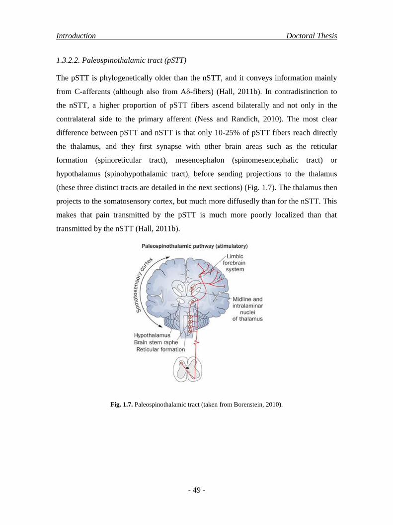
Introduction Doctoral Thesis
- 49 -
1.3.2.2. Paleospinothalamic tract (pSTT)
The pSTT is phylogenetically older than the nSTT, and it conveys information mainly
from C-afferents (although also from Aδ-fibers) (Hall, 2011b). In contradistinction to
the nSTT, a higher proportion of pSTT fibers ascend bilaterally and not only in the
contralateral side to the primary afferent (Ness and Randich, 2010). The most clear
difference between pSTT and nSTT is that only 10-25% of pSTT fibers reach directly
the thalamus, and they first synapse with other brain areas such as the reticular
formation (spinoreticular tract), mesencephalon (spinomesencephalic tract) or
hypothalamus (spinohypothalamic tract), before sending projections to the thalamus
(these three distinct tracts are detailed in the next sections) (Fig. 1.7). The thalamus then
projects to the somatosensory cortex, but much more diffusedly than for the nSTT. This
makes that pain transmitted by the pSTT is much more poorly localized than that
transmitted by the nSTT (Hall, 2011b).
Fig. 1.7. Paleospinothalamic tract (taken from Borenstein, 2010).

Doctoral Thesis Introduction
- 50 -
1.3.2.2.1. Spinoreticular tract
The spinoreticular tract transmit nociceptive, thermal (nonnociceptive), and
nondiscriminatory (crude) touch signals from the spinal cord to the thalamus indirectly,
by forming multiple synapses in the reticular formation prior to their thalamic
projections. This tract is furnished with input from C-fibers that synapse on interneurons
in laminae II and III, which in turn have polysynaptic influence on projection neurons in
laminae V-VIII. Also, the reticular formation receives collaterals from lamina I STT
axons. The third order neurons in the reticular formation project to intralaminar
thalamic nuclei and the posterior thalamus (reticulothalamic fibers). The intralaminar
nucleus then projects to the striatum and wide areas of cerebral cortex, whereas the
posterior nucleus projects to the secondary somatosensory cortex and posterior insular
cortex. These cortex areas are responsible to elicit the arousal and wakefulness after an
injury, that will enable the organism to evade the painful stimuli (Terman and Bonica,
2001; reviewed by Craig, 2003; Dostrovsky and Craig, 2008; Willis, 2009).
Spinoreticular axons also project to nuclei in the rostroventral medulla (RVM) such as
the nucleus gigantocellularis, paragigantocellularis and raphe magnus (Ness and
Randich, 2010), which are key areas involved in the descending modulatory pathways,
as it will be described in the Section 1.5.2.
1.3.2.2.2. Spinomesencephalic tract
The spinomesencephalic tract comprises axons of dorsal horn cells in laminae I and V.
This pathway ascends mainly in the contralateral anterolateral tract, although some
axons from lamina I ascend in the dorsolateral funiculus. The spinomesencephalic tract
projects to the periaqueductal gray (PAG), which contains neurons that are part of
descending pathway that regulates pain transmission (Calvino and Grilo, 2006). Part of
the spinomesencephalic tract ends just above the PAG, in the superior colliculus and
nucleus cuneiformis, located in the tectum of the midbrain. Therefore, this part of the
spinomesencephalic tract is also called spinotectal tract. The superior colliculus and
nucleus cuneiformis participate in coordination of motor avoidance behaviour evoked

Introduction Doctoral Thesis
- 51 -
by painful stimuli. Another prominent target of the spinomesencephalic tract is the
parabraquial nuclear complex nuclei. Neurons in the lateral parabranchial nucleus
project to the amygdala through the spino-parabrachio-amygdalar pathway, and might
therefore be involved in affective and emotional responses to pain (Calvino and Grilo,
2006; Ness and Randich, 2010).
1.3.2.2.3. Spinohypothalamic tract
This tract includes axons which projects to or through the hypothalamus.
Spinohypothalamic fibers transmit somatosensory information, including nociceptive
spinal input (somatic/visceral). These fibers arises from cells in laminae I, V, VII and X
of the dorsal horn and ascend in the anterolateral system, where the axon or collateral
branches of the spinohypothalamic neurons terminate in both sides of the hypothalamus,
thalamus, superior colliculus, and reticular formations in the midbrain, pons, and
medulla. As one of the main spinolimbic tracts, this pathway is associated with the
autonomic and reflex responses (i.e. neuroendocrine and cardiovascular) to nociception,
and to emotional changes caused by pain. (Basbaum and Jessel, 2000; Terman and
Bonica, 2001; Willis, 2009).
1.4. The somatosensory cortex
The cortex is divided in different areas according to their functional and anatomical
characteristics. There exist distinct cortical structures for motor, sensory, cognitive
emotional or autonomic functions (Ness and Randich, 2010). Only the somatosensory
cortex will be described here.
The somatosensory cortex (SSC) is involved in the processing of somatic sensory
information. The SSC comprises two different regions, the primary somatic sensory
cortex (S-I), and secondary somatic sensory cortex (S-II) (Purves et al., 2006; Ness and
Randich, 2010). The S-I is located in the postcentral gyrus of the parietal lobe and
consists of four functional areas: Brodmanns areas 1, 2, 3a, and 3b (Fig. 1.8A). Results

Doctoral Thesis Introduction
- 52 -
obtained in experiments in nonhuman primates indicated that neurons of each
Brodmann´s area respond depending on the type of stimulus. The areas 3b and 1 are
responsive to cutaneous stimuli, while the neurons in area 3a responds to stimulation of
the propioceptors; finally the neurons in area 2 process both tactile and proprioceptive
stimuli (Purves et al., 2006). S-I process the information from the contralateral part of
the body, receiving projections from the ventral posterior nucleus. The S-I is precisely
organized somatotopically. Thereby, in S-I certain areas of the body are better
represented than others (the face, oral cavity or hands, etc.) (Fig. 1.8B), which is related
to the receptors density in the body (Bonica, 1990; Martin and Jessel, 2000; Ness and
Randich, 2010). Importantly, direct intracerebral electrical stimulation of S-I failed to
elicit painful sensations (Lorenz and Hauck, 2010). Therefore, although S-I contribute
to the discriminative analysis of painful stimuli, it needs to act in conjunction with other
areas to perceive a stimulation as painful (Lorenz and Hauck, 2010).
Fig. 1.8. (A) Schematic anatomical localization of primary somatosensory cortex (modified from Kolb
and Whishan, 2003. (B) Somatotopic organization of human primary somatosensory cortex (taken from
Purves et al., 2006).
The S-II is situated lateral and posterior to S-I and in contradistinction to S-I, it
responds to sensory stimuli bilaterally (Purves et al., 2006; Ness and Randich 2010).
Stimuli frompropioiceptors
Cutaneousstimuli
Cutaneous andpropioceptive stimuli
A BPrimary somatosensorycortex

Introduction Doctoral Thesis
- 53 -
S-II receives convergent projections from the S-I and sends projections to limbic
structures such as the amygdala and hippocampus and to the somatic sensory fields in
the insular region (Martin and Jessel, 2000; Lorenz and Hauck, 2010). Therefore, S-II is
thought to play a major role in pain-induced attention, learning and memory, as well as
in the emotional aspects of pain experience (Chen et al., 2008).
1.5. Pain modulation: the gate control theory and the descending control systems
The sections above discussed the anatomical pathways involved in the transmission of
nociceptive information from the periphery to the central nervous system. However,
there is also a modulatory system within the central nervous system that influences pain
transmission and processing: the spinal gate control and the modulatory descending
pathways.
1.5.1. The gate control theory
It was in the early 1960s when neurophysiological studies provided evidence that
modulation of dorsal horn cells could be affected by input coming from the periphery
but also from supraespinal descending systems. This idea was introduced by Patrick
Wall and Ronald Melzack as the gate control theory (Melzack and Wall, 1965). They
found that the stimulation of low-threshold mechanoreceptors (LTM), corresponding to
myelinated Aβ-fibers, decreases the response of dorsal horn neurons to unmyelinated
nociceptors (C-fibers), whereas blockade of conduction in myelinated fibers enhances
the response of dorsal horn neurons. Therefore, the activity of spinal cord neurons in
response to nociceptor activation can be modulated by nonnociceptive fibers (reviewed
by Bonica et al., 1990; Jessel and Kelly, 2000). The action of LTM was explained by
the presence of inhibitory interneurons in the superficial laminae of the dorsal horn,
which receive inputs from both nociceptive and nonnociceptive primary afferents, and
synapse with the dorsal horn projection neurons. These interneurons are present in a
very high density in the superficial laminae of dorsal horn, and make up approximately
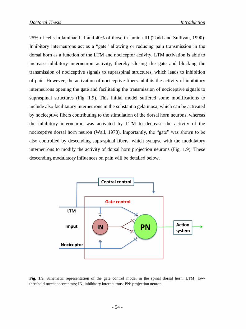
Doctoral Thesis Introduction
- 54 -
25% of cells in laminae I-II and 40% of those in lamina III (Todd and Sullivan, 1990).
Inhibitory interneurons act as a “gate” allowing or reducing pain transmission in the
dorsal horn as a function of the LTM and nociceptor activity. LTM activation is able to
increase inhibitory interneuron activity, thereby closing the gate and blocking the
transmission of nociceptive signals to supraspinal structures, which leads to inhibition
of pain. However, the activation of nociceptive fibers inhibits the activity of inhibitory
interneurons opening the gate and facilitating the transmission of nociceptive signals to
supraspinal structures (Fig. 1.9). This initial model suffered some modifications to
include also facilitatory interneurons in the substantia gelatinosa, which can be activated
by nociceptive fibers contributing to the stimulation of the dorsal horn neurons, whereas
the inhibitory interneuron was activated by LTM to decrease the activity of the
nociceptive dorsal horn neuron (Wall, 1978). Importantly, the “gate” was shown to be
also controlled by descending supraspinal fibers, which synapse with the modulatory
interneurons to modify the activity of dorsal horn projection neurons (Fig. 1.9). These
descending modulatory influences on pain will be detailed below.
Fig. 1.9. Schematic representation of the gate control model in the spinal dorsal horn. LTM: low-
threshold mechanoreceptors; IN: inhibitory interneurons; PN: projection neuron.
+
-
IN PN
Central control
Gate control
Actionsystem
+
+
-
-
LTM
Imput
Nociceptor

Introduction Doctoral Thesis
- 55 -
1.5.2. Descending modulatory influences from the brainstem.
Cerebral cortex projects to two key components of limbic system: the amygdala and the
hypothalamus which in turn send descending projections to the brainstem (Bonica et al.,
1990). There are three main brainstem areas involved in the descending modulation of
pain: the PAG, the RVM and the locus coeruleus (LC). Their relations are summarized
in Fig. 1.10, and detailed below.
As described in the preceding sections, the PAG receives afferent fibers from the ALS.
Reynolds (1969) demonstrated that electrical stimulation of PAG induced analgesia,
which was referred as “stimulation-produced analgesia”. The effects of stimulation-
produced analgesia may last from seconds to even hours.
Few neurons in the PAG project directly to the dorsal horn; instead they make
excitatory connections with neurons of the RVM, in particular with serotonergic
neurons in the nucleus raphe magnus (NRM) and nucleus reticularis gigantocellularis.
These nuclei are considered a final common relay of descending serotonergic
projections from supraspinal sites (Ossipov et al., 2010).
Activation of RVM neurons can generate either facilitation or inhibition of pain
transmission. This dual control results from the activity of different neuronal
subpopulations within the RVM. These populations are termed ON, OFF or neutral cells.
ON cells increase their activity immediately before a pain response occurs, and are
thought to exert a facilitating role for the nociceptive transmission. However, OFF cells
show a tonic activity which is stopped immediately before the pain response, and are
thought to be responsible for the descending inhibition of nociception. The net effect of
RVM on pain modulation depends therefore on the balance between ON and OFF cells
activity. Neutral cells were initially characterized by their absence of response to
nociceptive stimuli, although they might respond to nociceptive stimuli applied to other
body parts (Fields, 1992; Porreca et al., 2002; Calvino and Grilo, 2006).
Furthermore, both PAG and RVM project to the noradrenergic LC, which sends inputs
to different nuclei of PAG and RVM and also directly to spinal cord. Fibers from LC
Doctoral Thesis Introduction
- 56 -
are a major source of noradrenergic projections to the spinal cord to inhibit pain
transmission (Calvino and Grilo, 2006; Ossipov et al., 2010; Ness and Randich, 2010).
Descending serotonergic and noradrenergic projections from the brainstem course
through the dorsolateral and ventrolateral funiculi (DLF and VLF, respectively) to
synapse with primary afferent terminals, ascending projection neurons and modulatory
interneurons in the spinal cord, controlling therefore the “gate” of pain transmission
(Ossipov et al., 2010). The roles of the fibers in the DLF and VLF are distinct. While
DLF carries predominantly descending inhibitory signals, and consequently lesions in
the DLF disrupt the supraspinal inhibition of nociceptive stimuli (Ness and Randich,
2010), VLF transmits predominantly facilitatory influences from supraspinal origin
(Zhuo and Gebhart, 1992). Therefore, the balance between the inhibitory and
facilitatory descending systems determines the overall level of excitability of the
neurons in the dorsal horn.
There is another cerebral cortex descending modulatory system, the corticospinal tract
that sends terminals from sensory cortex directly to dorsal horn spinal cord. Neurons in
this pathway also project to several areas such as striatum, reticular ventrobasal
posterior and intralaminar thalamic nuclei, and to mesencephalon and reticular
formation. Although its function is not well known it is thought to enhance the
inhibitory function of the mesencephalic and medullary structures explained above
(Bonica et al., 1990).

Introduction Doctoral Thesis
- 57 -
Fig. 1.10. Spinal-medullary-spinal negative feedback loop underlying an endogenous analgesic system
called into play by nociceptive stimuli (modified from Basbaum and Fields, 1984).


Introduction Doctoral Thesis
- 59 -
2. µ-OPIOID DRUGS: MECHANISM OF ACTION, THERAPEUTIC USE AND
SIDE EFFECTS
2.1. Opioid system: historical overview
As described in the previous chapter, pain is a fundamental characteristic of the
physiological mechanisms of protection from tissue damage. However, when pain is
persistent or too severe it loses its protective role, and pain management is needed.
Therefore, trying to relieve pain has been one of the most constant human searches
(Haigh and Blake, 2001).
The opium, product extracted from poppy (Papaver somniferum) is among the oldest
medications known to treat a number of medical problems including pain but also cough
and diarrhoea. The psychoactive and the rewarding properties of this extract are also
well known since centuries ago (Kieffer, 1999, Pasternak and Pan, 2013). The active
ingredients of opium are alkaloid compounds. The most active component of opium is
morphine, which was isolated in 1805 by Friedrich Sertürner, a german pharmacy
apprentice, who called it morphine after the god of dreams Morpheus (Kieffer, 1999;
Gutstein and Akil, 2001). Subsequently, other alkaloids were isolated from opium such
as codeine (metilmorphine), thebaine (dimetilmorphine), papaverine and noscapine.
Afterwards semisynthetic opioids as heroine and also synthetic opioids such as fentanyl
were synthesized (reviewed by Florez, 2008).
Pharmacological studies developed for years shows the existence of an opioid system
expressed throughout the nociceptive neural circuitry and in regions of the central
nervous system included in reward and emotion-related brain structures, among other
areas (reviewed by Al-Hasani and Bruchas, 2011). The opioid system is composed of
three major receptor types known as µ-, δ- and κ-opioid receptors (reviewed by Kieffer,
1999; Pasternak and Pan, 2013). Genes encoding these opioid receptors have been
cloned and characterized (Akil et al., 1984, Satoh and Minami, 1995). Overall, µ
receptor agonists display the best antinociceptive activity, and are the ones most
frequently used in therapeutics (reviewed by Kieffer, 1999). Accordingly, this chapter
focuses on µ-opioid receptors and their clinically relevant uses.

Doctoral Thesis Introduction
- 60 -
2.2. Endogenous opioid peptides
The existence of a specific receptor for morphine and other opioid drugs led to think
that this receptor should play an important physiological role in pain control and in the
existence of endogenous ligands able to activate opioid receptors.
It was observed that the electrical stimulation of different brain areas produced
antinociception, which was reversed by the administration of the opioid antagonist
naloxone (Akil et al., 1972 and 1976). These findings indicated that endogenous opioids
were produced within the central nervous system, and led to subsequent experiments
aimed to search for the responsible molecules. These endogenous opioid ligands were
identified as peptides with opioid-like activity and affinity for opioid receptors
(reviewed by Pasternak and Pan, 2013).
The first endogenous opioid ligands discovered were the pentapeptides enkephalins,
Met- and Leu-enkephalin (Hughes et al., 1975). Soon after, other peptides were
aisolated: β-endorphin, dynorphin A and B, α-neoendorphin, and the, endomorphines 1
and 2. Remarkably, all endogenous opioids peptides except endomorphines share a four
amino acids sequence (Tyr-Gly-Gly-Phe) at the N-terminus, with differing extensions
and sequence at the C-terminus, which can vary from just one additional aminoacid (as
for Leu-enkephalin or Met-enkephalin) to as much as 27 extra aminoacids (as for
dynorphin A) (see Table 2.1.) (Florez, 2008; Pasternak and Pan, 2013).
The endogenous opioid ligands show different selectivity for the opioid receptor
subtypes. Enkephalins have higher affinity for δ-opioid receptors, Dynorphin A for κ1,
while β-endorphin binds with high affinity to µ receptors (and also to δ receptors,
although with a lower affinity) (Pasternak and Pan, 2013).

Introduction Doctoral Thesis
- 61 -
Table 2.1. Mammalian endogenous opioids peptides.
[Leu5]enkephalin Tyr-Gly-Gly-Phe-Met
[Met5]enkephalin Tyr-Gly-Gly-Phe-Leu
Dynorphin A Tyr-Gly-Gly-Phe-Met-Thr-Ser-Gly-Lys-Ser-Gln-Thr-Pro-Leu-Val-Thr-Leu-Phe-
Lys-Asn-Ala-lle-lle-Lys-Asn-Ala-Tyr-Lys-Lys-Gly-Glu
Dynorphin B Tyr-Gly-Gly-Phe-Leu-Arg-Arg-lle-Arg-Pro-Lys-Leu-Lys-Trp-Asp-Asn-Gln
α-Neoendorphin Tyr-Gly-Gly-Phe-Leu-Arg-Lys-Tyr-Pro-Lys
Endomorphin-1 Tyr-Pro-Trp-Phe-NH2
Endomorphin-2 Tyr-Pro-Phe-Phe-NH2
2.3. Opioid drugs: classification by intrinsic efficacy
Opioid drugs usually show affinity for more than one opioid receptor subtype, although
the opioid drugs used in the clinical practice mainly exert their effects through µ opioids
receptors (Kieffer, 1999; Pasternak and Pan, 2011; Pasternak and Pan, 2013). These
drugs can be classified according to their origin (natural, semisynthetic and synthetic),
chemical structure or intrinsic activity (Florez, 2008). Among these classifications, the
one based on the intrinsic activity of the compound is the widest used, since this
parameter clearly influences the effects of the drug (both in the clinical practice and in
preclinical research). According to their intrinsic activity the opioids drugs can be
classified as:
- Pure agonists: drugs which display a maximum intrinsic activity and therefore
the highest efficacy inducing signalling through its receptor. This group includes
known µ agonists such as morphine, oxycodone, fentanyl (and its derivatives
such as sufentanyl), heroin and methadone (Florez, 2008).
- Partial agonists: drugs that act with a lower intrinsic activity than a pure agonist.
The most relevant example is the partial µ agonist buprenorphine (Vadivelu and
Hines, 2007).
- Mixed agonist-antagonist: opioid drugs which act as agonists of a concrete
subtype of opioid receptor while acting as antagonists of a different subtype.

Doctoral Thesis Introduction
- 62 -
One representative example is pentazocine, which is a κ agonist but also a µ
antagonist (Hoskin and Hanks, 1991).
- Agonists with additional mechanisms: some drugs in addition to opioid agonism
show additional mechanisms which contribute to their analgesic effects. These
include drugs such as tapentadol and tramadol, which in addition to µ agonism
inhibits the reuptake of neurotransmitters. Specifically, tapentadol inhibits the
recaptation of noradrenalin (Hartrick and Rozek, 2011; Raffa et al., 2012),
whereas tramadol inhibits the reuptake of both serotonin and noradrenalin
(Mongin, 2007; Raffa et al., 2012).
- Antagonists: drugs that have affinity for opioid receptors but lack of intrinsic
activity and therefore of efficacy. The most widely used opioid antagonist is the
centrally-penetrant naloxone, which shows affinity for all three opioid receptor
subtypes (although with some preference for µ-opioid receptors). This drug is
used in preclinical research or in the clinical practice to reverse the effect of
opioid agonists (Mueller-Lissner, 2010; Cui et al., 2014; Mauger et al., 2014).
Another centrally-penetrant opioid antagonist with clinical use is naltrexone
(Corder et al., 2013; Syed and Keating, 2013). In addition, there have been
developed opioid antagonists with limited accessibility to the CNS, to reverse
exclusively the peripheral effects induced by opioid agonists. These drugs
include naloxone methiodide, which has been extensively used in preclinical
research (González-Rodríguez et al, 2010; Baamonde et al., 2006), and
methylnaltrexone which has been recently introduced in the clinical practice to
reverse clinically relevant peripheral opioid side effects (which will be described
later in the Section 2.6).
An additional classification of opioid drugs is done in base of their analgesic efficacy at
their therapeutic doses. This classification will be described in the Section 2.5.1.2.

Introduction Doctoral Thesis
- 63 -
2.4. The µ-opioid receptor and its molecular mechanisms of signaling
The µ-opioid receptor is a member of the G-protein-coupled receptor (GPCR)
superfamily (Quock, 1999; Costantino et al., 2012). It has a typical GPCR structure with
seven transmembrane helices, and with the N- and C-terminus facing at the extracellular
and the intracellular side, respectively (Connor and Christie, 1999; Knapman and
Connor, 2014) (Fig. 2.1).
The G proteins coupled to µ-opioid receptors are heterotrimeric proteins composed by a
Gα subunit (tipically of the inhibitory subtype) that binds guanosine diphosphate (GDP)
and is coupled to the receptor, and the Gβγ dimer which is anchored to the plasma
membrane and binds to Gα (Raehal et al, 2011). Once the opioid agonist binds to the
opioid receptor, GTP is exchanged by GDP in the Gα subunit. Then the Gα dissociates
from the Gβγ subunits, and is able to interact with other cytoplasmatic proteins. The
activated Gα subunit, inhibits the activity of adenylate cyclase (AC) with the consequent
decrease in cAMP production and thereby protein kinase A functioning (Al-Hashani
and Bruchas, 2011) (Fig. 2.1). In addition, the Gβγ dimer, which is still anchored in the
membrane, now is able to interact with several membrane targets. Specifically, the Gβγ
subunit inhibits the activity of several plasmalemmal voltage-dependent Ca2+
channels
(P/Q-type, N-type, and L-type), while opening G-protein-regulated inwardly rectifying
potassium channels (GIRKs) (Mark and Herlitze, 2000; Ocaña et al., 2004; Al-Hasani
and Bruchas, 2011) (Fig. 2.1). Importantly, the Gα subunit contributes to the
modulation of the effect of the Gβγ dimer to GIRKs (e.g. Mark and Herlitze 2000). All
these events contribute to the decrease of the activity of the neurons stimulated by µ
agonists, by hyperpolarizing the cell (Al-Hasani and Bruchas, 2011; Raehal et al., 2011)
(Fig. 2.1).

Doctoral Thesis Introduction
- 64 -
Fig. 2.1. Opioid signaling. The opioid agonists such as morphine (Mor) activate µ-opioid receptors
(µOR). The activated Gα subunit inhibits adenylate cyclase (AC), and so the AMPc production thereby
diminishing protein kinase A (PKA) activation. The βγ complex inhibits voltage dependent Ca+2
channels
and opens G-protein-regulated inwardly rectifying K+
channels (GIRKs). Activated Gα subunit also
modulates the effect of βγ complex on K+
channels.
2.4.1. µ-opioid receptor splice variants and the differences in the signaling induced by µ
agonists
The gene encoding for the µ-opioid receptor (MOR-1) is comprised by a number of
exons that can combine to form a wide range of splice variants (Fig. 2.2); MOR-1 itself
contains 4 exons. The first 3 exons encode both the N-terminus and all 7 transmembrane
domains, whereas exon 4 encodes only the last 12 amino acids in the C-terminus. The
major series of the variants of µ-opioid receptors are constituted by receptors with seven
transmembrane domains in which the exon 4 is replaced with a set of alternative exons
(Fig. 2.2). This splicing pattern is relatively conserved in mice, rats or humans
(Pasternak, 2010 and 2014). The binding pocket for all of these variants might be
identical, as it is comprised by the 7 transmembrane domains encoded by exons 1, 2,
and 3. However, the differences in the amino acid sequence at the C-terminus appear to
be important, since the signaling (measured as the increase in [35
S]GTPγS binding)
induced by several µ agonists differ between these spliced versions of the receptor, and

Introduction Doctoral Thesis
- 65 -
not equally for all µ agonists tested, indicating that these spliced variants could exhibit a
preferential activation by specific drugs (e.g. Pasternak et al., 2004; Bolan et al., 2004;
Pan et al., 2005 and 2009).
Fig. 2.2. Schematic representation of the MOR-1 splice variants (Taken from Pasternak, 2014).
Complementary detailed studies showed that not all µ agonists activate the same pattern
of Gα subunits in vivo. The array of Gα subunits known to be activated by µ opioids,
include the inhibitory Gi1, Gi2, Gi3, Go1, Go2 and Gz, but also Gq and G11 (Raffa et
al., 1994; Sánchez-Blázquez et al., 1995, 1999 and 2001; Garzón et al., 1996).
Exon
Spliced
Spliced
Spliced

Doctoral Thesis Introduction
- 66 -
Morphine and DAMGO were shown to predominantly activate Gi2 and Gz (and
additionally Gq for DAMGO). However, methadone and buprenorphine are able to
activate different combinations of a broader spectrum of Gα subunits, and in particular
heroin is able to activate all Gα subtypes mentioned above (Sánchez-Blázquez et al.,
1995 and 2001) (Fig. 2.3). Therefore, the spliced variants of the receptor exhibit a
preferential activation by specific drugs, and specific opioids induce distinct signaling
patterns. Importanty, the effectors of different µ-opioid drugs are also only partially
overlapping (see Urban et al., 2007 and Raehal et al., 2011 for reviews), which might be
influenced by the distinct signaling of the different opioid drugs. One clear example is
the recruitment of ATP-sensitive K+ channels for the antinociceptive effect of only
some µ agonists, which include morphine, buprenorphine or methadone, while other
agonists such as fentanyl or levorphanol do not need of the activation of these channels
for inducing antinociception (Ocaña et al., 1995).
Fig. 2.3. Activation patterns of Gα subunits depend on the µ agonist tested (Taken from Sánchez-
Blázquez et al., 2001, with slight modifications).
In addition, there are splice variants which code for opioid receptors of 6 or even only
one transmembrane domain (reviewed by Pasternak, 2014) (Fig. 2.2), which by
definition do not fit to the 7 transmembrane domain protein of a GPCR, and therefore
are not coupled to G proteins. The 6 transmembrane variant of the receptor still retains

Introduction Doctoral Thesis
- 67 -
binding to some known µ agonists, such as fentanyl, although do not bind other classic
drugs such as morphine (Majumdar et al., 2011 and 2012). Importantly, activation of
this truncated receptor retains antinociceptive activity while showing limited respiratory
depression or constipation (Majumdar et al., 2011 and 2012), which are troublesome
opioid side-effects, as it will be described in the Section 2.6. Although the role of the
single transmembrane domain versions of the µ receptor are still largely unclear, it has
been recently shown that they could exhibit a chaperone-like activity by minimizing the
degradation of the full length versions of the receptor, and therefore increasing the
protein levels (and therefore the actions) of the later (Xu et al., 2013).
2.5. Therapeutic use of µ-opioid drugs
Analgesia is the most extended therapeutic use of opioids, but they can be also
prescribed as antitussive and antidiarrheal drugs, among other clinical uses.
2.5.1. Opioid analgesia
Analgesia is the most important therapeutic effect induced by opioids. Although the
activation of all subtypes of opioid receptors leads to analgesia, the great majority of
opioid analgesic drugs used in therapeutics are µ agonists (Marker et al., 2005; Al-
Hasani and Bruchas, 2011; Pasternak, 2013). In this section we will summarize the
modulation of the pain pathways by µ-opioid drugs as well as their clinical use as
analgesics, highlighting their place in the World Health Organization (WHO) analgesic
ladder and their rational use in therapeutics.
2.5.1.1. Modulation of pain pathways by µ-opioid drugs
µ-opioid receptors are located along the pain pathways, including several areas of the
central nervous system (both in supraspinal nuclei or at spinal level) as well as in the
periphery (dorsal root ganglia, DRG) (Bigliardi-Qi et al., 2004; Khalefa et al., 2012). It
is thought that opioid drugs exert their analgesic effects mainly at central levels.
Although they can elicit a robust analgesia when administered at the spinal cord (e.g.

Doctoral Thesis Introduction
- 68 -
Yaksh and Rudy, 1976; Wagemans et al., 1997; Mercadante, 1999), they are
particularly active at supraspinal sites (e.g. Christie et al., 2000; Ossipov et al., 2010;
Khalefa et al., 2012; Ringkamp and Raja, 2012), where µ-opioid receptors are expressed
at their highest density (e.g. Arvidsson et al., 1995; Khalefa et al., 2012; Ringkamp and
Raja, 2012).
Among the several supraspinal structures which participate in opioid analgesia, the most
prominent ones are those constituting the modulatory descending pain pathways. As
described in the Section 1.5.2, these areas include the PAG, LC and RVM.
Administration of opioids into any of these three sites is enough to elicit analgesia (e.g.
Millan, 2002; Porreca et al., 2002; Mei and Pasternak, 2007). These three areas are
interconnected and participate coordinately in pain control (Fields, 1984). The
relationship between their actions is such that the simultaneous administration of low
(inactive) doses of morphine into the PAG and RVM is able to elicit strong
antinociceptive effects that are far higher than additive (i.e. synergistic) (Rossi et al.,
1993). RVM play a particular role on the descending modulation of pain, since it
receives afferents from both PAG and LC and is thought to be the final common relay
in both the descending inhibition and facilitation of pain (reviewed by Porreca et al.,
2002; Millan, 2002). This dual pain control is the result from the activity of two distinct
neuronal subpopulations within the RVM: the ON and OFF cells. ON cells are thought
to exert a facilitating role for the nociceptive transmission, and increase their activity
immediately before a pain response occurs; moreover, the activation of ON cells is able
to inhibit the activity of OFF cells. These latter neurons show a tonic activity which is
stopped immediately before the pain response, and they are thought to be responsible
for the descending inhibition of nociception. Therefore, these two cell types act
coordinately, and a nociceptive stimulation is able to induce simultaneously both an
increase in the activity of ON cells and a decrease in the activity of OFF cells, resulting
on pain (see Fig. 2.4 left panel) (Fields, 1992; Porreca et al,. 2002; Calvino and Grilo,
2006; Ossipov et al., 2010). Opioid effects are particularly well studied in the RVM.
Opioid administration is able to decrease the activation of ON cells (and not that of OFF

Introduction Doctoral Thesis
- 69 -
cells), resulting in the disinhibition of OFF cells and therefore eliciting analgesia
(reviewed by Porreca et al., 2002; Ossipov et al., 2010) (see Fig. 2.4 right panel).
Fig. 2.4. ON and OFF cells activity in the rostroventral medulla (RVM) during application of a
nociceptive stimulus and after opioid administration.
Although most of the analgesic effects of opioids are attributed to their actions at central
levels, peripheral opioid receptor activation can also elicit analgesia in rodents (Craft et
al., 1995; Kayser et al., 1995; Kolesnikov et al., 1996 and 2000; Shannon and Lutz,
2002) and humans (reviewed by Stein et al., 2003; Sehgal et al., 2011), but their effects
are thought to be limited in comparison to their central actions (Greenwood-Van
Meerveld and Standifer, 2008; Joshi et al., 2008; Thomas et al., 2008; Khalefa et al.,
2012; Ringkamp and Raja, 2012).
2.5.1.2. Clinical use as analgesics
The WHO developed a 3-step “analgesic ladder” to guide pain management, which is
based on the intensity of pain (see Fig. 2.5). In accordance with WHO
recommendations, if pain intensity is mild a nonopioid analgesic, such as a nonsteroidal
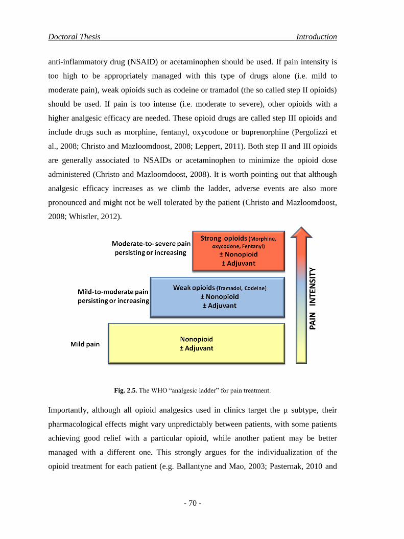
Doctoral Thesis Introduction
- 70 -
anti-inflammatory drug (NSAID) or acetaminophen should be used. If pain intensity is
too high to be appropriately managed with this type of drugs alone (i.e. mild to
moderate pain), weak opioids such as codeine or tramadol (the so called step II opioids)
should be used. If pain is too intense (i.e. moderate to severe), other opioids with a
higher analgesic efficacy are needed. These opioid drugs are called step III opioids and
include drugs such as morphine, fentanyl, oxycodone or buprenorphine (Pergolizzi et
al., 2008; Christo and Mazloomdoost, 2008; Leppert, 2011). Both step II and III opioids
are generally associated to NSAIDs or acetaminophen to minimize the opioid dose
administered (Christo and Mazloomdoost, 2008). It is worth pointing out that although
analgesic efficacy increases as we climb the ladder, adverse events are also more
pronounced and might not be well tolerated by the patient (Christo and Mazloomdoost,
2008; Whistler, 2012).
Fig. 2.5. The WHO “analgesic ladder” for pain treatment.
Importantly, although all opioid analgesics used in clinics target the µ subtype, their
pharmacological effects might vary unpredictably between patients, with some patients
achieving good relief with a particular opioid, while another patient may be better
managed with a different one. This strongly argues for the individualization of the
opioid treatment for each patient (e.g. Ballantyne and Mao, 2003; Pasternak, 2010 and

Introduction Doctoral Thesis
- 71 -
2014). These clinical findings could be replicated in different strains of mice, which
show striking differences in the analgesic sensitivity to opioid antinociception, and not
to the same degree or direction for all opioids; for instance, morphine had a much lower
antinociceptive effect in CXBK mice than in CD-1 mice, whereas heroin behaved in the
opposite way (reviewed by Pasternak, 2004). These clinical and preclinical observations
can hardly be explained by the existence of a common mechanism of action for all µ
opioids, and support that µ-opioid drugs do not bind to a single receptor, but they might
interact with a large number of receptor variants with different activation profiles
elicited by the different drugs, as previously commented.
2.5.2. Antitussive properties of opioid drugs
Cough is considered a defensive reflex mechanism, which protects the airways by
ejecting obstructive or potentially harmful substances. However, chronic cough is also a
distressing characteristic symptom of many inflammatory airways‟ diseases (Belvisi and
Hele, 2009). Both bronchopulmonary C-fibers and polymodal Aδ-fibers subsets (the so
called “cough receptors”) can participate in the cough process (Reynolds et al., 2004).
After their activation, they stimulate the brainstem cough centre through vagal afferent
pathways, eliciting the cough reflex (reviewed by Canning, 2011). It is thought that the
antitussive properties of opioids are predominantly central (e.g. Reynolds et al., 2004),
and a consequence of the decreased activity of the cough center induced by opioid
receptor activation (although the signal transduction involved is not as well described as
the one involved in analgesia) (Belvisi and Hele, 2009). Peripheral opioid actions in the
airway cough sensors might also participate in the antitussive effects of opioids,
however it is thought that their contribution is much limited in comparison to that from
central opioid receptors (Reynolds et al., 2004).
Although all centrally acting opioids effectively decrease the cough reflex, codeine is
often considered the „gold standard‟ in antitussive therapy since it has a better side-
effect profile than the stronger opioids (Reynolds et al., 2004; Chung 2008; Al-Hasani
and Bruchas, 2011).
Doctoral Thesis Introduction
- 72 -
2.5.3. Antidiarrheal
Although gastrointestinal transit can be influenced by both central and peripheral opioid
receptors, the central contribution to this effect is thought to be limited (Holzer et al.,
2009), contrary to analgesia and the antitussive effects of opioid drugs described above.
Opioid receptors are located in the gastrointestinal tract on the myenteric and
submucosal plexuses (Brock et al., 2012; Khalefa et al., 2012), and enteric neurons at
these locations synthesize and release opioid peptides, so the opioidergic system is
thought to have a physiological role on gastrointestinal transit function (Greenwood-
Van Meerveld et al., 2004; Holzer et al., 2009).
Opioid drugs can also interact with opioid receptors on the gastrointestinal tract (Holzer
et al., 2009; Brock et al., 2012). Opioid agonism in this location induces a decrease of
gut motility and fluid secretion as well as increase in sphinter contraction (DeHaven-
Hudkins et al., 2008; Holzer et al., 2009; Brock et al., 2012).
Although the inhibition of gastrointestinal transit is considered an adverse side effect of
opioid analgesics (see Section 2.6), this effect of opioid agonists is therapeutically used
for the symptomatic treatment of acute and chronic diarrhea (Holzer, 2009; Gallelli et
al., 2010). Not all opioid agonists have this clinical application, since their central
effects limit their use. The opioid most frequently used for this therapeutic purpose is
loperamide. This particular opioid agonist has little CNS effect due to its active efflux
transport from the CNS into the blood by transporters including P-glycoprotein (Upton,
2007), but as its peripheral actions are prominent it has a marked effect on
gastrointestinal transit (Layer et al., 2010; Gallelli et al., 2010).

Introduction Doctoral Thesis
- 73 -
2.6. Opioid side effects
Along with their therapeutic effects, opioids have the potential to produce important
side effects. Management of opioid-induced adverse events is still today a major clinical
challenge, since they occur frequently and reduce the quality of life of patients, being
often the cause of treatment discontinuation (Cherny et al., 2001; Al-Hashani and
Bruchas, 2011; Annemans, 2011; Brock et al., 2012). As for the therapeutically useful
effects of opioids, their side events (or most of them) derive from the inhibitory actions
of opioids on neuronal functioning. In most cases these side effects are produced by the
simultaneous actions of opioids at both central and peripheral levels, although for each
concrete effect the peripheral or central contribution varies, as it will be detailed below.
The most relevant opioid-induced side effects and their most prominent origins (central
or peripheral) are listed below (Table 2.2.).
Table 2.2. Opioid side effects distributed by their primary location of induction (central or peripheral).
Location of action Effects
Central Nervous System
↑Nausea and vomiting
↑Psychotomimetic alterations
↑Confusion
↑Sedation
↓Rate of respiration
↑Truncal rigidity
↓Pupil size (miosis)
↑Urinary retention
↑Pruritus
Peripheral Nervous
System
Gastrointestinal system
↓Gastric motility
↑Sphincter contraction
↓Intestinal secretion
↓Peristaltic waves in the colon
↑Gastroesophageal reflux
Cardiovascular system ↓Blood pressure and heart rate if cardiovascular system
is stressed
The actions summarized in this table are observed for all clinically available opioid agonists. Although
several of these effects are known to have both a central and a peripheral component, for clarity purposes
only the predominant location of each side effect was considered for this table.

Doctoral Thesis Introduction
- 74 -
Among the peripheral side effects of opioid drugs, those related to the gastrointestinal
tract are the most frequent, being the constipation the more prevalent symptom
produced by prolonged opioid treatment and one of the main reasons for patients‟
voluntary withdrawal from opioid medication (Brock, 2012; Dhingra et al., 2013).
Constipation is mainly caused by the inhibition of gastrointestinal motility, intestinal
secretion and sphincter contraction induced by opioid receptors localized on the enteric
nervous system, but might also present a central component (see Section 2.5.2 for
details). Constipation is also accompanied by other symptoms such as incomplete
evacuation, abdominal distention, abdominal discomfort and gastroesophageal reflux,
which can lead to secondary complications such as anorexia, which can altogether
profoundly impair the quality of life of patients (Holzer, 2010; Brock, 2012; Dhingra et
al., 2013). Hence, several treatment strategies are available to alleviate these side
effects. Traditionally, laxative therapy has been used to prevent constipation, however
clinical studies have showed that it is clearly insufficient (see Greenwood-Van
Meerveld and Standifer, 2008 for references). The recent introduction of the
peripherally-restricted opioid receptor antagonist methylnaltrexone in the clinical
practice (trade name Relistor) has opened up a new window for controlling opioid-
induced constipation without compromising the central analgesic actions of opioids
(Greenwood-Van Meerveld and Standifer, 2008; Thomas et al., 2008; Holzer, 2010;
Gatti and Sabato, 2012; Mori et al., 2013).
On the other hand, nausea and vomiting, which is one of the most frequent and severe
symptoms after acute treatment with opioids (Florez, 2008), are mainly produced
centrally by the stimulation of the chemoreceptor vomiting center in the area postrema
(Bastami, 2014). However, the inhibition of gastrointestinal transit is also thought to
play a role on the induction of nausea and vomiting, and in fact treatment with
methylnaltrexone is able to partially relieve these side effects of opioids (Rosow et al.,
2007; Gatti and Sabato, 2012; Sharma and Jamal, 2013).
Sedation and confusion, are centrally-mediated opioid side effects which mainly occur
with initiation of therapy or dose escalation (Florez, 2008), and both are among the

Introduction Doctoral Thesis
- 75 -
most commonly reported adverse effects of opioids (Davies et al., 2009). Opioid-
induced sedation occurs on a continuum ranging from full consciousness to complete
loss of consciousness (coma), the later happening more frequently in cases of
intoxication (Jarzyna et al., 2011; Mégarbane et al., 2007). During opioid intoxication,
coma is often accompanied with other two centrally-mediated opioid side effects: severe
respiratory depression and accused miosis (pinpoint pupils) (Sporer et al., 1996; Florez,
2008; Mégarbane et al., 2007). If this symptomatic triad (coma, respiratory depression
and pinpoint pupils) can be reversed by naloxone, it is considered a pathognomonic sign
of opioid intoxication (Sporer et al., 1996; Florez, 2008; Mégarbane et al., 2007).
Although respiratory depression induced by opioids is relatively uncommon, in some
cases (of opioid intoxication) is life-threatening (Dahan et al., 2013). Its frequency
increases when an underlying pulmonary disease, interactions with other drugs or renal
failure exits, and during an overdose by opioid misuse (Florez, 2008; Pasternak and
Pan, 2013; Dahan et al., 2013). Respiratory alterations caused by opioids are mainly
originated at central level by depression of central respiratory centers, but they also have
a peripheral component, as opioids can decrease the sensitivity of peripheral chemo-
receptors to carbon dioxide (Pattinson, 2008; Florez, 2008; Radke et al., 2014). Another
central effect of opioids that can aggravate the respiratory alterations induced by opioids
is trunk rigidity which although it is an uncommon effect, increases its frequency when
the opioid drug used is highly lipophilic, such as fentanyl or derivatives (Al-Hasani and
Bruchas, 2011; Coruh et al., 2013). Miosis is a central effect occurring at the autonomic
segment of the oculomotor nerve, and although it is present at analgesic doses of
opioids it does not induce any relevant distress. It is used clinically as an indicator of
possible opioid intoxication when is very pronounced, and experimentally as an
indicator of the central effects of opioids, since it is sensitive to naloxone but not to
methylnaltrexone (Rosow et al., 2007).
The psychotomimetic alterations induced by a chronic treatment with opioids are
directly related to the concentration reached in the CNS, and therefore vary depending
on the liposolubility of the specific opioid (Martín and Goicoecha, 2008).
Doctoral Thesis Introduction
- 76 -
In addition to these effects, opioids are well known to increase the tone of the trigone,
decrease detrusor tone, decrease the urge to void, and inhibit the voiding reflex, all of
them factors contributing to the urinary retention, which may lead to the overdistension
of the bladder producing pain and bladder injury (Kuipers et al., 2004; Rosow et al.,
2007; Harris, 2008; Zand et al., 2014). Although this adverse event of opioids is
clinically significant, in particular in elder men (Florez, 2008), its incidence has proven
surprisingly difficult to estimate (Rosow et al., 2007). Intrathecal administration (i.t.) of
opioids is well known to decrease urinary function, and therefore activation of
peripheral opioid receptors is not needed for the occurrence of this effect (e.g. Kuipers
et al., 2004; Zand et al., 2014). However, the association of methylnaltrexone with
systemic opioids was shown to decrease at some extent urinary retention, which might
be due to the reversion of the actions of opioids in parasympathetic nerves innervating
the bladder (Rosow et al., 2007; Reichle and Conzen, 2008).
Pruritus, a subjective unpleasant and irritating sensation that provokes an urge to
scratch, is also very often reported in patients with opioid treatment (Davies et al.,
2009). The symptoms typically start at the trunk, nose, around the eyes and in other
facial areas (Szarvas et al., 2003; Kumar and Indu-Sing, 2013). This opioid effect is
thought to be mainly central in origin as it happens often after epidural or intrathecal
administration (Szarvas et al., 2003; Kumar and Indu-Sing, 2013), but peripheral opioid
receptors might play a role as well, since topical treatment with opioid antagonists can
relieve some types of pruritus (Bigliardi et al., 2007).
The cardiovascular complications during a treatment with morphine are rare in
normotensive patients. However, opioids may affect cardiovascular function by acting
in the kidneys, blood vessels and even in the heart, decreasing blood pressure and heart
rate. The frequency of cardiovascular complications increases at high doses of opioids
or in patients with a preexistent heart disease (Florez, 2008; Martín and Goicoechea,
2008; Harris, 2008).
Interestingly, it is known that the chronic use of opioids, reduces the immune response
and promotes the development of infections. These actions of opioids in immune system

Introduction Doctoral Thesis
- 77 -
can be direct (by stimulation of opioid receptors expressed immune cells), or indirect
(by neuro-immune interactions) (Florez, 2008; Martín and Goicoechea, 2008; Shen et
al., 2014).
A major limitation to the long-term use of opioids is the development of tolerance and
dependence (Cherny et al., 2001; Harris, 2008), which will be detailed in the next
section.
2.7. Tolerance and dependence
Tolerance is a pharmacologic phenomenon that develops with the repeated used of some
drugs, including opioids. The development of tolerance reduces the effects of opioids
and therefore increased doses of opioids are needed to maintain the same level of
analgesia (Ballantyne and Mao, 2003; Raehal et al., 2011). Importantly, although
tolerance to the analgesic effect of opioids is often produced, tolerance is not (or it is
minimally) developed for other nonanalgesic effects of these drugs, such as constipation
(Brock et al., 2012; Pan and Pasternak, 2013). This is particularly worrisome, since the
dose escalation needed for the maintenance of analgesia increase in parallel the severity
of some opioid side effects that are not attenuated over time (Martín and Goicoecha,
2008; Schmittner et al., 2009).
The activation of opioid receptors by an agonist induce the phosphorylation of the
opioid receptor by G protein-coupled receptor kinases (GRKs), inhibiting the
association of the receptor to the G proteins. This process leads to the desensitization of
the receptor to further agonist stimulations. β-arrestins (of the subtype 2) can then bind
to the phosphorylated receptor promoting its internalization by endocytosis. Following
endocytosis receptors can be dephosphorylated and recycled to the cell surface in a fully
active state, thereby resensitizing cells to agonist. However, if agonist exposure is
persistent, the endocytosis process will end up in the degradation of the opioid receptor.
This will decrease the number of opioid receptors available in the plasma membrane
(downregulation), and consequently decreasing the effects of the opioids (reviewed by
Kohout and Lefkowitz, 2003; Waldhoer et al., 2004; Garzón et al., 2008; Pasternak and
Doctoral Thesis Introduction
- 78 -
Pan, 2013). Although this is thought to be a general mechanism for the development of
desensitization/tolerance, it is not applicable equally to all µ agonists. For instance
morphine is known to induce a minimal internalization of the receptor, while DAMGO
is shown to robustly induce this process (reviewed by Waldhoer et al., 2004; Garzón et
al., 2008; Pasternak and Pan, 2013).
Changes during tolerance not only involve changes in the opioid receptor, but has also
changes in second messenger systems (as adenyl cyclase superactivation), as well as in
glutamatergic neurotransmission (through the NMDA-nitric oxide cascade) (reviewed
by Waldhoer et al., 2004; Garzón et al., 2008; Pasternak and Pan, 2013). In addition,
prolonged treatment with opioids enhances the release of excitatory neurotransmitters
including glutamate, CGRP, and Substance P from nociceptive primary afferent fibers
within the spinal cord, which contribute to the analgesic tolerance (reviewed by King et
al., 2005; Waldhoer et al., 2004).
Interestingly, patients highly tolerant to the analgesic effect of one specific µ-opioid
drug may regain (at least partially) their sensitivity when switched to a different
µ-opioid. This observation led to the clinical practice of opioid rotation. When the dose
escalation with one opioid fails (the balance between analgesia and side effects is
decompensated), the incomplete cross-tolerance between opioid drugs may permit a
lower (relative) dose of a new opioid to be used (reviewed by Pasternak, 2010;
Pasternak and Pan, 2013). This clinical observation has been replicated in the preclinical
setting in which it was clearly shown that several µ opioid drugs show incomplete
cross-tolerance (reviewed by Pasternak, 2004). This does not reconcile with the
existence of a unique µ opioid receptor or with signalling pathways and mechanisms for
tolerance common for all µ opioid agonists, but might be influenced by the diversity of
µ opioid receptors and the known differences in the signalling and effectors between the
different µ opioid drugs (see Section 2.4.1), and of course by the differences in the
mechanisms of the desensitization/resensitization of the receptor by different µ agonists
(as previously commented).

Introduction Doctoral Thesis
- 79 -
In addition to tolerance, physical dependence, which results in the necessity for
continued administration of the drug to prevent the development of symptoms of opioid
withdrawal, can ensue in patients. The withdrawal syndrome can be precipitated by the
abrupt withdrawal of the opioid treatment or by the administration of opioid drugs with
lower intrinsic efficacy (including of course opioid antagonists) (Walsh and Eissenberg,
2003; Waldhoer et al., 2004; Paronis and Bergman, 2010). Tolerance and dependence
may share partially overlapping mechanisms (reviewed by Waldhoer et al., 2004); as an
example, both tolerance and dependence can be blocked by NMDA receptor antagonists
(Trujillo and Akil, 1991; Kolesnikov et al., 1993; Gónzalez et al., 1997). However, the
overlapping between the mechanisms of tolerance and dependence are not complete.
For instance it is known that β-arrestin-2 is needed for the development of opioid
tolerance, but does not influence dependence (Bohn et al., 2000).
From a clinical standpoint, opioid withdrawal is one of the most powerful factors
driving addictive behaviors. Addiction is a complex phenomenon, distinct to
dependence, which is characterized by intense drug-seeking behavior and the
compulsive use of the drug (Williams et al., 2001; Florez, 2008). Opioids are able to
modulate neural brain circuits associated with reward (positive reinforcement), so
opioid administration can motivate the repeated use of the drug simply for pleasure
(Waldhoer et al., 2004). It is generally thought that opioid drugs induce addiction when
they activate the rewarding areas in the absence of significant pain (i.e. during
recreational use), and that when they are used for treating severe pain the tendency to
abuse is exceptional (Williams et al., 2001; Florez, 2008). However, in the last few
years it has been detected a high increase in the abuse of opioid analgesics, which is
thought to be due to the recent increased number of their prescriptions (Højsted and
Sjøgren, 2007).

Introduction Doctoral Thesis
- 81 -
3. SIGMA-1 (1) RECEPTORS AND PAIN: FROM CENTER TO PERIPHERY
Despite the progress made in the understanding of pain mechanisms, pain management
is still a major clinical need (Cobos and Portillo-Salido, 2013; Finnerrup et al., 2013;
Drageset et al., 2014; Pergolizzi et al., 2014). Therefore, it is important to search for
new pharmacological targets to obtain new therapies or to improve existing treatments.
Here we will summarize the discovery of σ1 receptors, their pharmacology, anatomical
and subcellular distribution, as well as their modulatory role on neurotransmission.
Finally, we will extensively describe the experimental evidences which leaded to
propose σ1 receptors as a potential new pharmacological target for pain treatment from
two different perspectives: the modulation of opioid effects on acute pain, and the
modulation of pain hypersensitivity on pathological pain. In this latter section we will
place into an adequate frame the results obtained during this Doctoral Thesis,
highlighting our findings as the first evidences of a role of peripheral σ1 receptors on
pain modulation and the impact of these findings on the field.
3.1. Discovery of σ receptors
Initial studies misclassified σ receptors as a subtype of opioid receptors, and later with
the binding site for phencyclidine (PCP) within the N-methyl-D-aspartate (NMDA)
glutamate receptor (Martin et al., 1976; Zukin et al., 1984). These early confusions were
due to the complex pharmacology of the first σ ligand deeply characterized, (±)-
SKF-10,047. Whereas (–)-SKF-10,047 binds to opioid receptors, and therefore its
effects are reversible by naloxone, the (+)-isomer lacks affinity for opioid receptors but
binds to two different naloxone insensitive sites: the PCP binding site and to an
additional benzomorphan binding site (Walker et al., 1990; Cobos et al., 2008; Robson
et al., 2012). This latter binding site was termed σ receptor (Quirion et al., 1992) and
still retains the same designation (Cobos et al., 2008; Zamanillo et al., 2013 for
references).
Doctoral Thesis Introduction
- 82 -
Early studies demonstrated the existence of two subtypes of σ receptors, which differed
on their molecular mass (29 kDa and 18-21.5 kDa for σ1 and σ2, respectively)
(Hellewell and Bowen, 1990), and also in their pharmacological profile. While σ1
binding sites display stereospecificity towards dextrorotatory isomers of
benzomorphanes, σ2 binding sites display certain selectivity to levorotatory isomers
(Quirion et al., 1992). The cloning of the σ1 receptor strongly helped to establish σ1
receptors as a unique entity (as it will be described in the next section). Only the
characteristics and mechanisms of σ1 receptors will be discussed in the present chapter.
3.2. Cloning and structural characteristics of σ1 receptor
A significant progress in the knowledge of the molecular structure of σ1 receptors was
possible after its cloning. σ1 receptor was firstly cloned from guinea pig liver (Hanner et
al., 1996), and later from other tissues and species, as mouse kidney, a JAR human
choriocarcinoma cell line, and in the rat and mouse brain (see Cobos et al., 2008 for
references). The cloning of σ1 receptors allowed to unveil both its structure and
function. This receptor is a single polypeptide, composed by 223 amino acids with high
sequence homology between several species (Seth et al., 2001). Interestingly, this
receptor shows no homology with any other mammalian protein, but shares
approximately 30% identity with the yeast gene that encodes the C7–C8 sterol
isomerase (Moebius et al., 2001), which might explain at some extent its affinity for
neurosteroids (Maurice et al., 2001 and 2006), that as it will be described in the next
section are putative σ1 endogenous ligands. Although initial studies proposed a structure
of σ1 receptors with a single transmembrane region (Hanner et al., 1996; Seth et al.,
1997; Dussossoy et al., 1999), further studies made clear that σ1 receptors have two
transmembrane segments (Aydar et al., 2002). This later structure has been refined in
subsequent studies, reporting the existence of two additional hydrophobic segments
(one of them partially overlapping the second transmembrane domain), corresponding
to steroid binding domain-like sites and purportedly responsible for ligand binding
(Chen et al., 2007; Pal et al., 2007 and 2008). More recently, it was described the
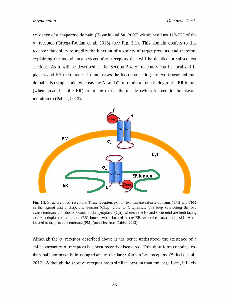
Introduction Doctoral Thesis
- 83 -
existence of a chaperone domain (Hayashi and Su, 2007) within residues 112-223 of the
σ1 receptor (Ortega-Roldan et al, 2013) (see Fig. 3.1). This domain confers to this
receptor the ability to modify the function of a variety of target proteins, and therefore
explaining the modulatory actions of σ1 receptors that will be detailed in subsequent
sections. As it will be described in the Section 3.4, σ1 receptors can be localized in
plasma and ER membranes. In both cases the loop connecting the two transmembrane
domains is cytoplasmic, whereas the N- and C- termini are both facing to the ER lumen
(when located in the ER) or to the extracellular side (when located in the plasma
membrane) (Pabba, 2013).
Fig. 3.1. Structure of σ1 receptors. These receptors exhibit two transmembrane domains (TM1 and TM2
in the figure) and a chaperone domain (Chap) close to C-terminus. The loop connecting the two
transmembrane domains is located in the cytoplasm (Cyt), whereas the N- and C- termini are both facing
to the endoplasmic reticulum (ER) lumen, when located in the ER, or to the extracellular side, when
located in the plasma membrane (PM) (modified from Pabba, 2013).
Although the σ1 receptor described above is the better understood, the existence of a
splice variant of σ1 receptors has been recently discovered. This short form contains less
than half aminoacids in comparison to the large form of σ1 receptors (Shioda et al.,
2012). Although the short σ1 receptor has a similar location than the large form, it likely

Doctoral Thesis Introduction
- 84 -
lacks chaperone activity and ligand binding ability, as the coding region of the
chaperone and ligand binding domains is truncated (Shioda et al., 2012).
Importantly, the cloning of σ1 receptor allowed the design of specific antisense
oligodeoxynucleotides (e.g. King et al., 1997; Pan et al., 1998) and the development of
σ1-receptor knockout mice (Langa et al., 2003). These tools have been extensively used
for studying the role of these receptors on pain (as it will be described in the Section
3.6).
3.3. Pharmacological profile of σ1 receptors
σ1 receptor binds bezomorphans displaying stereospecificity for their dextrorotatory
isomers, and also bind to a broad catalogue of compounds with high to moderate
affinity, including: antipsychotics (e.g. haloperidol), antidepressants (e.g. fluvoxamine),
antitussives (e.g. carbetapentane) and drugs of abuse (e.g. cocaine) (see Hayashi and Su,
2004; Cobos et al., 2008; Su et al., 2010 for references). Moreover, allosteric
modulators of σ1 receptors have also been described, such as the anticonvulsant drug
phenytoin (Musacchio et al., 1989; Cobos et al., 2005 and 2006).
Neurosteroids (pregnenolone, dehydroepiandrosterone, progesterone, allopregnanolone
and their sulfate esters) are considered the most likely naturally occurring σ1
endogenous ligands (Maurice et al., 2001; Hayashi and Su, 2004; Cobos et al., 2008). In
recent years it has been reported the existence of another putative endogenous ligand,
N,N-dimethyltryptamine (DMT), which is a natural hallucinogen (Fontanilla, 2009).
However, the affinities of most neurosteroids or DMT for σ1 receptors fall in the µM
range (Schwarz et al., 1989; Cobos et al., 2008; Fontanilla, 2009), and do not appear to be
high enough for endogenous ligands. Therefore, the endogenous σ1 ligands (if any) of σ1
receptors have not yet been unequivocally defined.
Although the endogenous σ1 ligands are still unclear, there have been developed some
selective and high affinity σ1 drugs for studying σ1 receptor function. These include the
prototypical σ1 agonists (+)-pentazocine and PRE-084, and the σ1 antagonists BD-1047,
Introduction Doctoral Thesis
- 85 -
NE-100, BD-1063 (reviewed by Hayashi and Su, 2004; Cobos et al., 2008), and more
recently the σ1 antagonist S1RA, which has been shown to exhibit an exquisite
selectivity for σ1 receptors lacking affinity for 170 additional targets (Romero et al.,
2012). This latter drug is currently in Phase II clinical trials for pain treatment, after
completing a Phase I study showing excellent kinetics and tolerability (Abadías et al.,
2013).
3.4. Anatomical and subcellular distribution of σ1 receptors
The distribution of σ1 receptor has been studied at anatomical and subcellular levels,
and the knowledge of its location has provided information for the understanding of its
possible functions.
At anatomical level, σ1 receptors are widely localized in the central nervous system
(CNS), including important areas for pain control, such as superficial layers of the
dorsal horn, the periaqueductal gray matter, the locus coeruleus and rostroventral
medulla (Roh et al., 2008b; Zamanillo et al., 2013; Sánchez-Fernández et al., 2014).
Recently, it was also reported that σ1 receptor is also present in the peripheral nervous
system in both neuronal bodies of the dorsal root ganglion (DRG) (Bangaru et al., 2013)
and in Schwann cells, specifically in their cytoplasm and in the paranodal region of
Ranvier nodes (Palacios et al., 2004). Interestingly, although most functional pain
studies focused on the role of central σ1 receptors on pain processing, we recently found
(as a part of this Doctoral Thesis) that σ1 receptors are present in the DRGs at much
higher density than in pain-related CNS areas (Sánchez-Fernández et al., 2014).
Besides to be found in the nervous system, σ1 receptor is also expressed in a variety of
nonnervous organs, including endocrine organs, gastrointestinal tract, liver, kidney,
spleen and heart, among others, although its function in nonnervous tissue is much less
investigated (e.g. Bowen, 2000; Stone et al., 2006; Bhuiyan and Fukunaga, 2011).
At subcellular level, the σ1 receptor is localized as highly clustered globular structures
enriched in cholesterol and neutral lipids in biological membranes, including

Doctoral Thesis Introduction
- 86 -
microsomal, mitochondrial, nuclear and plasma membranes (Alonso et al., 2000;
Hayashi and Su, 2004). Recent studies showed that σ1 receptors are found to be
particularly enriched in mitochondrion-associated endoplasmic reticulum (ER)
membrane (MAM) (Hayashi and Su, 2007; Hayashi et al., 2011), and its function there
will be detailed below.
3.5. σ1 receptor as a calcium-sensing and ligand-operated chaperone
The MAM plays an important role in the transfer of Ca2+
from the endoplasmic
reticulum to mitochondria, stimulating oxidative metabolism and regulating Ca2+
homeostasis (e.g. Pinton et al., 2008). σ1 receptors constitutively interact through its
chaperone domain with BiP (immunoglobulin heavy chain-binding protein) (Hayashi
and Su, 2007; Su and Hayashi, 2010; Ortega-Roldan et al., 2013). This latter protein is
another chaperone involved in the folding and assembly of proteins in the ER. In stress
situations, when Ca2+
decreases in the ER or when σ1 receptors bind to an agonist, the
complex σ1 receptor-BiP is dissociated and its chaperone activity increases. Then, σ1
receptor is translocated to the ER reticular network to bind unstable IP3 (inositol 1,4,5-
trisphosphate) receptors (Fig. 3.2A). On the other hand, σ1 antagonism is able to
completely prevent the effect of σ1 agonists by blocking the dissociation of σ1 receptors
from BiP (Fig. 3.2B) (Hayashi and Su, 2007; Ortega-Roldan et al., 2013).

Introduction Doctoral Thesis
- 87 -
Fig. 3.2. Proposed mechanism for the modulation of IP3 (inositol 1,4,5-trisphosphate) receptors at the
endoplasmic reticulum (ER) by σ1 receptors. (A) In the resting state, σ1 receptors interact to BiP
(immunoglobulin heavy chain-binding protein) through its chaperone (Chap) domain. The complex σ1
receptor-BiP can be dissociated in response to a σ1 agonist, with the consequent binding of σ1 receptors to
IP3 receptors. (B) Blockade of the action of σ1 agonists by σ1 antagonists.
It has been very recently reported that the binding of σ1 receptor to either agonists or
antagonists leads to distinct conformational changes in the receptor, so that σ1 agonists
increase the separation between the N- and C-termini, while σ1 antagonists produce the
opposite effect (Gómez-Soler et al., 2014). These conformational changes in response to
ligands might be related to the intracellular dynamics of σ1 receptor, modulating its
ability to form complexes with other proteins such as IP3 receptors. The association
between σ1 and IP3 receptors leads to the decrease of the degradation of the later, with
the consequent enhancement of Ca2+
signalling from the ER into the mitochondria (Fig.
3.3). All of the described above correspond to the functions of the large isoform of σ1
receptors. However, the short variant of σ1 receptors appears to have opposing effects,
holding a regulatory role on the functions of the large isoform by counteracting its
actions on Ca+2
influx (Shioda et al., 2013).
IP3Rσ1
Chap
BiP
Antagonist
Agonist
IP3Rσ1
Chap
BiP
Antagonist
IP3R
σ1
Chap
BiP
Agonist
IP3Rσ1
ChapBiP
Agonist
IP3R
σ1
ChapBiP
Agonist
ER ER ER
ERER
Agonist
A
B
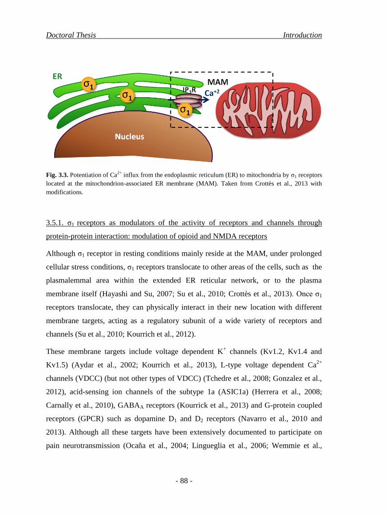
Doctoral Thesis Introduction
- 88 -
Fig. 3.3. Potentiation of Ca2+
influx from the endoplasmic reticulum (ER) to mitochondria by σ1 receptors
located at the mitochondrion-associated ER membrane (MAM). Taken from Crottès et al., 2013 with
modifications.
3.5.1. σ1 receptors as modulators of the activity of receptors and channels through
protein-protein interaction: modulation of opioid and NMDA receptors
Although σ1 receptor in resting conditions mainly reside at the MAM, under prolonged
cellular stress conditions, σ1 receptors translocate to other areas of the cells, such as the
plasmalemmal area within the extended ER reticular network, or to the plasma
membrane itself (Hayashi and Su, 2007; Su et al., 2010; Crottès et al., 2013). Once σ1
receptors translocate, they can physically interact in their new location with different
membrane targets, acting as a regulatory subunit of a wide variety of receptors and
channels (Su et al., 2010; Kourrich et al., 2012).
These membrane targets include voltage dependent K+ channels (Kv1.2, Kv1.4 and
Kv1.5) (Aydar et al., 2002; Kourrich et al., 2013), L-type voltage dependent Ca2+
channels (VDCC) (but not other types of VDCC) (Tchedre et al., 2008; Gonzalez et al.,
2012), acid-sensing ion channels of the subtype 1a (ASIC1a) (Herrera et al., 2008;
Carnally et al., 2010), GABAA receptors (Kourrick et al., 2013) and G-protein coupled
receptors (GPCR) such as dopamine D1 and D2 receptors (Navarro et al., 2010 and
2013). Although all these targets have been extensively documented to participate on
pain neurotransmission (Ocaña et al., 2004; Lingueglia et al., 2006; Wemmie et al.,

Introduction Doctoral Thesis
- 89 -
2006; Jaggi and Sing, 2011; Pradhan et al., 2012; Youn et al., 2013; Liu et al., 2014),
two additional receptors targeted by the chaperoning activity of σ1 receptors have been
strongly linked to the effects of σ1 receptors on animal models of pain: opioid receptors
(Kim et al., 2010) and NMDA receptors (Balasuriya et al., 2014; Sánchez-Blázquez et
al., 2014) (Fig. 3.4). As it will be extensively described in the next sections, σ1 receptors
have been proposed to impact on acute pain when opioid drugs are administered, and on
tonic/chronic pain through the modulation of NMDA receptors.
The direct interaction between σ1 and µ-opioid receptors has been reported recently
(Kim et al., 2010). These receptors can be co-immunoprecipitated and it has been shown
that σ1 receptors negatively modulate µ-opioid signalling, since σ1 antagonism is able to
increase GTPγS in response to the µ-opioid agonist DAMGO (Kim et al., 2010).
Although a direct coupling between σ1 and other opioid receptor subtypes has not been
demonstrated yet, it is likely that this occurs since σ1 inhibition is widely reported to
modulate κ- and δ-opioid mediated effects or signalling (e.g. Chien and Pasternak,
1994; King et al., 1997; Pan et al., 1998; Mei and Pasternak, 2002; Kim et al., 2010). It
is worth pointing out that in addition to opioid receptors, several protein targets of σ1
receptors are known to participate in opioid effects. For instance, L-type Ca2+
channels
are among the downstream effectors of opioid signalling (Al-Hasani and Bruchas,
2011), and opioid antinociception is known to be modulated by NMDA receptors
(reviewed by Pasternak and Pan, 2013). Therefore, the behavioral impact on the
modulation of opioid effects by σ1 receptors, which will be described in the next
section, might be the result from simultaneous complex interactions between several
membrane targets of the σ1 receptors and not exclusively from the direct modulation of
opioid receptors.
The modulation of NMDA receptors appears to be more complex. It has been reported
that σ1 receptors can modulate small conductance calcium-activated K+
(SK) channels,
which can be inhibited by σ1 agonism (Martina et al., 2007). The activity of SK
channels is known to inhibit NMDA receptors, and therefore σ1 receptor activation can
enhance the activity of NMDA receptors through this indirect mechanism (Martina et
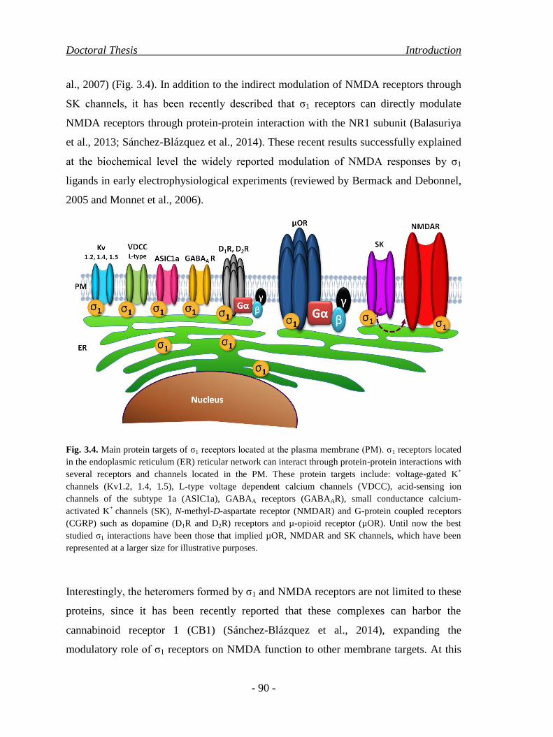
Doctoral Thesis Introduction
- 90 -
al., 2007) (Fig. 3.4). In addition to the indirect modulation of NMDA receptors through
SK channels, it has been recently described that σ1 receptors can directly modulate
NMDA receptors through protein-protein interaction with the NR1 subunit (Balasuriya
et al., 2013; Sánchez-Blázquez et al., 2014). These recent results successfully explained
at the biochemical level the widely reported modulation of NMDA responses by σ1
ligands in early electrophysiological experiments (reviewed by Bermack and Debonnel,
2005 and Monnet et al., 2006).
Fig. 3.4. Main protein targets of σ1 receptors located at the plasma membrane (PM). σ1 receptors located
in the endoplasmic reticulum (ER) reticular network can interact through protein-protein interactions with
several receptors and channels located in the PM. These protein targets include: voltage-gated K+
channels (Kv1.2, 1.4, 1.5), L-type voltage dependent calcium channels (VDCC), acid-sensing ion
channels of the subtype 1a (ASIC1a), GABAA receptors (GABAAR), small conductance calcium-
activated K+
channels (SK), N-methyl-D-aspartate receptor (NMDAR) and G-protein coupled receptors
(CGRP) such as dopamine (D1R and D2R) receptors and µ-opioid receptor (µOR). Until now the best
studied σ1 interactions have been those that implied µOR, NMDAR and SK channels, which have been
represented at a larger size for illustrative purposes.
Interestingly, the heteromers formed by σ1 and NMDA receptors are not limited to these
proteins, since it has been recently reported that these complexes can harbor the
cannabinoid receptor 1 (CB1) (Sánchez-Blázquez et al., 2014), expanding the
modulatory role of σ1 receptors on NMDA function to other membrane targets. At this

Introduction Doctoral Thesis
- 91 -
time it is unknown if these complexes can be formed with other proteins important for
pain processing. The discovery of these complexes, together with the variety of
receptors and channels involved in neurotransmission which are targeted by σ1
receptors, point to that the precise mechanisms by which σ1 receptors modulate pain
transmission are still at this time only very partially unveiled.
Rencently it has been reported that σ1 receptors besides forming heteromers such as
those described above, these receptors are also able to form homodimers and/or
homomultimers. However, their physiological significance is not clear at the moment
(Chu et al., 2013).
3.6. Evidence for the role of σ1 receptors on animal models of pain
The involvement of σ1 receptors on pain has been studied from several perspectives.
Initially, and probably because of their early confusion with opioid receptors, the
interaction between opioid antinociception and the effects of σ1 drugs was explored in
models of acute pain. The spectrum of pain models in which the role of σ1 receptors was
explored has been more recently greatly expanded to cover several types of pain from
different aetiologies. In this section we will summarize the most important findings on
the modulatory role of σ1 receptors on preclinical pain models either in combination
with opioid agonists or alone.
3.6.1. Modulation of opioid effects by σ1 receptors
In contradistinction to opioid drugs, which can relieve acute pain from either thermal or
mechanical stimuli (e.g. Ocaña et al., 2004; Pasternak and Pan, 2013), this is not the
case of σ1 drugs (e.g. Chien and Pasternak, 1994; Cendán et al., 2005b; Entrena et al.,
2009; Sánchez-Fernández et al., 2013; Tejada et al., 2014). However, σ1 receptors have
been thoroughly shown to modulate opioid-induced antinociception to acute thermal
stimuli (tail-flick test), particularly at central levels. We recently expanded these results
by testing the modulation of opioid antinociception in peripheral tissue and to
Doctoral Thesis Introduction
- 92 -
mechanical stimuli (paw pressure test), as part of this Doctoral Thesis. In addition to
opioid antinociception, the possible role on nonanalgesic effects of opioids has been
also explored (partially reviewed by Zamanillo et al., 2013). These findings will be
summarized in the present section.
3.6.1.1. Modulation by σ1 receptors of opioid-induced thermal antinociception
Although most experiments on the modulation of opioid antinociception by the σ1
system have been performed using µ agonists (mainly morphine), there is also a broad
body of evidence showing modulation of κ- and δ- opioid antinociception by σ1
receptors. These experiments and their most relevant conclusions are summarized
below.
3.6.1.1.1. Modulation by σ1 receptors of µ-opioid-induced thermal antinociception
Pasternak‟s research group was the first to report the modulation of opioid
antinociception by σ1 drugs, much earlier than the recent discovery of the physical
association between σ1 and opioid receptors. They reported that the systemic
administration of the selective σ1 agonist (+)-pentazocine and the non-selective σ1 drug
1,3-di(2-tolyl)guanidine (DTG) diminished the antinociceptive effect induced by the
systemic administration of the prototypical µ-opioid agonist morphine (Chien and
Pasternak, 1993 and 1994). On the other hand, haloperidol, a non-selective σ1
antagonist, greatly enhanced morphine antinociception (Chien and Pasternak, 1993,
1994 and 1995a), which was abolished by the selective µ receptor antagonist β-
funaltrexamine indicating that the activation of µ-opioid receptors was participating in
the enhanced antinociception (Chien and Pasternak, 1994).
Although it is now widely assumed that haloperidol acts as a potent σ1 antagonist
(Cobos et al., 2008; Hayashi and Su, 2004), this drug binds to several other receptors
such as dopamine receptors (mainly D2 receptors) (Jaen et al., 1993), σ2 receptors (e.g.
Bowen et al., 1990; Moison et al., 2003) and NMDA receptors (e.g. Shim et al., 1999).

Introduction Doctoral Thesis
- 93 -
Among all pharmacological targets of haloperidol, the main targets of this neuroleptic
are classically thought to be dopamine receptors, and in fact this drug is used in
therapeutics as an antipsychotic (Donnelly et al., 2013). Interestingly, (-)-sulpiride, a
known D2 antagonist devoid of affinity for σ1 sites (Bowen et al., 1990; Cobos et al.,
2007), had no effect on morphine antinociception (Chien and Pasternak, 1993 and
1994), and both (+)-pentazocine and haloperidol were still able to modulate the
antinociception induced by morphine in D2 knockout mice (King et al., 2001),
conclusively discarding the possible involvement of dopaminergic antagonism on the
enhanced morphine antinociception induced by haloperidol. These experiments greatly
contributed to the establishment of haloperidol as a prototypical σ1 antagonist which
although it is far from the desired selectivity on σ1 receptors is still widely used in basic
research on σ1 receptors (Cendán et al., 2005b; Entrena et al., 2009b; Caplan, 2012,
Delaunois et al., 2013). In addition, were the first evidences of the presence for a
tonically active anti-opioid σ system, and led to subsequent deeper studies on the
modulatory role of σ1 receptors on opioid antinociception.
More recently, experiments using the systemic administration of other more selective σ1
antagonists replicated the increase induced by haloperidol on the thermal
antinociception produced by systemic morphine. Morphine thermal antinociception was
enhanced by a spipethiane derivative (named as compound 9 in the original description
of its effects, in Piergentili et al., 2010), and very recently the highly selective σ1
antagonist S1RA (Vidal-Torres et al., 2014). Importantly, morphine is not the only µ
agonist used in clinics shown to be modulated by σ1 antagonism, since S1RA also
increases the antinociceptive effect to thermal stimuli induced by the systemic
administration of fentanyl, oxycodone, codeine, buprenorphine and tramadol (Vidal-
Torres et al., 2013). Therefore, the modulation of morphine thermal antinoception is not
restricted to this µ-opioid drug but is extensive to other relevant µ agonists.
The results of the studies testing the combination of systemic treatments with µ-opioids
and σ1 drugs on acute heat nociception (tail-flick test) are summarized in table 3.1.

Doctoral Thesis Introduction
- 94 -
Table 3.1. Summary of the effects on acute heat nociception (tail-flick test) of the combination of
systemic treatments with µ-opioids and σ1 drugs in WT mice.
Opioid
receptor
subtype
Opioid drug
σ1 receptor
agonist/antagonis
σ1 drug Effect on opioid
antinociception References
µ
Morphine
Agonist
(+)-Pentazocine
Inhibition
Chien and Pasternak,
1993 and 1994
DTG Chien and Pasternak,
1994
Antagonist
Haloperidol
Enhancement
Chien and Pasternak,
1993, 1994 and 1995a
Unnamed
spipethiane
derivative
Piergentili et al., 2010
S1RA Vidal-Torres et al., 2013
Fentanyl
Antagonist S1RA Enhancement Vidal-Torres et al., 2013
Oxycodone
Codeíne
Buprenorphine
Tramadol
Systemic treatments were performed via intraperitoneal (i.p.) administration in the study by Vidal-Torres
et al., 2013, and subcutaneously in the rest of studies.
In agreement with the lack of effect of σ1 drugs on acute thermal nociception (in the
absence of opioid drugs), σ1 receptor knockout (σ1-KO) mice do not exhibit any deficit
in the responses to thermal stimuli (De la Puente et al., 2009; Vidal-Torres et al., 2013;
Tejada et al., 2014). However, in contradistinction to the enhancement of µ-opioid
induced thermal antinociception by the pharmacological inhibition of σ1 receptors, σ1-
KO mice do not exhibit any alteration in the effect induced by the systemic
administration of morphine, sufentanyl, fentanyl or buprenorphine on thermal
nociception (Vidal-Torres et al., 2013) (Table 3.2.). Although these results are
apparently contradictory, might be explained by the development of compensatory
mechanisms in the mutant mice (Vidal-Torres et al., 2013). Conflicting results between
the pharmacological and genetic inhibition of σ1 receptors has been reported in other
pain models (as it will be described in the Section 3.6.2, and also in previous pain
studies of targets other than σ1 receptors (e.g., Petrus et al., 2007; Bonin et al., 2011), or
in fields different than pain research (e.g., Guscott et al., 2005; Voss et al., 2010).
Introduction Doctoral Thesis
- 95 -
Therefore, the development of compensatory mechanisms is not a peculiarity of σ1-KO
mice or pain research with mutant animals but extensive to other targets and to other
fields of study. Regardless of the nature of these compensatory mechanisms, σ1-KO
mice can be used to test the specificity of drugs, since their pharmacological target is
absent and it would be expected that a drug targeting the absent receptor will be devoid
of effect. In this context, the σ1 antagonist S1RA was unable to increase µ-opioid
induced thermal antinociception in σ1-KO mice (Vidal-Torres et al., 2013), supporting
on-target mechanisms for the effect of this drug.
The results of the studies testing the effects of µ opioids on σ1–KO mice on acute heat
nociception are summarized in table 3.2.
Further studies were conducted to determine the anatomical location of the modulation
of µ-opioid thermal antinociception by σ1 receptors. The antinociception induced by the
intracerebroventricular (i.c.v.) administration of morphine was decreased by the σ1
agonist (+)-pentazocine either systemically (Chien and Pasternak, 1994) or i.c.v.
administered (Mei and Pasternak, 2002). Conversely, the antinociceptive effect induced
by systemic morphine could be decreased by i.c.v. treatment with (+)-pentazocine (Mei
and Pasternak, 2002). These results indicate that the inhibitory effects on morphine
antinociception induced by σ1 agonism could be produced at supraspinal levels. The
presence of the tonically active anti-opioid σ1 system at supraspinal locations was
determined by several approaches. The antinociceptive effect elicited by the supraspinal
(i.c.v.) administration of DAMGO was enhanced by the systemic administration of the
σ1 antagonist (+)-MR200 (Marrazzo et al., 2006), and the antinociceptive effect of
supraspinal morphine was enhanced by the central administration of specific antisense
oligodeoxynucleotides (ASOs) to inhibit the expression of σ1 receptors (Pan et al., 1998;
Mei and Pasternak, 2002). Interestingly, the antinociceptive effect induced by spinal
morphine was not affected by either i.c.v. or i.t. treatment with the σ1 agonist (Mei and
Pasternak, 2002), although it could be decreased by systemic (+)-pentazocine (Chien
and Pasternak, 1994), which might indicate that the activation of both spinal and
supraspinal σ1 sites might be necessary to functionally impact on spinal morphine

Doctoral Thesis Introduction
- 96 -
antinociception (Mei and Pasternak, 2002), and that although σ1 receptors are expressed
at either supraspinal and spinal sites (Alonso et al., 2000; Mei and Pasternak, 2002;
Sánchez-Fernández et al., 2014), the modulation of opioid antinociception by spinal σ1
receptors is less promiment than that produced by supraspinal σ1 receptors.
Table 3.2. Summary of the effects on acute heat (tail-flick test) and mechanical (paw pressure)
nociception of µ opioids in σ1-KO mice.
Effect of σ1 receptor knockout on opioid antinociception
Opioid drug Route Thermal
Mechanical without S1RA with S1RA (s.c.)
Morphine s.c. No effects
abolishment of the potentiation
of µ-opioid antinociception
induced by S1RA
Enhancement i.pl n.t. n.t.
Fentanyl
s.c.
No effects
abolishment of the potentiation
of µ-opioid antinociception
induced by S1RA
Oxycodone
Buprenorphine
Tramadol n.t. n.t.
Loperamide
Abbreviations: s.c., subcutaneous; i.pl., intraplantar; n.t., not tested.
Codeine and sufentanyl were evaluated in the tail-flick test σ1-KO mice, with similar effects than the
other opioids tested, but these results have been omitted in this table for clarity. Results on thermal
nociception can be found in Vidal-Torres et al., 2013, and results on mechanical nociception can be found
in Sánchez-Fernández et al., 2013 and 2014.
The specific supraspinal sites involved in the modulation of morphine antinociception
by σ1 receptors were determined by subsequent experiments. The rostroventral medulla
(RVM), the periaqueductal gray (PAG) and the locus coeruleus (LC) constitute
important sites for the circuit responsible of the descending modulation of pain (Fields,
2000), and µ-opioids strongly act in these nuclei to elicit analgesia (Porreca et al.,
2002). Although σ1 receptor is expressed in these three areas (Alonso et al., 2000;
Sánchez-Fernández et al., 2014), the modulation of morphine antinociception differed
depending on the injection site (Mei and Pasternak, 2007). The microinjection of

Introduction Doctoral Thesis
- 97 -
morphine into any of these three regions elicited antinociception, which was decreased
by the co-administration of (+)-pentazocine in the same region (although the PAG was
much less sensitive than the other two sites). Blockade of the σ1 receptors with
haloperidol in the RVM markedly enhanced the antinociceptive actions of
coadministered morphine, implying a strong tonic activity of the σ1 system in this site.
This effect was mimicked by down-regulation of RVM σ1 receptors using specific
σ1-ASOs. However, no tonic σ1 activity was observed in either the LC or the PAG
(evidenced by the lack of response to haloperidol coadministered with morphine in
these regions). Highlighting the marked role of σ1 receptors in the RVM, the
pharmacological agonism of σ1 receptors in this site was able to modulate the
antinociception elicited from morphine microinjected into the PAG, whereas
haloperidol in the RVM enhanced PAG morphine analgesia (Mei and Pasternak, 2007).
These results illustrate the importance on morphine antinociception of brainstem σ1
receptors, and in particular of those located in the RVM.
The results of the studies testing the effects on acute heat nociception of the
combination of µ-opioids and σ1 drugs or σ1-ASOs, administered at specific central
nervous system sites, are summarized in table 3.3.

Table 3.3. Summary of the effects on acute heat nociception (tail-flick test) of the combination of µ-opioids and σ1 drugs or antisense
oligodeoxynucleotides (ASOs), administered at specific central nervous system sites.
Opioid receptor
subtype Opioid drug Route
σ1 receptor
agonist/antagonist σ1 drug or σ1-ASO Route
Effect on opioid
antinociception References
µ Morphine
s.c. Agonist (+)-Pentazocine i.c.v Inhibition Mei and Pasternak, 2002
i.c.v.
Agonist (+)-Pentazocine s.c.
Inhibition Chien and Pasternak, 1994
i.c.v Mei and Pasternak, 2002
- ASO i.c.v Enhancement Pan et al., 1998; Mei and
Pasternak, 2002
PAG
Agonist (+)-Pentazocine
PAG
Inhibition Mei and Pasternak, 2007 RVM
LC LC
RVM RVM
RVM
Antagonist Haloperidol
RVM Enhancement
Mei and Pasternak, 2007
PAG PAG No effect
RVM Enhancement
LC LC No effect
RVM - ASO RVM Enhancement
PAG
i.t. Agonist (+)-Pentazocine
s.c. Inhibition Chien and Pasternak, 1994
i.c.v . No effect
Mei and Pasternak, 2002
i.t. Mei and Pasternak, 2002
DAMGO i.c.v Antagonist (+)-MR200 s.c. Enhancement Marrazzo et al., 2006
Abbreviations: s.c., subcutaneous; i.c.v., intracerebroventricular; i.t., intrathecal; RVM, rostroventral medulla; PAG, periaqueductal gray matter; LC, locus
coeruleus; ASO, antisense oligodeoxynucleotide.
- 98 -

Introduction Doctoral Thesis
- 99 -
3.6.1.1.2. Modulation by σ1 receptors of κ-and δ-opioid-induced thermal antinociception
In addition to the modulation of µ-opioid antinociception by σ1 receptors, the effects of
the systemic administration of σ1 drugs on the thermal antinociception induced by the
systemic administration of agonists of other opioid receptors was tested. Similar to the
results found with the modulation of µ-opioid antinociception by σ1 drugs, systemic σ1
agonism (by the prototypic σ1 agonist (+)-pentazocine and the purported σ1 agonist (±)-
PPCC) inhibited the antinociception induced by the systemic administration of the κ1-
opioid agonist U50,488H (Chien and Pasternak, 1994; Ronsisvalle et al., 2001;
Prezzavento et al., 2008). On the other hand, systemic administration of the σ1
antagonists haloperidol, MR200 and (-)-MRV3 were able to enhance U50,488H-
induced antinociception (Chien and Pasternak, 1994 and 1995a; Ronsisvalle et al.,
2001; Marrazzo et al., 2011). However, the σ1 antagonist BD-1047 unexpectedly
induced a decrease of the antinociception induced by U50,488H (Prezzavento et al.,
2008). Although BD-1047 is widely used as a σ1 antagonist (e.g. Hayashi and Su, 2004;
Cobos et al., 2008), its selectivity has been tested in a panel of only 10 receptors
(Matsumoto et al., 1995). Therefore, a possible explanation of these discordant effects
induced by BD-1047 might be that this drug could be interacting to unknown target/s
interfering with opioid antinociception.
In addition, the σ1 antagonist haloperidol was also able to increase the antinociception
induced by the κ3-opioid agonist naloxone benzoylhydrazone (NalBzoH) (Chien and
Pasternak, 1994 and 1995a) and that induced by the nonselective κ-opioid analgesic
(-)-pentazocine (Chien and Pasternak, 1995b). (-)-Pentazocine is a peculiar drug
because it has high affinity for several other receptors including σ1 receptors (although
with a considerably lower affinity than its dextrogyre isomer) behaving as a σ1 agonist
(Chien and Pasternak., 1995b). Due to this agonistic activity over σ1 receptors, high
doses of (-)-pentazocine are thought to be able to inhibit its own opioid-induced
antinociception, which results in animals in a biphasic dose-response curve (Chien and
Pasternak, 1995b). The interest of this observation is that pentazocine (as a racemic
mixture) is used in humans patients as an opioid analgesic (Adeniji and Atanda, 2013;

Doctoral Thesis Introduction
- 100 -
King et al., 2013), and both (+)-pentazocine and (-)-pentazocine through σ1 agonism
could be interfering in the opioid analgesia induced by (-)-pentazocine. However, the
relevance in the clinical practice of the interaction of pentazocine with σ1 receptors is
still unknown.
The results of the studies testing the combination of systemic treatments with κ opioids
and σ1 drugs on acute heat nociception (tail-flick test) are summarized in Table 3.4.
Table 3.4. Summary of the effects on acute heat nociception (tail-flick test) of the combination of
systemic treatments with κ opioids and σ1 drugs.
Opioid
receptor
subtype
Opioid drug σ1 receptor
agonist/antagonist σ1 drug
Effect on opioid
antinociception References
κ
U50,488H
Agonist (+)-Pentazocine
Inhibition
Chien and Pasternak,
1994; Ronsisvalle et al.,
2001 and Prezzavento et
al., 2008
(±)-PPCC Prezzavento et al., 2008
Antagonist
Haloperidol
Enhancement
Chien and Pasternak,
1994 and 1995a
MR200 Ronsisvalle et al., 2001
(-)-MRV3 Marrazzo et al., 2011
BD-1047 Inhibition Prezzavento et al., 2008
NalBzoH Antagonist Haloperidol Enhancement Chien and Pasternak,
1994 and 1995a
(-)-Pentazocine Antagonist Haloperidol Enhancement Chien and Pasternak,
1995b
All systemic treatments were performed by the subcutaneous administration of the σ1 drugs.
As for the modulation of µ-opioid antinociception by σ1 receptors, it has been reported
that supraspinal σ1 receptors play a pivotal role on the antinociceptive effects induced
not only by the κ agonists U50,488H and NalBzoH, but also by the δ agonist DPDPE. It
was shown that the supraspinal administration of (+)-pentazocine inhibited the
antinociception induced by U50,488H and NalBzoH systemically administered, and that
induced by the i.c.v. administration of DPDPE (Mei and Pasternak, 2002), while σ1
antisense treatments increased the antinociceptive effects of all opioid drugs tested
Introduction Doctoral Thesis
- 101 -
(King et al., 1997; Pan et al., 1998; Mei and Pasternak, 2002), demonstrating the
presence of the tonic activation of the σ1 system inhibiting κ antinociception. In
agreement with the tonic supraspinal inhibitory control of κ and δ antinociception by σ1
receptors, it was found that the systemic administration of the σ1 antagonist (+)-MR200
was able to enhance the antinociception induced by the supraspinal administration of
either U50,488H or DPDPE (Marrazzo et al., 2006). Interestingly, the systemic
administration of haloperidol was able to enhance the antinociceptive effect induced by
spinal DPDPE (Chien and Pasternak, 1994), although it is unclear if the modulatory
effect of haloperidol was produced at spinal or supraspinal levels (or both).
The results of the studies testing the effects on acute heat nociception of the
combination of κ or δ opioids and σ1 drugs or σ1-ASOs, administered at specific central
nervous system sites, are summarized in table 3.5.
Taking into account all these studies together, it seems clear that in addition to the
modulation by central σ1 receptors (particularly supraspinally) of the thermal
antinociception by µ agonists, they also strongly modulate κ- and δ-mediated
antinociception.
.

Table 3.5. Summary of the effects on acute heat nociception (tail-flick test) of the combination of κ- or δ- opioids and σ1 drugs or antisense
oligodeoxynucleotides (ASOs), administered at specific central nervous system sites.
Opioid
receptor
subtype
Opioid
drug Route
σ1 receptor
agonist/antagonist σ1 drug or σ1-ASO Route
Effect on opioid
antinociception References
κ
U50,488H s.c.
Agonist (+)-Pentazocine i.c.v. Inhibition Mei and Pasternak, 2002
- ASO i.c.v. Enhancement King et al., 1997; Pan et al., 1998
i.c.v. Antagonist (+)-MR200 s.c. Enhancement Marrazzo et al., 2006
NalBzoH s.c.
Agonist (+)-Pentazocine i.c.v Inhibition Mei and Pasternak, 2002
- ASO i.c.v Enhancement Mei and Pasternak, 2002; Pan et al., 1998;
King et al., 1997
δ DPDPE
i.c.v. Agonist (+)-Pentazocine
i.c.v Inhibition Mei and Pasternak, 2002
- ASO Enhancement Mei and Pasternak, 2002; Pan et al.,1998
i.c.v. Antagonist
(+)-MR200 s.c. Enhancement
Marrazzo et al., 2006
i.t. Haloperidol Chien and Pasternak, 1994
Abbreviations: s.c., subcutaneous; i.c.v., intracerebroventricular; i.t., intrathecal; ASO, antisense oligodeoxynucleotide; NalBzoH, Naloxone
benzoylhydrazone; DPDPE, [D-Pen2, D-Pen
5]enkephalin.
- 102 -

Introduction Doctoral Thesis
- 103 -
3.6.1.2. Modulation by σ1 receptors of µ-opioid-induced mechanical antinociception
Bearing in mind that the neurochemical mechanisms underlying opioid-induced
mechanical and thermal antinociception differ (Kuraishi et al., 1985; Sato et al., 1999;
Tseng et al., 1995; Wegert et al., 1997), the previously described modulation of opioid-
induced thermal antinociception by σ1 receptors was not necessarily expected to be
applicable to mechanical stimulation. In addition, although the role of central σ1
receptors on opioid antinociception has been explored in the past (as thoroughly
described in the sections above), the possible role of peripheral σ1 receptors on opioid
antinociception was absolutely unknown. Therefore, the main goal of this Doctoral
Thesis was to explore the role of σ1 receptors on opioid mechanical antinociception, and
specifically focusing on the role of peripheral σ1 receptors in the modulation of the
antinociception. Since the most clinically relevant opioid drugs are agonists of the µ
subtype (Kieffer, 1999; Pasternak and Pan, 2011; Pasternak and Pan, 2013), we tested a
variety of µ opioids in clinical use
We found that σ1–KO mice showed an enhanced mechanical antinociception induced by
morphine, fentanyl, oxycodone, buprenorphine and tramadol (Sánchez-Fernández et al.,
2013 and 2014). These data are in contrast with the absence of modulation of opioid
thermal antinociception in these mutant mice purportedly by compensatory mechanisms
(Vidal-Torres et al., 2013), as described in the Section 3.6.1.1.1 and in Table 3.2., and
may be attributable to the known differences in the neurochemical mechanisms of
thermal and mechanical opioid antinociception mentioned above, which may be
affected differentially by possible compensatory mechanisms in σ1-KO mice. Systemic
σ1 pharmacological σ1 blockade replicated the increase in µ-opioid antinociception seen
in the σ1-KO for all opioids tested (Sánchez-Fernández et al., 2013 and 2014).
Interestingly, this σ1 inhibition (by either σ1 knockout or systemic σ1 pharmacological
antagonism) was sufficient to unmask strong antinociceptive effects by loperamide
(Sánchez-Fernández et al., 2014), a peripherally restricted µ-opioid agonist used in
therapeutics as an antidiarrheal drug (Layer et al., 2010; Gallelli et al., 2010), and the
intraplantar (i.pl.) administration of σ1 antagonists was able to increase the

Doctoral Thesis Introduction
- 104 -
antinociception to mechanical stimuli induced by the systemic administration of all µ
opioids tested (Sánchez-Fernández et al., 2014). Reinforcing the peripheral mediation of
the effects observed, the enhanced antinociception by σ1 inhibition was very sensitive to
peripheral opioidantagonism (i.e. to the peripherally restricted opioid antagonist
naloxone methiodide) (Sánchez-Fernández et al., 2014). In addition, and adding
evidence to the peripheral antinociceptive synergism between σ1 inhibition and opioid
agonism, we found that the intraplantar administration of morphine showed a marked
enhanced antinociception in σ1-KO mice and in wild-type mice coadministered with
several σ1 antagonists in the morphine-injected paw (Sánchez-Fernández et al., 2013).
These results clearly show that the modulation of µ-opioid antinociception by σ1
receptors is not restricted to either thermal stimuli or to central sites, and constituted the
first reported evidence of a role of peripheral σ1 receptors on pain.
The results obtained on the modulatory role of σ1 receptors of µ-opioid induced
mechanical antinociception are summarized in Tables 3.2. and 3.6. (for the experiments
performed in σ1-KO mice and using σ1 drugs, respectively), and will be exhaustively
presented in the Chapter "Published Papers” of this Doctoral Thesis.
Further research will be needed to test whether the modulation of peripheral opioid
antinociception by σ1 receptors is restricted to µ agonists or if it is extensive to other
types of opioids, and to test whether this peripheral modulation of opioid mechanical
antinociception is extensive to pain induced by thermal stimuli. Although there are
undoubtedly still some gaps on this field of research, the discovery of the modulatory
role of peripheral σ1 receptors on the effects of opioids on acute pain planted the seed
for subsequent studies on the modulation of other types of pain by peripheral σ1
receptors, described in the section below.

Introduction Doctoral Thesis
- 105 -
Table 3.6. Summary of the effects on acute mechanical nociception (paw pressure test) of the
combination of opioids and σ1 drugs
Opioid drug Route σ1 receptor
agonist/antagonist σ1 drug Route
Effect on opioid
antinociception References
Morphine
s.c.
Antagonist
BD-1063
s.c. Enhancement Sánchez-Fernández et al.,
2013 and 2014
i.pl. Enhancement Sánchez-Fernández et al.,
2014
BD-1047 s.c. Enhancement
Sánchez-Fernández et al.,
2013 NE-100
S1RA s.c. Enhancement Sánchez-Fernández et al.,
2013 and 2014
Agonist PRE-084 s.c. No effect Sánchez-Fernández et al.,
2013
i.pl. Antagonist
BD-1063
i.pl. Enhancement Sánchez-Fernández et al.,
2013
BD-1047
NE-100
S1RA
Fentanyl s.c. Antagonist
BD-1063 s.c.
Enhancement Sánchez-Fernández et al.,
2014
i.pl.
S1RA s.c.
i.pl.
Oxycodone s.c. Antagonist BD-1063 s.c.
Enhancement Sánchez-Fernández et al.,
2014 i.pl.
Buprenorphine s.c. Antagonist BD-1063 s.c.
Enhancement Sánchez-Fernández et al.,
2014 i.pl.
Tramadol s.c. Antagonist BD-1063 s.c.
Enhancement Sánchez-Fernández et al.,
2014 i.pl.
Loperamide s.c. Antagonist
BD-1063
s.c.
Enhancement Sánchez-Fernández et al.,
2014
i.pl.
S1RA
s.c.
i.pl.
Abbreviations: s.c., subcutaneous; i.pl., intraplantar
Doctoral Thesis Introduction
- 106 -
3.6.1.3. Modulation by σ1 receptors of non-analgesic (adverse) effects of opioids
As it has been described in the Chapter “µ-Opioid drugs: mechanism of action,
therapeutic use and side effects” although opioid drugs are widely used in therapeutics
for pain management, their well-known side effects strongly limit their use (Benyamin
et al., 2008; Al-Hasani and Bruchas, 2011; Ringkamp and Raja, 2012).
Before the experiments presented in this Doctoral Thesis were conducted very few
studies existed about the role of σ1 receptor in non-analgesic opioid effects. The only
previously published study was performed by Chien and Pasternak, who reported that
unlike the decrease on the antinociceptive effects of opioids induced by σ1 agonism, the
inhibition of gastrointestinal transit or lethality induced by morphine were not affected
by the administration of (+)-pentazocine (Chien and Pasternak, 1994). However, the
effect of σ1 inhibition in non-analgesic opioid effects remained untested. The
importance of the study of the modulation by σ1 antagonists of non-analgesic effects of
opioids is that if they would equally increase analgesic and non-analgesic opioid effects,
it would decrease the interest for the possible future clinical use of σ1 antagonists as
opioid adjuvants to enhance opioid-induced analgesia.
At present, the possible modulation by σ1 inhibition on a wide variety of non-analgesic
opioid effects has been explored in rodents. These include hyperlocomotion, mydriasis,
withdrawal, tolerance, dependence and gastrointestinal transit inhibition. Changes in pupil
size are a well-known centrally-induced non-analgesic effect of opioids. Although in
humans opioid drugs induce myosis (Al-Hasani and Bruchas, 2011), in mice they induce
mydriasis (Stav et al., 1992; Vidal-Torres et al., 2013). S1RA administration did not modify
the pupillary diameter when administered alone, and did not modify morphine-induced
mydriasis in rodents (Vidal-Torres et al., 2013). Other paradoxically and centrally-induced
non-analgesic opioid effect is the sedation which is often produced in humans (Al-Hasani
and Bruchas, 2011) while increasing the locomotor activity in mice (Hnasko et al., 2005).
We found that morphine-induced hyperlocomotion was not modified either by σ1
inhibition (σ1-KO) mice (Sánchez-Fernández et al., 2013; see Chapter "Published
Papers” for a detailed description of these findings). Furthermore, σ1 pharmacological

Introduction Doctoral Thesis
- 107 -
antagonism (by S1RA) did not change the severity of somatic manifestations of naloxone-
induced morphine withdrawal nor the development of morphine tolerance in mice (Vidal-
Torres et al., 2013), which are very relevant opioid side effects in humans (Al-Hasani and
Bruchas, 2011). σ1 antagonism was also effective in restoring morphine antinociception in
morphine-tolerant mice, and interestingly, the rewarding effects of morphine (evaluated by
place conditioning) were antagonized (Vidal-Torres et al., 2013). This later result although
in opposite direction to the widely reported enhanced opioid antinociception by σ1
antagonism, agree with additional preclinical studies which proposed the use of σ1
antagonists as promising tools for the treatment of addiction and dependence induced by
other drugs of abuse (e.g. Matsumoto, 2009).
As a part of this Doctoral Thesis, we reported that gastrointestinal transit inhibition
induced by morphine, fentanyl and loperamide, was unaltered by σ1 receptor inhibition
(Sánchez-Fernández et al., 2013 and 2014) (see Chapter "Published Papers”). The lack
of involvement of σ1 receptor in the inhibition of gastrointestinal transit has been
confirmed in a parallel study (Vidal-Torres et al., 2013). This opioid non-analgesic effect
has a high clinical relevance, since is one of the main reasons for patients‟ voluntary
withdrawal from opioid medication (Dhingra et al., 2013), and in contradistinction to the
other opioid side effects, it is produced mainly peripherally (Al-Hasani and Bruchas, 2011;
Ringkamp and Raja, 2012).
Therefore, σ1 receptors do not appear to modulate either centrally- or peripherally-induced
opioid non-analgesic effects. Although the mechanisms responsible of the differential
modulation of opiod antinociception and their non-analgesic effects by σ1 receptors are
unclear, it can be concluded that σ1 antagonism is a promising pharmacological tool for
differentially enhancing the analgesic effects of opioids while minimizing their side effects.
3.6.2. Pain modulation by σ1 receptors in the absence of opioid drugs
In addition to the described role of σ1 receptors on the modulation of opioid effects on
acute nociceptive pain induced by either thermal or mechanical stimuli, it has been
widely reported that σ1 inhibition might play a role in the absence of opioids to

Doctoral Thesis Introduction
- 108 -
ameliorate other types of pain, in particular those produced by chemical irritation or
sensitization of pain pathways.
3.6.2.1. σ1 receptors and pain induced by chemical irritants: formalin and capsaicin
Formalin is one of the chemical irritants most widely used in pain research (Le Bars et
al., 2001). Formalin injection induces a biphasic pain response in rodents: an initial
acute pain response (first phase) due to the direct activation of nociceptors, followed by
a prolonged tonic response (second phase) characterized by spontaneous activity of
primary afferent neurons together with functional changes in the spinal cord (central
sensitization) accompanied by an edematous process (Le Bars et al., 2001; Sawynok
and Liu, 2004). Although in pain studies formalin is classically administered in the paw,
it can be also injected into the trigeminal area (e.g. Chavelou et al., 1995) to study
orofacial pain, which is a distinct type of pain known to be resistant to analgesic
treatment (see Sarlani et al., 2005).
The first reported evidences on the role of σ1 receptors on a chemically-induced pain
model were performed in the formalin test. σ1-KO mice showed a significant decrease
of both nociceptive phases after intraplantar formalin (Cendán et al., 2005a). Moreover,
the systemic pharmacological antagonism of σ1 receptors, using either non-selective
(such as haloperidol) and selective drugs (such as S1RA), also decreased both phases of
formalin-induced pain when the chemical irritant was administered into the paw
(Cendán et al., 2005b; Romero et al., 2012; Díaz et al., 2013; Lan et al., 2014; Gómez-
Soler et al., 2014; Vidal-Torres et al., 2014) or into the trigeminal area (Roh and Yoon,
2014).
Since the involvement of central sensitization in the second phase of formalin-induced
pain is prominent, further studies focused on the role of spinal σ1 receptors on this pain
model. It was found that the i.t. administration of σ1 antagonists reduced formalin
induced pain (Kim et al., 2006; Vidal-Torres et al., 2014), and this was accompanied by
a reduction of the phosphorylation (activation) of the NR1 subunit of the NMDA at
either protein kinase C (PKC) and protein kinase A (PKA) sites (Kim et al., 2006),

Introduction Doctoral Thesis
- 109 -
which plays a pivotal role in central sensitization (Latremoliere and Woolf, 2009).
Although it is unknown whether σ1 receptors can directly modulate the activity of
intracellular kinases, taking into account that σ1 receptors can directly bind to the NR1
subunit of NMDA receptors (Balasuriya et al., 2013; Sánchez-Blázquez et al., 2014),
one tempting speculation to explain (at least partially) the decrease in pNR1 by σ1
antagonism could be that the accessibility of the phosphorylation sites of the NR1
would depend on the activation state of σ1 receptors, and that σ1 inhibition would lead to
a decrease in the accessibility to these sites by the intracellular kinases.
Recently it has been explored the role of σ1 receptors at other anatomical locations in
formalin-induced pain. It was found that supraspinal σ1 antagonism increased
noradrenaline levels in the spinal cord, suggesting the involvement of descending
inhibitory pathways in the mechanism of action of σ1 inhibition to decrease pain (Vidal-
Torres et al., 2014). Interestingly, it was recently reported that σ1 antagonism in the
place of the administration of formalin was also able to reduce pain behavior, indicating
that peripheral σ1 receptors also participate in the decrease of formalin-induced pain by
σ1 inhibition (Vidal-Torres et al., 2014). These results indicate that σ1 receptors
facilitate formalin-induced pain at central sites (both spinally and supraspinally) and
also peripherally.
Further experiments were performed using capsaicin as a chemical irritant. The
intradermal injection of capsaicin induces a decrease in the mechanical threshold of the
area surrounding the injection even if it was not stimulated by capsaicin (area of
secondary hypersensitivity), which is produced by central sensitization (Sang et al,
1996; Baron, 2000). The interest of exploring the effects of σ1 inhibition on capsaicin-
induced secondary mechanical hypersensitivity was that this model is widely used in
clinical research to test the effects of drugs on mechanical allodynia (e.g. Eisenach et
al., 2002; Gottrup et al., 2004) and it is considered to be a surrogate model of
neuropathic pain, since anti-neuropathic drugs show antiallodynic activity in this test in
both humans and rodents (Gottrup et al., 2004; Joshi et al., 2006). It has been shown
that σ1 knockout mice did not sensitize to mechanical stimuli in response to capsaicin

Doctoral Thesis Introduction
- 110 -
(Entrena et al., 2009a), and this phenotype was mimicked by σ1 antagonists, such as the
non-selective drug haloperidol or selective σ1 antagonists including BD-1063 or S1RA
(Entrena et al., 2009a and b; Romero et al., 2012; Díaz et al., 2013).
Importantly, the effects of σ1 inhibition on either capsaicin-induced secondary
mechanical hypersensitivity or formalin-induced pain were not reversed by the opioid
antagonist naloxone (Cendán et al., 2005b and Entrena et al., 2009b, respectively),
definitively indicating that these effects are independent to the modulation of the
opioidergic system, and that σ1 receptors can act through other mechanisms to decrease
pain transmission.
Both formalin and capsaicin are known C-fiber activators, although through distinct
receptors. While formalin activates TRPA1, capsaicin stimulates TRPV1 (Kerstein et
al., 2009; Dubin and Patapoutian, 2010). The repetitive activation of C-fibers is known
to increase the excitability of spinal cord neurons, a phenomenon named wind-up which
can contribute to the establishment of central sensitization (Herrero et al., 2000;
Latremoliere and Woolf, 2009). This process is known to be inhibited in σ1–KO mice
(de la Puente et al., 2009) or by S1RA in WT animals (Romero et al., 2012), which
support the role of σ1 receptors on the hyperexcitability of spinal neurons that contribute
to pain hypersensitivity after the injection of formalin or capsaicin.
The utilities of capsaicin administration as a pain model are not restricted to somatic
pain, since this chemical algogen can be also administered in the gut to produce visceral
pain. Visceral pain induced by capsaicin shows two distinct components: an intense
(acute) pain and refereed mechanical hyperalgesia, and these two components have been
reported in both humans (Drewes et al., 2003; Schmidt et al., 2004) and rodents (Laird
et al., 2001). The pathophysiological mechanisms and response to drug treatments are
different in somatic and visceral pain (Cerveró and Laird, 1999). In fact, in
contradistinction to somatic afferents, visceral afferents are known to be unable to
trigger wind-up in the spinal cord (reviewed by Herrero et al., 2000), hence the interest
of testing the effects of σ1 receptor inhibition on a visceral pain model. Systemic
administration of a variety of selective σ1 antagonists (BD-1063, S1RA and NE-100)
Introduction Doctoral Thesis
- 111 -
attenuated both acute pain-related behaviours and refereed mechanical hyperalgesia
induced by intracolonic capsaicin (González-Cano et al., 2013). Interestingly, σ1
knockout mimicked the effects of σ1 antagonists in the decrease of acute pain-like
behaviours induced by intracolonic capsaicin but did not show any amelioration in
refereed hyperalgesia. However, σ1 antagonists were devoid of effect in σ1-KO mice,
indicating that the effects of these drugs are specifically mediated by σ1 receptors, and
that the absence of phenotype in refereed hyperalgesia showed by σ1-KO mice was due
to the development of compensatory mechanisms (González-Cano et al., 2013).
There have not been yet performed studies to dissect the anatomical location of pain
modulation by σ1 receptors on nociceptive behaviors or sensory hypersensitivity
induced by capsaicin (either administered in somatic or visceral tissues), although
taking into account the accumulated evidence of the role of σ1 receptors on central
sensitization it is likely that central σ1 receptors play a role in the effects observed.
However, taking into account the mixed contribution of peripheral and central σ1
receptors to pain induced by other chemical irritants (formalin), the participation of
peripheral σ1 receptors to capsaicin-induced pain or sensitization cannot be discarded,
and further studies are needed to clarify this issue.
3.6.2.2. σ1 receptors and neuropathic pain
Neuropathic pain is one of the most challenging types of chronic pain conditions to
treat, and new therapeutic tools are strongly needed (Zhou et al., 2011; Attal, 2012). The
early results of the amelioration of formalin-induced pain and capsaicin-induced
secondary mechanical hypersensitivity by σ1 inhibition led to further research on the
role of σ1 receptors on neuropathic pain, since central sensitization is a key feature of
this important pathological state (Latremoliere and Woolf, 2009).
In experimental animals (and humans), peripheral neuropathic pain can be produced by
nerve trauma and by the administration of several agents with neurotoxic properties,
such as antineoplastics (reviewed by Campbell and Meyer, 2006; Cobos and Portillo-
Salido, 2013). It was reported that σ1-KO mice do not develop signs of either cold or

Doctoral Thesis Introduction
- 112 -
mechanical allodynia after traumatic nerve injury, although they showed a normal heat
hyperalgesia (De la Puente et al., 2009). However, systemic repeated administrations of
the selective σ1 antagonist S1RA, starting before the neuropathy was established
(immediately after the injury), avoided the development of not only neuropathic cold
and mechanical allodynia but also heat hyperalgesia (Romero et al., 2012). These
effects of σ1 antagonists disappeared after discontinuation of the treatment and their
repeated administration did not induce tolerance to the antihypersensitivity effects
(Romero et al., 2012). The efficacy in preventing neuropathic heat hyperalgesia by
S1RA and the normal development of this sensory alteration in injured σ1-KO mice
suggests again that compensatory mechanisms in pain pathways involved in heat
sensitivity might be developed in σ1-KO mice.
The ameliorative effects of S1RA on neuropathic hypersensitivity were not limited to its
preemptive administration, since systemic administration of this σ1 antagonist was able
to fully reverse sensory hypersensitivity once the neuropathy was fully established
(several days after the trauma) (Romero et al., 2012; Díaz et al., 2012; Bura et al.,
2012). Importantly, neuropathic rodents were shown to freely administer themselves
S1RA once neuropathy was established, to reverse not only mechanical and cold
allodynia and heat hyperalgesia, but also neuropathic anhedonia (measured as a
decreased preference to sweet solution) as an indicator of the emotional negative state
induced by the pain condition, which reflects both the self-assessment of the successful
efficacy of the treatment and the deep positive effects on the emotional state of the
rodents (Bura et al., 2012).
Similar to the results found after traumatic nerve injury, σ1-KO mice did not develop
sensory hypersensitivity (mechanical and cold) after the administration of the
antineoplastic drug paclitaxel (Nieto et al., 2012 and 2014), a first-line
chemotherapeutic agent for the treatment of several types of cancer which frequently
produces painful peripheral neuropathies as one of its major side-effects (Argyriou et
al., 2008). In addition, systemic treatment with σ1 antagonists (BD-1063 and S1RA) not
only abolished mechanical and cold allodynia once neuropathy was fully developed
Introduction Doctoral Thesis
- 113 -
(Nieto et al., 2012), but were able to fully prevent the hypersensitivity associated to the
neuropathy (which did not manifest again even after the discontinuation of the treatment
with the σ1 antagonists), suggesting that σ1 inhibition had a protective role for the
neuronal toxicity induced by the taxane (Nieto et al., 2012 and 2014).
Further studies were conducted to examine the role of spinal σ1 receptors on peripheral
neuropathic pain. It was shown that σ1 receptor protein was transiently up-regulated in
the dorsal spinal cord in the early days after the nerve injury (Roh et al., 2008b), and
that repeated i.t. administration of the σ1 antagonist BD-1047 prevented the full
development of neuropathic mechanical hypersensitivity (Roh et al., 2008b; Choi et al.,
2013), pointing to the importance of spinal σ1 receptors in the development of
neuropathic hypersensitivity. In fact, in neuropathic animals σ1 inhibition decreased the
phosphorylation of the NR1subunit of NMDA receptors in the spinal cord (Roh et al.,
2008b), the phosphorylation of different nitrogen-activated protein kinases, such as
extracellular signal-regulated kinase (ERK) 1/2 (De la Puente et al., 2009; Nieto et al.,
2012) and p38 (Moon et al., 2013), and the activity of NADPH oxidase 2 (Nox2) with
the subsequent reduction in the production of reactive oxygen species in neuropathic
animals (Choi et al., 2013). All these molecules are known to play a major role on
central sensitization (Latremoliere and Woolf, 2009). Conversely, intrathecal σ1
agonism (by the selective drugs PRE-084 or (+)-pentazocine) induces a neuropathic-like
phenotype in mice, resulting in sensory hypersensitivity (Roh et al., 2008a, 2010 and
2011; Ohsawa et al., 2011; Choi et al., 2013) and triggering similar mechanisms to
those ameliorated by σ1 inhibition in neuropathic animals (Roh et al., 2008a, 2010 and
2011; Ohsawa et al., 2011; Choi et al., 2013). Although the known modulation of
NMDA receptors by σ1 receptors could undoubtedly play a role in the effects induced
by σ1 drugs, it is hard to believe that all the above mentioned biochemical events fully
derive from this interaction. Bearing in mind the mechanism of action of σ1 receptor as
a chaperone able to modulate numerous of targets by protein-protein interaction,
including several ion channels and GPCRs involved in neurotransmission (see 3.5.1.), it
would not be surprising that in the next future it will be demonstrated the functional
relevance of these and other protein-protein interactions in an appropriate pain context.

Doctoral Thesis Introduction
- 114 -
Although there is no doubt for the pronociceptive role of σ1 receptors in the spinal cord,
it is worth pointing out that the sensory hypersensitivity induced by σ1 agonists when
administered i.t. (Roh et al., 2008a, 2010 and 2011; Choi et al., 2013) is not found when
the agonists are administered systemically (Prezzavento et al., 2008; Entrena et al.,
2009a and b; Tejada et al., 2014), supraspinally (e.g. Mei and Pasternak, 2002) or
peripherally (Tejada et al., 2014), at doses able to reverse the effect of σ1 antagonists;
which indicates that the drugs were interacting with σ1 receptors and producing effects.
Although it is hard to explain these apparently contradictory findings, it might indicate
that the concentrations of σ1 agonists obtained after i.t. administration to induce
hypersensitivity are higher in the spinal cord than those obtained by systemic
treatments, or that alternatively, the procedure for the spinal administration of the drugs
might be priming the nociceptive system making it more susceptible to the modulation
by σ1 receptor activation. These hypotheses need to be tested.
Interestingly, although the role of spinal σ1 receptors in the development of neuropathic
mechanical allodynia is clear, i.t. administration of the σ1 antagonist BD-1047 in the
induction phase of the neuropathy failed to alter thermal hyperalgesia (Roh et al.,
2008b), and when the σ1 antagonist was i.t. applied after the neuropathy was fully
established it was devoid of effect in both mechanical and thermal hypersensitivity (Roh
et al., 2008b). These results are in marked contrast with the previously commented high
efficacy of systemically administered σ1 antagonists on both mechanical and thermal
hypersensitivity either before the neuropathy was established or when it was fully
developed (Romero et al., 2012; Díaz et al., 2012; Bura et al., 2012; Nieto et al., 2012
and 2014), and point to that in addition to the participation of σ1 receptors at the spinal
level, these receptors at other locations might also contribute to the amelioration of the
neuropathic pain phenotype produced by systemically administered σ1 antagonists.
There are not studies yet that show the effects of σ1 antagonism at supraspinal levels in
neuropathic pain models, which would be clarifying for expanding the central effects of
σ1 receptors on neuropathic pain. However, it has been recently reported that σ1
receptors are down-regulated in DRG neurons after traumatic nerve injury (Bangaru et
al., 2013), and that σ1 knockout or systemic treatment with the σ1 antagonist BD-1063

Introduction Doctoral Thesis
- 115 -
were able to prevent mitochondria abnormalities (as a sign of the toxicity) in myelinated
A-fibers of the saphenous nerve in paclitaxel-treated mice (Nieto et al., 2014), which are
thought to play a major role to the neuropathic pain induced by this antineoplastic (e.g.
Flatters et al., 2006). These results are the first indicating the involvement of peripheral
σ1 receptors in the pathophysiological changes occurring during peripheral painful
neuropathy, although they need to be expanded to clarify the role of peripheral σ1
receptors on neuropathic pain processing.
In summary, σ1 receptors clearly modulate central sensitization during neuropathic pain.
Although most studies on neuropathic pain and σ1 receptors have been focused on their
role of at central levels, the evidence indicates that the spinal effects of σ1 antagonists
do not fully account for the effects seen in mice systemically treated with σ1
antagonists, and that σ1 receptors at other locations (such as in the peripheral nervous
system) might be contributing to the ameliorative effects of the systemic administration
of σ1 antagonists on neuropathic pain. Further studies are warranted to clarify the role of
peripheral σ1 receptors in the aberrant sensory gain occurring during neuropathic pain.
3.6.2.3. σ1 receptors and inflammatory pain
Inflammatory pain is a major type of clinical pain (e.g., Scholz and Woolf, 2002; Woolf,
2004). In contrast to neuropathic pain, inflammatory pain is characterized by a more
pronounced enhancement of nociceptor responsiveness (peripheral sensitization) in
response to the milieu of inflammatory mediators released at the inflammation site (see
Scholz and Woolf, 2002; Ji, 2004; Patapoutian et al., 2009; Latremoliere and Woolf, 2009,
for reviews). In contradistinction to the large amount of studies focused on σ1 receptors and
neuropathic pain, the role of these receptors on inflammatory pain has been only very
recently described.
Inflammatory pain can be induced in rodents by the administration of proinflammatory
agents. While the administration of carrageenan is used for studying acute inflammation,
other agents such as complete Freund‟s adjuvant (CFA) induce a longer lasting
inflammatory pain hypersensitivity (Sandkühler, 2009; Cobos and Portillo-Salido, 2013). It

Doctoral Thesis Introduction
- 116 -
was reported that the systemic administration of several σ1 antagonists, including the
selective σ1 drugs S1RA and BD-1063 and the well reported σ1 antagonist (+)-MR200,
were able to ameliorate acute inflammatory pain hypersensitivity (Parenti et al., 2014a and
b; Tejada et al., 2014; Gris et al., 2014). In addition, systemically administered S1RA was
also shown to decrease the sensory gain during chronic inflammation (Gris et al., 2014). As
for other aspects of the pain phenotype described in the preceding sections σ1-KO mice only
partially replicated the ameliorative effects induced by σ1 antagonists. While systemic σ1
pharmacological antagonism was able to abolish inflammatory mechanical and heat
hyperalgesia, and mechanical allodynia (Parenti et al., 2014a and b; Tejada et al., 2014; Gris
et al., 2014), σ1-KO mice showed an abolishment of inflammatory mechanical hyperalgesia
(Tejada et al., 2014) but a normal thermal hypersensitivity and mechanical allodynia in
response to inflammation (Tejada et al., 2014; Gris et al., 2014). However, σ1
pharmacological antagonism did not have any effect on σ1–KO mice (Tejada et al., 2014;
Gris et al., 2014), indicating that the presence of σ1 receptors is necessary for the
ameliorative effects of the drugs.
Importantly, we showed that peripheral σ1 pharmacological antagonism (by BD-1063 and
S1RA) in the inflamed site was enough to fully abolish inflammatory hyperalgesia (both to
heat and mechanical stimuli) (Tejada et al., 2014), indicating that the activity of σ1 receptors
during inflammation is needed for the sensory alterations induced by a painful
inflammation. However, we lack for a satisfactory mechanistic explanation of these
findings. It could be hypothesized that σ1 receptors might be chaperoning proteins involved
in the transduction of mechanical or thermal stimuli during inflammation, or that σ1
receptors modulate the signaling of algesic mediators released in the inflamed site. In fact,
σ1 agonism is known to enhances bradykinin-induced Ca+2
signaling in neuronal-like cell
cultures (Hayashi et al., 2000; Hong et al., 2004), and this compound is well known to be
released during inflammation and to contribute to peripheral sensitization (Chuang et al.
2001; Wang et al., 2006; Patapoutian et al., 2009). However, this hypothesis needs to be
tested in DRG neurons, and the specific pain mediators susceptible to this possible
modulation remain to be elucidated.

Introduction Doctoral Thesis
- 117 -
The role of central σ1 receptors on inflammatory hypersensitivity remains completely
unexplored up to date, but taking into account the extensively described role of σ1 receptors
on central sensitization (as described in the preceding sections), and that this process is also
prominent on inflammatory pain (Latremoliere and Woolf, 2009), it could be expected that
central σ1 receptors could also be participating on inflammatory pain.
In summary, these recent studies expand the therapeutic possibilities of σ1 receptors beyond
neuropathic pain states, showing that peripheral σ1 receptors are a promising target for
inflammatory pain treatment.
3.7. Conclusions and final remarks
We have reviewed the available evidence showing the prominent role of σ1 receptors on
different types of pain. At present time they are a very promising pharmacological target for
neuropathic and inflammatory pain treatment as well as for the amelioration of nociceptive
pain at either in an acute pain situation (lowering the doses of opioids necessary to reach
therapeutic effects). In addition, in spite of the disparities between the results obtained using
σ1 antagonists and σ1-KO mice by purported compensatory mechanisms, these mutant mice
has been successfully used as a tool for testing the specificity of drug effects by exploring
the expected lack of activity of σ1 drugs in the absence of σ1 receptors. Experiments using
σ1-KO mice also indicates that caution should be used in drawing conclusions when a
mutant mouse strain is the only tool available to study a putative therapeutic target (not only
σ1 receptors). Finally, although σ1 receptors have been considered classically a
neuromodulatory protein acting at central levels, the recently reported activity of peripheral
σ1 receptors as a biological brake for opioid antinociception, pathophysiological processes
in the peripheral nerve during neuropathy, and their role as pain facilitators during
inflammation, open both new possible therapeutic utilities for σ1 antagonists and a broad
new field of study on the peripheral mechanisms of pain.


RRRAAATTTIIIOOONNNAAALLLEEE,,, HHHYYYPPPOOOTTTHHHEEESSSIIISSS AAANNNDDD
GGGOOOAAALLLSSS


Rationale and goals Doctoral Thesis
- 119 -
1.1. Rationale
Opioid drugs, particularly µ agonists (such as morphine, fentanyl, oxycodone,
buprenorphine or tramadol), are widely used in therapeutics for treatment of moderate
to severe pain (Al-Hasani and Bruchas, 2011; Pasternak and Pan, 2011; Pergolizzi et al.,
2008; Schäfer, 2010). µ-opioid receptors are located along the pain pathways, including
several areas of the central nervous system (both in supraspinal nuclei or at spinal level)
as well as in the periphery (e.g. dorsal root ganglia, DRG) (Bigliardi-Qi et al., 2004;
Khalefa et al., 2012). It is thought that opioid drugs exert their analgesic effects mainly
at central levels, particularly supraspinally (e.g. Christie et al., 2000; Khalefa et al.,
2012). However, the study of the possible role of peripheral opioid receptors to opioid
analgesia has gained considerable interest in recent years (e.g. Stein et al., 2003; Sehgal
et al., 2011).
In addition to analgesia, opioid drugs produce other effects resulting from their actions
at central level, including nausea, mental confusion and respiratory depression, among
other clinically relevant side effects (reviewed in Waldhoer et al., 2004 and Al-Hasani
and Bruchas, 2011). Opioids also produce effects at peripheral levels, since these drugs
act on the myenteric plexus to decrease gastrointestinal transit (Holzer et al., 2009;
Brock et al., 2012). This latter opioid effect is used in therapeutics for the symptomatic
treatment of diarrhea, specifically by the use of loperamide. This µ-opioid agonist acts
exclusively at peripheral level (Menendez et al., 2005; Sevostianova et al., 2005; Parenti
et al., 2012) and is able to inhibit gastrointestinal transit without inducing central effects
(Gallelli et al., 2010; Layer et al., 2010); therefore it is not clinically used as an
analgesic. Unfortunately, inhibition of gastrointestinal transit is also produced by
centrally penetrant opioid analgesics and has a marked impact on the quality of life of
patients. In fact, constipation induced by opioid drugs is the main cause of voluntary
withdrawal of opioid medication by patients (Dhingra et al., 2013).
The sigma (σ) receptor was identified by Martin et al., in 1976. After its discovery, it
was misclassified as a subtype of opioid receptors, and later confused with the
phencyclidine (PCP) binding site within the N-methyl-D-aspartate (NMDA) receptor,
Doctoral Thesis Rationale and goals
- 120 -
but σ receptors are currently classified as a distinct entity (reviewed by Cobos et al.,
2008; Zamanillo et al., 2013). Biochemical and pharmacological studies show the
existence of two subtypes of σ receptors, termed σ1 y σ2 (see Cobos et al., 2008 for
references). The σ1 subtype is more deeply characterized and is the topic of study of this
Doctoral Thesis.
The pharmacology of σ1 receptors is currently well described, and there are available
several selective σ1 drugs. These drugs include the selective antagonists BD-1063, BD-
1047, NE-100 and S1RA, and the selective agonists PRE-084 and (+)-pentazocine
(Cobos et al., 2008; Zamanillo et al., 2013.). The σ1 receptor has been cloned and shows
no homology with opioid receptors or any other mammalian protein (see Guitart et al.,
2004; Hayashi and Su, 2003 and Cobos et al., 2008, for references.). Its cloning allowed
the development of σ1 knockout mice (σ1-KO) (Langa et al., 2003), highly facilitating
the study of the function of these receptors. σ1 receptors have a neuromodulatory role
attributable to its chaperone action on other receptors and channels involved in several
pathophysiological processes (Aydar et al., 2002; Kim et al., 2010, Navarro et al., 2010
and 2013; Su et al., 2010; Balasurilla et al., 2012; Kourrich et al., 2012 and 2013).
µ-opioid receptors are among the target proteins susceptible to the σ1 modulatory
activity. Both receptors physically interact, and the pharmacological antagonism of σ1
receptors is able to increase opioid signaling, measured as the increase in [35
S]GTPγS
binding in response to the μ agonist DAMGO (Kim et al., 2010).
The σ1 receptor is widely localized in important areas for pain processing and opioid
analgesia. These areas include the spinal cord dorsal horn, the periaqueductal gray
matter, rostroventral medulla or the DRGs (Alonso et al., 2000; Kitaichi et al., 2000;
Roh et al., 2008b; Ueda et al., 2001). However, the expression of the σ1 receptor has not
yet been quantitatively compared between areas of the central and peripheral nervous
system. Therefore, the locations where σ1 is more abundant are unknown.
Although the genetic or pharmacological inhibition of σ1 receptors do not alter
nociceptive pain induced by the application of acute thermal or punctate mechanical
stimuli (e.g. Chien and Pasternak, 1994; De La Puente et al., 2009; Entrena et al., 2009a

Rationale and goals Doctoral Thesis
- 121 -
and b; Marrazzo et al., 2011; Romero et al., 2012), it is able to potentiate the
antinociceptive effect of opioid agonists (reviewed by Zamanillo et al., 2013).
Importantly, the potentiation of opioid antinociception by σ1 receptor inhibition
described in previous studies was examined exclusively using thermal stimuli (e.g.
Chien and Pasternak, 1993 and 1994; Marrazzo et al., 2011). The ontogenesis and
neurochemical mechanisms of opioid analgesia to thermal and mechanical stimuli are
different (Kuraishi et al., 1985; Tseng et al., 1995; Wegert et al., 1997; Sato et al.,
1999). Therefore, the previously described modulation of morphine thermal
antinociception by σ1 receptors is not necessarily expected to be applicable to
mechanical stimulation, which remains unexplored.
In previous studies, the enhancement of opioid antinociception by the inhibition of σ1
receptors was attributed to central effects, particularly at supraspinal levels (King et al.,
1997; Pan et al., 1998; Mei and Pasternak, 2002 and 2007; Marrazzo et al., 2006).
However the possible role of peripheral σ1 receptor in the potentiation of opioid
antinociceptive effects was completely unexplored. A useful procedure for clarifying
the central or peripheral origin of opioid effects is to test their sensitivity to opioid
antagonists lacking of central penetrability, such as naloxone methiodide (Menendez et
al., 2005; Sevostianova et al., 2005; Parenti et al., 2012). However, the effect of
peripheral opioid antagonism on the potentiation of opioid antinociception by σ1
inhibition is unknown, as well as whether the inhibition of peripheral σ1 receptors is
enough to enhance opioid antinociception.
Furthermore, previous studies showed that pharmacological σ1 receptor agonism does
not modulate some adverse effects of opioids (inhibition of intestinal transit and
morphine-induced lethality) (Chien and Pasternak, 1994). However, it was unknown
whether the increase of opioid antinociception by σ1 receptor inhibition could be
accompanied by an increase in non-analgesic effects of opioids, limiting its potential
clinical use as an opioid adjuvant.

Doctoral Thesis Rationale and goals
- 122 -
1.2. Hypothesis and goals
Taking into account these antecedents, the main hypothesis of this Doctoral Thesis was
that σ1 receptors might be involved in the modulation of peripheral opioid
antinociception to a mechanical stimulus, and that the inhibition of σ1 receptors may
differentially potentiate opioid antinociception without altering other opioid non-
analgesic (adverse) effects.
To test this hypothesis, our first goal was to study the influence of σ1 receptor inhibition
on nociceptive pain induced by a blunt mechanical stimulus in the presence or absence
of the systemic (s.c.) administration of several clinically relevant opioid analgesics. To
fulfill this goal we used σ1-KO mice and systemically administered several σ1
antagonists (BD-1063, BD-1047, NE-100 or S1RA) to wild-type mice. The opioid
analgesics evaluated included the centrally penetrant drugs morphine, fentanyl,
oxycodone, buprenorphine and tramadol.
The second goal of this Doctoral Thesis was to determine the role of peripheral opioid
receptors in opioid antinociception to a blunt mechanical stimulus, in the presence or
absence of the inhibition of σ1 receptors. We used several experimental approaches to
achieve this goal:
1) To compare the effect of the inhibition by a peripheral opioid antagonist
(naloxone methiodide) on the antinociception induced by the systemic
administration (s.c.) of the above mentioned opioid agonists, in control
conditions and when σ1 receptors are inhibited (in σ1-KO mice or in wild-type
mice treated systemically with σ1 receptor antagonists).
2) To study whether σ1 receptor inhibition (in σ1-KO mice or by the systemic σ1
pharmacological blockade in wild-type mice) is able to unmask possible
antinociceptive actions induced by the peripheral opioid agonist loperamide.
3) To study whether the local pharmacological inhibition of σ1 receptors is able to
increase the peripheral mechanical antinociception induced by the systemic
administration of the opioid agonists tested. To do that, we administered the
opioids via s.c. and BD-1063 or S1RA via i.pl.; moreover, as an indicator of the

Rationale and goals Doctoral Thesis
- 123 -
involvement of peripheral opioid receptors in the resulting antinociceptive effect,
we evaluated its sensitivity to the peripherally-restricted opioid antagonist
naloxone methiodide.
4) To study whether the inhibition of σ1 receptors is able to allow the expression of
the antinociceptive effect induced by the local administration of morphine (used
as a prototype of opioid agonist). We studied the effects of i.pl. morphine both in
wild-type mice and in σ1-KO mice, as well as during the local pharmacological
blockade of σ1 receptors (by the i.pl. administration of BD-1063, BD-1047, NE-
100 or S1RA) in wild-type mice.
Since the experimental data of this study suggest that σ1 receptors are able to modulate
the peripheral antinociceptive effects of opioids, the third goal of this Doctoral Thesis
was to compare the expression of σ1 receptors in several areas of the central and
peripheral nervous system involved in opioid analgesia, to provide an anatomical
support for the behavioral effects of σ1 receptor inhibition on peripheral opioid
antinociception. To achieve this goal we performed Western blot experiments (using a
σ1 receptor specific antibody) in samples from central nervous system (basolateral
amygdala, rostroventral medulla, periaqueductal gray matter and dorsal spinal cord) and
from peripheral nervous tissue (DRG).
The fourth goal of this Doctoral Thesis was to demonstrate that the increase in the
antinociceptive effect of opioids found in σ1-KO mice is not due to adaptive changes in
the peripheral or central expression of μ-opioid receptors, as well as to demonstrate that
the modulation of opioid antinociception induced by σ1 receptor antagonists was not due
to crossed effects between opioid drugs and σ1 receptors or between σ1 ligands and μ
opioid receptors. To achieve this goal, we performed saturation [3H]DAMGO (μ
receptor selective radioligand) binding assays in brain, spinal cord and hind-paw plantar
skin samples from both genotypes. In addition, we carried out competition binding
assays of [3H]DAMGO and [
3H](+)-pentazocine (a selective σ1 receptor radioligand)
using cold σ1 and opioid ligands, respectively.

Doctoral Thesis Rationale and goals
- 124 -
Finally, taking into account the clinical relevance of the non-analgesic (adverse) effects
of opioids, the fifth goal of this Doctoral Thesis was to study the modulation by σ1
receptors of opioid effects different from their antinociceptive actions. To fulfill this
goal, we studied the effects of σ1 receptor inhibition in: a) morphine-induced
hyperlocomotion, which is a centrally mediated opioid effect in rodents (Hnasko et al.,
2005), and b) gastrointestinal transit inhibition induced by opioid analgesics (morphine
and fentanyl) and by the antidiarrheal drug loperamide. This second non-analgesic
opioid effect is of particular relevance for the possible future applicability of the
findings of this Doctoral Thesis, since as mentioned above, gastrointestinal transit
inhibition is an opioid side effect with a high clinical relevance and it is induced
peripherally, as well as the potentiation of opioid antinociception to mechanical stimuli
by σ1 receptor inhibition (as it is shown in this Doctoral Thesis).

PPPUUUBBBLLLIIISSSHHHEEEDDD PPPAAAPPPEEERRRSSS


- 125 -
1. Potentiation of morphine-induced mechanical
antinociception by σ1 receptor inhibition: role of
peripheral σ1 receptors
Sánchez-Fernández C, Nieto FR, González-Cano R, Artacho-Cordón A,
Romero L, Montilla-García A, Zamanillo D, Baeyens JM, Entrena JM,
Cobos EJ
Neuropharmacology, 2013; 70:348-58

Neuropharmacology (2013) Doctoral Thesis
- 127 -
1.1. ABSTRACT
We studied the modulation of morphine-induced mechanical antinociception and side
effects by σ1 receptor inhibition. Both wild-type (WT) and σ1 receptor knockout (σ1-
KO) mice showed similar responses to paw pressure (100-600 g), while systemically
(subcutaneously) or locally (intraplantarly) administered σ1 antagonists (BD-1063, BD-
1047, NE-100 and S1RA) were also devoid of antinociceptive effects. However, σ1-KO
mice exhibited an enhanced mechanical antinociception in response to systemic
morphine (1-16 mg/kg). Similarly, systemic treatment of WT mice with σ1 antagonists
markedly potentiated morphine-induced antinociception, and their effects were reversed
by the selective σ1 agonist PRE-084. Although the local administration of morphine
(50-200 μg) was devoid of antinociceptive effects in WT mice, it induced dose-
dependent antinociception in σ1-KO mice. This effect was limited to the injected paw.
Enhancement of peripheral morphine antinociception was replicated in WT mice locally
co-administered with σ1 antagonists and the opioid. None of the σ1 antagonists tested
enhanced morphine-antinociception in σ1-KO mice, confirming a σ1-mediated action.
Morphine-induced side-effects (hyperlocomotion and inhibition of gastrointestinal
transit) were unaltered in σ1-KO mice. These results cannot be explained by direct
interaction of σ1 ligands with μ-opioid receptors or adaptive changes of μ-receptors in
σ1-KO mice, given that [3H]DAMGO binding in forebrain, spinal cord, and hind-paw
skin membranes was unaltered in mutant mice, and none of the σ1 drugs tested bound to
μ-opioid receptors. These results show that σ1 receptor inhibition potentiates morphine-
induced mechanical analgesia but not its acute side effects, and that this enhanced
analgesia can be induced at peripheral level.

Doctoral Thesis Neuropharmacology (2013)
- 128 -
1.2. INTRODUCTION
Although sigma (σ) receptors were initially confused with opioid receptors, they are
now considered a distinct entity. In contradistinction to the seven transmembrane
domains of opioid receptors, typical of the G-protein coupled receptor family, the
sigma-1 (σ1) receptor only has two transmembrane domains. Furthermore, its sequence
does not resemble any other known mammalian protein, underscoring its uniqueness
(see Cobos et al., 2008; Guitart et al., 2004; Hayashi and Su, 2004, for reviews). The σ1
receptor has been pharmacologically characterized and is known to exert
neuromodulatory actions (Cobos et al., 2008), which may be attributable, at least in
part, to its physical association with other receptors and channels (Aydar et al., 2002;
Kim et al., 2010). σ1 receptors are widely distributed in both the central and peripheral
nervous system, including important pain control areas such as the spinal cord dorsal
horn, periaqueductal gray matter, and dorsal root ganglia (Alonso et al., 2000; Kitaichi
et al., 2000; Roh et al., 2008b; Ueda et al., 2001).
σ1 receptor inhibition does not influence acute pain induced by thermal or punctate
mechanical stimuli (e.g. Chien and Pasternak, 1994; De la Puente et al., 2009; Entrena
et al., 2009a; Marrazzo et al., 2011; Romero et al., 2012). However, it induces a marked
decrease in pain responses in experimental models of tonic/chronic pain involving
central sensitization, such as formalin-induced pain (Cendán et al., 2005a and b; Kim et
al., 2006; Romero et al., 2012), capsaicin-induced mechanical hypersensitivity (Entrena
et al., 2009a and b; Romero et al., 2012), and neuropathic pain (De la Puente et al.,
2009; Nieto et al., 2012; Roh et al., 2008b; Romero et al., 2012). Although σ1 inhibition
appears to have no effect on acute pain, it has been widely reported that σ1 antagonism
or downregulation (by antisense oligodeoxynucleotides) in the central nervous system
greatly increases opioid-induced thermal antinociception (King et al., 1997; Marrazzo et
al., 2006; Mei and Pasternak 2002 and 2007; Pan et al., 1998). These results indicate
that the σ1 system at central levels modulates both pain hypersensitivity and the effects
of opioids in acute thermal nociception.

Neuropharmacology (2013) Doctoral Thesis
- 129 -
Most of the antinociceptive effects of morphine and other opioids have been attributed
to their action on the central nervous system (e.g. Christie et al., 2000; Khalefa et al.,
2012), but they can also have local antinociceptive effects under some circumstances
(Sehgal et al., 2011; Stein et al., 2003). However, the possible modulatory role of σ1
receptors on peripherally-mediated opioid antinociception is not known. Moreover, the
ontogenesis and neurochemical mechanisms underlying morphine-induced mechanical
and thermal antinociception differ (Kuraishi et al., 1985; Sato et al., 1999; Tseng et al.,
1995; Wegert et al., 1997). Therefore, the previously described modulation of morphine
thermal antinociception by σ1 receptors is not necessarily expected to be applicable to
mechanical stimulation, which remains unexplored. Consequently, the main aim of this
study was to evaluate the role of σ1 receptors in the modulation of morphine-induced
mechanical antinociception, particularly in the periphery. To this end, we studied the
effects of σ1 receptor inactivation (σ1-knockout) and of the systemic (subcutaneous) and
local (intraplantar) administration of the known σ1 receptor antagonists BD-1063, BD-
1047, NE-100, and S1RA (Cobos et al., 2008; Díaz et al., 2012; Hayashi and Su 2004;
Romero et al., 2012) on the mechanical antinociception induced by the systemic and
local administration of morphine. We also assessed the specificity of the effects induced
by the σ1 antagonists by attempting to reverse them with the selective σ1 agonist PRE-
084 (Cobos et al., 2008; Su et al., 1991) and by testing their expected inactivity in σ1-
knockout (σ1-KO) mice.
Morphine is widely used to treat moderate-to-severe pain, despite its clinically relevant
side effects, which are mediated at both central (e.g. nausea, respiratory depression) and
peripheral (e.g. constipation) levels (Al-Hasani and Bruchas, 2011; Waldhoer et al.,
2004). An additional goal of the present study was to determine the possible influence
of σ1 receptor inhibition on some adverse effects of morphine. For this purpose, we
compared the responses of wild-type (WT) and σ1-KO mice to morphine-induced
hyperlocomotion, an acute centrally-mediated side effect of morphine in rodents (Hnasko
et al., 2005), and their responses to morphine-induced gastrointestinal transit inhibition, a
major peripherally-mediated adverse effect of this opioid (Al-Hasani and Bruchas, 2011).

Doctoral Thesis Neuropharmacology (2013)
- 130 -
Finally, in order to rule out any direct effects of σ1 ligands on μ-opioid receptors or any
adaptive changes in μ-receptors that could account for effects observed in σ1-KO
animals, we compared [3H]DAMGO binding properties between WT and σ1-KO mice
and studied the affinity of all σ1 ligands used in the behavioral experiments for μ-opioid
receptors in WT mice.
1.3. MATERIAL AND METHODS
1.3.1. Experimental animals
Experiments were performed in female wild-type (Charles River, Barcelona, Spain) and
σ1-KO CD-1 mice (Laboratorios Esteve, Barcelona, Spain) weighing 25-30 g. The
knockout mice were generated on a CD-1 background as previously described (Entrena
et al., 2009a). Animals were housed under a 12/12 h day/night cycle in temperature-
controlled rooms (22 ± 2ºC) and were fed a standard laboratory diet (Harlan Teklad
Research Diet, Madison, WI, USA) and tap water ad libitum. After arrival at our
vivarium, animals were allowed to acclimatize for at least 4 days before experimental
handling. Testing was performed during the light phase (from 9.00 h to 15.00 h)
randomly throughout the estrous cycle. Animal care was carried out in accordance with
institutional (Research Ethics Committee of the University of Granada, Spain) and
international standards (European Communities Council directive 86/609).
1.3.2. Radioligand, drugs, and drug administration
The 1 receptor antagonists used were: BD-1063 (1-[2-(3,4-dichlorophenyl)ethyl]-4-
methylpiperazine dihydrochloride), BD-1047 (N-[2-(3,4-dichlorophenyl)ethyl]-N-
methyl-2-(dimethylamino)ethylamine dihydrobromide), NE-100 (N,N-dipropyl-2-[4-
methoxy-3-(2-phenylethoxy)phenyl]-ethylamine monohydrochloride), and the new
selective 1 antagonist S1RA (4-[2-[[5-methyl-1-(2-naphtalenyl)1H-pyraol-3-
yl]oxy]ethyl] morpholine hydrochloride) (Cobos et al., 2008; Díaz et al. 2012; Hayashi
and Su 2004; Romero et al., 2012). BD-1047 and BD-1063 were purchased from Tocris
Neuropharmacology (2013) Doctoral Thesis
- 131 -
Cookson Ltd. (Bristol, United Kingdom), NE-100 was synthesized as previously
described (Nakazato et al., 1999), and S1RA was synthesized and kindly supplied by
Laboratorios Esteve. As 1 receptor agonist, we used PRE-084 ([2-(4-morpholinethyl)1-
phenylcyclohexanecarboxylate) hydrochloride]) provided by Tocris Cookson Ltd. The
µ-opioid receptor agonist morphine hydrochloride was obtained from the General
Directorate of Pharmacy and Drugs, Spanish Ministry of Health (Madrid, Spain).
For in vivo studies, all drugs were dissolved in sterile physiological saline (0.9% NaCl).
To evaluate the effects of systemic treatments, 5 ml/kg of the drugs or their solvent were
administered by subcutaneous injection (s.c.) into the interscapular zone. When the
systemic effect of the association of two drugs was assessed, each injection was
performed in different areas of the interscapular zone. The local effect of treatments was
evaluated through the intraplantar (i.pl.) injection of 20 μL of the drug into the right
hind-paw using a 1710 TLL Hamilton microsyringe (Teknokroma, Barcelona, Spain)
with a 301/2
-gauge needle. The control group received the same volume of sterile saline
in the same manner. When morphine and a 1 receptor antagonist were associated, they
were dissolved in the same solution and injected together to avoid paw lesions from
multiple injections. In one experiment, morphine was injected in the right hind-paw and
BD-1063 in the left hind-paw of the same animals.
For binding assays, the radioligand used to label µ-opioid receptors was [3H]DAMGO
(D-Ala2,Me-Phe
4,Gly-ol
5]enkephalin), with a specific activity of 50.0 Ci/mmol
(PerkinElmer Life Sciences, Boston, MA, USA). Naloxone HCl (Sigma-Aldrich,
Madrid, Spain) was used to measure the non-specific binding. Dilutions from the stock
[3H]DAMGO solution were prepared with incubation buffer (50 mM HCl-Tris buffer,
pH 7.44 at 25 ºC). Naloxone, morphine, BD-1063, BD-1047, NE-100, S1RA, and PRE-
084 were dissolved in ultrapure water to make up a 1 mM stock solution, from which
further dilutions were prepared with incubation buffer.

Doctoral Thesis Neuropharmacology (2013)
- 132 -
1.3.3. Evaluation of the behavioral response to paw pressure
Animals were placed in the experimental room for a 1-h acclimation period before
starting the experiments. Then, after gently restraining the animals, blunt mechanical
stimulation was applied to the hind-paws with an Analgesimeter (Model 37215, Ugo-
Basile, Varese, Italy), as previously described by Menéndez et al. (2005) with slight
modifications. Briefly, a cone-shaped paw-presser with a rounded tip was applied
carefully (to avoid alarming the animal) to the dorsal surface of the hind-paw. The
analgesimeter allowed a constant pressure (of varied intensity depending on the type of
experiment, see below) to be applied to the paw until the animal showed a struggle
reaction. A chronometer was manually activated at the start of the pressure application
and stopped at the onset of the struggle reaction, and the response latency was recorded
in seconds. The test was performed twice alternately in each hind-paw, with a 1-min
interval between each stimulation. A 50-sec cut-off was established for each
measurement to prevent tissue damage. In the systemic drug treatment experiments,
animals showed a similar struggle response latency during the stimulation of each hind-
paw, and the mean value of the two averaged measurements for each hind-paw was
considered in the analyses. In the local (i.pl.) treatment experiments, the average of the
two values was independently considered for each paw (injected and non-injected).
To test whether the lack of σ1 receptor alters the response to paw pressure stimulation,
we compared the responses of naïve WT and σ1-KO mice, applying a wide range of
pressures (100 - 600 g) to the hind-paws and recording the struggle response latency at
each pressure, as described above. Each pressure was tested in a different group of
animals to avoid paw sensitization from repeated stimulation. Based on these data, a
pressure-response curve (stimulus pressure vs. latency time) was constructed for each
genotype and was used to determine the optimal pressure for the subsequent
experiments. A pressure of 450 g was always used as nociceptive stimulus to test the
effect of the drugs because the response latency was markedly reduced at this pressure,
offering a wide window to observe an increase in the latency up to the cut-off time (see

Neuropharmacology (2013) Doctoral Thesis
- 133 -
Fig. 1). Furthermore, this pressure was used in previous research on the analgesic
effects of opioids in mice (Menéndez et al., 2005).
In experiments on the effects of the systemic administration of morphine or its solvent,
these were s.c injected at 30 min before application of the mechanical stimulus to the
hind-paws; in those on the effects of systemic administration of 1 drugs, these were s.c.
injected at 5 min before injection of the opioid or solvent. When PRE-084 was used to
reverse the effects of 1 receptor antagonists, it was s.c. injected immediately before the
1 antagonist solution. In the study of the local antinociceptive effects of morphine, 1
antagonist, or their combination, these were i.pl. injected at 5 min before application of
the mechanical stimulus to the hind-paw to minimize their systemic absorption.
1.3.4. Assessment of morphine-induced hyperlocomotion
Ambulatory locomotion was monitored by using an infrared detector (Med associated
Inc., St Albans, VT, USA) equipped with 48 infrared photocell emitters and detectors
(12 photo sensors on front and back walls and 12 on each side wall). Mice were
habituated to the evaluation chambers (27.5 cm wide x 27.5 cm long x 20 cm high) for
90 min before the s.c. administration of morphine or its solvent. After the injection, we
immediately returned the animal to its evaluation cage and recorded the distance it
travelled between 30 and 60 min post-injection. To avoid distracting factors that could
interfere with the locomotor activity, the evaluation chambers were always in a sound-
isolated testing room, and no experimenters were present at any time during the
habituation or evaluation period except for the time needed to inject the drug.
1.3.5. Assessment of morphine-induced inhibition of gastrointestinal transit
Evaluation of gastrointestinal transit was performed following a previously published
protocol (Chien and Pasternak, 1994) with modifications. Briefly, mice were fasted for
8 h with water available ad libitum before evaluation of the morphine effects. At 30 min
after the s.c. administration of morphine or its solvent (saline), 0.3 ml of 0.5% (w/v)

Doctoral Thesis Neuropharmacology (2013)
- 134 -
activated charcoal (2-12 μm powder, Sigma-Aldrich) suspended in distilled water was
intragastrically administered. At 30 min after ingestion of the activated charcoal, mice
were killed by cervical dislocation, and the small intestine from the pyloric sphincter to
the ileocecal junction was isolated. The distance travelled by the leading edge of the
charcoal meal was measured with a ruler for calculation of the gastrointestinal transit.
1.3.6. Membrane preparations for binding assays
Experiments were performed in crude synaptosomal membranes (P2 fraction) obtained
as previously described (Cobos et al., 2005, 2006) with slight modifications. Mice were
killed by cervical dislocation, and the forebrain, spinal cord, and hind-paw plantar skin
were rapidly removed. Forebrains and spinal cords were homogenized in 15 volumes
(w/v) of 0.32 M sucrose-10 mM Tris-HCl, pH 7.4, with a Polytron homogenizer (model
PT10-35, Kinematica AG, Basel, Switzerland). The same procedure was followed with
the paw plantar skin except that it was frozen with liquid nitrogen before
homogenization, as previously described (Baamonde et al., 2007). All homogenates
were centrifuged (Avanti 30, Beckman Coulter España S.A., Madrid, Spain) at 1,000×g
for 13 min, discarding the resulting pellets (P1 pellets) and then centrifuging the
supernatants at 21,000×g for 15 min to obtain the P2 pellets. Each P2 pellet (obtained
from 2 forebrains, 5 spinal cords or the plantar skin of 20 hind-paws) was washed by
resuspension in 15 mL of 10 mM Tris-HCl, pH 7.4, and centrifuged again at 21,000×g
for 15 min. The entire process was performed at 4°C. Finally, each pellet was
resuspended in 1 ml of 10 mM Tris-HCl, pH 7.4 and frozen in aliquots at −80°C. The
binding characteristics of the tissue were stable for at least 1 month when stored at
−80°C.
1.3.7. [3H]DAMGO binding assays
Binding assays were performed as previously described (Narita et al., 2001) with slight
modifications. Membrane aliquots, obtained as reported in the previous section, were
Neuropharmacology (2013) Doctoral Thesis
- 135 -
slowly thawed and resuspended in fresh incubation buffer to obtain a final protein
concentration of 400-500 µg/ml. Protein concentration was measured by the method of
Lowry et al. (1951) with some modifications, using bovine serum albumin as the
standard. Membrane solutions were incubated with 20 µl of [3H]DAMGO solution and
20 µl of the cold ligand solution or its solvent at 25°C for 120 min, at a final volume of
500 µl. The final concentration of [3H]DAMGO was 5 nM in competition assays and
0.25-45 nM in saturation assays. Because the amount of plantar skin sample was
limited, a single saturating concentration (20 nM) of [3H]DAMGO was used to estimate
the maximum number of binding sites (Bmax) labeled by the radioligand, as reported in
other binding assays (Barturen and Garcia-Sevilla, 1992; Cobos et al., 2007). The same
procedure was also used with forebrain and spinal cord samples to permit comparison of
the results. In all experiments, non-specific binding was defined as the binding retained
on the filter and membranes in the presence of 10 µM naloxone.
To stop the [3H]DAMGO binding, 5 ml ice-cold filtration buffer (10 mM Tris pH 7.4)
was added to the membrane solution. The bound and free radioligand were separated by
rapid filtration under a vacuum using a Brandel cell harvester (Model M-12 T Brandel
Instruments; SEMAT Technical Ltd., St Albans, Hertfordshire, UK) on Whatman GF/B
glass fiber filters (SEMAT Technical Ltd.), which were pre-soaked with 0.5%
polyethylenimine (Sigma-Aldrich) in 10 mM Tris pH 7.4, for at least 1 h prior to their
utilization to reduce non-specific binding. The filters were washed twice with 5 ml
volumes of the ice-cold filtration buffer and transferred to scintillation counting vials;
then, a 4 ml liquid scintillation cocktail (Opti-phase Hisafe II; PerkinElmer Wallac,
Loughborough, UK) was added and the mixture was equilibrated for at least 20 h. The
radioactivity retained in the filter was measured by using a liquid scintillation
spectrometer (Beckman Coulter España S.A.) with an efficiency of 52%. Each assay
was conducted in triplicate.

Doctoral Thesis Neuropharmacology (2013)
- 136 -
1.3.8. Data analysis
Data were analyzed with the SigmaPlot 12.0 program (Systat Software Inc., San Jose,
CA, USA). In behavioral assays, the dose-response curves of the drugs and the
pressure-response curves were estimated by using the equation for a sigmoid plot. In the
binding experiments, the equilibrium dissociation constant (KD) and the maximum
number of binding sites (Bmax) from saturation assays were calculated by non-linear
regression analysis of the results fitted to a rectangular hyperbola equation. The IC50
value (concentration of morphine that inhibited 50% of [3H]DAMGO-specific binding)
was calculated from competition assays using non-linear regression analysis, assuming
one-site competition. The Ki value of morphine in forebrain membranes (indicating the
affinity of the inhibitor for the receptor) was calculated with the Cheng-Prussoff
equation: Ki = IC50/(1 + [L]/KD), where [L] is the concentration of radioligand used, and
KD is the value obtained by nonlinear regression analysis from the saturation
experiment. Parameters obtained from non-linear regressions of binding assays were
compared with Snedecor‟s F test to check the goodness-of-fit of different models that
shared one or more parameters. When several means were compared, the statistical
analysis was carried out using a one-way or two-way analysis of variance (ANOVA),
depending on the experiment, followed by a Bonferroni post-hoc test. P < 0.05 was
considered significant in all tests.
1.4. RESULTS
1.4.1. Comparison of mechanical sensitivity in wild-type and σ1 knockout mice
The cone-shaped paw-presser was applied at different intensities (100 to 600 g) on the
dorsal hind-paw of the animals, and response latency values were compared between
WT and σ1-KO mice. The struggle response latency decreased as the mechanical
pressure on the dorsal hind-paw increased in both WT and σ1-KO mice, which did not
significantly differ in response latency at any pressure applied (Fig. 1); i.e., the WT and
σ1-KO mice showed equivalent responses to noxious paw pressure.

Neuropharmacology (2013) Doctoral Thesis
- 137 -
Fig. 1. Latency to struggle response evoked by
blunt mechanical stimulation (100-600 g
pressure) of the hind-paws of wild-type (WT)
and σ1 knockout (σ1-KO) mice. Each point and
vertical line represents the mean ± SEM of
values obtained in 8-10 animals. Each group
was stimulated with only one pressure. There
were no statistically significant differences
between the values obtained in WT and σ1-KO
mice at any pressure applied (two-way
ANOVA followed by Bonferroni test).
1.4.2. Effects of systemic (subcutaneous) morphine on mechanical nociception in
wild-type and σ1 knockout mice
The effects of s.c. morphine on mechanical nociception were evaluated as the increased
response latency with respect to solvent-treated mice when noxious pressure (450 g)
was applied to the hind-paw. The mechanical stimulus produced a fast and similar
response in both WT (1.50 ± 0.14 s) and σ1-KO (1.25 ± 0.25 s) mice treated with saline
(Fig. 2, dose 0). Morphine administration (0.5-16 mg/kg, s.c.) induced a dose-dependent
increase in response latency in WT mice, i.e., exerted mechanical antinociception, and
this effect was significant at doses of 4 mg/kg (P < 0.05) or higher (Fig. 2, closed
circles). We were not able to accurately test the effects on nociception of doses higher
than 16 mg/kg of morphine, because the mice exhibited behavioral abnormalities
(nervousness and stiffness) that hampered the behavioral evaluation. Morphine also
induced a dose-dependent antinociceptive effect in σ1-KO mice; however, in contrast to
WT mice, they showed a strong and highly significant (P < 0.001) antinociception from
a dose of 2 mg/kg, and latency values close to the cut-off time were obtained at doses of
4 mg/kg and higher (Fig. 2, open circles). Hence, the genetic inactivation of σ1 receptors
induced a clear and marked potentiation of systemic morphine-induced mechanical
antinociception.
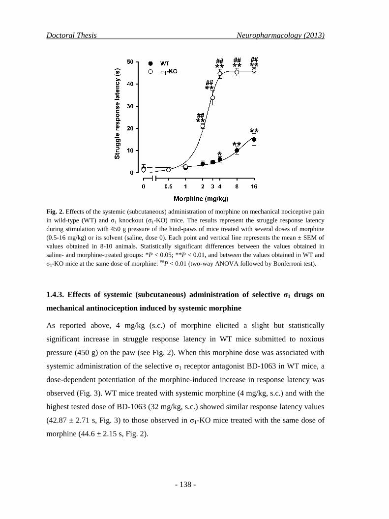
Doctoral Thesis Neuropharmacology (2013)
- 138 -
Fig. 2. Effects of the systemic (subcutaneous) administration of morphine on mechanical nociceptive pain
in wild-type (WT) and σ1 knockout (σ1-KO) mice. The results represent the struggle response latency
during stimulation with 450 g pressure of the hind-paws of mice treated with several doses of morphine
(0.5-16 mg/kg) or its solvent (saline, dose 0). Each point and vertical line represents the mean ± SEM of
values obtained in 8-10 animals. Statistically significant differences between the values obtained in
saline- and morphine-treated groups: *P < 0.05; **P < 0.01, and between the values obtained in WT and
σ1-KO mice at the same dose of morphine: ##
P < 0.01 (two-way ANOVA followed by Bonferroni test).
1.4.3. Effects of systemic (subcutaneous) administration of selective σ1 drugs on
mechanical antinociception induced by systemic morphine
As reported above, 4 mg/kg (s.c.) of morphine elicited a slight but statistically
significant increase in struggle response latency in WT mice submitted to noxious
pressure (450 g) on the paw (see Fig. 2). When this morphine dose was associated with
systemic administration of the selective σ1 receptor antagonist BD-1063 in WT mice, a
dose-dependent potentiation of the morphine-induced increase in response latency was
observed (Fig. 3). WT mice treated with systemic morphine (4 mg/kg, s.c.) and with the
highest tested dose of BD-1063 (32 mg/kg, s.c.) showed similar response latency values
(42.87 ± 2.71 s, Fig. 3) to those observed in σ1-KO mice treated with the same dose of
morphine (44.6 ± 2.15 s, Fig. 2).

Neuropharmacology (2013) Doctoral Thesis
- 139 -
We evaluated the specificity of the effects of systemic BD-1063 on morphine-induced
mechanical antinociception by testing whether the selective σ1 agonist PRE-084 was
able to reverse the effect of the σ1 antagonist. In contrast to the effects induced by BD-
1063, treatment with the selective σ1 agonist PRE-084 (16 mg/kg, s.c.) did not
significantly modify the antinociceptive effect of morphine, either at 4 mg/kg (Fig. 4) or
at 16 mg/kg (data not shown). However, when PRE-084 (4-16 mg/kg, s.c.) was co-
administered with BD-1063 (32 mg/kg, s.c.), it completely reversed the potentiation of
morphine-induced mechanical antinociception by the σ1 antagonist in a dose-dependent
manner (Fig. 4).
Fig. 3. Effects of systemic (subcutaneous)
administration of BD-1063 on mechanical
antinociception induced by systemic morphine
in wild-type mice. The results represent the
struggle response latency during stimulation
with 450 g pressure of the hind-paws of mice
treated with several doses of BD-1063 (1-32
mg/kg) or its solvent (dose 0) associated with
morphine (4 mg/kg). Each point and vertical
line represents the mean ± SEM of values
obtained in 8-10 animals. Statistically
significant differences between the values
obtained in the groups treated with BD-1063
and its solvent (dose 0): **P < 0.01 (one way
ANOVA followed by Bonferroni test).
We confirmed the effects of the pharmacological antagonism of σ1 receptors on
morphine-induced antinociception by using a panel of selective σ1 antagonists: BD-
1047 (32 mg/kg, s.c.), NE-100 (4 mg/kg, s.c.) and SR1A (32 mg/kg, s.c.). All of these
drugs mimicked the effects of BD-1063 on morphine-induced mechanical

Doctoral Thesis Neuropharmacology (2013)
- 140 -
antinociception, increasing the response latency of morphine-treated WT mice, and their
effects were dose-dependently reversed by PRE-084 (Fig. 4).
Fig. 4. Effect of the systemic (subcutaneous) administration of the σ1 agonist PRE-084 on the mechanical
antinociception induced by the systemic administration of morphine associated with several σ1
antagonists in wild-type mice. The results represent the struggle response latency during stimulation with
450 g pressure of the hind-paws of mice treated with PRE-084 (4-16 mg/kg) or its solvent and the σ1
antagonists BD-1063 (32 mg/kg), BD-1047 (32 mg/kg), NE-100 (4 mg/kg), or S1RA (32 mg/kg) or their
solvent, associated with morphine (4 mg/kg). Each bar and vertical line represents the mean ± SEM of
values obtained in 8-10 animals. Statistically significant differences between the values obtained in the
groups treated with morphine alone and associated with the σ1 drugs: **P < 0.01, and between the values
obtained in mice given each σ1 antagonist associated with PRE-084, with respect to each σ1 antagonist
associated with PRE-084 solvent (dose 0): ##
P < 0.01 (one-way ANOVA followed by Bonferroni test).
We further evaluated the selectivity of the effects induced by the σ1 antagonists by
testing their action on morphine-induced mechanical antinociception in σ1-KO mice. A
slightly lower morphine dose (3 mg/kg, s.c.) was used in this experiment in order to
facilitate the detection of possible non-specific increases in struggle response latency
induced by the σ1 antagonists. Response latencies were significantly higher in WT mice
co-administered s.c. with 3 mg/kg morphine and σ1 antagonist than in those treated with

Neuropharmacology (2013) Doctoral Thesis
- 141 -
morphine alone (Fig. 5, middle panel). However, none of the σ1 antagonists further
increased the morphine-induced mechanical antinociception in σ1-KO mice (Fig. 5,
right panel), suggesting that off-target effects do not significantly contribute to the
potentiation of morphine-induced antinociception by these drugs in our experimental
conditions. Response latency values did not significantly differ between those of WT
mice treated by any of the σ1 antagonists tested in combination with morphine and those
of σ1-KO mice treated with morphine alone (Fig. 5, middle and right panel), indicating
that similar levels of enhanced morphine analgesia were induced by the systemic
pharmacological blockade of σ1 receptors and by their genetic inactivation.
Fig. 5. Effects of the systemic (subcutaneous) administration of several σ1 antagonists on the mechanical
antinociceptive effect induced by systemic morphine in wild-type (WT) and σ1 knockout (σ1-KO) mice.
The results represent the struggle response latency during stimulation with 450 g pressure of the hind-
paws of mice treated with the σ1 antagonists BD-1063 (32 mg/kg), BD-1047 (32 mg/kg), NE-100 (4
mg/kg), or S1RA (32 mg/kg) or their solvent (saline), associated to morphine (3 mg/kg) or its solvent
(saline). Each bar and vertical line represents the mean ± SEM of values obtained in 8-10 animals.
Statistically significant differences between the values obtained in the groups treated with each σ1
antagonist and their solvent in morphine-treated WT mice: **P < 0.01. N.S.: no statistically significant
differences between the values (two-way ANOVA followed by Bonferroni test).

Doctoral Thesis Neuropharmacology (2013)
- 142 -
None of the σ1 antagonists administered alone was able to modify the struggle response
latency in WT mice, i.e., they did not affect the responses to noxious pressure in the
absence of morphine (Fig. 5, left panel). Therefore, the antinociceptive effects observed
in WT mice treated with the association of morphine and σ1 antagonist were synergistic
rather than merely additive.
1.4.4. Local antinociceptive effects induced by intraplantar administration of
morphine in wild-type and σ1 knockout mice
We tested whether σ1 receptors are able to modulate the antinociceptive effects of
morphine in the periphery by the i.pl. administration of this opioid in WT and σ1-KO
mice. The i.pl. administration of morphine (50-200 µg) did not significantly modify the
struggle response latency in WT mice in either the injected or non-injected paw (Fig. 6),
indicating that these doses were unable to locally induce a significant mechanical
antinociceptive effect in this genotype. In contrast, σ1-KO mice i.pl injected with the
same doses of morphine showed a marked dose-dependent increase in response latency
in the injected paw, reaching values close to the cut-off time at the highest dose (200
µg) (Fig. 6). Latency values in the contralateral non-injected paw of σ1-KO mice
remained unchanged and undistinguishable from control values in mice treated with the
solvent of morphine (Fig. 6), indicating that the antinociceptive effect of morphine in
σ1-KO mice was locally produced. Hence, the local effect of morphine against noxious
pressure was potentiated in mice globally lacking functional σ1 receptors.

Neuropharmacology (2013) Doctoral Thesis
- 143 -
Fig. 6. Effect of the local (intraplantar) injection of morphine (Mor) on mechanical nociceptive pain in
wild-type (WT) and σ1 knockout (σ1-KO) mice. The results represent the struggle response latency during
stimulation with 450 g pressure of the mice hind-paw injected with morphine (50-200 µg) or its solvent
(dose 0), in comparison to that obtained during stimulation of the non-injected hind-paw. Each bar and
vertical line represents the mean ± SEM of values obtained in 8-10 animals. Statistically significant
differences between the values obtained stimulating the morphine-injected hind-paw versus those
obtained stimulating the solvent-treated hind-paw (dose 0) in σ1-KO mice: **P < 0.01, and between the
values obtained in the injected and non-injected hind-paws in σ1-KO mice: ##
P < 0.01 (two-way ANOVA
followed by Bonferroni test). No statistically significant differences (N.S.) were found in the values from
WT groups between the injected and non-injected hind-paws or between any dose of morphine with
respect to its solvent (two-way ANOVA followed by Bonferroni test).
1.4.5. Potentiation of the local antinociceptive effect of morphine by
pharmacological blockade of σ1 receptors
We investigated whether local pharmacological antagonism of σ1 receptors could also
potentiate morphine-induced peripheral mechanical antinociception in WT mice by
testing the effects of the i.pl. co-administration of the selective σ1 receptor antagonist
BD-1063 (12.5-200 µg) with 200 µg morphine in WT mice.
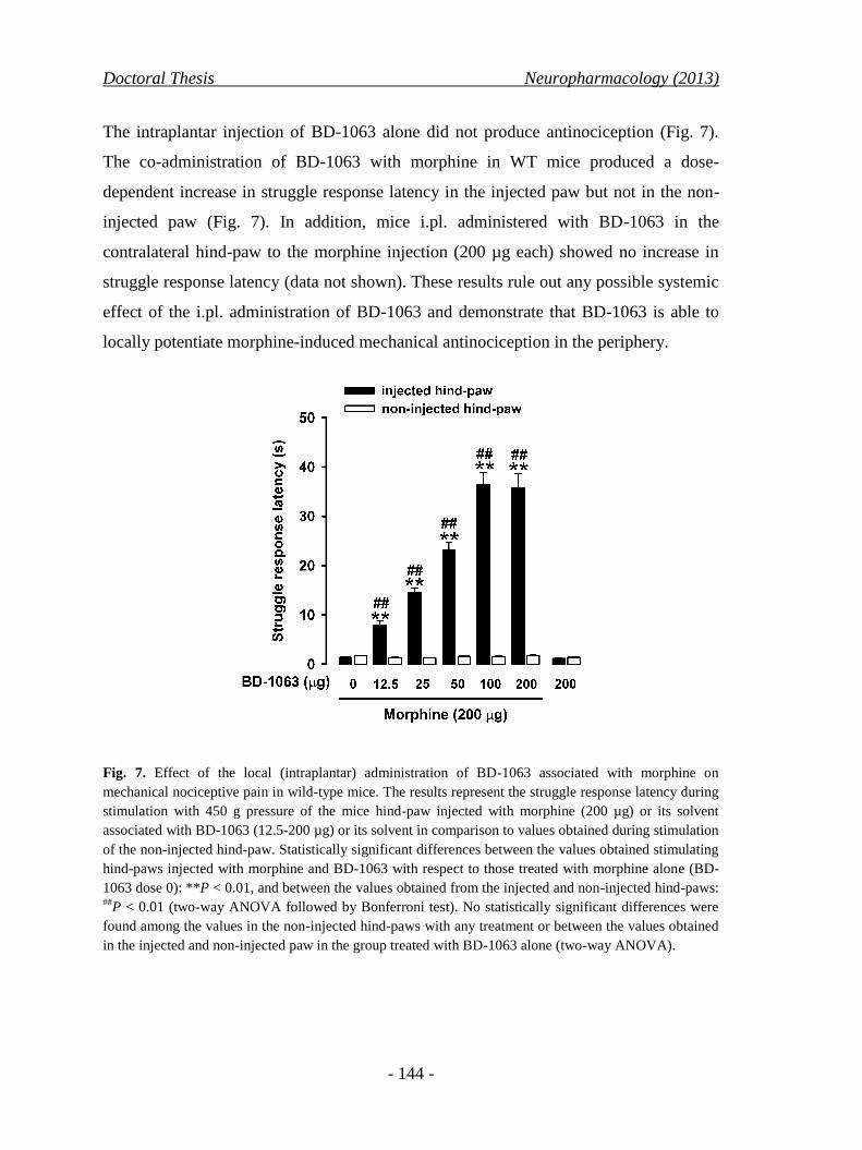
Doctoral Thesis Neuropharmacology (2013)
- 144 -
The intraplantar injection of BD-1063 alone did not produce antinociception (Fig. 7).
The co-administration of BD-1063 with morphine in WT mice produced a dose-
dependent increase in struggle response latency in the injected paw but not in the non-
injected paw (Fig. 7). In addition, mice i.pl. administered with BD-1063 in the
contralateral hind-paw to the morphine injection (200 µg each) showed no increase in
struggle response latency (data not shown). These results rule out any possible systemic
effect of the i.pl. administration of BD-1063 and demonstrate that BD-1063 is able to
locally potentiate morphine-induced mechanical antinociception in the periphery.
Fig. 7. Effect of the local (intraplantar) administration of BD-1063 associated with morphine on
mechanical nociceptive pain in wild-type mice. The results represent the struggle response latency during
stimulation with 450 g pressure of the mice hind-paw injected with morphine (200 µg) or its solvent
associated with BD-1063 (12.5-200 µg) or its solvent in comparison to values obtained during stimulation
of the non-injected hind-paw. Statistically significant differences between the values obtained stimulating
hind-paws injected with morphine and BD-1063 with respect to those treated with morphine alone (BD-
1063 dose 0): **P < 0.01, and between the values obtained from the injected and non-injected hind-paws: ##
P < 0.01 (two-way ANOVA followed by Bonferroni test). No statistically significant differences were
found among the values in the non-injected hind-paws with any treatment or between the values obtained
in the injected and non-injected paw in the group treated with BD-1063 alone (two-way ANOVA).

Neuropharmacology (2013) Doctoral Thesis
- 145 -
In order to establish the specificity of these effects, we tested the effect of BD-1063 in
σ1-KO mice. For this experiment, we used a lower dose of morphine (100 µg) to
facilitate detection of any non-specific increases in morphine-antinociception
attributable to the i.pl. injection of the σ1 antagonist. BD-1063 (100 µg) enhanced the
effects of morphine in WT mice but did not significantly alter the response of
morphine-treated σ1-KO mice (Fig. 8, middle and right panel).
Likewise, the i.pl. administration of BD-1047 (50 µg), NE-100 (50 µg), or S1RA (100
µg) had no effects on the behavioral response in the absence of morphine (Fig. 8, left
panel). However, these drugs increased the response latency in the injected paw of
morphine-treated (100 µg, i.pl.) WT mice (Fig. 8, middle panel), although not in their
non-injected paw (data not shown). In contrast, the co-administration of these σ1
antagonists with morphine did not potentiate the effect of the opioid in σ1-KO mice
(Fig. 8, right panel). These results support that the enhancement of locally-induced
morphine antinociception produced by the σ1 antagonists is mediated by their
interaction with σ1 receptors, and that no additional effects of these drugs are
participating in the effects observed. In addition, the response latency of WT mice
locally co-administered with σ1 antagonist and morphine did not significantly differ
from that obtained in σ1-KO mice treated with morphine alone (Fig. 8, middle and right
panel). This finding indicates that a similar potentiation of local morphine
antinociceptive effects was produced by local σ1 pharmacological blockade and by
genetic inactivation.
According to our results, the local pharmacological blockade of σ1 receptors potentiates
the mechanical antinociception induced locally by morphine but does not alter
nociceptive responses in the absence of the opioid.

Doctoral Thesis Neuropharmacology (2013)
- 146 -
Fig. 8. Effects of the local (intraplantar) administration of several σ1 antagonists on the mechanical
antinociceptive effect induced by intraplantar morphine in wild-type (WT) and σ1 knockout (σ1-KO)
mice. The results represent the struggle response latency during stimulation with 450 g pressure of the
hind-paws of mice treated with the σ1 antagonists BD-1063 (100 µg), BD-1047 (50 µg), NE-100 (50 µg),
or S1RA (100 µg) or their solvent (saline) associated with morphine (100 µg) or its solvent (saline). Each
bar and vertical line represents the mean ± SEM of values obtained in 8-10 animals. Statistically
significant differences in values between morphine-injected WT mice treated with each σ1 antagonist and
those treated with its solvent: **P < 0.01. N.S.: no statistically significant differences between the values
(two-way ANOVA followed by Bonferroni test). None of the treatments produced any effect in the non-
injected paw (data not represented in order to simplify the figure)
1.4.6. Morphine-induced side effects (hyperlocomotion and inhibition of
gastrointestinal transit) in wild-type and σ1 knockout mice
To determine whether the increased morphine-induced antinociception observed in σ1-
KO mice was accompanied by an increase in the non-analgesic effects of morphine, we
tested morphine-induced hyperlocomotion and gastrointestinal transit inhibition. WT
and σ1-KO mice showed a similar ambulatory locomotion when injected with saline
(Fig. 9A). Systemic administration of morphine (4-16 mg/kg, s.c.) induced a marked
and dose-dependent increase in the distance travelled by both WT mice and morphine-

Neuropharmacology (2013) Doctoral Thesis
- 147 -
injected σ1-KO mice, with no statistically significant differences between them (Fig.
9A).
Gastrointestinal transit values did not significantly differ between saline-treated WT and
σ1-KO mice, with the charcoal meal travelling about 30 cm of the small intestine in both
genotypes (Fig. 9B). Morphine (1-8 mg/kg, s.c.)-treated mice of both genotypes showed
a dose-dependent decrease in gastrointestinal transit of a similar magnitude (Fig. 9B).
Therefore, two different non-analgesic effects of morphine were unaltered by the
genetic inactivation of σ1 receptors.
Fig. 9. Effects of the systemic (subcutaneous) administration of morphine or saline on (A) locomotor
activity and (B) gastrointestinal transit of wild-type (WT) and σ1 knockout (σ1-KO) mice. (A) Mice were
injected with morphine or saline, and the distance that they travelled between 30 and 60 min post-
injection was recorded. Each bar and vertical line represents the mean ± SEM of values obtained in 8-13
mice. (B) At 30 min after the injection of morphine or saline, mice were intragastrically administered
with 0.5 % charcoal suspension. Transit of the charcoal was measured at 30 min after its ingestion. Each
bar and vertical line represents the mean ± SEM of values obtained in 6 mice. Statistically significant
differences between the values obtained in saline- and morphine-treated groups: **P <0.01. No
statistically significant differences were found between genotypes under the same treatment (two-way
ANOVA followed by Bonferroni test).

Doctoral Thesis Neuropharmacology (2013)
- 148 -
1.4.7. [3H]DAMGO saturation binding assays in spinal cord, forebrain, and hind-
paw skin membranes from wild-type and σ1 knockout mice
Saturation assays showed that the selective µ-opioid receptor radioligand [3H]DAMGO
bound in a saturable manner to forebrain and spinal cord membranes (P2 fraction) from
both WT and σ1-KO mice (Fig. 10A). Replicates were fitted by nonlinear regression
analysis to hyperbolic equations. In the case of the forebrain membranes, no significant
differences were found between WT and σ1-KO mice in [3H]DAMGO equilibrium
dissociation constant (KD) values (1.530 ± 0.212 nM vs. 1.172 ± 0.166 nM, respectively;
P > 0.05) or in the maximal number of receptors (Bmax) (0.137 ± 0.004 vs. 0.133 + 0.004
pmol/mg of protein, respectively; P > 0.05). Likewise, in the spinal cord membranes, no
significant differences between WT and σ1-KO mice were found in KD (2.028 ± 0.269
vs. 1.861 ± 0.226 nM, respectively; P > 0.05) or Bmax (0.179± 0.005 vs. 0.180 ± 0.004
pmol/mg of protein, respectively; P > 0.05) values.
The [3H]DAMGO Bmax in paw skin membranes was estimated by using a single
saturating concentration (20 nM) of this radioligand. For a better comparison, we used
the same concentration of the radioligand to determine the estimated Bmax in both spinal
cord and forebrain membranes. Using this approach, we obtained very similar
[3H]DAMGO-specific binding values to those obtained with the full saturation assay in
both WT (0.180 ± 0.012 and 0.1266 ± 0.003 pmol/mg of protein for spinal cord and
forebrain membranes, respectively) and σ1-KO (0.183 ± 0.013 and 0.122 ± 0.07
pmol/mg of protein for spinal cord and forebrain membranes, respectively) mice (Fig.
10B). [3H]DAMGO binding sites were much less abundant in the paw skin membranes
than in the tissues from the central nervous system, with no significant differences
between the genotypes (0.030 ± 0.002 and 0.030 ± 0.001 pmol/mg of protein for WT
and σ1-KO mice, respectively) (Fig. 10B).

Neuropharmacology (2013) Doctoral Thesis
- 149 -
Fig. 10. [3H]DAMGO binding assays in wild-type (WT) and σ1 knockout (σ1-KO) mice. (A)
[3H]DAMGO saturation assays in forebrain and spinal cord membranes (P2 fraction) from WT (closed
symbols) or σ1-KO (open symbols) mice. Experiments were performed by incubating the membranes for
120 min at 25ºC with several concentrations of [3H]DAMGO (0.25-45 nM). (B) [
3H]DAMGO Bmax values
in forebrain, spinal cord, and hind-paw skin membranes. Bmax values were estimated by incubating the
membranes with a single saturating concentration of [3H]DAMGO (20 nM) under the experimental
conditions described above. All experiments were carried out in the presence of 10 µM naloxone (to
define non-specific binding) or its solvent. The data shown represent three experiments carried out in
triplicate.

Doctoral Thesis Neuropharmacology (2013)
- 150 -
1.4.8. Affinity of selective σ1 ligands and morphine for [3H]DAMGO binding sites
in forebrain membranes from wild-type mice
We used competition binding assays to test the binding of the studied drugs to
[3H]DAMGO-labeled µ-receptors in forebrain membranes from WT mice. As expected,
the [3H]-DAMGO-specific binding was concentration-dependently inhibited by
morphine, which showed an affinity (Ki) value of 3.746 ± 0.319 nM. However, the
specific binding of [3H]DAMGO was not inhibited by any of the selective σ1-ligands
tested (BD-1063, BD-1047, NE-100, S1RA, or PRE-084), therefore demonstrating
negligible affinity of these drugs for [3H]DAMGO binding sites (Fig. 11).
Fig. 11. Inhibition by unlabeled drugs of
[3H]DAMGO binding to forebrain
membranes (P2 fraction) in wild-type mice.
Membranes were incubated for 120 min at
25ºC with 5 nM [3H]DAMGO and
increasing concentrations of morphine or
the σ1 ligands BD-1063, BD-1047, NE-
100, S1RA, or PRE-084. Naloxone (10
µM) was used to define the non-specific
binding. Data are the average of three
experiments carried out in triplicate.
1.5. DISCUSSION
In this study, pharmacological antagonism or genetic inactivation of σ1 receptors
induced a strong functional synergism with the mechanical antinociceptive effect of
morphine, without altering its non-analgesic effects (hyperlocomotion and inhibition of
gastrointestinal transit). Furthermore, this synergistic interaction occurred at the
peripheral level. None of these findings have been previously reported.

Neuropharmacology (2013) Doctoral Thesis
- 151 -
We found that the pain-like responses evoked by a blunt mechanical stimulus were
virtually identical between σ1-KO and WT mice over a wide range of pressure
intensities. Neither were they altered by the local or systemic administration of σ1
antagonists. These findings expand the results found in previous studies reporting that
σ1-KO mice or WT mice treated with σ1 antagonists showed unaltered responses against
different sensory modalities of acute nociceptive pain, including mechanical (punctate)
and thermal (cold and heat) stimulation (e.g. Chien and Pasternak, 1994; De la Puente et
al., 2009; Entrena et al., 2009a; Marrazzo et al., 2011; Nieto et al., 2012; Romero et al.,
2012). These results suggest that the basic mechanisms for perceiving these stimuli and
the motor response for producing the pain-like responses are intact in σ1-KO mice and
are not altered by the pharmacological blockade of σ1 receptors in WT mice. However,
despite the apparent absence of σ1 receptor involvement in acute nociception, we found
that the mechanical antinociceptive effect of morphine was markedly enhanced in σ1-
KO mice and that this effect was mimicked by the systemic administration of several
prototypic σ1 antagonists in WT mice. Our results extend previous reports on the
potentiation of opioid-induced thermal antinociception by σ1 inhibition (Chien and
Pasternak, 1993 and 1994; Marrazzo et al., 2011 and 2006; Mei and Pasternak, 2002 and
2007; Pan et al., 1998; Ronsisvalle et al., 2001). Taken together, these findings suggest
that a tonically active antiopioid σ1 system modulates both the mechanical and thermal
antinociception induced by morphine.
We found that the i.pl. administration of morphine had no analgesic effect against
mechanical stimuli in naïve mice over a wide range of doses. This finding is in contrast
to previous reports showing that local morphine is effective against thermal stimuli (e.g.
Kolesnikov et al., 1996, 2000), highlighting the differences between the effects of this
opioid against thermal and mechanical stimuli. The lack of effect of local morphine
demonstrated here is in agreement with clinical reports showing that the local
application of opioid agonists (including morphine) to uninjured tissue does not reliably
produce analgesic effects (reviewed by Stein et al., 2003), and it is consistent with the
preferentially central action of opioids to induce analgesia in either humans or rodents
(e.g. Christie et al., 2000; Thomas et al., 2008; Khalefa et al., 2012). Because of this

Doctoral Thesis Neuropharmacology (2013)
- 152 -
preferentially central localization of opioid-induced analgesia, previous studies focused
on the role of σ1 receptors at central levels in modulating this opioid-mediated effect,
demonstrating that the central administration of either σ1 antagonists or antisense
oligodeoxynucleotides enhances morphine-induced thermal antinociception (Mei and
Pasternak, 2002 and 2007; Pan et al., 1998). In the present study, we show that σ1-KO
mice locally treated with morphine and WT mice locally co-administered with this
opioid and σ1 receptor antagonist exhibit a strong synergistic mechanical
antinociceptive effect at the site of the administration of the combined drug solution but
not at a site distant from its injection (contralateral paw) which suggests that the
interaction is produced locally. This view is further supported by the fact that WT mice
treated with morphine (200 µg) in one paw and BD-1063 (200 µg) in the contralateral
one evidenced no antinociception in either paw (data not shown). Hence, our data reveal
for the first time that the tonic inhibition of morphine analgesia by σ1 receptors is also
present at the periphery and is strongly involved in mechanical nociceptive pain.
We found similar levels of ([3H]DAMGO-labeled) μ-opioid receptors in σ1-KO and WT
mice in all tissues examined, and the affinity of this radioligand for its binding site was
not altered in σ1-KO mice. In addition, all σ1 ligands tested showed a negligible affinity
for μ-opioid receptors from mouse forebrain membranes, as previously reported for
some of these σ1-ligands in other tissues or species (Kim et al., 2010; Matsumoto et al.,
1995; Romero et al., 2012). Hence, our results cannot be explained by abnormal μ-
opioid receptors in the σ1-KO mice or by a direct interaction of the σ1 drugs used in this
study with μ-opioid receptors.
Several of our findings indicate that a selective σ1 receptor action is involved in the
modulation of morphine-induced mechanical antinociception. Firstly, the enhancement
of morphine-induced mechanical antinociception in σ1-KO mice was replicated, at a
similar magnitude, by all of the selective σ1 antagonists tested in WT mice. Secondly, the
selective σ1 agonist PRE-084, which had no effect on morphine-induced antinociception,
was able to reverse the effects of the systemic σ1 antagonists. Finally, none of the σ1
antagonists tested (administered either systemically or locally) further enhanced morphine-
Neuropharmacology (2013) Doctoral Thesis
- 153 -
induced antinociception in σ1-KO mice, indicating that off-target effects do not account for
the effects observed. The similarities in the antinociceptive effects of morphine between
σ1-KO mice and σ1 antagonist-treated WT mice, together with the clear σ1 pharmacology
of these effects, strongly suggest that the effects observed are mediated by σ1 receptor
inhibition. A recent study indicates a possible mechanism for these effects. Thus, the
prototypic σ1 receptor antagonist BD-1047 was found to increase DAMGO-induced G-
protein couple receptor signaling (measured as the increase in [35
S]GTPγS binding)
without altering opioid receptor binding, and σ1 receptors and μ-opioid receptor were
shown to physically interact (Kim et al., 2010). Therefore, basal σ1 receptor activity
may tonically reduce μ-opioid receptor signaling, explaining the increase in morphine
analgesia by σ1 receptor inhibition.
Previous research on opioid-induced thermal antinociception showed that the selective
σ1 agonist (+)-pentazocine could decrease morphine-induced thermal antinociception,
indicating that tonic inhibition of this analgesic effect of morphine can be enhanced by
further σ1 activation (Chien and Pasternak, 1993 and 1994; Mei and Pasternak, 2002
and 2007). However, under our conditions, PRE-084 did not influence the effect of
morphine on mechanical stimuli, suggesting that the tonic inhibition of morphine-
induced mechanical antinociception by σ1 receptors was already maximal and could not
be further increased by exogenous σ1 activation. The role of σ1 receptors in animal
models of tonic/chronic pain has been thoroughly studied over the past decade (Cendán
et al., 2005a and b; De la Puente et al., 2009; Entrena et al., 2009a and b; Kim et al.,
2006; Nieto et al., 2012; Roh et al., 2008b; Romero et al., 2012). Modulation of these
pain behaviors by σ1 receptors is thought to be located at central levels, because σ1
receptor activation in the spinal cord triggers central sensitization to induce mechanical
allodynia (Kim et al., 2008; Roh et al., 2010 and 2011; Ohsawa et al., 2011), likely
contributing through this mechanism to the modulation of activity-induced spinal
sensitization (De la Puente et al., 2009; Romero et al., 2012). We previously
demonstrated that the effect of σ1 antagonists on some of those behavioral outcomes
(formalin-induced pain and capsaicin-induced mechanical hypersensitivity) was
insensitive to naloxone treatment (Cendán et al., 2005a; Entrena et al., 2009b).
Doctoral Thesis Neuropharmacology (2013)
- 154 -
Therefore, these effects of σ1 antagonists do not appear to depend on modulation of the
opioid system.
Opioids, and particularly morphine, are considered to be the “gold standard” for pain
management (Christie et al., 2000; Waldhoer et al., 2004). However, several serious
side effects associated with acutely administered morphine, including constipation,
sedation, respiratory depression, and nausea, represent substantial drawbacks to its use
(reviewed in Al-Hasani and Bruchas, 2011 and Waldhoer et al., 2004). Two strategies
have been proposed to minimize the adverse effects of opioids. One of these approaches
is to administer an adjuvant drug with synergistic analgesic effects in order to minimize
the dose of the opioid (and hence its side effects) while maintaining acceptable levels of
analgesia. One example is the combination of non-steroidal anti-inflammatory drugs or
anticonvulsants with opioids (Christie et al., 2000; Mao et al., 2011). In the present
study, we show that, despite the strong potentiation of the analgesic effects of morphine
observed in σ1-KO mice, σ1 receptors do not appear to influence two morphine-induced side
effects, i.e., hyperlocomotion and gastrointestinal transit inhibition. Hyperlocomotion is a
common side effect of opioids in rodents (e.g. Cobos et al., 2012; Elhabazi et al., 2012) and
reflects an increase in supraspinal dopamine release (Hnasko et al., 2005), whereas the
inhibition of gastrointestinal transit mainly results from opioid action at peripheral level (Al-
Hasani and Bruchas., 2011). In agreement with our data, a previous study found that the
selective σ1 agonist (+)-pentazocine did not modify the morphine-induced decrease in
gastrointestinal transit or lethality (Chien and Pasternak, 1994). The differential impact of σ1
receptors on the modulation of opioid analgesia and side effects may suggest that σ1
receptors are present in a specific subset of opioid receptor-expressing neurons that are
more involved in pain pathways or, alternatively, that further mechanisms besides the
direct modulation of opioid signaling may participate in the analgesia enhancement.
A different approach to reducing the side effects of opioids is to target peripheral opioid
antinociception in order to minimize undesirable centrally mediated effects (reviewed
by Sehgal et al., 2011; Stein et al., 2003). Here we show that σ1 inhibition is able to
potentiate local morphine analgesia, producing an even greater antinociceptive effect

Neuropharmacology (2013) Doctoral Thesis
- 155 -
than is induced by systemic morphine at the highest dose administered in this study (16
mg/kg). Consequently, the potentiation of peripherally mediated morphine analgesia by
σ1 receptor antagonists may offer safer and improved therapeutic outcomes in pain
management.
1.6. CONCLUSIONS
In summary, we found that systemic σ1 receptor inhibition synergistically enhances
morphine-induced mechanical antinociception without modifying a centrally-induced
(hyperlocomotion) or peripherally-induced (constipation) side effect of morphine.
Enhancement of morphine analgesia by σ1 inhibition can be produced locally, indicating
that σ1 receptors tonically inhibit peripheral opioid functioning. The local combination
of morphine with σ1 receptor blockade may represent a strategy to minimize the adverse
effects of morphine by differentially potentiating its therapeutic analgesic actions.
1.7. ACKNOWLEDGEMENTS
C. Sánchez-Fernández and R González-Cano were supported by FPU grants from the
Spanish Ministerio de Educación y Ciencia (MEC), and E. J. Cobos by the Research
Program of the University of Granada. This research was done in partial fulfillment of
the requirements for the doctoral thesis of C. Sánchez-Fernández. The authors thank
Richard Davies for revising the English style of the manuscript. This study was partially
supported by the Spanish Ministry of Education and Science (MEC) [grant SAF2006-
06122], Junta de Andalucía [grant CTS 109], Laboratorios Esteve, and The Centre for
Industrial Technological Development (Spanish Government) [Genius Pharma project].


- 157 -
2. Modulation of peripheral µ-opioid analgesia by
σ1 receptors
Sánchez-Fernández C, Montilla-García A, González-Cano R, Nieto FR,
Romero L, Artacho-Cordón A, Montes R, Fernández-Pastor B, Merlos M,
Baeyens JM, Entrena JM, Cobos EJ
The Journal of Pharmacology
and Experimental Therapeutics (JPET), 2014; 348(1):32-45
Highlighted article of the volume of January, 2014


JPET (2014) Doctoral Thesis
- 159 -
2.1. ABSTRACT
We evaluated the effects of σ1 receptor inhibition on µ-opioid-induced mechanical
antinociception and constipation. σ1-knockout mice exhibited marked mechanical
antinociception in response to several μ-opioid analgesics (fentanyl, oxycodone,
morphine, buprenorphine and tramadol) at systemic (subcutaneous, s.c.) doses that were
inactive in wild-type mice, and even unmasked the antinociceptive effects of the
peripheral µ-opioid agonist loperamide. Similarly, systemic (s.c.) or local (intraplantar)
treatment of wild-type mice with the selective σ1 antagonists BD-1063 or S1RA
potentiated µ-opioid antinociception; these effects were fully reversed by the σ1 agonist
PRE-084, showing the selectivity of the pharmacological approach. The µ-opioid
antinociception potentiated by σ1 inhibition (by σ1 receptor knockout or σ1
pharmacological antagonism) was more sensitive to the peripherally-restricted opioid
antagonist naloxone methiodide than opioid antinociception under normal conditions,
indicating a key role for peripheral opioid receptors in the enhanced antinociception.
Direct interaction between the opioid drugs and σ1 receptor cannot account for our
results, since the former lacked affinity for σ1 receptors (labeled with [3H](+)-
pentazocine). A peripheral role for σ1 receptors was also supported by their higher
density (western blot results) in peripheral nervous tissue (dorsal root ganglia) than in
several central areas involved in opioid antinociception (dorsal spinal cord, basolateral
amygdala, periaqueductal gray and rostroventral medulla). In contrast to its effects on
nociception, σ1 receptor inhibition did not alter fentanyl- or loperamide-induced
constipation, a peripherally-mediated nonanalgesic opioid effect. Therefore, σ1-receptor
inhibition may be used as a systemic or local adjuvant to enhance peripheral µ-opioid
analgesia without affecting opioid-induced constipation.

Doctoral Thesis JPET (2014)
- 160 -
2.2. INTRODUCTION
Opioid drugs, particularly agonists of the µ-receptor subtype, are widely used to treat
moderate-to-severe pain (Pasternak and Pan, 2011; Al-Hasani and Bruchas, 2011).
Opioid receptors are located at different sites along the pain-processing pathway,
including both the central (spinal cord and different supraspinal nuclei) and peripheral
(dorsal root ganglion [DRG] and peripheral nerve terminals) nervous system (Khalefa et
al., 2012, Bigliardi-Qi et al., 2004). The antinociceptive effect of systemic opioids is
thought to be produced mainly at the central (particularly supraspinal) level
(Greenwood-Van Meerveld and Standifer, 2008; Thomas et al., 2008; Joshi et al., 2008;
Khalefa et al., 2012), although peripheral opioid receptors might also participate
(Kayser et al., 1995; Craft et al., 1995; Shannon and Lutz, 2002).
The sigma-1 (σ1) receptor has been cloned and its sequence does not show homology
with opioid receptors or any other known mammalian protein; it is therefore currently
considered a unique entity (Cobos et al., 2008; Zamanillo et al., 2013). Inhibition of σ1
receptor function either by the systemic administration of σ1 antagonists or by σ1
receptor knockdown does not influence acute nociception per se (Cendán et al., 2005b;
Entrena et al., 2009b; De la Puente et al., 2009; Nieto et al., 2012; Romero et al., 2012;
Sánchez-Fernández et al., 2013). However, σ1 inhibition is able to enhance opioid
signaling (Kim et al., 2010) and to potentiate the antinociceptive effect of systemic
opioids (e.g. Chien and Pasternak, 1993 and 1994; Marrazzo et al., 2011; Sánchez-
Fernández et al., 2013; Vidal-Torres et al., 2013). Opioid antinociception can be
potentiated by central σ1 inhibition (King et al., 1997; Pan et al., 1998; Mei and
Pasternak 2002 and 2007; Marrazzo et al., 2006), and we recently reported that the local
peripheral coadministration of σ1 antagonists and morphine also resulted in markedly
enhanced antinociception (Sánchez-Fernández et al., 2013). However, it is unknown
whether peripheral mechanisms are involved in the antinociception induced by systemic
opioids when σ1 receptors are inhibited. We hypothesize that the contribution of
peripheral opioid receptors to overall antinociception induced by the combination of
systemic opioids and σ1 inhibition might be more relevant than the contribution of these

JPET (2014) Doctoral Thesis
- 161 -
receptors to the effect of systemic opioids under normal conditions. The main goal of
this study was to test this hypothesis.
To do so, we compared the effect of the peripherally restricted opioid antagonist
naloxone methiodide (Menéndez et al., 2005; Sevostianova et al., 2005; Parenti et al.,
2012) on the mechanical antinociception induced by systemic µ-opioids in the presence
or absence of σ1 receptor inhibition. We used several clinically relevant µ-opioids with
different intrinsic activities and blood–brain barrier permeabilities. These opioid drugs
include the centrally active analgesics fentanyl, morphine, oxycodone, tramadol and
buprenorphine (Pergolizzi et al., 2008; Schäfer 2010), and the peripherally restricted µ-
agonist loperamide, used clinically as an antidiarrheal drug (Layer et al., 2010; Gallelli
et al., 2010). To inhibit σ1 receptors we used σ1-knockout (σ1-KO) mice and systemic
and local treatments of wild-type (WT) mice with the selective σ1 antagonists BD-1063
and S1RA. Moreover, because the data from the present study support the importance of
peripheral σ1 receptors as modulators of opioid antinociception, we compared the
expression of σ1 receptors in peripheral (DRG) and central areas (dorsal spinal cord,
basolateral amygdala, periaqueductal gray and rostroventral medulla) known to be
involved in opioid antinociception (Millan, 2002). Finally, to rule out possible direct
interactions between the opioid drugs tested here and σ1 receptors, we determined their
affinity for σ1 receptors.
Opioid-induced constipation is the most clinically relevant peripheral side effect of
µ-opioids (Benyamin et al 2008; Al-Hasani and Bruchas, 2011; Ringkamp and Raja,
2012), and it is one of the main reasons for patients‟ voluntary withdrawal from opioid
medication (Dhingra et al., 2012). We recently showed that although morphine-induced
antinociception was potentiated in σ1-KO mice, morphine-induced constipation
remained unaltered (Sánchez-Fernández et al., 2013; Vidal-Torres et al., 2013).
However, it is unknown whether these differential effects of σ1 inhibition also occur
with other µ-opioids. Therefore, an additional goal of this study was to test the effects of
σ1 inhibition on constipation induced by two very different µ-opioids: fentanyl, a
centrally penetrant drug, and loperamide, a peripherally restricted drug.
Doctoral Thesis JPET (2014)
- 162 -
2.3. MATERIAL AND METHODS
2.3.1. Experimental animals
Most experiments were performed in female WT (Charles River, Barcelona, Spain) and
σ1-KO CD-1 mice (Laboratorios Esteve, Barcelona, Spain) weighing 25-30 g. The
knockout mice were backcrossed for 10 generations to a CD-1 genetic background as
described previously (Entrena et al., 2009a). Some experiments were also performed in
male WT mice to ensure that sex differences did not affect our results. All animals were
kept in our animal facilities for a minimum of 7 days before the experiments. Animals
were housed under controlled environmental conditions: 12/12 h day/night cycle,
constant temperature (22 ± 2 °C) with free access to water and food (standard laboratory
diet, Harlan Teklad Research Diet, Madison, WI, USA). Behavioral testing was done
during the light phase (from 9.00 h to 15.00 h) and randomly throughout the estrous
cycle. Animal care was provided in accordance with institutional (Research Ethics
Committee of the University of Granada, Spain) and international standards (European
Communities Council directive 86/609), and with the guidelines for the investigation of
experimental pain in conscious animals (Zimmermann 1983).
2.3.2. Radioligand, drugs and drug administration
The opioid drugs and their suppliers were: the µ-opioid agonists morphine
hydrochloride (from the General Directorate of Pharmacy and Drugs, Spanish Ministry
of Health), fentanyl citrate, oxycodone hydrochloride, buprenorphine hydrochloride and
loperamide (all from Sigma-Aldrich Química SA, Madrid, Spain), tramadol (supplied
by Laboratorios Esteve, Barcelona, Spain) and the opioid antagonists naloxone
hydrochloride and naloxone methiodide (Sigma-Aldrich Química SA). BD-1063 (1-[2-
(3,4-dichlorophenyl)ethyl]-4-methylpiperazine dihydrochloride) (Tocris Cookson Ltd.,
Bristol, UK), and S1RA (4-[2-[[5-methyl-1-(2-naphthalenyl)1H-pyrazol-3-yl]oxy]ethyl]
morpholine hydrochloride) (synthesized and kindly supplied by Laboratorios Esteve,
Barcelona, Spain) were used as selective σ1 antagonists (Cobos et al., 2008; Romero et
al., 2012). PRE-084 ([2-(4-morpholinethyl)1-phenylcyclohexanecarboxylate)

JPET (2014) Doctoral Thesis
- 163 -
hydrochloride]) (Tocris Cookson Ltd.) was used as a selective σ1 agonist (Hayashi and
Su, 2004; Cobos et al., 2008).
All drugs used for the in vivo studies were dissolved in sterile physiological saline
(NaCl 0.9%) except loperamide, which was dissolved in 1% dimethylsulfoxide (DMSO,
Merck KGaA, Darmstadt, Germany) in ultrapure water. The solutions of the σ1 ligands
BD-1063 and PRE-084 were appropriately alkalinized with NaOH. To evaluate the
effects of systemic treatments, drugs or their solvents were injected subcutaneously
(s.c.) into the interscapular zone in a volume of 5 mL/kg. When the effect of the
association of several drugs was tested, each drug was injected into a different area of
the interscapular zone. To study the local effects of BD-1063 or S1RA, these drugs
were injected intraplantarly (i.pl.) into the right hindpaw at a volume of 20 μL with a
1710 TLL Hamilton microsyringe (Teknokroma, Barcelona, Spain) with a 301/2–gauge
needle. Each control group received the same volume of drug vehicle.
For binding assays, the radioligand was the selective σ1 agonist [3H](+)-pentazocine
(Cobos et al., 2006), with a specific activity of 34.8 Ci/mmol (PerkinElmer Life
Sciences, Boston, MA, USA). Dilutions from the stock [3H](+)-pentazocine solution
were prepared with ice-cold incubation buffer (50 mM Tris–HCl buffer, pH 8 at 30 °C).
The cold drugs tested were the opioids: fentanyl, oxycodone, morphine, buprenorphine,
loperamide, tramadol, naloxone and naloxone methiodide. BD-1063 was used as a
control with known high-affinity for σ1 receptors (Cobos et al., 2007; Entrena et al.,
2009a). All drugs were dissolved at a concentration of 1 mM in ultrapure water with the
exception of loperamide, which was dissolved in absolute ethanol. Further dilutions
were prepared with incubation buffer. The final maximal concentration of ethanol in the
incubation medium was 0.1% (vol/vol), which did not affect [3H](+)-pentazocine
binding (Cobos et al., 2005 and 2006).
2.3.3. Evaluation of mechanical nociception (paw pressure)
The effects of the drugs on mechanical nociception were evaluated with an
Analgesimeter (Model 37215, Ugo-Basile, Varese, Italy) according to methods

Doctoral Thesis JPET (2014)
- 164 -
described previously (Menéndez et al., 2005; Sánchez-Fernández et al., 2013). Briefly,
the hindpaw of the mice was stimulated with a constant pressure of 450 g using a cone-
shaped paw-presser with a rounded tip until the animal showed a struggle reaction.
Immediately thereafter, the stimulus was stopped and the response latency (in seconds)
was recorded. The test was done twice alternately to each hindpaw at intervals of 1 min
between each stimulation, with a 50-s cutoff for each determination. The
antinociceptive effects of µ-agonists were evaluated 30 min after s.c. administration
except for buprenorphine, which was administered s.c. 1 h before the evaluation, since
the onset of its antinociceptive effect is known to be much slower than for other opioids
(Yassen et al., 2005). To study the effects of the systemic administration of BD-1063 or
S1RA on µ-opioid antinociception, these drugs (or its solvent) were administered s.c. 5
min before the µ-agonists. When drugs were administered systemically the struggle
response latency was calculated as the mean of the two averaged times in each hindpaw,
since no differences between sides were seen in the response of each hindpaw. To study
the local effects of σ1 inhibition on µ-opioid antinociception, BD-1063, S1RA or their
solvent were administered i.pl. and the animals were evaluated 5 min after the injection
to minimize possible systemic effects induced by the drug. In experiments to determine
the local effects of the σ1 antagonists, we calculated the average of the two values from
the injected and noninjected hindpaws independently. To test the effects of PRE-084,
naloxone or naloxone methiodide on µ-opioid antinociception in the presence or
absence of σ1 inhibition, these drugs or their solvent were administered s.c. 5 min before
the µ-agonist solution, when appropriate.
The experimenters who evaluated the behavioral responses were blinded to the
treatment group and genotype of each experimental animal.
2.3.4. Evaluation of opioid-induced inhibition of gastrointestinal transit
Gastrointestinal transit was estimated as previously described (Sánchez-Fernández et
al., 2013). Briefly, 8 h before the experiment, food was withheld and water was
available ad libitum. BD-1063 or its solvent was injected s.c., and 5 min later fentanyl,

JPET (2014) Doctoral Thesis
- 165 -
loperamide or their solvent was injected s.c. Thirty minutes after the opioid was given,
the mice received 0.3 mL of a 0.5% (wt/vol) activated charcoal suspension (2–12 μm
powder, Sigma-Aldrich Química SA) in distilled water. The mice were killed by
cervical dislocation 30 min after the activated charcoal was administered (i.e. 60 min
after administration of the opioid); then the small intestine was removed from the
pyloric sphincter to the ileocecal junction and straightened to measure the distance
travelled by the leading edge of the charcoal meal.
2.3.5. Western blotting
The basolateral amygdala (BLA), rostroventral medulla (RVM), periaqueductal grey
matter (PAG), dorsal spinal cord of the lumbar enlargement (dSC) and lumbar (L4-L5)
root ganglia (DRG) were carefully removed from naïve WT and σ1-KO mice. The tissue
was homogenized by sonication in a buffer solution (50 mM Tris, 150 nM NaCl, 2 mM
EDTA, 0.5% Triton X-100, 0.1% SDS, 1% NP-40, 1mM phenylmethylsulfonyl
fluoride, 1 mM Na3VO4, 25 mM NaF, 0.5% protease inhibitor cocktail, 1% phosphatase
inhibitor cocktail, all from Sigma-Aldrich Quımica SA). Protein concentration in the
tissue homogenate was measured with the Bradford assay. The samples were stored at
−80 °C until use.
Twenty-five micrograms of protein was loaded on 12% (wt/vol) SDS-polyacrylamide
gels and transferred electrophoretically to nitrocellulose membranes (Bio-Rad, Madrid,
Spain). The membranes were blocked at room temperature for 1 h with blocking buffer
containing 5% dry skim milk in T-TBS (10 mM Tris, 150 mM NaCl, 0.1% Tween-20,
pH 7.5). Then the membranes were incubated overnight at 4 °C with a mouse
monoclonal antibody that recognized σ1 receptor (1:1,000, sc-137075, Santa Cruz
Biotechnology, Dallas, TX, USA). Mouse monoclonal anti-β-actin antibody (1:2,500,
sc-81178, Santa Cruz Biotechnology) was used as a loading control. Both primary
antibodies were diluted in T-TBS containing 0.5% dry skim milk. The membranes were
washed (3 10 min) with T-TBS and incubated for 1 h at room temperature with
horseradish peroxidase-linked goat anti-mouse IgG (sc-2005, Santa Cruz

Doctoral Thesis JPET (2014)
- 166 -
Biotechnology), diluted to 1:2,500 in T-TBS containing 0.5% dry skim milk. The
membranes were washed (6 10 min) with T-TBS, and the bands were visualized with
an enhanced chemiluminescence method (ECL Prime Western Blotting Detection
Reagents, Amersham Biosciences, Buckinghamshire, UK) according to the
manufacturer‟s instructions (5 min incubation at room temperature). Densitometric
analysis of immunoreactive bands was done with Quantity One software (Bio-Rad). The
data are presented as the ratio of the intensity of the σ1 receptor bands to the β-actin
bands. To estimate the molecular weight of the resulting immunoreactive bands, when
the samples were loaded we included in the same gel a mixture of 10 blue-stained
recombinant proteins of different molecular weights (Precision Plus Protein All Blue
Standards, Bio-Rad).
2.3.6. [3H](+)-Pentazocine competition binding assays
Binding assays were done in WT mouse brain membranes (P2 fraction) as previously
described (Entrena et al., 2009a and b; Sánchez-Fernández et al., 2013). Briefly,
membrane solutions (460 μL) were incubated at a final protein concentration of 0.8
mg/mL with 20 μL of several concentrations of the cold drug or its solvent and 20 μL
[3H](+)-pentazocine (final concentration of 5 nM) for 240 min at 30 °C, pH 8. Five
milliliters of ice-cold filtration buffer (Tris 10 mM, pH 7.4) was added to the tubes, and
the solutions were rapidly filtered (Brandel cell harvester Model M-12 T, Brandel
Instruments, SEMAT Technical, St. Albans, Hertfordshire, UK) over Whatman GF/B
glass fiber filters (SEMAT Technical) pre-soaked with 0.5% polyethylenimine in Tris
10 mM, pH 7.4. The filters were washed twice with 5 mL of ice-cold filtration buffer.
Then 4 mL of liquid scintillation cocktail (CytoScint scintillation counting solution, MP
Biomedicals, Irvine, CA, USA) was added to each filter. On the next day, their
radioactivity level was measured by liquid scintillation spectrometry (Beckman Coulter
España SA, Madrid, Spain). Nonspecific binding was defined as the binding retained in
the presence of a high concentration of BD-1063 (1 µM), and was always less than 20%
of total binding.

JPET (2014) Doctoral Thesis
- 167 -
2.3.7. Data analysis
The data were analyzed with the SigmaPlot 12.0 program (Systat Software Inc., San
Jose, CA, USA). In behavioral assays, the dose-response curves of the drugs were
estimated with equations for a sigmoid plot. For binding assays, the concentration of
unlabeled BD-1063 that inhibited 50% of [3H](+)-pentazocine specific binding (IC50)
and its standard error were calculated from the inhibition curve with nonlinear
regression analysis from the equation for a sigmoid plot, assuming one-site competition.
To compare different mean values from in vivo studies, one-way or two-way analysis of
variance (ANOVA) was used depending on the experiment; a Bonferroni post hoc test
was done in both cases. P < 0.05 was considered significant in all tests.
2.4. RESULTS
2.4.1. Effects of systemic (subcutaneous) μ-opioid receptor agonists on mechanical
nociception in wild-type and σ1 knockout mice
Mechanical antinociception induced by the s.c. administration of several opioid drugs
was measured as the increase in struggle response latency in drug-treated mice with
respect to control (solvent-treated) mice when constant pressure (450 g) was applied to
the hindpaw. The struggle response latency was similarly short in WT and σ1-KO
solvent-treated mice (Fig. 1A and B, dose 0).
As expected, the s.c. administration of the opioids fentanyl (0.04 – 0.32 mg/kg),
oxycodone (0.75 – 4 mg/kg) or morphine (0.5 – 16 mg/kg) induced a dose-dependent
mechanical antinociceptive effect in WT mice. The highest doses tested of fentanyl,
oxycodone and morphine increased the latency values 20-fold, 13-fold and 10-fold,
respectively, compared to solvent-treated WT mice (Fig. 1A, closed symbols).
Buprenorphine (0.06 – 0.48 mg/kg, s.c.) evoked a significant mechanical
antinociceptive effect only at the highest dose tested, increasing the latency 6-fold
compared to solvent-treated WT mice. However, tramadol (5 – 40 mg/kg, s.c.) and the
peripheral µ-agonist loperamide (1 – 4 mg/kg, s.c.) did not induce a significant increase
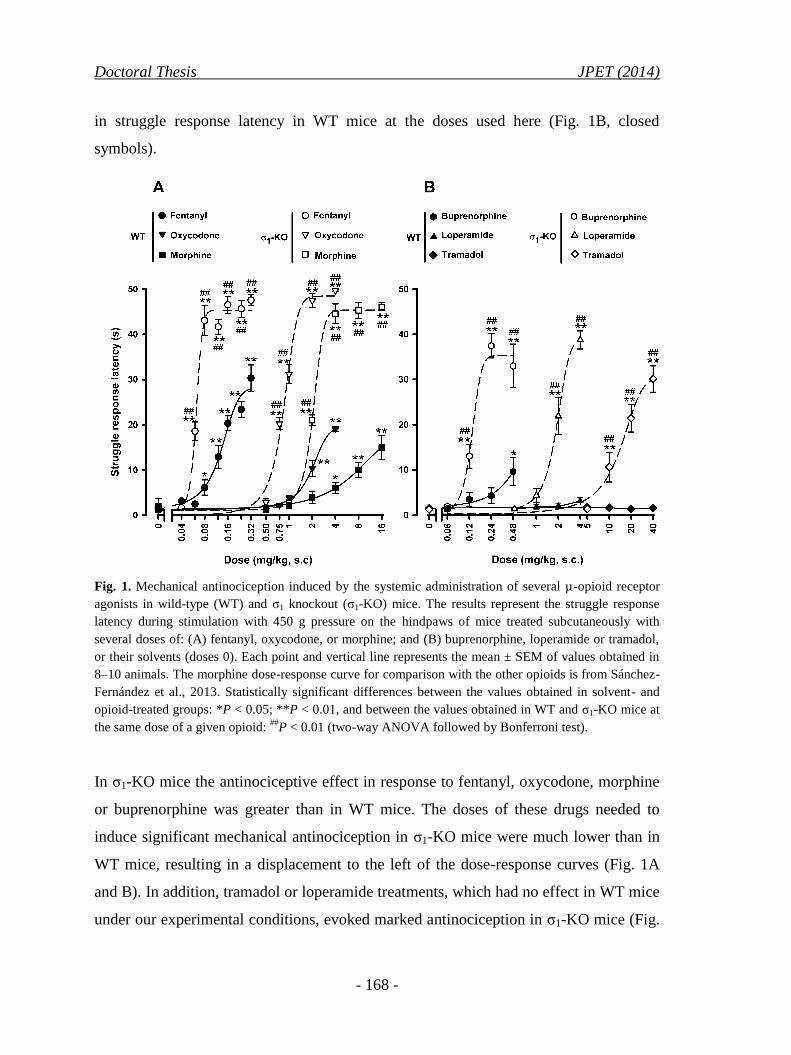
Doctoral Thesis JPET (2014)
- 168 -
in struggle response latency in WT mice at the doses used here (Fig. 1B, closed
symbols).
Fig. 1. Mechanical antinociception induced by the systemic administration of several µ-opioid receptor
agonists in wild-type (WT) and σ1 knockout (σ1-KO) mice. The results represent the struggle response
latency during stimulation with 450 g pressure on the hindpaws of mice treated subcutaneously with
several doses of: (A) fentanyl, oxycodone, or morphine; and (B) buprenorphine, loperamide or tramadol,
or their solvents (doses 0). Each point and vertical line represents the mean ± SEM of values obtained in
8–10 animals. The morphine dose-response curve for comparison with the other opioids is from Sánchez-
Fernández et al., 2013. Statistically significant differences between the values obtained in solvent- and
opioid-treated groups: *P < 0.05; **P < 0.01, and between the values obtained in WT and σ1-KO mice at
the same dose of a given opioid: ##
P < 0.01 (two-way ANOVA followed by Bonferroni test).
In σ1-KO mice the antinociceptive effect in response to fentanyl, oxycodone, morphine
or buprenorphine was greater than in WT mice. The doses of these drugs needed to
induce significant mechanical antinociception in σ1-KO mice were much lower than in
WT mice, resulting in a displacement to the left of the dose-response curves (Fig. 1A
and B). In addition, tramadol or loperamide treatments, which had no effect in WT mice
under our experimental conditions, evoked marked antinociception in σ1-KO mice (Fig.

JPET (2014) Doctoral Thesis
- 169 -
1B, open symbols). The largest antinociceptive effects in σ1-KO mice were recorded
after treatment with fentanyl, oxycodone or morphine, and struggle response latency
reached values close to the cutoff time (50 s) (Fig. 1A, open symbols). Interestingly, σ1-
KO animals treated with the highest doses of buprenorphine, tramadol or even the
peripheral µ-agonist loperamide showed latencies higher than those in WT mice treated
with fentanyl, oxycodone or morphine (Fig. 1B, open symbols). Therefore, the genetic
inactivation of σ1 receptors clearly potentiated the mechanical antinociception induced
by the systemic administration of µ-opioid agonists, including antinociception induced
by the peripherally restricted opioid loperamide.
2.4.2. Contribution of peripheral opioid receptors to antinociception induced by
the systemic (subcutaneous) administration of µ-opioid analgesics in wild-type
mice
To shed light on the role of peripheral opioid receptors in the mechanical
antinociception induced by µ-opioid analgesics in WT mice, we tested the sensitivity of
this antinociception to the peripherally restricted opioid antagonist naloxone
methiodide.
In WT mice treated s.c. with fentanyl (0.16 mg/kg), oxycodone (4 mg/kg), morphine
(16 mg/kg) or buprenorphine (0.48 mg/kg) and pretreated with the naloxone methiodide
solvent, struggle response latency increased markedly in comparison to untreated mice
(Fig. 2). Tramadol and loperamide were not evaluated in this experiment because they
did not induce any antinociceptive effect at any dose tested in WT mice (Fig. 1B).
Pretreatment with naloxone methiodide (2 – 8 mg/kg) did not alter the increase in
struggle response latency induced by fentanyl, morphine or buprenorphine (Fig. 2).
However, the antinociception induced by oxycodone was partially reversed by the
highest dose of this peripheral opioid antagonist (Fig. 2). As expected, the centrally-
penetrant opioid antagonist naloxone (0.5 mg/kg, s.c.) was able to completely reverse
the antinociceptive effect induced by all μ-agonists tested: latencies were
undistinguishable from those in untreated mice (Fig. 2).
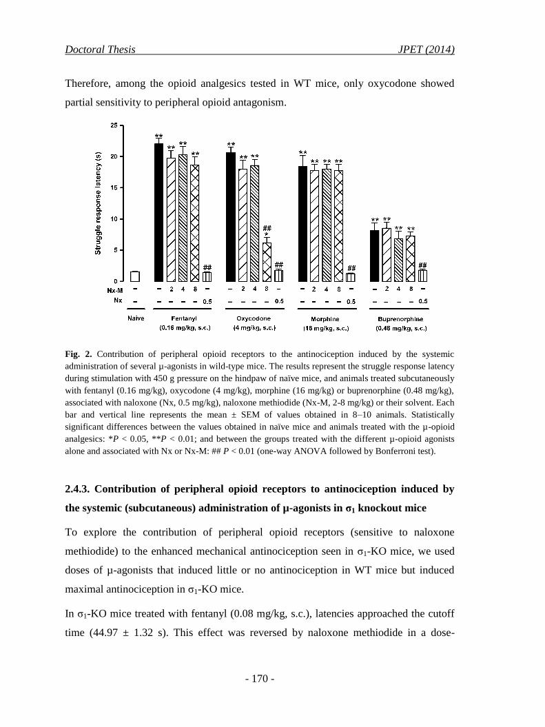
Doctoral Thesis JPET (2014)
- 170 -
Therefore, among the opioid analgesics tested in WT mice, only oxycodone showed
partial sensitivity to peripheral opioid antagonism.
Fig. 2. Contribution of peripheral opioid receptors to the antinociception induced by the systemic
administration of several µ-agonists in wild-type mice. The results represent the struggle response latency
during stimulation with 450 g pressure on the hindpaw of naïve mice, and animals treated subcutaneously
with fentanyl (0.16 mg/kg), oxycodone (4 mg/kg), morphine (16 mg/kg) or buprenorphine (0.48 mg/kg),
associated with naloxone (Nx, 0.5 mg/kg), naloxone methiodide (Nx-M, 2-8 mg/kg) or their solvent. Each
bar and vertical line represents the mean ± SEM of values obtained in 8–10 animals. Statistically
significant differences between the values obtained in naïve mice and animals treated with the µ-opioid
analgesics: *P < 0.05, **P < 0.01; and between the groups treated with the different µ-opioid agonists
alone and associated with Nx or Nx-M: ## P < 0.01 (one-way ANOVA followed by Bonferroni test).
2.4.3. Contribution of peripheral opioid receptors to antinociception induced by
the systemic (subcutaneous) administration of µ-agonists in σ1 knockout mice
To explore the contribution of peripheral opioid receptors (sensitive to naloxone
methiodide) to the enhanced mechanical antinociception seen in σ1-KO mice, we used
doses of µ-agonists that induced little or no antinociception in WT mice but induced
maximal antinociception in σ1-KO mice.
In σ1-KO mice treated with fentanyl (0.08 mg/kg, s.c.), latencies approached the cutoff
time (44.97 ± 1.32 s). This effect was reversed by naloxone methiodide in a dose-

JPET (2014) Doctoral Thesis
- 171 -
dependent manner (0.5 – 2 mg/kg, s.c), and a dose as low as 2 mg/kg completely
reversed the potentiation of fentanyl antinociception seen in σ1-KO mice (Fig. 3A). This
dose of naloxone methiodide was chosen to test the involvement of peripheral opioid
receptors in the mechanical antinociception induced by several other µ-agonists in σ1-
KO mice. The mechanical antinociception induced by the s.c. administration of
oxycodone (2 mg/kg), morphine (4 mg/kg), buprenorphine (0.24 mg/kg), loperamide (4
mg/kg) or tramadol (40 mg/kg) in σ1-KO mice was completely abolished by naloxone
methiodide (Fig. 3B).
Fig. 3. Contribution of peripheral opioid receptors to the antinociception induced by the systemic
administration of low doses of several µ-agonists in σ1 knockout mice. The results represent the struggle
response latency during stimulation with 450 g pressure on the hindpaws of (A) mice treated
subcutaneously with the combination of fentanyl (0.08 mg/kg), and naloxone methiodide (Nx-M, 0.5-2
mg/kg) or its solvent, and mice treated with the solvent of both drugs (white bar), or (B) naïve mice and
mice treated subcutaneously with oxycodone (2 mg/kg), morphine (4 mg/kg), buprenorphine (0.24
mg/kg), loperamide (4 mg/kg) or tramadol (40 mg/kg), associated with Nx-M (2 mg/kg) or its solvent.
Each bar and vertical line represents the mean ± SEM of values obtained in 8–10 animals. Statistically
significant differences between the values obtained in mice treated and not treated with the µ-opioid
analgesics: **P < 0.01; and between the groups treated with different µ-opioid agonists associated with
Nx-M or its solvent: ##
P < 0.01 (one-way ANOVA followed by Bonferroni test).

Doctoral Thesis JPET (2014)
- 172 -
Therefore, the peripheral opioid antagonist naloxone methiodide was able to fully
reverse the enhanced mechanical antinociception induced by several µ-agonists in σ1-
KO mice.
2.4.4. Effects of systemic (subcutaneous) administration of the selective σ1
antagonist BD-1063 on mechanical antinociception induced by µ-agonists in WT
mice: involvement of σ1- and peripheral opioid receptors
We tested whether the pharmacological blockade of σ1 receptors in WT mice would
replicate the phenotype seen in σ1-KO mice, potentiating µ-opioid analgesia.
BD-1063 (8 – 32 mg/kg) increased the antinociception induced by a low dose of
fentanyl (0.08 mg/kg, s.c.) in a dose-dependent manner (Fig. 4A). To confirm the
selectivity of this effect, we tested its sensitivity to treatment with PRE-084, a σ1
agonist. PRE-084 (8 – 32 mg/kg) completely reversed, in a dose-dependent manner, the
potentiation of fentanyl antinociception induced by BD-1063 (Fig. 4B).
To test the involvement of peripheral opioid receptors in the potentiation of fentanyl-
induced antinociception by systemic treatment with BD-1063, we assayed the
sensitivity of this effect to naloxone methiodide. A dose of 2 mg/kg (s.c.) of this
peripheral opioid antagonist completely reversed the antinociception induced by the
association of BD-1063 with fentanyl (Fig. 4B). BD-1063, PRE-084 and naloxone
methiodide did not modify struggle response latency per se (data not shown).
Our results were not affected by sex differences, since as described above for female
mice, the fentanyl-induced antinociception in response to the systemic administration of
BD-1063 (32 mg/kg, s.c.) was also greatly increased in male mice. This enhanced
antinociception was also abolished by PRE-084 (32 mg/kg, s.c.) or Nx-M (2 mg/kg,
s.c.) in male mice (data not shown).
We also studied the effects of the association of BD-1063 (32 mg/kg, s.c.) with other μ-
opioid receptor agonists administered at doses that induce little or no significant
antinociceptive activity in WT mice.

JPET (2014) Doctoral Thesis
- 173 -
Fig. 4. Contribution of σ1 and peripheral opioid receptors to the antinociception induced by the
association of fentanyl (0.08 mg/kg) and BD-1063, both administered systemically to wild-type mice. The
results represent the struggle response latency during stimulation with 450 g pressure on the hindpaws of
(A) mice treated subcutaneously (s.c.) with fentanyl and BD-1063 (8-32 mg/kg) or its solvent, and (B)
mice s.c. given fentanyl (0.08 mg/kg) or fentanyl (0.08 mg/kg) + BD-1063 (32 mg/kg) associated to PRE-
084 (8-32 mg/kg, s.c.), naloxone methiodide (Nx-M, 2 mg/kg, s.c.) or their solvent. Each bar and vertical
line represents the mean ± SEM of values obtained in 8–10 animals. Statistically significant differences
between the values obtained in the groups treated with fentanyl associated with BD-1063 or its solvent:
**P < 0.01; and between the values obtained in mice treated with the combination of fentanyl and BD-
1063, and its association with PRE-084 or Nx-M: ##
P < 0.01 (one-way ANOVA followed by Bonferroni
test).
The association of BD-1063 with oxycodone (2 mg/kg, s.c.), morphine (4 mg/kg, s.c.),
buprenorphine (0.24 mg/kg, s.c.), tramadol (40 mg/kg, s.c.) or even loperamide (4
mg/kg, s.c.) induced a statistically significant increase in struggle response latency
compared to animals treated with the opioid drugs alone (Fig. 5). This increase was
completely reversed by PRE-084 (32 mg/kg, s.c.) (Fig. 5). Naloxone methiodide (2
mg/kg, s.c.) decreased the effect of the association of BD-1063 and the µ-agonists to the
same extent as the effect of µ-opioids administered alone, with the exception of the

Doctoral Thesis JPET (2014)
- 174 -
combination of BD-1063 and oxycodone: the effect of this combination was decreased
more markedly than the effect of oxycodone alone (Fig. 5).
Fig. 5. Contribution of σ1 and peripheral opioid receptors to the antinociception induced by the
association of several µ-agonists and BD-1063, both administered systemically to wild-type mice. The
results represent the struggle response latency during stimulation with 450 g pressure on the hindpaw of
mice treated subcutaneously (s.c.) with BD-1063 (32 mg/kg) or its solvent in combination with
oxycodone (2 mg/kg), morphine (4 mg/kg), buprenorphine (0.24 mg/kg), loperamide (4 mg/kg) or
tramadol (40 mg/kg), and its association with the s.c. administration of PRE-084 (32 mg/kg), naloxone
methiodide (Nx-M, 2 mg/kg) or their solvent. Each bar and vertical line represents the mean ± SEM of
values obtained in 8–10 animals. Statistically significant differences between the values obtained in the
groups treated with different µ-opioid agonists associated with BD-1063 or its solvent: **P < 0.01; and
between the values obtained in mice treated with the combination of a given µ-agonist with BD-1063, and
its association with PRE-084 or Nx-M: ##
P < 0.01 (one-way ANOVA followed by Bonferroni test).
Therefore, systemic treatment with the σ1 antagonist BD-1063 synergistically enhanced
the antinociception induced by the systemic administration of all µ-agonists tested. This
effect was sensitive to both the σ1 receptor agonist PRE-084 and the peripheral opioid
antagonist naloxone methiodide.
JPET (2014) Doctoral Thesis
- 175 -
2.4.5. Effects of local (intraplantar) administration of the selective σ1 antagonist
BD-1063 on mechanical antinociception induced by µ-agonists in wild-type mice:
involvement of σ1 and peripheral opioid receptors
We also tested whether the local administration of BD-1063 into the hindpaw was
sufficient to enhance µ-opioid-induced mechanical antinociception. As in the
experiments described in the preceding section, low doses of systemic µ-agonists were
injected s.c. in WT mice, but BD-1063 was injected i.pl. to test for locally-induced
effects.
In WT mice, fentanyl injected s.c. (0.08 mg/kg) and BD-1063 solvent (saline) injected
i.pl. led to similarly short struggle response latencies in the treated paw and the
contralateral (untreated) paw (Fig. 6A). However, when this dose of fentanyl was
associated to BD-1063 (50-200 μg, i.pl.), a dose-dependent increase in latency was seen
only in the treated hindpaw (black bars in Fig. 6A).
To test the σ1 specificity of the effects induced by local treatment with BD-1063, we
used the σ1 agonist PRE-084, and also evaluated the effects of naloxone methiodide to
determine whether peripheral opioid receptors were involved in the enhanced
antinociception induced by the combination of fentanyl and local σ1 pharmacological
blockade. PRE-084 (8-32 mg, s.c.) completely reversed, and in a dose-dependent
manner, the mechanical antinociceptive effect induced by the combination of fentanyl
(0.08 mg/kg, s.c.) and BD-1063 (200 μg, i.pl.) (Fig. 6B). Treatment with naloxone
methiodide (2 mg/kg, s.c.) was also able to completely abolish the local potentiation of
fentanyl-induced antinociception by BD-1063 (200 μg, i.pl.) (Fig. 6B). Neither PRE-
084 nor naloxone methiodide induced any change in latencies of the noninjected paw in
animals treated with systemic fentanyl (white bars in Fig. 6B).

Doctoral Thesis JPET (2014)
- 176 -
Fig. 6. Contribution of σ1 and peripheral opioid receptors to the effects of the local administration of BD-
1063 on the antinociception induced by systemically administered fentanyl in wild-type mice. The results
represent the struggle response latency during stimulation with 450 g pressure on the hindpaw of mice
treated subcutaneously (s.c.) with: (A) fentanyl (0.08 mg/kg) + intraplantar (i.pl.) BD-1063 (50-200 µg)
or its solvent, and (B) fentanyl (0.08 mg/kg, s.c.) + BD-1063 (200 µg, i.pl.) in animals pretreated s.c. with
PRE-084 (8-32 mg/kg), naloxone methiodide (Nx-M, 2 mg/kg) or their solvent. Each bar and vertical line
represents the mean ± SEM of values obtained in 8–10 animal. (A and B) Statistically significant
differences between the values obtained upon stimulation after the injection of BD-1063 or its solvent in
the hindpaw: *P < 0.05, **P < 0.01; and between the values obtained from the injected and noninjected
hindpaws: ##
P < 0.01 (two-way ANOVA followed by Bonferroni test). (B) Statistically significant
differences between the values obtained in mice treated with the combination of fentanyl with BD-1063,
and its association with PRE-084 or Nx-M: †† P < 0.01 (two-way ANOVA followed by Bonferroni test).
We also tested the association of local treatment of BD-1063 (200 µg, i.pl.) with low
doses of the other µ-agonists. Local treatment with BD-1063 was also able to increase
struggle response latencies in animals that were given (s.c.) oxycodone (2 mg/kg),
morphine (4 mg/kg), buprenorphine (0.24 mg/kg), loperamide (4 mg/kg) or tramadol
(40 mg/kg) (Fig. 7). This effect was only seen in the treated hindpaw, whereas latencies
in the untreated hindpaw remained unaltered (data not shown). This increased latency in

JPET (2014) Doctoral Thesis
- 177 -
the hindpaw treated with BD-1063 in mice that were given µ-agonists was abolished by
treatment with either PRE-084 (32 mg/kg, s.c.) or naloxone methiodide (2 mg/kg, s.c.)
(Fig. 7). As in the experiments described in the preceding section, PRE-084 reversed the
effects of the local administration of BD-1063, but not beyond the level of the effects of
the µ-agonists alone (Fig. 7). Naloxone methiodide decreased latencies in animals
treated with BD-1063 together with µ-agonists to the same extent as the effect of the
opioids administered alone, with the exception of the combination of BD-1063 and
oxycodone: the effect of this combination was decreased more markedly than the effect
of oxycodone alone (Fig. 7).
Fig. 7. Contribution of σ1 and peripheral opioid receptors to the effects of the local administration of BD-
1063 on the antinociception induced by several µ-agonists administered systemically to wild-type mice.
The results represent the struggle response latency during stimulation with 450 g pressure on the injected
hindpaw in mice treated intraplantarly with BD-1063 (200 µg) or its solvent in combination with the
subcutaneous (s.c.) administration of oxycodone (2 mg/kg), morphine (4 mg/kg), buprenorphine (0.24
mg/kg), loperamide (4 mg/kg) or tramadol (40 mg/kg), and its association with the s.c. administration of
PRE-084 (32 mg/kg), naloxone methiodide (Nx-M, 2 mg/kg) or their solvent. Each bar and vertical line
represents the mean ± SEM of values obtained in 8–10 animals. Statistically significant differences
between the values obtained in the groups treated with the different µ-opioid agonists alone and
associated with BD-1063: **P < 0.01; and between the values obtained in mice treated with the
combination of a given µ-agonist with BD-1063, and its association with PRE-084 or Nx-M: ##
P < 0.01
(one-way ANOVA followed by Bonferroni test).

Doctoral Thesis JPET (2014)
- 178 -
Therefore, local treatment with the σ1 antagonist BD-1063 synergistically enhanced the
antinociception induced by all µ-agonists tested when they were given systemically.
This effect was observed only in the treated paw and was sensitive to both σ1 agonism
and peripheral opioid antagonism.
2.4.6. Effects of systemic (subcutaneous) and local (intraplantar) administration of
the selective σ1 antagonist S1RA on mechanical antinociception induced by
fentanyl and loperamide in wild-type mice: involvement of σ1- and peripheral
opioid receptors
To test whether the effects induced by BD-1063 were replicable by a different σ1
antagonist, we evaluated the effects of S1RA on the antinociceptive effects induced by
two different µ-opioids: the centrally active fentanyl and the peripherally restricted
loperamide.
The systemic administration of S1RA (64 mg/kg, s.c.) increased the struggle response
latencies in animals treated s.c. with either fentanyl (0.08 mg/kg) or loperamide (4
mg/kg) (Fig. 8A). The i.pl. administration of S1RA (200 µg) was also able to increase
the response latency in mice treated with these µ-opioid agonists (Fig. 8B), and this
enhanced antinociception was detectable in the injected but not in the contralateral paw
(data not shown). As described in the preceding sections regarding the effects of BD-
1063, treatment with either the σ1 agonist PRE-084 (32 mg/kg, s.c.) or the peripheral
opioid antagonist naloxone methiodide (2 mg/kg, s.c.) abolished the effects on opioid
antinociception of S1RA administered either systemically or locally (Fig. 8A and B,
respectively).
Therefore, systemic or local treatment with the σ1 antagonist S1RA synergistically
enhanced the antinociception induced by the systemic administration of fentanyl or
loperamide, and these effects were fully reversed by either σ1 agonism or peripheral
opioid antagonism.

JPET (2014) Doctoral Thesis
- 179 -
Fig. 8. Contribution of σ1 and peripheral opioid receptors to the effects of the systemic and local
administration of S1RA on the antinociception induced by systemically administered fentanyl and
loperamide in wild-type mice. Animals were treated (A) subcutaneously (s.c.) or (B) intraplantarly (i.pl.)
with S1RA (64 mg/kg and 200 µg, respectively) in combination with fentanyl (0.08 mg/kg, s.c.) or
loperamide (4 mg/kg, s.c.), and its association with the s.c. administration of PRE-084 (32 mg/kg),
naloxone methiodide (Nx-M, 2 mg/kg) or their solvent. The results represent the struggle response
latency in mice during stimulation with 450 g pressure on the hindpaws (A) or on the injected hindpaw
(B). Each bar and vertical line represents the mean ± SEM of values obtained in 8–10 animals.
Statistically significant differences between the values obtained in the groups treated with the µ-opioid
agonists associated with S1RA or its solvent: **P < 0.01; and between the values obtained in mice treated
with the combination of a given µ-agonist with S1RA, and its association with PRE-084 or Nx-M: ##
P <
0.01 (one-way ANOVA followed by Bonferroni test).
2.4.7. Sigma-1 receptor expression in the central and peripheral nervous system
To obtain anatomical support for the marked behavioral effects of local σ1 receptor
blockade on opioid antinociception, we compared the expression of σ1 receptors in
different areas of the nervous system involved in opioid analgesia, including supraspinal
(BLA, RVM and PAG), spinal (dSC) and peripheral nervous locations (DRG). All
samples from WT mice yielded immunoreactive bands at a molecular weight slightly

Doctoral Thesis JPET (2014)
- 180 -
higher than 25 kDa (Fig. 9A), which is consistent with the molecular weight of the
cloned σ1 receptor from the mouse (~28 kDa) (Pan et al., 1998).
Fig. 9. Expression of sigma-1 (σ1) receptors in the basolateral amygdala (BLA), rostroventral medulla
(RVM), periaqueductal grey matter (PAG), dorsal spinal cord from the lumbar enlargement (dSC), and
lumbar dorsal root ganglia (DRGs) in wild-type (WT) and σ1 knockout (σ1-KO) mice. (A) Representative
immunoblots for σ1 receptors in WT and σ1-KO mice. β-actin was used as the loading control. The
migration positions of molecular weight standards (in kDa) are shown to the left of the gel. (B)
Quantification of immunoblotting for the σ1 receptor in WT and σ1-KO mice. Each bar and vertical line
represents the mean ± SEM of the densitometric values obtained in 8 animals. The σ1 receptor band
intensities were relativized to those of their corresponding β-actin loading control bands. Statistically
significant differences between the values obtained in samples of central nervous system regions (BLA,
RVM, PAG, dSC) and the lumbar dorsal root ganglia (DRGs) from WT mice **P < 0.01. No σ1 receptor
expression was found in samples from σ1-KO mice.

JPET (2014) Doctoral Thesis
- 181 -
We found no significant differences in σ1 receptor band intensities among all central
areas in WT mice (Fig. 9B); however, σ1 receptor density was much higher in DRG
samples than in any of the central areas examined (Fig. 9A and B). To verify the
specificity of the anti-σ1 receptor antibody, we tested its immunoreactivity in samples
from σ1-KO mice. We found no immunoreactive σ1 receptor bands in PAG, dSC or
DRG samples from σ1-KO animals (Fig. 9A and B), or in samples of BLA or RVM
from these mice (data not shown). The absence of these bands in σ1-KO samples argues
for the specificity of the σ1 antibody used. In addition, we were unable to detect
immunoreactive bands in WT samples when tested in the absence of anti-σ1 antibody
(data not shown), which further confirms the specificity of the procedure.
The high level of σ1 receptor expression in peripheral nervous tissue argues for a
possible major role of peripheral σ1 receptors in nociception.
2.4.8. Effects of fentanyl and loperamide on gastrointestinal transit in wild-type
mice, wild-type mice treated with BD-1063, and σ1 knockout mice
Gastrointestinal transit distances did not differ significantly between WT mice, WT
mice treated with BD-1063 (32 mg/kg, s.c.) and σ1-KO mice that had been given the
fentanyl (Fig. 10A) or loperamide solvents (Fig. 10B). In all cases the charcoal meal
traversed about 30 cm of the small intestine. Both fentanyl (0.04 – 0.16 mg/kg, s.c.) and
loperamide (0.125 – 1 mg/kg, s.c.) induced a dose-dependent decrease in
gastrointestinal transit in WT mice (white bars in Fig. 10A and 10B). For each dose of
fentanyl (Fig. 10A) or loperamide (Fig. 10B), this decrease was similar in WT mice
treated with BD-1063 and in σ1-KO mice. Therefore, σ1 inhibition alone did not alter
gastrointestinal transit and did not modify the effects of fentanyl or loperamide on this
outcome.

Doctoral Thesis JPET (2014)
- 182 -
Fig. 10. Effects of the systemic (subcutaneous, s.c.) administration of several doses of fentanyl (A) and
loperamide (B) on gastrointestinal transit of wild-type mice (WT), WT mice treated with BD-1063 (32
mg/kg, s.c.), and σ1 knockout mice (σ1-KO). BD-1063 or its solvent was injected, and 5 min later the
animals were treated with fentanyl, loperamide or their solvents. 30 minutes after the administration of
the opioid drug, the mice were given a 0.5% charcoal suspension intragastrically. Transit of the charcoal
was measured 30 min after administration. Each bar and vertical line represents the mean ± SEM of
values obtained in 8-12 mice. Statistically significant differences between the values obtained in saline-
and opioid-treated groups: **P <0.01. No statistically significant differences were found between
genotypes with the same treatment, or between animals treated and not treated with BD-1063 (two-way
ANOVA followed by Bonferroni test).
2.4.9. Affinity of µ-opioid drugs for [3H](+)-pentazocine binding sites
We used competition binding assays to test the affinity of the opioid drugs assayed in
vivo for [3H](+)-pentazocine-labeled σ1 receptors in brain membranes from WT mice.
As expected, the known σ1 antagonist BD-1063 inhibited [3H](+)-pentazocine specific
binding in a concentration-dependent manner, with an IC50 value of 40.21 ± 3.24 nM.
This value was similar to that found in previous studies (Entrena 2009a; Cobos et al.,
2005, 2006 and 2007). However, none of the opioid drugs (fentanyl, oxycodone,
morphine, buprenorphine, tramadol, loperamide, naloxone or naloxone methiodide)
significantly inhibited [3H](+)-pentazocine binding at any concentration tested (ranging

JPET (2014) Doctoral Thesis
- 183 -
from 10-10 to 10-6 M), and therefore their affinity for [3H](+)-pentazocine binding sites
was considered negligible (Fig. 11).
Fig. 11. Inhibition by unlabeled drugs of
[3H](+)-pentazocine binding to brain
membranes (P2 fraction) in wild-type mice.
[3H](+)-pentazocine (5 nM) was incubated
with 0.8–1 mg/mL brain membrane protein
at 30 °C, pH 8, for 240 min and increasing
concentrations of BD-1063, fentanyl,
oxycodone, morphine, buprenorphine,
loperamide, tramadol, naloxone or naloxone
methiodide. The data shown are the
averages of two experiments carried out in
triplicate.
2.5. DISCUSSION
We found that several centrally penetrant µ-opioid analgesics (fentanyl, oxycodone,
morphine, buprenorphine and tramadol) and the peripheral µ-agonist loperamide, at
systemic doses which induce little or no antinociception in control animals, have a
marked antinociceptive effect when boosted by σ1 receptor inhibition (σ1–KO mice or
σ1 pharmacological blockade). We show that this enhanced opioid antinociception is
mediated peripherally, since it can be achieved by local σ1 pharmacological blockade,
and is sensitive to peripheral opioid antagonism. However, the increase in peripheral
opioid antinociception by σ1 receptor inhibition is not accompanied by increased
inhibition of opioid-induced gastrointestinal transit, a known peripherally–mediated side
effect of opioids.
It has been proposed that the analgesic mechanisms of different µ-opioids used in
clinical settings overlap only partially (Ocaña et al., 1995; Pasternak, 2004; Pasternak
and Pan 2011; Smith, 2008; Raehal et al., 2011). Hence the importance of testing
whether the widely reported enhancement of morphine antinociception by σ1 inhibition
(e.g. Chien and Pasternak 1993; Mei and Pasternak 2007; Díaz et al., 2009; Sánchez-

Doctoral Thesis JPET (2014)
- 184 -
Fernández et al., 2013) also occurs with other clinically relevant µ-opioids. It was
recently reported that the systemic (subcutaneous) administration of S1RA enhanced
antinociception against a thermal stimulus induced by morphine and other systemically
administered centrally-penetrant µ-opioid analgesics (fentanyl, oxycodone,
buprenorphine and tramadol) (Vidal-Torres et al., 2013). We now extend those results
by showing that this σ1 antagonist and also BD-1063 enhance opioid antinociception
against a different type of nociceptive (mechanical) stimulus, and more importantly, that
the enhanced antinociception is mediated peripherally (see below). Moreover, we show
that opioid-induced mechanical antinociception is clearly potentiated in σ1-KO mice,
which contrasts with the previously reported absence of modulation of opioid thermal
antinociception in σ1-KO mice (Díaz et al., 2009; Vidal-Torres et al., 2013). These
apparently contradictory results seem to be related to the type of sensory stimulation
used, and may be attributable to the known differences in the neurochemical
mechanisms of thermal and mechanical opioid antinociception (Kuraishi et al., 1995;
Wegert et al., 1997). These mechanisms may be affected differentially by possible
compensatory mechanisms in σ1-KO mice.
In addition to centrally-acting µ-opioid analgesics, we also tested the effects of the
peripheral µ-opioid agonist loperamide under normal conditions and during σ1
inhibition. In agreement with previous reports, loperamide had no antinociceptive
effects in WT mice (Menendez et al., 2005; Sevostianova et al., 2005). However, in σ1–
KO mice or WT mice treated systemically with BD-1063 or S1RA, we observed a
profound antinociceptive effect in response to loperamide. Therefore, σ1 inhibition is
sufficient to unmask the strong antinociceptive effects of this peripherally acting µ-
opioid. This finding evidences that peripheral µ-opioid analgesia is enhanced by σ1
inhibition. In fact, the local (intraplantar) administration of σ1 antagonists was able to
enhance µ-antinociception in response to all µ-agonists tested. This enhanced
antinociception occurred only in the paw injected with the σ1 antagonist, but not in the
contralateral paw, clearly indicating that the effects of these σ1 antagonists occurred
locally. The selective σ1 agonist PRE-084 reversed the effects of BD-1063 or S1RA
(administered either systemically or locally), arguing for a σ1-mediated action.

JPET (2014) Doctoral Thesis
- 185 -
Interestingly, PRE-084 was unable to reverse the enhanced antinociception seen in σ1-
KO mice treated with fentanyl, supporting on-target mechanisms in the effects induced
by this σ1 agonist (data not shown).
To investigate the role of peripheral opioid receptors in the antinociceptive effect of
systemically administered µ-agonists under normal conditions and during σ1 inhibition,
we tested the sensitivity of antinociception to peripheral opioid antagonism in both
situations. The antinociceptive effects of the opioids fentanyl, morphine and
buprenorphine in WT mice (in the absence of σ1 blockade) were sensitive to the
centrally penetrant opioid antagonist naloxone. However, they were resistant to the
peripherally restricted antagonist naloxone methiodide. These findings support a
preferential location of the antinociceptive effects of µ-opioids at central levels under
our experimental conditions, and are consistent with previous studies (Greenwood-Van
Meerveld and Standifer, 2008; Thomas et al., 2008; Joshi et al., 2008; Khalefa et al.,
2012; Ringkamp and Raja, 2012). Among the opioids tested in WT mice (in the absence
of σ1 blockade), only oxycodone had an antinociceptive effect that was partially
reversible by naloxone methiodide, and this only occurs at the highest dose (8 mg/kg) of
the antagonist. Our findings are consistent with clinical data suggesting that part of the
analgesic effects of oxycodone may be mediated peripherally (Olesen et al., 2010). The
sensitivity to peripheral opioid antagonism is diametrically different when opioid
antinociception is enhanced by σ1 inhibition. A dose as low as 2 mg/kg of naloxone
methiodide was enough to completely abolish the opioid antinociception induced by all
µ-agonists tested, not only in WT mice treated locally with a σ1 antagonist (in which its
effects are clearly peripherally mediated), but also in σ1–KO mice or WT mice treated
systemically with the σ1 antagonists. This does not argue against the widely reported
potentiation of opioid antinociception by central σ1 receptor inhibition (King et al.,
1997; Pan et al., 1998; Mei and Pasternak 2002 and 2007; Marrazzo et al., 2006), but
indicates that when both opioid agonism and σ1 inhibition are induced systemically, the
increase in antinociception occurs mainly at the peripheral level. In support of the
peripheral location of these modulatory effects of σ1 receptors, we found much higher

Doctoral Thesis JPET (2014)
- 186 -
levels of these receptors in the DRG than in several areas of the central nervous system
that play a key role in opioid antinociception.
Among the opioid drugs tested in the present study, only morphine and naloxone had
previously been shown to lack affinity for σ1 receptors (Walker et al., 1990). Here we
show that this lack of σ1 affinity is shared by other opioid drugs (fentanyl, oxycodone,
buprenorphine, tramadol, loperamide and naloxone methiodide). In addition, it is known
that the σ1 drugs tested here (BD-1063, S1RA and PRE-084) do not bind to µ-opioid
receptors (Matsumoto et al., 1995; Sánchez-Fernández et al., 2013). Therefore, our
results support a functional link between peripheral σ1 receptors and the µ-opioid
system rather than interactions of σ1 ligands with µ opioid receptors or opioid drugs
with σ1 receptors.
It was recently reported that σ1 antagonism potentiates µ-opioid signaling (measured as
the increase in DAMGO-induced [35
S]GTPγS binding), providing a mechanistic
explanation for the enhanced opioid antinociception by σ1 inhibition (Kim et al. 2010).
Previous studies of thermal nociception show that σ1 inhibition potentiates µ, κ and δ
opioid antinociception at central levels (King et al., 1997; Mei and Pasternak, 2002).
Therefore, the modulation of peripheral µ opioid antinociception by σ1 inhibition that
we report here might occur with other subtypes of opioid receptors, although this
hypothesis remains to be tested.
Clinically, a dose-limiting factor in obtaining maximal analgesia with systemic opioids
is the risk of adverse side effects. Some of these effects are mediated through peripheral
opioid receptors (constipation), whereas others are mediated at supraspinal sites
(addiction, dependency, nausea, somnolence, respiratory depression) (Benyamin et al
2008; Greenwood-Van Meerveld and Standifer, 2008; Al-Hasani and Bruchas, 2011;
Ringkamp and Raja, 2012). Targeting peripheral opioid receptors has been proposed as
a strategy to minimize opioid-induced side effects that are produced centrally (Sehgal et
al., 2011; Ringkamp and Raja, 2012). However, since most of the analgesia from
systemic opioids is produced normally at central sites, it seems difficult to dissociate
antinociceptive effects from centrally-induced side effects. In this regard, we previously

JPET (2014) Doctoral Thesis
- 187 -
reported that despite the potentiation of antinociception by σ1 inhibition, the central side
effects of morphine (such as hyperlocomotion, physical dependence or mydriasis) were
not altered (Sánchez-Fernández et al., 2013; Vidal-Torres et al, 2013). Here we show
that the enhancement of opioid antinociception by systemic σ1 inhibition occurs
primarily at peripheral levels, which might explain the lack of potentiation of morphine-
induced central side effects. Interestingly, morphine-induced constipation, which is the
most clinically relevant peripheral side effect of µ-opioids, was also not modulated by
σ1 receptors (Chien and Pasternak, 1994; Sánchez-Fernández et al., 2013; Vidal-Torres
et al, 2013); however, it is unknown whether this characteristic is shared by other
opioids. To find out, we tested two additional µ-opioids of very different characteristics:
the central analgesic fentanyl and the peripherally-acting loperamide. Importantly, the
gastrointestinal transit inhibition induced by these opioids was unaffected in either σ1–
KO mice or WT mice treated with BD-1063. Regardless of the exact mechanistic nature
of the differential modulation of opioid antinociception and adverse events by σ1
inhibition, our findings point to a potentially beneficial avenue of research aimed at
improving the safety profile of opioid drugs.
In summary, we found that σ1 receptor inhibition enhanced the peripheral opioid
antinociception induced by clinically relevant µ-agonists, but did not increase opioid-
induced constipation. These data support the conclusion that peripheral σ1 receptors are
a biological brake to µ-opioid antinociception, and that either systemic or local σ1
receptor inhibition is potentially useful as an adjuvant to enhance peripheral µ-opioid
analgesia. Combinations of σ1 antagonists and µ-opioid agonists may be of therapeutic
interest in terms of both efficacy and safety, and merit clinical studies.

Doctoral Thesis JPET (2014)
- 188 -
2.6. ACKNOWLEDGEMENTS
C. Sánchez-Fernández and R. González-Cano were supported by FPU grants from the
Spanish Ministerio de Educación y Ciencia (MEC), and E. J. Cobos was supported by
the Research Program of the University of Granada. This research was done in partial
fulfillment of the requirements for the doctoral thesis of C. Sánchez-Fernández. The
authors thank K. Shashok for revising the English style of the manuscript.

CCCOOONNNCCCLLLUUUSSSIIIOOONNNSSS


Conclusions Doctoral Thesis
- 189 -
SPECIFIC CONCLUSIONS
1) Inhibition of σ1 receptor function (in σ1-KO mice or by the systemic or local σ1
pharmacological antagonism in wild-type mice) does not alter the nociceptive
response to a blunt mechanical stimulation.
2) The mechanical antinociceptive effect induced by the systemic administration of
µ-opioid agonists clinically used as analgesics (fentanyl, morphine, oxycodone,
buprenorphine and tramadol) is increased in σ1-KO mice and in wild-type mice
treated systemically with σ1 antagonists.
3) Inhibition of σ1 receptor function, in σ1-KO mice or by the systemic σ1
pharmacological antagonism in wild-type mice, is able to unmask
antinociceptive effects induced by the peripherally restricted opioid agonist
loperamide.
4) Mechanical antinociception induced by the systemic administration of opioid
analgesics to wild-type mice under normal conditions (in the absence of σ1
receptor inhibition) is mainly produced at central levels. However, the
potentiation of opioid antinociception in σ1-KO mice and in wild-type mice
treated systemically with σ1 antagonists completely depends on the activation of
peripheral opioid receptors, because it is abolished by naloxone methiodide.
5) Local σ1-pharmacological blockade is enough to enhance the peripheral
antinociceptive effects induced by the systemic administration of opioid drugs
clinically used as analgesics as well as to unmask an antinociceptive effect of
loperamide.
6) Although the local administration of morphine does not produce any
antinociceptive effect against a mechanical stimulus, it induces a marked effect
in σ1-KO or in wild-type mice locally treated with σ1 antagonists.
7) The expression of σ1 receptors is higher in the peripheral nervous system (DRG)
than in several central areas involved in opioid analgesia (at either supraspinal or
spinal levels), which supports the role of σ1 receptors in nociception at the
peripheral level.

Doctoral Thesis Conclusions
- 190 -
8) Inhibition of σ1 receptor function does not interfere with other non-analgesic
opioid effects, including morphine-induced hyperlocomotion (a centrally
mediated effect) and the decrease of gastrointestinal transit (a peripherally
mediated effect) induced by morphine, fentanyl and loperamide.
9) σ1-KO mice do not present adaptive changes in density or affinity of μ-opioid
receptors in the brain, spinal cord or hind-paw plantar skin which could account
for their enhanced opioid antinociception.
10) The μ-opioid ligands used (fentanyl, morphine, oxycodone, buprenorphine,
tramadol, loperamide, naloxone and naloxone methiodide) lack affinity for σ1
receptors. Also, the σ1 ligands used (BD-1063, BD 1047, NE-100, S1RA and
PRE-084) are devoid of affinity for μ opioid receptors. Therefore, our behavioral
results cannot be explained by direct interaction of opioid drugs with σ1
receptors, or σ1 ligands with µ-opioid receptors.
GENERAL CONCLUSION
Systemic or local inhibition of σ1 receptor function increases peripheral opioid
antinociception to mechanical stimuli, suggesting that the σ1 receptor is a biological
brake to peripheral opioid analgesia. This potentiation of opioid analgesia is not
accompanied by an increase of non-analgesic central or peripheral (hyperlocomotion
and inhibition of gastrointestinal transit, respectively) opioid effects. Therefore, σ1
antagonism might be clinically useful as a systemic or local adjuvant to increase opioid
analgesia without increasing opioid side effects.

LLLIIISSSTTT OOOFFF AAABBBBBBRRREEEVVVIIIAAATTTIIIOOONNNSSS


Abbreviations Doctoral Thesis
- 191 -
AC: adenylate cyclase
AH: heat-sensitive A-fibers
ALS: anterolateral system
AM: mechanosensitive A-fibers
AMH: mechano-heat sensitive A-fibers
AMHI: heat-mechano-sensitive A-fibers type I
AMHII: heat-mechano-sensitive A-fibers type II
ANOVA: analysis of variance
ASIC1a: acid-sensing ion channels of the subtype 1a
ASOs: antisense oligodeoxynucleotides
ATP: adenosine triphosphate
BD-1063: 1-[2-(3,4-dichlorophenyl)ethyl]-4-methylpiperazine dihydrochloride
BD-1047: N-[2-(3,4-dichlorophenyl)ethyl]-N-methyl-2-(dimethylamino)ethylamine
dihydrobromide
BiP: immunoglobulin heavy chain-binding protein
BLA: basolateral amygdala
Bmax: maximum number of binding sites
ºC: celsius degrees
cAMP: 3´-5´cyclic adenosine monophosphate
CGRP: calcitonin-gene related peptide
Chap: chaperone domain
CM: mechanosensitive C-fibers
CMHs: mechano-heat sensitive C-fibers
CNS: central nervous system
Doctoral Thesis Abbreviations
- 192 -
CT: C-tactile afferents
Cyt: cytoplasm
DAMGO: [3H][D-Ala
2,N-Me-Phe
4,Gly-ol
5]enkephalin
DCML: dorsal column-medial lemniscal
DLF: dorsolateral funiculi.
DMT: N,N-dimethyltryptamine
DMSO: dimethylsulfoxide
DPDPE: [D-Pen2,DPen
5] enkephalin
DRG: dorsal root ganglia
dSC: dorsal spinal cord;
DTG: 1,3-di(2-tolyl)guanidine
D1R: dopamine 1 receptor
D2R: dopamine 2 receptor
GABA: gamma aminobutyric acid
GABAAR: gamma aminobutyric acid receptor type A
GTPγS: guanosine 5'-O-(γ-thio)triphosphate
ER: endoplasmic reticulum
ERK: extracellular signal-regulated kinase
Fig.: Figure
GDP: guanosine diphosphate
GIRKs: G-protein-regulated inwardly rectifying potassium channels
H: heat sensitive nociceptors
IB4: isolectin B4

Abbreviations Doctoral Thesis
- 193 -
IC50: concentration of unlabeled drug that inhibited 50% of radioligand-specific binding
i.c.v.: intracerebroventricular
IN: interneuron
i.pl.: intraplantar
IP3: inositol 1,4,5-trisphosphate
i.t.: intrathecal
KD: equilibrium dissociation constant
Ki: inhibition constant
KO: knockout
Kv: voltage-gated K+ channels
kDa: kilodalton
LC: locus coeruleus
LTM: low-threshold mechanoreceptors
L4-L5: lumbar vertebrae 4 and 5
M: mechanosensitive nociceptors
MAM: mitochondrion-associated endoplasmic reticulum membrane
mg: milligrams
MH: mechano-heat sensitive nociceptors
MIA: mechanically insensitive afferent
MiHi: mechanically insensitive and heat insensitive afferents
mM: millimolar
µM: micromolar
µm: micrometer

Doctoral Thesis Abbreviations
- 194 -
m/s: meters per second
Mor: morphine
MOR-1: gene encoding for the µ-opioid receptor
µOR: µ-opioid receptor
Mrgprd: mas-related G-protein-coupled receptor subtype d
MSA: mechanical sensitive afferent
NADPH: nicotinamide adenine dinucleotide phosphate
NalBzoH: naloxone benzoylhydrazone
NE-100: N,N-dipropyl-2-[4-methoxy-3-(2-phenylethoxy)phenyl]-ethylamine
monohydrochloride
NF-200: neurofilament 200
NMDA: N-methyl-D-aspartate
NMDAR: N-methyl-D-aspartate receptor
Nox2: NADPH oxidase 2
NRM: nucleus raphe magnus
NSAID: nonsteroidal anti-inflammatory drug
nSTT: neospinothalamic tract
n.t.: not tested
Nx: naloxone
Nx-M: naloxone methiodide
PAG: periaqueductal gray
PCP: phencyclidine
PM: plasma membrane

Abbreviations Doctoral Thesis
- 195 -
PN: projection neuron
pNR1: phosphorylated NMDA receptor NR1 subunit
PRE-084: [2-(4-morpholinethyl)1-phenylcyclohexanecarboxylate) hydrochloride]
PSDC: postsynaptic dorsal column
pSTT: paleospinothalamic tract
P1: crude nuclear fraction
P2: crude synaptosomal fraction
RVM: rostroventral medulla
s.c.: subcutaneous
STT: neospinothalamic tract
SSC: somatosensory cortex
SK: small conductance calcium-activated K+
channels,
SP: substance P
S1RA: 4-[2-[[5-methyl-1-(2-naphtalenyl)1H-pyraol-3-yl]oxy]ethyl] morpholine
hydrochloride
S-I: primary somatic sensory cortex
S-II: secondary somatic sensory cortex
σ receptor: sigma receptor
σ1 receptor: sigma-1 receptor
σ2 receptor: sigma-2 receptor
TM1: transmembrane domain 1
TM2: transmembrane domain 2
TRP: transient receptor potential
TRPA1: transient receptor potential type A1

Doctoral Thesis Abbreviations
- 196 -
TRPC1: transient receptor potential canonical 1
TRPC3: transient receptor potential canonical 3
TRPC6: transient receptor potential canonical 6
TRPM8: transient receptor potential type M8
TRPV1: transient receptor potential vanilloid type 1
TRPV2: transientreceptor potential vanilloid type 2
T-TBS: tween 20 in Tris-buffered saline
U50,488H: trans-3,4-dichloro-N-methyl-N-[2-(1-pyrrolidinyl)-cyclohexyl]-
benzeneacetamide
VDCC: L-type voltage dependent calcium channels
VLF: ventrolateral funiculi
WDR: wide-dynamic range
WHO: World Health Organization
WT: wild-type
IIi: inner part of lamina II
IIo: outer of lamina II
(+)-MR200: (+)-methyl (1R,2S)-2-{[4-(4-chlorophenyl)-4-hydroxypiperidin-1-
yl]methyl}-1-phenylcyclopropanecarboxylate
(±)-PPCC: 1R,2S/1S,2R)-2-[4-hydroxy-4-phenylpiperidin-1-yl)methyl]-1-(4-
methylphenyl

RRREEEFFFEEERRREEENNNCCCEEESSS


References Doctoral Thesis
- 197 -
Abadias M, Escriche M, Vaqué A, Sust M,
Encina G (2013) Safety, tolerability and
pharmacokinetics of single and multiple doses of
a novel sigma-1 receptor antagonist in three
randomized phase I studies. Br J Clin
Pharmacol. 75:103-17.
Abraham TS, Chen ML, Ma SX (2011) TRPV1
expression in acupuncture points: response to
electroacupuncture stimulation. J Chem
Neuroanat. 41:129-36.
Adeniji AO and Atanda OO (2013) Randomized
comparison of effectiveness of unimodal opioid
analgesia with multimodal analgesia in post-
cesarean section pain management. J Pain Res.
6:419-24.
Argyriou AA, Koltzenburg M, Polychronopoulos
P, Papapetropoulos S, Kalofonos HP (2008)
Peripheral nerve damage associated with
administration of taxanes in patients with cancer.
Crit Rev Oncol Hematol. 66:218-28.
Akil H, Mayer DJ, Liebeskind JC (1972)
Monoaminergic mechanisms involved in
analgesia caused by stimulation of the brain stem
in rats J Physiol (Paris). 65:185A.
Akil H, Mayer DJ, Liebeskind JC (1976)
Antagonism of stimulation-produced analgesia
by naloxone, a narcotic antagonist. Science.
191:961-2.
Akil H, Watson SJ, Young E, Lewis ME,
Khachaturian H, Walker JM (1984) Endogenous
opioids: biology and function. Annu Rev
Neurosci. 7:223-55.
Al-Chaer ED, Lawand NB, Westlund KN, Willis
WD (1996) Pelvic visceral input into the nucleus
gracilis is largely mediated by the postsynaptic
dorsal column pathway. J Neurophysiol.
76:2675-90.
Al-Hasani R and Bruchas MR (2011) Molecular
mechanisms of opioid receptor-dependent
signaling and behavior. Anesthesiology.
115:1363-1381.
Alonso G, Phan V, Guillemain I, Saunier M,
Legrand A, Anoal M, Maurice T (2000)
Immunocytochemical localization of the sigma1
receptor in the adult rat central nervous system.
Neuroscience. 97: 155-170.
Annemans L (2011) Pharmacoeconomic impact
of adverse events of long-term opioid treatment
for the management of persistent pain. Clin Drug
Investig. 31:73-86.
Arvidsson U, Riedl M, Chakrabarti S, Lee JH,
Nakano AH, Dado RJ, Loh HH, Law PY,
Wessendorf MW, Elde R (1995) Distribution and
targeting of a mu-opioid receptor (MOR1) in
brain and spinal cord. J Neurosci. 15:3328-3341.
Attal N (2012) Neuropathic pain: mechanisms,
therapeutic approach, and interpretation of
clinical trials. Continuum (Minneap Minn).
18:161-75.
Aydar E, Palmer CP, Klyachko VA, Jackson MB
(2002) The sigma receptor as a ligand-regulated
auxiliary potassium channel subunit. Neuron.
34:399-410.
Baamonde A, Lastra A, Juárez L, García-Suárez
O, Meana A, Hidalgo A, Menéndez L (2006)
Endogenous beta-endorphin induces thermal
analgesia at the initial stages of a murine
osteosarcoma. Peptides. 27:2778-85.
Baamonde A, Curto-Reyes V, Juárez L, Meana
A, Hidalgo A, Menéndez L (2007)
Antihyperalgesic effects induced by the IL-1
receptor antagonist anakinra and increased IL-
1beta levels in inflamed and osteosarcoma-
bearing mice. Life Sci. 81:673-682.
Balasuriya D, Stewart AP, Edwardson JM (2013)
The σ-1 receptor interacts directly with GluN1
but not GluN2A in the GluN1/GluN2A NMDA
receptor. J Neurosci. 33:18219-24.
Ballantyne JC and Mao J (2003) Opioid therapy
for chronic pain. N Engl J Med. 349:1943-53.
Bandell M, Story GM, Hwang SW, Viswanath
V, Eid SR, Petrus MJ, Earley TJ, Patapoutian A
(2004) Noxious cold ion channel TRPA1 is
activated by pungent compounds and bradykinin.
Neuron. 41:849-57.

Doctoral Thesis References
- 198 -
Bangaru ML, Weihrauch D, Tang QB, Zoga V,
Hogan Q, Wu HE (2013) Sigma-1 receptor
expression in sensory neurons and the effect of
painful peripheral nerve injury. Mol Pain. 9:47.
Baron R (2000) Capsaicin and nociception: from
basic mechanisms to novel drugs. Lancet.
356:785-7.
Barturen F and Garcia-Sevilla JA (1992) Long
term treatment with desipramine increases the
turnover of α2-adrenoceptors in the rat brain. Mol
Pharmacol. 42:846-855.
Basbaum AI and Jessell TM (2000) The
perception of pain. In: Principles of Neural
Science. 4th ed. Kandel ER, Schwartz JH and
Jessell TM eds. McGraw-Hill Interamericana.
Madrid.
Basbaum AI, Bautista DM, Scherrer G, Julius D
(2009) Cellular and molecular mechanisms of
pain. Cell. 139:267-84.
Bastami S, Gupta A, Zackrisson AL, Ahlner J,
Osman A, Uppugunduri S (2014) Influence of
UGT2B7, OPRM1 and ABCB1 Gene
Polymorphisms on Morphine Use. Basic Clin
Pharmacol Toxicol. [Epub ahead of print]
Bautista DM, Jordt SE, Nikai T, Tsuruda PR,
Read AJ, Poblete J, Yamoah EN, Basbaum AI,
Julius D (2006) TRPA1 mediates the
inflammatory actions of environmental irritants
and proalgesic agents. Cell. 124:1269-82.
Bautista DM, Siemens J, Glazer JM, Tsuruda
PR, Basbaum AI, Stucky CL, Jordt SE, Julius D
(2007) The menthol receptor TRPM8 is the
principal detector of environmental cold. Nature.
448:204-8.
Beissner F, Brandau A, Henke C, Felden L,
Baumgärtner U, Treede RD, Oertel BG, Lötsch J
(2010) Quick discrimination of Adelta and C fiber
mediated pain based on three verbal descriptors.
PLoS One. 5:e12944.
Belvisi MG and Hele DJ (2009) Cough sensors.
III. Opioid and cannabinoid receptors on vagal
sensory nerves. Handb Exp Pharmacol. 187:63-
76.
Benyamin R, Trescot AM, Datta S,
Buenaventura R, Adlaka R, Sehgal N, Glaser SE,
Vallejo R (2008) Opioid complications and side
effects. Pain Physician. 11:105-120.
Bigliardi PL, Stammer H, Jost G, Rufli T,
Büchner S, Bigliardi-Qi M (2007) Treatment of
pruritus with topically applied opiate receptor
antagonist. J Am Acad Dermatol. 56:979-88.
Bohn LM, Gainetdinov RR, Lin FT, Lefkowitz
RJ, Caron MG (2000) Mu-opioid receptor
desensitization by β-arrestin-2 determines
morphine tolerance but not dependence. Nature.
408:720-3.
Bolan EA, Pan YX, Pasternak GW (2004)
Functional analysis of MOR-1 splice variants of
the mouse mu opioid receptor gene Oprm.
Synapse. 51:11-8.
Bonica JJ (1990) Anatomic and physiologic basis
of nociception and pain. In: The Management of
Pain. 4th ed. Fissheman S, Ballantyne and
Rathmell JP eds. Lippincott Williams & Wilkins.
Philadelphia.
Bonin RP, Labrakakis C, Eng DG, Whissell PD,
De KoninckY, Orser BA (2011) Pharmacological
enhancement of δ-subunit-containing GABAA
receptors that generate a tonic inhibitory
conductance in spinal neurons attenuates acute
nociception inmice. Pain. 152:1317-1326
Borenstein D (2010) The role of the
rheumatologist in managing pain therapy. Nat
Rev Rheumatol. 6:227-31.
Boulais N and Misery L (2008) The epidermis: a
sensory tissue. Eur J Dermatol. 18:119-27.
Bowen WD, Moses EL, Tolentino PJ, Walker
JM (1990) Metabolites of haloperidol display
preferential activity at sigma receptors compared
to dopamine D-2 receptors. Eur J Pharmacol.
177:111-8.
Bowen WD (2000) Sigma receptors: recent
advances and new clinical potentials. Pharm
Acta Helv. 74:211-8.

References Doctoral Thesis
- 199 -
Bowen WD, Moses EL, Tolentino PJ, Walker
JM (1990) Metabolites of haloperidol display
preferential activity at sigma receptors compared
to dopamine D-2 receptors. Eur J Pharmacol.
177:111-8.
Breese NM, George AC, Pauers LE, Stucky CL
(2005) Peripheral inflammation selectively
increases TRPV1 function in IB4-positive
sensory neurons from adult mouse. Pain. 115:37-
49.
Brock C, Olesen SS, Olesen AE, Frøkjaer JB,
Andresen T, Drewes AM (2012) Opioid-induced
bowel dysfunction: pathophysiology and
management. Drugs. 72:1847-65.
Bura AS, Guegan T, Zamanillo D, Vela JM,
Maldonado R (2012) Operant self-administration
of a sigma ligand improves nociceptive and
emotional manifestations of neuropathic pain.
Eur J Pain. 17:832-43.
Byers MR and Bonica JJ (2001). Peripheral pain
mechanisms and nociceptor plasticity. In:
Bonica‟s Management of Pain. 4th ed.
Fissheman S, Ballantyne and Rathmell JP eds.
Lippincott Williams & Wilkins. Philadelphia.
Calvino B and Grilo RM (2006) Central pain
control. Joint Bone Spine. 73:10-6.
Campbell JN and Meyer RA (2006) Mechanisms
of neuropathic pain. Neuron. 52:77-92.
Campero M, Serra J, Ochoa JL (1996). C-
polymodal nociceptors activated by noxious low
temperature in human skin. J Physiol. 497:565-
72.
Caplan JP (2012) Delirium, sigma-1 receptors,
dopamine, and glutamate: how does haloperidol
keep the genie in the bottle?. Crit Care Med.
40:982-3.
Caterina MJ, Rosen TA, Tominaga M, Brake AJ,
Julius D (1999) A capsaicin-receptor homologue
with a high threshold for noxious heat. Nature.
398:436-41.
Caterina MJ, Schumacher MA, Tominaga M,
Rosen TA, Levine JD, Julius D (1997) The
capsaicin receptor: a heat-activated ion channel
in the pain pathway. Nature. 389:816-24.
Cavanaugh DJ, Lee H, Lo L, Shields SD, Zylka
MJ, Basbaum AI, Anderson DJ (2009) Distinct
subsets of unmyelinated primary sensory fibers
mediate behavioral responses to noxious thermal
and mechanical stimuli. Proc Natl Acad Sci.
106:9075-80.
Cendán CM, Pujalte JM, Portillo-Salido E,
Montoliu L, Baeyens JM (2005a) Formalin-
induced pain is reduced in σ1 receptor knockout
mice. Eur J Pharmacol. 511:73-74.
Cendán CM, Pujalte JM, Portillo-Salido E,
Baeyens JM (2005b) Antinociceptive effects of
haloperidol and its metabolites in the formalin
test in mice. Psychopharmacology (Berl).
182:485-493.
Cervero F and Laird JM (1999) Visceral pain.
Lancet. 353:2145-8.
Chao MV and Julius D (2001) Bradykinin and
nerve growth factor release the capsaicin
receptor from PtdIns (4,5) P2-mediated
inhibition. Nature 411:957-962
Chen CC, Akopian AN, Sivilotti L, Colquhoun
D, Burnstock G, Wood JN (1995) A P2X
purinoceptor expressed by a subset of sensory
neurons. Nature. 377:428-31.
Chen Y, Hajipour AR, Sievert MK, Arbabian M,
Ruoho AE (2007) Characterization of the
cocaine binding site on the sigma-1 receptor.
Biochemistry. 46:3532-42.
Cherny N, Ripamonti C, Pereira J, Davis C,
Fallon M, McQuay H, Mercadante S, Pasternak
G, Ventafridda V (2001) Strategies to manage
the adverse effects of oral morphine: an
evidence-based report. J Clin Oncol. 19:2542-54.
Chien CC and Pasternak GW (1993) Functional
antagonism of morphine analgesia by (+)-
pentazocine: evidence for an anti-opioid sigma-1
system. Eur J Pharmacol. 250:R7-R8.

Doctoral Thesis References
- 200 -
Chien CC and Pasternak GW (1994) Selective
antagonism of opioid analgesia by a sigma
system. J Pharmacol Exp Ther. 271: 1583-1590.
Chien CC, and Pasternak GW (1995a) Sigma
antagonists potentiate opioid analgesia in rats.
Neurosci Lett. 190:137-9.
Chien CC and Pasternak GW (1995b) (-)-
Pentazocine analgesia in mice: interactions with
a sigma receptor system. Eur J Pharmacol.
294:303-8.
Choi SR, Roh DH, Yoon SY, Kang SY, Moon
JY, Kwon SG, Choi HS, Han HJ, Beitz AJ, Oh
SB, Lee JH (2013) Spinal sigma-1 receptors
activate NADPH oxidase 2 leading to the
induction of pain hypersensitivity in mice and
mechanical allodynia in neuropathic rats.
Pharmacol Res. 74:56-67.
Christie MJ, Connor M, Vaughan CW, Ingram
SL, Bagley EE (2000) Cellular actions of opioids
and other analgesics: implications for synergism
in pain relief. Clin Exp Pharmacol Physiol.
27:520-523.
Christo PJ and Mazloomdoost D (2008) Cancer
pain and analgesia. Ann N Y Acad Sci. 1138:278-
98.
Chuang HH, Prescott ED, Kong H, Shields S,
Jordt SE, Basbaum AI, Wang H, Ehnerta C,
Brenner GJ, Woolf C (2006) Bradykinin and
peripheral sensitization. Biol Chem. 387:11–14
Chung KF (2008) Currently available cough
suppressants for chronic cough. Lung. 186:S82-
7.
Clavelou P, Dallel R, Orliaguet T, Woda A,
Raboisson P (1995) The orofacial formalin test
in rats: effects of different formalin
concentrations. Pain. 62:295-301
Cobos EJ, Baeyens JM, Del Pozo E (2005)
Phenytoin differentially modulates the affinity of
agonist and antagonist ligands for σ1 receptors of
guinea pig brain. Synapse. 55: 192-195.
Cobos EJ, Lucena G, Baeyens JM, Del Pozo E
(2006) Differences in the allosteric modulation
by phenytoin of the binding properties of the σ1
ligands [3H](+)-pentazocine and [
3H]NE-100.
Synapse. 59:152-161.
Cobos EJ, Del Pozo E, Baeyens JM (2007)
Irreversible blockade of sigma-1 receptors by
haloperidol and its metabolites in guinea pig
brain and SH-SY5Y human neuroblastoma cells.
J Neurochem. 102: 812-825.
Cobos EJ, Entrena JM, Nieto FR, Cendán CM,
Del Pozo E (2008) Pharmacology and
therapeutic potential of sigma1 receptor ligands.
Curr Neuropharmacol. 6:344-366.
Cobos EJ, Ghasemlou N, Araldi D, Segal D,
Duong K, Woolf CJ (2012) Inflammation-
induced decrease in voluntary wheel running in
mice: a nonreflexive test for evaluating
inflammatory pain and analgesia. Pain. 153:876-
884.
Cobos EJ and Portillo-Salido E (2013) "Bedside-
to-Bench" Behavioral Outcomes in Animal
Models of Pain: Beyond the Evaluation of
Reflexes. Curr Neuropharmacol. 11:560-91.
Connor M and Christie MD (1999) Opioid
receptor signalling mechanisms. Clin Exp
Pharmacol Physiol. 26:493-9.
Corder G, Doolen S, Donahue RR, Winter MK,
Jutras BL, He Y, Hu X, Wieskopf JS, Mogil JS,
Storm DR, Wang ZJ, McCarson KE, Taylor BK
(2013) Constitutive μ-opioid receptor activity
leads to long-term endogenous analgesia and
dependence. Science. 341:1394-9.
Coruh B, Tonelli MR, Park DR (2013) Fentanyl-
induced chest wall rigidity. Chest. 143:1145-6.
Costantino CM, Gomes I, Stockton SD, Lim MP,
Devi LA (2012) Opioid receptor heteromers in
analgesia. Expert Rev Mol Med. 14:e9.
Coste B, Mathur J, Schmidt M, Earley TJ,
Ranade S, Petrus MJ, Dubin AE, Patapoutian A
(2010) Piezo1 and Piezo2 are essential
components of distinct mechanically activated
cation channels. Science. 330:55-60.

References Doctoral Thesis
- 201 -
Craft RM, Henley SR, Haaseth RC, Hruby VJ,
Porreca F (1995) Opioid antinociception in a rat
model of visceral pain: systemic versus local
drug administration. J Pharmacol Exp Ther.
275:1535-42.
Craig AD (2003) Pain mechanisms: labeled lines
versus convergence in central processing. Annu
Rev Neurosci. 26:1-30.
Cui Y, Ostlund SB, James AS, Park CS, Ge W,
Roberts KW, Mittal N, Murphy NP, Cepeda C,
Kieffer BL, Levine MS, Jentsch JD, Walwyn
WM, Sun YE, Evans CJ, Maidment NT, Yang
XW (2014) Targeted expression of μ-opioid
receptors in a subset of striatal direct-pathway
neurons restores opiate reward. Nat Neurosci.
17:254-61.
Dahan A, Overdyk F, Smith T, Aarts L, Niesters
M (2013) Pharmacovigilance: a review of
opioid-induced respiratory depression in chronic
pain patients. Pain Physician. 16:E85-94.
Davies EC, Green CF, Taylor S, Williamson PR,
Mottram DR, Pirmohamed M (2009) Adverse
drug reactions in hospital in-patients: a
prospective analysis of 3695 patient-episodes.
PLoS One. 4:e4439.
Davis KD, Meyer RA, Campbell JN (1993)
Chemosensitivity and sensitization of
nociceptive afferents that innervate the hairy skin
of monkey. J Neurophysiol. 69:1071-81.
Davis KD (1998) Cold-induced pain and prickle
in the glabrous and hairy skin. Pain. 75:47-57.
Davis KD and Pope GE (2002) Noxious cold
evokes multiple sensations with distinct time
courses. Pain. 98:179-85.
De la Puente B, Nadal X, Portillo-Salido E,
Sanchez-Arroyos R, Ovalle S, Palacios G, Muro
A, Romero L, Entrena JM, Baeyens JM, Lopez-
Garcia JA, Maldonado R, Zamanillo D, Vela JM
(2009) Sigma-1 receptors regulate activity-
induced spinal sensitization and neuropathic pain
after peripheral nerve injury. Pain. 145: 294-303.
DeHaven-Hudkins DL, DeHaven RN, Little PJ,
Techner LM (2008) The involvement of the mu-
opioid receptor in gastrointestinal
pathophysiology: therapeutic opportunities for
antagonism at this receptor. Pharmacol Ther.
117:162-87.
Delaunois A, De Ron P, Detrait E, Guyaux M
(2013) Inhibitory effects of sigma-1 ligands on
handling-induced tachycardia in conscious
tethered rats. Fundam Clin Pharmacol. 27:354-
63.
Dhaka A, Earley TJ, Watson J, Patapoutian A
(2008) Visualizing cold spots: TRPM8-
expressing sensory neurons and their projections.
J Neurosci. 28:566-75.
Dhingra L, Shuk E, Grossman B, Strada A, Wald
E, Portenoy A, Knotkova H, Portenoy R (2013)
A qualitative study to explore psychological
distress and illness burden associated with
opioid-induced constipation in cancer patients
with advanced disease. Palliat Med. 27: 447-56
Díaz JL, Zamanillo D, Corbera J, Baeyens JM,
Maldonado R, Pericàs MA, Vela JM, Torrens A
(2009) Selective sigma1 receptor antagonists:
emerging target for the treatment of neuropathic
pain. Cent Nerv Syst Agents Med Chem. 9:172-
83.
Díaz JL, Cuberes R, Berrocal J, Contijoch M,
Christmann U, Fernández A, Port A, Holenz J,
Buschmann H, Laggner C, Serafini MT,
Burgueño J, Zamanillo D, Merlos M, Vela JM,
Almansa C (2012) Synthesis and biological
evaluation of the 1-arylpyrazole class of σ(1)
receptor antagonists: identification of 4-{2-[5-
methyl-1-(naphthalen-2-yl)-1H-pyrazol-3-
yloxy]ethyl}morpholine (S1RA, E-52862). J
Med Chem. 55:8211-24.
Díaz JL, Christmann U, Fernández A, Luengo
M, Bordas M, Enrech R, Carro M, Pascual R,
Burgueño J, Merlos M, Benet-Buchholz J,
Cerón-Bertran J, Ramírez J, Reinoso RF,
Fernández de Henestrosa AR, Vela JM, Almansa
C (2013) Synthesis and biological evaluation of a
new series of hexahydro-2H-pyrano[3,2-
c]quinolines as novel selective σ1 receptor
ligands. J Med Chem. 56:3656-65.

Doctoral Thesis References
- 202 -
Donnelly L, Rathbone J, Adams CE (2013)
Haloperidol dose for the acute phase of
schizophrenia. Cochrane Database Syst Rev.
28;8:CD001951.
Dostrovsky JO and Craig AD (2008) Ascending
projection systems. In: Wall and Melzack's
Textbook of Pain. 5th ed. Saunders ed. Churchill
Livingstone Elsevier. London.
Drageset J, Corbett A, Selbaek G, Husebo BS
(2013) Cancer-Related Pain and Symptoms
Among Nursing Home Residents: A Systematic
Review. J Pain Symptom Manage. [Epub ahead
of print]
Drewes AM, Schipper KP, Dimcevski G,
Petersen P, Gregersen H, Funch-Jensen P,
Arendt-Nielsen L (2003) Gut pain and
hyperalgesia induced by capsaicin: A human
experimental model. Pain. 104:333-41
Dubin AE and Patapoutian A (2010)
Nociceptors: the sensors of the pain pathway. J
Clin Invest. 120:3760-72.
Dussossoy D, Carayon P, Belugou S, Feraut D,
Bord A, Goubet C, Roque C, Vidal H, Combes
T, Loison G, Casellas P (1999) Colocalization of
sterol isomerase and sigma1 receptor at
endoplasmic reticulum and nuclear envelope
level. Eur J Biochem. 263:377-86.
Edin B (2001) Cutaneous aferents provide
information about knee joint movements in
humans. J Physiol. 531:289-297.
Eisenach JC, Hood DD, Curry R (2002)
Preliminary efficacy assessment of intrathecal
injection of an American formulation of
adenosine in humans. Anesthesiology. 96:29-34.
Elhabazi K, Trigo JM, Mollereau C, Moulédous
L, Zajac JM, Bihel F, Schmitt M, Bourguignon
JJ, Meziane H, Petit-demoulière B, Bockel F,
Maldonado R, Simonin F (2012) Involvement of
neuropeptide FF receptors in neuroadaptive
responses to acute and chronic opiate treatments.
Br J Pharmacol. 165: 424-435.
Entrena JM, Cobos EJ, Nieto FR, Cendán CM,
Gris G, Del PE, Zamanillo D, Baeyens JM
(2009a) Sigma-1 receptors are essential for
capsaicin-induced mechanical hypersensitivity:
studies with selective sigma-1 ligands and sigma-
1 knockout mice. Pain. 143: 252-261.
Entrena JM, Cobos EJ, Nieto FR, Cendán CM,
Baeyens JM, Del Pozo E (2009b) Antagonism by
haloperidol and its metabolites of mechanical
hypersensitivity induced by intraplantar
capsaicin in mice: role of sigma-1 receptors.
Psychopharmacology (Berl). 205:21-33.
Farquhar-Smith WP (2008) Anatomy,
physiology and pharmacology of pain. Pain. 9:3-
7.
Fields HL (1984) Neurophysiology of pain and
pain modulation. Am J Med. 77:2-8.
Fields HL (1992) Is there a Facilitating
Component to Central Pain Modulation?. APS
Journal. 1:139-141.
Finnerup NB, Scholz J, Attal N, Baron R,
Haanpää M, Hansson P, Raja SN, Rice AS, Rief
W, Rowbotham MC, Simpson DM, Treede RD
(2013) Neuropathic pain needs systematic
classification. Eur J Pain. 17:953-6.
Flatters SJ and Bennett GJ (2006) Studies of
peripheral sensory nerves in paclitaxel-induced
painful peripheral neuropathy: evidence for
mitochondrial dysfunction. Pain. 122:245-57.
Florez J (2008) Anatomic and physiologic basis
of nociception and pain. In Farmacología
Humana. 5th ed. Flórez J, Aermijo JA,
Mediavilla A eds. Elsevier, Barcelona, España.
Fontanilla D, Johannessen M, Hajipour AR,
Cozzi NV, Jackson MB, Ruoho AE (2009) The
hallucinogen N,N-dimethyltryptamine (DMT) is
an endogenous sigma-1 receptor regulator.
Science. 323:934-7.
References Doctoral Thesis
- 203 -
Gallelli L, Colosimo M, Tolotta GA, Falcone D,
Luberto L, Curto LS, Rende P, Mazzei F,
Marigliano NM, De Sarro G, Cucchiara S (2010)
Prospective randomized double-blind trial of
racecadotril compared with loperamide in elderly
people with gastroenteritis living in nursing
homes. Eur J Clin Pharmacol. 66:37-144.
Gardner EP, Martin JH, Jessell TM (2000) The
Bodily Senses. In: Principles of Neural Science.
3th ed. Kandel ER, Schwartz JH, Jessell TM eds.
McGraw-Hill. New York.
Garrison SR, Dietrich A, Stucky CL (2012)
TRPC1 contributes to light-touch sensation and
mechanical responses in low-threshold cutaneous
sensory neurons. J Neurophysiol. 107:913-22.
Garzón J, García-España A, Sánchez-Blázquez P
(1997) Opioids binding mu and delta receptors
exhibit diverse efficacy in the activation of Gi2
and G(x/z) transducer proteins in mouse
periaqueductal gray matter. J Pharmacol Exp
Ther. 281:549-57.
Garzón J, Rodríguez-Muñoz M, Sánchez-
Blázquez P (2008) Do pharmacological
approaches that prevent opioid tolerance target
different elements in the same regulatory
machinery?. Curr Drug Abuse Rev. 1:222-38.
Gatti A and Sabato AF (2012) Management of
opioid-induced constipation in cancer patients:
focus on methylnaltrexone. Clin Drug Investig.
32:293-301.
Gómez-Soler M, Fernández-Dueñas V, Portillo-
Salido E, Pérez P, Zamanillo D, Vela JM,
Burgueño J, Ciruela F (2014) Predicting the
antinociceptive efficacy of σ1 receptor ligands by
a novel receptor fluorescence resonance energy
transfer (FRET) based biosensor. J Med Chem.
57:238-42.
González P, Cabello P, Germany A, Norris B,
Contreras E (1997) Decrease of tolerance to, and
physical dependence on morphine by, glutamate
receptor antagonists. Eur J Pharmacol. 332:257-
62.
González-Rodríguez S, Hidalgo A, Baamonde A,
Menéndez L (2010) Spinal and peripheral
mechanisms involved in the enhancement of
morphine analgesia in acutely inflamed mice.
Cell Mol Neurobiol. 30:113-21.
González-Cano R, Merlos M, Baeyens JM,
Cendán CM (2013) σ1 receptors are involved in
the visceral pain induced by intracolonic
administration of capsaicin in mice.
Anesthesiology. 118:691-700.
Gottrup H, Juhl G, Kristensen AD, Lai R, Chizh
BA, Brown J, Bach FW, Jensen TS (2004)
Chronic oral gabapentin reduces elements of
central sensitization in human experimental
hyperalgesia. Anesthesiology. 101:1400-8.
Greenwood-Van Meerveld B, Gardner CJ, Little
PJ, Hicks GA, Dehaven-Hudkins DL (2004)
Preclinical studies of opioids and opioid
antagonists on gastrointestinal function.
Neurogastroenterol Motil. 2:46-53.
Greenwood-Van Meerveld B, Standifer KM
(2008) Methylnaltrexone in the treatment of
opioid-induced constipation. Clin Exp
Gastroenterol. 1:49-58.
Gris G, Merlos M, Vela JM, Zamanillo D,
Portillo-Salido E (2014) S1RA, a selective
sigma-1 receptor antagonist, inhibits
inflammatory pain in the carrageenan and
complete Freund's adjuvant models in mice.
Behav Pharmacol. 25:226-35.
Guitart X, Codony X, Monroy X (2004) Sigma
receptors: biology and therapeutic potential.
Psychopharmacology (Berl). 174: 301-319.
GuscottM, BristowLJ, HadinghamK, RosahlTW,
BeerMS, Stanton JA, Bromidge F, Owens AP,
Huscroft I, Myers J, Rupniak NM, Patel S,
Whiting PJ, Hutson PH, Fone KC, Biello SM,
Kulagowski JJ, McAllister G (2005) Genetic
knockout and pharmacological blockade studies
of the 5-HT7 receptor suggest therapeutic
potential in depression. Neuropharmacology
48:492–502
Doctoral Thesis References
- 204 -
Gutstein HB and Akil H (2001) Opioid
Analgesics. In: The Pharmacological Basis of
Therapeutics. 10th ed. Hardman JG, Limbird LE,
Gilman AG, Goodman and Gilman‟s eds.
McGraw-Hill. New York.
Haigh RC and Blake DR (2001) Understanding
pain. Clin Med. 1:44-8. Drugs. 41:326-44.
Hall JE (2011a) Sensibilidades Somáticas: I.
Organización general, las sensaciones táctil y
posicional. In Tratado de Fisiología Médica. Hall
JE eds. Elsevier. Barcelona.
Hall JE (2011b) Sensibilidades Somáticas: II.
Dolor, cefalea y sensibilidad térmica. In Tratado
de Fisiología Médica. Hall JE eds. Elsevier.
Barcelona.
Handwerker HO (2006) Chapter 3 Nociceptors:
neurogenic inflammation. Handb Clin Neurol.
81:23-33.
Hanner M, Moebius FF, Flandorfer A, Knaus
HG, Striessnig J, Kempner E, Glossmann H
(1996) Purification, molecular cloning, and
expression of the mammalian sigma1-binding
site. Proc Natl Acad Sci. 93:8072-7.
Harris JD (2008) Management of expected and
unexpected opioid-related side effects. Clin J
Pain. Suppl 10:S8-S13.
Harrison JL and Davis KD (1999). Cold-evoked
pain varies with skin type and cooling rate: a
psychophysical study in humans. Pain. 83:123-
135.
Hartrick CT and Rozek RJ (2011) Tapentadol in
pain management: a μ-opioid receptor agonist
and noradrenaline reuptake inhibitor. CNS
Drugs. 25:359-70.
Hayashi T, Maurice T, Su TP (2000) Ca2+
signaling via σ1-Receptors: novel regulatory
mechanism affecting intracellular Ca2+
concentration. J Pharmacol Exp Ther. 293:788-
98.
Hayashi T and Su TP (2003) Sigma-1 receptors
(σ1 binding sites) form raft-like microdomains
and target lipid droplets on the endoplasmic
reticulum: roles in endoplasmic reticulum lipid
compartmentalization and export. J Pharmacol
Exp Ther. 306:718-25.
Hayashi T and Su TP (2004) Sigma-1 receptor
ligands: potential in the treatment of
neuropsychiatric disorders. CNS Drugs. 18:269-
284.
Hayashi T and Su TP (2007) Sigma-1 receptor
chaperones at the ER-mitochondrion interface
regulate Ca2+
signaling and cell survival. Cell.
131:596-610.
Hellewell SB and Bowen WD (1990) A sigma-
like binding site in rat pheochromocytoma
(PC12) cells: decreased affinity for (+)-
benzomorphans and lower molecular weight
suggest a different sigma receptor form from that
of guinea pig brain. Brain Res. 527:244-53.
Herrero JF, Laird JM, López-García JA (2000)
Wind-up of spinal cord neurones and pain
sensation: much ado about something?. Prog
Neurobiol. 61:169-203.
Hnasko TS, Sotak BN, Palmiter RD (2005)
Morphine reward in dopamine-deficient mice.
Nature. 438:854-857.
Højsted J and Sjøgren P (2007) Addiction to
opioids in chronic pain patients: a literature
review. Eur J Pain. 11:490-518.
Holzer P (2009) Opioid receptors in the
gastrointestinal tract. Regul Pept. 155:11-7.
Holzer P (2010) Opioid antagonists for
prevention and treatment of opioid-induced
gastrointestinal effects. Curr Opin Anaesthesiol.
23:616-22.
Hong W, Nuwayhid SJ, Werling LL (2004)
Modulation of bradykinin-induced calcium
changes in SH-SY5Y cells by neurosteroids and
sigma receptor ligands via a shared mechanism.
Synapse. 54:102-10.

References Doctoral Thesis
- 205 -
Hoskin PJ and Hanks GW (1991) Opioid
agonist-antagonist drugs in acute and chronic
pain states. Drugs. 41:326-44.
Hu ZL, Zhang X, Shi M, Tian ZW, Huang Y,
Chen JY, Ding YQ (2013) Delayed but not loss
of gliogenesis in Rbpj-deficient trigeminal
ganglion. Int J Clin Exp Pathol 6:1261-71.
Hughes J, Smith TW, Kosterlitz HW, Fothergill
LA, Morgan BA, Morris HR (1975)
Identification of two related pentapeptides from
the brain with potent opiate agonist activity.
Nature. 258:577-80.
Jarzyna D, Jungquist CR, Pasero C, Willens JS,
Nisbet A, Oakes L, Dempsey SJ, Santangelo D,
Poloma (2011) American Society for Pain
Management Nursing guidelines on monitoring
for opioid-induced sedation and respiratory
depression. Pain Manag Nurs. 12:118-145.
Jessell TM and Kelly DD (2000) Pain and
Analgesia. In: Principles of Neural Science 4th
ed. Kandel ER, Schwartz JH and Jessell TM eds.
McGraw-Hill Interamericana. Madrid.
Ji RR (2004) Peripheral and central mechanisms
of inflammatory pain, with emphasis on MAP
kinases. Curr Drug Targets Inflamm Allergy.
3:299-303.
Joshi SK, Hernandez G, Mikusa JP, Zhu CZ,
Zhong C, Salyers A, Wismer CT, Chandran P,
Decker MW, Honore P. (2006) Comparison of
antinociceptive actions of standard analgesics in
attenuating capsaicin and nerve-injury-induced
mechanical hypersensitivity. Neuroscience
143:587–96.
Joshi SK, Mikusa JP, Weaver B, Honore P
(2008) Morphine and ABT-594 (a nicotinic
acetylcholine agonist) exert centrally mediated
antinociception in the rat cyclophosphamide
cystitis model of visceral pain. J Pain. 9:146-56.
Kayser V, Lee SH, Guilbaud G (1995) Evidence
for a peripheral component in the enhanced
antinociceptive effect of a low dose of systemic
morphine in rats with peripheral
mononeuropathy. Neuroscience. 64:537-45.
Kerstein PC, del Camino D, Moran MM, Stucky
CL (2009) Pharmacological blockade of TRPA1
inhibits mechanical firing in nociceptors. Mol
Pain. 5:19.
Khalefa BI, Shaqura M, Al-Khrasani M, Fürst S,
Mousa SA, Schäfer M (2012) Relative
contributions of peripheral versus supraspinal or
spinal opioid receptors to the antinociception of
systemic opioids. Eur J Pain. 16:690-705.
Kieffer BL (1999) Opioids: first lessons from
knockout mice. Trends Pharmacol Sci. 20:19-26.
Kim FJ, Kovalyshyn I, Burgman M, Neilan C,
Chien CC, Pasternak GW (2010) Sigma 1
receptor modulation of G-protein-coupled
receptor signaling: potentiation of opioid
transduction independent from receptor binding.
Mol Pharmacol. 77:695-703.
Kim HW, Kwon YB, Roh DH, Yoon SY, Han
HJ, Kim KW, Beitz AJ, Lee JH (2006)
Intrathecal treatment with σ1 receptor antagonists
reduces formalin-induced phosphorylation of
NMDA receptor subunit 1 and the second phase
of formalin test in mice. Br J Pharmacol.
148:490-498.
Kim HW, Roh DH, Yoon SY, Seo HS, Kwon
YB, Han HJ, Kim KW, Beitz AJ, Lee JH, (2008)
Activation of the spinal sigma-1 receptor
enhances NMDA-induced pain via PKC- and
PKA-dependent phosphorylation of the NR1
subunit in mice. B J Pharmacol. 154:1125-1134.
King MA, Bradshaw S, Chang AH, Pintar JE,
Pasternak GW (2001) Potentiation of opioid
analgesia in dopamine2 receptor knock-out mice:
evidence for a tonically active anti-opioid
system. J Neurosci. 21:7788-92.
King CD, Goodin B, Glover TL, Riley JL, Hou
W, Staud R, Fillingim RB (2013) Is the pain-
reducing effect of opioid medication reliable? A
psychophysical study of morphine and
pentazocine analgesia. Pain. 154:476-83.
King M, Pan YX, Mei J, Chang A, Xu J,
Pasternak GW (1997) Enhanced kappa-opioid
receptor-mediated analgesia by antisense
targeting the sigma1 receptor. Eur J Pharmacol.
331:R5-R6.

Doctoral Thesis References
- 206 -
King T, Ossipov MH, Vanderah TW, Porreca F,
Lai J (2005) Is paradoxical pain induced by
sustained opioid exposure an underlying
mechanism of opioid antinociceptive tolerance?.
Neurosignals. 14:194-205.
Kitaichi K, Chabot JG, Moebius FF, Flandorfer
A, Glossmann H, Quirion R (2000) Expression
of the purported sigma1 (σ1) receptor in the
mammalian brain and its possible relevance in
deficits induced by antagonism of the NMDA
receptor complex as revealed using an antisense
strategy. J Chem Neuroanat. 20:375-387.
Knapman A and Connor M (2014) Cellular
Signaling of Non-synonymous Single Nucleotide
Polymorphisms of the Human μ-Opioid Receptor
(OPRM1). Br J Pharmacol. [Epub ahead of
print]
Kobayashi K, Fukuoka T, Obata K, Yamanaka
H, Dai Y, Tokunaga A, Noguchi K (2005)
Distinct expression of TRPM8, TRPA1, and
TRPV1 mRNAs in rat primary afferent neurons
with Aδ/C-fibers and colocalization with trk
receptors. J Comp Neurol. 493:596-606.
Kohout TA and Lefkowitz RJ (2003) Regulation
of G protein-coupled receptor kinases and
arrestins during receptor desensitization. Mol
Pharmacol. 63:9-18.
Kolesnikov YA, Chereshnev I, Pasternak GW
(2000) Analgesic synergy between topical
lidocaine and topical opioids. J Pharmacol Exp.
Ther. 295:546-51.
Kolesnikov YA, Ferkany J, Pasternak GW
(1993). Blockade of mu and kappa 1 opioid
analgesic tolerance by NPC17742, a novel
NMDA antagonist. Life Sci. 53:1489-94.
Kolesnikov YA, Jain S, Wilson R, Pasternak GW
(1996) Peripheral morphine analgesia: synergy
with central sites and a target of morphine
tolerance. J Pharmacol Exp Ther. 279: 502-506.
Kourrich S, Su TP, Fujimoto M, Bonci A (2012)
The sigma-1 receptor: roles in neuronal plasticity
and disease. Trends Neurosci. 35:762-71.
Kourrich S, Hayashi T, Chuang JY, Tsai SY, Su
TP, Bonci A (2013) Dynamic interaction
between sigma-1 receptor and Kv1.2 shapes
neuronal and behavioral responses to cocaine.
Cell. 152:236-47.
Kuipers PW, Kamphuis ET, van Venrooij GE,
van Roy JP, Ionescu TI, Knape JT, Kalkman CJ
(2004) Intrathecal opioids and lower urinary tract
function: a urodynamic evaluation.
Anesthesiology. 100:1497-503.
Kumar K and Singh SI (2013) Neuraxial opioid-
induced pruritus: An update. J Anaesthesiol Clin
Pharmacol. 29:303-307.
Kuraishi Y, Hirota N, Satoh M, Takagi H (1985)
Antinociceptive effects of intrathecal opioids,
noradrenaline and serotonin in rats: mechanical
and thermal algesic tests. Brain Res. 326:168-71.
Kwan KY, Allchorne AJ, Vollrath MA,
Christensen AP, Zhang DS, Woolf CJ, Corey DP
(2006) TRPA1 contributes to cold, mechanical,
and chemical nociception but is not essential for
hair-cell transduction. Neuron. 50:277-89.
Kwan KY, Glazer JM, Corey DP, Rice FL,
Stucky CL (2009) TRPA1 modulates
mechanotransduction in cutaneous sensory
neurons. J Neurosci. 29:4808-19.
Laird JM, Martinez-Caro L, Garcia-Nicas E,
Cervero F (2001) A new model of visceral pain
and referred hyperalgesia in the mouse. Pain.
92:335-42.
Lan Y, Chen Y, Xu X, Qiu Y, Liu S, Liu X, Liu
BF, Zhang G (2014) Synthesis and biological
evaluation of a novel sigma-1 receptor antagonist
based on 3,4-dihydro-2(1H)-quinolinone scaffold
as a potential analgesic. Eur J Med Chem.
79:216-30.
Langa F, Codony X, Tovar V, Lavado A,
Giménez E, Cozar P, Cantero M, Dordal A,
Hernández E, Pérez R, Monroy X, Zamanillo D,
Guitart X, Montoliu L (2003) Generation and
phenotypic analysis of sigma receptor type I
(sigma 1) knockout mice. Eur J Neurosci.
18:2188-96.

References Doctoral Thesis
- 207 -
Latremoliere A and Woolf CJ (2009) Central
sensitization: a generator of pain hypersensitivity
by central neural plasticity. J Pain. 10:895-926.
Lawson JJ, McIlwrath SL, Woodbury CJ, Davis
BM, Koerber HR (2008) TRPV1 unlike TRPV2
is restricted to a subset of mechanically
insensitive cutaneous nociceptors responding to
heat. J Pain. 9:298-308.
Layer P and Andresen V (2010) Review article:
rifaximin, a minimally absorbed oral
antibacterial, for the treatment of travellers‟
diarrhoea. Aliment Pharmacol Ther 31:1155-
1164.
Le Bars D, Gozariu M, Cadden SW (2001)
Animal models of nociception. Pharmacol Rev.
53:597-652.
Leffler A, Linte RM, Nau C, Reeh P, Babes A
(2007) A high-threshold heat-activated channel
in cultured rat dorsal root ganglion neurons
resembles TRPV2 and is blocked by gadolinium.
Eur J Neurosci. 26:12-22
Liu Q, Vrontou S, Rice FL, Zylka MJ, Dong X,
Anderson DJ (2007) Molecular genetic
visualization of a rare subset of unmyelinated
sensory neurons that may detect gentle touch.
Nat Neurosci. 10:946-8.
Lorenz J and Hauck M (2010) Supraspinal
mechanisms of pain and nociception. In Bonica‟s
Management of Pain. 4th ed. Fissheman S,
Ballantyne and Rathmell JP eds. Lippincott
Williams & Wilkins. Philadelphia.
Lowry OH, Rosebrough NJ, Farr AL, Randall RJ
(1951) Protein measurements with the folin
phenol reagent. J Biol Chem. 193:265-271.
Majumdar S, Grinnell S, Le Rouzic V, Burgman
M, Polikar L, Ansonoff M, Pintar J, Pan YX,
Pasternak GW (2011) Truncated G protein-
coupled mu opioid receptor MOR-1 splice
variants are targets for highly potent opioid
analgesics lacking side effects. Proc Natl Acad
Sci. 108:19778-83.
Majumdar S, Subrath J, Le Rouzic V, Polikar L,
Burgman M, Nagakura K, Ocampo J, Haselton
N, Pasternak AR, Grinnell S, Pan YX, Pasternak
GW (2012) Synthesis and evaluation of aryl-
naloxamide opiate analgesics targeting truncated
exon 11-associated μ opioid receptor (MOR-1)
splice variants. J Med Chem. 55:6352-62.
Mao J, Gold MS, Backonja MM (2011)
Combination drug therapy for chronic pain: a
call for more clinical studies. J Pain. 12:157-166.
Mark MD and Herlitze S (2000) G-protein
mediated gating of inward-rectifier K+
channels.
Eur J Biochem. 267:5830-6.
Marker CL, Luján R, Loh HH, Wickman K
(2005) Spinal G-protein-gated potassium
channels contribute in a dose-dependent manner
to the analgesic effect of mu- and delta- but not
kappa-opioids. J Neurosci. 25:3551-9.
Marrazzo A, Parenti C, Scavo V, Ronsisvalle S,
Scoto GM, Ronsisvalle G (2006) In vivo
evaluation of (+)-MR200 as a new selective
sigma ligand modulating MOP, DOP and KOP
supraspinal analgesia. Life Sci. 78:449-53.
Marrazzo A, Cobos EJ, Parenti C, Arico G,
Marrazzo G, Ronsisvalle S, Pasquinucci L,
Prezzavento O, Colabufo NA, Contino M,
Gonzalez LG, Scoto GM, Ronsisvalle G (2011)
Novel potent and selective sigma ligands:
evaluation of their agonist and antagonist
properties. J Med Chem. 54:3669-3673.
Martin WR, Eades CG, Thompson JA, Huppler
RE, Gilbert PE (1976) The effects of morphine-
and nalorphine- like drugs in the nondependent
and morphinedependent chronic spinal dog. J
Pharmacol Exp Ther. 197:517-532.
Martín MI and Goicoecha G (2008) Fármacos
analgésicos opioides. In Farmacología Básica y
Clínica. 18th ed. Lorenzo P, Moreno A,
Lizasoain I, Leza JC, Moro MA and Portolés A
eds. Panamericana. Madrid
Martin JM and Jessel TM. (2000) Anatomy of
the Somatic Sensory System. In Principles of
Neural Science 4th ed. Kandel ER, Schwartz JH
and Jessell TM eds. McGraw-Hill
Interamericana. Madrid.

Doctoral Thesis References
- 208 -
Matsumoto RR, Bowen WD, Tom MA, Vo VN,
Truong DD, and De Costa BR (1995)
Characterization of two novel sigma receptor
ligands: antidystonic effects in rats suggest sigma
receptor antagonism. Eu J Pharmacol. 280:301-
310.
Matsumoto RR (2009) Targeting sigma
receptors: novel medication development for
drug abuse and addiction. Expert Rev Clin
Pharmacol. 2:351-8.
Mauger S, Fraser R, Gill K (2014) Utilizing
buprenorphine-naloxone to treat illicit and
prescription-opioid dependence. Neuropsychiatr
Dis Treat. 10:587-598.
Maurice T, Urani A, Phan VL, Romieu P (2001)
The interaction between neuroactive steroids and
the sigma1 receptor function: behavioral
consequences and therapeutic opportunities.
Brain Res Brain Res Rev. 37:116-32.
Maurice T, Grégoire C, Espallergues J (2006)
Neuro(active)steroids actions at the
neuromodulatory sigma1 (σ1) receptor:
biochemical and physiological evidences,
consequences in neuroprotection. Pharmacol
Biochem Behav. 84:581-97.
McGlone F and Reilly D (2010) The cutaneous
sensory system. Neurosci Biobehav Rev. 34:148-
59.
Mei J and Pasternak GW (2002) 1 receptor
modulation of opioid analgesia in the mouse. J.
Pharmacol Exp Ther. 300:1070-1074.
Mei J and Pasternak GW (2007) Modulation of
brainstem opiate analgesia in the rat by 1
receptors: a microinjection study. J Pharmacol
Exp Ther. 322:1278-1285.
Melzack R and Wall PD (1965) Pain
mechanisms: a new theory. Science. 150:971-9.
Menéndez L, Lastra A, Meana A, Hidalgo A,
Baamonde A (2005) Analgesic effects of
loperamide in bone cancer pain in mice.
Pharmacol Biochem Behav. 81:114-121.
Mercadante S (1999) Problems of long-term
spinal opioid treatment in advanced cancer
patients. Pain. 79:1-13.
Mégarbane B, Declèves X, Bloch V, Bardin C,
Chast F, Baud FJ (2007) Case report:
quantification of methadone-induced respiratory
depression using toxicokinetic/toxicodynamic
relationships. Crit Care. 11:R5.
Meyer RA, Ringkamp M, Campbell JN, Raja SN
(2008) Peripheral mechanisms of cutaneous
nociception. In: Wall and Melzack's Textbook of
Pain. 5th ed. Saunders ed. Churchill Livingstone
Elsevier. London.
Millan MJ (1999) The induction of pain: an
integrative review. Prog Neurobiol. 57:1-164.
Millan MJ (2002) Descending control of pain
Prog. Neurobiol. 66:355-474.
Mitchell K, Bates BD, Keller JM, Lopez M,
Scholl L, Navarro J, Madian N, Haspel G,
Nemenov MI, Iadarola MJ (2010) Ablation of rat
TRPV1-expressing Adelta/C-fibers with
resiniferatoxin: analysis of withdrawal behaviors,
recovery of function and molecular correlates.
Mol Pain. 6:94.
Moison D, De Deurwaerdère P, Cagnotto A,
Marrazzo A, Prezzavento O, Ronsisvalle G,
Mennini T, Spampinato U (2003) Intrastriatal
administration of sigma ligands inhibits basal
dopamine release in vivo. Neuropharmacology.
45:945-53.
Mongin G (2007) Tramadol extended-release
formulations in the management of pain due to
osteoarthritis. Expert Rev Neurother. 7:1775-84.
Moon JY, Roh DH, Yoon SY, Kang SY, Choi
SR, Kwon SG, Choi HS, Han HJ, Beitz AJ, Lee
JH (2013) Sigma-1 receptor-mediated increase in
spinal p38 MAPK phosphorylation leads to the
induction of mechanical allodynia in mice and
neuropathic rats. Exp Neurol. 247:383-91.

References Doctoral Thesis
- 209 -
Mori T, Shibasaki Y, Matsumoto K, Shibasaki
M, Hasegawa M, Wang E, Masukawa D,
Yoshizawa K, Horie S, Suzuki T (2013)
Mechanisms that underlie μ-opioid receptor
agonist-induced constipation: differential
involvement of μ-opioid receptor sites and
responsible regions. J Pharmacol Exp Ther.
347:91-9.
Moebius FF, Reiter RJ, Hanner M, Glossmann H
(1997) High affinity of sigma1-binding sites for
sterol isomerization inhibitors: evidence for a
pharmacological relationship with the yeast
sterol C8-C7 isomerase. Br J Pharmacol. 121:1-6.
Mueller-Lissner S (2010) Fixed combination of
oxycodone with naloxone: a new way to prevent
and treat opioid-induced constipation. Adv Ther.
27:581-90.
Musacchio JM, Klein M, Paturzo JJ (1989)
Effects of dextromethorphan site ligands and
allosteric modifiers on the binding of (+)-[3H]3-
(-3-hydroxyphenyl)-N-(1-propyl)piperidine. Mol
Pharmacol. 35:1-5.
Nakamura A, Hasegawa M, Minami K, Kanbara
T, Tomii T, Nishiyori A, Narita M, Suzuki T,
Kato A (2013) Differential activation of the μ-
opioid receptor by oxycodone and morphine in
pain-related brain regions in a bone cancer pain
model. Br J Pharmacol 168:375-88.
Nakazato A, Kumagai T, Ohta K, Chaki S,
Okuyama S, Tomisawa K (1999) Synthesis and
SAR of 1-alkyl-2-phenylethylamine derivatives
designed from N,N-dipropyl-4-methoxy-3-(2-
phenylethoxy)phenylethylamine to discover σ1
ligands. J Med Chem. 42:3965-3970.
Narita M, Ozaki S, Ioka M, Mizoguchi H.,
Nagase H, Tseng L, Suzuki T (2001) Different
motivational effects induced by the endogenous
µ-opioid receptor ligands endomorphin-1 and -2
in the mouse. Neuroscience. 105:213-218.
Navarro G, Moreno E, Aymerich M, Marcellino
D, McCormick PJ, Mallol J, Cortés A, Casadó V,
Canela EI, Ortiz J, Fuxe K, Lluís C, Ferré S,
Franco R (2010) Direct involvement of σ-1
receptors in the dopamine D1 receptor-mediated
effects of cocaine. Proc Natl Acad Sci. U S A.
107:18676-81.
Navarro G, Moreno E, Bonaventura J,
Brugarolas M, Farré D, Aguinaga D, Mallol J,
Cortés A, Casadó V, Lluís C, Ferre S, Franco R,
Canela E, McCormick PJ (2013) Cocaine
inhibits dopamine D2 receptor signaling via
sigma-1-D2 receptor heteromers. PLoS One.
8:e61245.
Ness T and Randich A (2010) Substrates of
Spinal Cord Nociceptive Processing. In Bonica‟s
Management of Pain. 4th ed. Fissheman S,
Ballantyne and Rathmell JP eds. Lippincott
Williams & Wilkins. Philadelphia.
Nieto FR, Cendán CM, Sánchez-Fernández C,
Cobos EJ, Entrena JM, Tejada MA, Zamanillo
D, Vela JM, Baeyens JM (2012) Role of sigma-1
receptors in paclitaxel-induced neuropathic pain
in mice. J Pain. 13:1107-1121.
Nieto FR, Cendán CM, Cañizares FJ, Cubero
MA, Vela JM, Fernández-Segura E, Baeyens JM
(2014) Genetic inactivation and pharmacological
blockade of sigma-1 receptors prevent paclitaxel-
induced sensory-nerve mitochondrial
abnormalities and neuropathic pain in mice. Mol
Pain. 10:11.
Nilius B and Honoré E (2012) Sensing pressure
with ion channels. Trends Neurosci. 35:477-86
Nordin M (1990) Low-threshold
mechanoreceptive and nociceptive units with
unmyelinated (C) fibres in the human
supraorbital nerve. J Physiol (Lond.). 426:229-
240.
Ocaña M, Del Pozo E, Barrios M, Baeyens JM
(1995) Subgroups among mu-opioid receptor
agonists distinguished by ATP-sensitive K+
channel-acting drugs. Br J Pharmacol.
114:1296-302.
Ocaña M, Cendán CM, Cobos EJ, Entrena JM,
Baeyens JM (2004) Potassium channels and
pain: present realities and future opportunities.
Eur J Pharmacol. 500:203-19.

Doctoral Thesis References
- 210 -
Ohsawa M, Carlsson A, Asato M, Koizumi T,
Nakanishi Y, Fransson R, Sandström A, Hallberg
M, Nyberg F, Kamei J (2011) The dipeptide Phe-
Phe amide attenuates signs of hyperalgesia,
allodynia and nociception in diabetic mice using
a mechanism involving the sigma receptor
system. Mol Pain. 31:7-11.
Olausson H, Cole J, Rylander K, McGlone F,
Lamarre Y, Wallin BG, Krämer H, Wessberg J,
Elam M, Bushnell MC, Vallbo A (2008)
Functional role of unmyelinated tactile afferents
in human hairy skin: sympathetic response and
perceptual localization. Exp Brain Res. 184:135-
40.
Olesen AE, Upton R, Foster DJ, Staahl C,
Christrup LL, Arendt-Nielsen L, Drewes AM
(2010) A pharmacokinetic and
pharmacodynamic study of oral oxycodone in a
human experimental pain model of hyperalgesia.
Clin Pharmacokinet. 49:817-827.
Ortega-Roldan JL, Ossa F, Schnell JR (2013)
Characterization of the human sigma-1 receptor
chaperone domain structure and binding
immunoglobulin protein (BiP) interactions. J
Biol Chem. 288:21448-57.
Ossipov MH, Dussor GO, Porreca F (2010)
Central modulation of pain. J Clin Invest.
120:3779-87.
Pabba M (2013) The essential roles of protein-
protein interaction in sigma-1 receptor functions.
Front Cell Neurosci. 7:50.
Pal A, Hajipour AR, Fontanilla D,
Ramachandran S, Chu UB, Mavlyutov T, Ruoho
AE (2007) Identification of regions of the
sigma-1 receptor ligand binding site using a
novel photoprobe. Mol Pharmacol. 72:921-33.
Pal A, Chu UB, Ramachandran S, Grawoig D,
Guo LW, Hajipour AR, Ruoho AE (2008)
Juxtaposition of the steroid binding domain-like I
and II regions constitutes a ligand binding site in
the sigma-1 receptor. J Biol Chem. 283:19646-
56.
Palacios G, Muro A, Verdú E, Pumarola M, Vela
JM (2004) Immunohistochemical localization of
the sigma1 receptor in Schwann cells of rat
sciatic nerve. Brain Res. 1007:65-70.
Paleček J and Willis WD (2003) The dorsal
column pathway facilitates visceromotor
responses to colorectal distention after colon
inflammation in rats. Pain. 104:501-7.
Paleček J (2004) The role of dorsal columns
pathway in visceral pain. Physiol Res. 1:S125-
30.
Pan YX, Mei J, Xu J, Wan BL, Zuckerman A,
Pasternak GW (1998) Cloning and
characterization of a mouse sigma1 receptor. J
Neurochem. 70:2279-2285.
Pan YX; Xu JY; Xu MM; Yu R; Bolan E;
Gilbert AK; Pasternak GW (2009). Isolation and
expression of three new splice variants, mMOR-
1A, mMOR-1O and mMOR-1P, from mouse mu
opioid receptor gene. Synapse. 68:144-52.
Pan L, Xu J, Yu R, Xu MM, Pan YX, Pasternak
GW (2005) Identification and characterization of
six new alternatively spliced variants of the
human mu opioid receptor gene. Oprm.
Neurosci. 133, 209e220
Parenti C, Turnaturi R, Arico G, Marrazzo A,
Prezzavento O, Ronsisvalle S, Scoto GM,
Ronsisvalle G, Pasquinucci L (2012)
Antinociceptive profile of LP1, a non-peptide
multitarget opioid ligand. Life Sci. 90:957-961.
Parenti C, Marrazzo A, Aricò G, Cantarella G,
Prezzavento O, Ronsisvalle S, Scoto GM,
Ronsisvalle G (2014) Effects of a selective sigma
1 antagonist compound on inflammatory pain.
Inflammation. 37:261-6
Paronis CA and Bergman J (2011)
Buprenorphine and opioid antagonism, tolerance,
and naltrexone-precipitated withdrawal. J
Pharmacol Exp Ther. 336:488-95.
Patapoutian A, Tate S, Woolf CJ (2009)
Transient receptor potential channels: targeting
pain at the source. Nat Rev Drug Discov. 8:55-
68.

References Doctoral Thesis
- 211 -
Pasternak GW (2004) Multiple opiate receptors:
deja vu all over again. Neuropharmacology.
47:312-323.
Pasternak GW (2010) Molecular insights into mu
opioid pharmacology: From the clinic to the
bench. Clin J Pain. Suppl. 10:S3-9.
Pasternak GW and Pan YX (2011) Mu opioid
receptors in pain management. Acta Anaesthesiol
Taiwan. 49:21-25.
Pasternak GW and Pan YX (2013) Mu opioids
and their receptors: evolution of a concept.
Pharmacol Rev. 65:1257-317.
Pasternak GW (2014) Opioids and their
receptors: Are we there yet?
Neuropharmacology. 76:198-203.
Pattinson KT (2008) Opioids and the control of
respiration. Br J Anaesth. 100:747-58.
Pergolizzi J, Boger RH, Budd K, Dahan A,
Erdine S, Hans G, Kress HG, Langford R, Likar
R, Raffa RB, Sacerdote P (2008) Opioids and the
management of chronic severe pain in the
elderly: consensus statement of an International
Expert Panel with focus on the six clinically
most often used World Health Organization Step
III opioids (buprenorphine, fentanyl,
hydromorphone, methadone, morphine,
oxycodone). Pain Pract. 8:287-313.
Pergolizzi JV, Raffa RB, Taylor R J (2014)
Treating Acute Pain in Light of the
Chronification of Pain. Pain Manag Nurs.
15:380-390.
Petrus M, Peier AM, Bandell M, Hwang SW,
Huynh T, Olney N, Jegla T, Patapoutian A
(2007) A role of TRPA1 in mechanical
hyperalgesia is revealed by pharmacological
inhibition. Mol Pain.17;3:40.
Piergentili A, Amantini C, Del Bello F,
Giannella M, Mattioli L, Palmery M, Perfumi M,
Pigini M, Santoni G, Tucci P, Zotti M, Quaglia
W (2010) Novel highly potent and selective
sigma 1 receptor antagonists related to
spipethiane. J Med Chem. 53:1261-9.
Porreca F, Ossipov MH, Gebhart GF (2002)
Chronic pain and medullary descending
facilitation. Trends Neurosc. 25:319-25.
Price TJ and Flores CM (2007) Critical
evaluation of the colocalization between
calcitonin gene-related peptide, substance P,
transient receptor potential vanilloid subfamily
type 1 immunoreactivities, and isolectin B4
binding in primary afferent neurons of the rat and
mouse. J Pain. 8:263-72
Priestley JV (2009) Neuropeptides: Sensory
Systems. In Synaptic Plasticity in Pain textbook.
Malcangio Marzia ed. Springer. UK.
Prezzavento O, Parenti C, Marrazzo A,
Ronsisvalle S, Vittorio F, Aricò G, Scoto GM,
Ronsisvalle G (2008) A new sigma ligand, (+/-)-
PPCC, antagonizes kappa opioid receptor-
mediated antinociceptive effect. Life Sci. 82:549-
53.
Purves D, Augustine GJ, Fitzpatrick D, Hall WC,
Lamantia AS, Mcnamara JO, Williams SM
(2006) Sistema Somatosensitivo. In Tratado de
Neurociencia. 3th Ed. Purves D eds.
Panamericana. Madrid.
Quick K, Zhao J, Eijkelkamp N, Linley JE,
Rugiero F, Cox JJ, Raouf R, Gringhuis M,
Sexton JE, Abramowitz J, Taylor R, Forge A,
Ashmore J, Kirkwood N, Kros CJ, Richardson
GP, Freichel M, Flockerzi V, Birnbaumer L,
Wood JN. (2012) TRPC3 and TRPC6 are
essential for normal mechanotransduction in
subsets of sensory neurons and cochlear hair
cells. Open Biol. 2:120068.
Quirion R, Bowen WD, Itzhak Y, Junien JL,
Musacchio JM, Rothman RB, Su TP, Tam SW,
Taylor DP (1992) A proposal for the
classification of sigma binding sites. Trends
Pharmacol Sci. 13:85-6.
Quock RM, Burkey TH, Varga E, Hosohata Y,
Hosohata K, Cowell SM, Slate CA, Ehlert FJ,
Roeske WR, Yamamura HI (1999) The delta-
opioid receptor: molecular pharmacology, signal
transduction, and the determination of drug
efficacy. Pharmacol Rev. 51:503-32.

Doctoral Thesis References
- 212 -
Radke JB, Owen KP, Sutter ME, Ford JB,
Albertson TE (2014) The effects of opioids on
the lung. Clin Rev Allergy Immunol. 46:54-64
Raehal KM, Schmid CL, Groer CE, Bohn LM
(2011) Functional selectivity at the µ-opioid
receptor: implications for understanding opioid
analgesia and tolerance. Pharmacol Rev. 63:
1001-1019.
Raffa RB, Martinez RP, Connelly CD (1994) G-
protein antisense oligodeoxyribonucleotides and
mu-opioid supraspinal antinociception. Eur J
Pharmacol. 258:R5-7.
Raffa RB, Buschmann H, Christoph T,
Eichenbaum G, Englberger W, Flores CM,
Hertrampf T, Kögel B, Schiene K, Straßburger
W, Terlinden R, Tzschentke TM (2012)
Mechanistic and functional differentiation of
tapentadol and tramadol. Expert Opin
Pharmacother. 13:1437-49.
Reichle FM and Conzen PF (2008)
Methylnaltrexone, a new peripheral mu-receptor
antagonist for the prevention and treatment of
opioid-induced extracerebral side effects. Curr
Opin Investig Drugs. 9:90-100.
Reynolds DV (1969) Surgery in the rat during
electrical analgesia induced by focal brain
stimulation. Science. 164:444-5.
Reynolds SM, Mackenzie AJ, Spina D, Page CP
(2004) The pharmacology of cough. Trends
Pharmacol Sci. 25:569-76.
Ringkamp M and Raja SN (2012) Dissecting the
relative contribution of central versus peripheral
opioid analgesia: are the analgesic and adverse
effects of opioids inseparable?. Eur J Pain.
16:621-3.
Roh DH, Choi SR, Yoon SY, Kang SY, Moon
JY, Kwon SG, Han HJ, Beitz AJ, Lee JH (2011)
Spinal neuronal NOS activation mediates
sigma-1 receptor-induced mechanical and
thermal hypersensitivity in mice: involvement of
PKC-dependent GluN1 phosphorylation. Br J
Pharmacol. 163: 1707-1720.
Roh DH, Kim HW, Yoon SY, Seo HS, Kwon
YB, Kim KW, Han HJ, Beitz AJ, Lee JH (2008a)
Intrathecal administration of sigma-1 receptor
agonists facilitates nociception: involvement of a
protein kinase C-dependent pathway (2008) J
Neurosci Res. 86:3644-54.
Roh DH, Kim HW, Yoon SY, Seo HS, Kwon
YB, Kim KW, Han HJ, Beitz AJ, Na HS, Lee JH
(2008b) Intrathecal injection of the σ1 receptor
antagonist BD1047 blocks both mechanical
allodynia and increases in spinal NR1 expression
during the induction phase of rodent neuropathic
pain. Anesthesiology. 109:879-889.
Roh DH, Yoon SY, Seo, HS, Kang SY, Moon
JY, Song S, Beitz AJ, Lee JH (2010) Sigma-1
receptor-induced increase in murine spinal NR1
phosphorylation is mediated by the PKCα and ,
but not the PKC, isoforms. Neurosci Lett.
477:95-99.
Roh DH and Yoon SY (2014) Sigma-1 receptor
antagonist, BD1047 reduces nociceptive
responses and phosphorylation of p38 MAPK in
mice orofacial formalin model. Biol Pharm Bull.
37:145-51.
Romero L, Zamanillo D, Nadal X, Sanchez-
Arroyos R, Rivera-Arconada I, Dordal A,
Montero A, Muro A, Bura A, Segales C, Laloya
M, Hernandez E, Portillo-Salido E, Escriche M,
Codony X, Encina G, Burgueño J, Merlos M,
Baeyens JM, Giraldo J, Lopez-Garcia JA,
Maldonado R, Plata-Salaman CR, Vela JM
(2012) Pharmacological properties of S1RA, a
new sigma-1 receptor antagonist that inhibits
neuropathic pain and activity-induced spinal
sensitization. Br J Pharmacol. 166:2289-2306.
Ronsisvalle G, Marrazzo A, Prezzavento O,
Cagnotto A., Mennini T, Parenti C, Scoto GM
(2001) Opioid and sigma receptor studies. New
developments in the design of selective sigma
ligands. Pure Appl Chem. 73:1499-1509.
Robson MJ, Noorbakhsh B, Seminerio MJ,
Matsumoto RR (2012) Sigma-1 receptors:
potential targets for the treatment of substance
abuse. Curr Pharm Des. 18:902-19.

References Doctoral Thesis
- 213 -
Rosow CE, Gomery P, Chen TY, Stefanovich P,
Stambler N, Israel R (2007) Reversal of opioid-
induced bladder dysfunction by intravenous
naloxone and methylnaltrexone. Clin Pharmacol
Ther. 82:48-53.
Rossi GC, Pasternak GW, Bodnar RJ (1993)
Synergistic brainstem interactions for morphine
analgesia. Brain Res. 624:171-80.
Sánchez-Blázquez P, García-España A, Garzón J
(1995) In vivo injection of antisense
oligodeoxynucleotides to G alpha subunits and
supraspinal analgesia evoked by mu and delta
opioid agonists. J Pharmacol Exp Ther.
275:1590-6.
Sánchez-Blázquez P, Rodríguez-Díaz M,
DeAntonio I, Garzón J (1999) Endomorphin-1
and endomorphin-2 show differences in their
activation of mu opioid receptor-regulated G
proteins in supraspinal antinociception in mice. J
Pharmacol Exp Ther. 291:12-8.
Sánchez-Blázquez P, Gómez-Serranillos P,
Garzón J (2001) Agonists determine the pattern
of G-protein activation in mu-opioid receptor-
mediated supraspinal analgesia. Brain Res Bull.
54:229-35.
Sánchez-Blázquez P, Rodríguez-Muñoz M,
Herrero-Labrador R, Burgueño J, Zamanillo D,
Garzón J (2014) The calcium-sensitive Sigma-1
receptor prevents cannabinoids from provoking
glutamate NMDA receptor hypofunction:
implications in antinociception and psychotic
diseases. In J Neuropsychopharmacol. 31:1-13.
Sánchez-Fernández C, Nieto FR, González-Cano
R, Artacho-Cordón A, Romero L, Montilla-
García A, Zamanillo D, Baeyens JM, Entrena
JM, Cobos EJ (2013) Potentiation of morphine-
induced mechanical antinociception by σ1
receptor inhibition: Role of peripheral σ1
receptors. Neuropharmacology. 70:348-58.
Sánchez-Fernández C, Montilla-García Á,
González-Cano R, Nieto FR, Romero L,
Artacho-Cordón A, Montes R, Fernández-Pastor
B, Merlos M, Baeyens JM, Entrena JM, Cobos
EJ (2014) Modulation of peripheral μ-opioid
analgesia by σ1 receptors. J Pharmacol Exp
Ther. 348:32-45.
Sandkühler J (2009) Models and mechanisms of
hyperalgesia and allodynia. Physiol Rev. 89:707-
58.
Sang CN, Gracely RH, Max MB, Bennett GJ
(1996) Capsaicin-evoked mechanical allodynia
and hyperalgesia cross nerve territories.
Evidence for a central mechanism.
Anesthesiology. 85:491-6.
Sarlani E, Balciunas BA, Grace EG (2005)
Orofacial pain--Part I: Assessment and
management of musculoskeletal and neuropathic
causes. AACN Clin Issues. 16:333-46.
Satoh M and Minami M (1995) Molecular
pharmacology of the opioid receptors.
Pharmacol Ther. 68:343-64.
Sato T, Sakurada S, Takahashi N, Sakurada T,
Tan-No K., Wako K, Kisara K (1999)
Contribution of spinal µ1-opioid receptors to
morphine-induced antinociception. Eur J
Pharmacol. 369:183-187.
Sawynok J and Liu XJ (2003) The Formalin
Test: Characteristics and Usefulness of the
Model. In Reviews in Analgesia. 7:145-163.
Schäfer M (2009) Opioids in Pain Medicine. In
Guide to Pain Management in Low-Resources
Setting. Kopf A and Patel NB eds. IASP Press.
Seattle.
Schaible HG and Grubb BD (1993) Afferent and
spinal mechanisms of joint pain. Pain. 55:5-54.
Schepers RJ and Ringkamp M (2009)
Thermoreceptors and thermosensitive afferents.
Neurosci Biobehav Rev. 33:205-12.
Schmittner J, Schroeder JR, Epstein DH, Krantz
MJ, Eid NC, Preston KL (2009)
Electrocardiographic effects of lofexidine and
methadone coadministration: secondary findings
from a safety study. Pharmacotherapy. 29:495-
502.
Scholz J and Woolf CJ (2002) Can we conquer
pain?. Nat Neurosci. Suppl 5:1062-7.

Doctoral Thesis References
- 214 -
Schwarz S, Pohl P, Zhou GZ (1989) Steroid
binding at sigma-"opioid" receptors. Science.
246:1635-8.
Sehgal N, Smith HS, Manchikanti L (2011)
Peripherally acting opioids and clinical
implications for pain control. Pain Physician
14:249-258.
Seth P, Leibach FH, Ganapathy V (1997)
Cloning and structural analysis of the cDNA and
the gene encoding the murine type 1 sigma
receptor. Biochem Biophys Res Commun.
241:535-40.
Seth P, Ganapathy ME, Conway SJ, Bridges CD,
Smith SB, Casellas P, Ganapathy V (2001)
Expression pattern of the type 1 sigma receptor
in the brain and identity of critical anionic amino
acid residues in the ligand-binding domain of the
receptor. Biochim Biophys Acta. 1540:59-67.
Sevostianova N, Danysz W, Bespalov AY (2005)
Analgesic effects of morphine and loperamide in
the rat formalin test: interactions with NMDA
receptor antagonists. Eur J Pharmacol 525:83-
90.
Shannon HE and Lutz EA (2002) Comparison of
the peripheral and central effects of the opioid
agonists loperamide and morphine in the
formalin test in rats. Neuropharmacology.
42:253-61.
Sharma A and Jamal MM (2013) Opioid induced
bowel disease: a twenty-first century physicians'
dilemma. Considering pathophysiology and
treatment strategies. Curr Gastroenterol Rep.
15:334.
Shen JC, Sun HL, Zhang MQ, Liu XY, Wang
ZY, Yang JJ (2014) Flurbiprofen improves
dysfunction of T-lymphocyte subsets and natural
killer cells in cancer patients receiving post-
operative morphine analgesia. Int J Clin
Pharmacol Ther. [Epub ahead of print]
Shim SS, Grant ER, Singh S, Gallagher MJ,
Lynch DR (1999) Actions of butyrophenones
and other antipsychotic agents at NMDA
receptors: relationship with clinical effects and
structural considerations. Neurochem Int.
34:167-75.
Shioda N, Ishikawa K, Tagashira H, Ishizuka T,
Yawo H, Fukunaga K (2012) Expression of a
truncated form of the endoplasmic reticulum
chaperone protein, σ1 receptor, promotes
mitochondrial energy depletion and apoptosis. J
Biol Chem. 287:23318-31.
Simone DA and Kajander KC (1996) Excitation
of rat cutaneous nociceptors by noxious cold.
Neurosci Lett. 213:53-6.
Simone DA and Kajander KC (1997) Responses
of cutaneous A-fiber nociceptors to noxious cold.
J Neurophysiol. 77:2049-60.
Smith MT (2008) Differences between and
combinations of opioids re-visited. Curr Opin
Anaesthesiol. 21:596-601.
Schmidt B, Hammer J, Holzer P, Hammer HF
(2004) Chemical nociception in the jejunum
induced by capsaicin. Gut. 53:1109-16
Sporer KA, Firestone J, Isaacs SM (1996) Out-
of-hospital treatment of opioid overdoses in an
urban setting. Acad Emerg Med. 3:660-7.
Stein C, Schäfer M, Machelska H (2003)
Attacking pain at its source: new perspectives on
opioids. Nat Med. 9:1003-1008.
Stav A, Rabinowitz R, Korczyn AD (1992)
Action of opioid agonist-antagonist drugs on the
pupil and nociceptive responses in mice. J
Anesth. 6:439-45.
Stanfield CL and Germann WJ (2006) Principles
of Human Physiology. 3th ed. Etanfield C ed.
Benjamin-Cummings Publishing Company.
Massachusetts.
Szarvas S, Harmon D, Murphy D (2003)
Neuraxial opioid-induced pruritus: a review. J
Clin Anesth. 15:234-9.
Su TP, Wu XZ, Cone EJ, Shukla K, Gund TM,
Dodge AL, Parish DW (1991) Sigma compounds
derived from phencyclidine: identification of
PRE-084, a new, selective sigma ligand. J
Pharmacol Exp Ther. 259:543-550.

References Doctoral Thesis
- 215 -
Su TP, Hayashi T, Maurice T, Buch S, Ruoho
AE (2010) The sigma-1 receptor chaperone as an
inter-organelle signaling modulator. Trends
Pharmacol Sci. 31:557-66.
Syed YY and Keating GM (2013) Extended-
release intramuscular naltrexone (VIVITROL®):
a review of its use in the prevention of relapse to
opioid dependence in detoxified patients. CNS
Drugs. 27:851-61.
Tejada MA, Montilla-García A, Sánchez-
Fernández C, Entrena JM, Perazzoli G, Baeyens
JM, Cobos EJ (2014) Sigma-1 receptor inhibition
reverses acute inflammatory hyperalgesia in
mice: role of peripheral sigma-1 receptors.
Psychopharmacology (Berl). [Epub ahead of
print]
Terman GW and Bonica JJ (2001). Spinal
mechanism and its modulation. In: Bonica’s
Management of pain. Loeser JD, Butler SH,
Chapman CR. Turk D, eds. 3th ed. Lippincott
Williams & Wilkins. Philadelphia.
Thomas J, Karver S, Cooney GA, Chamberlain
BH, Watt CK, Slatkin NE, Stambler N, Kremer
AB, Israel RJ (2008) Methylnaltrexone for
opioid-induced constipation in advanced illness.
N Engl J Med. 358:2332-43.
Todd AJ and Sullivan AC (1990) Light
microscope study of the coexistence of GABA-
like and glycine-like immunoreactivities in the
spinal cord of the rat. J Comp Neurol. 296:496-
505.
Todd AJ (2010) Neuronal circuitry for pain
processing in the dorsal horn. Nat Rev Neurosci.
11:823-36.
Tominaga M and Caterina MJ (2004)
Thermosensation and pain. J Neurobiol. 61:3-12.
Treede RD, Meyer RA, Raja SN, Campbell JN
(1995) Evidence for two different heat
transduction mechanisms in nociceptive primary
afferents innervating monkey skin. J Physiol.
483:747-58.
Trujillo KA and Akil H (1991) Inhibition of
morphine tolerance and dependence by the
NMDA receptor antagonist MK-801. Science.
251:85-7.
Tseng LF, Collins KA, Wang Q (1995)
Differential ontogenesis of thermal and
mechanical antinociception induced by morphine
and β-endorphin. Eur J Pharmacol. 277: 71-76.
Ueda H, Inoue M, Yoshida A, Mizuno K,
Yamamoto H, Maruo J, Matsuno K, Mita S
(2001) Metabotropic neurosteroid/σ-receptor
involved in stimulation of nociceptor endings of
mice. J Pharmacol Exp Ther. 298:703-710.
Upton RN (2007) Cerebral uptake of drugs in
humans. Clin Exp Pharmacol Physiol. 34:695-
701.
Urban JD, Clarke WP, von Zastrow M, Nichols
DE, Kobilka B, Weinstein H, Javitch JA, Roth
BL, Christopoulos A, Sexton PM, Miller KJ,
Spedding M, Mailman RB (2007) Functional
selectivity and classical concepts of quantitative
pharmacology. J Pharmacol Exp Ther. 320:1-13.
Vadivelu N and Hines RL (2007)
Buprenorphine: a unique opioid with broad
clinical applications. J Opioid Manag. 3:49-58.
Vallbo ÅB, Olausson H, Wessberg J, Norrsell U
(1993) A system of unmyelinated afferents for
innocuous mechanoreception in the human skin.
Brain Res. 628:301-304.
Vallbo ÅB, Olausson H, Wessberg J (1999)
Unmyelinated afferents constitute a second
system coding tactile stimuli of the human hairy
skin. J Neurophysiol. 81:2753–2763.
Vidal-Torres A, de la Puente B, Rocasalbas M,
Touriño C, Andreea Bura S, Fernández-Pastor B,
Romero L, Codony X, Zamanillo D, Buschmann
H, Merlos M, Baeyens JM, Maldonado R, Vela
JM (2013) Sigma-1 receptor antagonism as
opioid adjuvant strategy: Enhancement of opioid
antinociception without increasing adverse
effects. Eur J Pharmacol. 711: 63-72.
Doctoral Thesis References
- 216 -
Vidal-Torres A, Fernández-Pastor B, Carceller
A, Vela JM, Merlos M, Zamanillo D (2014)
Effects of the selective sigma-1 receptor
antagonist S1RA on formalin-induced pain
behavior and neurotransmitter release in the
spinal cord in rats. J Neurochem. 129:484-94.
Voss LJ, Melin S, Jacobson G, Sleigh JW (2010)
GABAergic compensation in connexin36 knock-
out mice evident during lowmagnesium seizure-
like event activity. Brain Res 1360:49–55
Wagemans MF, Zuurmond WW, de Lange JJ
(1997) Long-Term Spinal Opioid Therapy in
Terminally Ill Cancer Pain Patients. Oncologist.
2:70-75.
Waldhoer M, Bartlett SE, Whistle JL (2004)
Opioid receptors. Annu Rev Biochem. 73:953-
990.
Walker JM, Bowen WD, Walker FO, Matsumoto
RR, De Costa B, Rice KC (1990) Sigma
receptors: biology and function. Pharmacol Rev.
42:355-402.
Wall PD (1978) The gate control theory of pain
mechanisms. A re-examination and re-statement.
Brain. 101:1-18.
Walsh SL and Eissenberg T (2003) The clinical
pharmacology of buprenorphine: extrapolating
from the laboratory to the clinic. Drug Alcohol
Depend. 70 (Suppl 2):S13-27.
Wegert S, Ossipov MH, Nichols ML, Bian D,
Vanderah TW, Malan TP Jr, Porreca F (1997)
Differential activities of intrathecal MK-801 or
morphine to alter responses to thermal and
mechanical stimuli in normal or nerve-injured
rats. Pain. 71:57-64.
Wenk HN, Brederson JD, Honda CN (2005)
Morphine directly inhibits nociceptors in
inflamed skin. J Neurophysiol. 95:2083-97.
Wessberg J, Olausson H, Fernström KW, Vallbo
AB (2003) Receptive field properties of
unmyelinated tactile afferents in the human skin.
J Neurophysiol. 89:1567-1575.
Whistler JL (2012) Examining the role of mu
opioid receptor endocytosis in the beneficial and
side-effects of prolonged opioid use: from a
symposium on new concepts in mu-opioid
pharmacology. Drug Alcohol Depend. 121:189-
204.
Williams J, Christie MJ, Manzoni O (2001)
Cellular and synaptic adaptations mediating
opioid dependence. Physiol Rev. 81:299-343.
Willis WD Jr (2007) The somatosensory system,
with emphasis on structures important for pain.
Brain Res Rev. 55:297-313.
Willis WD Jr (2009) Spinal cord pain systems.
In: Encyclopedia of Neuroscience online. Squire
LR, ed. online. Elsevier.
http://www.sciencedirect.com/science/reference
works/9780080450469.
Willis WD and Coggeshall RE (2004) Primary
Afferente Neurons and the Spinal Dorsal Horn.
In: Sensory mechanisms of the Spinal Cord. 3th
ed. Kluwer Academic/Plenum Publishers. New
York.
Woolf CJ (2004) American College of
Physicians; American Physiological Society.
Pain: moving from symptom control toward
mechanism-specific pharmacologic management.
Ann Intern Med. 140:441-51.
Xu J, Xu M, Brown T, Rossi GC, Hurd YL,
Inturrisi CE, Pasternak GW, Pan YX (2013)
Stabilization of the μ-opioid receptor by
truncated single transmembrane splice variants
through a chaperone-like action. J Biol Chem.
288:21211-27.
Yaksh TL, and Rudy TA (1976) Analgesia
mediated by a direct spinal action of narcotics.
Science. 192:1357–8.
Yassen A, Olofsen E, Dahan A, Danhof M
(2005) Pharmacokinetic-pharmacodynamic
modeling of the antinociceptive effect of
buprenorphine and fentanyl in rats: role of
receptor equilibration kinetics. J Pharmacol Exp
Ther. 313:1136-1149.

References Doctoral Thesis
- 217 -
Zamanillo D, Romero L, Merlos M, Vela JM
(2013) Sigma 1 receptor: A new therapeutic
target for pain. Eur J Pharmacol.16:78-93.
Zand F, Amini A, Asadi S, Farbood A (2014)
The Effect of Methylnaltrexone on the Side
Effects of Intrathecal Morphine after Orthopedic
Surgery under Spinal Anesthesia. Pain Pract.
[Epub ahead of print]
Zhou HY, Chen SR, Pan HL (2011) Targeting N-
methyl-D-aspartate receptors for treatment of
neuropathic pain. Expert Rev Clin Pharmacol.
4:379-88.
Zhuo M, Gebhart GF (1992) Characterization of
descending facilitation and inhibition of spinal
nociceptive transmission from the nuclei
reticularis gigantocellularis and gigantocellularis
pars alpha in the rat. J Neurophysiol. 67:1599-
614.
Zimmermann M (1983) Ethical guidelines for
investigations of experimental pain in conscious
animals. Pain. 16: 109-110.
Zukin SR, Brady KT, Slifer BL, Balster RL
(1984) Behavioral and biochemical
stereoselectivity of sigma opiate/PCP receptors.
Brain Res. 294:174-7.
Zwick M, Davis BM, Woodbury CJ, Burkett JN,
Koerber HR, Simpson JF, Albers KM (2002)
Glial cell line-derived neurotrophic factor is a
survival factor for isolectin B4-positive, but not
vanilloid receptor 1-positive, neurons in the
mouse. J Neurosci. 22:4057-65.
.