unit 6: cellular division biology i daysheet 70: mitosis
TRANSCRIPT

UNIT 6: Cellular Division Biology I DAYSHEET 70: Mitosis Name _____________________________________ Date: __________ Bellringer:
1. What are the three sub-phases that are apart of interphase? ______________________________ _____________________________________________________________________________
2. What important event takes place during the Synthesis (S) phase of interphase? _______________________________________________________________________________________________________________________________________________________________________________________________________________________________________
3. How many phases is the Cell Cycle composed of? ________________________________
4. What is the difference between mitosis and cytokinesis? _______________________________
_______________________________________________________________________________________________________________________________________________________________________________________________________________________________________
Objective: I can name, identify, and explain the steps of each phase of cellular division. Homework: Complete HW70

Activity 1: Stages of Mitosis Drawings Cornell Notes
Activity 2: Modeling Mitosis! Before Mitosis: Make sure there are 2 different strips of paper on your plate Each strip of paper represents a chromosome
o How many chromosomes does your cell have before mitosis?
DNA Replication § Before mitosis can start, the DNA has to replicate. § Place a matching paper next to each one that is already on your plate.
o What does it mean when the DNA “replicates?”
o How many chromosomes does your cell have after DNA replication?
o Why does the DNA have to replicate before mitosis? Prophase: “Package”
o During prophase, the DNA gets bundled into _______________________
o Why is it useful for the DNA to get packaged into chromosomes? Station 1: Mitosis Graphic Organizer
Main Ideas / Questions: 1. ______________________ 2.
3. 4. 5.
Notes: Prophase: In prophase:
• Chromosomes become visible • Nuclear envelope starts to dissolve • The spindle forms
___________________________________________________________________ Metaphase: In Metaphase:
• Chromosomes line up on the equator (middle) of the cell
___________________________________________________________________ Anaphase: In Anaphase:
• Chromosomes are split Apart at the centromere
• The spindle shortens • Chromatids are pulled to
Opposite ends of the cell
___________________________________________________________________ Telophase: In Telophase:
• Nuclear envelope reforms • Chromosomes uncoil • Spindle dissolves • Cytoplasm begins to pinch in
___________________________________________________________________ Cytokinesis In Cytokinesis
• The cytoplasm splits down the middle forming two daughter cells • Nuclear envelope (membrane) starts to reform

Directions: You and your group members must revisit your Mitosis Cornell Notes from DS44. Use
the information in your notes to fill in the graphic organizer below. Make sure your organizer is as
detailed as possible!
Station 2: Identifying the Phases of the Cell Cycle
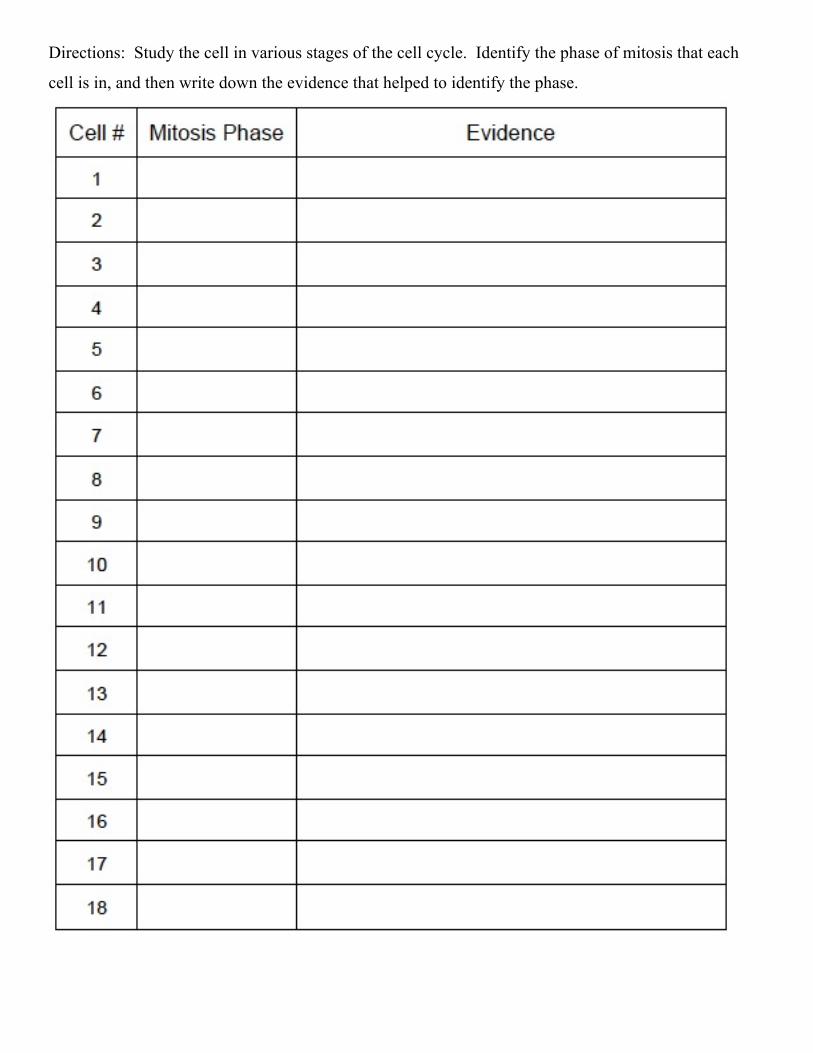
Directions: Study the cell in various stages of the cell cycle. Identify the phase of mitosis that each
cell is in, and then write down the evidence that helped to identify the phase.
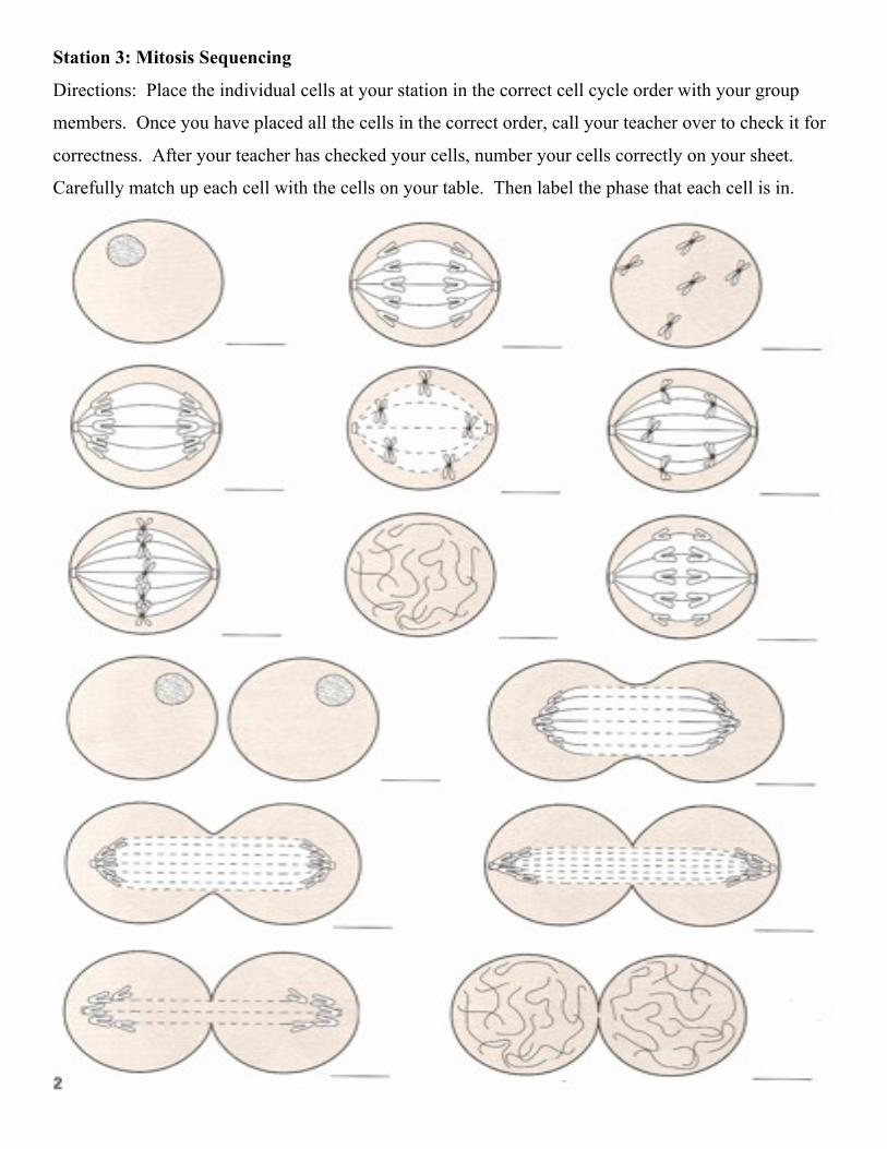
Station 3: Mitosis Sequencing
Directions: Place the individual cells at your station in the correct cell cycle order with your group
members. Once you have placed all the cells in the correct order, call your teacher over to check it for
correctness. After your teacher has checked your cells, number your cells correctly on your sheet.
Carefully match up each cell with the cells on your table. Then label the phase that each cell is in.
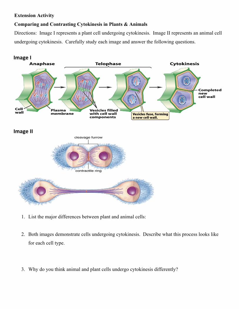
Extension Activity
Comparing and Contrasting Cytokinesis in Plants & Animals
Directions: Image I represents a plant cell undergoing cytokinesis. Image II represents an animal cell
undergoing cytokinesis. Carefully study each image and answer the following questions.
1. List the major differences between plant and animal cells:
2. Both images demonstrate cells undergoing cytokinesis. Describe what this process looks like
for each cell type.
3. Why do you think animal and plant cells undergo cytokinesis differently?
Image I
Image II
HW70: Cytokinesis Review Biology I
Name: ___________________________ Date: _____________________