unilateral zygoma implant in pterygoid area
DESCRIPTION
Long iomplants as alternative to sinus augmentationTRANSCRIPT

Case reportDr. Dov Kischinovsky
Jerusalem - Israel

Preoperative PanorexWith radiopaque ScannoGuide

Diagnosis:Female,54 y
Main complaint: Upper edentulous left area
Medical History:IHDDiabetes Mellitus
Treatment plan:
27 Unilateral Pterygoid implant positioned with Tooth- Mucosa-Supported SurgiGuide
Standard Implants 24 25Permanent bridge 24 25 27
.

Pre-treatment CT scan

To plan
Conversion of the CT data to 3D file: Surgery planning with SimPlant®
SurgiGuide® type
SurgiGuide® solution
Immediateloading
Temporary prosthesis
Final prosthesis
Navigator SurgiGuide®
NO No Bridge

Simplant Panoramic View choosing preferred cross section plane
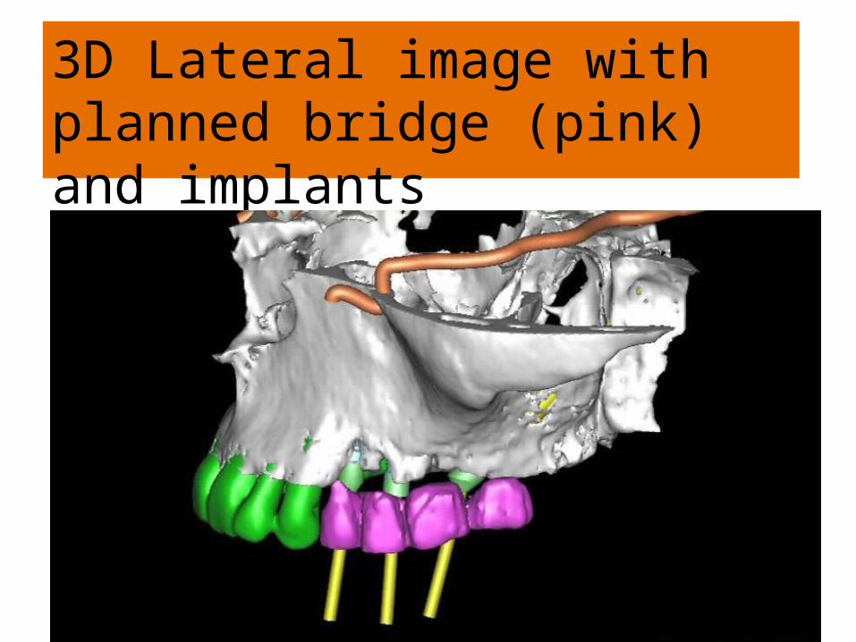
3D Lateral image with planned bridge (pink) and implants

3D Lateral image with planned bridge (pink) and implants


Axial view with pterygoid(Zygoma) implant head on alveolar ridge . One-stage placed implants

Axial view

To plan

To plan

Modification from zygomatic implant position to posterior pterygoid position

To plan

Sagittal view

To plan
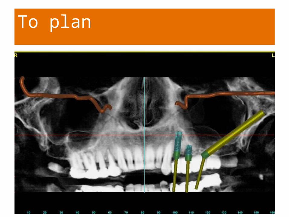
To plan
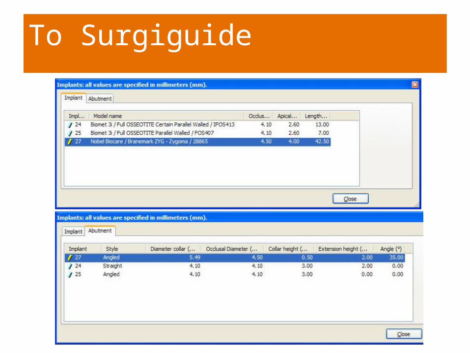
To Surgiguide

To Surgiguide - Plaster Model

SurgiGuide for Pterygoid placed Zygomatic Implant
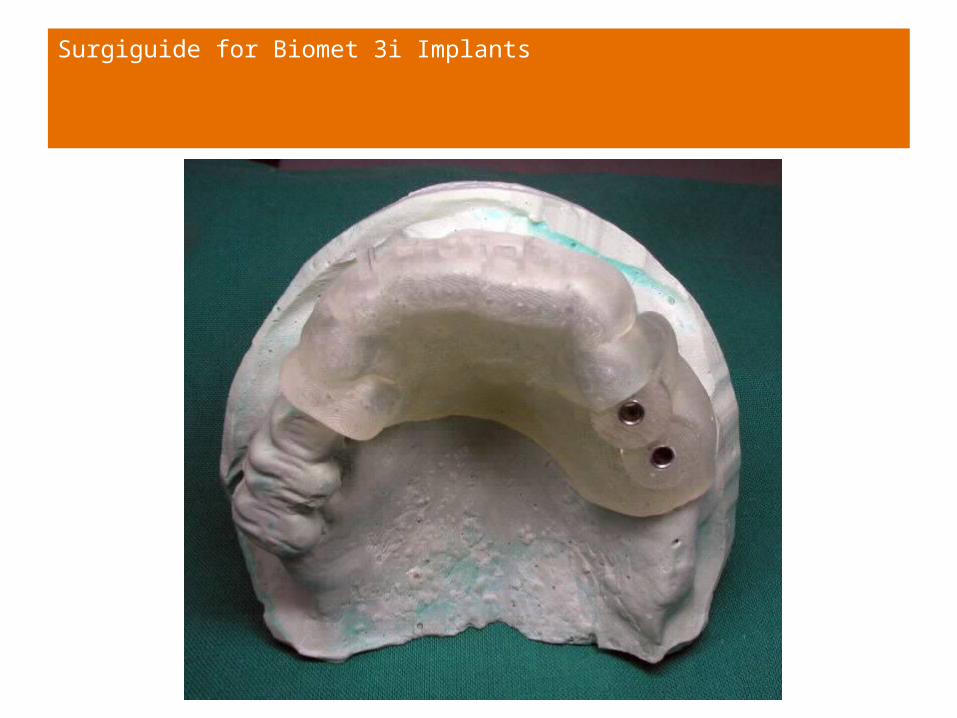
Surgiguide for Biomet 3i Implants

To guide – Visualisation using plaster model

– Showing Zygoma Implant with angulated head and angulated abutment

Pre-operative

Drilling with long drill through SurgiGuide with pterygoid(Zygoma) implant: 10mm high metal sleeve.

AAAAa
Angulated 45 degree of head of pterygoid(zygomatic )implant demonstrating parallelism of abutment to implants placed anteriolly

Clinical Photograph of surgery – One stage Healing Abutments
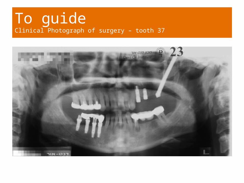
To guideClinical Photograph of surgery – tooth 37

Clinical Photograph Multi-unit Abutment on Implant 27

Photograph of Prosthetic Restoration (Dr.Moshe Kaplan,Jerusalem)

To guideClinical Photograph of surgery – tooth 37
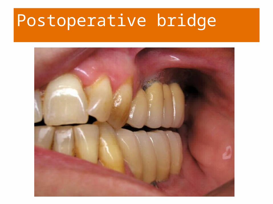
Postoperative bridge

Virtual planning versus Post-operative stage
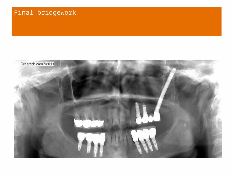
Final bridgework

Conclusion
This new modification of the zygomatic implant technique ,where the pterygoid placed implant emerges on the dento-alveolar crest makes it an excellent supportfor prosthetic work.By the use of a drill guide the implant is placed in exact position without raising a mucosal flap reducing operation time and morbidiy,as an alternative to sinus augmentation.