unexpected host dependency of antarctic nanohaloarchaeota€¦ · antarctic nanohaloarchaeota...
TRANSCRIPT
Unexpected host dependency ofAntarctic NanohaloarchaeotaJoshua N. Hamma, Susanne Erdmanna,1, Emiley A. Eloe-Fadroshb, Allegra Angelonia, Ling Zhongc,Christopher Brownleed, Timothy J. Williamsa, Kirston Bartone, Shaun Carswelle, Martin A. Smithe,f, Sarah Brazendalea,2,Alyce M. Hancocka,3, Michelle A. Allena, Mark J. Rafteryc, and Ricardo Cavicchiolia,4
aSchool of Biotechnology and Biomolecular Sciences, University of New South Wales, Sydney, NSW 2052, Australia; bDepartment of Energy, Joint GenomeInstitute, Walnut Creek, CA 94598; cBioanalytical Mass Spectrometry Facility, University of New South Wales, Sydney, NSW 2052, Australia; dBiologicalResources Imaging Laboratory, University of New South Wales, Sydney, NSW 2052, Australia; eKinghorn Centre for Clinical Genomics, Garvan Institute ofMedical Research, Darlinghurst, NSW 2010, Australia; and fSt. Vincent’s Clinical School, University of New South Wales, Sydney, Darlinghurst, NSW 2010,Australia
Edited by Norman R. Pace, University of Colorado Boulder, Boulder, CO, and approved June 5, 2019 (received for review March 25, 2019)
In hypersaline environments, Nanohaloarchaeota (Diapherotrites,Parvarchaeota, Aenigmarchaeota, Nanoarchaeota, Nanohaloarch-aeota [DPANN] superphylum) are thought to be free-livingmicroorganisms. We report cultivation of 2 strains of AntarcticNanohaloarchaeota and show that they require the haloarchaeonHalorubrum lacusprofundi for growth. By performing growth us-ing enrichments and fluorescence-activated cell sorting, we dem-onstrated successful cultivation of Candidatus Nanohaloarchaeumantarcticus, purification of Ca. Nha. antarcticus away from otherspecies, and growth and verification of Ca. Nha. antarcticus withHrr. lacusprofundi; these findings are analogous to those requiredfor fulfilling Koch’s postulates. We use fluorescent in situ hybridiza-tion and transmission electron microscopy to assess cell structuresand interactions; metagenomics to characterize enrichment taxa,generate metagenome assembled genomes, and interrogate Ant-arctic communities; and proteomics to assess metabolic pathwaysand speculate about the roles of certain proteins. Metagenomeanalysis indicates the presence of a single species, which is endemicto Antarctic hypersaline systems that support the growth of hal-oarchaea. The presence of unusually large proteins predicted tofunction in attachment and invasion of hosts plus the absence ofkey biosynthetic pathways (e.g., lipids) in metagenome assembledgenomes of globally distributed Nanohaloarchaeota indicate that allmembers of the lineage have evolved as symbionts. Our work ex-pands the range of archaeal symbiotic lifestyles and provides a ge-netically tractable model system for advancing understanding of thefactors controlling microbial symbiotic relationships.
archaea | symbiont | DPANN
Recent exploration into the genomic landscape of the domainArchaea has shed light on the ecological and evolutionary
prominence of uncultivated lineages (1). The Nanohaloarchaeawere first identified from metagenomic data as a class of un-cultivated halophilic archaea composed of 6 clades (2) and weresubsequently placed in the phylum Nanohaloarchaeota withinthe Diapherotrites, Parvarchaeota, Aenigmarchaeota, Nano-archaeota, Nanohaloarchaeota (DPANN) superphylum (3). Thelineage has since been identified in data from a range of hyper-saline environments including: Australian thalassohaline lake (2, 4),Spanish saltern (5), Russian soda brine (6), Californian saltern (7),and Chilean halite (8). From these data, 1 complete metagenome-assembled genome (MAG; Candidatus [Ca.] Nanopetramus SG9)(8) and 12 incomplete MAGs have been generated. Nano-haloarchaeal cells in environmental samples appear as small cocciwith an average diameter of ∼0.6 μm (0.1 to 0.8 μm size range) (2,4) and a genome size smaller (e.g., 1.1 Mb for Ca. NanopetramusSG9) (8) than haloarchaea (phylum Euryarchaeota, class Hal-obacteria; ∼3 to 4 Mb).Despite the monophyly of the DPANN superphylum being called
into question (9), members, such as Candidatus Nanoarchaeumequitans (0.4-μm diameter, <0.5 Mb) (10), CandidatusNanopusillus
acidilobi (<0.3-μm diameter, ∼0.6 Mb) (11), CandidatusParvarchaeum acidophilus (0.6-μm diameter, <1 Mb) (12), andCandidatusMancarchaeum acidophilum (∼1-μm diameter, ∼1Mb)(13), have all been shown to possess small cell and genome sizes;these are traits that have been proposed to be typical of allDPANN phyla (3). Due to their reduced genomic capacity, theseNanoarchaeota and Pavarchaeota require cell–cell contact withlarger (10–12) or similarly (13) sized hosts to proliferate and canbe described as ectoparasites (11). The DPANN CandidatusHuberarchaeum crystalense has also been described as a possibleepibiotic symbiont of Candidatus Altiarchaeum sp. (14). Althoughmost DPANN genomes lack some biosynthetic pathways requiredfor autonomous growth, it has been reasoned that certain genetictraits (diversity generating retroelements) may enhance the abilityof DPANN lineages (e.g., Pacearchaeota and Woesearchaeota) to
Significance
We demonstrate that Candidatus Nanohaloarchaeum antarc-ticus requires Halorubrum lacusprofundi for growth, illustrat-ing that Nanohaloarchaeota require a host rather than beingfree living as previously proposed. Developing the means ofcultivating Nanohaloarchaeota in the laboratory provides thecapacity to advance understanding of how archaea interactand the factors that control their symbiotic relationship (e.g.mutualism, commensalism, antagonism). Our findings amplifythe view that Antarctic lakes are a treasure trove for the dis-covery of microbes with previously unknown properties.
Author contributions: J.N.H. and R.C. designed research; J.N.H., S.E., A.A., L.Z., C.B., K.B.,S.C., M.A.S., S.B., A.M.H., and M.J.R. performed research; J.N.H., E.A.E.-F., T.J.W., M.A.A.,and R.C. analyzed data; and J.N.H., S.E., E.A.E.-F., A.A., L.Z., C.B., T.J.W., K.B., S.C., M.A.S.,S.B., A.M.H., M.A.A., M.J.R., and R.C. wrote the paper.
The authors declare no conflict of interest.
This article is a PNAS Direct Submission.
This open access article is distributed under Creative Commons Attribution License 4.0(CC BY).
Data deposition: The mass spectrometry data have been deposited to the ProteomeXchangeConsortium via the PRIDE partner repository with project name Cultivation of an AntarcticNanohaloarchaea (accession no. PXD010625). Integrated Microbial Genomes identificationnumbers are 3300005925, 2643221421, 3300028663, 3300028522, 2791354821, and3300031914.1Present address: Department of Microbiology, Max Planck Institute for Marine Microbi-ology, 28359 Bremen, Germany.
2Present address: Private address, Pegarah, TAS 7256, Australia.3Present address: University of Tasmania Institute of Marine and Antarctic Studies, Ant-arctic Gateway Partnership and Antarctic Climate & Ecosystem Research Centre, BatteryPoint, TAS 7004, Australia.
4To whom correspondence may be addressed. Email: [email protected].
This article contains supporting information online at www.pnas.org/lookup/suppl/doi:10.1073/pnas.1905179116/-/DCSupplemental.
www.pnas.org/cgi/doi/10.1073/pnas.1905179116 PNAS Latest Articles | 1 of 10
ECOLO
GY
Dow
nloa
ded
by g
uest
on
May
29,
202
0

have dynamic interactions with their hosts that possibly enableshifts between mutualism, predation, and parasitism (15).In contrast to host dependence, analyses of an ∼1.24-Mb
single amplified genome of Candidatus Iainarchaeum andersonii(Diapherotrites) (16) and ∼1.2-Mb metagenome assemblies ofCandidatus Nanosalina sp. J07AB43 and Candidatus Nanosalinarumsp. J07AB56 (Nanohaloarchaea) (2) have drawn the conclusionthat these DPANN members could be capable of autonomousgrowth. The Nanohaloarchaea were predicted to be free livingbased on their genome size being larger than other knownDPANN representatives and nanohaloarchaeal cells not beingobserved to associate with potential host cells in environmentalsamples (2). The view that Nanohaloarchaeota may be capableof leading an independent lifestyle is reflected in the currentliterature (17).Here, we report the cultivation of Antarctic strains of nano-
haloarchaea (proposed as Candidatus Nanohaloarchaeum ant-arcticus) from 2 different hypersaline lakes and show that anAntarctic haloarchaeon, Halorubrum lacusprofundi, is requiredfor growth. Examination of the nanohaloarchaea–haloarchaeainteractions and analysis of available MAGs of nanohaloarchaealead us to conclude that Nanohaloarchaeota have evolved assymbionts requiring hosts rather than as free-living cells.
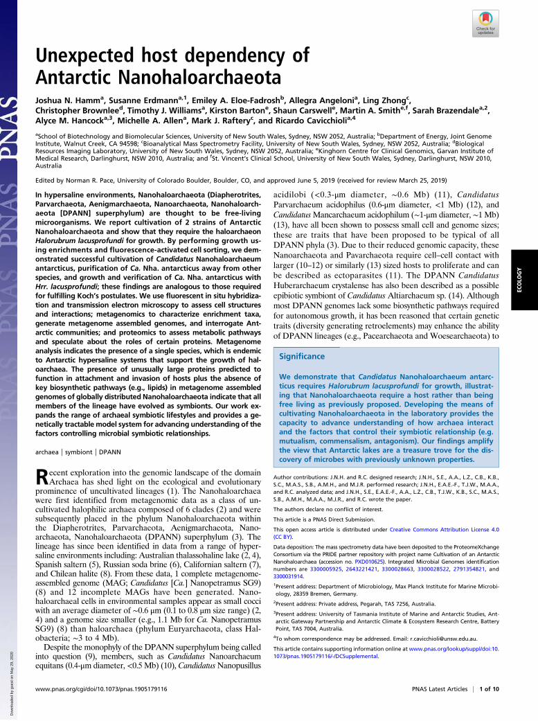
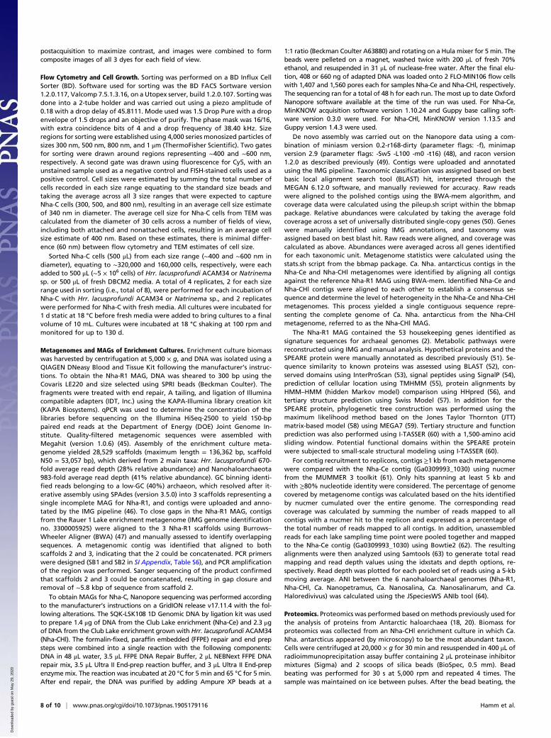
Cultivation of Antarctic NanohaloarchaeotaEnrichment cultures were previously established using hypersa-line water from Rauer 1 Lake, East Antarctica, resulting in theisolation of Hrr. lacusprofundi strain R1S1 (18) (SI Appendix,Table S1). Metagenome data of an enrichment culture revealedhigh relative abundance of Hrr. lacusprofundi (47%) and Nano-haloarchaeota (43%) (SI Appendix, Fig. S1A and Table S1).From guanine-cytosine (GC) binning, iterative assembly, andadditional PCR sequencing, 2 scaffolds resolved representing aMAG for Rauer 1 Nanohaloarchaeota, herein referred to as Ca.Nha. antarcticus R1 (“Nha-R1”). Transmission electron microscopy(TEM) of the enrichment culture (SI Appendix, Fig. S1 A–D) and0.22-μm filtrate (SI Appendix, Fig. S1 E and F) revealed small cocci(0.3 to 1.0 μm) resembling the morphology of nanohaloarchaea(2, 4), including small cocci associated with larger, pleomorphicrod-shaped cells characteristic of Hrr. lacusprofundi (SI Appen-dix, Fig. S1 A and C) (18–20).While Nha-R1 grew in liquid enrichment and on solid me-
dium, multiple attempts to propagate Nha-R1 using the 0.22-μmfiltrate in liquid or on solid medium were unsuccessful (Materialsand Methods). Isolation attempts included the use of spent mediafrom the Nha-R1 enrichment, fresh medium supplemented witha cell extract of the enrichment, and a diffusion chamber in-oculated with the Nha-R1 filtrate and placed into a growingenrichment culture. The growth experiments provided nutrientsfrom the enrichment community while preventing Nha-R1 fromhaving physical contact with other species, indicating that cell–cellcontact was required for Nha-R1 to proliferate.To confirm the cultivability of Nanohaloarchaeota and obtain
a second MAG, an enrichment culture was established usingwater from Club Lake, East Antarctica (see Fig. 2). TEM revealedmorphologies similar to those from the Rauer 1 Lake enrichment(SI Appendix, Fig. S2). After metagenome sequencing (SI Ap-pendix, Table S1), a single scaffold was obtained, which had >99%average nucleotide identity (ANI) and an identical 16S ribosomalribonucleic acid (rRNA) gene sequence to the Nha-R1 MAG,indicating that the enrichments contain strains of the sameNanohaloarchaeota species. The Club Lake NanohaloarchaeotaCa. Nha. antarcticus C is referred to as “Nha-C,” and its meta-genome “Nha-Ce” denotes Ca. Nha. antarcticus Club Lake en-richment (SI Appendix, Table S1). Hrr. lacusprofundi (41%) andNanohaloarchaeota (26%) were the 2 most abundant taxa in theenrichment (SI Appendix, Fig. S1B and Table S1).
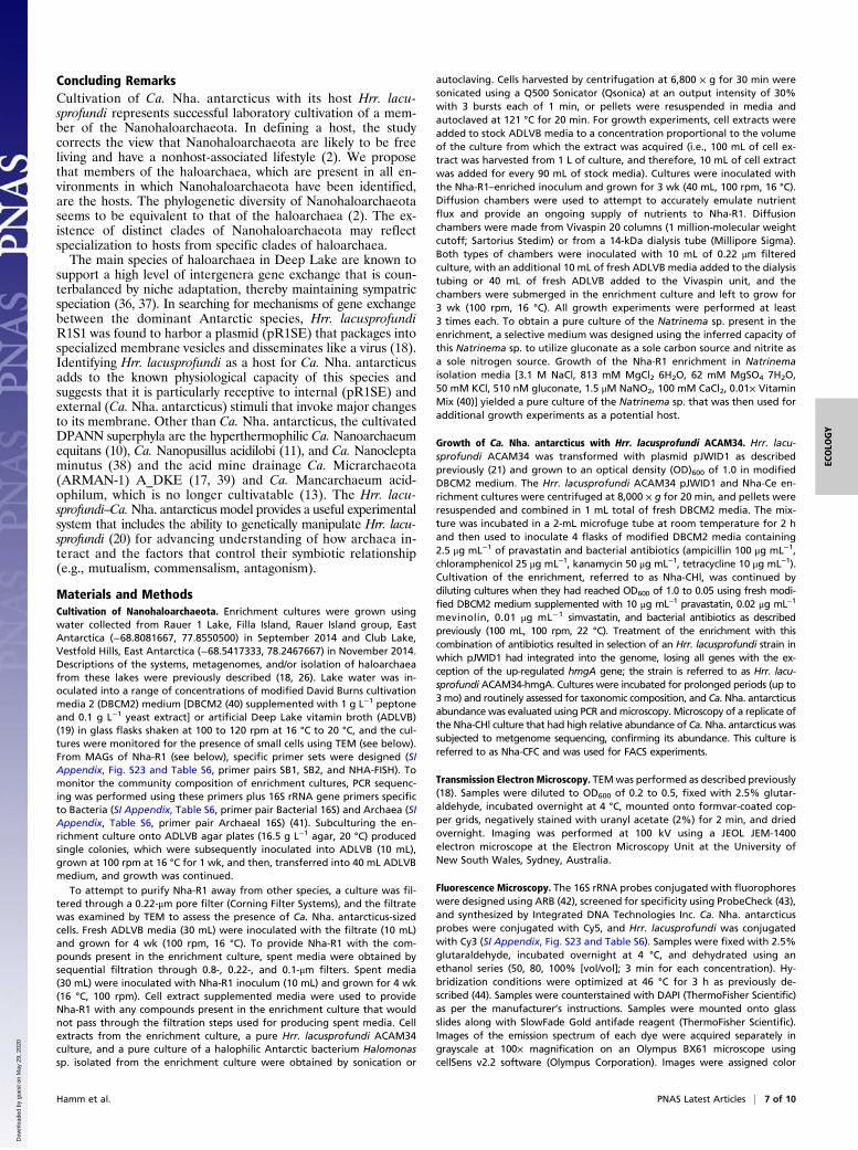
Growth of Nha-C with Hrr. lacusprofundi ACAM34-hmgAIn the enrichments from both lakes, Hrr. lacusprofundi was themost abundant species (Fig. 1 A and B), indicating that it couldbe the host. The up-regulated 3-hydroxy-3-methylglutaryl coenzymeA reductase gene (hmgA) gene in plasmid pJWID1 confers re-sistance to 2.5 μg mL−1 pravastatin in Hrr. lacusprofundi ACAM34(21). Pravastatin inhibits HmgA and hence, lipid synthesis via themevalonate pathway, which is absent from the Ca. Nha. antarcti-cus MAGs (see below). To inhibit the growth of other archaea inthe enrichment, the transformed ACAM34 strain was grown withan aliquot of the Club Lake enrichment in pravastatin (2.5 μg mL−1)and bacterial antibiotic (ampicillin 100 μg mL−1, chloramphenicol25 μg mL−1, kanamycin 50 μg mL−1, tetracycline 10 μg mL−1)containing media (Fig. 2). To provide stronger selection pressure forHrr. lacusprofundi ACAM34, after several rounds of growth, prava-statin concentration was increased up to 10 μg mL−1, and addi-tional archaeal antibiotics were added (mevinolin 0.02 μg mL−1,simvastatin 0.01 μg mL−1). This resulted in selection of a strain(ACAM34-hmgA) where the hmgA gene from the plasmid (flankedby Hrr. lacusprofundi transposases) relocated into the chromosome,and all other plasmid genes were lost or rendered nonfunctional (SIAppendix, Fig. S3).The metagenome of cultures (grown with 10 μg mL−1 prava-
statin) harvested during midlog phase growth showed a highrelative abundance of Nha-C (30%) and Hrr. lacusprofundi(56%), with a relatively low abundance of Natrinema (8%) andbacterial Marinobacter (6%) (SI Appendix, Fig. S1C and TableS1). The Ca. Nha. antarcticus MAG is referred to as “Nha-CHl,”
Ca. Nha. antarcticusHrr. lacusprofundiNatrinemaHaloferax
HalogeometricumOther archaeaHalobacterium sp. DL1Proteobacteria
Nha-R1 Nha-Ce
Nha-CFCNha-CHl
A B
C D
Fig. 1. Relative abundances of the major taxa present in Ca. Nha. antarcticusenrichment cultures. Metagenomes for Nha-R1 (A), Nha-Ce (B), Nha-CHl (C), andNha-CFC (D). Quadrants show relative abundances of the major taxa identifiedin each enrichment metagenome. Abundances were calculated using averageread depth across a set of universally distributed single-copy genes. Area of thecolored sections is directly proportional to the relative abundance of eachtaxonomic unit. The only taxa identified in all 4 metagenomes were Ca. Nha.antarcticus (Nha-R1: 41%; Nha-Ce: 19%; Nha-CHl: 30%; Nha-CFC: 47%), Hrr.lacusprofundi (Nha-R1: 28%; Nha-Ce: 59%; Nha-CHl: 56%; Nha-CFC: 29%), andNatrinema sp. (Nha-R1: 1%; Nha-Ce: 14%; Nha-CHl: 8%; Nha-CFC: 16%).
2 of 10 | www.pnas.org/cgi/doi/10.1073/pnas.1905179116 Hamm et al.
Dow
nloa
ded
by g
uest
on
May
29,
202
0
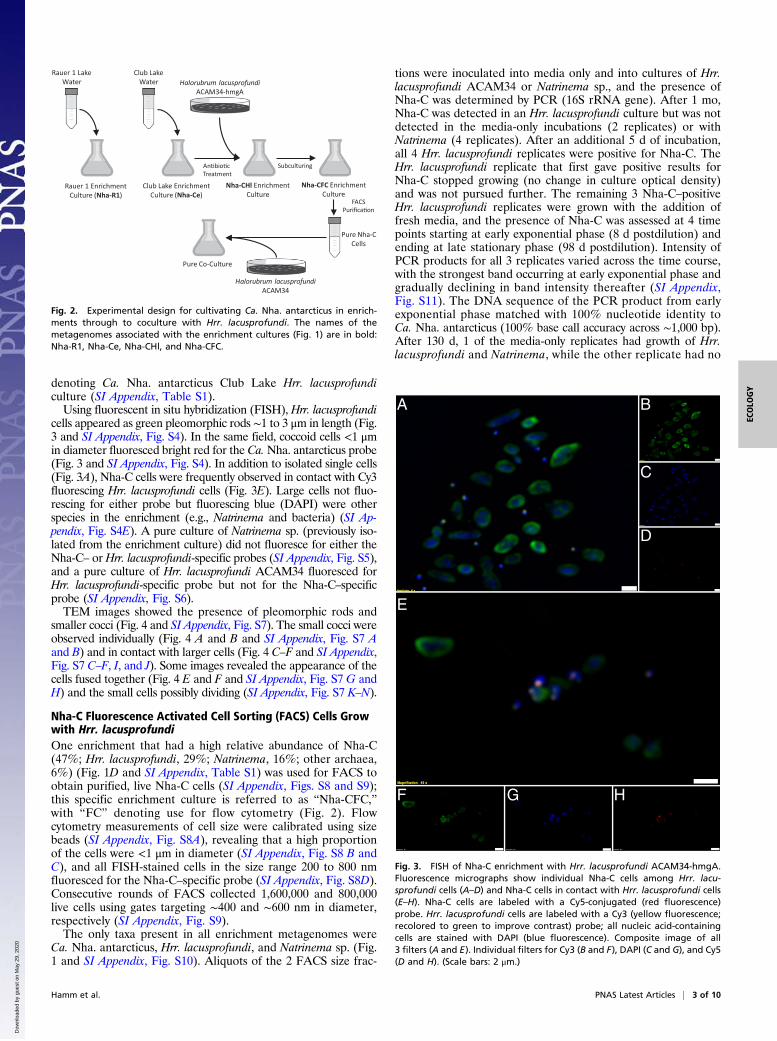
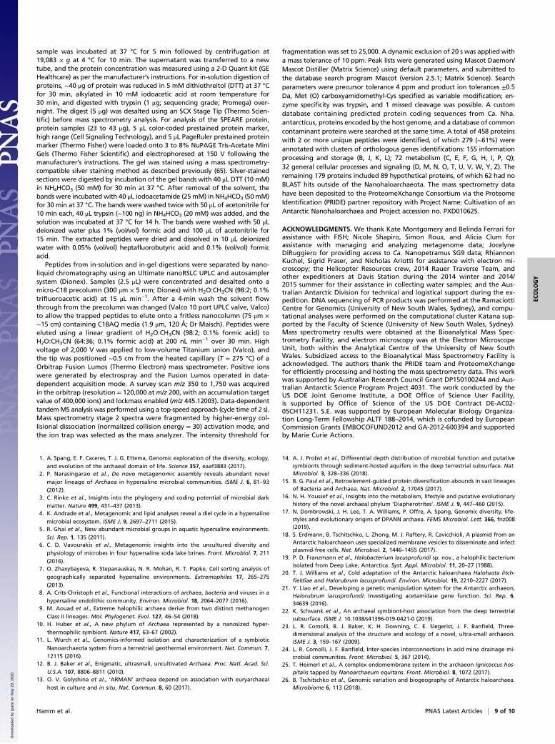
denoting Ca. Nha. antarcticus Club Lake Hrr. lacusprofundiculture (SI Appendix, Table S1).Using fluorescent in situ hybridization (FISH), Hrr. lacusprofundi
cells appeared as green pleomorphic rods ∼1 to 3 μm in length (Fig.3 and SI Appendix, Fig. S4). In the same field, coccoid cells <1 μmin diameter fluoresced bright red for the Ca.Nha. antarcticus probe(Fig. 3 and SI Appendix, Fig. S4). In addition to isolated single cells(Fig. 3A), Nha-C cells were frequently observed in contact with Cy3fluorescing Hrr. lacusprofundi cells (Fig. 3E). Large cells not fluo-rescing for either probe but fluorescing blue (DAPI) were otherspecies in the enrichment (e.g., Natrinema and bacteria) (SI Ap-pendix, Fig. S4E). A pure culture of Natrinema sp. (previously iso-lated from the enrichment culture) did not fluoresce for either theNha-C– or Hrr. lacusprofundi-specific probes (SI Appendix, Fig. S5),and a pure culture of Hrr. lacusprofundi ACAM34 fluoresced forHrr. lacusprofundi-specific probe but not for the Nha-C–specificprobe (SI Appendix, Fig. S6).TEM images showed the presence of pleomorphic rods and
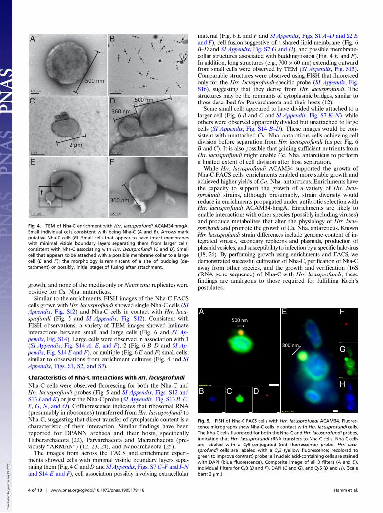
smaller cocci (Fig. 4 and SI Appendix, Fig. S7). The small cocci wereobserved individually (Fig. 4 A and B and SI Appendix, Fig. S7 Aand B) and in contact with larger cells (Fig. 4 C–F and SI Appendix,Fig. S7 C–F, I, and J). Some images revealed the appearance of thecells fused together (Fig. 4 E and F and SI Appendix, Fig. S7 G andH) and the small cells possibly dividing (SI Appendix, Fig. S7 K–N).
Nha-C Fluorescence Activated Cell Sorting (FACS) Cells Growwith Hrr. lacusprofundiOne enrichment that had a high relative abundance of Nha-C(47%; Hrr. lacusprofundi, 29%; Natrinema, 16%; other archaea,6%) (Fig. 1D and SI Appendix, Table S1) was used for FACS toobtain purified, live Nha-C cells (SI Appendix, Figs. S8 and S9);this specific enrichment culture is referred to as “Nha-CFC,”with “FC” denoting use for flow cytometry (Fig. 2). Flowcytometry measurements of cell size were calibrated using sizebeads (SI Appendix, Fig. S8A), revealing that a high proportionof the cells were <1 μm in diameter (SI Appendix, Fig. S8 B andC), and all FISH-stained cells in the size range 200 to 800 nmfluoresced for the Nha-C–specific probe (SI Appendix, Fig. S8D).Consecutive rounds of FACS collected 1,600,000 and 800,000live cells using gates targeting ∼400 and ∼600 nm in diameter,respectively (SI Appendix, Fig. S9).The only taxa present in all enrichment metagenomes were
Ca. Nha. antarcticus, Hrr. lacusprofundi, and Natrinema sp. (Fig.1 and SI Appendix, Fig. S10). Aliquots of the 2 FACS size frac-
tions were inoculated into media only and into cultures of Hrr.lacusprofundi ACAM34 or Natrinema sp., and the presence ofNha-C was determined by PCR (16S rRNA gene). After 1 mo,Nha-C was detected in an Hrr. lacusprofundi culture but was notdetected in the media-only incubations (2 replicates) or withNatrinema (4 replicates). After an additional 5 d of incubation,all 4 Hrr. lacusprofundi replicates were positive for Nha-C. TheHrr. lacusprofundi replicate that first gave positive results forNha-C stopped growing (no change in culture optical density)and was not pursued further. The remaining 3 Nha-C–positiveHrr. lacusprofundi replicates were grown with the addition offresh media, and the presence of Nha-C was assessed at 4 timepoints starting at early exponential phase (8 d postdilution) andending at late stationary phase (98 d postdilution). Intensity ofPCR products for all 3 replicates varied across the time course,with the strongest band occurring at early exponential phase andgradually declining in band intensity thereafter (SI Appendix,Fig. S11). The DNA sequence of the PCR product from earlyexponential phase matched with 100% nucleotide identity toCa. Nha. antarcticus (100% base call accuracy across ∼1,000 bp).After 130 d, 1 of the media-only replicates had growth of Hrr.lacusprofundi and Natrinema, while the other replicate had no
Rauer 1 Lake Water
Club LakeWater
Rauer 1 EnrichmentCulture (Nha-R1)
Club Lake EnrichmentCulture (Nha-Ce)
Halorubrum lacusprofundiACAM34-hmgA
Nha-CHl EnrichmentCulture
An�bio�c Treatment
Nha-CFC EnrichmentCulture
Pure Nha-CCells
FACSPurifica�on
Pure Co-Culture
Halorubrum lacusprofundiACAM34
Subculturing
Fig. 2. Experimental design for cultivating Ca. Nha. antarcticus in enrich-ments through to coculture with Hrr. lacusprofundi. The names of themetagenomes associated with the enrichment cultures (Fig. 1) are in bold:Nha-R1, Nha-Ce, Nha-CHl, and Nha-CFC.
A
D
C
B
E
HGF
Fig. 3. FISH of Nha-C enrichment with Hrr. lacusprofundi ACAM34-hmgA.Fluorescence micrographs show individual Nha-C cells among Hrr. lacu-sprofundi cells (A–D) and Nha-C cells in contact with Hrr. lacusprofundi cells(E–H). Nha-C cells are labeled with a Cy5-conjugated (red fluorescence)probe. Hrr. lacusprofundi cells are labeled with a Cy3 (yellow fluorescence;recolored to green to improve contrast) probe; all nucleic acid-containingcells are stained with DAPI (blue fluorescence). Composite image of all3 filters (A and E). Individual filters for Cy3 (B and F), DAPI (C and G), and Cy5(D and H). (Scale bars: 2 μm.)
Hamm et al. PNAS Latest Articles | 3 of 10
ECOLO
GY
Dow
nloa
ded
by g
uest
on
May
29,
202
0
growth, and none of the media-only or Natrinema replicates werepositive for Ca. Nha. antarcticus.Similar to the enrichments, FISH images of the Nha-C FACS
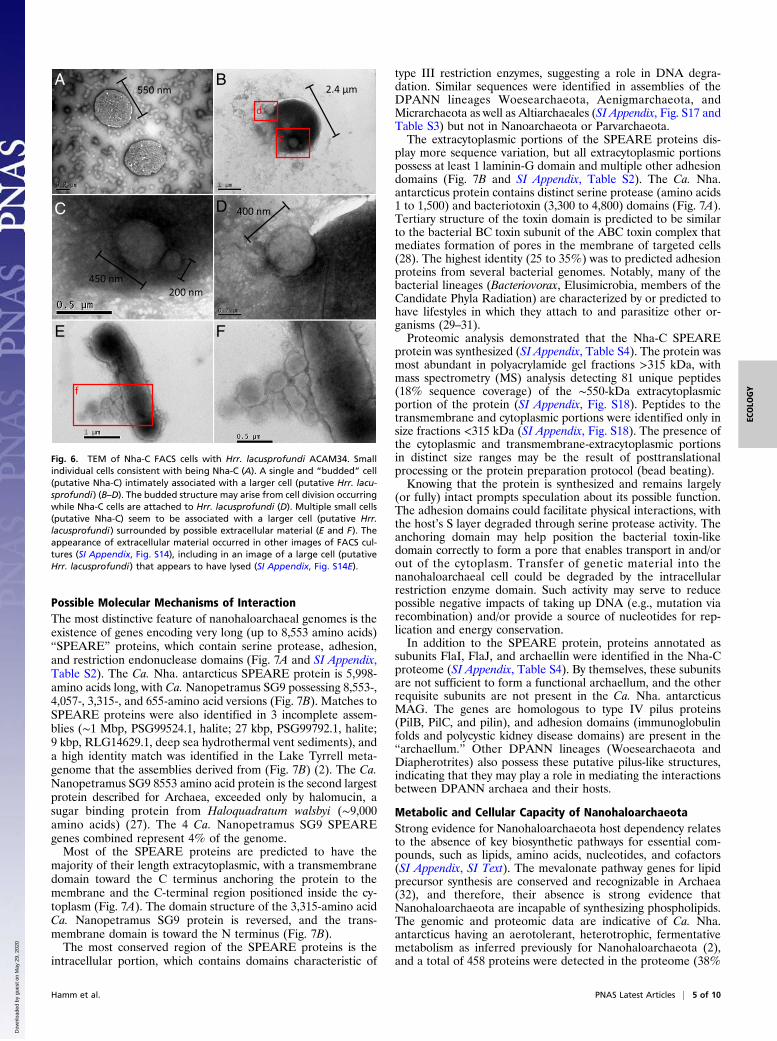
cells grown with Hrr. lacusprofundi showed single Nha-C cells (SIAppendix, Fig. S12) and Nha-C cells in contact with Hrr. lacu-sprofundi (Fig. 5 and SI Appendix, Fig. S12). Consistent withFISH observations, a variety of TEM images showed intimateinteractions between small and large cells (Fig. 6 and SI Ap-pendix, Fig. S14). Large cells were observed in association with 1(SI Appendix, Fig. S14 A, E, and F), 2 (Fig. 6 B–D and SI Ap-pendix, Fig. S14 E and F), or multiple (Fig. 6 E and F) small cells,similar to observations from enrichment cultures (Fig. 4 and SIAppendix, Figs. S1, S2, and S7).
Characteristics of Nha-C Interactions with Hrr. lacusprofundiNha-C cells were observed fluorescing for both the Nha-C andHrr. lacusprofundi probes (Fig. 5 and SI Appendix, Figs. S12 andS13 J and K) or just the Nha-C probe (SI Appendix, Fig. S13 B, C,F, G, N, and O). Cofluorescence indicates that ribosomal RNA(presumably in ribosomes) transferred from Hrr. lacusprofundi toNha-C, suggesting that direct transfer of cytoplasmic content is acharacteristic of their interaction. Similar findings have beenreported for DPANN archaea and their hosts, specificallyHuberarchaeota (22), Parvarchaeota and Micrarchaeota (pre-viously “ARMAN”) (12, 23, 24), and Nanoarchaeota (25).The images from across the FACS and enrichment experi-
ments showed cells with minimal visible boundary layers sepa-rating them (Fig. 4 C andD and SI Appendix, Figs. S7 C–F and I–Nand S14 E and F), cell association possibly involving extracellular
material (Fig. 6 E and F and SI Appendix, Figs. S1 A–D and S2 Eand F), cell fusion suggestive of a shared lipid membrane (Fig. 6B–D and SI Appendix, Fig. S7 G and H), and possible membrane-collar structures associated with budding/fission (Fig. 4 E and F).In addition, long structures (e.g., 700 × 60 nm) extending outwardfrom small cells were observed by TEM (SI Appendix, Fig. S15).Comparable structures were observed using FISH that fluorescedonly for the Hrr. lacusprofundi-specific probe (SI Appendix, Fig.S16), suggesting that they derive from Hrr. lacusprofundi. Thestructures may be the remnants of cytoplasmic bridges, similar tothose described for Parvarchaeota and their hosts (12).Some small cells appeared to have divided while attached to a
larger cell (Fig. 6 B and C and SI Appendix, Fig. S7 K–N), whileothers were observed apparently divided but unattached to largecells (SI Appendix, Fig. S14 B–D). These images would be con-sistent with unattached Ca. Nha. antarcticus cells achieving celldivision before separation from Hrr. lacusprofundi (as per Fig. 6B and C). It is also possible that gaining sufficient nutrients fromHrr. lacusprofundi might enable Ca. Nha. antarcticus to performa limited extent of cell division after host separation.While Hrr. lacusprofundi ACAM34 supported the growth of
Nha-C FACS cells, enrichments enabled more stable growth andachieved higher yields of Ca. Nha. antarcticus. Enrichments havethe capacity to support the growth of a variety of Hrr. lacu-sprofundi strains, although presumably, strain diversity wouldreduce in enrichments propagated under antibiotic selection withHrr. lacusprofundi ACAM34-hmgA. Enrichments are likely toenable interactions with other species (possibly including viruses)and produce metabolites that alter the physiology of Hrr. lacu-sprofundi and promote the growth of Ca.Nha. antarcticus. KnownHrr. lacusprofundi strain differences include genome content of in-tegrated viruses, secondary replicons and plasmids, production ofplasmid vesicles, and susceptibility to infection by a specific halovirus(18, 26). By performing growth using enrichments and FACS, wedemonstrated successful cultivation of Nha-C, purification of Nha-Caway from other species, and the growth and verification (16SrRNA gene sequence) of Nha-C with Hrr. lacusprofundi; thesefindings are analogous to those required for fulfilling Koch’spostulates.
2 μm
360 nm
f300 nm
C
E
A B
500 nm
500 nmD
F
Fig. 4. TEM of Nha-C enrichment with Hrr. lacusprofundi ACAM34-hmgA.Small individual cells consistent with being Nha-C (A and B). Arrows markputative Nha-C cells (B). Small cells that appear to have intact membraneswith minimal visible boundary layers separating them from larger cells,consistent with Nha-C associating with Hrr. lacusprofundi (C and D). Smallcell that appears to be attached with a possible membrane collar to a largecell (E and F); the morphology is reminiscent of a site of budding (de-tachment) or possibly, initial stages of fusing after attachment.
800 nm
500 nmA E
DCBH
G
F
Fig. 5. FISH of Nha-C FACS cells with Hrr. lacusprofundi ACAM34. Fluores-cence micrographs show Nha-C cells in contact with Hrr. lacusprofundi cells.The Nha-C cells fluoresced for both the Nha-C and Hrr. lacusprofundi probes,indicating that Hrr. lacusprofundi rRNA transfers to Nha-C cells. Nha-C cellsare labeled with a Cy5-conjugated (red fluorescence) probe. Hrr. lacu-sprofundi cells are labeled with a Cy3 (yellow fluorescence; recolored togreen to improve contrast) probe; all nucleic acid-containing cells are stainedwith DAPI (blue fluorescence). Composite image of all 3 filters (A and E).Individual filters for Cy3 (B and F), DAPI (C and G), and Cy5 (D and H). (Scalebars: 2 μm.)
4 of 10 | www.pnas.org/cgi/doi/10.1073/pnas.1905179116 Hamm et al.
Dow
nloa
ded
by g
uest
on
May
29,
202
0
Possible Molecular Mechanisms of InteractionThe most distinctive feature of nanohaloarchaeal genomes is theexistence of genes encoding very long (up to 8,553 amino acids)“SPEARE” proteins, which contain serine protease, adhesion,and restriction endonuclease domains (Fig. 7A and SI Appendix,Table S2). The Ca. Nha. antarcticus SPEARE protein is 5,998-amino acids long, with Ca. Nanopetramus SG9 possessing 8,553-,4,057-, 3,315-, and 655-amino acid versions (Fig. 7B). Matches toSPEARE proteins were also identified in 3 incomplete assem-blies (∼1 Mbp, PSG99524.1, halite; 27 kbp, PSG99792.1, halite;9 kbp, RLG14629.1, deep sea hydrothermal vent sediments), anda high identity match was identified in the Lake Tyrrell meta-genome that the assemblies derived from (Fig. 7B) (2). The Ca.Nanopetramus SG9 8553 amino acid protein is the second largestprotein described for Archaea, exceeded only by halomucin, asugar binding protein from Haloquadratum walsbyi (∼9,000amino acids) (27). The 4 Ca. Nanopetramus SG9 SPEAREgenes combined represent 4% of the genome.Most of the SPEARE proteins are predicted to have the
majority of their length extracytoplasmic, with a transmembranedomain toward the C terminus anchoring the protein to themembrane and the C-terminal region positioned inside the cy-toplasm (Fig. 7A). The domain structure of the 3,315-amino acidCa. Nanopetramus SG9 protein is reversed, and the trans-membrane domain is toward the N terminus (Fig. 7B).The most conserved region of the SPEARE proteins is the
intracellular portion, which contains domains characteristic of
type III restriction enzymes, suggesting a role in DNA degra-dation. Similar sequences were identified in assemblies of theDPANN lineages Woesearchaeota, Aenigmarchaeota, andMicrarchaeota as well as Altiarchaeales (SI Appendix, Fig. S17 andTable S3) but not in Nanoarchaeota or Parvarchaeota.The extracytoplasmic portions of the SPEARE proteins dis-
play more sequence variation, but all extracytoplasmic portionspossess at least 1 laminin-G domain and multiple other adhesiondomains (Fig. 7B and SI Appendix, Table S2). The Ca. Nha.antarcticus protein contains distinct serine protease (amino acids1 to 1,500) and bacteriotoxin (3,300 to 4,800) domains (Fig. 7A).Tertiary structure of the toxin domain is predicted to be similarto the bacterial BC toxin subunit of the ABC toxin complex thatmediates formation of pores in the membrane of targeted cells(28). The highest identity (25 to 35%) was to predicted adhesionproteins from several bacterial genomes. Notably, many of thebacterial lineages (Bacteriovorax, Elusimicrobia, members of theCandidate Phyla Radiation) are characterized by or predicted tohave lifestyles in which they attach to and parasitize other or-ganisms (29–31).Proteomic analysis demonstrated that the Nha-C SPEARE
protein was synthesized (SI Appendix, Table S4). The protein wasmost abundant in polyacrylamide gel fractions >315 kDa, withmass spectrometry (MS) analysis detecting 81 unique peptides(18% sequence coverage) of the ∼550-kDa extracytoplasmicportion of the protein (SI Appendix, Fig. S18). Peptides to thetransmembrane and cytoplasmic portions were identified only insize fractions <315 kDa (SI Appendix, Fig. S18). The presence ofthe cytoplasmic and transmembrane-extracytoplasmic portionsin distinct size ranges may be the result of posttranslationalprocessing or the protein preparation protocol (bead beating).Knowing that the protein is synthesized and remains largely
(or fully) intact prompts speculation about its possible function.The adhesion domains could facilitate physical interactions, withthe host’s S layer degraded through serine protease activity. Theanchoring domain may help position the bacterial toxin-likedomain correctly to form a pore that enables transport in and/orout of the cytoplasm. Transfer of genetic material into thenanohaloarchaeal cell could be degraded by the intracellularrestriction enzyme domain. Such activity may serve to reducepossible negative impacts of taking up DNA (e.g., mutation viarecombination) and/or provide a source of nucleotides for rep-lication and energy conservation.In addition to the SPEARE protein, proteins annotated as
subunits FlaI, FlaJ, and archaellin were identified in the Nha-Cproteome (SI Appendix, Table S4). By themselves, these subunitsare not sufficient to form a functional archaellum, and the otherrequisite subunits are not present in the Ca. Nha. antarcticusMAG. The genes are homologous to type IV pilus proteins(PilB, PilC, and pilin), and adhesion domains (immunoglobulinfolds and polycystic kidney disease domains) are present in the“archaellum.” Other DPANN lineages (Woesearchaeota andDiapherotrites) also possess these putative pilus-like structures,indicating that they may play a role in mediating the interactionsbetween DPANN archaea and their hosts.
Metabolic and Cellular Capacity of NanohaloarchaeotaStrong evidence for Nanohaloarchaeota host dependency relatesto the absence of key biosynthetic pathways for essential com-pounds, such as lipids, amino acids, nucleotides, and cofactors(SI Appendix, SI Text). The mevalonate pathway genes for lipidprecursor synthesis are conserved and recognizable in Archaea(32), and therefore, their absence is strong evidence thatNanohaloarchaeota are incapable of synthesizing phospholipids.The genomic and proteomic data are indicative of Ca. Nha.antarcticus having an aerotolerant, heterotrophic, fermentativemetabolism as inferred previously for Nanohaloarchaeota (2),and a total of 458 proteins were detected in the proteome (38%
E
f
F
D 400 nm
550 nmB
c
d
A
C
450 nm200 nm
2.4 μm
Fig. 6. TEM of Nha-C FACS cells with Hrr. lacusprofundi ACAM34. Smallindividual cells consistent with being Nha-C (A). A single and “budded” cell(putative Nha-C) intimately associated with a larger cell (putative Hrr. lacu-sprofundi) (B–D). The budded structure may arise from cell division occurringwhile Nha-C cells are attached to Hrr. lacusprofundi (D). Multiple small cells(putative Nha-C) seem to be associated with a larger cell (putative Hrr.lacusprofundi ) surrounded by possible extracellular material (E and F). Theappearance of extracellular material occurred in other images of FACS cul-tures (SI Appendix, Fig. S14), including in an image of a large cell (putativeHrr. lacusprofundi) that appears to have lysed (SI Appendix, Fig. S14E).
Hamm et al. PNAS Latest Articles | 5 of 10
ECOLO
GY
Dow
nloa
ded
by g
uest
on
May
29,
202
0

of protein-encoding genes) (SI Appendix, Table S4). Overall, theproteome is consistent with the genomic evidence that Ca. Nha.antarcticus can synthesize its own carbohydrates (including forstorage) but otherwise, has extremely limited biosynthetic capa-bilities (SI Appendix, SI Text). Thus, Ca. Nha. antarcticus wouldrequire certain biopolymers from its host, including polypeptidesand nucleic acids as sources of amino acids and nucleotides,respectively. Conceivably, Ca. Nha. antarcticus could gain accessto essential metabolic products when it is attached to hosts, withsurplus carbon stored as polysaccharides that could be mobilizedwhen cells are nonattached. It is possible that Hrr. lacusprofundimay gain some benefit from by-products produced by Ca. Nha.antarcticus that the latter apparently cannot assimilate, such asammonium or acetate (33, 34).
Genome Variation and Evolution of NanohaloarchaeotaMetagenome read and contig mapping identified Nano-haloarchaeota in 2 Vestfold Hills lakes (Deep Lake and ClubLake) and 4 Rauer Island lakes (3, 6, 11, 13) (SI Appendix, Figs.S19–S21). The samples from Deep Lake represented 37 meta-genomes spanning 8 y (December 2006 to January 2015) andcovering a seasonal cycle (December 2013 to January 2015) anddepth profile (2008: surface and 5, 13, 24, and 36 m) (26). Readcoverage across the Deep Lake metagenomes was ∼0.5%, simi-lar to Club Lake, with the Rauer Island lakes having more var-iable coverage, and highest overall coverage (3.5%) was fromRauer 3 Lake (SI Appendix, Fig. S19). All of the lakes withNanohaloarchaeota contained high levels of haloarchaea, withthe highest representation in the Vestfold Hills lakes (26).Contig and read coverage were high (average genome coverage
68%), although the relative abundance of Nanohaloarchaeota waslow (≤3.5%), and contigs had high identity (>99%), illustratingthat little genomic variation existed within the nanohaloarchaealpopulations (SI Appendix, Figs. S19–S21). Short regions existedwith high read depth (SI Appendix, Figs. S20 D–G and S21) andcontained mobile elements (e.g., transposases) with high identityto Antarctic haloarchaea (e.g., 100% match to Halorubraceae
IS5-like element) (SI Appendix, Table S5), indicating that theyarose through sharing with haloarchaea. Gaps between contigs(∼270, 930, and 1,025 kb) coincided with low read recruitment(SI Appendix, Figs. S20 D–G and S21) and contained similartypes of sequences (transposases, viral genes, hypothetical pro-teins), most of which had best matches to haloarchaea. Of notewas a complete type 5 Bacteriophage Exclusion (BREX) systemthat was absent in Nha-CHl but present in Nha-R1 (SI Appendix,Fig. S20C and Table S5). BREX functions by inhibiting viralreplication (35). One additional gene was present (2643307549)downstream from the BREX genes, which is annotated as con-served Nanohaloarchaeota hypothetical protein and may possi-bly be a Nanohaloarchaeota-specific BREX gene. The BREXcluster was flanked by a gene annotated as a viral integrase(2643307539 integrated microbial genomes [IMG] gene identifica-tion), with average coverage indicating that the BREX cluster itselfmay be part of a mobile element. The read depth coverage acrossall lake metagenomes was ∼25% of average, indicating that theBREX-containing phylotype was present throughout all of the lakesin approximately 1/4 of the Nanohaloarchaeota population.While genomic variation across the Antarctic Nano-
haloarchaeota is minimal, the genomes of non-Antarctic Nano-haloarchaeota have much lower similarity (≤70% ANI, <90%16S rRNA gene identity) (SI Appendix, Fig. S22), indicating thatthe other known nanohaloarchaeal lineages represent distinctgenera from a range of diverse types of hypersaline environ-ments. This level of diversity (i.e., genera) is similar to the di-versity present within haloarchaea (36) and may arise within theNanohaloarchaeota as a result of host specificity. If coevolutionoccurs, each lineage of Nanohaloarchaeota would be expected tointeract with a specific lineage of haloarchaea. In this regard, wepredict that a host for some of the Lake Tyrrell Nanohaloarchaeotais Hqr. walsbyi, as it is an abundant species of haloarchaea in thelake, and nanohaloarchaea appear in FISH images associatedwith square-shaped cells (figure 2 in ref. 2).
Ca. Nha. antarc�cusSPEARE
Ca. Nanopetramus SG9SPEARE 1
Ca. Nanopetramus SG9SPEARE 2
Lake TyrrellSPEARE
Ca. Nanopetramus SG9SPEARE 3
600055004800 53004600330015000
Serine ProteaseType 3 Restric�on Enzyme
TransmembraneAdhesion
Pore-Forming Bacteriotoxin
Immunoglobulin Fold
A
B
Ca. Nanopetramus SG9SPEARE 4
Fig. 7. Domain structure of SPEARE proteins. (A) Protein domains predicted for the 5,998 amino acid Ca. Nha. antarcticus SPEARE protein (IMG geneidentification no. 2643306914; locus tag: NAR1_1133). (B) Domain structure comparison of SPEARE proteins from Nha-R1, Ca. Nanopetramus SG9 (8), and LakeTyrrell metagenome data (2) depicted approximately to scale. Ca. Nanopetramus SG9 SPEARE 1 (8,553 amino acids; hypothetical protein AQV86_04780;accession no. AOV94740.1). Ca. Nanopetramus SG9 SPEARE 2 (4,057 amino acids; hypothetical protein AQV86_02335; accession no. AOV95205.1). Lake TyrrellSPEARE (717 amino acids; IMG gene identification no. LTJ07AB_218510; locus tag: LTJ07AB_218510). Ca. Nanopetramus SG9 SPEARE 3 (3,315 amino acids;hypothetical protein AQV86_02330; accession no. AOV94739.1).
6 of 10 | www.pnas.org/cgi/doi/10.1073/pnas.1905179116 Hamm et al.
Dow
nloa
ded
by g
uest
on
May
29,
202
0
Concluding RemarksCultivation of Ca. Nha. antarcticus with its host Hrr. lacu-sprofundi represents successful laboratory cultivation of a mem-ber of the Nanohaloarchaeota. In defining a host, the studycorrects the view that Nanohaloarchaeota are likely to be freeliving and have a nonhost-associated lifestyle (2). We proposethat members of the haloarchaea, which are present in all en-vironments in which Nanohaloarchaeota have been identified,are the hosts. The phylogenetic diversity of Nanohaloarchaeotaseems to be equivalent to that of the haloarchaea (2). The ex-istence of distinct clades of Nanohaloarchaeota may reflectspecialization to hosts from specific clades of haloarchaea.The main species of haloarchaea in Deep Lake are known to
support a high level of intergenera gene exchange that is coun-terbalanced by niche adaptation, thereby maintaining sympatricspeciation (36, 37). In searching for mechanisms of gene exchangebetween the dominant Antarctic species, Hrr. lacusprofundiR1S1 was found to harbor a plasmid (pR1SE) that packages intospecialized membrane vesicles and disseminates like a virus (18).Identifying Hrr. lacusprofundi as a host for Ca. Nha. antarcticusadds to the known physiological capacity of this species andsuggests that it is particularly receptive to internal (pR1SE) andexternal (Ca. Nha. antarcticus) stimuli that invoke major changesto its membrane. Other than Ca. Nha. antarcticus, the cultivatedDPANN superphyla are the hyperthermophilic Ca. Nanoarchaeumequitans (10), Ca. Nanopusillus acidilobi (11), and Ca. Nanocleptaminutus (38) and the acid mine drainage Ca. Micrarchaeota(ARMAN-1) A_DKE (17, 39) and Ca. Mancarchaeum acid-ophilum, which is no longer cultivatable (13). The Hrr. lacu-sprofundi–Ca.Nha. antarcticus model provides a useful experimentalsystem that includes the ability to genetically manipulate Hrr. lacu-sprofundi (20) for advancing understanding of how archaea in-teract and the factors that control their symbiotic relationship(e.g., mutualism, commensalism, antagonism).
Materials and MethodsCultivation of Nanohaloarchaeota. Enrichment cultures were grown usingwater collected from Rauer 1 Lake, Filla Island, Rauer Island group, EastAntarctica (−68.8081667, 77.8550500) in September 2014 and Club Lake,Vestfold Hills, East Antarctica (−68.5417333, 78.2467667) in November 2014.Descriptions of the systems, metagenomes, and/or isolation of haloarchaeafrom these lakes were previously described (18, 26). Lake water was in-oculated into a range of concentrations of modified David Burns cultivationmedia 2 (DBCM2) medium [DBCM2 (40) supplemented with 1 g L−1 peptoneand 0.1 g L−1 yeast extract] or artificial Deep Lake vitamin broth (ADLVB)(19) in glass flasks shaken at 100 to 120 rpm at 16 °C to 20 °C, and the cul-tures were monitored for the presence of small cells using TEM (see below).From MAGs of Nha-R1 (see below), specific primer sets were designed (SIAppendix, Fig. S23 and Table S6, primer pairs SB1, SB2, and NHA-FISH). Tomonitor the community composition of enrichment cultures, PCR sequenc-ing was performed using these primers plus 16S rRNA gene primers specificto Bacteria (SI Appendix, Table S6, primer pair Bacterial 16S) and Archaea (SIAppendix, Table S6, primer pair Archaeal 16S) (41). Subculturing the en-richment culture onto ADLVB agar plates (16.5 g L−1 agar, 20 °C) producedsingle colonies, which were subsequently inoculated into ADLVB (10 mL),grown at 100 rpm at 16 °C for 1 wk, and then, transferred into 40 mL ADLVBmedium, and growth was continued.
To attempt to purify Nha-R1 away from other species, a culture was fil-tered through a 0.22-μm pore filter (Corning Filter Systems), and the filtratewas examined by TEM to assess the presence of Ca. Nha. antarcticus-sizedcells. Fresh ADLVB media (30 mL) were inoculated with the filtrate (10 mL)and grown for 4 wk (100 rpm, 16 °C). To provide Nha-R1 with the com-pounds present in the enrichment culture, spent media were obtained bysequential filtration through 0.8-, 0.22-, and 0.1-μm filters. Spent media(30 mL) were inoculated with Nha-R1 inoculum (10 mL) and grown for 4 wk(16 °C, 100 rpm). Cell extract supplemented media were used to provideNha-R1 with any compounds present in the enrichment culture that wouldnot pass through the filtration steps used for producing spent media. Cellextracts from the enrichment culture, a pure Hrr. lacusprofundi ACAM34culture, and a pure culture of a halophilic Antarctic bacterium Halomonassp. isolated from the enrichment culture were obtained by sonication or
autoclaving. Cells harvested by centrifugation at 6,800 × g for 30 min weresonicated using a Q500 Sonicator (Qsonica) at an output intensity of 30%with 3 bursts each of 1 min, or pellets were resuspended in media andautoclaved at 121 °C for 20 min. For growth experiments, cell extracts wereadded to stock ADLVB media to a concentration proportional to the volumeof the culture from which the extract was acquired (i.e., 100 mL of cell ex-tract was harvested from 1 L of culture, and therefore, 10 mL of cell extractwas added for every 90 mL of stock media). Cultures were inoculated withthe Nha-R1–enriched inoculum and grown for 3 wk (40 mL, 100 rpm, 16 °C).Diffusion chambers were used to attempt to accurately emulate nutrientflux and provide an ongoing supply of nutrients to Nha-R1. Diffusionchambers were made from Vivaspin 20 columns (1 million-molecular weightcutoff; Sartorius Stedim) or from a 14-kDa dialysis tube (Millipore Sigma).Both types of chambers were inoculated with 10 mL of 0.22 μm filteredculture, with an additional 10 mL of fresh ADLVB media added to the dialysistubing or 40 mL of fresh ADLVB added to the Vivaspin unit, and thechambers were submerged in the enrichment culture and left to grow for3 wk (100 rpm, 16 °C). All growth experiments were performed at least3 times each. To obtain a pure culture of the Natrinema sp. present in theenrichment, a selective medium was designed using the inferred capacity ofthis Natrinema sp. to utilize gluconate as a sole carbon source and nitrite asa sole nitrogen source. Growth of the Nha-R1 enrichment in Natrinemaisolation media [3.1 M NaCl, 813 mM MgCl2 6H2O, 62 mM MgSO4 7H2O,50 mM KCl, 510 nM gluconate, 1.5 μM NaNO2, 100 mM CaCl2, 0.01× VitaminMix (40)] yielded a pure culture of the Natrinema sp. that was then used foradditional growth experiments as a potential host.
Growth of Ca. Nha. antarcticus with Hrr. lacusprofundi ACAM34. Hrr. lacu-sprofundi ACAM34 was transformed with plasmid pJWID1 as describedpreviously (21) and grown to an optical density (OD)600 of 1.0 in modifiedDBCM2 medium. The Hrr. lacusprofundi ACAM34 pJWID1 and Nha-Ce en-richment cultures were centrifuged at 8,000 × g for 20 min, and pellets wereresuspended and combined in 1 mL total of fresh DBCM2 media. The mix-ture was incubated in a 2-mL microfuge tube at room temperature for 2 hand then used to inoculate 4 flasks of modified DBCM2 media containing2.5 μg mL−1 of pravastatin and bacterial antibiotics (ampicillin 100 μg mL−1,chloramphenicol 25 μg mL−1, kanamycin 50 μg mL−1, tetracycline 10 μg mL−1).Cultivation of the enrichment, referred to as Nha-CHl, was continued bydiluting cultures when they had reached OD600 of 1.0 to 0.05 using fresh modi-fied DBCM2 medium supplemented with 10 μg mL−1 pravastatin, 0.02 μg mL−1
mevinolin, 0.01 μg mL−1 simvastatin, and bacterial antibiotics as describedpreviously (100 mL, 100 rpm, 22 °C). Treatment of the enrichment with thiscombination of antibiotics resulted in selection of an Hrr. lacusprofundi strain inwhich pJWID1 had integrated into the genome, losing all genes with the ex-ception of the up-regulated hmgA gene; the strain is referred to as Hrr. lacu-sprofundi ACAM34-hmgA. Cultures were incubated for prolonged periods (up to3mo) and routinely assessed for taxonomic composition, and Ca.Nha. antarcticusabundance was evaluated using PCR andmicroscopy. Microscopy of a replicate ofthe Nha-CHl culture that had high relative abundance of Ca. Nha. antarcticus wassubjected to metgenome sequencing, confirming its abundance. This culture isreferred to as Nha-CFC and was used for FACS experiments.
Transmission Electron Microscopy. TEMwas performed as described previously(18). Samples were diluted to OD600 of 0.2 to 0.5, fixed with 2.5% glutar-aldehyde, incubated overnight at 4 °C, mounted onto formvar-coated cop-per grids, negatively stained with uranyl acetate (2%) for 2 min, and driedovernight. Imaging was performed at 100 kV using a JEOL JEM-1400electron microscope at the Electron Microscopy Unit at the University ofNew South Wales, Sydney, Australia.
Fluorescence Microscopy. The 16S rRNA probes conjugated with fluorophoreswere designed using ARB (42), screened for specificity using ProbeCheck (43),and synthesized by Integrated DNA Technologies Inc. Ca. Nha. antarcticusprobes were conjugated with Cy5, and Hrr. lacusprofundi was conjugatedwith Cy3 (SI Appendix, Fig. S23 and Table S6). Samples were fixed with 2.5%glutaraldehyde, incubated overnight at 4 °C, and dehydrated using anethanol series (50, 80, 100% [vol/vol]; 3 min for each concentration). Hy-bridization conditions were optimized at 46 °C for 3 h as previously de-scribed (44). Samples were counterstained with DAPI (ThermoFisher Scientific)as per the manufacturer’s instructions. Samples were mounted onto glassslides along with SlowFade Gold antifade reagent (ThermoFisher Scientific).Images of the emission spectrum of each dye were acquired separately ingrayscale at 100× magnification on an Olympus BX61 microscope usingcellSens v2.2 software (Olympus Corporation). Images were assigned color
Hamm et al. PNAS Latest Articles | 7 of 10
ECOLO
GY
Dow
nloa
ded
by g
uest
on
May
29,
202
0
postacquisition to maximize contrast, and images were combined to formcomposite images of all 3 dyes for each field of view.
Flow Cytometry and Cell Growth. Sorting was performed on a BD Influx CellSorter (BD). Software used for sorting was the BD FACS Sortware version1.2.0.117, Valcomp 7.5.1.3.16, on a Utopex server, build 1.2.0.107. Sorting wasdone into a 2-tube holder and was carried out using a piezo amplitude of0.18 with a drop delay of 45.8111. Mode used was 1.5 Drop Pure with a dropenvelope of 1.5 drops and an objective of purify. The phase mask was 16/16,with extra coincidence bits of 4 and a drop frequency of 38.40 kHz. Sizeregions for sorting were established using 4,000 series monosized particles ofsizes 300 nm, 500 nm, 800 nm, and 1 μm (ThermoFisher Scientific). Two gatesfor sorting were drawn around regions representing ∼400 and ∼600 nm,respectively. A second gate was drawn using fluorescence for Cy5, with anunstained sample used as a negative control and FISH-stained cells used as apositive control. Cell sizes were estimated by summing the total number ofcells recorded in each size range equating to the standard size beads andtaking the average across all 3 size ranges that were expected to captureNha-C cells (300, 500, and 800 nm), resulting in an average cell size estimateof 340 nm in diameter. The average cell size for Nha-C cells from TEM wascalculated from the diameter of 30 cells across a number of fields of view,including both attached and nonattached cells, resulting in an average cellsize estimate of 400 nm. Based on these estimates, there is minimal differ-ence (60 nm) between flow cytometry and TEM estimates of cell size.
Sorted Nha-C cells (500 μL) from each size range (∼400 and ∼600 nm indiameter), equating to ∼320,000 and 160,000 cells, respectively, were eachadded to 500 μL (∼5 × 10⁶ cells) of Hrr. lacusprofundi ACAM34 or Natrinemasp. or 500 μL of fresh DBCM2 media. A total of 4 replicates, 2 for each sizerange used in sorting (i.e., total of 8), were performed for each incubation ofNha-C with Hrr. lacusprofundi ACAM34 or Natrinema sp., and 2 replicateswere performed for Nha-C with fresh media. All cultures were incubated for1 d static at 18 °C before fresh media were added to bring cultures to a finalvolume of 10 mL. Cultures were incubated at 18 °C shaking at 100 rpm andmonitored for up to 130 d.
Metagenomes and MAGs of Enrichment Cultures. Enrichment culture biomasswas harvested by centrifugation at 5,000 × g, and DNA was isolated using aQIAGEN DNeasy Blood and Tissue Kit following the manufacturer’s instruc-tions. To obtain the Nha-R1 MAG, DNA was sheared to 300 bp using theCovaris LE220 and size selected using SPRI beads (Beckman Coulter). Thefragments were treated with end repair, A tailing, and ligation of Illuminacompatible adapters (IDT, Inc.) using the KAPA-Illumina library creation kit(KAPA Biosystems). qPCR was used to determine the concentration of thelibraries before sequencing on the Illumina HiSeq-2500 to yield 150-bppaired end reads at the Department of Energy (DOE) Joint Genome In-stitute. Quality-filtered metagenomic sequences were assembled withMegahit (version 1.0.6) (45). Assembly of the enrichment culture meta-genome yielded 28,529 scaffolds (maximum length = 136,362 bp, scaffoldN50 = 53,057 bp), which derived from 2 main taxa: Hrr. lacusprofundi 670-fold average read depth (28% relative abundance) and Nanohaloarchaeota983-fold average read depth (41% relative abundance). GC binning identi-fied reads belonging to a low-GC (40%) archaeon, which resolved after it-erative assembly using SPAdes (version 3.5.0) into 3 scaffolds representing asingle incomplete MAG for Nha-R1, and contigs were uploaded and anno-tated by the IMG pipeline (46). To close gaps in the Nha-R1 MAG, contigsfrom the Rauer 1 Lake enrichment metagenome (IMG genome identificationno. 3300005925) were aligned to the 3 Nha-R1 scaffolds using Burrows–Wheeler Aligner (BWA) (47) and manually assessed to identify overlappingsequences. A metagenomic contig was identified that aligned to bothscaffolds 2 and 3, indicating that the 2 could be concatenated. PCR primerswere designed (SB1 and SB2 in SI Appendix, Table S6), and PCR amplificationof the region was performed. Sanger sequencing of the product confirmedthat scaffolds 2 and 3 could be concatenated, resulting in gap closure andremoval of ∼5.8 kbp of sequence from scaffold 2.
To obtain MAGs for Nha-C, Nanopore sequencing was performed accordingto the manufacturer’s instructions on a GridION release v17.11.4 with the fol-lowing alterations. The SQK-LSK108 1D Genomic DNA by ligation kit was usedto prepare 1.4 μg of DNA from the Club Lake enrichment (Nha-Ce) and 2.3 μgof DNA from the Club Lake enrichment grownwith Hrr. lacusprofundi ACAM34(Nha-CHl). The formalin-fixed, paraffin embedded (FFPE) repair and end prepsteps were combined into a single reaction with the following components:DNA in 48 μL water, 3.5 μL FFPE DNA Repair Buffer, 2 μL NEBNext FFPE DNArepair mix, 3.5 μL Ultra II End-prep reaction buffer, and 3 μL Ultra II End-prepenzymemix. The reaction was incubated at 20 °C for 5 min and 65 °C for 5 min.After end repair, the DNA was purified by adding Ampure XP beads at a
1:1 ratio (Beckman Coulter A63880) and rotating on a Hula mixer for 5 min. Thebeads were pelleted on a magnet, washed twice with 200 μL of fresh 70%ethanol, and resuspended in 31 μL of nuclease-free water. After the final elu-tion, 408 or 660 ng of adapted DNA was loaded onto 2 FLO-MIN106 flow cellswith 1,407 and 1,560 pores each for samples Nha-Ce and Nha-CHl, respectively.The sequencing ran for a total of 48 h for each run. The most up to date OxfordNanopore software available at the time of the run was used. For Nha-Ce,MinKNOW acquisition software version 1.10.24 and Guppy base calling soft-ware version 0.3.0 were used. For Nha-CHl, MinKNOW version 1.13.5 andGuppy version 1.4.3 were used.
De novo assembly was carried out on the Nanopore data using a com-bination of miniasm version 0.2-r168-dirty (parameter flags: -f), minimapversion 2.9 (parameter flags: -Sw5 -L100 -m0 -t16) (48), and racon version1.2.0 as described previously (49). Contigs were uploaded and annotatedusing the IMG pipeline. Taxonomic classification was assigned based on bestbasic local alignment search tool (BLAST) hit, interpreted through theMEGAN 6.12.0 software, and manually reviewed for accuracy. Raw readswere aligned to the polished contigs using the BWA-mem algorithm, andcoverage data were calculated using the pileup.sh script within the bbmappackage. Relative abundances were calculated by taking the average foldcoverage across a set of universally distributed single-copy genes (50). Geneswere manually identified using IMG annotations, and taxonomy wasassigned based on best blast hit. Raw reads were aligned, and coverage wascalculated as above. Abundances were averaged across all genes identifiedfor each taxonomic unit. Metagenome statistics were calculated using thestats.sh script from the bbmap package. Ca. Nha. antarcticus contigs in theNha-Ce and Nha-CHl metagenomes were identified by aligning all contigsagainst the reference Nha-R1 MAG using BWA-mem. Identified Nha-Ce andNha-CHl contigs were aligned to each other to establish a consensus se-quence and determine the level of heterogeneity in the Nha-Ce and Nha-CHlmetagenomes. This process yielded a single contiguous sequence repre-senting the complete genome of Ca. Nha. antarcticus from the Nha-CHlmetagenome, referred to as the Nha-CHl MAG.
The Nha-R1 MAG contained the 53 housekeeping genes identified assignature sequences for archaeal genomes (2). Metabolic pathways werereconstructed using IMG and manual analysis. Hypothetical proteins and theSPEARE protein were manually annotated as described previously (51). Se-quence similarity to known proteins was assessed using BLAST (52), con-served domains using InterProScan (53), signal peptides using SignalP (54),prediction of cellular location using TMHMM (55), protein alignments byHMM–HMM (hidden Markov model) comparison using HHpred (56), andtertiary structure prediction using Swiss Model (57). In addition for theSPEARE protein, phylogenetic tree construction was performed using themaximum likelihood method based on the Jones Taylor Thornton (JTT)matrix-based model (58) using MEGA7 (59). Tertiary structure and functionprediction was also performed using I-TASSER (60) with a 1,500-amino acidsliding window. Potential functional domains within the SPEARE proteinwere subjected to small-scale structural modeling using I-TASSER (60).
For contig recruitment to replicons, contigs ≥1 kb from each metagenomewere compared with the Nha-Ce contig (Ga0309993_1030) using nucmerfrom the MUMMER 3 toolkit (61). Only hits spanning at least 5 kb andwith ≥80% nucleotide identity were considered. The percentage of genomecovered by metagenome contigs was calculated based on the hits identifiedby nucmer cumulated over the entire genome. The corresponding readcoverage was calculated by summing the number of reads mapped to allcontigs with a nucmer hit to the replicon and expressed as a percentage ofthe total number of reads mapped to all contigs. In addition, unassembledreads for each lake sampling time point were pooled together and mappedto the Nha-Ce contig (Ga0309993_1030) using Bowtie2 (62). The resultingalignments were then analyzed using Samtools (63) to generate total readmapping and read depth values using the idxstats and depth options, re-spectively. Read depth was plotted for each pooled set of reads using a 5-kbmoving average. ANI between the 6 nanohaloarchaeal genomes (Nha-R1,Nha-CHl, Ca. Nanopetramus, Ca. Nanosalina, Ca. Nanosalinarum, and Ca.Haloredivivus) was calculated using the JSpeciesWS ANIb tool (64).
Proteomics. Proteomics was performed based on methods previously used forthe analysis of proteins from Antarctic haloarchaea (18, 20). Biomass forproteomics was collected from an Nha-CHl enrichment culture in which Ca.Nha. antarcticus appeared (by microscopy) to be the most abundant taxon.Cells were centrifuged at 20,000 × g for 30 min and resuspended in 400 μL ofradioimmunoprecipitation assay buffer containing 2 μL proteinase inhibitormixtures (Sigma) and 2 scoops of silica beads (BioSpec, 0.5 mm). Beadbeating was performed for 30 s at 5,000 rpm and repeated 4 times. Thesample was maintained on ice between pulses. After the bead beating, the
8 of 10 | www.pnas.org/cgi/doi/10.1073/pnas.1905179116 Hamm et al.
Dow
nloa
ded
by g
uest
on
May
29,
202
0
sample was incubated at 37 °C for 5 min followed by centrifugation at19,083 × g at 4 °C for 10 min. The supernatant was transferred to a newtube, and the protein concentration was measured using a 2-D Quant kit (GEHealthcare) as per the manufacturer’s instructions. For in-solution digestion ofproteins, ∼40 μg of protein was reduced in 5 mM dithiothreitol (DTT) at 37 °Cfor 30 min, alkylated in 10 mM iodoacetic acid at room temperature for30 min, and digested with trypsin (1 μg; sequencing grade; Promega) over-night. The digest (5 μg) was desalted using an SCX Stage Tip (Thermo Scien-tific) before mass spectrometry analysis. For analysis of the SPEARE protein,protein samples (23 to 43 μg), 5 μL color-coded prestained protein marker,high range (Cell Signaling Technology), and 5 μL PageRuler prestained proteinmarker (Thermo Fisher) were loaded onto 3 to 8% NuPAGE Tris-Acetate MiniGels (Thermo Fisher Scientific) and electrophoresed at 150 V following themanufacturer’s instructions. The gel was stained using a mass spectrometry-compatible silver staining method as described previously (65). Silver-stainedsections were digested by incubation of the gel bands with 40 μL DTT (10 mM)in NH4HCO3 (50 mM) for 30 min at 37 °C. After removal of the solvent, thebands were incubated with 40 μL iodoacetamide (25 mM) in NH4HCO3 (50mM)for 30 min at 37 °C. The bands were washed twice with 50 μL of acetonitrile for10 min each, 40 μL trypsin (∼100 ng) in NH4HCO3 (20 mM) was added, and thesolution was incubated at 37 °C for 14 h. The bands were washed with 50 μLdeionized water plus 1% (vol/vol) formic acid and 100 μL of acetonitrile for15 min. The extracted peptides were dried and dissolved in 10 μL deionizedwater with 0.05% (vol/vol) heptafluorobutyric acid and 0.1% (vol/vol) formicacid.
Peptides from in-solution and in-gel digestions were separated by nano-liquid chromatography using an Ultimate nanoRSLC UPLC and autosamplersystem (Dionex). Samples (2.5 μL) were concentrated and desalted onto amicro-C18 precolumn (300 μm × 5 mm; Dionex) with H2O:CH3CN (98:2; 0.1%trifluoroacetic acid) at 15 μL min−1. After a 4-min wash the solvent flowthrough from the precolumn was changed (Valco 10 port UPLC valve, Valco)to allow the trapped peptides to elute onto a fritless nanocolumn (75 μm ×∼15 cm) containing C18AQ media (1.9 μm, 120 Å; Dr Maisch). Peptides wereeluted using a linear gradient of H2O:CH3CN (98:2; 0.1% formic acid) toH2O:CH3CN (64:36; 0.1% formic acid) at 200 nL min−1 over 30 min. Highvoltage of 2,000 V was applied to low-volume Titanium union (Valco), andthe tip was positioned ∼0.5 cm from the heated capillary (T = 275 °C) of aOrbitrap Fusion Lumos (Thermo Electron) mass spectrometer. Positive ionswere generated by electrospray and the Fusion Lumos operated in data-dependent acquisition mode. A survey scan m/z 350 to 1,750 was acquiredin the orbitrap (resolution = 120,000 atm/z 200, with an accumulation targetvalue of 400,000 ions) and lockmass enabled (m/z 445.12003). Data-dependenttandemMS analysis was performed using a top-speed approach (cycle time of 2 s).Mass spectrometry stage 2 spectra were fragmented by higher-energy col-lisional dissociation (normalized collision energy = 30) activation mode, andthe ion trap was selected as the mass analyzer. The intensity threshold for
fragmentation was set to 25,000. A dynamic exclusion of 20 s was applied witha mass tolerance of 10 ppm. Peak lists were generated using Mascot Daemon/Mascot Distiller (Matrix Science) using default parameters, and submitted tothe database search program Mascot (version 2.5.1; Matrix Science). Searchparameters were precursor tolerance 4 ppm and product ion tolerances ±0.5Da, Met (O) carboxyamidomethyl-Cys specified as variable modification; en-zyme specificity was trypsin, and 1 missed cleavage was possible. A customdatabase containing predicted protein coding sequences from Ca. Nha.antarcticus, proteins encoded by the host genome, and a database of commoncontaminant proteins were searched at the same time. A total of 458 proteinswith 2 or more unique peptides were identified, of which 279 (∼61%) wereannotated with clusters of orthologous genes identifications: 155 informationprocessing and storage (B, J, K, L); 72 metabolism (C, E, F, G, H, I, P, Q);32 general cellular processes and signaling (D, M, N, O, T, U, V, W, Y, Z). Theremaining 179 proteins included 89 hypothetical proteins, of which 62 had noBLAST hits outside of the Nanohaloarchaeota. The mass spectrometry datahave been deposited to the ProteomeXchange Consortium via the ProteomeIdentification (PRIDE) partner repository with Project Name: Cultivation of anAntarctic Nanohaloarchaea and Project accession no. PXD010625.
ACKNOWLEDGMENTS. We thank Kate Montgomery and Belinda Ferrari forassistance with FISH; Nicole Shapiro, Simon Roux, and Alicia Clum forassistance with managing and analyzing metagenome data; JocelyneDiRuggiero for providing access to Ca. Nanopetramus SG9 data; RhiannonKuchel, Sigrid Fraser, and Nicholas Ariotti for assistance with electron mi-croscopy; the Helicopter Resources crew, 2014 Rauer Traverse Team, andother expeditioners at Davis Station during the 2014 winter and 2014/2015 summer for their assistance in collecting water samples; and the Aus-tralian Antarctic Division for technical and logistical support during the ex-pedition. DNA sequencing of PCR products was performed at the RamaciottiCentre for Genomics (University of New South Wales, Sydney), and compu-tational analyses were performed on the computational cluster Katana sup-ported by the Faculty of Science (University of New South Wales, Sydney).Mass spectrometry results were obtained at the Bioanalytical Mass Spec-trometry Facility, and electron microscopy was at the Electron MicroscopeUnit, both within the Analytical Centre of the University of New SouthWales. Subsidized access to the Bioanalytical Mass Spectrometry Facility isacknowledged. The authors thank the PRIDE team and ProteomeXchangefor efficiently processing and hosting the mass spectrometry data. This workwas supported by Australian Research Council Grant DP150100244 and Aus-tralian Antarctic Science Program Project 4031. The work conducted by theUS DOE Joint Genome Institute, a DOE Office of Science User Facility,is supported by Office of Science of the US DOE Contract DE-AC02-05CH11231. S.E. was supported by European Molecular Biology Organiza-tion Long-Term Fellowship ALTF 188–2014, which is cofunded by EuropeanCommission Grants EMBOCOFUND2012 and GA-2012-600394 and supportedby Marie Curie Actions.
1. A. Spang, E. F. Caceres, T. J. G. Ettema, Genomic exploration of the diversity, ecology,
and evolution of the archaeal domain of life. Science 357, eaaf3883 (2017).2. P. Narasingarao et al., De novo metagenomic assembly reveals abundant novel
major lineage of Archaea in hypersaline microbial communities. ISME J. 6, 81–93
(2012).3. C. Rinke et al., Insights into the phylogeny and coding potential of microbial dark
matter. Nature 499, 431–437 (2013).4. K. Andrade et al., Metagenomic and lipid analyses reveal a diel cycle in a hypersaline
microbial ecosystem. ISME J. 9, 2697–2711 (2015).5. R. Ghai et al., New abundant microbial groups in aquatic hypersaline environments.
Sci. Rep. 1, 135 (2011).6. C. D. Vavourakis et al., Metagenomic insights into the uncultured diversity and
physiology of microbes in four hypersaline soda lake brines. Front. Microbiol. 7, 211
(2016).7. O. Zhaxybayeva, R. Stepanauskas, N. R. Mohan, R. T. Papke, Cell sorting analysis of
geographically separated hypersaline environments. Extremophiles 17, 265–275
(2013).8. A. Crits-Christoph et al., Functional interactions of archaea, bacteria and viruses in a
hypersaline endolithic community. Environ. Microbiol. 18, 2064–2077 (2016).9. M. Aouad et al., Extreme halophilic archaea derive from two distinct methanogen
Class II lineages. Mol. Phylogenet. Evol. 127, 46–54 (2018).10. H. Huber et al., A new phylum of Archaea represented by a nanosized hyper-
thermophilic symbiont. Nature 417, 63–67 (2002).11. L. Wurch et al., Genomics-informed isolation and characterization of a symbiotic
Nanoarchaeota system from a terrestrial geothermal environment. Nat. Commun. 7,
12115 (2016).12. B. J. Baker et al., Enigmatic, ultrasmall, uncultivated Archaea. Proc. Natl. Acad. Sci.
U.S.A. 107, 8806–8811 (2010).13. O. V. Golyshina et al., ‘ARMAN’ archaea depend on association with euryarchaeal
host in culture and in situ. Nat. Commun. 8, 60 (2017).
14. A. J. Probst et al., Differential depth distribution of microbial function and putativesymbionts through sediment-hosted aquifers in the deep terrestrial subsurface. Nat.Microbiol. 3, 328–336 (2018).
15. B. G. Paul et al., Retroelement-guided protein diversification abounds in vast lineagesof Bacteria and Archaea. Nat. Microbiol. 2, 17045 (2017).
16. N. H. Youssef et al., Insights into the metabolism, lifestyle and putative evolutionaryhistory of the novel archaeal phylum ‘Diapherotrites’. ISME J. 9, 447–460 (2015).
17. N. Dombrowski, J. H. Lee, T. A. Williams, P. Offre, A. Spang, Genomic diversity, life-styles and evolutionary origins of DPANN archaea. FEMS Microbiol. Lett. 366, fnz008(2019).
18. S. Erdmann, B. Tschitschko, L. Zhong, M. J. Raftery, R. Cavicchioli, A plasmid from anAntarctic haloarchaeon uses specialized membrane vesicles to disseminate and infectplasmid-free cells. Nat. Microbiol. 2, 1446–1455 (2017).
19. P. D. Franzmann et al., Halobacterium lacusprofundi sp. nov., a halophilic bacteriumisolated from Deep Lake, Antarctica. Syst. Appl. Microbiol. 11, 20–27 (1988).
20. T. J. Williams et al., Cold adaptation of the Antarctic haloarchaea Halohasta litch-fieldiae and Halorubrum lacusprofundi. Environ. Microbiol. 19, 2210–2227 (2017).
21. Y. Liao et al., Developing a genetic manipulation system for the Antarctic archaeon,Halorubrum lacusprofundi: Investigating acetamidase gene function. Sci. Rep. 6,34639 (2016).
22. K. Schwank et al., An archaeal symbiont-host association from the deep terrestrialsubsurface. ISME J. 10.1038/s41396-019-0421-0 (2019).
23. L. R. Comolli, B. J. Baker, K. H. Downing, C. E. Siegerist, J. F. Banfield, Three-dimensional analysis of the structure and ecology of a novel, ultra-small archaeon.ISME J. 3, 159–167 (2009).
24. L. R. Comolli, J. F. Banfield, Inter-species interconnections in acid mine drainage mi-crobial communities. Front. Microbiol. 5, 367 (2014).
25. T. Heimerl et al., A complex endomembrane system in the archaeon Ignicoccus hos-pitalis tapped by Nanoarchaeum equitans. Front. Microbiol. 8, 1072 (2017).
26. B. Tschitschko et al., Genomic variation and biogeography of Antarctic haloarchaea.Microbiome 6, 113 (2018).
Hamm et al. PNAS Latest Articles | 9 of 10
ECOLO
GY
Dow
nloa
ded
by g
uest
on
May
29,
202
0
27. D. G. Burns et al., Haloquadratum walsbyi gen. nov., sp. nov., the square haloarchaeonof Walsby, isolated from saltern crystallizers in Australia and Spain. Int. J. Syst. Evol.Microbiol. 57, 387–392 (2007).
28. J. N. Busby, S. Panjikar, M. J. Landsberg, M. R. H. Hurst, J. S. Lott, The BC component ofABC toxins is an RHS-repeat-containing protein encapsulation device. Nature 501,547–550 (2013).
29. H. Chen, S. Young, T. K. Berhane, H. N. Williams, Predatory Bacteriovorax commu-nities ordered by various prey species. PLoS One 7, e34174 (2012).
30. D. P. R. Herlemann, O. Geissinger, A. Brune, The termite group I phylum is highlydiverse and widespread in the environment. Appl. Environ. Microbiol. 73, 6682–6685(2007).
31. L. A. Hug et al., A new view of the tree of life. Nat. Microbiol. 1, 16048 (2016).32. Y. Koga, H. Morii, Biosynthesis of ether-type polar lipids in archaea and evolutionary
considerations. Microbiol. Mol. Biol. Rev. 71, 97–120 (2007).33. T. J. Williams et al., Microbial ecology of an Antarctic hypersaline lake: Genomic as-
sessment of ecophysiology among dominant haloarchaea. ISME J. 8, 1645–1658(2014).
34. B. Tschitschko et al., Ecophysiological distinctions of haloarchaea from a hypersalineAntarctic lake determined using metaproteomics. Appl. Environ. Microbiol. 82, 3165–3173 (2016).
35. T. Goldfarb et al., BREX is a novel phage resistance system widespread in microbialgenomes. EMBO J. 34, 169–183 (2015).
36. M. Z. DeMaere et al., High level of intergenera gene exchange shapes the evolutionof haloarchaea in an isolated Antarctic lake. Proc. Natl. Acad. Sci. U.S.A. 110, 16939–16944 (2013).
37. R. Cavicchioli, Microbial ecology of Antarctic aquatic systems. Nat. Rev. Microbiol. 13,691–706 (2015).
38. E. St John et al., A new symbiotic nanoarchaeote (Candidatus Nanoclepta minutus)and its host (Zestosphaera tikiterensis gen. nov., sp. nov.) from a New Zealand hotspring. Syst. Appl. Microbiol. 42, 94–106 (2019).
39. S. Krause, A. Bremges, P. C. Münch, A. C. McHardy, J. Gescher, Characterisation of astable laboratory co-culture of acidophilic nanoorganisms. Sci. Rep. 7, 3289 (2017).
40. M. Dyall-Smith, “The Halohandbook—Protocols for Haloarchaeal Genetics.” (2009). https://haloarchaea.com/wp-content/uploads/2018/10/Halohandbook_2009_v7.3mds.pdf. Accessed10 November 2017.
41. S. Takahashi, J. Tomita, K. Nishioka, T. Hisada, M. Nishijima, Development of a pro-karyotic universal primer for simultaneous analysis of Bacteria and Archaea usingnext-generation sequencing. PLoS One 9, e105592 (2014).
42. W. Ludwig et al., ARB: A software environment for sequence data. Nucleic Acids Res.32, 1363–1371 (2004).
43. A. Loy et al., ProbeCheck–A central resource for evaluating oligonucleotide probecoverage and specificity. Environ. Microbiol. 10, 2894–2898 (2008).
44. J. Pernthaler, F. Glöckner, W. Schönhuber, R. Amann, Fluorescence in situ hybridiza-tion (FISH) with rRNA-targeted oligonucleotide probes. Methods Microbiol. 30, 201–210 (2001).
45. D. Li et al., MEGAHIT v1.0: A fast and scalable metagenome assembler driven byadvanced methodologies and community practices. Methods 102, 3–11 (2016).
46. I. A. Chen et al., IMG/M: Integrated genome and metagenome comparative data
analysis system. Nucleic Acids Res. 45, D507–D516 (2017).47. H. Li, Aligning sequence reads, clone sequences and assembly contigs with BWA-
MEM. arXiv:1303.3997v2 (16 March 2013).48. H. Li, Minimap and miniasm: Fast mapping and de novo assembly for noisy long se-
quences. Bioinformatics 32, 2103–2110 (2016).49. R. Vaser, I. Sovi�c, N. Nagarajan, M. �Siki�c, Fast and accurate de novo genome assembly
from long uncorrected reads. Genome Res. 27, 737–746 (2017).50. C. J. Creevey, T. Doerks, D. A. Fitzpatrick, J. Raes, P. Bork, Universally distributed
single-copy genes indicate a constant rate of horizontal transfer. PLoS One 6, e22099
(2011).51. M. A. Allen et al., The genome sequence of the psychrophilic archaeon, Meth-
anococcoides burtonii: The role of genome evolution in cold adaptation. ISME J. 3,
1012–1035 (2009).52. S. F. Altschul, W. Gish, W. Miller, E. W. Myers, D. J. Lipman, Basic local alignment
search tool. J. Mol. Biol. 215, 403–410 (1990).53. P. Jones et al., InterProScan 5: Genome-scale protein function classification. Bio-
informatics 30, 1236–1240 (2014).54. T. N. Petersen, S. Brunak, G. von Heijne, H. Nielsen, SignalP 4.0: Discriminating signal
peptides from transmembrane regions. Nat. Methods 8, 785–786 (2011).55. A. Krogh, B. Larsson, G. von Heijne, E. L. L. Sonnhammer, Predicting transmembrane
protein topology with a hidden markov model: Application to complete genomes.
J. Mol. Biol. 305, 567–580 (2001).56. L. Zimmermann et al, A completely reimplemented MPI bioinformatics toolkit with a
new HHpred server at its core. J. Mol. Biol. 430, 2237–2243 (2018).57. A. Waterhouse et al., SWISS-MODEL: Homology modelling of protein structures and
complexes. Nucleic Acids Res. 46, W296–W303 (2018).58. D. T. Jones, W. R. Taylor, J. M. Thornton, The rapid generation of mutation data
matrices from protein sequences. Comput. Appl. Biosci. 8, 275–282 (1992).59. S. Kumar, G. Stecher, K. Tamura, MEGA7: Molecular evolutionary genetics analysis
version 7.0 for bigger datasets. Mol. Biol. Evol. 33, 1870–1874 (2016).60. J. Yang et al., The I-TASSER suite: Protein structure and function prediction. Nat.
Methods 12, 7–8 (2015).61. S. Kurtz et al., Versatile and open software for comparing large genomes. Genome
Biol. 5, R12 (2004).62. B. Langmead, S. L. Salzberg, Fast gapped-read alignment with Bowtie 2. Nat. Methods
9, 357–359 (2012).63. H. Li et al.; 1000 Genome Project Data Processing Subgroup, The sequence alignment/
map format and SAMtools. Bioinformatics 25, 2078–2079 (2009).64. M. Richter, R. Rosselló-Móra, F. Oliver Glöckner, J. Peplies, JSpeciesWS: A web server
for prokaryotic species circumscription based on pairwise genome comparison. Bio-
informatics 32, 929–931 (2016).65. E. Mortz, T. N. Krogh, H. Vorum, A. Görg, Improved silver staining protocols for high
sensitivity protein identification using matrix-assisted laser desorption/ionization-
time of flight analysis. Proteomics 1, 1359–1363 (2001).
10 of 10 | www.pnas.org/cgi/doi/10.1073/pnas.1905179116 Hamm et al.
Dow
nloa
ded
by g
uest
on
May
29,
202
0