unconventional aqueous humor outflow: a revie · unconventional aqueous humor outflow: ... 1977)....
TRANSCRIPT
lable at ScienceDirect
Experimental Eye Research 158 (2017) 94e111
Contents lists avai
Experimental Eye Research
journal homepage: www.elsevier .com/locate/yexer
Review
Unconventional aqueous humor outflow: A review
Mark Johnson a, b, c, *, Jay W. McLaren d, Darryl R. Overby e
a Department of Biomedical Engineering, Northwestern University, Evanston, IL, USAb Department of Mechanical Engineering, Northwestern University, Evanston, IL, USAc Department of Ophthalmology, Northwestern University, Chicago, IL, USAd Department of Ophthalmology, Mayo Clinic, Rochester, MN, USAe Department of Bioengineering, Imperial College London, London, England, UK
a r t i c l e i n f o
Article history:Received 17 September 2015Received in revised form4 January 2016Accepted in revised form 26 January 2016Available online 2 February 2016
Keywords:Aqueous humor dynamicsTrabecularUveoscleralUveovortexMousePrimateTracersIndirect
* Corresponding author. Department of BiomedicaUniversity, Evanston, IL, USA.
E-mail address: [email protected] (M
http://dx.doi.org/10.1016/j.exer.2016.01.0170014-4835/© 2016 Elsevier Ltd. All rights reserved.
a b s t r a c t
Aqueous humor flows out of the eye primarily through the conventional outflow pathway that includesthe trabecular meshwork and Schlemm's canal. However, a fraction of aqueous humor passes through analternative or ‘unconventional’ route that includes the ciliary muscle, supraciliary and suprachoroidalspaces. From there, unconventional outflow may drain through two pathways: a uveoscleral pathwaywhere aqueous drains across the sclera to be resorbed by orbital vessels, and a uveovortex pathwaywhere aqueous humor enters the choroid to drain through the vortex veins. We review the anatomy,physiology and pharmacology of these pathways. We also discuss methods to determine unconventionaloutflow rate, including direct techniques that use radioactive or fluorescent tracers recovered from tis-sues in the unconventional pathway and indirect methods that estimate unconventional outflow basedon total outflow over a range of pressures. Indirect methods are subject to a number of assumptions andgenerally give poor agreement with tracer measurements. We review the variety of animal models thathave been used to study conventional and unconventional outflow. The mouse appears to be a promisingmodel because it captures several aspects of conventional and unconventional outflow dynamics com-mon to humans, although questions remain regarding the magnitude of unconventional outflow in mice.Finally, we review future directions. There is a clear need to develop improved methods for measuringunconventional outflow in both animals and humans.
© 2016 Elsevier Ltd. All rights reserved.
Contents
1. Introduction . . . . . . . . . . . . . . . . . . . . . . . . . . . . . . . . . . . . . . . . . . . . . . . . . . . . . . . . . . . . . . . . . . . . . . . . . . . . . . . . . . . . . . . . . . . . . . . . . . . . . . . . . . . . . . . . . . . . . . . 952. Historical basis of unconventional outflow . . . . . . . . . . . . . . . . . . . . . . . . . . . . . . . . . . . . . . . . . . . . . . . . . . . . . . . . . . . . . . . . . . . . . . . . . . . . . . . . . . . . . . . . . . . . . 953. The anatomy of the unconventional outflow pathway . . . . . . . . . . . . . . . . . . . . . . . . . . . . . . . . . . . . . . . . . . . . . . . . . . . . . . . . . . . . . . . . . . . . . . . . . . . . . . . . . . 964. Methods of measurement . . . . . . . . . . . . . . . . . . . . . . . . . . . . . . . . . . . . . . . . . . . . . . . . . . . . . . . . . . . . . . . . . . . . . . . . . . . . . . . . . . . . . . . . . . . . . . . . . . . . . . . . . . . 96
4.1. Direct measurements using tracers . . . . . . . . . . . . . . . . . . . . . . . . . . . . . . . . . . . . . . . . . . . . . . . . . . . . . . . . . . . . . . . . . . . . . . . . . . . . . . . . . . . . . . . . . . . . . 964.1.1. Tracer-based estimates of conventional outflow: tracer accumulation in the general circulation . . . . . . . . . . . . . . . . . . . . . . . . . . . . . . . . . . 974.1.2. Tracer-based estimates of unconventional outflow: tracer accumulation in ocular tissues . . . . . . . . . . . . . . . . . . . . . . . . . . . . . . . . . . . . . . . 974.1.3. Tracer-based estimates of unconventional outflow: tracer concentration in the vortex veins . . . . . . . . . . . . . . . . . . . . . . . . . . . . . . . . . . . . 984.1.4. Tracer-based estimates of uveolymphatic outflow: tracer accumulation in lymphatic vessels and lymph nodes . . . . . . . . . . . . . . . . . . . 984.1.5. Limitations of tracer-based methods . . . . . . . . . . . . . . . . . . . . . . . . . . . . . . . . . . . . . . . . . . . . . . . . . . . . . . . . . . . . . . . . . . . . . . . . . . . . . . . . . . . . . 98
4.2. Indirect estimates of unconventional outflow . . . . . . . . . . . . . . . . . . . . . . . . . . . . . . . . . . . . . . . . . . . . . . . . . . . . . . . . . . . . . . . . . . . . . . . . . . . . . . . . . . . . 994.2.1. Limitations of indirect estimates of unconventional outflow . . . . . . . . . . . . . . . . . . . . . . . . . . . . . . . . . . . . . . . . . . . . . . . . . . . . . . . . . . . . . . 100
5. Physiological characteristics of unconventional outflow . . . . . . . . . . . . . . . . . . . . . . . . . . . . . . . . . . . . . . . . . . . . . . . . . . . . . . . . . . . . . . . . . . . . . . . . . . . . . . . . 1015.1. The final route of unconventional outflow: uveoscleral vs uveovortex flow . . . . . . . . . . . . . . . . . . . . . . . . . . . . . . . . . . . . . . . . . . . . . . . . . . . . . . . . . 1015.2. Pressure insensitivity of unconventional outflow . . . . . . . . . . . . . . . . . . . . . . . . . . . . . . . . . . . . . . . . . . . . . . . . . . . . . . . . . . . . . . . . . . . . . . . . . . . . . . . . 1035.3. Role of the ciliary muscle: a principal site of unconventional outflow resistance . . . . . . . . . . . . . . . . . . . . . . . . . . . . . . . . . . . . . . . . . . . . . . . . . . . . . 103
l Engineering, Northwestern
. Johnson).

M. Johnson et al. / Experimental Eye Research 158 (2017) 94e111 95
5.4. Unconventional outflow in living and post-mortem eyes . . . . . . . . . . . . . . . . . . . . . . . . . . . . . . . . . . . . . . . . . . . . . . . . . . . . . . . . . . . . . . . . . . . . . . . . . 1046. Characteristics of unconventional flow in different species . . . . . . . . . . . . . . . . . . . . . . . . . . . . . . . . . . . . . . . . . . . . . . . . . . . . . . . . . . . . . . . . . . . . . . . . . . . . . . 104
6.1. Human . . . . . . . . . . . . . . . . . . . . . . . . . . . . . . . . . . . . . . . . . . . . . . . . . . . . . . . . . . . . . . . . . . . . . . . . . . . . . . . . . . . . . . . . . . . . . . . . . . . . . . . . . . . . . . . . . . . . 1046.2. Monkeys . . . . . . . . . . . . . . . . . . . . . . . . . . . . . . . . . . . . . . . . . . . . . . . . . . . . . . . . . . . . . . . . . . . . . . . . . . . . . . . . . . . . . . . . . . . . . . . . . . . . . . . . . . . . . . . . . . . 1056.3. Rabbit . . . . . . . . . . . . . . . . . . . . . . . . . . . . . . . . . . . . . . . . . . . . . . . . . . . . . . . . . . . . . . . . . . . . . . . . . . . . . . . . . . . . . . . . . . . . . . . . . . . . . . . . . . . . . . . . . . . . . 1056.4. Dog . . . . . . . . . . . . . . . . . . . . . . . . . . . . . . . . . . . . . . . . . . . . . . . . . . . . . . . . . . . . . . . . . . . . . . . . . . . . . . . . . . . . . . . . . . . . . . . . . . . . . . . . . . . . . . . . . . . . . . . . 1056.5. Cat . . . . . . . . . . . . . . . . . . . . . . . . . . . . . . . . . . . . . . . . . . . . . . . . . . . . . . . . . . . . . . . . . . . . . . . . . . . . . . . . . . . . . . . . . . . . . . . . . . . . . . . . . . . . . . . . . . . . . . . . 1066.6. Mouse . . . . . . . . . . . . . . . . . . . . . . . . . . . . . . . . . . . . . . . . . . . . . . . . . . . . . . . . . . . . . . . . . . . . . . . . . . . . . . . . . . . . . . . . . . . . . . . . . . . . . . . . . . . . . . . . . . . . . 106
7. Clinical significance of unconventional outflow . . . . . . . . . . . . . . . . . . . . . . . . . . . . . . . . . . . . . . . . . . . . . . . . . . . . . . . . . . . . . . . . . . . . . . . . . . . . . . . . . . . . . . . 1077.1. Drug effects . . . . . . . . . . . . . . . . . . . . . . . . . . . . . . . . . . . . . . . . . . . . . . . . . . . . . . . . . . . . . . . . . . . . . . . . . . . . . . . . . . . . . . . . . . . . . . . . . . . . . . . . . . . . . . . . . 1077.2. Surgery . . . . . . . . . . . . . . . . . . . . . . . . . . . . . . . . . . . . . . . . . . . . . . . . . . . . . . . . . . . . . . . . . . . . . . . . . . . . . . . . . . . . . . . . . . . . . . . . . . . . . . . . . . . . . . . . . . . . 1087.3. Other clinical issues . . . . . . . . . . . . . . . . . . . . . . . . . . . . . . . . . . . . . . . . . . . . . . . . . . . . . . . . . . . . . . . . . . . . . . . . . . . . . . . . . . . . . . . . . . . . . . . . . . . . . . . . . 108
8. Summary and future directions . . . . . . . . . . . . . . . . . . . . . . . . . . . . . . . . . . . . . . . . . . . . . . . . . . . . . . . . . . . . . . . . . . . . . . . . . . . . . . . . . . . . . . . . . . . . . . . . . . . . . 108Acknowledgments . . . . . . . . . . . . . . . . . . . . . . . . . . . . . . . . . . . . . . . . . . . . . . . . . . . . . . . . . . . . . . . . . . . . . . . . . . . . . . . . . . . . . . . . . . . . . . . . . . . . . . . . . . . . . . . . 108References . . . . . . . . . . . . . . . . . . . . . . . . . . . . . . . . . . . . . . . . . . . . . . . . . . . . . . . . . . . . . . . . . . . . . . . . . . . . . . . . . . . . . . . . . . . . . . . . . . . . . . . . . . . . . . . . . . . . . . . . 108
1. Introduction
The pathway of aqueous humor drainage has long been of in-terest and is of great importance because it provides the fluidresistance that maintains a proper intraocular pressure (IOP). IOPthat is too low can impair vision by distorting the retina, cornea,and lens; IOP that is too high can lead to glaucomatous opticneuropathy.
Aqueous humor drainage from the anterior chamber throughthe trabecular meshwork, Schlemm's canal, collector channels,aqueous veins, and into the episcleral veins was first proposed byLeber (1873), Schwalbe (1870), and Knies (1875), and finallydemonstrated by Seidel (1921) and Ascher (1942). The latter's ob-servations of clear fluid in aqueous veins established this path asthe primary route of aqueous humor outflow from the eye. Afterexploring aqueous humor drainage in more detail by using tracermolecules, investigators eventually realized that aqueous humoralso left the eye through another route, passing through the uvea,the ciliary body and muscle, and into the choroid and sclera. Thispathway has been called the uveoscleral, uveovortex, or uncon-ventional pathway to distinguish it from the trabecular pathway,and has been estimated to carry 3e82% of the total aqueous humoroutflow in different species. This paper will review the historicalbasis of our understanding of unconventional outflow, its proper-ties and characteristics, how it is measured, and its significance toglaucoma research.
Fig. 1. Meridional section through the uveal tract of a Macaca fascicularis. Arrowheadsshow supraciliary and suprachoroidal space. Unconventional outflow passes from theanterior chamber (AC), through themost posterioraspects of the uvealmeshwork, entersthe open spaces between longitudinal aspects of the ciliarymuscle (CM) and then entersthe suprachoroidal space. SCe Schlemm's canal; PPe pars plana. Reprinted from (Woodet al., 1990) with permission from Springer.
2. Historical basis of unconventional outflow
Leber reported in 1903 that tracers introduced into the anteriorchamber passed not only into the conventional trabecular outflowpathway but also were found in the suprachoroidal space(Nesterov, 1986). Other early investigators (Nuel and Benoit, 1900;Erdmann, 1907; Seidel, 1921; Kiss, 1943) reported that colloidaltracer accumulated outside the conventional outflow pathway,often deep within the peripheral ciliary body, posterior sclera, andchoroid after perfusion with these tracers. These studies led in-vestigators to infer the existence of a secondary aqueous humoroutflow pathway (Fine, 1964) now known as the “unconventional”outflow pathway.
It was not until studies inmonkeys by Anders Bill and colleaguesin the 1960's that our functional understanding of unconventionaloutflow solidified. Bill explored this pathway quantitatively byperfusing radiolabeled molecules of various sizes through theanterior chamber and examining the different pathways by whichthe tracers left the eye (Bill, 1962, 1965, 1966a, 1966c, 1966d). By
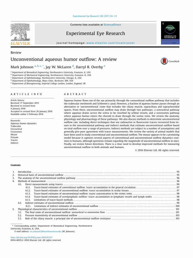
Fig. 2. Light micrograph of fluorescein dextran in the unconventional outflow pathwayof a rabbit after intracameral perfusion (AC, anterior chamber). Fluorescence is visiblein the conventional aqueous drainage pathway (a), comprising angular meshwork,angular aqueous plexus, and collector channels and aqueous veins, and in the un-conventional outflow in the outer aspects of the ciliary body (b) and muscle, and in thesuprachoroidal space (arrows). Tracer is also seen in the iris (c), which has beenartefactually deflected posteriorly. Reprinted from (Tripathi, 1977) with permissionfrom Elsevier.
M. Johnson et al. / Experimental Eye Research 158 (2017) 94e11196
collecting tracer in the blood, Bill could account for only about 80%of the tracer that left the eye, presumably through the trabecularoutflow pathway. The remaining tracer accumulated in the ciliarybody, choroid, and sclera. Because tracer accumulated at a constantrate in the sclera and other tissues and because this accumulationwas not correlated with the tracer's diffusion coefficient, Billconcluded that there must be a flow from the anterior chamberthrough the uvea and into the sclera by way of the choroid andsuprachoroid (Bill, 1965; Bill and Hellsing, 1965; Bill, 1966a). Billreferred to this as the “unconventional route” to distinguish it fromthe trabecular, or “conventional route”.
The definition of unconventional outflow has been expanded toinclude any of the pathways through which aqueous humor mightleave the eye other than through the trabecular pathway, includinga corneal route, an iridial route (Fine, 1964) and a retinal route(Fowlks et al., 1963). Flow through the corneal and iridial routes hasbeen shown to be negligible (Bill, 1977). Flow through the retinalroute is generated by the pumping capacity of the retinal pigmentepithelium, but is considered to be small as long as the retina re-mains attached (Pederson and Cantrill, 1984). This reviewwill focuson the flow exiting the posterior aspect of the uveal meshwork,passing through the ciliarymuscle, and entering the suprachoroidalspace.
Bill (1966a,d) found that in cynomolgus and vervet monkeys,the rate of outflow through the unconventional pathway rangedfrom 25 to 40% of total aqueous humor production. He also notedthat the magnitude of this flow changed little when IOP wasincreased, and concluded that this flow was relatively pressure-insensitive (Bill, 1966a). Since the time of these early experimentsthat demonstrated the unconventional pathway, much effort hasbeen put into determining its flow rate in humans and animalsunder a variety of experimental conditions. The rate of unconven-tional outflow is clinically important because it partly determinesIOP and it provides a means to reduce IOP for glaucoma therapy.
3. The anatomy of the unconventional outflow pathway
Because the interstitial spaces of the anterior uvea communicatewith the intertrabecular spaces (Fig. 1), a fraction of the aqueoushumor outflow that passes into the uveal meshwork can directly
enter the interstitial spaces of the ciliary muscle (Henderson, 1950).Perfusion studies with microspheres (Inomata et al., 1972) andfluorescein dextran (Tripathi, 1977; Lindsey and Weinreb, 2002)have demonstrated flow of aqueous humor from this regionthrough the interstitial spaces between the longitudinal ciliarymuscle bundles and into the supraciliary and suprachoroidal spaces(Fig. 2). However, where the fluid travels from here has beendebated. Bill (Bill, 1966a; Bill and Phillips, 1971; Bill, 1975) andGabelt and Kaufman (1989) postulate that the fluid seeps throughsclera and episclera, and passes into the orbit where it is absorbedby the orbital vasculature (the uveoscleral pathway). B�ar�any (1967)and others (Pederson et al., 1977; Sherman et al., 1978) have pro-posed that a significant fraction of this fluid is absorbed osmoticallyby the choroid and passes into the vortex veins (the uveovortexpathway). As a third potential route, recent studies have proposeddrainage into lymphatic vessels located within the ciliary body (theso-called ‘uveolymphatic’ pathway) (Yücel et al., 2009). However,the existence of lymphatic vessels in the eye under normal physi-ological conditions remains controversial (Schroedl et al., 2014).Wewill address the evidence for flow through each of these pathwayslater in this review.
Unconventional outflow, whether through the uveoscleral,uveovortex or uveolymphatic pathway, must pass through theinterstitial spaces of the ciliary muscle. Pilocarpine, which causesciliary muscle contraction and decreases the size of spaces betweenthe muscle bundles, decreases unconventional outflow (Crawfordand Kaufman, 1987) while atropine, a muscarinic antagonist, re-laxes the ciliary muscle and increases unconventional outflow (Billand Wålinder, 1966). This indicates that the ciliary muscle likelyrepresents a significant site of flow resistance along this pathway,and hence ciliary muscle tone can modulate unconventional flow.PGF2a, which increases unconventional outflow, is thought to act byreducing the amount of extracellular matrix between ciliarymusclebundles (Lütjen-Drecoll and Tamm, 1988) allowing increased flowthrough these spaces.
4. Methods of measurement
The greatest challenge facing studies of unconventional aqueoushumor outflow has been measurement of its flow rate. By defini-tion, this pathway encompasses any outflow of aqueous humor thatdoes not pass through the trabecular (conventional) pathway. Un-like the trabecular route, the path of egress is diffuse and difficult totrace, and this complicates measurement of flow through thispathway.
Measurements of unconventional outflow rate have been clas-sified as: (i) direct, tracer-based methods, or (ii) indirect methods.Direct methods are used to estimate both conventional and un-conventional outflow from the rate of accumulation of a tracermolecule in ocular tissues and in the blood after introducing thetracer in the anterior chamber. They are believed to be the mostaccurate, but because they are invasive and may require histologicanalysis, they are generally not suitable for use in humans. Indirectmethods are used to infer unconventional outflow from the dif-ference between aqueous humor production and aqueous humoroutflow through the trabecular pathway, each determined inde-pendently. Bothmethods of estimating unconventional flowwill beexplored here in more detail along with their underlying assump-tions and limitations.
4.1. Direct measurements using tracers
Anders Bill introduced tracer-based methods to determineaqueous humor outflow through both the conventional and un-conventional pathways in experimental animals and, to a limited

Fig. 3. Fluorescein concentration in the vortex veins and in general circulation in arhesus monkey after introduction into the anterior chamber. IOP was held at 20 mmHgfor 60 min and then increased to 32 mmHg. Reprinted from (Pederson et al., 1977) withpermission from the Association for Research in Vision and Ophthalmology.
M. Johnson et al. / Experimental Eye Research 158 (2017) 94e111 97
extent, in human eyes (Bill, 1962, 1965; Bill and Phillips, 1971). Hismethods still provide themost definitivemeasurements of aqueoushumor outflow through both pathways.
Any tracer introduced into the anterior chamber will be carriedaway by aqueous humor drainage. However, two characteristics ofthe tracer can be used to distinguish outflow through the conven-tional versus unconventional pathway: (i) transit time out of theeye and (ii) filtration based on tracer size. Outflow through theconventional pathway is relatively fast (a minute or less) and fairlyinsensitive to tracer molecular size, such that the rate of tracerappearance in the blood can be used to determine the flow ratethrough the conventional outflow pathway. In contrast, tracersdraining through the unconventional pathway are retarded orcaptured as they move through the unconventional outflowpathway such that their transit may take up to two hoursdepending on tracer size, animal species and dimensions of the eye.Tracer accumulation in the uveal tissues thereby provides a meansto estimate the unconventional outflow rate as well as identifyingthe anatomical route of unconventional outflow (as discussed inSection 3).
4.1.1. Tracer-based estimates of conventional outflow: traceraccumulation in the general circulation
Tracers are used to estimate conventional outflow directly bymeasuring the rate of tracer accumulation in the blood whilemaintaining a constant tracer concentration in the anterior cham-ber. Macromolecules of roughly 40 kDa (Toris et al., 1987) or larger(Nilsson, 1997) pass quickly through the conventional outflowpathway but pass slowly through or are captured within the un-conventional outflow pathway. Under these conditions, the rate ofconventional outflow is directly related to the rate of appearance oftracer in the blood.
Tracer concentration in the anterior chamber is maintainedconstant by using an infusion-withdrawal system that continuouslyinfuses tracer-containing solution (at concentration Co) into theanterior chamber of an anesthetized animal while simultaneouslywithdrawing at the same rate (Gabelt and Kaufman, 1989). Thissystem maintains a constant IOP and a constant tracer concentra-tion in the anterior chamber despite production and drainage ofaqueous humor during the perfusion (Bill, 1962, 1965, 1966a, 1966c,1966d). When the rate of appearance of tracer in the blood, dCb=dt,becomes relatively constant, then the conventional outflow rate Qc
can be estimated as:
Qc ¼ Vb
C0
dCbdt
(1)
where Vb is the distribution volume of the tracer in the blood thataccounts for tracer dilution in the blood after it leaves the eye. Vb
can be estimated asM/Cb, after injecting a known quantity,M, of thetracer into the general circulation and measuring its concentration,Cb, in the blood after mixing. For 131I-albumin tracer in cynomolgusmonkeys, Bill found that, Vb was the volume equivalent of
Table 1Distribution of unconventional outflow in cynomolgus mon131I-albumin after 2 h of perfusion. Uncertainties are stand
Tissue Estimated
Anterior sclera 14.5 ± 2.4Extraocular tissues 14.3 ± 5.5Posterior sclera 10.8 ± 3.8Choroid 5.1 ± 2.0Ciliary body 4.3 ± 0.5Iris 1.2 ± 0.2Remainder of eye 5.1 ± 1.4
approximately 7% of the total body weight (Bill, 1966a).Gabelt and Kaufman (1990) used this technique to show that
PGF2a does not increase conventional outflow in cynomolgusmonkeys despite increasing unconventional outflow in this species.Alternatively, this methodmay be combinedwithmeasurements ofaqueous humor production (Qin) to estimate unconventionaloutflow indirectly as the difference between Qin and Qc.
4.1.2. Tracer-based estimates of unconventional outflow: traceraccumulation in ocular tissues
This is the most common method of directly determining un-conventional outflow and is considered definitive. The flow rate iscalculated from the rate of accumulation of tracers in the tissues ofthe unconventional pathway after maintaining a constant con-centration in the anterior chamber for a fixed time. Typically, asolution with tracer is exchanged with aqueous humor in theanterior chamber of an anesthetized animal and tracer concentra-tion is maintained for 0.5e2 h at constant pressure using aninfusion-withdrawal system such as described in Section 4.1.1. Atthe end of a fixed perfusion time, the anterior chamber isexchanged with a tracer-free solution, and perfused for a short timeto remove tracer from the conventional outflow pathway.
The animal is then sacrificed, the eye enucleated and the tissuesseparated. Themass of the tracer in each tissue (typically, the sclera,choroid, ciliary body, iris and orbital tissue) is then determined bymethods specific to the tracer used in the study. The volume of fluidVi required to move the measured quantity of tracer mass mi intoeach tissue is given by Vi ¼mi/CAC, where CAC is the concentration oftracer in the anterior chamber and i ¼ 1 through n, where n is thenumber of tissues examined (e.g. Table 1). The average unconven-tional outflow during a time T is then:
key at normal IOP (12 mmHg) based on distribution ofard errors of the mean (Bill, 1966a).
volume of AC fluid passing into each tissue (Vi : ml)
M. Johnson et al. / Experimental Eye Research 158 (2017) 94e11198
Qu ¼Pn
i¼1 Vi
T¼
Pni¼1 mi
T CAC(2)
Tracers that have been used to study unconventional flow bythis method include 131I-g-globulin (Bill, 1966a), 131I-albumin (Billand Phillips, 1971), FITC-dextran (Suguro et al., 1985), and 3H-labeled dextran (Barrie et al., 1985). The tissue distribution of 131I-albumin used to estimate unconventional flow in cynomologusmonkeys by Bill (1966a) are given in Table 1. In this example, thetotal unconventional outflow was 0.46 ml/min, while conventionaloutflow measured by tracer accumulation in the blood was0.71± 0.09 ml/min, yielding a total outflow rate of 1.17± 0.13 ml/min.Thus, roughly 40% of the aqueous outflow passed through the un-conventional outflow pathway in this species, typical of othermeasurements in non-human primates.
The distribution of tracer recovered in the various tissues isthought to reflect the anatomical distribution of unconventionaloutflow. For example, the relatively high tracer content in theanterior sclera suggests that this was a preferential outflow route,as would be expected based on the organization of this tissue (seeFigs. 1 and 2). Similarly, the high tracer content in the extraoculartissues suggests that a significant fraction of tracer passed acrossthe sclera, consistent with uveoscleral outflow. In contrast, therewas very little tracer in the iris, suggesting negligible iridialoutflow. The retinal pathway was not separately assessed in thesestudies. However, while these measurements clearly indicate traceraccumulation in the various tissues, the tracer distribution does notnecessarily reflect the distribution of aqueous humor drainagethrough those tissues. This is because filtration and diffusion of thetracer affect its transport and may thereby influence tracer accu-mulation in the various tissues of the unconventional outflowpathway. We will consider these issues further in Section 5.1.
4.1.3. Tracer-based estimates of unconventional outflow: tracerconcentration in the vortex veins
Tracers introduced into the anterior chamber also have beenused to study the component of unconventional outflow thatpasses through the suprachoroidal space into the choroidal circu-lation and vortex veins (the uveovortex pathway). According to thistechnique, a tracer small enough to pass relatively unimpededthrough the vessel walls of the choriocapillaris (e.g. fluorescein) isintroduced into the anterior chamber and maintained at constantconcentration by the infusion-withdrawal system previouslydescribed. One of the four vortex veins is isolated and cannulated.The increased concentration of tracer in the vortex veins, Cvv,relative to that in the general circulation, Cb, is used to estimate theuveovortex outflow rate, Quv (Pederson et al., 1977):
Quv ¼ 4QCvv � CbCAC
(3)
where Q is the rate of blood flow out of the cannulated vein and thefactor of four accounts for the number of vortex veins. Pedersonet al. (1977) found that uveovortex outflow could account for asignificant fraction of unconventional outflow (Fig. 3).
The size of the tracer molecule is critical for measuring uveo-vortex outflow. In contrast to small molecular tracers, such asfluorescein, that are able to enter the choroidal vasculaturewith theaqueous humor flow, large tracers such as albumin are effectivelyexcluded from passing into the choroid (Bill, 1962, 1964) andaccumulate in the suprachoroidal space. From there, these largetracers diffuse across the sclera and into the orbit. Thus, thepathway taken by tracers the size of albumin or larger likely differsfrom the pathway taken by aqueous humor and small molecules.
4.1.4. Tracer-based estimates of uveolymphatic outflow: traceraccumulation in lymphatic vessels and lymph nodes
Traditionally, the eye has been considered to be devoid oflymphatics (Streilein, 2003). This view has recently been chal-lenged with reports of vessels that express lymphatic markerswithin the ciliary body (Yücel et al., 2009) and choroid (Koina et al.,2015) of human eyes, although these reports have been met withconsiderable controversy (Schroedl et al., 2014; Chan-Ling et al.,2015; Heindl et al., 2015). If lymphatics are normally present withinthe eye, then these vessels may contribute to unconventionaloutflow.
To measure outflow via the putative uveolymphatic route, Yücelet al. (2009) administered radio-labeled albumin into the anteriorchamber of sheep and measured its accumulation in lymph nodesand in cannulated lymphatic vessels (Kim et al., 2011). Traceraccumulated preferentially in the cervical lymph nodes (Yücelet al., 2009), with ~1e2% of tracer passing into the cervical andthoracic lymphatic vessels (Kim et al., 2011). Following intra-cameral injection, fluorescent tracer nanospheres were foundwithin vessels of the ciliary body that expressed lymphaticmarkers, suggestive of drainage via these vessels (Yücel et al.,2009). In mice, quantum dots injected unilaterally into the ante-rior chamber accumulated in ipsilateral lymph nodes in the neck(Tam et al., 2011) and the rate of accumulation was increased bylatanoprost (Tam et al., 2013).
When measuring uveolymphatic outflow, it should be recog-nized that not all tracer recovered within cervical lymphatics hadnecessarily originated from lymphatic vessels within the eyeproper, as some tracer may have entered lymphatic vessels fromwithin the periocular tissues. Albumin used to measure uveolym-phatic outflow (Kim et al., 2011) will also follow a uveoscleral route(Bill, 1966a) and enter the conjunctiva where lymphatic vessels areplentiful. Consistent with this notion, radiolabelled albumin wasmeasured in the periocular tissues following intracameral injectionin sheep (Kim et al., 2011). Once within the periocular space, al-bumin may enter conjunctival lymphatics that drain into the cer-vical lymphatics. Quantum dots, while significantly larger thanalbumin, also accumulate in lymph nodes of mice following intra-cameral injection (Tam et al., 2011, 2013). However, quantum dotscan diffuse through sclera as well (Amrite et al., 2008), although at arate slower than smaller molecules the size of albumin (Pease et al.,2014).
Currently, there is no clear consensus on the presence of lym-phatics in the eye under normal physiological conditions, althoughlymphatics may be recruited during inflammation or if the scleralborder is compromised (Schroedl et al., 2014). In light of this lack ofconsensus and concerns regarding the methods used to measureuveolymphatic outflow, further work is necessary to demonstratewhether uveolymphatic outflow represents a physiologically sig-nificant outflow pathway.
4.1.5. Limitations of tracer-based methodsThe use of tracers to measure unconventional outflow is based
on two fundamental assumptions. First, all tracer that enters atissue is retained or trapped within the tissue and that no tracer islost before the measurement. Second, all tracer enters the tissue bythe flow of aqueous humor (advection) and not by diffusion.
4.1.5.1. Retention of tracer. The first assumption will be satisfied ifthe tracer used is small enough to enter the tissue freely with theaqueous humor but too large to pass through the tissue during thetime of the experiment. Toris et al. (1987) reported that dextrans of40 kDa or larger were retained by the tissue whereas smallerdextrans (4 kDa) passed into the circulation or easily crossed thesclera and were lost in the orbit. Bill (1966a) found similarly that
M. Johnson et al. / Experimental Eye Research 158 (2017) 94e111 99
albumin (65 kDa) crossed the sclera easily but left the orbit at a lowrate, while g-globulin (150 kDa) was significantly delayed passingthrough the sclera.
4.1.5.2. Flow vs diffusion. Tracers can enter a tissue by advectionwhere they are transported at a rate dictated by the velocity of thefluid in which they are carried, or by diffusion where they aretransported at a rate dictated by molecular Brownian motion, or bya combination of both advection and diffusion. Tracers that enter atissue primarily by diffusion therefore cannot be used to make in-ferences regarding the quantity or velocity of flow. As diffusionbecomes more significant for decreasing molecular size, smallertracers tend to overestimate unconventional outflow based ontracer accumulation within tissue.
Over time t, a tracer will diffuse a distance of roughlyffiffiffiffiffiffiDt
p,
where D is the diffusion coefficient of the tracer, whereas a tracercarried by advectionwill move an average distance of V t, where V isthe average velocity of the fluid. Several consequences of theserelationships allow us to evaluate the impact of tracer diffusion onthe estimate of unconventional flow. First, because diffusionalmotion increases with the square root of time while advectivemotion increases linearly with time, the rate of accumulation of atracer in a tissue can indicate whether diffusion or advectiondominates the transport process. In Bill's study of cynomologusmonkeys, tracer accumulation in all unconventional outflow tissuesexamined after 30 min was equivalent to that contained in 13 ml ofsolution in the anterior chamber, while after 2 h, it was 55 ml (Bill,1966a). Thus, tracer accumulated roughly linearly over time, whichis consistent with an advection-dominated process.
Second, the influence of diffusion on estimating unconventionalflow can be explored using tracers of different size that havedifferent diffusion coefficients. If tracers are carried into a tissueprimarily by advection, then their rate of accumulation in the tissueshould be unaffected by the diffusion coefficient provided that theirsize is sufficiently small so as not to be excluded from this pathway.Bill (1966a) showed in cynomolgus monkey eyes that roughly thesame unconventional outflow rate was found with albumin as withg-globulin. Pederson and Toris (1987) reported similar findingsusing 40 and 150 kD dextran.
Third, the diffusion distance, estimated byffiffiffiffiffiffiDt
p, relative to the
length of the unconventional pathway, can also indicate theimportance of diffusion in tracer transport. In large animals, such asmonkeys, the length of the unconventional pathway is on the order
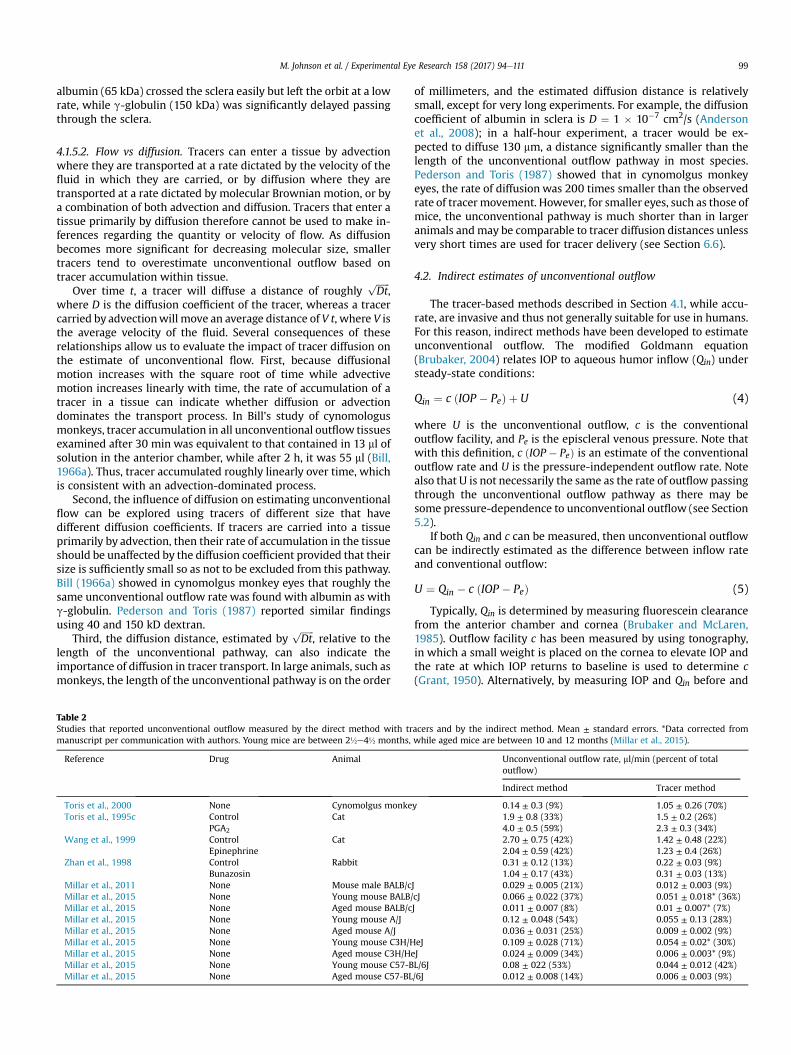
Table 2Studies that reported unconventional outflow measured by the direct method with trmanuscript per communication with authors. Young mice are between 2½e4½ months,
Reference Drug Animal
Toris et al., 2000 None Cynomolgus monkeToris et al., 1995c Control
PGA2
Cat
Wang et al., 1999 ControlEpinephrine
Cat
Zhan et al., 1998 ControlBunazosin
Rabbit
Millar et al., 2011 None Mouse male BALB/cMillar et al., 2015 None Young mouse BALB/Millar et al., 2015 None Aged mouse BALB/cMillar et al., 2015 None Young mouse A/JMillar et al., 2015 None Aged mouse A/JMillar et al., 2015 None Young mouse C3H/HMillar et al., 2015 None Aged mouse C3H/HeMillar et al., 2015 None Young mouse C57-BMillar et al., 2015 None Aged mouse C57-BL
of millimeters, and the estimated diffusion distance is relativelysmall, except for very long experiments. For example, the diffusioncoefficient of albumin in sclera is D ¼ 1 � 10�7 cm2/s (Andersonet al., 2008); in a half-hour experiment, a tracer would be ex-pected to diffuse 130 mm, a distance significantly smaller than thelength of the unconventional outflow pathway in most species.Pederson and Toris (1987) showed that in cynomolgus monkeyeyes, the rate of diffusion was 200 times smaller than the observedrate of tracermovement. However, for smaller eyes, such as those ofmice, the unconventional pathway is much shorter than in largeranimals andmay be comparable to tracer diffusion distances unlessvery short times are used for tracer delivery (see Section 6.6).
4.2. Indirect estimates of unconventional outflow
The tracer-based methods described in Section 4.1, while accu-rate, are invasive and thus not generally suitable for use in humans.For this reason, indirect methods have been developed to estimateunconventional outflow. The modified Goldmann equation(Brubaker, 2004) relates IOP to aqueous humor inflow (Qin) understeady-state conditions:
Qin ¼ c ðIOP � PeÞ þ U (4)
where U is the unconventional outflow, c is the conventionaloutflow facility, and Pe is the episcleral venous pressure. Note thatwith this definition, c ðIOP � PeÞ is an estimate of the conventionaloutflow rate and U is the pressure-independent outflow rate. Notealso that U is not necessarily the same as the rate of outflow passingthrough the unconventional outflow pathway as there may besome pressure-dependence to unconventional outflow (see Section5.2).
If both Qin and c can be measured, then unconventional outflowcan be indirectly estimated as the difference between inflow rateand conventional outflow:
U ¼ Qin � c ðIOP � PeÞ (5)
Typically, Qin is determined by measuring fluorescein clearancefrom the anterior chamber and cornea (Brubaker and McLaren,1985). Outflow facility c has been measured by using tonography,in which a small weight is placed on the cornea to elevate IOP andthe rate at which IOP returns to baseline is used to determine c(Grant, 1950). Alternatively, by measuring IOP and Qin before and
acers and by the indirect method. Mean ± standard errors. *Data corrected fromwhile aged mice are between 10 and 12 months (Millar et al., 2015).
Unconventional outflow rate, ml/min (percent of totaloutflow)
Indirect method Tracer method
y 0.14 ± 0.3 (9%) 1.05 ± 0.26 (70%)1.9 ± 0.8 (33%) 1.5 ± 0.2 (26%)4.0 ± 0.5 (59%) 2.3 ± 0.3 (34%)2.70 ± 0.75 (42%) 1.42 ± 0.48 (22%)2.04 ± 0.59 (42%) 1.23 ± 0.4 (26%)0.31 ± 0.12 (13%) 0.22 ± 0.03 (9%)1.04 ± 0.17 (43%) 0.31 ± 0.03 (13%)
J 0.029 ± 0.005 (21%) 0.012 ± 0.003 (9%)cJ 0.066 ± 0.022 (37%) 0.051 ± 0.018* (36%)J 0.011 ± 0.007 (8%) 0.01 ± 0.007* (7%)
0.12 ± 0.048 (54%) 0.055 ± 0.13 (28%)0.036 ± 0.031 (25%) 0.009 ± 0.002 (9%)
eJ 0.109 ± 0.028 (71%) 0.054 ± 0.02* (30%)J 0.024 ± 0.009 (34%) 0.006 ± 0.003* (9%)L/6J 0.08 ± 022 (53%) 0.044 ± 0.012 (42%)/6J 0.012 ± 0.008 (14%) 0.006 ± 0.003 (9%)
M. Johnson et al. / Experimental Eye Research 158 (2017) 94e111100
after administering a drug (e.g. acetazolamide) that suppressesaqueous humor production, c can be determined by solving Equa-tion (4) at two flow rates and the corresponding IOPs (Yablonskiet al., 1985; Hayashi et al., 1989). A value of episcleral venouspressure, Pe, is required to calculate U. This measurement is difficultand values between 8 and 10 mmHg are typically chosen, and thesame value is used for all subjects in a trial. More recent evidencesuggests that the mean episcleral venous pressure is 6.3 mm Hg innormal subjects (Sit et al., 2011). Importantly, the lower the valueused for Pe, the smaller the estimate of U.
Estimates of unconventional flow determined by using the in-direct method have varied considerably and, in general, show pooragreement with unconventional outflow measured by directmethods that used tracers. Even in the same study, Toris et al.(2000) reported an unconventional outflow rate in cynomolgusmonkeys of 0.14 ± 1.2 ml/min by using indirect methods, but1.05 ± 0.6 ml/min by using tracers in this same population, almostan order of magnitude greater. Toris et al. also noted a lack of cor-relation between unconventional outflow estimated indirectly andthosemeasured with tracers. Similarly, studies inmice (Millar et al.,2011, 2015) showed poor agreement between tracer-based andindirect estimates of unconventional outflow. In general, mea-surements with the indirect method are considerably higher thanthose measured using tracers (Table 2), although measurements byToris et al. (2000) represent a significant exception. While bothdirect and indirect methods have been shown to detect changes inresponse to drug treatment, the absence of any quantitativeagreement between the two approaches is troubling and raisesquestions as to what specifically is being measured by the indirectmethod.
4.2.1. Limitations of indirect estimates of unconventional outflowThe conventional outflow rate is similar to the aqueous humor
production rate, and the difference between these (Equation (5)) is,under most circumstances, significantly less than either.
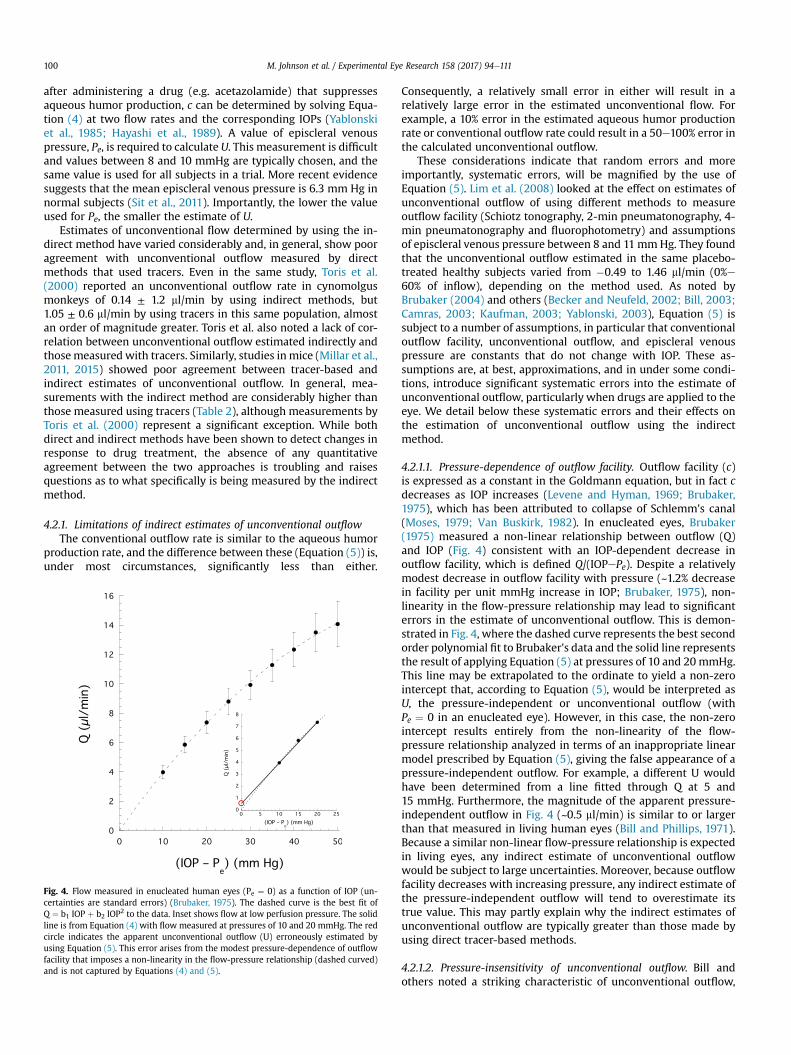
Fig. 4. Flow measured in enucleated human eyes (Pe ¼ 0) as a function of IOP (un-certainties are standard errors) (Brubaker, 1975). The dashed curve is the best fit ofQ ¼ b1 IOP þ b2 IOP2 to the data. Inset shows flow at low perfusion pressure. The solidline is from Equation (4) with flow measured at pressures of 10 and 20 mmHg. The redcircle indicates the apparent unconventional outflow (U) erroneously estimated byusing Equation (5). This error arises from the modest pressure-dependence of outflowfacility that imposes a non-linearity in the flow-pressure relationship (dashed curved)and is not captured by Equations (4) and (5).
Consequently, a relatively small error in either will result in arelatively large error in the estimated unconventional flow. Forexample, a 10% error in the estimated aqueous humor productionrate or conventional outflow rate could result in a 50e100% error inthe calculated unconventional outflow.
These considerations indicate that random errors and moreimportantly, systematic errors, will be magnified by the use ofEquation (5). Lim et al. (2008) looked at the effect on estimates ofunconventional outflow of using different methods to measureoutflow facility (Schiotz tonography, 2-min pneumatonography, 4-min pneumatonography and fluorophotometry) and assumptionsof episcleral venous pressure between 8 and 11mmHg. They foundthat the unconventional outflow estimated in the same placebo-treated healthy subjects varied from �0.49 to 1.46 ml/min (0%e60% of inflow), depending on the method used. As noted byBrubaker (2004) and others (Becker and Neufeld, 2002; Bill, 2003;Camras, 2003; Kaufman, 2003; Yablonski, 2003), Equation (5) issubject to a number of assumptions, in particular that conventionaloutflow facility, unconventional outflow, and episcleral venouspressure are constants that do not change with IOP. These as-sumptions are, at best, approximations, and in under some condi-tions, introduce significant systematic errors into the estimate ofunconventional outflow, particularly when drugs are applied to theeye. We detail below these systematic errors and their effects onthe estimation of unconventional outflow using the indirectmethod.
4.2.1.1. Pressure-dependence of outflow facility. Outflow facility (c)is expressed as a constant in the Goldmann equation, but in fact cdecreases as IOP increases (Levene and Hyman, 1969; Brubaker,1975), which has been attributed to collapse of Schlemm's canal(Moses, 1979; Van Buskirk, 1982). In enucleated eyes, Brubaker(1975) measured a non-linear relationship between outflow (Q)and IOP (Fig. 4) consistent with an IOP-dependent decrease inoutflow facility, which is defined Q/(IOPePe). Despite a relativelymodest decrease in outflow facility with pressure (~1.2% decreasein facility per unit mmHg increase in IOP; Brubaker, 1975), non-linearity in the flow-pressure relationship may lead to significanterrors in the estimate of unconventional outflow. This is demon-strated in Fig. 4, where the dashed curve represents the best secondorder polynomial fit to Brubaker's data and the solid line representsthe result of applying Equation (5) at pressures of 10 and 20 mmHg.This line may be extrapolated to the ordinate to yield a non-zerointercept that, according to Equation (5), would be interpreted asU, the pressure-independent or unconventional outflow (withPe ¼ 0 in an enucleated eye). However, in this case, the non-zerointercept results entirely from the non-linearity of the flow-pressure relationship analyzed in terms of an inappropriate linearmodel prescribed by Equation (5), giving the false appearance of apressure-independent outflow. For example, a different U wouldhave been determined from a line fitted through Q at 5 and15 mmHg. Furthermore, the magnitude of the apparent pressure-independent outflow in Fig. 4 (~0.5 ml/min) is similar to or largerthan that measured in living human eyes (Bill and Phillips, 1971).Because a similar non-linear flow-pressure relationship is expectedin living eyes, any indirect estimate of unconventional outflowwould be subject to large uncertainties. Moreover, because outflowfacility decreases with increasing pressure, any indirect estimate ofthe pressure-independent outflow will tend to overestimate itstrue value. This may partly explain why the indirect estimates ofunconventional outflow are typically greater than those made byusing direct tracer-based methods.
4.2.1.2. Pressure-insensitivity of unconventional outflow. Bill andothers noted a striking characteristic of unconventional outflow,

Fig. 5. Computed tomography oblique sections through the orbital region of an adultrhesus monkey perfused with radio-opaque contrast medium in the anterior chamber.(A) After 1 h of continuous perfusion while the monkey was alive, contrast mediumwas confined to the anterior chamber and there was no posterior motion of the me-dium. Similar images were recorded for up to 8 h. (B) In the same monkey 35 min afterdeath by pentobarbital overdose, contrast medium migrated posteriorly (red arrows).Black voids within the anterior chamber are an imaging artifact. Both images werefrom the same section plane in the same monkey. The right eye was set to 20 mmHgand left eye to 40 mmHg by using a fluid column. Reprinted from (Butler et al., 1984)with permission from Elsevier.
M. Johnson et al. / Experimental Eye Research 158 (2017) 94e111 101
namely its relative insensitivity to the pressure difference thatdrives the flow (Section 5.2). As measured using the indirectmethod, unconventional outflow is assumed to be entirely inde-pendent of IOP. However, direct tracer-based measurements indi-cate that unconventional outflow is not entirely pressure-independent, although it is relatively pressure-insensitive (Bill,1966a; Becker and Neufeld, 2002; Bill, 2003; Kaufman, 2003).Any pressure-dependent changes in unconventional outflow willbe attributed to conventional outflow in Equation (5). This will leadto a moderate underestimate of unconventional outflow andoverestimate of conventional outflow as measured using indirecttechniques. Kaufman (2003) suggests that the unconventionaloutflow facility is approximately 5% of total outflow facility in non-human primates. While this may seem small, this is roughlyequivalent to 20e50% of total unconventional outflow at physio-logical IOP, and therefore the pressure-dependent contribution tounconventional outflow cannot be ignored.
This problem may be a particular concern for drug studies if thecompounds used increase the pressure dependence of unconven-tional outflow (e.g. PGF2a (Gabelt and Kaufman, 1990; Kaufman,2003)). The assumption of a purely pressure-independent uncon-ventional outflow can greatly confuse interpretation of data fromsuch experiments and may explain why some studies have foundthat prostaglandins lower IOP primarily by increasing unconven-tional outflow (Gabelt and Kaufman, 1990; Toris et al., 1993;Stjernschantz, 2001; Woodward et al., 2010), while other studiessuggest that these agents also have a significant effect on conven-tional outflow (Christiansen et al., 2004;Wan et al., 2007; Lim et al.,2008).
4.2.1.3. Effect of episcleral venous pressure. The indirect method ofestimating unconventional outflow requires an accurate estimateof episcleral venous pressure (Christiansen et al., 2004) and re-quires that the episcleral pressure be independent of IOP. Studies inmonkeys support the latter assumption (M€aepea and Bill, 1989),but accurately measuring episcleral venous pressure proves to bevery difficult.
Mean reported episcleral venous pressures have ranged from6.3 mmHg to 11.4 mmHg depending on the method of measure-ment (Sit et al., 2011; Sit andMcLaren, 2011). Because of difficulty inits measurement, some investigators have assumed a single valuefor episcleral venous pressure for all subjects when calculatingunconventional outflow (Toris et al., 2000; Zhao et al., 2010). Errorsin episcleral venous pressure directly impact the estimate of un-conventional outflow determined using Equation (5). For example,a 1 mmHg error in episcleral venous pressure leads to an error ofapproximately 0.3 ml/min in unconventional outflow (assumingc ¼ 0.3 ml/min/mmHg), which is 25e100% of the typical uncon-ventional outflow. Assuming too low a value of episcleral venouspressure in this calculation (Equation (5)) would lead to under-estimated unconventional outflow whereas assuming too high avalue of episcleral venous pressure would lead to overestimatedunconventional outflow.
The assumptions inherent in Equations (4) and (5), combinedwith measurement inaccuracies and systematic errors, lead toconsiderable uncertainties in indirect estimates of unconventionaloutflow. Note that while both the direct and indirect methods ofcalculating unconventional outflow are subject to considerableexperimental variability, the systematic errors inherent in thedirect method are significantly smaller than those inherent to theindirect method. But unfortunately, there is currently no othermethod to assess unconventional outflow in humans other than bythe indirect methods, and thus they are used despite their limita-tions. In animals, where unconventional outflow can be directlymeasured, the lack of agreement between direct and indirect
measures casts doubt on the utility of the indirect methods. Theseissues apply a fortiori in studies on the effects of drugs on uncon-ventional outflow, because drugs could directly affect the pressuredependence of unconventional flow and could alter episcleralvenous pressure, thereby leading to erroneous conclusionsregarding the effects of these drugs on aqueous humor dynamics.Indirect estimates of unconventional outflow must therefore beinterpreted with caution.
5. Physiological characteristics of unconventional outflow
Several features of unconventional outflow are particularlyinteresting from a physiological perspective. These features includethe route of aqueous humor drainage along this pathway, theinsensitivity to changes in intraocular pressure, the role of theciliary muscle, and changes in this pathway after death.
5.1. The final route of unconventional outflow: uveoscleral vsuveovortex flow
Unconventional outflow passes through the ciliary muscle andinto the suprachoroidal space, but the pathway taken from thesuprachoroidal space to exit the eye has been debated. Bill identi-fied this path by tracing the route of radioactive-labeled proteinsand other large molecules as they left the eye. These tracers movedthrough the ciliary muscle, into the suprachoroid space, andaccumulated in the sclera and orbit, a route he termed the“uveoscleral” pathway (Bill, 1965). This suggested fluid movementacross the sclera where it could be collected and drained by theextraocular lymph vessels. Bill calculated the flow rate through thisroute based on tracer accumulation in these tissues, but he recog-nized that this method would only give an accurate estimate ofunconventional flow if the tracer followed the same path as theaqueous humor and also was not washed away into the generalcirculation during the course of the experiment.
The use of smaller tracers suggested a somewhat different route.Pederson et al. (1977) perfused the anterior chamber of monkeyswith fluorescein, a small molecule, and found that a significantfraction of this tracer entered the vortex veins (Fig. 3). The fluo-rescein concentration in these vessels was somewhat dependent onperfusion pressure, although it was not as pressure-dependent aswas conventional outflow. The sensitivity to pressure suggestedthat movement through this pathway required ultrafiltration ofwater and small molecules into the uveal capillaries. They

Table 3Unconventional flow in different species determined by recovering labeled tracers (either collection of tracers in ocular tissues, or collection of tracer in general circulationwitha separate measurement of aqueous inflow).
Species Unconventional flow (ml/min) Fraction of inflow (%) References
Cynomolgus monkey 0.48e1.07 27e65 (Bill (1966a, 1967, 1971))(Bill and Wålinder, 1966)(Gabelt and Kaufman, 1989)(Nilsson et al. (1989, 2006))(Toris et al., 2000)(Gabelt et al., 2005)
Vervet monkey 0.23e0.74 19e38 (Bill (1966d, 1969a, 1969b, 1970, 1971))
Rhesus monkey 0.63 37 (Gabelt et al., 2003)
Cat 0.36e1.5 3e26 (Bill, 1966b)(Toris et al., 1995c)(Wang et al., 1999)(Goh et al., 1994)(Toris, 2008)
Rabbit (albino) 0.11e0.53 3e22 (Bill, 1966c)(Cole and Monro, 1976)(Goh et al., 1989)(Poyer et al., 1992)(Takashima et al., 1996)(Zhan et al., 1998)(Kanno et al., 1998)(Sugiyama et al., 2001)(Honjo et al., 2001)(Zhan et al., 2002)(Oka et al., 2006)
Dog (beagle) e 15 (Barrie et al., 1985)
Human 0.28 0e14 (Bill and Phillips, 1971)
Mouse (BALB/cJ) 0.01e0.051 7e36 (Millar et al., 2011)(Millar et al., 2015)
Mouse (A/J) 0.009e0.055 9e28 (Millar et al., 2011)(Millar et al., 2015)
Mouse (C3H/HeJ) 0.006e0.054 9e30 (Millar et al., 2011)(Millar et al., 2015)
Mouse (C57-BL/6J) 0.006e0.044 9e42 (Millar et al., 2011)(Millar et al., 2015)
M. Johnson et al. / Experimental Eye Research 158 (2017) 94e111102
suggested that Starling forces were responsible for fluid resorptioninto the uveal capillaries, with these forces arising from the highercolloidal osmotic pressure in these vessels relative to the uvealinterstitial fluid, a possibility first raised by B�ar�any (1967).
Bill also found tracer in the vortex veins in his early studies ofrabbits perfused through the anterior chamber with radio-labeledalbumin, but he concluded that less than 1.2% of the tracer withinthe anterior chamber drained via this route (Bill, 1962). In makingthis conclusion, Bill assumed that albumin could freely enter theuveal vessels once within the suprachoroidal space, an assumptionthat is inconsistent with the relatively low protein concentration insuprachoroidal fluid compared to plasma (Emi et al., 1989; Toriset al., 1990). The large size of albumin (67 kDa), as compared tothat of fluorescein (0.3 kDa), likely excluded albumin from enteringthe choriocapillaris and the uveal capillaries, such that albuminwould accumulate in the suprachoroidal space, diffuse across thesclera and enter the orbit, thereby explaining Bill's experimentalobservations (Yablonski, 2002). Larger molecular tracers such asalbumin therefore appear to travel along a separate pathway thandoes aqueous humor and smaller molecular tracers.
Visualization of unconventional outflow in living anesthetizedrhesus monkeys by using small tracers has provided indirect evi-dence for the role of these vessels in unconventional outflow. Butleret al. (1984) perfused the anterior chamber with a small radio-opaque contrast agent (789 Da) and created images of this agentby X-ray computed tomography. Despite perfusion for up to 8 h
while the animal was alive, tracer was restricted to the anteriorchamber, but immediately after death, tracer was seen migratingfrom the anterior chamber towards the posterior pole (Fig. 5). Thisexperiment suggests that small molecular tracers that permeatethe unconventional pathway are rapidly swept away by circulatingblood, but when blood flow ceases soon after death, these tracersaccumulate within the tissue.
Yablonski (2003) suggested that flow through the uveovortexpathway could be increased by chronic prostaglandin exposure.Prostaglandins have been shown to modify the extracellular matrixof the uvea, and if these modifications also increased the hydraulicpermeability of these vessels, then flow through this route wouldalso increase.
Based on the concentrations of fluorescein in the vortex veins,Pederson et al. (1977) estimated that the uveovortex pathway couldaccount for roughly 10% of aqueous humor outflow in the non-human primate eye. This estimate was similar to that ofMcMaster and Macri (1968), who found 8% of aqueous outflow topass through the uveovortex pathway in an arterially-perfusedenucleated rabbit eye. These estimates of outflow through theuveovortex pathway are significantly less than the total uncon-ventional flow that has been measured using direct tracer-basedtechniques in animals (Table 3). In the living eye, unconventionaloutflow therefore likely drains through both the uveoscleral anduveovortex routes, where the distribution of outflow through eachroute likely depends on IOP, ciliary muscle tone, and other factors
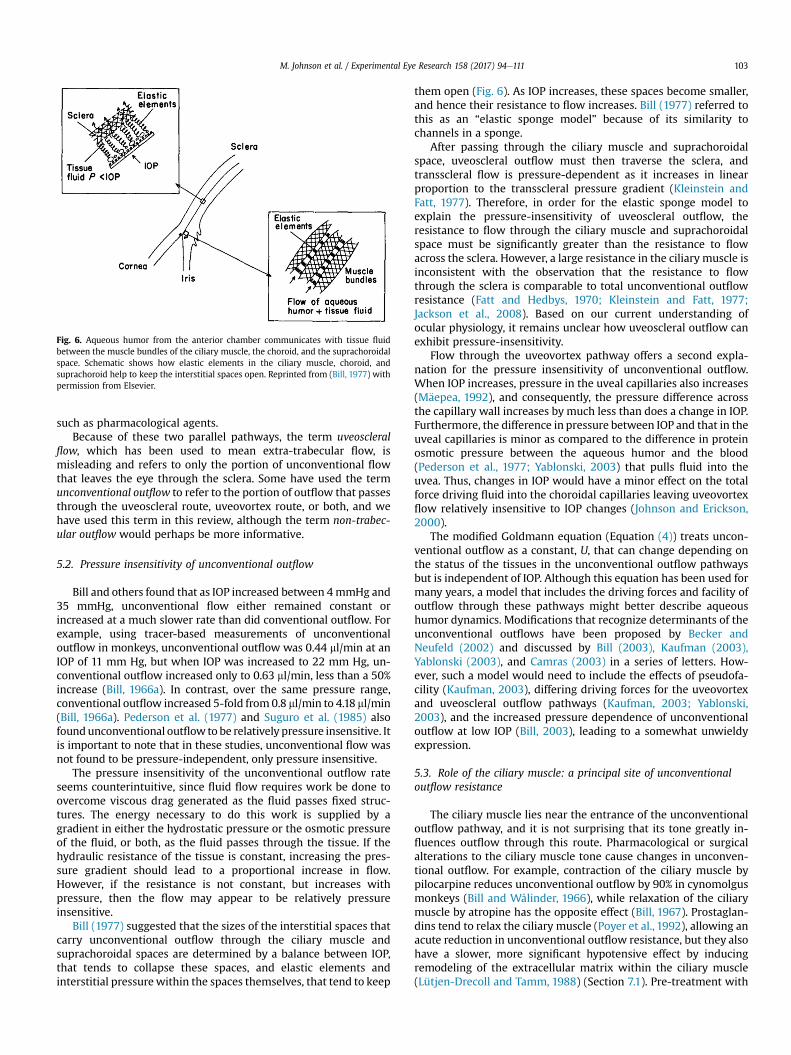
Fig. 6. Aqueous humor from the anterior chamber communicates with tissue fluidbetween the muscle bundles of the ciliary muscle, the choroid, and the suprachoroidalspace. Schematic shows how elastic elements in the ciliary muscle, choroid, andsuprachoroid help to keep the interstitial spaces open. Reprinted from (Bill, 1977) withpermission from Elsevier.
M. Johnson et al. / Experimental Eye Research 158 (2017) 94e111 103
such as pharmacological agents.Because of these two parallel pathways, the term uveoscleral
flow, which has been used to mean extra-trabecular flow, ismisleading and refers to only the portion of unconventional flowthat leaves the eye through the sclera. Some have used the termunconventional outflow to refer to the portion of outflow that passesthrough the uveoscleral route, uveovortex route, or both, and wehave used this term in this review, although the term non-trabec-ular outflow would perhaps be more informative.
5.2. Pressure insensitivity of unconventional outflow
Bill and others found that as IOP increased between 4mmHg and35 mmHg, unconventional flow either remained constant orincreased at a much slower rate than did conventional outflow. Forexample, using tracer-based measurements of unconventionaloutflow in monkeys, unconventional outflow was 0.44 ml/min at anIOP of 11 mm Hg, but when IOP was increased to 22 mm Hg, un-conventional outflow increased only to 0.63 ml/min, less than a 50%increase (Bill, 1966a). In contrast, over the same pressure range,conventional outflow increased 5-fold from 0.8 ml/min to 4.18 ml/min(Bill, 1966a). Pederson et al. (1977) and Suguro et al. (1985) alsofoundunconventional outflow to be relatively pressure insensitive. Itis important to note that in these studies, unconventional flow wasnot found to be pressure-independent, only pressure insensitive.
The pressure insensitivity of the unconventional outflow rateseems counterintuitive, since fluid flow requires work be done toovercome viscous drag generated as the fluid passes fixed struc-tures. The energy necessary to do this work is supplied by agradient in either the hydrostatic pressure or the osmotic pressureof the fluid, or both, as the fluid passes through the tissue. If thehydraulic resistance of the tissue is constant, increasing the pres-sure gradient should lead to a proportional increase in flow.However, if the resistance is not constant, but increases withpressure, then the flow may appear to be relatively pressureinsensitive.
Bill (1977) suggested that the sizes of the interstitial spaces thatcarry unconventional outflow through the ciliary muscle andsuprachoroidal spaces are determined by a balance between IOP,that tends to collapse these spaces, and elastic elements andinterstitial pressurewithin the spaces themselves, that tend to keep
them open (Fig. 6). As IOP increases, these spaces become smaller,and hence their resistance to flow increases. Bill (1977) referred tothis as an “elastic sponge model” because of its similarity tochannels in a sponge.
After passing through the ciliary muscle and suprachoroidalspace, uveoscleral outflow must then traverse the sclera, andtransscleral flow is pressure-dependent as it increases in linearproportion to the transscleral pressure gradient (Kleinstein andFatt, 1977). Therefore, in order for the elastic sponge model toexplain the pressure-insensitivity of uveoscleral outflow, theresistance to flow through the ciliary muscle and suprachoroidalspace must be significantly greater than the resistance to flowacross the sclera. However, a large resistance in the ciliary muscle isinconsistent with the observation that the resistance to flowthrough the sclera is comparable to total unconventional outflowresistance (Fatt and Hedbys, 1970; Kleinstein and Fatt, 1977;Jackson et al., 2008). Based on our current understanding ofocular physiology, it remains unclear how uveoscleral outflow canexhibit pressure-insensitivity.
Flow through the uveovortex pathway offers a second expla-nation for the pressure insensitivity of unconventional outflow.When IOP increases, pressure in the uveal capillaries also increases(M€aepea, 1992), and consequently, the pressure difference acrossthe capillary wall increases by much less than does a change in IOP.Furthermore, the difference in pressure between IOP and that in theuveal capillaries is minor as compared to the difference in proteinosmotic pressure between the aqueous humor and the blood(Pederson et al., 1977; Yablonski, 2003) that pulls fluid into theuvea. Thus, changes in IOP would have a minor effect on the totalforce driving fluid into the choroidal capillaries leaving uveovortexflow relatively insensitive to IOP changes (Johnson and Erickson,2000).
The modified Goldmann equation (Equation (4)) treats uncon-ventional outflow as a constant, U, that can change depending onthe status of the tissues in the unconventional outflow pathwaysbut is independent of IOP. Although this equation has been used formany years, a model that includes the driving forces and facility ofoutflow through these pathways might better describe aqueoushumor dynamics. Modifications that recognize determinants of theunconventional outflows have been proposed by Becker andNeufeld (2002) and discussed by Bill (2003), Kaufman (2003),Yablonski (2003), and Camras (2003) in a series of letters. How-ever, such a model would need to include the effects of pseudofa-cility (Kaufman, 2003), differing driving forces for the uveovortexand uveoscleral outflow pathways (Kaufman, 2003; Yablonski,2003), and the increased pressure dependence of unconventionaloutflow at low IOP (Bill, 2003), leading to a somewhat unwieldyexpression.
5.3. Role of the ciliary muscle: a principal site of unconventionaloutflow resistance
The ciliary muscle lies near the entrance of the unconventionaloutflow pathway, and it is not surprising that its tone greatly in-fluences outflow through this route. Pharmacological or surgicalalterations to the ciliary muscle tone cause changes in unconven-tional outflow. For example, contraction of the ciliary muscle bypilocarpine reduces unconventional outflow by 90% in cynomolgusmonkeys (Bill and Wålinder, 1966), while relaxation of the ciliarymuscle by atropine has the opposite effect (Bill, 1967). Prostaglan-dins tend to relax the ciliary muscle (Poyer et al., 1992), allowing anacute reduction in unconventional outflow resistance, but they alsohave a slower, more significant hypotensive effect by inducingremodeling of the extracellular matrix within the ciliary muscle(Lütjen-Drecoll and Tamm, 1988) (Section 7.1). Pre-treatment with
M. Johnson et al. / Experimental Eye Research 158 (2017) 94e111104
pilocarpine acutely abolishes the effect of prostaglandins on IOPand unconventional outflow in monkeys (Crawford and Kaufman,1987; Nilsson et al., 1989).
Bill (1977) suggested that compression of the ciliarymusclewithincreasing IOP could increase its flow resistance and therebycontribute to pressure insensitivity of unconventional outflow(Fig. 6). When the ciliary muscle is removed by cyclodialysis, mostof the resistance it offers is lost (Bill, 1966c) and unconventionaloutflow increases four-fold (Suguro et al., 1985) and becomespressure dependent (Toris and Pederson, 1985). Experimentsexploring the role of the ciliary muscle in unconventional outflowhave been reviewed by Alm and Nilsson (2009).
5.4. Unconventional outflow in living and post-mortem eyes
Flow through the conventional outflow pathway is pressure-driven and thought to have largely the same characteristics inliving and post-mortem eyes (Grant and Trotter, 1955; Grant, 1963),when corrected for the loss of episcleral venous pressure afterdeath. The unconventional outflow pathway, however, is morecomplicated. Bill found that there was no correlation between rateof unconventional outflow in the eye of a living animal and that inthe contralateral eye from the same animal after death (Bill, 1966a).Following death, unconventional outflow was approximately 150%higher than in eyes of living cynomolgus monkeys (Bill, 1966a),rabbits (Bill, 1966c), and cats (Bill, 1966b), and increased by nearly400% in vervet monkeys (Bill, 1966d). In mice, however, there wasno evidence for a difference in unconventional outflow betweenliving and post-mortem (in situ) eyes (Millar et al., 2011). Thepressure-insensitivity of unconventional flow in the physiologicalpressure range suggests that the increase in unconventionaloutflow after death was not likely caused by the loss of episcleralvenous pressure. Unconventional outflow, however, may haveincreased after death due to loss of ciliary muscle tone and the
Table 4Unconventional outflow in humans as determined by using the indirect method. Qu is uerages. “Fluorophotometric” or “tonographic” refers to the method used to measure aqu
Condition of subjects (method) Qu % Qin Refe
Normal (tonographic) 0.8 36 (TowNormal (tonographic) 1.06 41 (CoaYoung normal [21e23 y] (tonographic) �0.38 0 (Mis
1.23 85Young normal [21e23 y] (fluorophotometric) 0.57 25
0.78 54Normals (tonographic) 0.85e1.52 34e62 (BruNormal (fluorophotometric) 1.5 45 (Wa
1.1 361.1 39
Normal (fluorophotometric) 1.09 (TorNormal [47e76 y] (tonographic) 0.94 38 (Nau
0.07 6Glaucoma patients before LTP (fluorophotometric) 1.1 78 (Yab
0.51 400.96 64
Ocular hypertension (fluorophotometric) 0.60 30 (HayOcular hypertension (fluorophotometric) 0.66 (TorOcular hypertension (fluorophotometric) 1.14 46 (Tor
0.99 480.86 371.24 52
Ocular hypertension (fluorophotometric) 0.67 25 (Tor0.62 240.58 23
Ocular hypertension (fluorophotometric) 0.35 14 (Tor0.5 220.12 4.8
Ocular hypertension or glaucoma (tonographic) 0.64e0.96 25e40 (Chr
associated decrease in outflow resistance offered by the ciliarymuscle (Section 5.3).
6. Characteristics of unconventional flow in different species
Anatomical [see Henderson (1950) and Tripathi (1974)] orfunctional differences in the unconventional outflow pathway canaffect the drainage of aqueous humor through this route andcontribute to differences in unconventional outflow between spe-cies. Lower placentals, for example, exhibit a deep ciliary cleft witha relatively undeveloped ciliary muscle that allows open fluidcommunication to the posterior ciliary body. Primates, in contrast,have a well-developed ciliary muscle without a ciliary cleft thatrenders a strong dependence of unconventional outflow on ciliarymuscle tone and accommodation.
Table 3 shows the unconventional outflow rate and its fractionof total outflow in a variety of species, as measured by tracertechniques. In general, non-human primates have the highestfraction of unconventional outflow. We explore the difference inunconventional outflow between species, focusing on primates andmice because of their utility as models for human eyes and becauseof considerable discrepancies in literature regarding unconven-tional flow in mice.
6.1. Human
Only one study (Bill and Phillips, 1971) has reported directmeasurements of unconventional outflow in living human eyes byusing tracer methods. In patients' eyes destined for enucleation,unconventional outflow was estimated from radioactive albumintracer accumulation in the posterior scleral shell after injection intothe anterior chamber. In 3 eyes, unconventional outflow was esti-mated to be 0%, 4% and 14% of total outflow from the anteriorchamber (an absolute value of unconventional outflow was
nconventional outflow (ml/min) and Qin is inflow rate. All values are population av-eous humor outflow facility (LTP e laser trabeculoplasty).
rence Notes
nsend and Brubaker, 1980)kes and Siah, 1984) Control (Placebo)hima et al., 1997) Daytime
NighttimeDaytimeNighttime
baker et al., 2001) Control (Placebo)ng et al., 2002) Control (Baseline)
Control (Placebo)Experimental (Baseline)
is et al., 2002b)et al., 2013) Daytime
Nighttimelonski et al., 1985) Control (Baseline)
Control (Placebo)Experimental (Baseline)
ashi et al., 1989)is et al., 2002b)is et al., 2004) Control (Baseline)
Control (Placebo 1)Control (Placebo 2)Experimental (Baseline)
is et al., 1995b) Control (Baseline)Control (Placebo)Experimental (Baseline)
is et al., 1995a) Control (Baseline)Control (Placebo)Experimental (Baseline)
istiansen et al., 2004) Varying assumptions on episcleral venous press.
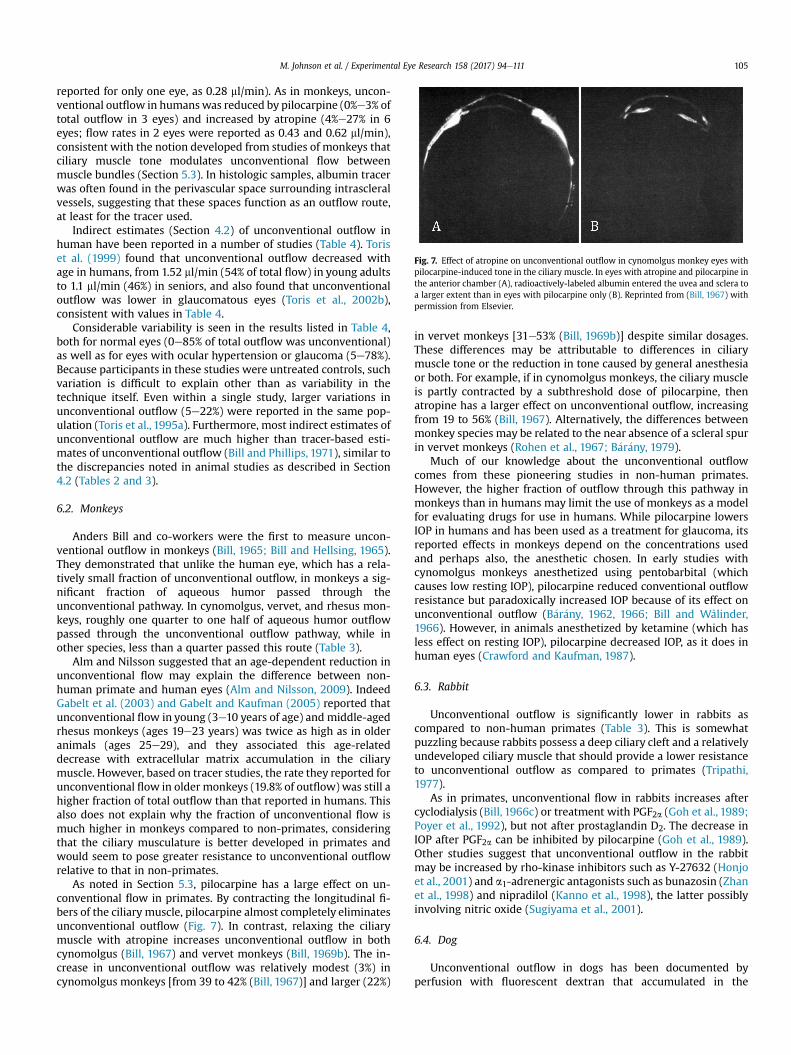
Fig. 7. Effect of atropine on unconventional outflow in cynomolgus monkey eyes withpilocarpine-induced tone in the ciliary muscle. In eyes with atropine and pilocarpine inthe anterior chamber (A), radioactively-labeled albumin entered the uvea and sclera toa larger extent than in eyes with pilocarpine only (B). Reprinted from (Bill, 1967) withpermission from Elsevier.
M. Johnson et al. / Experimental Eye Research 158 (2017) 94e111 105
reported for only one eye, as 0.28 ml/min). As in monkeys, uncon-ventional outflow in humanswas reduced by pilocarpine (0%e3% oftotal outflow in 3 eyes) and increased by atropine (4%e27% in 6eyes; flow rates in 2 eyes were reported as 0.43 and 0.62 ml/min),consistent with the notion developed from studies of monkeys thatciliary muscle tone modulates unconventional flow betweenmuscle bundles (Section 5.3). In histologic samples, albumin tracerwas often found in the perivascular space surrounding intrascleralvessels, suggesting that these spaces function as an outflow route,at least for the tracer used.
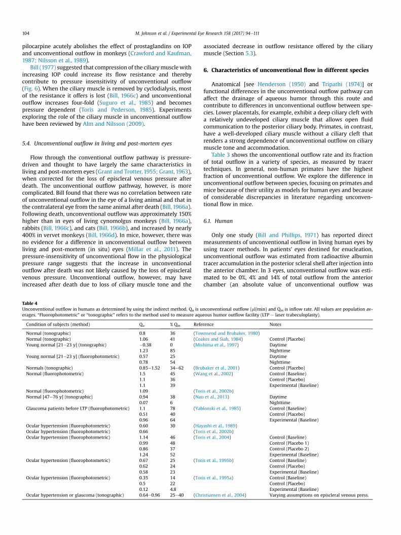
Indirect estimates (Section 4.2) of unconventional outflow inhuman have been reported in a number of studies (Table 4). Toriset al. (1999) found that unconventional outflow decreased withage in humans, from 1.52 ml/min (54% of total flow) in young adultsto 1.1 ml/min (46%) in seniors, and also found that unconventionaloutflow was lower in glaucomatous eyes (Toris et al., 2002b),consistent with values in Table 4.
Considerable variability is seen in the results listed in Table 4,both for normal eyes (0e85% of total outflow was unconventional)as well as for eyes with ocular hypertension or glaucoma (5e78%).Because participants in these studies were untreated controls, suchvariation is difficult to explain other than as variability in thetechnique itself. Even within a single study, larger variations inunconventional outflow (5e22%) were reported in the same pop-ulation (Toris et al., 1995a). Furthermore, most indirect estimates ofunconventional outflow are much higher than tracer-based esti-mates of unconventional outflow (Bill and Phillips, 1971), similar tothe discrepancies noted in animal studies as described in Section4.2 (Tables 2 and 3).
6.2. Monkeys
Anders Bill and co-workers were the first to measure uncon-ventional outflow in monkeys (Bill, 1965; Bill and Hellsing, 1965).They demonstrated that unlike the human eye, which has a rela-tively small fraction of unconventional outflow, in monkeys a sig-nificant fraction of aqueous humor passed through theunconventional pathway. In cynomolgus, vervet, and rhesus mon-keys, roughly one quarter to one half of aqueous humor outflowpassed through the unconventional outflow pathway, while inother species, less than a quarter passed this route (Table 3).
Alm and Nilsson suggested that an age-dependent reduction inunconventional flow may explain the difference between non-human primate and human eyes (Alm and Nilsson, 2009). IndeedGabelt et al. (2003) and Gabelt and Kaufman (2005) reported thatunconventional flow in young (3e10 years of age) and middle-agedrhesus monkeys (ages 19e23 years) was twice as high as in olderanimals (ages 25e29), and they associated this age-relateddecrease with extracellular matrix accumulation in the ciliarymuscle. However, based on tracer studies, the rate they reported forunconventional flow in older monkeys (19.8% of outflow) was still ahigher fraction of total outflow than that reported in humans. Thisalso does not explain why the fraction of unconventional flow ismuch higher in monkeys compared to non-primates, consideringthat the ciliary musculature is better developed in primates andwould seem to pose greater resistance to unconventional outflowrelative to that in non-primates.
As noted in Section 5.3, pilocarpine has a large effect on un-conventional flow in primates. By contracting the longitudinal fi-bers of the ciliary muscle, pilocarpine almost completely eliminatesunconventional outflow (Fig. 7). In contrast, relaxing the ciliarymuscle with atropine increases unconventional outflow in bothcynomolgus (Bill, 1967) and vervet monkeys (Bill, 1969b). The in-crease in unconventional outflow was relatively modest (3%) incynomolgus monkeys [from 39 to 42% (Bill, 1967)] and larger (22%)
in vervet monkeys [31e53% (Bill, 1969b)] despite similar dosages.These differences may be attributable to differences in ciliarymuscle tone or the reduction in tone caused by general anesthesiaor both. For example, if in cynomolgus monkeys, the ciliary muscleis partly contracted by a subthreshold dose of pilocarpine, thenatropine has a larger effect on unconventional outflow, increasingfrom 19 to 56% (Bill, 1967). Alternatively, the differences betweenmonkey species may be related to the near absence of a scleral spurin vervet monkeys (Rohen et al., 1967; B�ar�any, 1979).
Much of our knowledge about the unconventional outflowcomes from these pioneering studies in non-human primates.However, the higher fraction of outflow through this pathway inmonkeys than in humans may limit the use of monkeys as a modelfor evaluating drugs for use in humans. While pilocarpine lowersIOP in humans and has been used as a treatment for glaucoma, itsreported effects in monkeys depend on the concentrations usedand perhaps also, the anesthetic chosen. In early studies withcynomolgus monkeys anesthetized using pentobarbital (whichcauses low resting IOP), pilocarpine reduced conventional outflowresistance but paradoxically increased IOP because of its effect onunconventional outflow (B�ar�any, 1962, 1966; Bill and Wålinder,1966). However, in animals anesthetized by ketamine (which hasless effect on resting IOP), pilocarpine decreased IOP, as it does inhuman eyes (Crawford and Kaufman, 1987).
6.3. Rabbit
Unconventional outflow is significantly lower in rabbits ascompared to non-human primates (Table 3). This is somewhatpuzzling because rabbits possess a deep ciliary cleft and a relativelyundeveloped ciliary muscle that should provide a lower resistanceto unconventional outflow as compared to primates (Tripathi,1977).
As in primates, unconventional flow in rabbits increases aftercyclodialysis (Bill, 1966c) or treatment with PGF2a (Goh et al., 1989;Poyer et al., 1992), but not after prostaglandin D2. The decrease inIOP after PGF2a can be inhibited by pilocarpine (Goh et al., 1989).Other studies suggest that unconventional outflow in the rabbitmay be increased by rho-kinase inhibitors such as Y-27632 (Honjoet al., 2001) and a1-adrenergic antagonists such as bunazosin (Zhanet al., 1998) and nipradilol (Kanno et al., 1998), the latter possiblyinvolving nitric oxide (Sugiyama et al., 2001).
6.4. Dog
Unconventional outflow in dogs has been documented byperfusion with fluorescent dextran that accumulated in the
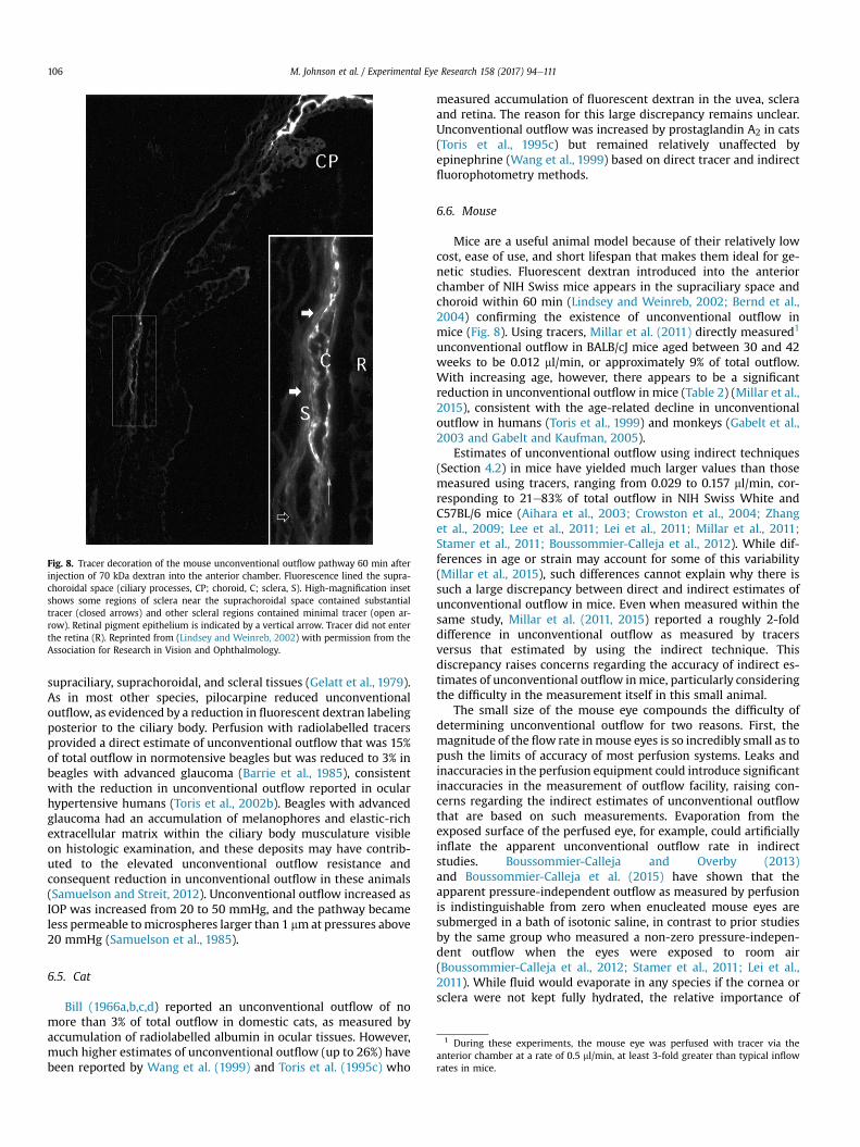
Fig. 8. Tracer decoration of the mouse unconventional outflow pathway 60 min afterinjection of 70 kDa dextran into the anterior chamber. Fluorescence lined the supra-choroidal space (ciliary processes, CP; choroid, C; sclera, S). High-magnification insetshows some regions of sclera near the suprachoroidal space contained substantialtracer (closed arrows) and other scleral regions contained minimal tracer (open ar-row). Retinal pigment epithelium is indicated by a vertical arrow. Tracer did not enterthe retina (R). Reprinted from (Lindsey and Weinreb, 2002) with permission from theAssociation for Research in Vision and Ophthalmology.
M. Johnson et al. / Experimental Eye Research 158 (2017) 94e111106
supraciliary, suprachoroidal, and scleral tissues (Gelatt et al., 1979).As in most other species, pilocarpine reduced unconventionaloutflow, as evidenced by a reduction in fluorescent dextran labelingposterior to the ciliary body. Perfusion with radiolabelled tracersprovided a direct estimate of unconventional outflow that was 15%of total outflow in normotensive beagles but was reduced to 3% inbeagles with advanced glaucoma (Barrie et al., 1985), consistentwith the reduction in unconventional outflow reported in ocularhypertensive humans (Toris et al., 2002b). Beagles with advancedglaucoma had an accumulation of melanophores and elastic-richextracellular matrix within the ciliary body musculature visibleon histologic examination, and these deposits may have contrib-uted to the elevated unconventional outflow resistance andconsequent reduction in unconventional outflow in these animals(Samuelson and Streit, 2012). Unconventional outflow increased asIOP was increased from 20 to 50 mmHg, and the pathway becameless permeable tomicrospheres larger than 1 mmat pressures above20 mmHg (Samuelson et al., 1985).
1 During these experiments, the mouse eye was perfused with tracer via theanterior chamber at a rate of 0.5 ml/min, at least 3-fold greater than typical inflowrates in mice.
6.5. Cat
Bill (1966a,b,c,d) reported an unconventional outflow of nomore than 3% of total outflow in domestic cats, as measured byaccumulation of radiolabelled albumin in ocular tissues. However,much higher estimates of unconventional outflow (up to 26%) havebeen reported by Wang et al. (1999) and Toris et al. (1995c) who
measured accumulation of fluorescent dextran in the uvea, scleraand retina. The reason for this large discrepancy remains unclear.Unconventional outflow was increased by prostaglandin A2 in cats(Toris et al., 1995c) but remained relatively unaffected byepinephrine (Wang et al., 1999) based on direct tracer and indirectfluorophotometry methods.
6.6. Mouse
Mice are a useful animal model because of their relatively lowcost, ease of use, and short lifespan that makes them ideal for ge-netic studies. Fluorescent dextran introduced into the anteriorchamber of NIH Swiss mice appears in the supraciliary space andchoroid within 60 min (Lindsey and Weinreb, 2002; Bernd et al.,2004) confirming the existence of unconventional outflow inmice (Fig. 8). Using tracers, Millar et al. (2011) directly measured1
unconventional outflow in BALB/cJ mice aged between 30 and 42weeks to be 0.012 ml/min, or approximately 9% of total outflow.With increasing age, however, there appears to be a significantreduction in unconventional outflow in mice (Table 2) (Millar et al.,2015), consistent with the age-related decline in unconventionaloutflow in humans (Toris et al., 1999) and monkeys (Gabelt et al.,2003 and Gabelt and Kaufman, 2005).
Estimates of unconventional outflow using indirect techniques(Section 4.2) in mice have yielded much larger values than thosemeasured using tracers, ranging from 0.029 to 0.157 ml/min, cor-responding to 21e83% of total outflow in NIH Swiss White andC57BL/6 mice (Aihara et al., 2003; Crowston et al., 2004; Zhanget al., 2009; Lee et al., 2011; Lei et al., 2011; Millar et al., 2011;Stamer et al., 2011; Boussommier-Calleja et al., 2012). While dif-ferences in age or strain may account for some of this variability(Millar et al., 2015), such differences cannot explain why there issuch a large discrepancy between direct and indirect estimates ofunconventional outflow in mice. Even when measured within thesame study, Millar et al. (2011, 2015) reported a roughly 2-folddifference in unconventional outflow as measured by tracersversus that estimated by using the indirect technique. Thisdiscrepancy raises concerns regarding the accuracy of indirect es-timates of unconventional outflow inmice, particularly consideringthe difficulty in the measurement itself in this small animal.
The small size of the mouse eye compounds the difficulty ofdetermining unconventional outflow for two reasons. First, themagnitude of the flow rate inmouse eyes is so incredibly small as topush the limits of accuracy of most perfusion systems. Leaks andinaccuracies in the perfusion equipment could introduce significantinaccuracies in the measurement of outflow facility, raising con-cerns regarding the indirect estimates of unconventional outflowthat are based on such measurements. Evaporation from theexposed surface of the perfused eye, for example, could artificiallyinflate the apparent unconventional outflow rate in indirectstudies. Boussommier-Calleja and Overby (2013)and Boussommier-Calleja et al. (2015) have shown that theapparent pressure-independent outflow as measured by perfusionis indistinguishable from zero when enucleated mouse eyes aresubmerged in a bath of isotonic saline, in contrast to prior studiesby the same group who measured a non-zero pressure-indepen-dent outflow when the eyes were exposed to room air(Boussommier-Calleja et al., 2012; Stamer et al., 2011; Lei et al.,2011). While fluid would evaporate in any species if the cornea orsclera were not kept fully hydrated, the relative importance of
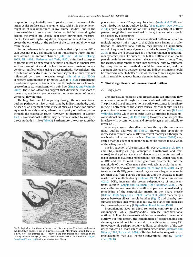
M. Johnson et al. / Experimental Eye Research 158 (2017) 94e111 107
evaporation is potentially much greater in mice because of thelarger ocular surface area to volume ratio. While this phenomenonmight be of less importance in live animal studies (due to thepresence of the extraocular muscles and orbital fat surrounding thesclera), the eyelids are usually kept open during such measure-ments. Even with hydrating drops, evaporation would tend to in-crease the osmolarity at the surface of the cornea and draw waterfrom the eye.
Second, whereas in larger eyes, such as that of primates, diffu-sion does not play a significant role in transporting tracer into tis-sues around the anterior chamber (Bill, 1965; Bill and Hellsing,1965; Bill, 1966a; Pederson and Toris, 1987), diffusional transportof tracers might be expected to be more significant in smaller eyessuch as those of mice and this leads to an overestimate of uncon-ventional outflow when using direct methods. Nevertheless, thedistribution of dextrans in the anterior segment of mice was notinfluenced by tracer molecular weight (Bernd et al., 2004),consistent with findings in primates (Section 4.1.5.2). Furthermore,the observed spread of tracer over time through the suprachoroidalspace of mice was consistent with bulk flow (Lindsey and Weinreb,2002). These considerations suggest that diffusional transport oftracer may not be a major concern in the measurement of uncon-ventional flow in mice.
The large fraction of flow passing through the unconventionaloutflow pathway in mice, as estimated by indirect methods, couldbe seen as an argument against use of mice as a model for humanaqueous humor dynamics, where the majority of outflow passesthrough the trabecular route. However, as discussed in Section4.2.1, unconventional outflow may be overestimated by using in-direct methods in mice (Table 2). Furthermore, the observation that
Fig. 9. Sagittal section through the anterior ciliary body. (A) Vehicle-treated controleye. CM, Ciliary muscle; I, iris. CP, ciliary processes. (B) After treatment with PGF2a, for4 days. Note the enlarged spaces between the thin muscle fiber bundles in theprostaglandin-treated eye (arrows). Asterisk: Greeff's vesicle. Reprinted from (Lütjen-Drecoll and Tamm, 1988) with permission from Elsevier.
pilocarpine reduces IOP in young black Swiss (Avila et al. 2001) andCD1mice by increasing outflow facility (Li et al., 2014; Overby et al.,2014) argues against the notion that a large quantity of outflowpasses through the unconventional pathway in mice (which wouldbe blocked by pilocarpine).
The age-related decline in unconventional outflow observed inmultiple mouse strains suggests that older mice having a smallerfraction of unconventional outflow may provide an appropriatemodel of aqueous humor dynamics in older humans (Millar et al.,2015). If mice are to be accepted as a model for human aqueous hu-mor dynamics, then like humans, the bulk of outflow inmice shouldpass through the conventional or trabecular outflow pathway. Thus,the accuracy of the reports of highunconventional outflowestimatedby using the indirect method should be reexamined, and theapparent discrepancy between direct and indirect estimates shouldbe resolved in order to better assesswhethermice are an appropriateanimal model for aqueous humor dynamics in humans.
7. Clinical significance of unconventional outflow
7.1. Drug effects
Cholinergics, adrenergics, and prostagladins can affect the flowof aqueous humor through the unconventional outflow pathway.The principal site of unconventional outflow resistance is the ciliarymuscle. Contraction of the ciliary muscle by cholinergics such aspilocarpine decrease the rate of unconventional outflow (Bill andWålinder, 1966) while cycloplegics such as atropine increase un-conventional outflow (Bill, 1967, 1969b). However, cholinergics alsointerfere with accommodation and are no longer used clinically tolower IOP.
Adrenergic agents also affect outflow through the unconven-tional outflow pathway. Bill (1969a) showed that epinephrineincreased unconventional outflow in vervet monkeys, although themechanism of action was unclear. Alm and Nilsson (2009) sug-gested that the effect of epinephrine might be related to relaxationof the ciliary muscle.
The introduction of the prostaglandin PGF2a (Camras et al., 1977)and PGF2a-analogues (e.g. latanoprost, bimatoprost, and trav-oprost) to the pharmacopeia of glaucoma treatments marked amajor change in glaucomamanagement. Not only is their reductionof IOP additive to most other glaucoma treatments, but themagnitude of their effect made them valuable as ocular hypoten-sive agent in their own right (Nilsson, 1997; Bron et al., 2001). Dailytreatment with PGF2a over several days causes a larger decrease inIOP than that from a single application, and the decrease is moremarked after multiple dosing (Nilsson, 1997). As noted in Section4.2.1.2, PGF2a increases the pressure-dependency of unconven-tional outflow (Gabelt and Kaufman, 1990; Kaufman, 2003). Themajor effect on unconventional outflow appears to be mediated byremodeling of the extracellular matrix in the ciliary muscle(Ocklind, 1998; Sagara et al., 1999; Gaton et al., 2001) that enlargesspaces between ciliary muscle bundles (Fig. 9) and thereby pre-sumably reduces unconventional outflow resistance and increasesits pressure-dependency (Lütjen-Drecoll and Tamm, 1988).
Prostaglandins have an effect somewhat contrary to that ofcholinergics; while prostaglandins increase unconventionaloutflow, cholinergics decrease it while also increasing conventionaloutflow. For this reason, the combination of prostaglandins andcholinergics would not be expected to be additive in reducing IOP.However, while perhaps not fully additive, combination of the twodrugs reduces IOP more effectively than either alone (Fristrom andNilsson,1993; Toris et al., 2002a). This has led to the suggestion thatprostaglandins may also increase conventional outflow (Bahleret al., 2008).
M. Johnson et al. / Experimental Eye Research 158 (2017) 94e111108
7.2. Surgery
In cyclodialysis, the attachment of the ciliary body to the scleralspur is disrupted, providing unimpeded access of aqueous humor tothe suprachoroidal space, bypassing the ciliary muscle. Cyclo-dialysis is associated with hypotony caused by a decreased aqueousinflow (Chandler and Maumenee, 1961) and a greatly increasedunconventional outflow (Bill, 1966c; Suguro et al., 1985). Cyclo-dialysis is usually the result of trauma or a surgical complication,although it can be created surgically in desperate circumstances.However, it is not routinely used to treat ocular hypertensionbecause of the very low resulting pressures (7 mmHg or less) andthe risk of acute IOP elevation should the cyclodialysis cleft spon-taneously close.
While the cyclodialysis procedure is rarely used clinically, thebenefits of greatly increased unconventional outflow withoutdecreased aqueous production can be realized by using a supra-choroidal stent to bypass the ciliary muscle (Melamed et al., 2009;Figus et al., 2011; Hoeh et al., 2013; Oatts et al., 2013). These deviceshave been used successfully to lower IOP, although there have beenreports of hypotony and of reactive fibrosis that blocks the bypasspathway (Brandao and Grieshaber, 2013). Nonetheless, they offer apotential means of achieving low IOPs that may be of particularbenefit to cases of normal tension glaucoma.
7.3. Other clinical issues
The unconventional outflow pathway may be affected by pa-thologies of the anterior segment. In uveitis, accumulation of cells,protein and debris may block the conventional outflow pathway,and it has been suggested that the unconventional outflowpathway may have developed as an adaption to provide an alter-nate drainage route for aqueous humor under these circumstances(Alm and Nilsson, 2009). Indeed, in uveitis, along with a reductionof aqueous production to roughly one half of its normal rate, thereis also a profound increase in unconventional drainage because of afour-fold increase in pressure-dependent unconventional outflowfacility (Toris and Pederson, 1987; Nilsson, 1997; Alm and Nilsson,2009). These changes produce hypotony until the uveitis sub-sides. It has been suggested that this effect is mediated by PGF2a(Crawford and Kaufman, 1987).
Unconventional outflowmay also play a role in the developmentof uveal effusion, an accumulation of fluid in the uvea that isthought to arise from impaired fluid drainage through the posteriorsegment and usually associated with abnormal thickening of thesclera, particularly in nanophthalmos (Ward et al., 1988; Jin andAnderson, 1990; Lam et al., 2005); uveal effusion can lead todetachment of choroid, ciliary body and retina. If a significantfraction of unconventional outflow passes into the vortex vein, thenthe proteins in the aqueous humor will be blocked from passingthrough these vessel walls, and they can only exit the eye bydiffusion across the sclera (Section 5.1). Thickening of the sclera,such as occurs in nanophthalmos, would reduce this proteindiffusion, potentially causing an accumulation of these proteinsalong the inner surface of the sclera. This in turn would lead to anincrease in the local oncotic pressure causing fluid accumulation, asis seen in uveal effusion (Gass, 1983; Johnson and Erickson, 2000;Elagouz et al., 2010). This disease is treated by full-thicknesssclerectomy to remove the barrier to protein transport.
8. Summary and future directions
Anders Bill was the first investigator to explore and characterizethe physiologic processes of the unconventional outflow pathways.His pioneering measurement techniques are still the standard for
identifying the pathway and estimating the amount of flowthrough this route. Originally this pathway was a scientific curi-osity, but it became clinically important when Camras and Bitofound that PGF2a could profoundly redirect outflow through thispathway from the conventional pathway, reducing IOP. Our un-derstanding of the unconventional outflow pathway has provided anew approach toward treating elevated IOP in glaucoma patients.
As unconventional outflow is pressure-insensitive under normalconditions, and because at least a fraction of this outflow passesthrough the uveovortex pathway, we would suggest that the terms“uveoscleral” and “pressure-independent flow” are misleading andcan lead to incorrect conclusions about the nature of this route,particularly when considering the effects of drugs. Distinctionsbetween pressure sensitive and anatomical pathways have impor-tant ramifications on the methods to be used for measuring andcharacterizing flow through this route. The more appropriate termfor this route of outflow is “unconventional” or “non-trabecular”.
Future animal studies of the unconventional outflow pathwayshould focus on species whose anatomic and physiologic propertiesof this route are close to those of humans. The similarity of themouse outflow pathways to that of the human (Smith et al., 2001;Overby et al., 2014) and particularly the fraction of flow passingthrough the unconventional outflow pathway, make the mouse anattractive model. However, the method for accurate measurementof flow through the unconventional outflow pathway (the tracermethod) is difficult in mice, cumbersome in larger animals, andvirtually impossible in humans. Indirect methods to estimate un-conventional outflow, while easily used, have serious flaws and theaccuracy of such approaches are questionable. Thus, a more robustand accurate method to measure unconventional outflow, both inanimals and in living humans, is necessary.
Development of a reliable non-invasive method for accuratemeasurement of unconventional flow would provide a greatadvance for characterizing the dynamics of the anterior segmentand for developing clinical treatments of glaucoma. One possibilitymight make use of fluorophotometry. A large macromolecule thatwould be trapped in or pass very slowly through the unconven-tional pathway could be introduced into the anterior chamberthrough the cornea and tracked to the general circulation as anindicator of trabecular flow, while its dilution in the anteriorchamber is simultaneously measured to determine aqueous humorinflow. The difference between trabecular outflow and inflow rateswould be a direct measure of the unconventional outflow rate.Alternatively, tracers could be tracked through the outflow path-ways by using high-resolution MRI or OCT.
Anders Bill developed the methods to establish the route andrate of unconventional outflow; our challenge now is to developmethods to measure this flow rate accurately and non-invasively inhumans.
Acknowledgments
NIH E019696 (MJ&DRO), NIH EY022359 (DRO), The BrightFocusFoundation (DRO), Research to Prevent Blindness, New York, NY(JWM), and the Mayo Foundation, Rochester, MN (JWM).
References
Aihara, M., Lindsey, J.D., et al., 2003. Aqueous humor dynamics in mice. Invest.Ophthalmol. Vis. Sci. 44 (12), 5168e5173.
Alm, A., Nilsson, S.F.E., 2009. Uveoscleral outflow e a review. Exp. Eye Res. 88 (4),760e768.
Amrite, A.C., Edelhauser, H.F., et al., 2008. Effect of circulation on the disposition andocular tissue distribution of 20 nm nanoparticles after periocular administra-tion. Mol. Vis. 14, 150e160.
Anderson, O.A., Jackson, T.L., et al., 2008. Human transscleral albumin permeabilityand the effect of topographical location and donor age. Invest. Ophthalmol. Vis.
M. Johnson et al. / Experimental Eye Research 158 (2017) 94e111 109
Sci. 49 (9), 4041e4045.Ascher, K.W., 1942. Aqueous veins. Am. J. Ophthalmol. 25, 31.Avila, M.Y., Carre, D.A., et al., 2001. Reliable measurement of mouse intraocular
pressure by a servo-null micropipette system. Invest. Ophthalmol. Vis. Sci. 42(8), 1841e1846.
Bahler, C.K., Howell, K.G., et al., 2008. Prostaglandins increase trabecular meshworkoutflow facility in cultured human anterior segments. Am. J. Ophthalmol. 145(1), 114e119.
B�ar�any, E., 1966. Dissociation of accommodation effects from outflow effects ofpilocarpine. In: Paterson, G., Miller, S., Paterson, G. (Eds.), Drug Mechanisms inGlaucoma, pp. 275e284. London, Churchill.
B�ar�any, E., 1967. Pseudofacility and uveoscleral outflow routes. In: Leydhecker, W.,Karger, S. (Eds.), Glaucoma, Tutzing Symposium, Basel, p. 35.
B�ar�any, E.H., 1962. The mode of action of pilocarpine on outflow resistance in theeye of a primate (Cercopithecus Ethiops). Invest. Ophthalmol. Vis. Sci. 1 (6),712e727.
B�ar�any, E.H., 1979. A pharmacologist looks at medical treatment in glaucomaeinretrospect and in prospect. Ophthalmology 86 (1), 80e94.
Barrie, K.P., Gum, G.G., et al., 1985. Quantitation of uveoscleral outflow in normo-tensive and glaucomatous beagles by H-3-labeled dextran. Am. J. Vet. Res. 46(1), 84e88.
Becker, B., Neufeld, A.H., 2002. Pressure dependence of uveoscleral outflow.J. Glaucoma 11 (6), 545.
Bernd, A.S., Aihara, M., et al., 2004. Influence of molecular weight on intracameraldextran movement to the posterior segment of the mouse eye. Invest. Oph-thalmol. Vis. Sci. 45 (2), 480e484.
Bill, A., 1962. The drainage of blood from the uvea and the elimination of aqueoushumour in rabbits. Exp. Eye Res. 1 (3), 200e205.
Bill, A., 1964. The drainage of albumin from the uvea. Exp. Eye Res. 3, 179e187.Bill, A., 1965. The aqueous humor drainage mechanism in the cynomolgus monkey
(Macaca irus) with evidence for unconventional routes. Invest. Ophthalmol. 4,911e919.
Bill, A., 1966a. Conventional and uveo-scleral drainage of aqueous humour in thecynomolgus monkey (Macaca irus) at normal and high intraocular pressures.Exp. Eye Res. 5, 45e54.
Bill, A., 1966b. Formation and drainage of aqueous humour in cats. Exp. Eye Res. 5(3), 185e190.
Bill, A., 1966c. The routes for bulk drainage of aqueous humor in rabbits with andwithout cyclodialysis. Doc. Ophthalmol. 20, 157e169.
Bill, A., 1966d. The routes for bulk drainage of aqueous humour in the vervetmonkey (Ceropithecus ethiops). Exp. Eye Res. 5, 55e57.
Bill, A., 1967. Effects of atropine and pilocarpine on aqueous humour dynamics incynomolgus monkeys (Macaca irus). Exp. Eye Res. 6 (2), 120e125.
Bill, A., 1969a. Early effects of epinephrine on aqueous humor dynamics in vervetmonkeys (Cercopithecus ethiops). Exp. Eye Res. 8, 35e43.
Bill, A., 1969b. Effects of atropine on aqueous humor dynamics in the vervet monkey(Cercopithecus ethiops). Exp. Eye Res. 8 (3), 284e291.
Bill, A., 1970. Effects of norepinephrine, isoproterenol, and sympathetic stimulationon aqueous humor dynamics on vervet monkeys. Exp. Eye Res. 10, 31e46.
Bill, A., 1971. Aqueous humor dynamics in monkeys (Macaca irus and Cercopithecusethiops). Exp. Eye Res. 11 (2), 195e206.
Bill, A., 1975. Blood circulation and fluid dynamics in the eye. Physiol. Rev. 55,383e416.
Bill, A., 1977. Basic physiology of the drainage of aqueous humor. In: Bito, L.Z.,Davson, H., Fenstermacher, J.D. (Eds.), The Ocular and Cerebrospinal Fluids.Academic Press, London, New York, San Francisco, pp. 291e303.
Bill, A., 2003. Some thoughts on the pressure dependence of uveoscleral flow.J. Glaucoma 12 (1), 88e89.
Bill, A., Hellsing, K., 1965. Production and drainage of aqueous humor in the cyn-omolgus monkey (Macaca irus). Invest. Ophthalmol. 4, 920e926.
Bill, A., Phillips, C.I., 1971. Uveoscleral drainage of aqueous humor in human eyes.Exp. Eye Res. 12, 275e281.
Bill, A., Wålinder, P., 1966. The effects of pilocarpine on the dynamics of aqueoushumor in a primate (Macaca irus). Invest. Ophthalmol. 5 (2), 170e175.
Boussommier-Calleja, A., Bertrand, J., et al., 2012. Pharmacologic manipulation ofconventional outflow facility in ex vivo mouse eyes. Invest. Ophthalmol. Vis. Sci.53 (9), 5838e5845.
Boussommier-Calleja, A., Li, G., et al., 2015. Physical factors affecting outflow facilitymeasurements in mice. Invest. Ophthalmol. Vis. Sci. 56 (13), 8331e8339.
Boussommier-Calleja, A., Overby, D.R., 2013. The influence of genetic background onconventional outflow facility in mice. Invest. Ophthalmol. Vis. Sci. 54 (13),8251e8258.
Brandao, L.M., Grieshaber, M.C., 2013. Update on Minimally Invasive GlaucomaSurgery (MIGS) and new implants. J. Ophthalmol. 2013, 705915.
Bron, A.M., Denis, P., et al., 2001. Additive IOP-reducing effect of latanoprost inpatients insufficiently controlled on timolol. Acta Ophthalmol. Scand. 79 (3),289e293.
Brubaker, R.F., 1975. The effect of intraocular pressure on conventional outflowresistance in the enucleated human eye. Invest. Ophthalmol. Vis. Sci. 14,286e292.
Brubaker, R.F., 2004. Goldmann's equation and clinical measures of aqueous dy-namics. Exp. Eye Res. 78 (3), 633e637.
Brubaker, R.F., McLaren, J.W., 1985. Uses of fluorophotometry in glaucoma research.Ophthalmology 92 (7), 884e890.
Brubaker, R.F., Schoff, E.O., et al., 2001. Effects of AGN 192024, a new ocular
hypotensive agent, on aqueous dynamics1. Am. J. Ophthalmol. 131 (1), 19e24.Butler, J.M., Raviola, G., et al., 1984. Computed tomography of aqueous humour
outflow pathways. Exp. Eye Res. 39 (6), 709e719.Camras, C.B., 2003. Some thoughts on the pressure dependence of uveoscleral flow.
J. Glaucoma 12 (1), 92e93 author reply 93e4.Camras, C.B., Bito, L.Z., et al., 1977. Reduction of intraocular pressure by prosta-
glandins applied topically to the eyes of conscious rabbits. Invest. Ophthalmol.Vis. Sci. 16 (12), 1125e1134.
Chan-Ling, T., Koina, M.E., et al., 2015. Author response: sufficient evidence forlymphatics in the developing and adult human choroid? LettersIOVS j MonthYear j Vol. 56 j No. 10 j page number Invest. Ophthalmol. Vis. Sci. 56 (11),6711e6713.
Chandler, P.A., Maumenee, A.E., 1961. A major cause of hypotony. Am. J. Ophthalmol.52, 609e618.
Christiansen, G.A., Nau, C.B., et al., 2004. Mechanism of ocular hypotensive action ofbimatoprost (Lumigan) in patients with ocular hypertension or glaucoma.Ophthalmology 111 (9), 1658e1662.
Coakes, R.L., Siah, P.B., 1984. Effects of adrenergic drugs on aqueous humour dy-namics in the normal human eye. I. Salbutamol. Br. J. Ophthalmol. 68 (6),393e397.
Cole, D.F., Monro, P.A., 1976. The use of fluorescein-labelled dextrans in investiga-tion of aqueous humour outflow in the rabbit. Exp. Eye Res. 23 (6), 571e585.
Crawford, K., Kaufman, P.L., 1987. Pilocarpine antagonizes prostaglandin F2 alpha-induced ocular hypotension in monkeys. Evidence for enhancement ofuveoscleral outflow by prostaglandin F2 alpha. Arch. Ophthamol. 105 (8),1112e1116.
Crowston, J.G., Aihara, M., et al., 2004. Effect of latanoprost on outflow facility in themouse. Invest. Ophthalmol. Vis. Sci. 45 (7), 2240e2245.
Elagouz, M., Stanescu-Segall, D., et al., 2010. Uveal effusion syndrome. Surv. Oph-thalmol. 55 (2), 134e145.
Emi, K., Pederson, J.E., et al., 1989. Hydrostatic pressure of the suprachoroidal space.Invest. Ophthalmol. Vis. Sci. 30, 233e238.
Erdmann, P., 1907. Über experimentelles Glaukom nebst Untersuchungen amglaukomat€osen Tierauge. Albr. Graefes Arch. Ophthalmol. 66 (2), 325e390.
Fatt, I., Hedbys, B.O., 1970. Flow of water in the sclera. Exp. Eye Res. 10, 243e249.Figus, M., Lazzeri, S., et al., 2011. Supraciliary shunt in refractory glaucoma. Br. J.
Ophthalmol. 95 (11), 1537e1541.Fine, B.S., 1964. Observations on the drainage angle in man and rhesus monkey: a
concept of the pathogenesis of chronic simple glaucoma: a light and electronmicroscopic study. Invest. Ophthalmol. Vis. Sci. 3 (6), 609e646.
Fowlks, W.L., Havener, V.R., et al., 1963. Meridional flow from the cornea ciliaristhrough the pararetinal zone of the rabbit vitreous. Invest. Ophthalmol. 2,63e71.
Fristrom, B., Nilsson, S.E., 1993. Interaction of PhXA41, a new prostaglandinanalogue, with pilocarpine. A study on patients with elevated intraocularpressure. Arch. Ophthalmol. 111 (5), 662e665.
Gabelt, B., Kaufman, P., 1989. Prostaglandin F increases uveoscleral outflow in thecynomolgus monkey. Exp. Eye Res. 49, 389e402.
Gabelt, B.A.T., Gottanka, J., et al., 2003. Aqueous humor dynamics and trabecularmeshwork and anterior ciliary muscle morphologic changes with age in rhesusmonkeys. Invest. Ophthalmol. Vis. Sci. 44 (5), 2118e2125.
Gabelt, B.A.T., Okka, M., et al., 2005. Aqueous humor dynamics in monkeys aftertopical R-DOI. Invest. Ophthalmol. Vis. Sci. 46 (12), 4691e4696.
Gabelt, B.T., Kaufman, P.L., 1990. The effect of prostaglandin F2 alpha on trabecularoutflow facility in cynomolgus monkeys. Exp. Eye Res. 51 (1), 87e91.
Gabelt, B.T., Kaufman, P.L., 2005. Changes in aqueous humor dynamics with age andglaucoma. Prog. Retin. Eye Res. 24 (5), 612e637.
Gass, J.D., 1983. Uveal effusion syndrome: a new hypothesis concerning patho-genesis and technique of surgical treatment. Trans. Am. Ophthalmol. Soc. 81,246e260.
Gaton, D.D., Sagara, T., et al., 2001. Increased matrix metalloproteinases 1, 2, and 3 inthe monkey uveoscleral outflow pathway after topical prostaglandinF2aeisopropyl ester treatment. Arch. Ophthalmol. 119 (8), 1165e1170.
Gelatt, K.N., Gum, G.G., et al., 1979. Uveoscleral flow of aqueous humor in thenormal dog. Am. J. Vet. Res. 40 (6), 845e848.
Goh, Y., Araie, M., et al., 1989. Effect of topical prostaglandin D2 on the aqueoushumor dynamics in rabbits. Graefes Arch. Clin. Exp. Ophthalmol. 227 (5),476e481.
Goh, Y., Oshima, T., et al., 1994. Mechanism of intraocular pressure reductionobserved after topical application of S-1033 in animals. Jpn. J. Ophthalmol. 38(3), 228e235.
Grant, W., Trotter, R., 1955. Tonographic measurements in enucleated eye. Arch.Ophthalmol. 53, 191e200.
Grant, W.M., 1950. Tonographic method for measuring the facility and rate ofaqueous flow in human eyes. Arch. Ophthalmol. 44, 204e214.
Grant, W.M., 1963. Experimental aqueous perfusion in enucleated human eyes.Arch. Ophthalmol. 69, 783e801.
Hayashi, M., Yablonski, M.E., et al., 1989. Trabecular outflow facility determined byfluorophotometry in human subjects. Exp. Eye Res. 48, 621e625.
Heindl, L.M., Kaser-Eichberger, A., et al., 2015. Sufficient evidence for lymphatics inthe developing and adult human choroid? LettersIOVS j Month Year j Vol. 56 jNo. 10 j page number Invest. Ophthalmol. Vis. Sci. 56 (11), 6709e6710.
Henderson, T., 1950. Principles of Ophthalmology. Wm. Heinemann, London.Hoeh, H., Ahmed, I.I., et al., 2013. Early postoperative safety and surgical outcomes
after implantation of a suprachoroidal micro-stent for the treatment of open-
M. Johnson et al. / Experimental Eye Research 158 (2017) 94e111110
angle glaucoma concomitant with cataract surgery. J. Cataract Refract. Surg. 39(3), 431e437.
Honjo, M., Inatani, M., et al., 2001. EFfects of protein kinase inhibitor, ha1077, onintraocular pressure and outflow facility in rabbit eyes. Arch. Ophthalmol. 119(8), 1171e1178.
Inomata, H., Bill, A., et al., 1972. Unconventional routes of aqueous humor outflow incynomolgus monkey (Macaca irus). Am. J. Ophthalmol. 73, 893.
Jackson, T.L., Hussain, A., et al., 2008. Scleral hydraulic conductivity and macro-molecular diffusion in patients with uveal effusion syndrome. Invest. Oph-thalmol. Vis. Sci. 49 (11), 5033e5040.
Jin, J.C., Anderson, D., 1990. Laser and unsutured sclerotomy in nanophthalmos. Am.J. Ophthalmol. 109, 575e580.
Johnson, M., Erickson, K., 2000. Mechanisms and routes of aqueous humor drainage.In: Albert, D.M., Jakobiec, F.A. (Eds.), Principles and Practice of Ophthalmology,vol. 4. Saunders, Philadelphia, pp. 2577e2595.
Kanno, M., Araie, M., et al., 1998. Effects of topical nipradilol, a beta-blocking agentwith alpha-blocking and nitroglycerin-like activities, on aqueous humor dy-namics and fundus circulation. Invest. Ophthalmol. Vis. Sci. 39 (5), 736e743.
Kaufman, P.L., 2003. Some thoughts on the pressure dependence of uveoscleralflow. J. Glaucoma 12 (1), 89 author reply 93e4.
Kim, M., Johnston, M.G., et al., 2011. A model to measure lymphatic drainage fromthe eye. Exp. Eye Res. 93 (5), 586e591.
Kiss, F., 1943. Der Blutkreislauf des Auges. Ophthalmologica 106 (5e6), 225e250.Kleinstein, R.N., Fatt, I., 1977. Pressure dependency of trans-scleral flow. Exp. Eye
Res. 24, 335.Knies, M., 1875. Die resorption von blut in der vorderen augenkammer. Arch. Pathol.
Anat. Physiol. Klin. Med. 62, 537e553.Koina, M.E., Baxter, L., et al., 2015. Evidence for lymphatics in the developing and
adult human choroid. Invest. Ophthalmol. Vis. Sci. 56 (2), 1310e1327.Lam, A., Sambursky, R.P., et al., 2005. Measurement of scleral thickness in uveal
effusion syndrome. Am. J. Ophthalmol. 140 (2), 329e331.Leber, T., 1873. Studien über den Flüssigkeitswechsel im Auge. Albr. v. Graefes Arch.
Ophthalmol. 19, 87e106.Lee, Y.S., Tresguerres, M., et al., 2011. Regulation of anterior chamber drainage by
bicarbonate-sensitive soluble adenylyl cyclase in the ciliary body. J. Biol. Chem.286 (48), 41353e41358.
Lei, Y., Overby, D.R., et al., 2011. Outflow physiology of the mouse eye: pressuredependence and washout. Invest. Ophthalmol. Vis. Sci. 52 (3), 1865e1871.
Levene, R., Hyman, B., 1969. The effect of intraocular pressure on the facility ofoutflow. Exp. Eye Res. 8, 116e121.
Li, G., Farsiu, S., et al., 2014. Pilocarpine-induced dilation of Schlemm's canal andprevention of lumen collapse at elevated intraocular pressures in living micevisualized by OCT. Invest. Ophthalmol. Vis. Sci. 55, 3737e3746.
Lim, K.S., Nau, C.B., et al., 2008. Mechanism of action of bimatoprost, latanoprost,and travoprost in healthy subjects. A crossover study. Ophthalmology 115 (5),790e795 e4.
Lindsey, J.D., Weinreb, R.N., 2002. Identification of the mouse uveoscleral outflowpathway using fluorescent dextran. Invest. Ophthalmol. Vis. Sci. 43 (7),2201e2205.
Lütjen-Drecoll, E., Tamm, E., 1988. Morphological study of the anterior segment ofcynomolgus monkey eyes following treatment with prostaglandin F2a. Exp. EyeRes. 47 (5), 761e769.
M€aepea, O., 1992. Pressures in the anterior ciliary arteries, choroidal veins andchoriocapillaris. Exp. Eye Res. 54 (5), 731e736.
M€aepea, O., Bill, A., 1989. The pressures in the episcleral veins, Schlemm's canal andthe trabecular meshwork in monkeys: effects of changes in intraocular pres-sure. Exp. Eye Res. 49 (4), 645e663.
McMaster, P.R.B., Macri, F.J., 1968. Secondary aqueous humor outflow pathways inthe rabbit, cat, and monkey. Arch. Ophthamol. 79, 297e303.
Melamed, S., Ben Simon, G.J., et al., 2009. Efficacy and safety of gold micro shuntimplantation to the supraciliary space in patients with glaucoma: a pilot study.Arch. Ophthalmol. 127 (3), 264e269.
Millar, J.C., Clark, A.F., et al., 2011. Assessment of aqueous humor dynamics in themouse by a novel method of constant-flow infusion. Invest. Ophthalmol. Vis.Sci. 52 (2), 685e694.
Millar, J.C., Phan, T.N., et al., 2015. Strain and age effects on aqueous humor dy-namics in the mouse. Invest. Ophthalmol. Vis. Sci. 56 (10), 5764e5776.
Mishima, H.K., Kiuchi, Y., et al., 1997. Circadian intraocular pressure managementwith latanoprost: diurnal and nocturnal intraocular pressure reduction andincreased uveoscleral outflow. Surv. Ophthalmol. 41 (Suppl. 2), S139eS144.
Moses, R.A., 1979. Circumferential flow in Schlemm's canal. Am. J. Ophthalmol. 88,585e591.
Nau, C.B., Malihi, M., et al., 2013. Circadian variation of aqueous humor dynamics inolder healthy adults. Invest. Ophthalmol. Vis. Sci. 54, 7623e7629.
Nesterov, A.P., 1986. Chapter 13: the future for surgery in the glaucomas byincreasing uveoscleral outflow. In: Cairns, J.E. (Ed.), Glaucoma. Grune & Strat-ton, pp. 257e273.
Nilsson, S.F., 1997. The uveoscleral outflow routes. Eye 11 (Pt 2), 149e154.Nilsson, S.F., Drecoll, E., et al., 2006. The prostanoid EP2 receptor agonist butaprost
increases uveoscleral outflow in the cynomolgus monkey. Invest. Ophthalmol.Vis. Sci. 47 (9), 4042e4049.
Nilsson, S.F.E., Samuelsson, M., et al., 1989. Increased uveoscleral outflow as apossible mechanism of ocular hypotension caused by prostaglandin F2a-1-isopropylester in the cynomolgus monkey. Exp. Eye Res. 48, 707e716.
Nuel, J., Benoit, F., 1900. Des voies d'elimination des liquides intra-oculaires hors de
la chambre anterieure et au fond de l'oeil. Arch. d'Opth. 20, 161e228.Oatts, J.T., Zhang, Z., et al., 2013. In vitro and in vivo comparison of two supra-
choroidal shunts. Invest. Ophthalmol. Vis. Sci. 54 (8), 5416e5423.Ocklind, A., 1998. Effect of latanoprost on the extracellular matrix of the ciliary
muscle. A study on cultured cells and tissue sections. Exp. Eye Res. 67 (2),179e191.
Oka, T., Taniguchi, T., et al., 2006. Aqueous humor dynamics associated with thephorbol ester-induced decrease in intraocular pressure in the rabbit. Jpn. J.Ophthalmol. 50 (6), 497e503.
Overby, D.R., Bertrand, J., et al., 2014. The structure of the trabecular meshwork, itsconnections to the ciliary muscle, and the effect of pilocarpine on outflow fa-cility in mice. Invest. Ophthalmol. Vis. Sci. 55 (6), 3727e3736.
Pease, M.E., Oglesby, E.N., et al., 2014. Scleral permeability varies by mouse strainand is decreased by chronic experimental glaucoma. Invest. Ophthalmol. Vis.Sci. 55 (4), 2564e2573.
Pederson, J.E., Cantrill, H.L., 1984. Experimental retinal detachment: V. Fluidmovement through the retinal hole. Arch. Ophthalmol. 102 (1), 136e139.
Pederson, J.E., Gaasterland, D.E., et al., 1977. Uveoscleral aqueous outflow in therhesus monkey: importance of uveal reabsorption. Invest. Ophthalmol. Vis. Sci.16, 1008e1017.
Pederson, J.E., Toris, C.B., 1987. Uveoscleral outflow: diffusion or flow? Invest.Ophthalmol. Vis. Sci. 28 (6), 1022e1024.
Poyer, J.F., Gabelt, B., et al., 1992. The effect of topical PGF(2a) on uveoscleral outflowand outflow facility in the rabbit eye. Exp. Eye Res. 54 (2), 277e283.
Rohen, J.W., Lütjen, E., et al., 1967. The relationship between the ciliary muscle andthe trabecular meshwork and its importance for the effect of miotics onaqueous outflow resistance. Albr. v. Graefes Arch. Klin. Exp. Ophthalmol. 172,23e47.
Sagara, T., Gaton, D.D., et al., 1999. Topical prostaglandin F2a treatment reducescollagen types I, III, and IV in the monkey uveoscleral outflow pathway. Arch.Ophthalmol. 117 (6), 794e801.
Samuelson, D., Streit, A., 2012. Microanatomy of the anterior uveoscleral outflowpathway in normal and primary open-angle glaucomatous dogs. Vet. Oph-thalmol. 15 (Suppl. 1), 47e53.
Samuelson, D.A., Gum, G.G., et al., 1985. Aqueous outflow in the beagle: uncon-ventional outflow, using different-sized microspheres. Am. J. Vet. Res. 46 (1),242e248.
Schroedl, F., Kaser-Eichberger, A., et al., 2014. Consensus statement on the immu-nohistochemical detection of ocular lymphatic vessels. Invest. Ophthalmol. Vis.Sci. 55 (10), 6440e6442.
Schwalbe, G., 1870. Untersuchen über die Lymphbahnen des Auges und ihreBegrenzungen. Arch. Mikrosk. Anat. 6, 261e362.
Seidel, E., 1921. Weitere experimentelle Untersuchungen über die Quelle und denVerlauf der intraokularen Saftstr€omung. IX Mitteilung. Über den Abfluss desKammerwassers aus der vorderen AugenKammer. Graefe's Arch. Clin. Exp.Ophthalmol. 104, 357e402.
Sherman, S.H., Green, K., et al., 1978. The fate of anterior chamber fluorescein in themonkey eye. I. The anterior chamber outflow pathways. Exp. Eye Res. 27,159e173.
Sit, A.J., Ekdawi, N.S., et al., 2011. A novel method for computerized measurement ofepiscleral venous pressure in humans. Exp. Eye Res. 92 (6), 537e544.
Sit, A.J., McLaren, J.W., 2011. Measurement of episcleral venous pressure. Exp. EyeRes. 93 (3), 291e298.
Smith, R.S., Zabaleta, A., et al., 2001. The mouse anterior chamber angle andtrabecular meshwork develop without cell death. BMC Dev. Biol. 1, 3.
Stamer, W.D., Lei, Y., et al., 2011. eNOS, a pressure-dependent regulator of intraoc-ular pressure. Invest. Ophthalmol. Vis. Sci. 52 (13), 9438e9444.
Stjernschantz, J.W., 2001. From PGF(2alpha)-isopropyl ester to latanoprost: a reviewof the development of xalatan: the Proctor Lecture. Invest. Ophthalmol. Vis. Sci.42 (6), 1134e1145.
Streilein, J.W., 2003. Ocular immune privilege: therapeutic opportunities from anexperiment of nature. Nat. Rev. Immunol. 3 (11), 879e889.
Sugiyama, T., Kida, T., et al., 2001. Involvement of nitric oxide in the ocular hypo-tensive action of nipradilol. Curr. Eye Res. 23 (5), 346e351.
Suguro, K., Toris, C.B., et al., 1985. Uveoscleral outflow following cyclodialysis in themonkey eye using a fluorescent tracer. Invest. Ophthalmol. Vis. Sci. 26 (6),810e813.
Takashima, Y., Taniguchi, T., et al., 1996. Ocular hypotensive mechanism of intra-vitreally injected brain natriuretic peptide in rabbit. Invest. Ophthalmol. Vis. Sci.37 (13), 2671e2677.
Tam, A.L., Gupta, N., et al., 2011. Quantum dots trace lymphatic drainage from themouse eye. Nanotechnology 22 (42), 425101.
Tam, A.L.C., Gupta, N., et al., 2013. Latanoprost stimulates ocular lymphaticdrainage: an in vivo nanotracer study. Transl. Vis. Sci. Technol. 2 (5), 3.
Toris, C.B., 2008. Aqueous humor dynamics I. Current topics in membranes. In:Civan, M.M. (Ed.), The Eye's Aqueous Humor, vol. 62, pp. 193e229.
Toris, C.B., Alm, A., et al., 2002a. Latanoprost and cholinergic agonists in combina-tion. Surv. Ophthalmol. 47 (Suppl. 1), S141eS147.
Toris, C.B., Camras, C.B., et al., 1993. Effects of PhXA41, a new prostaglandin F2 alphaanalog, on aqueous humor dynamics in human eyes. Ophthalmology 100 (9),1297e1304.
Toris, C.B., Gleason, M.L., et al., 1995a. Effects of brimonidine on aqueous humordynamics in human eyes. Arch. Ophthalmol. 113 (12), 1514e1517.
Toris, C.B., Gregerson, D.S., et al., 1987. Uveoscleral outflow using different-sizedfluorescent tracers in normal and inflamed eyes. Exp. Eye Res. 45 (4), 525e532.
M. Johnson et al. / Experimental Eye Research 158 (2017) 94e111 111
Toris, C.B., Koepsell, S.A., et al., 2002b. Aqueous humor dynamics in ocular hyper-tensive patients. J. Glaucoma 11 (3), 253e258.
Toris, C.B., Pederson, J.E., 1985. Effect of intraocular pressure on uveoscleral outflowfollowing cyclodialysis in the monkey eye. Invest. Ophthalmol. Vis. Sci. 26,1745e1749.
Toris, C.B., Pederson, J.E., 1987. Aqueous humor dynamics in experimental iridocy-clitis. Invest. Ophthalmol. Vis. Sci. 28 (3), 477e481.
Toris, C.B., Pederson, J.E., et al., 1990. Extravascular albumin concentration of theuvea. Invest. Ophthalmol. Vis. Sci. 31 (1), 43e53.
Toris, C.B., Tafoya, M.E., et al., 1995b. Effects of apraclonidine on aqueous humordynamics in human eyes. Ophthalmology 102 (3), 456e461.
Toris, C.B., Yablonski, M.E., et al., 1999. Aqueous humor dynamics in the aging hu-man eye. Am. J. Ophthalmol. 127 (4), 407e412.
Toris, C.B., Yablonski, M.E., et al., 1995c. Prostaglandin A2 increases uveoscleraloutflow and trabecular outflow facility in the cat. Exp. Eye Res. 61 (6), 649e657.
Toris, C.B., Zhan, G., et al., 2004. Increase in outflow facility with unoprostonetreatment in ocular hypertensive patients. Arch. Ophthalmol. 122 (12),1782e1787.
Toris, C.B., Zhan, G.L., et al., 2000. Aqueous humor dynamics in monkeys with laser-induced glaucoma. J. Ocul. Pharmacol. Ther. 16 (1), 19e27.
Townsend, D.J., Brubaker, R.F., 1980. Immediate effect of epinephrine on aqueousformation in the normal human eye as measured by fluorophotometry. Invest.Ophthalmol. Vis. Sci. 19 (3), 256e266.
Tripathi, R.C., 1974. Ch 3: comparative physiology and anatomy of the aqueousoutflow pathway. In: Davson, H., Graham, L.T. (Eds.), The Eye. ComparativePhysiology, vol. 5. Academic Press, London, England; New York, NY,pp. 163e356.
Tripathi, R.C., 1977. Uveoscleral drainage of aqueous humour. Exp. Eye Res. 25(Suppl), 305e308.
Van Buskirk, E.M., 1982. Anatomic correlates of changing aqueous outflow facility inexcised human eyes. Invest. Ophthalmol. Vis. Sci. 22, 625e632.
Wan, Z., Woodward, D.F., et al., 2007. Bimatoprost, prostamide activity, and
conventional drainage. Invest. Ophthalmol. Vis. Sci. 48 (9), 4107e4115.Wang, Y.L., Hayashi, M., et al., 2002. Effects of multiple dosing of epinephrine on
aqueous humor dynamics in human eyes. J. Ocul. Pharmacol. Ther. 18 (1),53e63.
Wang, Y.L., Toris, C.B., et al., 1999. Effects of topical epinephrine on aqueous humordynamics in the cat. Exp. Eye Res. 68 (4), 439e445.
Ward, R.C., Gragoudas, E.S., et al., 1988. Abnormal scleral findings in uveal effusionsyndrome. Am. J. Ophthalmol. 106, 139e146.
Wood, R.L., Koseki, T., et al., 1990. Structural analysis of potential barriers to bulk-flow exchanges between uvea and sclera in eyes of Macaque monkeys. CellTissue Res. 260, 459e468.
Woodward, D.F., Krauss, A.H.-P., et al., 2010. Bimatoprost effects on aqueous humordynamics in monkeys. J. Ophthalmol. 2010.
Yablonski, M.E., 2002. Letter to the editor. J. Glaucoma 12 (1), 90e92.Yablonski, M.E., 2003. Some thoughts on the pressure dependence of uveoscleral
flow. J. Glaucoma 12 (1), 90e92 author reply 93e4.Yablonski, M.E., Cook, D.J., et al., 1985. A fluorophotometric study of the effect of
argon laser trabeculoplasty on aqueous humor dynamics. Am. J. Ophthalmol.99, 579e582.
Yücel, Y.H., Johnston, M.G., et al., 2009. Identification of lymphatics in the ciliarybody of the human eye: a novel “uveolymphatic” outflow pathway. Exp. EyeRes. 89 (5), 810e819.
Zhan, G.L., Lee, P.Y., et al., 2002. Time dependent effects of sympathetic denervationon aqueous humor dynamics and choroidal blood flow in rabbits. Curr. Eye Res.25 (2), 99e105.
Zhan, G.L., Toris, C.B., et al., 1998. Bunazosin reduces intraocular pressure in rabbitsby increasing uveoscleral outflow. J. Ocul. Pharmacol. Ther. 14 (3), 217e228.
Zhang, Y., Davidson, B.R., et al., 2009. Enhanced inflow and outflow rates despitelower IOP in bestrophin-2-deficient mice. Invest. Ophthalmol. Vis. Sci. 50 (2),765e770.
Zhao, M., Hejkal, J.J., et al., 2010. Aqueous humor dynamics during the day and nightin juvenile and adult rabbits. Invest. Ophthalmol. Vis. Sci. 51 (6), 3145e3151.