ultrastructure of azotobacter vinelandii - digital library/67531/metadc284557/m2/1/high... ·...
TRANSCRIPT

JOURNAL OF BACTERIOLoGY, Nov. 1970, p. 933-939 Vol. 104, No. 2Copyright a 1970 American Society for Microbiology Printed in U.S.A.
Ultrastructure of Azotobacter vinelandiiG. R. VELA, G. D. CAGLE, AND P. R. HOLMGREN
Department of Biological Sciences, North Texas State University, Denton, Texas 76203
Received for publication 8 August 1970
Vegetative cells and cysts of Azotobacter vinelandii 12837 were prepared for elec-tron microscopy by several methods assumed to preserve structural details destroyedby techniques previously reported in the literature. Examination of large numbersof cells and cysts by these methods revealed four structural details not reportedpreviously: intine fibrils, intine vesicles, intine membrane, and microtubules. Theintine fibrils form a network in the gel-like homogeneous matrix of the CC2 layer.Intine vesicles which seem to originiate in the cell wall complex of the central bodyare seen in the intine and exine of cysts. Analogous structures are found on vegeta-tive cells. The intine is divided into two chemically distinct areas by the two-layeredintine membrane. Microtubules, previously reported only in vegetative cells, werefound in cysts.
Electron microscopy of thin sections of cells hasbeen used to study the subcellular structure ofmany bacteria including members of the genusAzotobacter. Bisset and Hale (1) published thefirst electron micrographs of A. chroococcumcells. These were gold-shadowed preparations of"gonidia." In 1961 (11), thin sections of the celland cyst of A. vinelandii 12837 were preparedfrom material imbedded in epoxy resin afterKMnO4 fixation. These preparations showedthat vegetative cells have a granular vacuolatedcytoplasm and that cysts contain a central bodywith nuclear region, lipid inclusions, an intine,and an outer exine. A subsequent report by thesame group (15) using essentially the same meth-ods revealed the presence of cytoplasmic mem-brane, cell wall, nuclear material, and peripheralbodies in vegetative cells and cysts ofA . vinelandii12837. These findings were confirmed by Tchan,Birch-Andersen, and Jensen (12), who used cellsfixed with OS04 or KMnO4 and imbedded inAraldite. They also described the triple-layeredlamellar architecture of the cyst exine. Pope andJurtshuk (7) reported the presence of micro-tubules in A. vinelandii strain 0 treated withpotassium phosphotungstate and glutaraldehyde.The function of these microtubules has not beendemonstrated; they were, however, associatedonly with metabolically active vegetative cells.Koo, Lin, and Sadoff (4, 5) employed carbon rep-licas to show that the surface structure of vegeta-tive cells walls is smooth and regular, whereas thatof the cyst coat is wrinkled and irregularly folded.Lin and Sadoff showed (6) that the exine is a com-plex multilayered lamellar structure which can bereadily separated from the gel-like viscous intine.
This morphology appears to be drastically alteredduring endogenous metabolism (16).
This report shows additional structural detailsin A. vinelandii 12837 obtained by methods notpreviously used in the study of Azotobacter.
MATERIALS AND METHODSCells. A. vinelandii 12837 was grown on Burks
medium (14) with 1% glucose as carbon source and2% agar as solidifying agent; glucose was replaced by0.3% n-butanol to induce encystment.
Electron microscopy. All vegetative cells were ob-tained from the Burks glucose medium after 18 to 24hr of incubation at 30 C, and all cysts from the n-butanol medium after 4 to 5 days of incubation at thesame temperature. To rule out artifacts, cells from thesame culture were treated by the different proceduresdescribed below and compared to results obtainedwith a more conventional method (13). The variousmethods of pretreatment, fixation, and staining usedare described below; ultimately, however, all cellswere dehydrated by passage through 30, 50, 75, 95,and 100% ethanol solutions and imbedded in Epon812 by using graded amounts of propylene oxide asthe carrier. Polymerization was accomplished at 60 Cin 24 to 48 hr in a drying oven. Thin sections wereobtained with the Porter-Blum ultramicrotomeequipped with diamond knife. All sections werepoststained with uranyl acetate for 45 min and thenby lead tartrate for 30 min and examined with anRCA-EMU-3G microscope at magnifications ofX 11,000 diameters.Method I. Cells were removed from the growth
medium, washed three times in water, fixed for 1 hrin 3% glutaraldehyde buffered with 0.1 M cacodylatebuffer, transferred to Bouin's fluid for 20 min, washedseveral times with buffer, placed in 1% OS04 incacodylate buffer for 1 hr at 4 C, and finally washedand dehydrated.
933

VELA, CAGLE, AND HOLMGREN
FIG. 1. Intine vesicles in cysts ofAzotobacter vinelandii treated by method III. Cysts show a clear empty intinespace (in) plainly separatedfrom the lamellar exine (ex). Arrows point to the intine vesicles. A reticular structuredesignated nuclear area (na) occupies a large part of the cell and the large transparent areas are sites where poly-,f-hydroxybutyrate (hb) was located in the intact cysts. The bar is 0.5 Wn.
Method II. Washed cysts were suspended for 1 hrin 3% glutaraldehyde in a buffer solution containing0.1 M cacodylate and 1% (w/v) K2Cr207 adjusted topH 7.4 with NaOH (2). They were transferred to asolution of 1% OS04 in 0.1 M cacodylate buffer for 1hr at 4 C, washed, and dehydrated.Method III. Cells or cysts were removed from the
growth medium, washed, and lyophilized. They wereresuspended in 3% glutaraldehyde buffered with 0.1M cacodylate buffer, fixed for 1 hr, washed, placed in1% OS04 in cacodylate buffer for another hour at 4 C,and finally washed and dehydrated.Method IV. Lyophilized cells or cysts were treated
as described for method I.
RESULTSCysts prepared by method III reveal a clear
intine, separate and distinct from the lamellarexine. The exine lamellae appear more organizedand compacted at the periphery of the cell thanthey do adjacent to the intine (Fig. 1). An ana-tomical detail associated with these structuresis seen routinely in cysts and vegetative cellsprepared by this method and also by methods Iand IV. These structural elements are particulatespherical bodies (intine vesicles) generally seenat the juncture of the intine and exine and on thesurface ofthe central body of cysts (Fig. 1, Fig. 2).
They are also evident in vegetative cells (Fig. 3),in which they appear on the cell wail surface andfree in the medium surrounding the cell. Theyseem to be extruded from the cell membrane-cellwall complex of cells (Fig. 3) and cysts (Fig. 4)outward into the medium. Electron micrographsof cysts treated by method IV (Fig. 5) show thatthe intine vesicles are circumscribed by a double-layered membrane.A second structural detail of the intine is shown
in Fig. 2. A network of electron-dense fibrils(intine fibrils) imbedded in a less electron-densematrix is seen only in cysts pretreated withglutaraldehyde and Bouin's fluid. These fibrilshave not been specifically shown before, buttheir existence has been inferred from many elec-tron micrographs (12, 13, 15). Examination ofmany cysts prepared by method IV, identical tomethod I except that the cysts are lyophilized,failed to show the intine fibrils. We infer fromthis observation that intine fibrils form a fragilestructural component of the intine which isdestroyed by lyophilization.Method II, considered a carbohydrate stabi-
lizing procedure, was used to study the differencebetween the CCl and CC2 layers first described
934 J. BACTERIOL.
4k .e

VOL. 104, 1970 ULTRASTRUCTURE OF A. VINELANDII 935
A .A
4,
I.
4w,~~~~~~~~~
i
w
.~~~~14~k, _ *f WtXS-h K
FiG. 2. Cyst treated by method I showing the exine (ex), intine (in), and intine fibrils (if). The long arrowspoint to intine vesicles. The bar is 0.1 jAm.
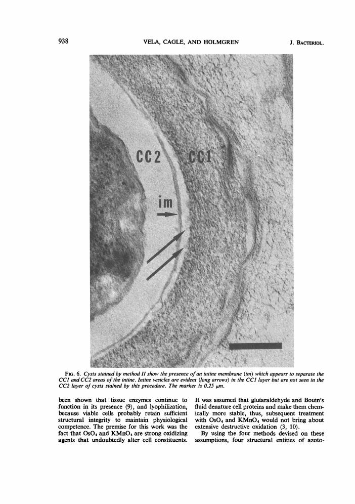
by Tchan et al. (12). It was found that the two layer is such that cysts stained by this method dolayers are separated by a continuous membrane not show structural elements like those seen in(Fig. 6) and that the chemical nature of the CC2 the CC1 layer. The membrane appears to be a
".e-.Z., -
v., -. .., A...*v.:.
t'.
Vr. ."
"espIt j
.t ";1

FIG. 3. Vegetative cell of Azotobacter vinelandii treated by method III. Arrows point to structures very similarto intine vesicles. In vegetative cells, they appear at cell wall andfree in the medium. The nuclear area (na) appears
identical to that ofthe cysts and seems to extend the length ofthe cell. The bar is 0.5 jAm.25lFFIL!I 4 1l. BIE JNL
FIG. 4. Cyst treated by method IV shows the intine vesicles (arrows) associated with the outer layers of theexine. The nuclear area is designated na. Dark gray material (ar) in the intine is assumed to be an artifact sinceit lacked consistency in different preparations. The bar is 0.25 jsm.
936

ULTRASTRUCTURE OF A. VINELANDII
FIG. 5. Cyst treated by method IV. Arrows point to spherical intine vesicles with double-layered surroundingmembrane. In lower center is a cyst microtubule (mt). The marker is 0.25 ,&m.
double-layered structure which effectively sepa-
rates the intine into two chemically distinct enti-ties, the CC1 and CC2 layers. This membranecould not be observed by the other methods oftreatment.
Microtubules, structural elements previouslyreported only in vegetative cells (7, 9), were
observed in cysts treated by method IV (Fig. 5).Only a few normal cysts had microtubules; in a
related experiment, however, most cysts of A.vinelandii grown in the presence of low concen-
trations of penicillin contained such micro-
tubules. We infer from this comparison that mic-rotubules can be observed by this method butthat normal cysts do not have large numbers ofthese subcellular elements.
DISCUSSIONThe use of various treatments to stabilize
specific constituents for examination revealedstructural details of A. vinelandii 12837 not seenin cells prepared by other methods. We chosetreatment with glutaraldehyde, assuming that itwould preserve structural cell proteins since it has
937VOL. 104, 1970
VELA, CAGLE, AND HOLMGREN
C.
*IA" V!,
itI:
"v\
4 Al ,
FiG. 6. Cysts stained by method 11 show the presence ofan inttine membrane (im) which appears to separate theCC] and CC2 areas of the intine. Intine vesicles are evident (long arrows) in the CC] layer but are not seen in theCC2 layer of cysts stained by this procedure. The marker is 0.25 um.
been shown that tissue enzymes continue tofunction in its presence (9), and lyophilization,because viable cells probably retain sufficientstructural integrity to maintain physiologicalcompetence. The premise for this work was thefact that OS04 and KMnO4 are strong oxidizingagents that undoubtedly alter cell constituents.
It was assumed that glutaraldehyde and Bouin'sfluid denature cell proteins and make them chem-ically more stable, thus, subsequent treatmentwith OS04 and KMnO4 would not bring aboutextensive destructive oxidation (3, 10).By using the four methods devised on these
assumptions, four structural entities of azoto-
938 J. BACTERIOL.
ULTRASTRUCTURE OF A. VINELANDII
bacter (i) intine fibrils, (ii) intine vesicles, (iii)intine membrane, and (iv) cyst microtubules werediscovered.
Intine vesicles (8) appear to be produced by thecell membrane-cell wall complex of cysts andvegetative cells. In cysts, the free vesicles appearto migrate toward the exine and eventually tothe cyst exterior. This phenomenon gives theimpression that neither intine nor exine are rigidimpermeable structures. Spherical structures iden-tical to intine vesicles are also found on vegetativecells (Fig. 3). We infer from this observation thatthe vesicles are not specific cyst organelles and thatthe Azotobacter cyst is essentially a resting vegeta-tive cell not analogous to bacterial spores. Thisassumption is further supported by the finding ofmicrotubules in cysts.The complexity of the cyst coat is indicated by
the presence of intine fibrils and the intine mem-brane. The fibrils are not seen in lyophilizedcysts and are presumably destroyed by thismanipulation. They appear to be homogeneouswith regard to electron density regardless of theirposition in the intine, whereas the CC1 layer isobviously chemically different from the CC2layer. Numerous examinations of cysts preparedby method II indicate the presence of a mem-branous structure separating the CC1 from theCC2 areas of intine. This membrane is doublelayered and is found in all cysts treated with thechrome-osmium preparation.The existence of intine vesicles, intine fibrils,
and intine membrane in cysts of A. vinelandiinegates the concept of the cyst coat as simply thepolymerized capsule of the vegetative cell. It mustbe replaced by a concept which is more in keepingwith the observations reported by Lin and Sadoff(6), by Pope and Wyss (8), and by us in this work.It is reasonable to assume that the Azotobactercyst is a complex metabolically active entity andthat the cyst coat is a complex functional part ofthe encysted cell.
ACKNOWLEDGMENT
We acknowledge the help and counsel of Orville Wyss of theUniversity of Texas, Austin, Tex., during the course of this work.
This work was supported by the Faculty Research Fund, NorthTexas State University, Denton, Tex.
LITERATURE CITED
1. Bisset, K. A., and C. M. F. Hale. 1953. The cytology and life-cycle of Azotobacter chroococcum. J. Gen. Microbiol.8:442-448.
2. Dalton, A. J. 1955. A chrome-osmium fixative for electronmicroscopy. Anat. Rec. 121:281.
3. Galigher, A. E. 1934. The essentials of practical microtech-nique in animal biology. Albert E. Galigher, Inc., Berkley.
4. Koo, V. M., L. P. Lin, and H. L. Sadoff. 1969. Surface struc-ture of Azotobacter vinelandii cysts as revealed by freeze-cleaving. J. Bacteriol. 100:1105-1107.
5. Lin, L. P., and H. L. Sadoff. 1969. Preparation and ultrastruc-ture of the outer coats of Azobacter vinelandii cysts. J.Bacteriol. 98:1335-1341.
6. Lin, L. P., and H. L. Sadoff. 1969. Chemical composition ofAzotobacter vinelandii cysts. J. Bacteriol. 100:480-486.
7. Pope, L. M., and P. Jurtshuk. 1967. Microtubule in Azoto-bacter vinelandii strain 0. J. Bacteriol. 94:2062-2064.
8. Pope, L. M., and 0. Wyss. 1970. Outer layers of the Azoto-bacter vinelandii cyst. J. Bacteriol. 102:234-239.
9. Robrish, S. A., and A. G. Marr. 1962. Location of enzymes inAzotobacter agilis. J. Bacteriol. 83:158-168.
10. Sabatini, D. D., K. Bensch, and R. J. Barrnett. 1963. Cyto-chemistry and electron microscopy; the preservation ofcellular ultrastructure and enzymatic activity by aldehydefixation. J. Cell Biol. 17:19-58.
11. Socolofsky, M. D. and 0. Wyss. 1961. Cysts of Azotobacter.J. Bacteriol. 81:946-954.
12. Tchan, Y. T., A. Birch-Andersen, and H. L. Jensen. 1962.The ultrastructure of vegetative cells and cysts of Azoto-bacter chroococcum. Arch. Mikrobiol. 43:50-66.
13. Vela, G. R., and G. D. Cagle. 1969. Formation of fragile cystsby a strain of Azotobacter chroococcum. J. Gen. Microbiol.57:365-368.
14. Wilson, P. W., and S. G. Knight. 1952. Experiments in bac-terial physiology. Burgess Publishing Co., Minneapolis.
15. Wyss, O., M. G. Neumann, and M. D. Socolofsky. 1961.Development and germination of the Azotobacter cyst.J. Biophys. Biochem. Cytol. 10:555-565.
16. Wyss, O., D. D. Smith, L. M. Pope, and K. W. Olson. 1969.Endogenous encystment of Azotobacter vinelandii. J. Bac-teriol. 100:475-479.
VOL. 104, 1970 939