characterization of free nitrogen fixing … · characterization of free nitrogen fixing bacteria...
TRANSCRIPT

846
Brazilian Journal of Microbiology (2011) 42: 846-858 ISSN 1517-8382
CHARACTERIZATION OF FREE NITROGEN FIXING BACTERIA OF THE GENUS Azotobacter IN ORGANIC
VEGETABLE-GROWN COLOMBIAN SOILS
Diego Javier Jiménez 1; José Salvador Montaña 1*; María Mercedes Martínez 2
1 Department of Microbiology, Faculty of Sciences, Pontificia Universidad Javeriana, Bogotá, D.C., Colombia 7th Avenue 43-82,
Building 50, Lab.106, Tel. 57-1-3208320 Ext. 4173, Bogotá, D.C., Colombia; 2 Institute of Crop Science and Resource
Conservation, Faculty of Agriculture, University of Bonn, Bonn, Germany, Auf dem Hugel 6, 53121.
Submitted: June 18, 2010; Returned to authors for corrections: January 13, 2011; Approved: March 14, 2011.
ABSTRACT
With the purpose of isolating and characterizing free nitrogen fixing bacteria (FNFB) of the genus
Azotobacter, soil samples were collected randomly from different vegetable organic cultures with neutral pH
in different zones of Boyacá-Colombia. Isolations were done in selective free nitrogen Ashby-Sucrose agar
obtaining a recovery of 40%. Twenty four isolates were evaluated for colony and cellular morphology,
pigment production and metabolic activities. Molecular characterization was carried out using amplified
ribosomal DNA restriction analysis (ARDRA). After digestion of 16S rDNA Y1-Y3 PCR products (1487pb)
with AluI, HpaII and RsaI endonucleases, a polymorphism of 16% was obtained. Cluster analysis showed
three main groups based on DNA fingerprints. Comparison between ribotypes generated by isolates and in
silico restriction of 16S rDNA partial sequences with same restriction enzymes was done with Gen
Workbench v.2.2.4 software. Nevertheless, Y1-Y2 PCR products were analysed using BLASTn. Isolate C5T
from tomato (Lycopersicon esculentum) grown soils presented the same in silico restriction patterns with A.
chroococcum (AY353708) and 99% of similarity with the same sequence. Isolate C5CO from cauliflower
(Brassica oleracea var. botrytis) grown soils showed black pigmentation in Ashby-Benzoate agar and high
similarity (91%) with A. nigricans (AB175651) sequence. In this work we demonstrated the utility of
molecular techniques and bioinformatics tools as a support to conventional techniques in characterization of
the genus Azotobacter from vegetable-grown soils.
Key words: ARDRA; Azotobacter; FNFB; UPGMA.
INTRODUCTION
The genus Azotobacter belongs to the �-subclass of the
Proteobacteria (4, 34) and comprises seven species: A.
chroococcum, A. vinelandii, A. beijerinckii, A. paspali, A.
armeniacus, A. nigricans and A. salinestri. In 2005 sequences
of 16S rDNA were identified that corresponded to a new
endemic soil species from Thailand (unpublished data).
Azotobacter are heterotrophic and aerobic bacteria and their
main property is the ability to fix nitrogen non-symbiotically,
with a genomic content of G-C of 63-67.5 % (Tm) (4, 32), and
distributed in soils, water and sediments (35, 36). Their
*Corresponding Author. Mailing address: Department of Microbiology, Faculty of Sciences, Pontificia Universidad Javeriana, Bogotá, D.C., Colombia 7th Avenue 43-82, Building 50, Lab.106, Tel. 57-1-3208320 Ext. 4173, Bogotá, D.C., Colombia.; Tel.: 49-228-735135.; E-mail: [email protected]
847
Jiménez, D.J. et al. Free nitrogen fixing bacteria of the genus Azotobacter agronomic importance is due to the capability of synthesizing
antibiotics, plant growth promotion substances (21, 24, 25, 30),
vitamins, exopolysaccharides and pigment production (8, 28),
besides their antagonist effect against pathogens (33). They
also have the ability to solubilize phosphates in aquaculture
systems, vermicompost production and potato (Solanum
tuberosum) grown soils (9, 12, 19). In Colombia, it has been
used as bacterial inoculants in the culture of Dendranthema
grandiflora, Stevia rebaudiana, vegetables and cotton (6, 11,
29). Characterization of Azotobacter species becomes difficult
due to the morphological similarity that exists with other FNFB
like Derxia, Azomonas and Beijerinckia, in addition to its
interspecific biochemical ambiguity. For that reason, in the last
years authors like Aquilanti et al. (1, 2), and Becking (4),
among others, justify the use of molecular techniques and
bioinformatics tools allowing fast identification and
characterization. This interest is focused towards the validation
of fast and reliable molecular methodologies for quality control
on agricultural biofertilizers. In this context, amplified
ribosomal DNA restriction analysis (ARDRA) was evaluated
for the characterization of Azotobacter isolated from organic
vegetable-grown soils, with the purpose of generating a
molecular approach to this genus in Colombian agro-
ecosystems.
MATERIALS AND METHODS
Isolation and biochemical characterization
Soil samples from organic broccoli (Brassica oleracea
var. italica), zucchini (Cucurbita pepo), cauliflower (B.
oleracea var. botrytis), spinach (Spinacia oleracea), tomato
(Lycopersicon esculentum) and carrot (Daucus carota) grown
soils were collected during October 2006 in Sogamoso
(5°43’N, 72°55’W) and Tibasosa (5°44’N, 73°00’W) (Boyacá-
Colombia) (Table 1). Five random samples each of 500 g were
withdrawn from 10–15 cm depth and sieved through a 4.75
mm-mesh sieve. Soil pH was measured according to Van
Lierop (38), analyzing samples fully suspended in distilled
water 1:1 (v/v). The primary isolation was made using the
grain soil technique (Figure 1A) according to the methodology
previously described by Aquilanti et al. (1) and Becking (4) in
Ashby-Sucrose agar (Agar 1.5%, Sucrose 0.5%, CaCO3 0.5%,
MgSO4 0.02%, NaCl 0.02%, KH2PO4 0.02%, FeSO4 0.0005%).
Plates were incubated at 28°C for 7 days until observing sticky
and glistening colonies around grains (Figure 1F). Counts were
made for each cultivated soil (% Recovery = Number of grains
with sticky and glistening colonies/total x 100). Isolates were
purified by streaking on free nitrogen Ashby-Sucrose agar and
morphological observations were done and subsequently stored
at -80ºC in nutrient broth (Scharlau®, Barcelona, Spain)
containing 50% glycerol. Pigment production was observed by
growing the cultures in Ashby agar with 0.5% (w/v) of
Benzoate (4, 22) and incubation at 28°C for 7 days. A.
vinelandii ATCC12518 and Azotobacter sp. strain (CCT)
previously isolated were used for biochemical identification
and molecular characterization. Biochemical identification was
carried out in triplicate using different sources of carbon:
glucose (GLU), maltose (MAL), mannitol (MAN), ramnose
(RAM) 1% (w/v) and benzoate (BNZ) 0.5% (w/v) in phenol
red broth (Merck®, Germany). Oxidase (OXI), catalase (CAT)
and nitrate reduction tests were done (34) and biochemical sets
were incubated at 28°C and evaluated after 24 h.
Genomic DNA extraction
Cultures were grown overnight in 5 ml nutrient broth,
shaked at 150 rpm and 20°C for 48 h. A 1.5 ml of the culture
was centrifuged at 10 000 g for 2 min and the resultant pellet
was resuspended in 1050 µl 1X TE buffer (10 mM Tris-HCl, 1
mM EDTA) and centrifuged at 10 000 g for 2 min,
resuspended again in 350 µl 1X TE buffer, 5 µl lysozyme (50
mg/ml) and 2 µl RNAse A (10 mg/ml) (Sigma®, St Louis,
USA), and incubated at 37ºC for 10 min. Finally, 3 µl
proteinase K (20 mg/ml) (Invitrogen®, Steinheim, Germany)
and 30 µl SDS 10% were added and incubated at 65ºC for 15
848
Jiménez, D.J. et al. Free nitrogen fixing bacteria of the genus Azotobacter
min. Crude DNA template was PCI (Phenol - Chloroform -
Isoamyl alcohol) (25:24:1) and CI (Chloroform - Isoamyl
alcohol) (24:1) extracted, precipitated in 1 ml absolute ethanol
and washed with 500 µl 70% ethanol and resuspended in 50 µl
of sterilized ultrapure water and stored at -20°C.
ARDRA
A 1487 bp fragment was amplified from the 16S rDNA
gene using universal primers Y1 (5'-
TGGCTCAGAACGAACGCTGGCGGC-3') and Y3 (5'-
TACCTTGTTACGACTTCACCCCAGTC-3'). (20, 40). PCR
reaction was done in a 100 µl reaction mixture containing: 1 U
of Taq DNA polymerase (Biolase®, Luckenwalde, Germany),
1X NH4 reaction buffer (160 mM (NH4)2 SO4, 670 mM Tris-
HCl pH 8.8, 0.1% Tween 20), 2 mM of MgCl2, 50 µM of
dNTPs, 0.2 µM of each primer and 50-100 ng of DNA. PCR
reactions were run for 35 cycles on a My cycler (Biorad®,
Hercules, CA, USA) as follows: denaturation at 93°C for 45 s,
annealing at 62°C for 45 s, and elongation at 72°C for 2 min.
An initial denaturation step at 93°C for 2 min and a final
extension step at 72°C for 5 min (16) were also done. DNA
amplification was checked by electrophoresis of 10 µl of each
PCR product in a 1.0% (w/v) agarose gel, in 1X TBE buffer
(0.09M Tris base, 0.09M sodium borate, 2.5 mM EDTA, pH
8.3) for 1 h at 3.2 V/cm on a Primo Thermo EC 330 Midcell
(Waltham®, MA, USA). A 25 µl-aliquot of the PCR product
was digested in a final volume of 40 µl for 3 h at 37°C with 2.5
U of restriction enzyme (AluI, HpaII or RsaI), 1X reaction
buffer, and 0.1 mg/µl of BSA, according to the manufacturer
(Promega®, Madison, USA). PCR products digests were
separated in a 2.5% (w/v) agarose gel electrophoresis in 1X
TBE buffer for 3 h at 3.2 V/cm and gels were stained in
ethidium bromide 0.5 µg/ml. DNA fragments were visualized
at 312 nm with an UV-transilluminator. Gels electronic images
were captured and analysed in the Quantity One 1-D Analysis
software (Biorad®, Hercules, CA, USA). For data scoring only
50 bp fragments or highest were considered to binary data
matrix built. Molecular weights of each band were determined
by means of a linear regression analysis, having as reference a
100 bp ladder (Promega®, Madison, USA). Genetic distance
between isolates was calculated using the DICE coefficient of
similarity and resulting distance matrix was used for the
unweighted pair group method with arithmetic mean
(UPGMA) analysis using NTSYSpc v.2.2 software for
Windows (27). The consistency of each node was estimated by
bootstrapping across markers (10) using 1000
pseudoreplications. In silico restriction analysis was carried
out with AluI, HpaII and RsaI using partial sequences of 16S
rDNA obtained from the following GenBank accessions:
AAY336565 (A. vinelandii); AY353708 (A. chroococcum);
AJ308318 (A. paspali); AB175656 (A. salinestris); AB175655
(A. armeniacus); E F100152 (A. beijerinckii); AB175651 (A.
nigricans); AF112477 (Azospirillum); AB175654 (Azomonas
macrocytogenes); AJ563934 (Beijerinckia derxi); AF164045
(Burkholderia tropica); AB089482 (Derxia gummosa);
AY191275 (Herbaspirillum seropedicae); AY509900
(Rhizobium leguminosarum) and AB305017 (Escherichia coli),
using Gene Workbench v.2.2.4 software for Windows®
(CLCbio®, Maryland, USA).
Sequence analyses
Primers Y1 (5'-TGGCTCAGAACGAACGCTGGCGGC-
3') and Y2 (5'-CCCACTGCTGCCTCCCGTAGGAGT-3')
were used for the analysis of a 318 bp partial sequence of 16S
rDNA (2). PCR reaction was done in a 100 µl reaction mixture
containing: 1 U of Taq DNA polymerase (Biolase®,
Luckenwalde, Germany), 1X NH4 reaction buffer (160mM
(NH4)2SO4, 670mM Tris-HCl pH 8.8, 0.1% Tween 20), 2 mM
of MgCl2, 200 µM of dNTPs, 0.5 µM of each primer and 50-
100 ng of DNA. PCR reactions were run for 35 cycles on My
Cycler (Biorad®, Hercules, CA, USA) as follows: denaturation
at 93°C for 45 s, annealing at 62°C for 45 s, and elongation at
72°C for 2 min. An initial denaturation step at 93°C for 5 min
and a final extension step at 72°C for 5 min were also done.
849
Jiménez, D.J. et al. Free nitrogen fixing bacteria of the genus Azotobacter
Amplification products were quantified using a Beckman DU
500 spectrophotometer (Fullerton®, CA, USA) and sequenced.
Sequences were aligned in Combined Workbench v.3.6.1
software for Windows (CLCbio®, Maryland, USA) and
compared with the data bases of GenBank using BLASTn (5).
RESULTS
Morphological and biochemical identification
The soil pH is within the range 6.25-7.44, which has been
reported optimal for the growth of Azotobacter species (3, 4).
Those pH values facilitated the recovery, allowing obtainment
percentages between 31% and 45% for the crop fields grown
with six different crops. A total of twenty-four isolates were
obtained. Most isolates presented whitish (cream color),
smooth, irregular, shining, 3-8mm diameter colonies;
nevertheless, colonies with transparent, glistening, shining, 2-5
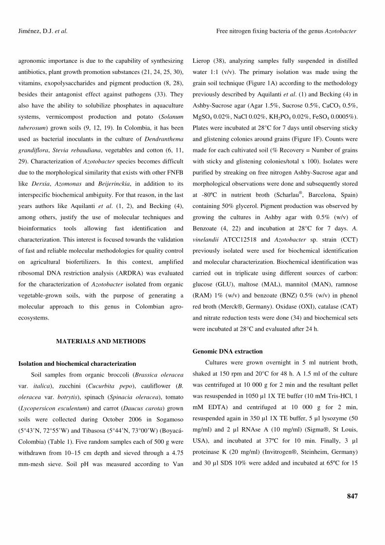
mm diameter also appeared. Three cell type morphologies were
identified: Gram-negative bacilli short and large; Gram-
negative bacilli short and small; and cysts (Figure 1). Some
showed a brown pigmentation and others presented a dark
brown pigmentation; a single one displayed a yellowish green
pigmentation, which is a characteristic of both A. vinelandii
and A. paspali (2, 4). Twelve isolates were glucose
fermentation positive, six showed a doubtful result (Table 1),
and twenty two were catalase positive. Isolate C5T was
ramnose negative; nevertheless it presented a doubtful result
for glucose fermentation. For preliminary identification (Table
1), morphological traits (cell and colony morphology) and
pigment production in the Ashby-Benzoate agar were
considered. Additionally, both control A. vinelandii
ATCC12518 and CCT strains identities were confirmed with
the same test.
Figure 1. Isolation, morphological traits and pigment production of nitrogen fixing bacteria isolates from vegetable-grown soils.
a) Primary isolation using soil grain technique. b) Whitish (cream color), smooth, irregular, shining, 3-8mm in diameter colonies,
isolate C5T. c) Cysts, isolate C5CA. d) Short and large Gram-negative bacilli, isolate C5T. e) Small Gram-negative bacilli isolate
C1BR. f) Sticky and glistening colonies growing around soil grains g) Transparent, glistening, shining 2-5mm in diameter
colonies, isolate C1BR. h) Brown pigmentation, isolate C1T. i) Brown-black pigmentation, isolate C5T. j) Yellow-green
pigmentation, isolate C5CA.

850
Jiménez, D.J. et al. Free nitrogen fixing bacteria of the genus Azotobacter
Table 1. Crop-grown soil samples, pH, recovery percentage, location, morphological characteristics, biochemical and preliminary identification of nitrogen fixing bacteria isolates
from vegetable-grown soils.
Crop pH %Ra Location Isolate Colony morphology
Cellular morphology Pigment GLU MAL MAN RAM BNZ OXI CAT NITRATE Preliminary identification
ND ND ND ND ATCC 12518 + + + + D + +
Potato ND ND ND CCT Y
+ + + + + + + A. vinelandii
C1BR
A
- - - - D + -
NITRITE
C2BR X
- + + D - - + AMMONIUM ND
C3BR Y + D + - - + + NITRITE A. nigricans C4BR
B X - - - - - - + AMMONIUM ND
Broccoli 7.06 36.3 Tibasosa
C5BR D + + + - + + NITRITE C1CA
Y
NP
+ + D + + + + A. vinelandii
C3CA Brown-Yellow - - D - D + + ND
C4CA X
Brown D - - - - + +
AMMONIUM
A. nigricans Zucchini 6.82 35 Sogamoso
C5CA Z Yellow-Green + + + + + + + A. vinelandii
C1CO Black D + + - D D + A. chroococcum/ A. nigricans
C4CO
A
D - - - D + +
NITRITE
Cauliflower 7.12 33.6 Tibasosa
C5CO + - - D - + + C1E
B D - - - - + +
AMMONIUM A. nigricans
C2E
NP
+ + + + + + + C3E
Y
+ + + + + + + NITRITE A. vinelandii
C4E X Brown
- - - - D + + ND Spinach 6.79 45
C5E Brown-Black + D + - D D + A. chroococcum/ A.
nigricans C1T
Y Brown + - - - - + +
AMMONIUM
A. nigricans C2T
A
X NP - D - - - - + NITRITE
C3T B Brown-Black - - - - - + + AMMONIUM
ND
C4T Brown + + + + + + + NITRITE A. vinelandii Tomato 7.44 37.6
Sogamoso
C5T
Y Brown-Black D + + - D D + AMMONIUM A. chroococcum/ A.
nigricans C1Z
A
Z + - - - - - + ND Carrot 6.25 31.3 Tibasosa C2Z B X
NP - - - - - + -
NITRITE A. paspali
a Recovery percentage; ND: Not determined; A: whitish (cream color), smooth, irregular, shining, 3-8mm in diameter colonies; B: transparent, glistening, shining, 2-5mm in diameter colonies; X: Gram-negative bacillus, short and small; Y: Gram-negative bacillus, short and large; Z: Cysts; NP: Negative pigment; (+): Positive test.; (-): Negative test; D: Doubtful; GLU: Glucose; MAL: Maltose; MAN: Mannitol; RAM: Ramnose; BNZ: Benzoate; OXI: Oxidase test; CAT: Catalase test.

851
Jiménez, D.J. et al. Free nitrogen fixing bacteria of the genus Azotobacter
ARDRA
Based on the preliminary identification, thirteen
presumptive Azotobacter isolates (C3BR, C5BR, C1CA,
C5CA, C5CO, C1E, C2E, C3E, C5E, C1T, C4T, C5T and
C1Z) were selected for DNA extraction and restriction
analysis. Isolate C1BR was identified as not belonging to
Azotobacter, therefore it was included in our study to observe
genetic differences with the other isolates that were catalase
positive (Table 1). A. vinelandii ATCC12518 and CCT strains
were included in the molecular analysis. Restriction
endonuclease AluI generated eight rybotypes, a total of 78
bands and a polymorphism of 16.6%; with HpaII twelve
rybotypes were obtained, a total of 92 bands and a
polymorphism of 17.3%; and RsaI generated seven rybotypes,
a total of 82 bands and a polymorphism of 15.8% and 1.2% of
unique bands (Figure 2). UPGMA showed that the isolates
exhibit three main groups (Figure 3). Group I, with a
coefficient of similarity of approximately 0.75%. In this group,
A. vinelandii ATCC12518 presented a greater coefficient of
similarity (1.0) with respect to CCT strain and a similarity of
0.97 with respect to C5CA. Group II, with a coefficient of
similarity of approximately 0.69 includes all the isolates
obtained from broccoli-grown soils. Therefore, group III, with
a coefficient of similarity of approximately 0.65 includes C5T,
C1Z and C3E, the ones that showed the most dissimilar
restriction patterns. On the other hand, UPGMA for the in
silico restriction analysis showed that Azotobacter species have
similar restriction patterns except for A. chroococcum
(AY353708) (Figure 4). The in silico restriction analysis with
AluI generated different restriction patterns for all Azotobacter
sequences and a polymorphism of 48.3%. HpaII generated
identical restriction patterns for A. nigricans (AB175651) and
A. vinelandii (AY336565) and a polymorphism of 42.4%.
Finally RsaI generated the same restriction patterns for A.
armeniacus (AB175655) and A. salinestris (AB175656) and a
polymorphism of 50%.
Figure 2. Agarose 2.5% gel electrophoresis of restriction fragments obtained after the digestion of 16S rDNA amplicons of
nitrogen fixing bacteria isolates from vegetable-grown soils. Restriction patterns with: a) AluI. b) HpaII. c) RsaI. 1. ATCC 12518.
2. CCT. 3. C1BR. 4. C3BR. 5. C5BR. 6. C1CA. 7. C5CA. 8. C5CO. 9. Negative control (sterilized ultra pure water). 10.
Molecular weight ladder (100pb Promega®). 11. C1E. 12. C2E. 13. C3E. 14. C5E. 15. C1T. 16. C4T. 17. C5T. 18. C1Z.

852
Jiménez, D.J. et al. Free nitrogen fixing bacteria of the genus Azotobacter
Figure 3. UPGMA for ARDRA of the FNFB isolates from vegetable-grown soils using NTSYSpc v.2.2. software for Windows®.
Figure 4. UPGMA for in silico restriction of the sequences of FNFB obtained from GenBank database using Gene Workbench®
v.3.6.1 software for Windows®.
853
Jiménez, D.J. et al. Free nitrogen fixing bacteria of the genus Azotobacter
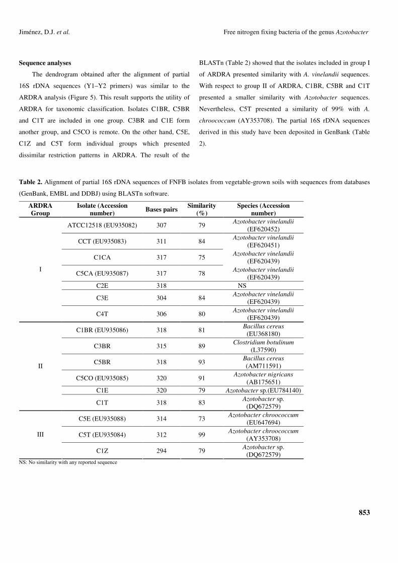
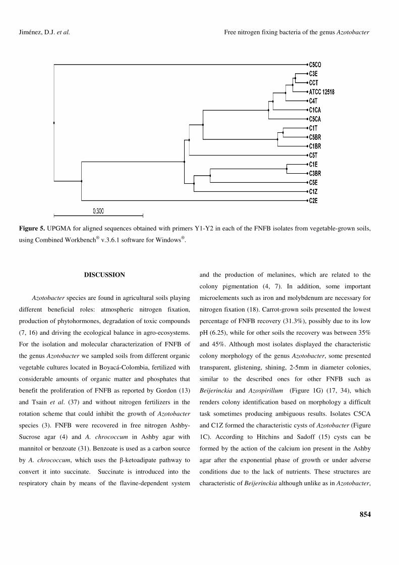
Sequence analyses
The dendrogram obtained after the alignment of partial
16S rDNA sequences (Y1–Y2 primers) was similar to the
ARDRA analysis (Figure 5). This result supports the utility of
ARDRA for taxonomic classification. Isolates C1BR, C5BR
and C1T are included in one group. C3BR and C1E form
another group, and C5CO is remote. On the other hand, C5E,
C1Z and C5T form individual groups which presented
dissimilar restriction patterns in ARDRA. The result of the
BLASTn (Table 2) showed that the isolates included in group I
of ARDRA presented similarity with A. vinelandii sequences.
With respect to group II of ARDRA, C1BR, C5BR and C1T
presented a smaller similarity with Azotobacter sequences.
Nevertheless, C5T presented a similarity of 99% with A.
chroococcum (AY353708). The partial 16S rDNA sequences
derived in this study have been deposited in GenBank (Table
2).
Table 2. Alignment of partial 16S rDNA sequences of FNFB isolates from vegetable-grown soils with sequences from databases
(GenBank, EMBL and DDBJ) using BLASTn software.
ARDRA Group
Isolate (Accession number) Bases pairs Similarity
(%) Species (Accession
number)
ATCC12518 (EU935082) 307 79 Azotobacter vinelandii (EF620452)
CCT (EU935083) 311 84 Azotobacter vinelandii (EF620451)
C1CA 317 75 Azotobacter vinelandii (EF620439)
C5CA (EU935087) 317 78 Azotobacter vinelandii (EF620439)
C2E 318 NS
C3E 304 84 Azotobacter vinelandii (EF620439)
I
C4T 306 80 Azotobacter vinelandii (EF620439)
C1BR (EU935086) 318 81 Bacillus cereus (EU368180)
C3BR 315 89 Clostridium botulinum (L37590)
C5BR 318 93 Bacillus cereus (AM711591)
C5CO (EU935085) 320 91 Azotobacter nigricans (AB175651)
C1E 320 79 Azotobacter sp.(EU784140)
II
C1T 318 83 Azotobacter sp. (DQ672579)
C5E (EU935088) 314 73 Azotobacter chroococcum (EU647694)
C5T (EU935084) 312 99 Azotobacter chroococcum (AY353708)
III
C1Z 294 79 Azotobacter sp. (DQ672579)
NS: No similarity with any reported sequence
854
Jiménez, D.J. et al. Free nitrogen fixing bacteria of the genus Azotobacter
Figure 5. UPGMA for aligned sequences obtained with primers Y1-Y2 in each of the FNFB isolates from vegetable-grown soils,
using Combined Workbench® v.3.6.1 software for Windows®.
DISCUSSION
Azotobacter species are found in agricultural soils playing
different beneficial roles: atmospheric nitrogen fixation,
production of phytohormones, degradation of toxic compounds
(7, 16) and driving the ecological balance in agro-ecosystems.
For the isolation and molecular characterization of FNFB of
the genus Azotobacter we sampled soils from different organic
vegetable cultures located in Boyacá-Colombia, fertilized with
considerable amounts of organic matter and phosphates that
benefit the proliferation of FNFB as reported by Gordon (13)
and Tsain et al. (37) and without nitrogen fertilizers in the
rotation scheme that could inhibit the growth of Azotobacter
species (3). FNFB were recovered in free nitrogen Ashby-
Sucrose agar (4) and A. chrococcum in Ashby agar with
mannitol or benzoate (31). Benzoate is used as a carbon source
by A. chrococcum, which uses the �-ketoadipate pathway to
convert it into succinate. Succinate is introduced into the
respiratory chain by means of the flavine-dependent system
and the production of melanines, which are related to the
colony pigmentation (4, 7). In addition, some important
microelements such as iron and molybdenum are necessary for
nitrogen fixation (18). Carrot-grown soils presented the lowest
percentage of FNFB recovery (31.3%), possibly due to its low
pH (6.25), while for other soils the recovery was between 35%
and 45%. Although most isolates displayed the characteristic
colony morphology of the genus Azotobacter, some presented
transparent, glistening, shining, 2-5mm in diameter colonies,
similar to the described ones for other FNFB such as
Beijerinckia and Azospirillum (Figure 1G) (17, 34), which
renders colony identification based on morphology a difficult
task sometimes producing ambiguous results. Isolates C5CA
and C1Z formed the characteristic cysts of Azotobacter (Figure
1C). According to Hitchins and Sadoff (15) cysts can be
formed by the action of the calcium ion present in the Ashby
agar after the exponential phase of growth or under adverse
conditions due to the lack of nutrients. These structures are
characteristic of Beijerinckia although unlike as in Azotobacter,

855
Jiménez, D.J. et al. Free nitrogen fixing bacteria of the genus Azotobacter
in this case they do not appear forming pairs. An important
characteristic of Azotobacter is the Gram-negative bacillary
morphology, with cells between 2 µm and 4 µm in diameter
(Figure 1D). Some isolates presented this morphology, while
others consisted of small Gram-negative bacilli but with a
morphology very similar to that displayed by Azospirillum,
Beijerinckia, Herbaspirillum and Derxia (Figure 1E).
However, it is known that Azotobacter is a pleiomorphic
microorganism (4). In general, morphological identification
and characterization are not necessarily useful in defining the
genus as it was the case of isolates C3BR and C5BR, which
exhibited morphological characteristics similar to those
reported for Azotobacter besides growing in nitrogen-free
culture media but the molecular identification revealed that
they do not belong to that genus. It has been reported that
Azotobacter has the capacity to produce soluble pigments (1,
4), this can be a useful tool in the characterization of some
Azotobacter species. Brown or black pigmentation in the
Ashby-Benzoate agar allowed in this study the differentiation
between A. chroococcum and A. nigricans (22). Isolates C1CO,
C5E, C1T, C3T and C5T exhibited morphological traits and
similar pigmentation to those displayed by A. chroococcum and
A. nigricans (Figure 1H and Table 1). On the another side,
isolates C3CA and C4E presented colonies with brown-yellow
pigmentation, Gram-negative bacilli, and catalase and oxidase
negative tests, suggesting that they can be aerobic FNFB that
do not belong to the genus Azotobacter. In the biochemical
identification, tests allowing the differentiation between genera
and species were used. An example is that A. vinelandii is the
only species capable of assimilating ramnose and using it as
carbon source. In addition, all Azotobacter species have the
capacity to produce oxidases and catalases for the protection of
their nitrogenase. Universal primers (Y1-Y3) have been used in
FNFB 16S rDNA amplification (2, 17, 20) although not
specific for the genus Azotobacter. FNFB characterization can
be done based on several different genes coding for many
functions, as is the case of the gene nif that codifies the
nitrogenase enzyme which drives the atmospheric nitrogen
fixation (21, 26), in this work a partial sequence of nifH gene
was amplified in several isolates including in the tree groups of
ARDRA (data not shown). Nevertheless, both the 16S rDNA
characteristics and its vertical transmission render this gene as
an excellent molecular marker (23, 39). ARDRA is a molecular
technique that allows establishing phylogenetic relationships
between individuals. The enzymes used must generate high
levels of polymorphism for specific groups to facilitate the
detection of intra or inter-specific significant differences (14).
The comparison between ARDRA and in silico analysis
revealed that ARDRA is highly useful in establishing
phylogenetic relationships between isolates and in clarifying
the taxonomy. In general, the percentages of polymorphism of
enzymes used in this work were relatively high, 16.5% on
average, and similar to those reported by Aquilanti et al. (2)
who obtained polymorphisms of 22%, 23% and 16% for AluI,
HpaII and RsaI, respectively. A directly proportional
relationship was demonstrated between the percentages of
polymorphism and the ribotypes produced. UPGMA showed
three main groups of similarity (Figure 3). We noted that group
I isolates are found only in crop-grown soils located in
Sogamoso, whereas group II and III isolates occur in crop-
grown soils in both localities. Based solely on soil
characteristics and the grown crops, clustering may differ from
that one obtained with ARDRA. On the other hand, there was
no relationship between the groups and the pH of crop-grown
soils, although most isolates of group II were recovered in pH
with values between 7.06 and 7.44. C5T and C1Z always
showed irregular patterns for each enzyme and isolate C5E
displayed a unique band with RsaI (Figure 2); these unique
bands are particularly important since they can be useful for
designing specific primers. When comparing the patterns
generated in ARDRA with those obtained from the in silico
restriction, a great consistency between number of bands and
their corresponding molecular weights was observed in some
cases. In other cases, this consistency results only for one or

856
Jiménez, D.J. et al. Free nitrogen fixing bacteria of the genus Azotobacter
two enzymes. Inconsistencies could be due to the fact that the
lengths of some of the GenBank 16S rDNA sequences do not
fully match with the lengths of the sequences of our isolates,
probably because the primers used differed from ours. By this
reason the molecular weights of the bands could differ by a few
base pairs, especially in fragments of the flanking regions of
the gene, considering as similar those bands that differed by
15-25 bp. On the other hand, in some isolates the presence of
bands of greater molecular weights was observed after the
digestion, suggesting that some copies of the gene were not
completely digested. Isolates of group I of the ARDRA (Figure
3) showed restriction patterns similar to those obtained from
the sequences of Azotobacter species used in the in silico
restriction, whereas the majority of isolates of group II did not
show this similarity. With respect to isolates of group III, a
special finding was isolate C5T that produced patterns identical
to A. chroococcum (accession number AY353708). In general,
the BLASTn analysis carried out with partial sequences of the
16S rDNA gene obtained from every isolate revealed for group
I similarity values between 74% and 84% with sequences of A.
vinelandii; isolate C5CA showed a similarity of 78%
(EF620439). Strain C5CO showed a similarity of 91% with A.
nigricans, confirming its preliminary identification. Regarding
isolates of group III from ARDRA, isolate C5T showed a
similarity of 99% with A. chroococcum (AY353708). In
conclusion, from the 15 isolates obtained from vegetable-
grown fields, 14 exhibited phenotypical similarity with species
of the genus Azotobacter and 13 showed restriction patterns
similar to the ones obtained with the sequences of Azotobacter
in the in silico restriction. Isolate C5T obtained from a tomato-
grown field (Lycopersicon esculentum) could be identified as
A. chroococcum. It is important to note that the short length of
the sequences analyzed with BLASTn did not suffice to obtain
a clarifying view of the taxonomic position of the majority of
isolates. However, ARDRA proved to be a molecular tool that
allows the identification of interspecific ribotypes of FNFB
using a few polymorphic enzymes. The analysis of diversity is
an important factor in the measurement of quality of
agricultural soils. On the other hand, it is necessary to design
species-specific primers for the genus Azotobacter because of
its large importance as atmospheric nitrogen fixing and plant
growth promoting rhizobacteria.
ACKNOWLEDGMENT
This research was supported by the International
Federation of Catholic Universities (CIC-FIUC). We thank the
Environmental Biotechnology Unit and the Plant
Biotechnology Unit of the Pontificia Universidad Javeriana for
their collaboration.
REFERENCES
1. Aquilanti, L.; Favilli, F.; Clemeti, F. (2004). Comparison of different
strategies for isolation and preliminary identification of Azotobacter from
soil samples. Soil. Biol. Biochem. 36, 1475–1483.
2. Aquilanti, L.; Mannazzu, I.; Papa, R.; Cavalca, L.; Clementi, F. (2004).
Amplified ribosomal DNA restriction analysis for the characterization of
Azotobacteraceae: a contribution to the study of these free-living
nitrogen-fixing bacteria. J. Microbiol. Methods. 57, 197-206.
3. Balandreau, J. (1986). Ecological factors and adaptive processes in N2-
fixing bacterial populations of the plant environment. Plant Soil. 90, 73-
80.
4. Becking, J. (2006). The family Azotobacteraceae. Prokaryotes. 6, 759-
783.
5. Benson, D.A.; Karsch, I.; Lipman, D.J.; Ostell, J.; Rapp, B.A.; Wheeler,
D.L. (2003). GenBank. Nucleic. Acids. Res. 31, 23–27.
6. Borda, D.; Pardo, J.M. (2008). Determinación de la influencia de
materia orgánica y Azotobacter spp. en un cultivo de Stevia rebaudiana.
B. Bogotá, Colombia, 125p. (Microbiology Undergraduate. Dissertation.
Science Faculty. Pontificia Javeriana University).
7. Castillo, J. (2005). Evaluación de la degradación de endosulfan por
Azotobacter chroococcum y determinación del efecto plaguicida sobre la
fijación biológica de nitrógeno y sobre la producción de auxinas.
Bogotá, Colombia, 121p. (Microbiology Undergraduate. Dissertation.
Science Faculty. Pontificia Javeriana University).
8. Cuesta, A. (2006). Estrategias de cultivo en la producción de alginatos
por Azotobacter vinelandii. Medellín, Colombia, 105p. (M.Sc.
Dissertation. National University of Colombia).

857
Jiménez, D.J. et al. Free nitrogen fixing bacteria of the genus Azotobacter
9. Faccini, G.; Garzon, S. (1996). Evaluación del efecto de un inóculo dual
de bacterias solubilizadoras de fosfato y Azotobacter chroococcum en el
cultivo de papa “criolla” yema de huevo (Solanum phureja). Bogotá,
Colombia, 97p. (Bacteriology Undergraduate. Dissertation. Science
Faculty. Pontificia Javeriana University).
10. Felsenstein, J. (1985). Confidence limits on phylogenies: an
approachusing the bootstrap. Evolution. 39, 783–791.
11. Galindo, T.; Polania, J.; Sánchez, J.; Moreno, N.; Vanegas, J.; Holgin, G.
(2006). Efecto de inoculantes biológicos en el crecimiento de manglar y
Citrullus vulgaris. San Andrés Isla, Colombia. Acta. Biol. Colomb. 11,
83 – 97.
12. Garg, S.; Bhatnagar, A.; Kalla, A.; Narula, N. (2001). In vitro nitrogen
fixation, phosphate solubilization, survival and nutrient release by
Azotobacter strains in an aquatic system. Bioresour. Technol. 80, 101–
109.
13. Gordon, J. (1981). Introduction to the nitrogen fixing prokaryotes. In:
Starr, M.P., Stolp, H., Pr�per, N.G., Balows, A., Schlegel, H.G. (eds.),
The Prokaryotes: A Handbook on Habitats, Isolation and Identification
of Bacteria. Springer, Heidelberg, Germany p. 781–794.
14. Heyndrickx, M.; Vauterin, L.; Bañadme, P.; Kesters, P.; De Vos, P.
(1996). Applicability of combined amplified ribosomal DNA restriction
analysis (ARDRA) patterns in bacterial phylogeny and taxonomy. J.
Microbiol. Methods. 26, 247-259.
15. Hitchins, V.; Sadoff, H. (1973). Sequential metabolic events during
encystment of Azotobacter vinelandii. J. Bacteriol. 113, 1273-1279.
16. Juárez, B.; Martínez, M.; González, J. (2004). Growth of Azotobacter
chroococcum in chemically defined media containing p-hydroxybenzoic
acid and protocatechuic acid. Chemosphere. 59, 1361–1365
17. Junior, F.; Silva, M.; Texeira, K.; Urquiaga, S.; Reis, M. (2004).
Identification of Azospirillum amazonense isolates associated to
Brachiaria spp. at different stages and growth conditions and bacterial
plant hormone production. Rev. Bras. Ciênc Solo. 28, 103-113.
18. Kumar, U.; Narula, N. (1999). Solubilization of inorganic phosphate and
grown emergencia of wheats as affected by Azotobacter chroococcum
mutants. Biol. Fertil. Soils. 28, 301-305.
19. Kumar, V.; Singh, K. (2001). Enriching vermicompost by nitrogen fixing
and phosphate solubilizing bacteria. Bioresour. Technol. 76, 173–175.
20. Magalhães, L.; Maltempi, De Souza.; Baler, O.; Baldani, J.; Dobereiner,
J.; De Oliveira, F. (2001). 16S ribosomal DNA characterization of
nitrogen-fixing bacteria isolated from Banana (Musa spp.) and Pineapple
(Ananas comosus (L.) Merril). Appl. Environ. Microbiol. 67, 2375-2379.
21. Mantilla, J. (2008). Distribución y capacidad fijadora de nitrógeno de
bacterias diazótrofos aisladas en suelos del sur del trapecio
Colombiano.155p. (M.Sc. Dissertation. Science Faculty. Pontificia
Javeriana University).
22. Martyniuk, S.; Martyniuk, M. (2002). Occurrence of Azotobacter spp. in
some polish soils. Pol. J. Environ. Stud. 12, 371-374.
23. Nogales, B. (2005). Microbiología del suelo en la era de la biología
molecular: Descubriendo la punta del iceberg. Ecosistemas. 2, 1-10.
24. Pandey, A.; Kumar, S. (1990). Inhibitory effects of Azotobacter
chroococcum and Azospirillum brasiliense on a range of rhizosphere
fungi. Indian J. Exp. Biol. 28, 52–54.
25. Pandey, A.; Sharma, E.; Palni, L. (1998). Influence of bacterial
inoculation on maize in upland farming systems of the Sikkim Himalaya.
Soil. Biol. Biochem. 30, 379-384.
26. Roesch, L.; Olivares, F.; Pereira, L.; Selbach, P.; Saccol, De Sae.;
Oliveira, F. (2006). Characterization of diazotrophic bacteria associated
with maize: effect of plant genotype, ontogeny and nitrogen-supply.
World. J. Microbiol.Biotechnol. 22, 967–974.
27. Rohlf, F.J. (1993). NTSYS. PC. Numerical taxonomy and multivariate
analysis system, Version 1.8 Applied Biostatistics. New York. USA.
28. Sabra, W.; Zeng, A.; Deckwer, W. (2001). Bacterial alginate:
physiology, product quality and process aspects. Appl. Microbiol.
Biotechnol. 56, 315–325.
29. Santana, M.; Vásquez, C. (2002). Evaluación de cepas de Azotobacter
spp. y de bacterias solubilizadoras de fosfato (BFS), como biofertilizante
mixto en cultivo de crisantemo (Chrysanthemum morifolium var. Regal
Suerte). Bogotá, Colombia. 94p. (Microbiology Undergraduate.
Dissesrtation. Science Faculty. Pontificia Javeriana University).
30. Salmeron, V.; Martinez, M.V.; Gonzalez, J. (1990). Nitrogen fixation
and production of auxins, gibberellins and cytokinin by Azotobacter
chroococcum strain isolated from root of Zea mays in presence of
insoluble phosphate. Chemosphere. 20, 417–422.
31. Saribay, G. (2003). Growth and nitrogen fixation dynamics of
Azotobacter chroococcum nitrogen-free and own containing medium.
Ankara, Turkia, 45p. (M.Sc. Dissertation. The Middle East Technical
University).
32. Setubal, J.; Dos Santos, P.; Goldman, B.; Ertesvåg, H.; Espin, G.; Rubio,
L.; Valla, S.; Almeida, N.; Balasubramanian, D.; Cromes, L.; Curatti, L.;
Du, Z.; Godsy, E.; Goodner, B.; Hellner-Burris, K.; Hernandez, J.;
Houmiel, K.; Imperial, J.; Kennedy, C.; Larson, T.; Latreille, P.; Ligon,
L.; Lu, J.; Mærk, M.; Miller, N.; Norton, S.; O'Carroll, I.; Paulsen, I.;
Raulfs, E.; Roemer, R.; Rosser, J.; Segura, D.; Slater, S.; Stricklin, S.;
Studholme, D.; Sun, J.; Viana, C.; Wallin, E.; Wang, B.; Wheeler, C.;
Zhu, H.; Dean, D.; Dixon, R.; Wood. D. (2009). Genome sequence of
Azotobacter vinelandii, an obligate aerobe specialized to support diverse
anaerobic metabolic processes. J. Bacteriol. 191, 4534-4545.
33. Sudhir, U.; Meshram.; Jager, G. (1983). Antagonism of Azotobacter
chroococcum isolates to Rhizoctonia solani. Eur. J. Plant Phatol. 89,
191-197.
34. Tchan, Y.T.; New, P.B. (1984). Genus I Azotobacter, Beijerinck 1901,
567AL. In: Krieg, N.R., Holt, J.G. (eds.). Bergey’s Manual of
858
Jiménez, D.J. et al. Free nitrogen fixing bacteria of the genus Azotobacter
Determinative Bacteriology, vol. 1. Williams & Wilkins, Baltimore,
USA, p. 220–229.
35. Tejera, N.; Lluch, C.; Martínez, M.; González, J. (2005). Isolation and
characterization of Azotobacter and Azospirillum strains from the
sugarcane rhizosphere. Plant Soil. 27, 223–232.
36. Torres, M.; Valencia, S.; Bernal, J.; Martínez, P. (2000). Isolation of
Enterobacteria, Azotobacter sp. and Pseudomonas sp., producers of
indole-3-acetic acid and siderophores, from Colombian rice rhizosphere.
Rev. Latin. Microbiol. 42, 171-176.
37. Tsain, J.; Aladegbami, S.; Vela, G. (1979). Phosphate-limited culture of
Azotobacter vinelandii. J. Bacteriol. 139, 639–645.
38. Van Lierop, W. (1981). Conversion of organic soil pH values measured
in water, 0.01 M CaCl2 or 1N KCl. Can. J. Soil. Sci. 6, 577–579.
39. Weisburg, W.; Barns, M.; Pelleteried, D.; Lane, D. (1991). 16S
Ribosomal DNA amplification for the phylogenetic study. J. Bacteriol.
173, 697-703.
40. Young, J.; Downer, H.; Eardly, B. (1991). Phylogeny of the phototrophic
Rhizobium Strain BTAil by polymerase chain reaction-based sequencing
of a16S rRNA gene segment. J. Bacteriol. 173, 2271-2277.
All the content of the journal, except where otherwise noted, is licensed under a Creative Commons License