ubiquitination of lysine-331 by kaposi s sarcoma ... · the conserved lysine k331 the likely target...
TRANSCRIPT
Ubiquitination of lysine-331 by Kaposi’s sarcoma-associated herpesvirus protein K5 targets HFE forlysosomal degradationDavid A. Rhodesa,1, Louise H. Boylea, Jessica M. Bonameb, Paul J. Lehnerb, and John Trowsdalea
Departments of aPathology, Immunology Division, and bMedicine, University of Cambridge, Cambridge Institute for Medical Research, Cambridge CB2 0XY,United Kingdom
Edited by Pamela J. Bjorkman, California Institute of Technology, Pasadena, CA, and approved August 6, 2010 (received for review March 16, 2010)
The nonclassical MHC class I-related (MHC-I) molecule HFE controlscellular iron homeostasis by a mechanism that has not been fullyelucidated. We examined the regulation of HFE by K5, the E3ubiquitin ligase encoded by Kaposi’s sarcoma-associated herpesvi-rus (KSHV/HHV8), that is known to down-regulate classical MHC-I.K5 down-regulated HFE efficiently, using polyubiquitination of themembrane proximal lysine in the HFE cytoplasmic tail (K331), totarget themolecule for degradation via ESCRT1/TSG101-dependentsorting from endosomes to multivesicular bodies (MVBs)/lyso-somes. In the primary effusion lymphoma cell line BC-3, which car-ries latent KSHV, HFEwas degraded rapidly upon virus reactivation.HFE was ubiquitinated on lysine-331 in unactivated BC-3 cells, con-ditions where K5 was not detectable, consistent with an endoge-nous E3 ubiquitin ligase controlling HFE expression. The resultsshow regulated expression of HFE by ubiquitination, consistentwith a role in cellular iron homeostasis, a molecular mechanismtargeted by KSHV to achieve a positive iron balance.
hemochromatosis | iron | immunology
Iron is an essential cofactor for host cells and pathogenicmicroorganisms, but causes cellular damage when in excess.
Molecular mechanisms have evolved to maintain optimal ironlevels in host cells while restricting the supply to pathogens,which also require iron for growth and replication (1, 2).The major histocompatibility complex class I-related (MHC-I)
molecule HFE (3) regulates iron homeostasis at the cellular leveland systemically (4). Initial studies were consistent with HFEforming a complex with β2 microglobulin (β2m) and transferrinreceptor 1 (TfR1) in the endoplasmic reticulum. Upon traffickingto the cell surface, HFE recycled in an endosomal compartmentof intermediate pH and competed with transferrin for binding toTfR1 to limit cellular iron uptake (5–7). At physiological concen-trations of iron-bound transferrin (10 μMholo-Tf), it was shown thatHFE could not compete for binding to TfR1 because of its loweraffinity, but that cellular iron levels were reduced by HFE ex-pression. HFE therefore appeared to be regulating some aspect ofiron import in the endosome, independent of TfR1 binding (8–10).HFE may also regulate iron export in some cell types (11, 12).HFE regulates iron levels systemically by influencing expres-
sion of the peptide hormone hepcidin (13). This pathway ismediated by BMP6 and SMAD4 signaling in response to in-creased iron, proinflammatory stimuli, and hypoxia (14, 15).Hepcidin binds to ferroportin, an iron efflux channel expressedin intestinal epithelium, hepatocytes, and macrophages, therebypromoting ferroportin degradation, so that iron export is pre-vented (16, 17). Regulation of hepcidin transcription by hep-atocytes required HFE to form a complex with transferrinreceptor 2, not TfR1, a switch mediated by holo-Tf, which dis-places HFE from TfR1 due to its greater affinity (18, 19).Kaposi’s sarcoma (KS)-associated herpesvirus (KSHV or
HHV8) encodes two proteins, K3 and K5, which regulate MHC-I(20). Areas of endemic KS in Africa correlate with red–brownvolcanic soils rich in iron, suggesting that iron represents an en-vironmental variable in KS pathogenesis (21, 22). Iron is alsoassociated with KS lesions by immunohistochemistry (23).
Given the homology of HFE to MHC-I, we asked whether K3and K5 could regulate HFE. We show that K5, but not K3,down-regulated surface expression by targeting lysine-331 in thecytoplasmic tail of HFE for ubiquitination, leading to lysosomaldegradation. In BC-3 cells, wild-type HFE was degraded rapidlyupon viral reactivation, whereas an HFE K331R mutant line wasunaffected. HFE was ubiquitinated on lysine-331 in unactivatedBC-3 cells, providing evidence for an endogenous E3 ligase withcharacteristics similar to K5. The results are consistent withcontrol of cellular iron homeostasis by ubiquitinated HFE, anendogenous pathway co-opted by KSHV to increase cytosoliciron levels required for lytic infection.
ResultsLysine 331 Is Required for K5-Mediated Regulation of HFE. HeLa-Mcells expressing FLAG-HFE were cotransfected with plasmidsfor K3 and K5 fused to GFP. Analysis by flow cytometry showedthat both K3 and K5 down-regulated MHC-I, but that only K5and not K3 regulated FLAG-HFE (Fig.1A).Amino acid alignment of the cytoplasmic tails of HLA-A2 and
HFE highlighted six conserved residues (Fig.1B). We consideredthe conserved lysine K331 the likely target of K5. The moredistal lysine in the HLA-A2 sequence, K340, shown to be thetarget for K3-mediated regulation, is not conserved (24). Theother conserved residues S335 and Y342 may be targets forphosphorylation or trafficking motifs. We modified these aminoacids and those associated with hemochromatosis, C282Y andH63D, by site-directed mutagenesis and tested regulation by K5.A stop codon was introduced to produce a truncated HFEmolecule lacking the cytoplasmic tail, L329*. Anti-FLAG anti-body staining in HFE variants cotransfected with K5GFP showedthat S335A, H63D, and Y342F were sensitive to regulation byK5, but that K331R and L329* were resistant (Fig.1C). There-fore the membrane proximal lysine residue K331 was identifiedas the critical determinant of K5 regulation of HFE.
Increased Binding and Uptake of Transferrin in K5 Cells. TfR1 ex-pression was not significantly affected by transient transfection ofK5 in HeLa-M cells expressing FLAG-HFE (Fig.1A). Usingstable lines expressing FLAG-HFE and K5, HFE K331R/K5,and HFE wild-type alone, we compared staining of anti-FLAG,anti-MHC-I, anti-TfR1, and anti-β2m (Fig. 2A). Expression ofuntagged K5 in the lentiviral system was monitored using a sec-ond promoter driving GFP. Anti-FLAG staining on HFE/K5cells was decreased by two orders of magnitude in comparison
Author contributions: D.A.R. designed research; D.A.R. and L.H.B. performed research;J.M.B., P.J.L., and J.T. contributed new reagents/analytic tools; D.A.R. analyzed data; andD.A.R. wrote the paper.
The authors declare no conflict of interest.
This article is a PNAS Direct Submission.
Freely available online through the PNAS open access option.1To whom correspondence should be addressed. E-mail: [email protected].
This article contains supporting information online at www.pnas.org/lookup/suppl/doi:10.1073/pnas.1003421107/-/DCSupplemental.
16240–16245 | PNAS | September 14, 2010 | vol. 107 | no. 37 www.pnas.org/cgi/doi/10.1073/pnas.1003421107

with cells without K5. Surface staining was rescued completely inK331R/K5 cells, confirming the potent effect of K5 on HFE andthe critical dependence of lysine-331. MHC-I and β2m stainingswere both decreased by an order of magnitude in K5+ cells.Flow cytometry (Fig. 2A) and confocal microscopy (Fig. 2B)confirmed that TfR1 was not affected significantly either by HFEexpression or by K5 down-regulating HFE, as only slight varia-tion in surface TfR1 was detected. Binding and uptake of Tf onthe other hand were increased in K5+ cells (Fig. 2 C and D).Therefore, HFE competed effectively with Tf (at 60 nM con-centration) for binding to TfR1, consistent with publishedreports (9). K5-mediated regulation of HFE relieved this com-peting interaction, allowing for increased Tf uptake.
K5 Targets HFE “High” for Degradation in a Post-Golgi Compartment.No change in FLAG-HFE protein levels was detected by im-munoblot (IB) in lysates from HFE cells ± K5 (Fig. S1A), al-though by microscopy, surface HFE was profoundly reduced(Fig. S1B). By this analysis, K5 removed specifically surface HFEwithout affecting total protein.Maturation of HFE and MHC-I through the secretory path-
way was examined by immunoprecipitation (IP) from metaboli-cally labeled cells using antibody to β2m. TfR1, HFE, MHC-I,and β2m were recovered at the expected molecular weights (Fig.S1C). Two forms of HFE, low (L) and high (H), were detected,with conversion to the higher molecular weight (MW) form overtime. HFE H was endoglycosidase H resistant (Fig. S1C), butPNGaseF sensitive (Fig. S1D), consistent with HFE glycosylationat the three predicted N-linked sites, in the endoplasmic re-ticulum, with further maturation occurring in the Golgi. Com-parison of HFE and HFE/K5 cells by β2m IP showed that onlyHFE H was disrupted in K5+ cells (Fig. 3), particularly evidentat the 45-min time point, where HFE H was absent. At 180 minthe HFE H band was reduced in intensity, with slight smearingtoward higher MW forms not seen in K5-negative cells. The datashow that HFE/β2m/TfR1 complexes formed and traffickednormally in K5 cells, consistent with degradation of HFE high ina post-Golgi compartment.
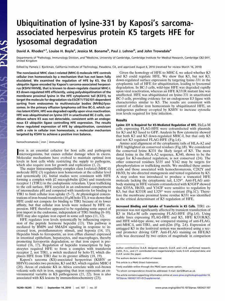
HFE Is Ubiquitinated on K331 and Degraded in Lysosomes. Thesmearing detected in HFE/K5 cells at the 180-min time point wasconsistent with HFE ubiquitination. We looked for ubiquitina-
tion of HFE in K5 cells by IP using anti-FLAG antibody and IBfor ubiquitin (Fig. 4A). HC10 antibody was used similarly for thedetection of ubiquitinated MHC-I. The ladder of higher MWbands indicative of polyubiquitination was detected for bothMHC-I and FLAG-HFE in K5 cells. HFE ubiquitination wasdependent on lysine-331 because the K331R variant was notubiquitinated (Fig. 4A Upper). IB of anti-FLAG IPs showed thatonly HFE H was degraded (Fig. 4A Lower).Ubiquitination of classical MHC-I by K3 leads to degradation
in lysosomes (20, 25). To determine whether HFE shared thisfate, HFE/K5 cells were treated with lysosomal inhibitors andthe effect on HFE investigated (Fig. 4B). Treatment with chlo-roquine was effective at rescuing HFE H, correlating with accu-mulation of a lower MW band, consistent with monoubiquitinatedHFE. Similar results were obtained using ammonium chloride,but the inhibitor of proteosomes MG132 was ineffective (Fig. S2).These results were consistent with the targeting of ubiquitinatedHFE for degradation in lysosomes. Treatment with concanamycinA (conA), a V-type ATPase inhibitor, was less effective, with lessubiquitination detected in conA-treated cells (Fig. 4B). ConA actsby preventing the acidification of endosomes, implying that ef-fective ubiquitination required endosome acidification.
Lysine-63 Linked Polyubiquitinated HFE Recruits TSG101. K5 pro-motes complex lysine-63 linked polyubiquitin chains to down-regulate MHC-I (26). To see whether HFE was regulated simi-larly, we used mutant ubiquitin (Ub) molecules to outcompeteendogenous Ub (Fig. 4D). Rescue of surface FLAG-HFE andMHC-I was detected with Ub K63R, but not with wild-type orK48R Ub variants. We concluded that HFE molecules weretargeted by K5 for lysine-63 linked polyubiquitination.Lysine-63 linked ubiquitination typically targets cell surface
receptors to endolysosomes via the ESCRT pathway. The threeprotein complexes ESCRTI, -II, and -III are recruited sequentiallyto endosomal membranes by ubiquitinated cargo, driving in-ternalization into multivesicular bodies (MVBs), which fuse withlysosomes (27). A key step is recruitment of ESCRT1/TSG101. Todetermine whether ubiquitinated HFE was targeted in this way,we used siRNA to TSG101 (24). FLAG-HFE and MHC-I mole-cules were restored to the cell surface by TSG101 knockdown,implicating the ESCRT pathway in the regulation of ubiquitinatedHFE (Fig. 4 E and F). An alternative model invokes sorting ofubiquitinated cargo from theGolgi byGGAs (Golgi-localized, γ-ear-containing, Arf-binding proteins) (28). siRNA targeting GGAs didnot significantly affect cell surfaceFLAG-HFEorMHC-I expressionin K5 cells (Fig. 4 E and F).
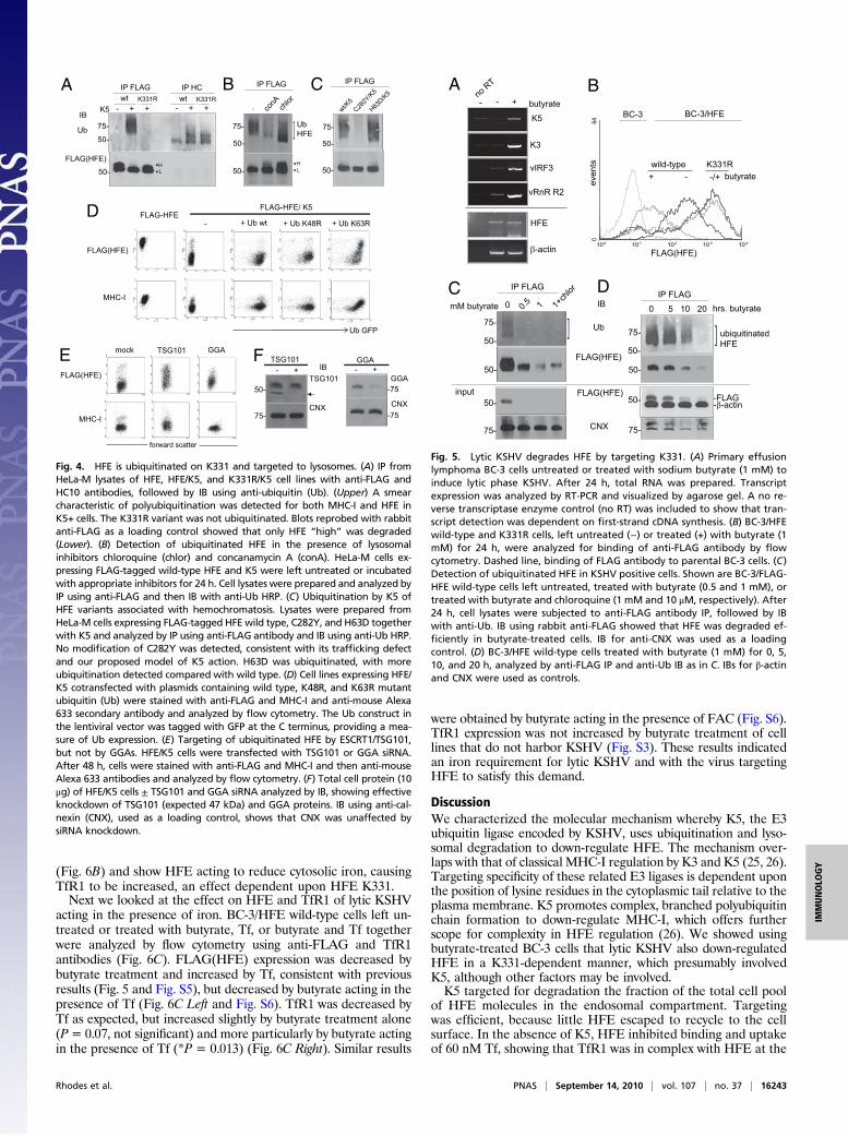
Reactivated KSHV Targets K331 to Degrade HFE. The effect of lyticKSHV on HFE was examined in the primary effusion lymphomacell line BC-3, which is latently infected with KSHV. Sodiumbutyrate treatment of BC-3 cells was used to induce expression oflytic cycle genes, including K5 and K3 (Fig. 5A). KSHV transcriptsvIRF3 and vRnR R2 subunit were also strongly induced, but en-dogenous transcripts β-actin and HFE were not. BC-3 cellsexpressed low levels of HFE mRNA, but no antibody is availableto detect endogenous HFE protein. Therefore we created FLAG-HFE wild-type and K331R BC-3 cells. Butyrate treatment of BC-3/HFE wild-type cells showed down-regulation of anti-FLAGstaining compared with untreated cells, whereas HFE K331R cellswere unaffected (Fig. 5B). FLAG(HFE) staining was not de-creased by butyrate treatment of cell lines that do not harborKSHV (Fig. S3). We noted that surface FLAG staining was notcomparable in untreated cells, as wild-type HFE exhibited in-termediate expression compared with K331R. Expression ofFLAG-HFE in this lentiviral system was monitored using a secondpromoter driving GFP, which showed no variation (Fig. S4).
HFE Is Ubiquitinated in Unactivated BC-3 Cells. We tested the effectof butyrate on HFE ubiquitination, by IP from BC-3/HFE wild-type cells, left untreated, treated with butyrate, or treated withbutyrate and chloroquine. We detected ubiquitinated HFE in
K5GFP
K3GFP
K5GFP
K331RL329*
GFP
328 348
MHC-I
S335A Y342F
FLAG(HFE))
EFH(
GALF
B
C
A
H63D
TfR1
Fig. 1. HFE down-regulation by K5 requires lysine-331. (A) HeLa-M cellsexpressingFLAG-taggedHFEwere transiently transfectedwithK3andK5asGFPfusions. After 48 h, cells were stained with anti-FLAG, MHC-I, and TfR1 primaryand anti-mouse Alexa 633 secondary antibodies and analyzed by flow cytom-etry. (B) Comparisonof the cytosolic tailsofHLA-A2andHFE identifiesconservedamino acids. (C) Conserved residuesweremodifiedby in vitromutagenesis. HFEvariants were expressed in HeLa-M cells and cotransfected with K5GFP. Cellswere stained with anti-FLAG antibody and analyzed by flow cytometry.
Rhodes et al. PNAS | September 14, 2010 | vol. 107 | no. 37 | 16241
IMMUNOLO
GY

untreated BC-3 cells (Fig. 5C). This observation may indicatelow K5 expression in unactivated cells, although K5 transcriptswere not detected by RT-PCR (Fig. 5A). Alternatively, an en-dogenous E3 ligase could be ubiquitinating HFE. Such an ac-tivity would explain the intermediate expression detected by flowcytometry of wild-type HFE compared with K331R (Fig. 5B andFig. S4). An endogenous E3 ligase could be acting upon K331 tocontrol HFE expression, a characteristic distinct from K5, whichtargets HFE for ubiquitination and degradation. In contrast, noFLAG-HFE was detected by IB in butyrate-treated cells, show-ing that HFE was degraded efficiently by lytic KSHV. By IB of
anti-FLAG IP, protein was detectable, with degradation of HFEby increasing butyrate concentration. Chloroquine rescued someprotein as assessed by band intensity relative to control IB forcalnexin (CNX).In similar IP experiments, we examined the effect of 1 mM
butyrate over a 20-h time course (Fig. 5D). We detected ubiq-uitinated HFE in untreated BC-3/HFE wild-type cells and degra-dation of HFE over time, compared with IB for β-actin and CNX.These data are consistent with an endogenous E3 ligase ubiquiti-nating HFE in untreated BC-3 cells, with K5 targeting HFE fordegradation upon KSHV induction. Alternatively, ubiquitinationin untreated cells is by K5 and other factors are induced by buty-rate, which targets HFE molecules for degradation.
Regulation of TfR1 by Iron in HFE Wild-Type and K331R Cells. KSHVpresumably down-regulates HFE to affect iron homeostasis. Welooked for evidence for this by monitoring changes in TfR1 inBC-3 cells. TfR1 expression is controlled at the posttranscrip-tional level by cytosolic iron acting to modulate the IRE-BP sys-tem (29). Low intracellular iron leads to stabilization of TfR1mRNA and increased translation (8).The regulation of TfR1 in BC-3, BC-3/HFE wild-type, and
K331R cells, either left untreated or treated with the ferric ironchelator desferrioxamine (DFO), was compared with the re-sponse to iron delivery by ferric ammonium citrate (FAC) orholo-transferrin (Tf). Untreated HFE wild-type cells showedincreased TfR1 surface expression compared with BC-3 andK331R cells (Fig. 6A Left). This increase in TfR1 was mimickedby DFO treatment (Fig. 6A Center Left) indicative of low cyto-solic iron. In response to iron delivery by FAC, the cell linesresponded by decreasing TfR1 (Fig. 6A Center Right). In re-sponse to Tf, TfR1 levels were again reduced, but the responsewas less profound in the HFE wild-type cells (Fig. 6A Right).These observations were consistent over multiple experiments
TfR1
M-aleH.1
EFH-GALF.25K/EFH.3
5K/R133K.4
12
31
23
12
3
K5GFP FLAG(HFE) merge
TfR1 merge
MHC-IFLAGFLAFLA
K5GFP
A
B
stneve
K5GFP Tf merge
0
20
40
60
80
100
120
0
5
10
15
20
K5GFP
FLAG(HFE)
FLAG(HFE) Tf
K5- K5+llec/ecne cseroulfnae
M)stinu
yrarti bra (
C
D
Tf binding
Tf uptake
Fig. 2. K5 down-regulation of HFE but not TfR1. (A) Com-parison of surface staining for anti-FLAG, MHC-I, β2m, andTfR1 antibodies by flow cytometry. Cell lines: 1, HeLa-M(shaded histogram); 2, HeLa FLAG-HFE plus empty lentiviralvector (red); 3, HFE/K5 (green); 4, K331R/K5 (blue). Dashedhistogram, isotype control. (B) A mixed population of FLAG-HFE and HFE/K5 HeLa-M cells were seeded on coverslips. After24 h, cells were fixed and stained with anti-FLAG (Upper) andanti-TfR1 (Lower) and anti-mouse Alexa 568 antibodies andthen analyzed by confocal microscopy. Merged images: K5/GFP (green) and anti-FLAG and TfR1 (red). (C) FLAG-HFE andHFE/K5 HeLa-M cells growing on coverslips were processed forconfocal microscopy as in B, except that Alexa 633 holo-transferrin (60 nM Tf) was used. (Upper) Anti-FLAG andtransferrin binding to fixed cells, with K5GFP. (Lower) Anti-FLAG and transferrin uptake after 1 h at 37 °C. Cells were thenfixed and permeabilized, and then secondary anti-mouseAlexa 568 antibody was applied. Merged images: (Upper) K5/GFP (green), anti-FLAG (red), and transferrin (purple); (Lower)anti-FLAG (red) and transferrin (green). (D) Images from Cwere analyzed using NIH Image software and relative fluo-rescence per cell was determined. Anti-FLAG staining in K5+cells represented a decrease in relative fluorescence of 80%over K5-negative cells. An increase in Tf binding of 40% wasdetected in K5+ cells. Error bars represent SD by analysis offive fields per image.
0 45 180 chase (min)0 45 180
TfR1
HFE
b2m
..HL
*
1st IP b2m- + K5
97-
66-
45-
14-
FLAG(HFE)
..HL
..HL
2nd IP
MHC-I
MHC-I
Fig. 3. K5 targets HFE “high” from a post-Golgi compartment. FLAG-HFEplus empty lentiviral vector and HFE/K5 cells were metabolically labeled andsubjected to primary IP using anti-β2m at different chase time points andthen secondary IP using anti-MHC-I and anti-FLAG antibodies and analyzedby SDS/PAGE. The appearance of the HFE high form was disrupted by K5expression, with smearing toward higher molecular weight forms (indicatedby *). Size markers are in kilodaltons.
16242 | www.pnas.org/cgi/doi/10.1073/pnas.1003421107 Rhodes et al.
(Fig. 6B) and show HFE acting to reduce cytosolic iron, causingTfR1 to be increased, an effect dependent upon HFE K331.Next we looked at the effect on HFE and TfR1 of lytic KSHV
acting in the presence of iron. BC-3/HFE wild-type cells left un-treated or treated with butyrate, Tf, or butyrate and Tf togetherwere analyzed by flow cytometry using anti-FLAG and TfR1antibodies (Fig. 6C). FLAG(HFE) expression was decreased bybutyrate treatment and increased by Tf, consistent with previousresults (Fig. 5 and Fig. S5), but decreased by butyrate acting in thepresence of Tf (Fig. 6C Left and Fig. S6). TfR1 was decreased byTf as expected, but increased slightly by butyrate treatment alone(P= 0.07, not significant) and more particularly by butyrate actingin the presence of Tf (*P = 0.013) (Fig. 6C Right). Similar results
were obtained by butyrate acting in the presence of FAC (Fig. S6).TfR1 expression was not increased by butyrate treatment of celllines that do not harbor KSHV (Fig. S3). These results indicatedan iron requirement for lytic KSHV and with the virus targetingHFE to satisfy this demand.
DiscussionWe characterized the molecular mechanism whereby K5, the E3ubiquitin ligase encoded by KSHV, uses ubiquitination and lyso-somal degradation to down-regulate HFE. The mechanism over-laps with that of classical MHC-I regulation by K3 and K5 (25, 26).Targeting specificity of these related E3 ligases is dependent uponthe position of lysine residues in the cytoplasmic tail relative to theplasma membrane. K5 promotes complex, branched polyubiquitinchain formation to down-regulate MHC-I, which offers furtherscope for complexity in HFE regulation (26). We showed usingbutyrate-treated BC-3 cells that lytic KSHV also down-regulatedHFE in a K331-dependent manner, which presumably involvedK5, although other factors may be involved.K5 targeted for degradation the fraction of the total cell pool
of HFE molecules in the endosomal compartment. Targetingwas efficient, because little HFE escaped to recycle to the cellsurface. In the absence of K5, HFE inhibited binding and uptakeof 60 nM Tf, showing that TfR1 was in complex with HFE at the
FLAG-HFE
MHC-I
FLAG-HFE/ K5
Ub GFP
- + Ub wt + Ub K63R+ Ub K48R
TSG101
CNX
50-
75-
- +
FLAG(HFE)
TSG101
FLAG(HFE)
MHC-I
GGAmock
forward scatter
IBTSG101F
D
E
CNX
GGA- +GGA
IB
FLAG(HFE)
Ub
IP FLAG IP HCwt wt
75-
50-
50-
K331R K331RK5 - + + - + +
..HL
IP FLAG
75-
50-
wt/K5
C282Y
/K5
H63D/K
5
-
IP FLAG
conA ch
lor
75-
50-
50-..H
L
50-
CBA
UbHFE]
-75
-75
Fig. 4. HFE is ubiquitinated on K331 and targeted to lysosomes. (A) IP fromHeLa-M lysates of HFE, HFE/K5, and K331R/K5 cell lines with anti-FLAG andHC10 antibodies, followed by IB using anti-ubiquitin (Ub). (Upper) A smearcharacteristic of polyubiquitination was detected for both MHC-I and HFE inK5+ cells. The K331R variant was not ubiquitinated. Blots reprobed with rabbitanti-FLAG as a loading control showed that only HFE “high” was degraded(Lower). (B) Detection of ubiquitinated HFE in the presence of lysosomalinhibitors chloroquine (chlor) and concanamycin A (conA). HeLa-M cells ex-pressing FLAG-tagged wild-type HFE and K5 were left untreated or incubatedwith appropriate inhibitors for 24 h. Cell lysates were prepared and analyzed byIP using anti-FLAG and then IB with anti-Ub HRP. (C) Ubiquitination by K5 ofHFE variants associated with hemochromatosis. Lysates were prepared fromHeLa-M cells expressing FLAG-tagged HFE wild type, C282Y, and H63D togetherwith K5 and analyzed by IP using anti-FLAG antibody and IB using anti-Ub HRP.No modification of C282Y was detected, consistent with its trafficking defectand our proposed model of K5 action. H63D was ubiquitinated, with moreubiquitination detected compared with wild type. (D) Cell lines expressing HFE/K5 cotransfected with plasmids containing wild type, K48R, and K63R mutantubiquitin (Ub) were stained with anti-FLAG and MHC-I and anti-mouse Alexa633 secondary antibody and analyzed by flow cytometry. The Ub construct inthe lentiviral vector was tagged with GFP at the C terminus, providing a mea-sure of Ub expression. (E) Targeting of ubiquitinated HFE by ESCRT1/TSG101,but not by GGAs. HFE/K5 cells were transfected with TSG101 or GGA siRNA.After 48 h, cells were stained with anti-FLAG and MHC-I and then anti-mouseAlexa 633 antibodies and analyzed by flow cytometry. (F) Total cell protein (10μg) of HFE/K5 cells ± TSG101 and GGA siRNA analyzed by IB, showing effectiveknockdown of TSG101 (expected 47 kDa) and GGA proteins. IB using anti-cal-nexin (CNX), used as a loading control, shows that CNX was unaffected bysiRNA knockdown.
50-
50-
IP FLAG
]75-
75-
1+ch
lor
0 0.5 1
50-input
mM butyrate
-+ butyrate
BC-3/HFEBC-3
FLAG(HFE)
A B
K5
K3
b-actin
no RT
- + butyrate
C
-
vIRF3 wild-type K331R-/+
vRnR R2
HFE
even
ts
100 101 102 103 104
064
50-
50-
IB
FLAG(HFE)
UbubiquitinatedHFE]75-
CNX
-FLAG50- -b-actin
IP FLAG0 5 10 20 hrs. butyrate
D
FLAG(HFE)
75-
Fig. 5. Lytic KSHV degrades HFE by targeting K331. (A) Primary effusionlymphoma BC-3 cells untreated or treated with sodium butyrate (1 mM) toinduce lytic phase KSHV. After 24 h, total RNA was prepared. Transcriptexpression was analyzed by RT-PCR and visualized by agarose gel. A no re-verse transcriptase enzyme control (no RT) was included to show that tran-script detection was dependent on first-strand cDNA synthesis. (B) BC-3/HFEwild-type and K331R cells, left untreated (−) or treated (+) with butyrate (1mM) for 24 h, were analyzed for binding of anti-FLAG antibody by flowcytometry. Dashed line, binding of FLAG antibody to parental BC-3 cells. (C)Detection of ubiquitinated HFE in KSHV positive cells. Shown are BC-3/FLAG-HFE wild-type cells left untreated, treated with butyrate (0.5 and 1 mM), ortreated with butyrate and chloroquine (1 mM and 10 μM, respectively). After24 h, cell lysates were subjected to anti-FLAG antibody IP, followed by IBwith anti-Ub. IB using rabbit anti-FLAG showed that HFE was degraded ef-ficiently in butyrate-treated cells. IB for anti-CNX was used as a loadingcontrol. (D) BC-3/HFE wild-type cells treated with butyrate (1 mM) for 0, 5,10, and 20 h, analyzed by anti-FLAG IP and anti-Ub IB as in C. IBs for β-actinand CNX were used as controls.
Rhodes et al. PNAS | September 14, 2010 | vol. 107 | no. 37 | 16243
IMMUNOLO
GY

plasma membrane. In K5 cells, binding of Tf increased, sug-gesting that K5 was targeting HFE molecules dissociated fromTfR1. Structural studies show that binding of the two moleculesvia their extracellular domains is pH dependent (5). Con-canamycin A, which inhibits endosome acidification, preventedeffective K5-mediated ubiquitination of HFE (Fig. 4B). Our datatherefore suggest a model whereby pH-dependent dissociation ofthe HFE/TfR1 complex occurs in early endosomes, allowing forK5-mediated ubiquitination on TfR1-free HFE. Instead ofrecycling to the cell surface, ubiquitinated HFE is sorted toMVBs via ESCRT machinery and degraded in lysosomes.Down-regulation of classical MHC-I by K3/K5 serves to mask-
virus infected cells from surveillance by circulating cytotoxic Tand NK cells (30). K5 down-regulation of HFE could be actingsimilarly to prevent engagement of another receptor. Alterna-tively, KSHV may manipulate iron levels to aid its replication.Induction of lytic KSHV required iron, as evidenced by the in-crease in TfR1 on viral induction (Fig. 6C). By inspection of theKSHV genome (NC_009333), the only molecule with an obviousiron requirement is orf60 ribonucleotide reductase R2 subunit(vRnR R2), the enzyme that provides nucleotide substrates forDNA biosynthesis. Transcripts for this gene were induced in lyticphase (Fig. 5A). It is possible that KSHV down-regulates HFE toincrease iron availability for DNA biosynthesis.By comparing the response to iron delivery of wild-type HFE
and K331R BC-3 cells, we detected differences in the regulationof TfR1, which was used to monitor changes in cytosolic iron (Fig.6). The increase in TfR1 by expression of wild-type HFE alonewas indicative of low cytosolic iron, presumably by increased ironexport. The response of wild-type HFE cells to 10 μMholo-Tf, butnot to FAC, also indicated a low iron phenotype compared withparental BC-3 and K331R cells, possibly as a result of controllediron uptake. Taken together, the results suggest that HFE acts to
balance iron import and export, effects dependent on lysine-331and iron delivery from Tf. The mechanism(s) did not rely on in-hibition of Tf binding, because TfR1 was increased by HFE ex-pression in untreated cells. Physiological concentrations of Tfwere used, as these have been shown to outcompete HFE forTfR1 binding (6) and K331R cells did not inhibit iron delivery byTf, despite high cell surface expression. These observations aremore consistent with regulation of iron import/export in theendosome, with the transporter DMT1/Nramp2 or ferric re-ductase being potential targets, as proposed by other workers (6).Ubiquitination of HFE lysine-331 leading to intermediate HFE
expression was detected in unactivated BC-3 cells. Expression ofK5 at low levels or of an endogenous E3 ligase with characteristicssimilar toK5would account for this observation.Moreworkwill berequired to resolve these alternatives. HFE protein has beendetected in few tissues (31–33) and it will also be important to as-sess whether HFE ubiquitination represents a general mechanismcontrolling HFE expression and iron homeostasis in these sites.
Materials and MethodsAntibodies. Monoclonal M2 and rabbit polyclonal anti-FLAG (Sigma), anti-HLA class I W6/32 and HC10 (lab stocks), anti-β2 microglobulin (Dako), anti-EEA1 (Molecular Probes), anti-TfR1 (Pharmingen), anti-calnexin (Stressgen),anti-TSG101 (Abcam), and anti-ubiquitin HRP (P4D1; Santa Cruz) were used.Secondary antibodies were goat anti-mouse Alexa 488, 568, and 647 andgoat anti-rabbit Alexa 488, 568, and 647 (Molecular Probes) and goat anti-rabbit and anti-mouse as HRP conjugates (Dako).
DNA Constructs. HFE cDNA was cloned into the pFLAG vector (Sigma) usingBglII/BamHI to produce FLAG-HFE. Primers were HFE.F agatctgcgttca-cactctctgcac and HFE.R ggatcctcactcacgttcagctaagac. HFE variants wereproduced by site-directed mutagenesis. K3 and K5 as GFP fusion constructs(24) and the pHRsin lentivirus system were used (34). FLAG-HFE was cloned
0
100
200
300
400
500 BC-3K331RHFE wild-type
0
50
100
150
200
250
0
50
100
150
200
250
300
100 101 102 103 104
064
100 101 102 103 104
064A
TfR1
FAC 1mM Tf 10mM
iso
untreated
100 101 102 103 104
064
BC-3HFE wild-typeK331R
even
ts
100 101 102 103 104
064 DFO 10mM
B
FLA
G(H
FE) e
xpre
ssio
n (M
FI)
TfR
1 ex
pres
sion
(MFI
)
untre
ated
butyr
ate Tf
Tf+buty
rate
p<0.001
p<0.001p=0.07
p=0.013
******
p<0.001
*
***
untre
ated DFO FAC Tf
TfR
1 ex
pres
sion
(MFI
)
C
TfR1 TfR1 TfR1
untre
ated
butyr
ate Tf
Tf+buty
rate
p<0.001
***- /+ Tf
Fig. 6. Regulation of TfR1 by iron in HFE wild-type and K331R cells. (A) Comparison of TfR1 expression on parental BC-3 and cells expressing wild-type andK331R HFE, left untreated (Left) and treated with desferrioxamine (DFO, Center Left), ferric ammonium citrate (FAC, Center Right), and holo-transferrin (Tf,Right). Cells were treated for 24 h, stained with anti-TfR1 and anti-mouse Alexa 633 antibodies, and analyzed by flow cytometry. Binding of isotype control(iso) is shown. (B) Chart shows mean fluorescence values for TfR1 expression from three experiments carried out as in A (except DFO treatment, whichrepresents duplicate data). Error bars show SD. (C) Effect of lytic KSHV acting in the presence of iron on expression of FLAG(HFE) (Left) and TfR1 (Right). BC-3/HFE wild-type cells were left untreated or treated with butyrate, Tf, or butyrate and Tf together for 24 h; stained with anti-FLAG and anti-TfR1 antibodies;and then analyzed by flow cytometry. Charts showmean fluorescence from triplicate experiments. P values show significance tested by paired Student’s t test.A P value <0.05 was considered significant.
16244 | www.pnas.org/cgi/doi/10.1073/pnas.1003421107 Rhodes et al.

into the pHRsin vector. Internal NotI and BamHI sites were removed byrounds of restriction digestion followed by T4 DNA polymerase treatment tocreate blunt ends, which were ligated. Resulting inserts were resequencedand then subcloned using BamHI/NotI into pHRsin.
Tissue Culture, Transfection, and Flow Cytometry. HeLa-M and BC-3 cells weremaintained in RPMI1640 medium plus 10% FCS, pen/strep (100 units), and L-glutamine (200 mM). HeLa cells growing in six-well plates were transfectedusing Fugene (Roche). Stable lines were produced by cell sorting of anti-FLAGpositive cells maintained in medium supplemented with G418 (1 mg/mL).FACScalibur was used for flow cytometry. For lentiviral transduction, 293Tcells were cotranfected with packaging vectors pCMVR8.91 and pMDG to-gether with pHRsin UbEm lentivirus plasmids. After 48 h, culture supernatantswere harvested, filtered (0.2-μm filter), and applied to cells. Reagents for de-pletion of TSG101 by siRNAwere described previously (24). HFE/K5 cells growingin six-well plates in complete medium lacking pen/strep were transfected with200 pmol TSG101 siRNA (Dharmacon), using Dharmafect 1. After 48 h cells wereanalyzed by flow cytometry and by IB to confirm TSG101 depletion. Depletionof GGAs was achieved similarly except using Smartpool siRNA (Dharmacon)targeted to GGAs (kindly provided by J. Hirst andM. S. Robinson, Department ofClinical Biochemistry, Cambridge University, Cambridge, UK). For induction ofKSHV, BC-3 cells were treated with sodium butyrate. Human holo-transferrinand ferric ammonium chloride were used. Desferrioxamine was a gift fromT. M. Cox (Cambridge University, UK).
Metabolic Labeling, Immunoprecipitation, and Immunoblot. HeLa-M cells werestarved for 1 h in methionine/cysteine-free medium and then labeled with[35S]methionine/cysteine for 20 min. Cells were removed to unlabeledcomplete medium for the indicated times to chase. Cells were lysed in IPbuffer [50 mM Tris-Cl (pH 7.5), 150 mM NaCl, 0.5% Triton-X, 2 mM PMSF, 5mM iodoacetamide, EDTA-free protease inhibitor] for 10 min at 4 °C andprecleared using protein A-Sepharose, followed by IP using the indicatedantibodies and protein A. For re-IP, sample beads from primary IP wereresuspended in 1% SDS, incubated at 70 °C for 5 min, and diluted to 0.1% SDSusing IP buffer, before adding secondary antibodies and protein A. Sampleswere treated with endoglycosidase H or PNGaseF, before analysis on 10% SDS/
PAGE. Gels were fixed (10% acetic acid, 20% methanol) and then dried andexposed to X-ray film (Kodak BiomaxMR). For IBs, proteins were transferred toPVDF membrane, blocked with dried milk (5% Marvel/PBS, 0.1% Tween20)and incubated for 1 h with primary and HRP-conjugated secondary antibodies.Blots were visualized with ECL reagent. The following inhibitors were used:ammonium chloride (50 mM), chloroquine (100 μM), MG132 (10 μM), andconcanamycin A (50 nM). For ubiquitination assays, 2 × 10⁶ cells were lysed in100 μl IP buffer, containing 1% SDS and 100 units of benzonase, and incubatedat 70 °C for 10 min. Lysates were diluted to 1 ml with IP buffer and precleared,followed by IP at 4 °C using anti-FLAG and protein A.
Antibody and Transferrin Binding and Internalization Assay. HeLa-M cellsexpressing FLAG-HFE with/without K5GFPwere grown on coverslips. Cells werefixed for 10 min (3% parafomaldehyde/1× PBS) and treated for 5 min in 0.1%Triton-X100 before staining with primary and secondary antibodies. Coverslipswere mounted (Fluoromount G) and cells were visualized using confocal mi-croscopy (Zeiss LSM510 META), using a 63× objective, and analyzed using LSMImage Browser software (Zeiss) before saving in Adobe Photoshop. For in-ternalization, cells growing on coverslips were stained with anti-FLAG M2antibody and directly labeled holo-transferrin (Tf Alexa633; Molecular Probes)for 30 min at 4 °C, washed twice with cold 1× PBS, and then incubated for 1 hat 37 °C. Samples were removed into 3% paraformaldehye/1× PBS to fix andwashed twice (1× PBS) before staining with appropriate secondary antibody.
RT-PCR. Total RNA was purified from cell pellets using TriReagent. TranscriptswereamplifiedusingtheOne-TubeRT-PCRSystem(Stratagene).PrimerswereK5Ftccaaggacgtagaagaggg K5R caccggcttttttgtgggcgc, K3F atggaagatgaggatgttccK3R ggagacactataagccccatcg, vIRF3F atggcgggacgcaggcttacc vIRF3R gtcatcaca-tgtaactgaacgc, VRNRF ctgtatacaagcgatcacgacgg VRNRR caaatcgtcagtcacacacg-tgg, and HFEF atgggcccgcgagccaggccgg HFER ctcacgttcagctaagacgtag.
ACKNOWLEDGMENTS. We thank Simon McCallum and Anna Petrunkina–Harrison (MoFlow), Matthew Gratian and Mark Bowen (microscopy), andDr. M. R. Wills (Department of Medicine, Cambridge University) for help withBC-3 experiments. We also thank Drs. Adrian Kelly and Howard Davidsonfor helpful discussion. This work was supported by the Wellcome Trust.
1. Schaible UE, Kaufmann SH (2004) Iron and microbial infection. Nat Rev Microbiol 2:946–953.
2. Drakesmith H, Prentice A (2008) Viral infection and iron metabolism. Nat RevMicrobiol 6:541–552.
3. Feder JN, et al. (1996) A novel MHC class I-like gene is mutated in patients withhereditary haemochromatosis. Nat Genet 13:399–408.
4. Hentze MW, Muckenthaler MU, Andrews NC (2004) Balancing acts: Molecular controlof mammalian iron metabolism. Cell 117:285–297.
5. Lebrón JA, et al. (1998) Crystal structure of the hemochromatosis protein HFE andcharacterization of its interaction with transferrin receptor. Cell 93:111–123.
6. Roy CN, Penny DM, Feder JN, Enns CA (1999) The hereditary hemochromatosisprotein, HFE, specifically regulates transferrin-mediated iron uptake in HeLa cells.J Biol Chem 274:9022–9028.
7. Giannetti AM, Björkman PJ (2004) HFE and transferrin directly compete for transferrinreceptor in solution and at the cell surface. J Biol Chem 279:25866–25875.
8. Riedel HD, et al. (1999) HFE downregulates iron uptake from transferrin and inducesiron-regulatory protein activity in stably transfected cells. Blood 94:3915–3921.
9. Zhang AS, Davies PS, Carlson HL, Enns CA (2003) Mechanisms of HFE-inducedregulation of iron homeostasis: Insights from the W81A HFE mutation. Proc Natl AcadSci USA 100:9500–9505.
10. Carlson H, Zhang AS, Fleming WH, Enns CA (2005) The hereditary hemochromatosisprotein, HFE, lowers intracellular iron levels independently of transferrin receptor 1 inTRVb cells. Blood 105:2564–2570.
11. Drakesmith H, et al. (2002) The hemochromatosis protein HFE inhibits iron exportfrom macrophages. Proc Natl Acad Sci USA 99:15602–15607.
12. Davies PS, Enns CA (2004) Expression of the hereditary hemochromatosis protein HFEincreases ferritin levels by inhibiting iron export in HT29 cells. J Biol Chem 279:25085–25092.
13. BridleKR, et al. (2003)Disruptedhepcidin regulation inHFE-associatedhaemochromatosisand the liver as a regulator of body iron homoeostasis. Lancet 361:669–673.
14. Wang RH, et al. (2005) A role of SMAD4 in iron metabolism through the positiveregulation of hepcidin expression. Cell Metab 2:399–409.
15. Andriopoulos B, Jr, et al. (2009) BMP6 is a key endogenous regulator of hepcidinexpression and iron metabolism. Nat Genet 41:482–487.
16. Park CH, Valore EV, Waring AJ, Ganz T (2001) Hepcidin, a urinary antimicrobialpeptide synthesized in the liver. J Biol Chem 276:7806–7810.
17. Nemeth E, et al. (2004) Hepcidin regulates cellular iron efflux by binding toferroportin and inducing its internalization. Science 306:2090–2093.
18. Gao J, et al. (2009) Interaction of the hereditary hemochromatosis protein HFE withtransferrin receptor 2 is required for transferrin-induced hepcidin expression. CellMetab 9:217–227.
19. Schmidt PJ, Toran PT, Giannetti AM, Bjorkman PJ, Andrews NC (2008) The transferrinreceptor modulates Hfe-dependent regulation of hepcidin expression. Cell Metab 7:205–214.
20. Coscoy L, Ganem D (2000) Kaposi’s sarcoma-associated herpesvirus encodes twoproteins that block cell surface display of MHC class I chains by enhancing theirendocytosis. Proc Natl Acad Sci USA 97:8051–8056.
21. Simonart T (2004) Iron: A target for the management of Kaposi’s sarcoma? BMCCancer 4:1–8.
22. Ziegler JL (1993) Endemic Kaposi’s sarcoma in Africa and local volcanic soils. Lancet342:1348–1351.
23. Wada DA, Perkins SL, Tripp S, Coffin CM, Florell SR (2007) Human herpesvirus 8 andiron staining are useful in differentiating Kaposi sarcoma from interstitial granulomaannulare. Am J Clin Pathol 127:263–270.
24. Hewitt EW, et al. (2002) Ubiquitylation of MHC class I by the K3 viral protein signalsinternalization and TSG101-dependent degradation. EMBO J 21:2418–2429.
25. Duncan LM, et al. (2006) Lysine-63-linked ubiquitination is required forendolysosomal degradation of class I molecules. EMBO J 25:1635–1645.
26. Boname JM, et al. (2010) Efficient internalization of MHC I requires lysine-11 andlysine-63 mixed linkage polyubiquitin chains. Traffic 11:210–220.
27. Hurley JH (2008) ESCRT complexes and the biogenesis of multivesicular bodies. CurrOpin Cell Biol 20:4–11.
28. Pelham HR (2004) Membrane traffic: GGAs sort ubiquitin. Curr Biol 14:R357–R359.29. Rouault TA (2006) The role of iron regulatory proteins in mammalian iron
homeostasis and disease. Nat Chem Biol 2:406–414.30. Coscoy L (2007) Immune evasion by Kaposi’s sarcoma-associated herpesvirus. Nat Rev
Immunol 7:391–401.31. Waheed A, et al. (1999) Association of HFE protein with transferrin receptor in crypt
enterocytes of human duodenum. Proc Natl Acad Sci USA 96:1579–1584.32. Parkkila S, et al. (1997) Association of the transferrin receptor in human placenta with
HFE, the protein defective in hereditary hemochromatosis. Proc Natl Acad Sci USA 94:13198–13202.
33. Bastin J, Drakesmith H, Rees M, Sargent I, Townsend A (2006) Localisation of proteinsof iron metabolism in the human placenta and liver. Br J Haematol 134:532–543.
34. Demaison C, et al. (2002) High-level transduction and gene expression inhematopoietic repopulating cells using a human immunodeficiency [correction ofimmunodeficiency] virus type 1-based lentiviral vector containing an internal spleenfocus forming virus promoter. Hum Gene Ther 13:803–813.
Rhodes et al. PNAS | September 14, 2010 | vol. 107 | no. 37 | 16245
IMMUNOLO
GY