types of microscopes simple microscope - contains only one lens. ex. magnifying glass compound light...
TRANSCRIPT

Types of Microscopes
Simple Microscope - contains only one lens.ex. magnifying glass
Compound Light Microscope - a system of two lens that work together.
Electron Microscopes - uses electrons to provide detailed views of specimens
ex. TEM and SEM
Dissecting Microscope - allows for the viewing of specimens without the use of a slide.

Label the following parts of the Microscope
Ocular / Eyepiece
Arm
Bodytube
Revolving Nose Piece
Low Power Objective Lens
Medium Power Objective Lens
High Power Objective Lens
Stage Clips
Diaphragm
Light Source
Stage
Coarse Adjustment Knob
Fine Adjustment Knob
Base

Calculating Magnification
To calculate magnification in a compound light microscope you multiply the magnification power of the objective by the magnification power of the ocular.
Example:
Ocular = 10 x Objective = 4 x
Magnification = 10 x * 4 x = 40 x
Calculate the magnification values in the following situations
1) Objective = 10 x Ocular = 10 x100 x
2) Objective = 40 x Ocular = 10 x400 x
3) Objective = 100 x Ocular = 10 x1000 x

Total Magnification Example
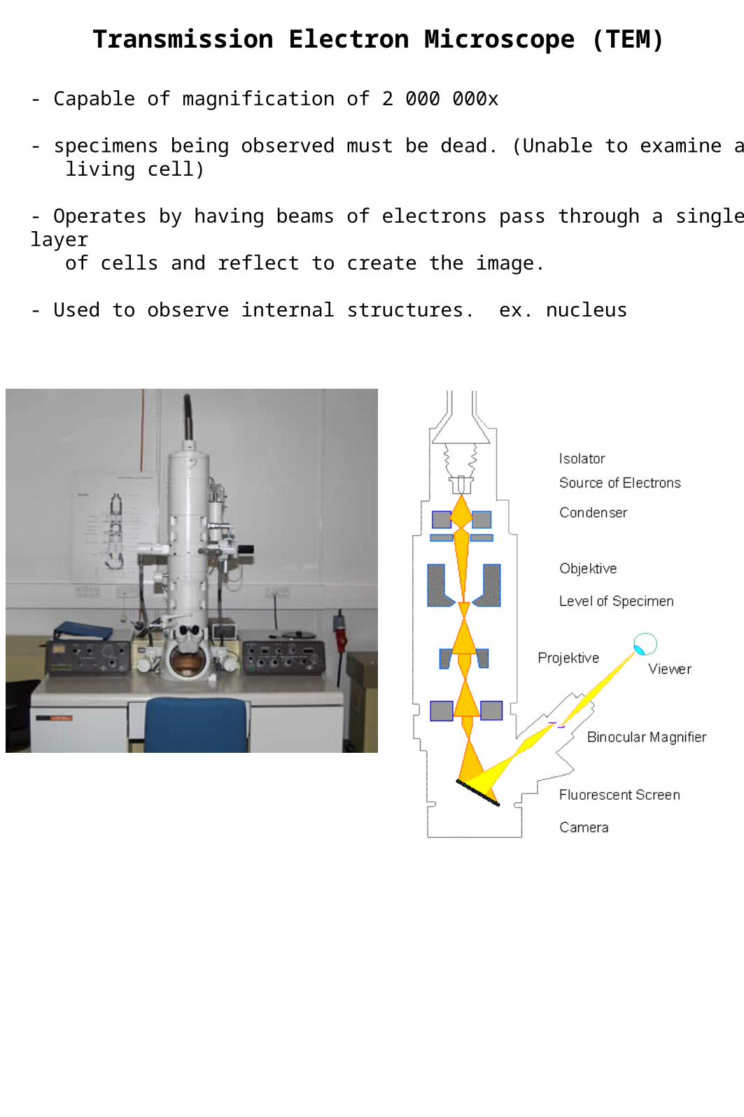
Transmission Electron Microscope (TEM)
- Capable of magnification of 2 000 000x
- specimens being observed must be dead. (Unable to examine a living cell)
- Operates by having beams of electrons pass through a single layer of cells and reflect to create the image.
- Used to observe internal structures. ex. nucleus

TEM SPECIMEN IMAGES

SCANNING ELECTRON MICROSCOPE (SEM)
- Capable of magnification up to 100 000x
- Specimens must be dead.
- Used to observe the external structures of a specimen. (Electrons are reflected off the surface of the specimen being observed.)

SEM Specimen Image
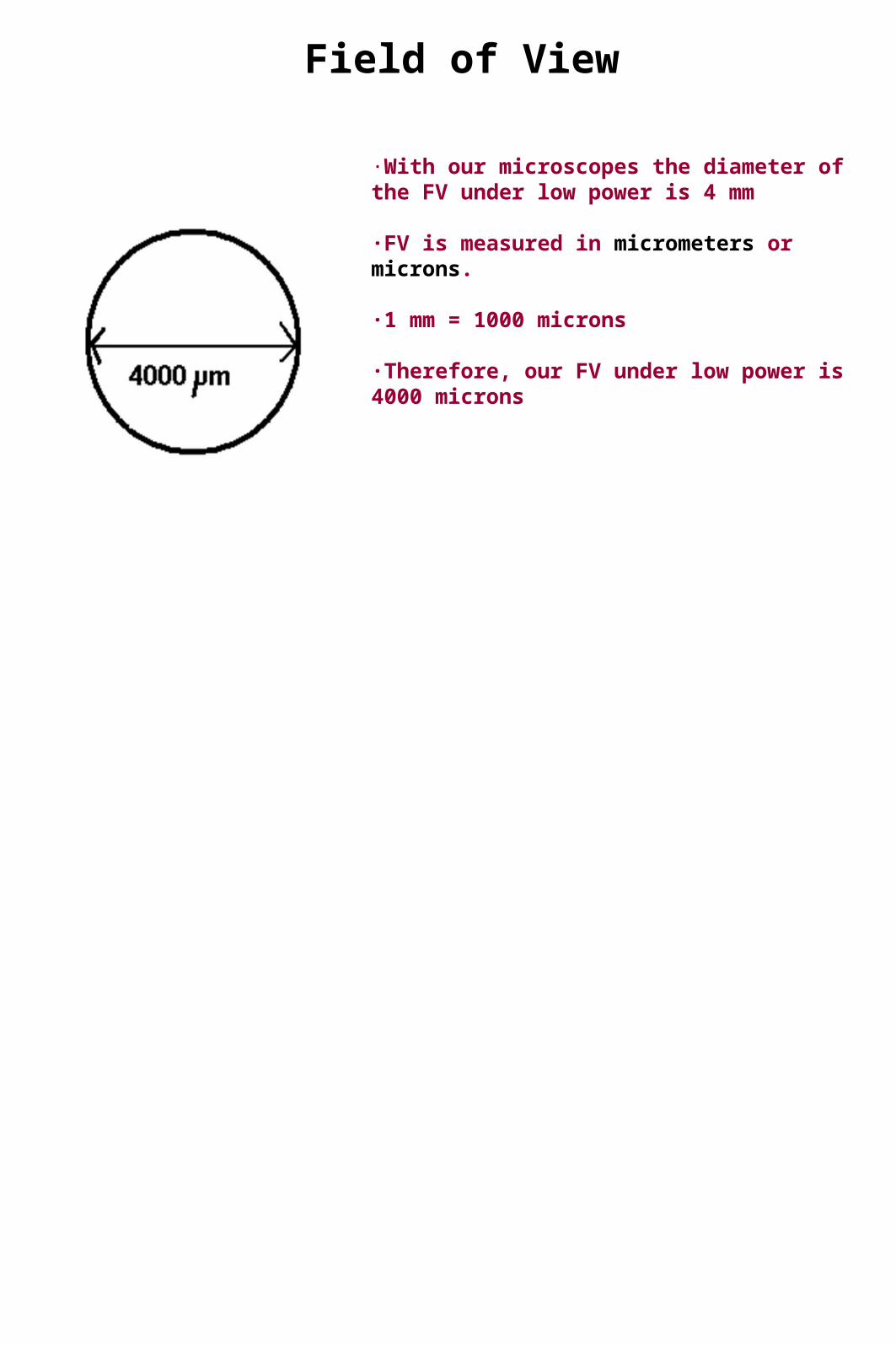
Field of View
·With our microscopes the diameter of the FV under low power is 4 mm
·FV is measured in micrometers or microns.
·1 mm = 1000 microns
·Therefore, our FV under low power is 4000 microns


Calculating Changes in the Field of View
- Each time magnification is increased the field of view decreases.
- To determine the size of the new field of view:
1) Calculate the increase in magnification.
ex. from low power (4x) to high power (10x) magnification increase = 10x/4x
magnification increase = 2.5 x
2) To calculate the amount the field of view has decreased by divide the old field of
view size by the value of the increase in magnification.
ex. 4000um/2.5x = 1600um

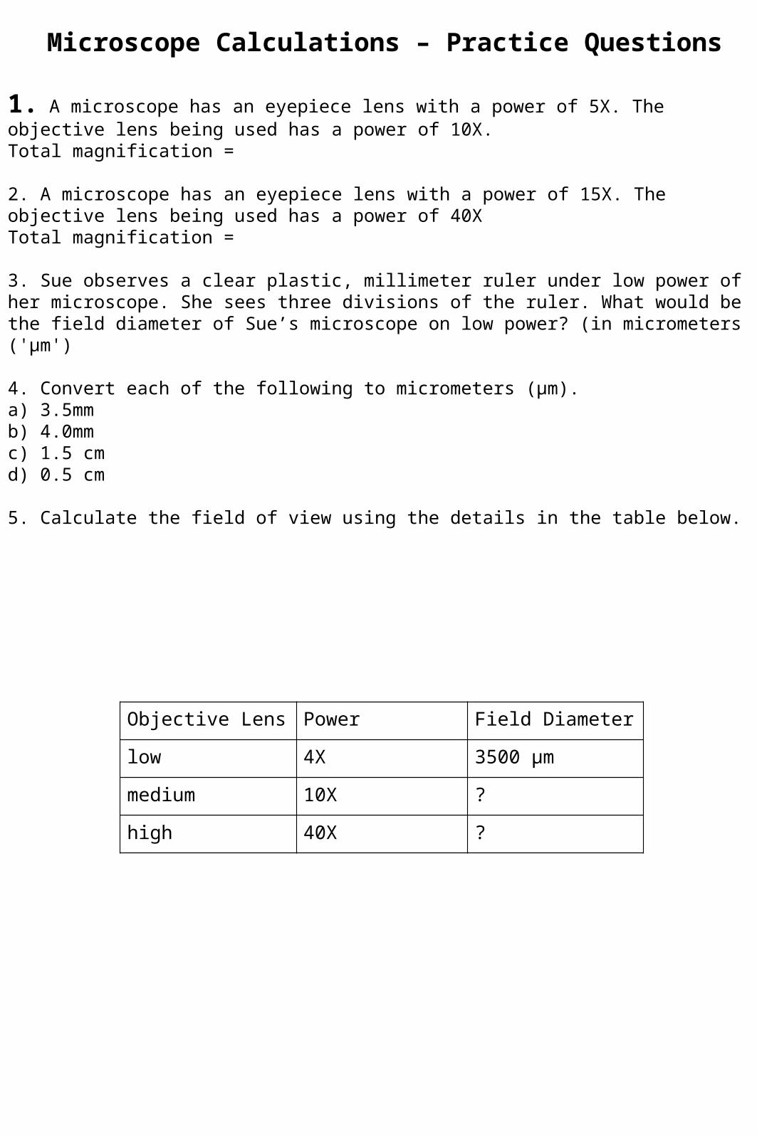
Objective Lens Power Field Diameter
low 4X 3500 µm
medium 10X ?
high 40X ?
Microscope Calculations – Practice Questions
1. A microscope has an eyepiece lens with a power of 5X. The objective lens being used has a power of 10X.Total magnification =
2. A microscope has an eyepiece lens with a power of 15X. The objective lens being used has a power of 40XTotal magnification =
3. Sue observes a clear plastic, millimeter ruler under low power of her microscope. She sees three divisions of the ruler. What would be the field diameter of Sue’s microscope on low power? (in micrometers ('µm')
4. Convert each of the following to micrometers (µm).a) 3.5mmb) 4.0mmc) 1.5 cmd) 0.5 cm
5. Calculate the field of view using the details in the table below.
