tumor cell independent estrogen signaling drives disease...
TRANSCRIPT

OF1 | CANCER DISCOVERY JANUARY 2017 www.aacrjournals.org
RESEARCH ARTICLE
ABSTRACT The role of estrogens in antitumor immunity remains poorly understood. Here, we show that estrogen signaling accelerates the progression of different estrogen-
insensitive tumor models by contributing to deregulated myelopoiesis by both driving the mobiliza-tion of myeloid-derived suppressor cells (MDSC) and enhancing their intrinsic immunosuppressive activity in vivo. Differences in tumor growth are dependent on blunted antitumor immunity and, cor-respondingly, disappear in immunodeficient hosts and upon MDSC depletion. Mechanistically, estrogen receptor alpha activates the STAT3 pathway in human and mouse bone marrow myeloid precursors by enhancing JAK2 and SRC activity. Therefore, estrogen signaling is a crucial mechanism underlying pathologic myelopoiesis in cancer. Our work suggests that new antiestrogen drugs that have no agonis-tic effects may have benefits in a wide range of cancers, independently of the expression of estrogen receptors in tumor cells, and may synergize with immunotherapies to significantly extend survival.
SIGNIFICANCE: Ablating estrogenic activity delays malignant progression independently of the tumor cell responsiveness, owing to a decrease in the mobilization and immunosuppressive activity of MDSCs, which boosts T-cell–dependent antitumor immunity. Our results provide a mechanistic rationale to block estrogen signaling with newer antagonists to boost the effectiveness of anticancer immuno-therapies. Cancer Discov; 7(1); 1–14. ©2016 AACR.
1Tumor Microenvironment and Metastasis Program, The Wistar Institute, Philadelphia, Pennsylvania. 2The Graduate School of Biomedical Sciences, The University of Texas Health Science Center, San Antonio, Texas. 3Department of Medicine, The University of Texas Health Science Center, San Antonio, Texas. 4Cancer Therapy and Research Center, The University of Texas Health Science Center, San Antonio, Texas. 5Helen F. Graham Can-cer Center, Christiana Care Health System, Newark, Delaware. 6Division of Thoracic Surgery, University of Pennsylvania, Philadelphia, Pennsylvania. 7Department of Pathology and Laboratory Medicine, University of Penn-sylvania, Philadelphia, Pennsylvania. 8Division of Endocrine and Oncologic Surgery, Department of Surgery, University of Pennsylvania, Philadelphia,
Pennsylvania. 9Gene Expression and Regulation Program, The Wistar Insti-tute, Philadelphia, Pennsylvania.Note: Supplementary data for this article are available at Cancer Discovery Online (http://cancerdiscovery.aacrjournals.org/).Corresponding Author: Jose R. Conejo-Garcia, H. Lee Moffitt Cancer Center, 12902 Magnolia Drive, MRC—Annex 2nd floor 2067D, Tampa, FL 33612. Phone: 802-299-8881; Fax: 215-898-0847; E-mail: Jose.Conejo- [email protected]: 10.1158/2159-8290.CD-16-0502©2016 American Association for Cancer Research.
Tumor Cell–Independent Estrogen Signaling Drives Disease Progression through Mobilization of Myeloid-Derived Suppressor CellsNikolaos Svoronos1, Alfredo Perales-Puchalt1, Michael J. Allegrezza1, Melanie R. Rutkowski1, Kyle K. Payne1, Amelia J. Tesone1, Jenny M. Nguyen1, Tyler J. Curiel2,3,4, Mark G. Cadungog5, Sunil Singhal6, Evgeniy B. Eruslanov6, Paul Zhang7, Julia Tchou8, Rugang Zhang9, and Jose R. Conejo-Garcia1
Research. on February 3, 2019. © 2016 American Association for Cancercancerdiscovery.aacrjournals.org Downloaded from
Published OnlineFirst September 30, 2016; DOI: 10.1158/2159-8290.CD-16-0502
JANUARY 2017 CANCER DISCOVERY | OF2
INTRODUCTION
Estrogens are pleiotropic steroid hormones known to influence many biological processes that ultimately affect homeostasis, such as development and metabolism. Estro-gens bind to two high-affinity receptors (ERα and ERβ) that activate similar but not identical response elements and are differentially expressed in multiple tissues. Due to their pathogenic role in accelerated malignant progression, ER+ breast cancers have been commonly treated with tamoxifen. Tamoxifen, however, has mixed antagonist/agonist effect on ERs, depending on cell type (1). Correspondingly, alternative interventions are currently evolving as results from clinical testing emerge (2). In contrast to breast cancer, antiestrogen therapies have proven to be effective in only some patients with ovarian cancer (3–7). However, these studies were exclu-sively focused on patients with ER+ cancer, who represent only 31% of patients with ovarian cancer for ERα and 60% of patients for ERβ, and did not provide any insight into the effects of estrogen activity on nontumor cells.
Besides tumor cells, the tumor microenvironment plays a critical role in determining malignant progression as well as response to various therapies. In particular, it is becoming evident that tumors elicit immune responses that ultimately affect survival. In ovarian cancer, for instance, the presence of tumor-infiltrating lymphocytes is a major positive prognostic indicator of tumor survival (8), and multiple T-cell inhibitory pathways have been identified (9–11).
In addition to tumor cells, both ERs are expressed by most immune cell types, including T cells, B cells, and natural killer cells, in which ERα46 is the predominant isoform (12). Correspondingly, estrogens influence helper CD4 T-cell
differentiation favoring humoral Th2 over cell-mediated Th1 responses (13). Premenopausal women have higher levels of estrogen than men, which may contribute to differences in the incidence of certain autoimmune diseases. Notably, vari-ous cancers exhibit sex biases that are at least partly explained by hormonal differences. Obesity, which is associated with increased adipocyte production of estrogens, is also a risk factor for a number of cancers. Changes in estrogen levels in women caused by menstruation, menopause, and pregnancy are associated with changes in the immune system, which could ultimately affect disease susceptibility. Despite grow-ing evidence implicating estrogen as a fundamental mediator of inflammation, currently little is known about its potential role in antitumor immune responses, and particularly in patients without direct estrogen signaling on tumor cells but with a strongly responsive immuno-environment.
Among suppressors of antitumor immune responses, fac-tors driving tumor-associated inflammation universally induce aberrant myelopoiesis in solid tumors, which fuels malignant progression in part by generating immunosup-pressive myeloid cell populations (14). In ovarian cancer, deregulated myelopoiesis results in the mobilization of mye-loid-derived suppressor cells (MDSC) from the bone marrow (BM; ref. 15) and, eventually, the accumulation of tumor-promoting inflammatory dendritic cells (DC) with immuno-suppressive activity in solid tumors (16, 17), while canonical macrophages build up in tumor ascites (16, 18). Although all these cell types express at least ERα and are influenced by estrogen signaling (19–21), how estrogens affect the orches-tration and maintenance of protective antitumor immunity remains elusive. Here, we show that estrogens, independently of the sensitivity of tumor cells to estrogen signaling, are a
Research. on February 3, 2019. © 2016 American Association for Cancercancerdiscovery.aacrjournals.org Downloaded from
Published OnlineFirst September 30, 2016; DOI: 10.1158/2159-8290.CD-16-0502

Svoronos et al.RESEARCH ARTICLE
OF3 | CANCER DISCOVERY JANUARY 2017 www.aacrjournals.org
crucial mechanism underlying pathologic myelopoiesis in ovar-ian cancer. We report that estrogens drive MDSC mobilization and augment their immunosuppressive activity, which directly facilitates malignant progression. Our data provide mechanis-tic insight into how augmented estrogenic activity could con-tribute to tumor initiation (e.g., in BRCA1-mutation carriers; ref. 22), and provide a rationale for blocking estrogen signals to boost the effectiveness of anticancer immunotherapies.
RESULTSEstrogen Signaling Impairs Protective Immunity Against Ovarian Cancer Independent of Tumor Cell Signaling
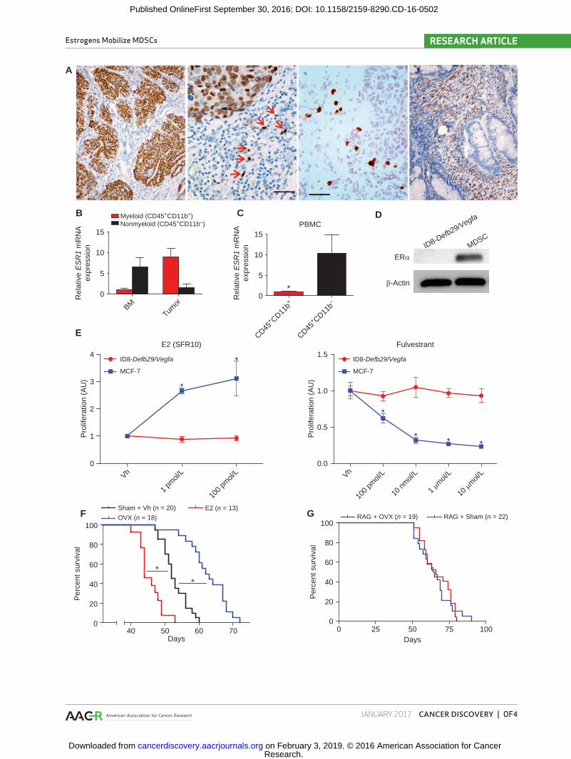
Nuclear expression of ERs specifically in neoplastic cells has been identified in human ovarian carcinomas of all histo-logic subtypes, with positive signal in approximately 60% of high-grade serous tumors (23). ERα is the predominant estro-gen receptor in mouse hematopoietic cells (12). To define the expression of ERα in human ovarian cancer–infiltrating leukocytes, we performed immunohistochemical analysis in 54 serous ovarian carcinomas. Supporting previous reports, we found specific nuclear staining in tumor cells in approxi-mately 35% of tumors (Fig. 1A, left). In addition, we identified weaker signals in individual cells in the stroma with leukocyte morphology (different from tumor cell nuclei) in approxi-mately 20% of ovarian tumors, independent of the ERα sta-tus of tumor cells (Fig. 1A, center). We finally identified two specimens that showed specific signals restricted to stromal fibroblasts (Fig. 1A, right). To confirm that hematopoietic cells at tumor beds express ERα, we sorted (CD45+) cells from 7 different dissociated human ovarian tumors. As shown in Fig. 1B and Supplementary Fig. S1A, both tumor-infiltrating (CD11b+) myeloid cells and (CD11b−) nonmyeloid leukocytes express variable levels of ERα. In addition, both myeloid and lymphoid cells sorted from either the BM of a patient with cancer or the peripheral blood of 5 patients with ovarian cancer were also ERα+ (Fig. 1B and C; and Supplementary Fig. S1A and S1B), suggesting that in addition to potentially having tumor cell–intrinsic effects, estrogens may also play a wider role in shaping the tumor immuno-environment. To determine the role of estrogen signaling in tumor-promoting inflammation or antitumor immunity, we used a preclinical model of aggressive ovarian cancer in which syngeneic epithe-lial ovarian tumor cells (ID8-Defb29/Vegfa) develop intraperi-toneal tumors and ascites that recapitulate the inflammatory microenvironment of human ovarian tumors (9, 15, 17, 24). Importantly, no ERα was detected in these tumor cells, unlike tumor-associated myeloid cells (Fig. 1D). Of note,
ID8-Defb29/Vegfa cells fail to respond to estradiol (E2) treat-ment or the ER antagonist fulvestrant in vitro, unlike estab-lished estrogen-responsive MCF-7 cells (Fig. 1E). Supporting a tumor cell–independent role of estrogen signaling in malig-nant progression, oöphorectomized (estrogen-depleted) wild-type (WT) mice survived significantly longer than non-oöphorectomized, aged-matched controls after orthotopic tumor challenge in multiple independent experiments (Fig. 1F), whereas estrogen supplementation further accelerated malignant progression and reversed the protective effects of oöphorectomy (Fig. 1F; Supplementary Fig. S1C). Strikingly, the survival benefit imparted by oöphorectomy disappeared in tumor-bearing immunodeficient RAG1-deficient KO mice (Fig. 1G), indicating that an adaptive immune response is required for the protective effects of estrogen depletion.
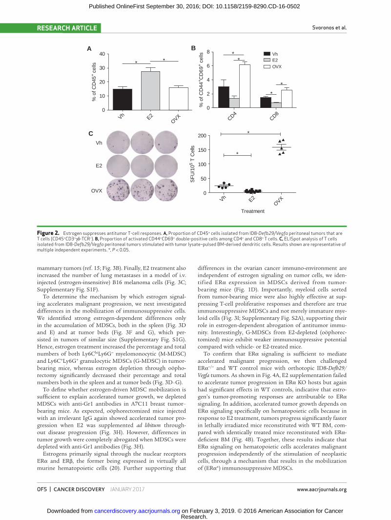
Interestingly, ad libitum E2 supplementation resulted in augmented T-cell inflammation at tumor (peritoneal) beds (Fig. 2A). However, the proportions of antigen experienced (CD44+), recently activated (CD69+) tumor-associated CD4 and CD8 T cells were significantly higher in oöphorecto-mized tumor-bearing hosts, with corresponding decreases in E2-supplemented animals (Fig. 2B). Accordingly, the frequen-cies of T cells isolated from the peritoneal cavity of oöpho-rectomized tumor-bearing mice producing IFNγ in response to cognate tumor antigens were significantly higher than those generated by control (non-oöphorectomized) mice in conventional ELISpot analysis (Fig. 2C), indicative of supe-rior T-cell–dependent antitumor immunity in the former. Consistently, tumor-associated T cells from E2-treated mice responded significantly worse than either group (Fig. 2C). Taken together, these results demonstrate that human ovar-ian cancer microenvironmental hematopoietic cells express ERα, and that, independent of a direct effect on tumor cells, estrogens accelerate ovarian cancer progression through a mechanism that blunts protective antitumor immunity.
ERα Signaling in Hematopoietic Cells Enhances Ovarian Cancer–Induced Myelopoietic Expansion
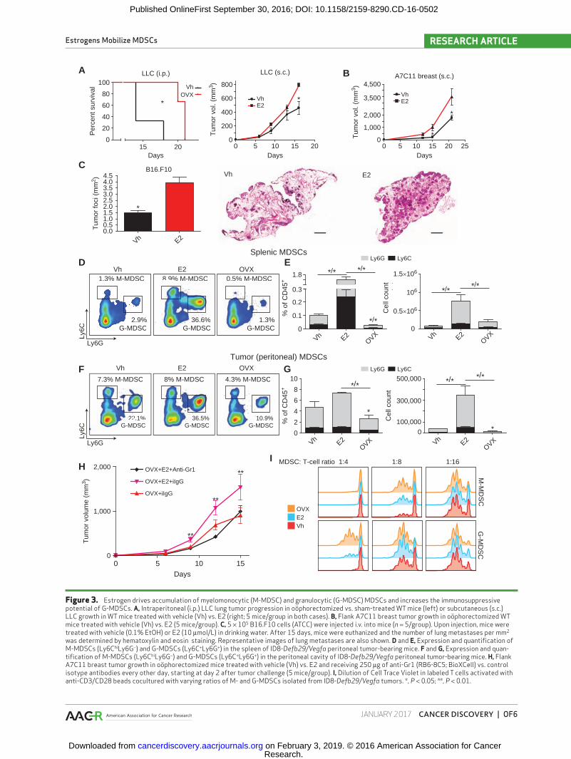
The benefits of estrogen depletion were not restricted to ID8-Defb29/Vegfa tumors, because the progression of estrogen- insensitive (Supplementary Fig. S1D), intraperitoneal Lewis lung carcinomas (LLC) was also significantly delayed in oöphorectomized mice, ultimately resulting in decreased sur-vival (Fig. 3A, left), whereas E2 supplementation accelerated flank tumor growth (Fig. 3A, right). Further supporting the general applicability of this mechanism, E2 supplementa-tion also accelerated the growth of estrogen-insensitive (Sup-plementary Fig. S1E) syngeneic A7C11 mammary tumor cells, derived from autochthonous p53/KRAS-dependent
Figure 1. Estrogen depletion impairment of ovarian tumor progression is independent of tumor cell signaling and is immune dependent. A, Frozen human ovarian tumor sections stained for ERα. Red arrows indicate ER+ nuclei with a typical leukocyte morphology that is different from the morphol-ogy of adjacent tumor cells. Scale bar indicates 1 μm. B–C, Reverse transcription qPCR for ESR1 expression in myeloid (CD45+CD11b+) and nonmyeloid (CD45+CD11b−) cells isolated from dissociated ovarian tumor or BM (B) or the peripheral blood (PBMC; C) of 5 patients with ovarian cancer. D, Western blot for ERα (66 kDa) expression by ID8-Defb29/Vegfa tumor cells and MDSCs isolated from mouse tumors. E, Proliferation relative to vehicle of ID8-Defb29/Vegfa and MCF-7 (positive control) cells in response increasing doses of E2 (in steroid-free media) and the ER antagonist fulvestrant as determined by MTS assay. Vh, vehicle. F, Survival of WT oöphorectomized (OVX) or sham-operated mice challenged with intraperitoneal ID8-Defb29/Vegfa tumors and supplemented or not with E2, pooled from three independent experiments. Total number of mice per group is depicted. G, Survival of OVX or sham-operated Rag1 KO mice challenged with i.p. ID8-Defb29/Vegfa, pooled from six independents. Total number of mice per group is depicted. *, P < 0.05.
Research. on February 3, 2019. © 2016 American Association for Cancercancerdiscovery.aacrjournals.org Downloaded from
Published OnlineFirst September 30, 2016; DOI: 10.1158/2159-8290.CD-16-0502
Estrogens Mobilize MDSCs RESEARCH ARTICLE
JANUARY 2017 CANCER DISCOVERY | OF4
B
F G
A
E2 (SFR10)
Vh
1 pm
ol/L
100
pmol/
L0
1
2
3
4ID8-Defb29/Vegfa
MCF-7
*
*
Pro
lifer
atio
n (A
U)
Fulvestrant
Vh
100
pmol/
L
10 n
mol/
L
1 µm
ol/L
10 µm
ol/L
0.0
0.5
1.0
1.5ID8-Defb29/Vegfa
MCF-7
*
** *
Pro
lifer
atio
n (A
U)
ID8-Defb29/Vegfa
MDSC
β-Actin
ERα
D
BMTum
or0
5
10
15
Myeloid (CD45+CD11b+)Nonmyeloid (CD45+CD11b–)
Rel
ativ
e E
SR
1 m
RN
Aex
pres
sion
Rel
ativ
e E
SR
1 m
RN
Aex
pres
sion
E
CPBMC
0
5
10
15
CD45+ CD11
b+
CD45+ CD11
b–
*
0 25 50 75 1000
20
40
60
80
100RAG + Sham (n = 22)RAG + OVX (n = 19)
0
20
40
60
80
100
Sham + Vh (n = 20) E2 (n = 13)OVX (n = 18)
40 50 60 70
*
*
Days
Per
cent
sur
viva
l
Days
Per
cent
sur
viva
l
Research. on February 3, 2019. © 2016 American Association for Cancercancerdiscovery.aacrjournals.org Downloaded from
Published OnlineFirst September 30, 2016; DOI: 10.1158/2159-8290.CD-16-0502
Svoronos et al.RESEARCH ARTICLE
OF5 | CANCER DISCOVERY JANUARY 2017 www.aacrjournals.org
Figure 2. Estrogen suppresses antitumor T-cell responses. A, Proportion of CD45+ cells isolated from ID8-Defb29/Vegfa peritoneal tumors that are T cells (CD45+CD3+γδ-TCR−). B, Proportion of activated CD44+CD69+ double-positive cells among CD4+ and CD8+ T cells. C, ELISpot analysis of T cells isolated from ID8-Defb29/Vegfa peritoneal tumors stimulated with tumor lysate–pulsed BM-derived dendritic cells. Results shown are representative of multiple independent experiments. *, P < 0.05.
VhE2
OVX0
50
100
150
200
*
*
Treatment
SF
U/1
05 T
Cel
ls
Vh
E2
OVX
% o
f CD
44+ C
D69
+ ce
lls
CD4CD8
0
2
4
6
8 VhE2OVX
**
VhE2
OVX0
10
20
30
40
% o
f CD
45+
cel
ls
A B
C
*
*
* *
mammary tumors (ref. 15; Fig. 3B). Finally, E2 treatment also increased the number of lung metastases in a model of i.v. injected (estrogen-insensitive) B16 melanoma cells (Fig. 3C; Supplementary Fig. S1F).
To determine the mechanism by which estrogen signal-ing accelerates malignant progression, we next investigated differences in the mobilization of immunosuppressive cells. We identified strong estrogen-dependent differences only in the accumulation of MDSCs, both in the spleen (Fig. 3D and E) and at tumor beds (Fig. 3F and G), which per-sisted in tumors of similar size (Supplementary Fig. S1G). Hence, estrogen treatment increased the percentage and total numbers of both Ly6ChiLy6G− myelomonocytic (M-MDSC) and Ly6C+Ly6G+ granulocytic MDSCs (G-MDSC) in tumor-bearing mice, whereas estrogen depletion through oöpho-rectomy significantly decreased their percentage and total numbers both in the spleen and at tumor beds (Fig. 3D–G).
To define whether estrogen-driven MDSC mobilization is sufficient to explain accelerated tumor growth, we depleted MDSCs with anti-Gr1 antibodies in A7C11 breast tumor–bearing mice. As expected, oöphorectomized mice injected with an irrelevant IgG again showed accelerated tumor pro-gression when E2 was supplemented ad libitum through-out disease progression (Fig. 3H). However, differences in tumor growth were completely abrogated when MDSCs were depleted with anti-Gr1 antibodies (Fig. 3H).
Estrogens primarily signal through the nuclear receptors ERα and ERβ, the former being expressed in virtually all murine hematopoietic cells (20). Further supporting that
differences in the ovarian cancer immuno-environment are independent of estrogen signaling on tumor cells, we iden-tified ERα expression in MDSCs derived from tumor- bearing mice (Fig. 1D). Importantly, myeloid cells sorted from tumor-bearing mice were also highly effective at sup-pressing T-cell proliferative responses and therefore are true immunosuppressive MDSCs and not merely immature mye-loid cells (Fig. 3I; Supplementary Fig. S2A), supporting their role in estrogen-dependent abrogation of antitumor immu-nity. Interestingly, G-MDSCs from E2-depleted (oöphorec-tomized) mice exhibit weaker immunosuppressive potential compared with vehicle- or E2-treated mice.
To confirm that ERα signaling is sufficient to mediate accelerated malignant progression, we then challenged ERα−/− and WT control mice with orthotopic ID8-Defb29/Vegfa tumors. As shown in Fig. 4A, E2 supplementation failed to accelerate tumor progression in ERα KO hosts but again had significant effects in WT controls, indicative that estro-gen’s tumor-promoting responses are attributable to ERα signaling. In addition, accelerated tumor growth depends on ERα signaling specifically on hematopoietic cells because in response to E2 treatment, tumors progress significantly faster in lethally irradiated mice reconstituted with WT BM, com-pared with identically treated mice reconstituted with ERα-deficient BM (Fig. 4B). Together, these results indicate that ERα signaling on hematopoietic cells accelerates malignant progression independently of the stimulation of neoplastic cells, through a mechanism that results in the mobilization of (ERα+) immunosuppressive MDSCs.
Research. on February 3, 2019. © 2016 American Association for Cancercancerdiscovery.aacrjournals.org Downloaded from
Published OnlineFirst September 30, 2016; DOI: 10.1158/2159-8290.CD-16-0502
Estrogens Mobilize MDSCs RESEARCH ARTICLE
JANUARY 2017 CANCER DISCOVERY | OF6
Figure 3. Estrogen drives accumulation of myelomonocytic (M-MDSC) and granulocytic (G-MDSC) MDSCs and increases the immunosuppressive potential of G-MDSCs. A, Intraperitoneal (i.p.) LLC lung tumor progression in oöphorectomized vs. sham-treated WT mice (left) or subcutaneous (s.c.) LLC growth in WT mice treated with vehicle (Vh) vs. E2 (right; 5 mice/group in both cases). B, Flank A7C11 breast tumor growth in oöphorectomized WT mice treated with vehicle (Vh) vs. E2 (5 mice/group). C, 5 × 10⁵ B16.F10 cells (ATCC) were injected i.v. into mice (n = 5/group). Upon injection, mice were treated with vehicle (0.1% EtOH) or E2 (10 μmol/L) in drinking water. After 15 days, mice were euthanized and the number of lung metastases per mm2 was determined by hematoxylin and eosin staining. Representative images of lung metastases are also shown. D and E, Expression and quantification of M-MDSCs (Ly6ChiLy6G−) and G-MDSCs (Ly6C+Ly6G+) in the spleen of ID8-Defb29/Vegfa peritoneal tumor-bearing mice. F and G, Expression and quan-tification of M-MDSCs (Ly6ChiLy6G−) and G-MDSCs (Ly6C+Ly6G+) in the peritoneal cavity of ID8-Defb29/Vegfa peritoneal tumor-bearing mice. H, Flank A7C11 breast tumor growth in oöphorectomized mice treated with vehicle (Vh) vs. E2 and receiving 250 μg of anti-Gr1 (RB6-8C5; BioXCell) vs. control isotype antibodies every other day, starting at day 2 after tumor challenge (5 mice/group). I, Dilution of Cell Trace Violet in labeled T cells activated with anti-CD3/CD28 beads cocultured with varying ratios of M- and G-MDSCs isolated from ID8-Defb29/Vegfa tumors. *, P < 0.05; **, P < 0.01.
Vh E2OVX
0
0.1
0.2
0.3
1.8 1.5×10⁶
0.5×10⁶
106
Vh E2OVX
0
500,000
300,000
100,000
Ly6C
Ly6G
% o
f CD
45+
VhE2
OVX VhE2
OVX0
2
4
6
8
10
0 *
Cel
l cou
nt
% o
f CD
45+
Cel
l cou
nt
Ly6CLy6G
Splenic MDSCs
Tumor (peritoneal) MDSCs
D
F
E
G
A
*/**/*
*/**/*
*/*
*/**/*
*/*
*
H
M-M
DS
CG
-MD
SC
MDSC: T-cell ratio 1:4 1:8 1:16
OVXE2Vh
LLC (i.p.)
0
20
40
60
80
100
15 20
VhOVX
*
Days
Per
cent
sur
viva
l
36.6%6 6%G-MDSC
8.9% M-MDSC8.9%
1.3%3%G-MDSC
0.5% M-MDSC0.5%
2.9%2 9%G-MDSC
1.3% M-MDSC1.3%
366.5%G-MDSC
100.9%G-MDSC
4.3% M-MDSC4 3%
222.1%G-MDSC
Ly6C
Ly6G
Vh E2 OVX
Vh E2 OVX
B
0 5 10 15 20 250
1,000
2,000
3,500
4,500
VhE2
*
*Tum
or v
ol. (
mm
3 )
Tum
or v
ol. (
mm
3 )
Tum
or v
olum
e (m
m3 )
Tum
or fo
ci (
mm
2 )
0 5 10 15 200
200
400
600
800
VhE2
*
C B16.F10
Vh E20.00.51.01.52.02.53.03.54.04.5
*
Vh E2
I
0 5 10 150
1,000
2,000
OVX+iIgG
OVX+E2+iIgG
OVX+E2+Anti-Gr1
Days
**
**
**
A7C11 breast (s.c.)
DaysDays
LLC (s.c.)
8% M-MDSC8%7.3% M-MDSC7 3%
Ly6CLy6G
Research. on February 3, 2019. © 2016 American Association for Cancercancerdiscovery.aacrjournals.org Downloaded from
Published OnlineFirst September 30, 2016; DOI: 10.1158/2159-8290.CD-16-0502
Svoronos et al.RESEARCH ARTICLE
OF7 | CANCER DISCOVERY JANUARY 2017 www.aacrjournals.org
Estrogens Signal through ERα on Human and Mouse Myeloid Progenitors to Boost the Proliferation of Regulatory Myeloid Cells and Enhance Their Immunosuppressive Activity
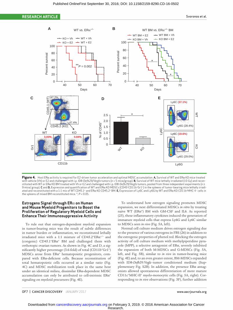
To rule out that estrogen-dependent myeloid expansion in tumor-bearing mice was the result of subtle differences in tumor burden or inflammation, we reconstituted lethally irradiated mice with a 1:1 mixture of CD45.2+ERα−/− and (congenic) CD45.1+ERα+ BM and challenged them with orthotopic ovarian tumors. As shown in Fig. 4C and D, a sig-nificantly higher percentage (3.6-fold) of total (CD11b+Gr1+) MDSCs arose from ERα+ hematopoietic progenitors, com-pared with ERα-deficient cells. Because reconstitution of total hematopoietic cells occurred at a similar ratio (Fig. 4C) and MDSC mobilization took place in the same host under an identical milieu, dissimilar ERα-dependent MDSC accumulation can only be attributed to cell-intrinsic ERα+ signaling on myeloid precursors (Fig. 4E).
To understand how estrogen signaling promotes MDSC expansion, we next differentiated MDSCs in vitro by treating naïve WT (ERα+) BM with GM-CSF and IL6. As reported (25), these inflammatory cytokines induced the generation of immature myeloid cells that express Ly6G and Ly6C similar to MDSCs seen in vivo (Fig. 5A, left).
Normal cell culture medium drives estrogen signaling due to the presence of various estrogens in FBS (26) in addition to the estrogenic properties of phenol red. Blocking the estrogen activity of cell culture medium with methylpiperidino pyra-zole (MPP), a selective antagonist of ERα, severely inhibited the expansion of both M-MDSCs and G-MDSCs (Fig. 5A, left, and Fig. 5B), similar to in vivo in tumor-bearing mice (Fig. 4E) and, to an even greater extent, BM-MDSCs expanded with ID8-Defb29/Vegfa–tumor conditioned medium (Sup-plementary Fig. S2B). In addition, the presence ERα antag-onists allowed spontaneous differentiation of more mature CD11c+MHC-II+ myelo-monocytic cells (Fig. 5A, right). Cor-responding to in vivo observations (Fig. 3F), further addition
Figure 4. Host ERα activity is required for E2-driven tumor acceleration and optimal MDSC accumulation. A, Survival of WT and ERα KO mice treated with vehicle (Vh) or E2 and challenged with i.p. ID8-Defb29/Vegfa tumors (n = 5 mice/group). B, Survival of WT mice lethally irradiated (10 Gy) and recon-stituted with WT or ERα KO BM treated with Vh or E2 and challenged with i.p. ID8-Defb29/Vegfa tumors, pooled from three independent experiments (n ≥ 9 mice/ group). C and D, Expression and quantification of WT and ERα KO MDSCs (CD45+CD11b+Gr1+) in the spleens of tumor-bearing mice lethally irradi-ated and reconstituted with a 1:1 mix of WT CD45.1+ and ERα KO CD45.2+ BM. E, Expression of Ly6C and Ly6G by WT and ERα KO CD11b+MHC-II− cells in the spleens of mixed BM reconstituted mice. *, P < 0.05.
47.4%
47.2%
CD45.2 (KO)
CD
45.1
(W
T)
WT BM + Vh
KO BM + E2WT BM + E2
KO BM + Vh
0
20
40
60
80
100
40 50 60 70
WT + Vh
WT + E2
KO + Vh
KO + E2
Days
Per
cent
sur
viva
l
WT BM vs. ERα–/– BMWT vs. ERα–/–
WT
KO0
0.5
1.0
1.5
2.0
2.5 *
Gr1
CD11b
% o
f CD
45+
C D E
A B
WT KO
Ly6GLy
6C
WT
KO
**P = 0.002
3.03% 0.84%
Ly6G (28.4%)
Ly6G (29.0%)
0
20
40
60
80
100
40 50 60 70
*
*
Days
Per
cent
sur
viva
l
y6C (Ly 37.1%)
Ly6C (19.6%)
Research. on February 3, 2019. © 2016 American Association for Cancercancerdiscovery.aacrjournals.org Downloaded from
Published OnlineFirst September 30, 2016; DOI: 10.1158/2159-8290.CD-16-0502

Estrogens Mobilize MDSCs RESEARCH ARTICLE
JANUARY 2017 CANCER DISCOVERY | OF8
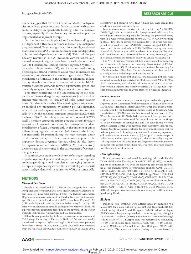
Figure 5. Optimal MDSC expansion and suppressive activity is dependent on estrogen signaling. A, Expression of Ly6C and Ly6G (left) or MHC-II and CD11c (right) by naïve mouse WT BM cultured with GM-CSF + IL6 and treated with Vh or 2 μmol/L antiestrogen MPP for 3 and 6 days. B, Total number of M-MDSCs and G-MDSCs after culturing naïve WT mouse BM with GM-CSF+IL6 and treating with 2 μmol/L MPP for 6 days. Cumulative results of three independent experiments. C, Dilution of Cell Trace Violet by labeled T cells activated with anti-CD3/CD28 beads cocultured with varying ratios of G-MDSCs or M-MDSCs isolated from 6-day BM cultures treated with Vh, 100 ng/mL E2, or 2 μmol/L MPP. D, Expansion of human M-MDSCs (CD45+HLA-DR−CD11b+CD33+CD14+) and G-MDSCs (CD45+HLA-DR−CD11b+CD33+CD15+) from lung cancer patient BM cultured in GM-CSF+IL6 and treated with Vh, 2 μmol/L, or 10 μmol/L MPP. E, Total number of human M-MDSCs and G-MDSCs derived from lung cancer patient BM. *, P < 0.05. F, scatter plot of the level of CD3E and PRF1 mRNA (measured as FKPM) in 266 serous ovarian cancers from TCGA datasets, separated by CYP19A1 expression above or below the median.
Gated on HUMAN CD45+HLA-DR–CD11b+CD33+
SCG-MDS45%
M-MDSC7%
SCG-MDS36%
M-MDSC6%
SCG-MDS56%
M-MDSC15%
Human M-MDSC yield
0 *
Tot
al c
ells
*
CD
15
Vh 2 µmol/L MPP 10 µmol/L MPP
Vh
2 µm
ol/L
MPP
10 µm
ol/L
MPP
0
*
*
MPPE2Vh
1:4 1:8 1:16 1:32
G-MDSCSuppression
E2Vh
M-MDSCSuppression
1:4 1:8 1:16 1:32
Human G-MDSC yield
MDSC: T-cell ratio
MDSC: T-cell ratio
A
B
D E
89.1%%3.9%
43.3%%32.1%
35.0%23.3%
68.2%0.80%
%1.6% %11.6%
30.9%30.9%55.9%55.9%
%3.5% %14.7%
28.5%28.5%53.4%53.4%
%3.9% 29.5%
23.9%23.9%42.6%42.6%
%1.51% 58.7%
15.3%15.3%24.5%24.5%
Vh Vh MPPMPP
Day
3D
ay 6
Day
3D
ay 6
Ly6G CD11c
Ly6C
MH
C-I
I
Mouse MDSC yield
M-M
DSC
G-MDSC
0
Tot
al c
ells
200,000
500,000
300,000
100,000
500,000
2.5×10⁶
1.5×10⁶
400,000
600,000
800,000
1,000,000 VhMPP
**
C
Tot
al c
ells
0100200300400500600
Perforin
Lowaromatase
Higharomatase
Nor
mal
ized
mR
NA
expr
essi
on
P < 0.01
0
1,000
2,000
Lowaromatase
Higharomatase
CD3e
P < 0.05
F
Nor
mal
ized
mR
NA
expr
essi
on
CD14
Research. on February 3, 2019. © 2016 American Association for Cancercancerdiscovery.aacrjournals.org Downloaded from
Published OnlineFirst September 30, 2016; DOI: 10.1158/2159-8290.CD-16-0502

Svoronos et al.RESEARCH ARTICLE
OF9 | CANCER DISCOVERY JANUARY 2017 www.aacrjournals.org
of E2 resulted in G-MDSCs that were more potently immuno-suppressive whereas abrogation of ERα signaling prevented the acquisition of stronger immunosuppressive activity by G-MDSCs (Fig. 5C, top). In contrast, E2 did not affect the inhibitory activity of M-MDSCs (Fig. 5C, bottom), suggesting that the role of estrogens in the accumulation of M-MDSCs is primarily to drive their expansion, although the low yields of BM-MDSCs obtained in the presence of estrogen antagonists preclude testing their suppressive activity.
To support the relevance of ERα signaling in boosting pathologic expansion of MDSCs, we finally procured BM from 5 different patients with lung cancer and expanded myeloid cells with GM-CSF and IL6 (25), in the presence of different concentrations of the ERα-selective antagonist MPP. As shown in Fig. 5D, this system results in reproducible expan-sion of CD11b+CD33+CD15+CD14−MHC-II− granulocytes and CD11b+CD33+CD15−/loCD14+MHC-II− monocytic cells, corresponding to the human counterparts of granulocytic and monocytic MDSCs, along with more mature myeloid cells (Supplementary Fig. S2C). Notably, blockade of ERα signaling resulted in a dramatic dose-dependent reduction in the expan-sion of both MDSC lineages, at the level of both proportions (Fig. 5D) and, especially, absolute numbers (Fig. 5E). Equally important, analysis of 266 patients with serous ovarian cancer in The Cancer Genome Atlas (TCGA) datasets confirmed that patients with expression levels of the aromatase gene CYP19A1 (the enzyme responsible for a key step in the biosynthesis of estrogens) above the median also exhibit lower expression of CD3e and perforin, indicators of cytotoxic activity and total T-cell infiltration, respectively (Fig. 5F).
Together, these data show that estrogen signaling through ERα influences myelopoiesis in both mice and humans to boost the expansion of highly immunosuppressive MDSCs in response to inflammatory signals and block their differentia-tion into MHC-II+ myeloid cells. These combined functions of ERα signaling in myeloid cells promote malignant progres-sion through MDSC-mediated immune-suppression.
Estrogen Signaling Enhances pSTAT3 Activity through Transcriptional Upregulation of JAK2 and Increased Total STAT3 Expression in Myeloid Progenitors
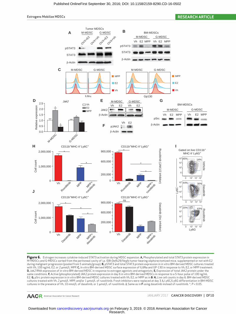
To determine the mechanism by which estrogen signaling promotes MDSC mobilization, we focused on the effect of estrogen signaling on STAT3 signaling, which plays a major role in regulating myeloid lineage cells and MDSC expansion (14). As shown in Fig. 6A, levels of pSTAT3Y705 were significantly increased in both monocytic and granulocytic MDSCs immu-nopurified from the peritoneal cavity of oöphorectomized ovarian cancer–bearing mice supplemented with E2, compared with control oöphorectomized mice receiving vehicle. Accord-ingly, antiestrogen treatment of in vitro BM-MDSC cultures also inhibited STAT3 signaling, resulting in lower phospho-STAT3 (pSTAT3) in both M-MDSCs and G-MDSCs (Fig. 6B), confirm-ing that pSTAT3 signaling is enhanced by estrogen activity.
Because STAT3 activation is triggered by IL6, which was used for in vitro MDSC expansion, we next investigated the role of estrogen signaling on IL6R. Treating BM-MDSCs with E2 or antiestrogens did not elicit changes in surface expression of the IL6Rα chain (Fig. 6C, left), whereas gp130
was paradoxically upregulated by MPP (Fig. 6C, right). We therefore focused on downstream JAK and SRC kinases, both of which mediate STAT3 phosphorylation, subsequent dimerization, and nuclear translocation following cytokine receptor engagement (27, 28). As shown in Fig. 6D, E2 sup-plementation induced transcriptional upregulation of JAK2 in cytokine-induced BM MDSCs of both lineages, whereas no detectable expression or changes were identified for other JAK members (not shown). Accordingly, E2 also induced a reproducible JAK2 upregulation at the protein level, includ-ing higher levels of active (phosphorylated) JAK2 after a short pulse (Fig. 6E and F). Notably, estrogen antagonists also reduced the levels of (active) phospho-Src in both M-MDSCs and G-MDSCs, whereas E2 supplementation also increased Src activity in the former (Fig. 6G).
To define which kinase (JAK2 vs. Src) plays a predominant role in estrogen-dependent MDSC expansion, we expanded BM-MDSCs in the presence of specific Src (dasatinib), JAK1/2 (ruxolitinib), or ERα (MPP) inhibitors. As shown in Fig. 6H and I, JAK1/2 inhibition had a dramatic negative effect in the dif-ferentiation of M-MDSCs. G-MDSC expansion was also heavily decreased upon JAK1/2 inhibition, but concurrent use of ERα antagonists significantly potentiated these suppressive effects (Fig. 6H and I). On the other hand, Src inhibition had no effect on preventing G-MDSC differentiation, but resulted in a sig-nificant decrease in M-MDSC mobilization, which was further enhanced by additional ERα inhibition (Fig. 6I and J). Therefore, concomitant inhibition of ERα signaling and STAT3-activating kinases has stronger negative effects on the expansion of both monocytic and granulocytic MDSC mobilization than inhi-bition of either pathway individually. Taken together, these data indicate that ERα signaling on myeloid precursors pro-motes MDSC expansion by driving STAT3 phosphorylation. In M-MDSCs, this occurs through both enhanced (phosphoryl-ated) Src activity and the necessary function of JAK2, whereas only JAK2 activity is relevant in G-MDSCs.
Estrogen Also Affects Other Components of the Tumor Immuno-Environment
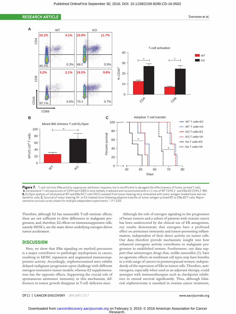
Finally, to rule out that differences in malignant pro-gression are due to the direct effect of estrogens on effector T cells, we performed mixed BM chimera experiments in which mice received a 1:1 mixture of ERα−/− and congenic WT BM. Compared with ERα−/− T cells, E2-responsive WT CD4 and CD8 T cells displayed a less activated phenotype character-ized by lower expression of CD44 (Fig. 7A). Correspondingly, the frequencies of WT T cells responding to tumor antigens in IFNγ ELISpot rechallenge assays were lower than those of their counterpart ERα−/− T cells, sorted from the same micro-environment (Fig. 7B).
To determine the relative importance of these differences in direct ERα signaling in T cells, independent of estrogen-dependent MDSC activity, WT and ERα−/− T-cell splenocytes were identically enriched for tumor-reactive populations by ex vivo priming against tumor lysate-pulsed bone marrow den-dritic cells (BMDC; refs. 29, 30), and then adoptively transferred into ovarian cancer–bearing mice. Confirming previous reports (29, 30), both WT and ERα−/− T cells significantly extended sur-vival. However, there was no difference between WT and ERα KO T cells regardless of whether mice were treated with E2 (Fig. 7C).
Research. on February 3, 2019. © 2016 American Association for Cancercancerdiscovery.aacrjournals.org Downloaded from
Published OnlineFirst September 30, 2016; DOI: 10.1158/2159-8290.CD-16-0502
Estrogens Mobilize MDSCs RESEARCH ARTICLE
JANUARY 2017 CANCER DISCOVERY | OF10
Figure 6. Estrogen increases cytokine-induced STAT3 activation during MDSC expansion. A, Phosphorylated and total STAT3 protein expression in M-MDSCs and G-MDSCs sorted from the peritoneal cavity of i.p. ID8-Defb29/Vegfa tumor-bearing oöphorectomized mice, supplemented or not with E2 during malignant progression (pooled from 5 animals/group). B, pSTAT3 and total STAT3 protein expression in in vitro BM-derived MDSC cultures treated with Vh, 100 ng/mL E2, or 2 μmol/L MPP. C, In vitro BM-derived MDSC surface expression of IL6Rα and GP-130 in response to Vh, E2, or MPP treatment. D, Jak2 RNA expression of in vitro BM-derived MDSC in response to estrogen agonists and antagonists. E, Expression of total JAK2 protein under the same conditions. F, Active (phosphorylated) JAK2 protein expression in day 6 in vitro BM-derived MDSCs in response to a 5-hour pulse of 100 ng/mL E2. G, pSrc protein expression in in vitro BM-derived MDSC cultures treated with Vh, E2, or MPP as in B. H, Live cell counts in day 6. BM-derived MDSC cultures treated with Vh, 2 μmol/L MPP, and/or 1 μmol/L of ruxolitinib. Fresh inhibitors were replaced at day 3. I, Ly6C/Ly6G differentiation in BM-MDSC cultures in the presence of Vh, 10 nmol/L of dasatinib, or 1 μmol/L of ruxolitinib. J, Same as in F using dasatinib instead of ruxolitinib. *, P < 0.05.
β-Actin
pSTAT3
STAT3
BM-MDSCsM-MDSC G-MDSC
M-MDSC G-MDSC
Vh E2 MPP Vh E2 MPP
A B
Jak2
M-M
DSC
G-MDSC
0.0
0.5
1.0
1.5
2.0
2.5 VhE2MPP
*
*
*
Rel
ativ
e ex
pres
sion
D
JAK2
M-MDSC G-MDSCVh E2 Vh E2
β-Actin
C
IL6rα
M-MDSC G-MDSC
Vh
E2
MPP
M-MDSC G-MDSC
Gp130
Vh
E2
MPP
OVX+Vh
OVX+Vh
OVX+E2
OVX+E2
Tumor MDSCs
G-MDSC
Vh E2 MPP
β-Actin
M-MDSC
Vh E2 MPP
BM-MDSCs
F
pSrc
CD11b+MHC-II–Ly6G+
CD11b+MHC-II–Ly6G+
CD11b+MHC-II–Ly6C+
CD11b+MHC-II–Ly6C+H
J
Dasatinib (S
rc inhibitor)R
uxolitinib (JAK
1/2 inhibitor)
Cel
l cou
nt
*
*
*
*
0
1,000,000
2,000,000 900,000
600,000
200,000
0
1,000,000
2,000,000
Vh
Ruxoli
tinib
MPP+
ruxo
litinibM
PP Vh
Ruxoli
tinib
MPP+
ruxo
litinibM
PP
Vh
Dasat
inib
MPP+
dasa
tinibM
PPVh
Dasat
inib
MPP+
dasa
tinibM
PP
ns
Cel
l cou
nt
0
900,000
600,000
200,000
0
*
*
20% 63%
44% 30%
45% 14%
Ly6G
Ly6C
Gated on live CD11b+
MHC-II–Ly6G+
Dasatinib
Ruxolitinib
Vehicle
I
*
*
pJAK2
β-Actin
Vh E2
E G
β-Actin
pSTAT3
STAT3
Research. on February 3, 2019. © 2016 American Association for Cancercancerdiscovery.aacrjournals.org Downloaded from
Published OnlineFirst September 30, 2016; DOI: 10.1158/2159-8290.CD-16-0502
Svoronos et al.RESEARCH ARTICLE
OF11 | CANCER DISCOVERY JANUARY 2017 www.aacrjournals.org
Therefore, although E2 has measurable T-cell–intrinsic effects, these are not sufficient to drive differences in malignant pro-gression, and, therefore, E2 effects on immunosuppressive cells, namely MDSCs, are the main driver underlying estrogen-driven tumor acceleration.
DISCUSSIONHere, we show that ERα signaling on myeloid precursors
is a major contributor to pathologic myelopoiesis in cancer, resulting in MDSC expansion and augmented immunosup-pressive activity. Accordingly, oöphorectomized mice exhibit delayed malignant progression upon challenge with different estrogen-insensitive tumor models, whereas E2 supplementa-tion has the opposite effects. Supporting the crucial role of spontaneous antitumor immunity in this mechanism, dif-ferences in tumor growth disappear in T-cell–deficient mice.
Although the role of estrogen signaling in the progression of breast tumors and a subset of patients with ovarian cancer has been underscored by the clinical use of ER antagonists, our results demonstrate that estrogens have a profound effect on antitumor immunity and tumor-promoting inflam-mation, independent of their direct activity on tumor cells. Our data therefore provide mechanistic insight into how enhanced estrogenic activity contributes to malignant pro-gression in established tumors. Furthermore, our data sup-port that antiestrogen drugs that, unlike tamoxifen (1), have no agonistic effects on nonbreast cell types may have benefits in a wide range of cancers in premenopausal women, indepen-dently of the expression of ERs in tumor cells. Therefore, anti-estrogens, especially when used as an adjuvant therapy, could synergize with immunotherapies such as checkpoint inhibi-tors to extend survival significantly. Thus, although bilat-eral oöphorectomy is standard in ovarian cancer treatment,
Figure 7. T-cell–intrinsic ERα activity suppresses antitumor response, but is insufficient to abrogate the effectiveness of tumor-primed T cells. A, Intratumoral T-cell expression of CD44 and CD69 in mice lethally irradiated and reconstituted with a 1:1 mix of WT CD45.1+ and ERα KO CD45.2+ BM. B, ELISpot analysis of intratumoral WT and ERα KO T cells FACS-isolated from tumor-bearing mice stimulated with tumor antigen–loaded bone marrow dendritic cells. C, Survival of tumor-bearing Vh- or E2-treated mice following adoptive transfer of tumor antigen–primed WT or ERα KO T cells. Repre-sentative survival curves shown for multiple independent experiments. *, P < 0.05.
A
10.1% 4.1%
%0.3%5%85.5
19.9% 11.7%
%0.5%068.0
5.2% 2.1%
%0.6%1%92.1
19.2% 9.8%
%0.7%370.3
WT KO
CD
4C
D8
T-cell activation
CD4CD8
0
10
20
30
40WT
KO*
% C
D44
+B
Mixed BM chimera T-cell ELISpot
WT
KO
0
20
40
60
80
100 *
SF
U/2
×104
T c
ells
Adoptive T-cell transfer
0
20
40
60
80
100
40 50 60 70 80
WT T cells+Vh
KO T cells+E2
WT T cells+E2
KO T cells+Vh
No T cells+Vh
No T cells+E2
Days
Per
cent
sur
viva
l
C
*
*
CD69
CD
44
*
Research. on February 3, 2019. © 2016 American Association for Cancercancerdiscovery.aacrjournals.org Downloaded from
Published OnlineFirst September 30, 2016; DOI: 10.1158/2159-8290.CD-16-0502
Estrogens Mobilize MDSCs RESEARCH ARTICLE
JANUARY 2017 CANCER DISCOVERY | OF12
our data suggest that ER− breast tumors and other malignan-cies in at least premenopausal female patients with cancer could be delayed by specifically blocking ERα in a systemic manner, especially if complementary immunotherapies are implemented as adjuvant therapy.
Our results also have implications for understanding gen-der-dependent differences in tumor initiation and malignant progression in different malignancies. For example, we showed that responses to αPD-L1 immunotherapy were sex-dependent in hormone-independent melanoma (31). This could be par-ticularly relevant in BRCA1-mutation carriers, where aug-mented estrogenic signals have been recently demonstrated (22, 32). Furthermore, ERα expression is regulated by BRCA1-dependent ubiquitination (33), so that cancer-predisposing heterozygous BRCA1 mutations could result in increased ER expression, and therefore increase estrogen activity. Whether mobilization of MDSCs in the context of additional inflam-matory signals contributes to tumor initiation in BRCA1 mutation carriers demands further experimental proof, but our study suggests this as a likely pathogenic mechanism.
This study contributes to the understanding of the com-plexity of factors deregulating myelopoiesis (and therefore antigen presentation) in virtually all solid tumor–bearing hosts. Our data indicate that ERα signaling has a triple effect on myeloid BM progenitors by altering pSTAT3 signaling, which drives both expansion and increased survival in these cells (14): On the one hand, estrogens upregulate JAK2, which mediates STAT3 phosphorylation, as well as total STAT3 itself. Therefore, estrogenic activity prepares the BM for acute expansion of myeloid precursors, but estrogen-dependent mobilization of MDSCs occurs only in the presence of direct inflammatory signals that activate JAK kinases, which may not necessarily be present during the high estrogen phase of the menstrual cycle. These mechanisms appear to be important during pregnancy though, where E2 also drives the expansion and activation of MDSCs (21), but our study demonstrates their relevance in the pathogenesis of women’s malignancies.
In summary, our study unveils the role of estrogen signaling in pathologic myelopoiesis and supports that more specific antiestrogen drugs could complement emerging immuno-therapies to significantly extend the survival of patients with cancer, independently of the expression of ERs in tumor cells.
METHODSMice and Cell Lines
Female 5- to 8-week-old WT C57BL/6 and congenic Ly5.1 mice were purchased from the Charles River Frederick facility. ESR1 knock-out (ERα KO) mice were purchased from The Jackson Laboratory. Oöphorectomies were performed by Charles River staff at 5 weeks of age. Mice were treated with vehicle (0.1% ethanol) or 10 μmol/L E2 (USP grade; Sigma) in drinking water refreshed every 3 to 4 days. All mice were maintained in specific pathogen-free barrier facilities. All experiments were conducted according to the approval of the Wistar Institute Institutional Animal Care and Use Committee.
ID8 cells were provided by K. Roby (Department of Anatomy and Cell Biology, University of Kansas, in 2011; ref. 34) and retrovirally transduced to express Defb29 and Vegfa (24). Clones were passaged fewer than 4 times. MCF-7, B16.F10, and LLC1 cells were obtained from the American Type Culture Collection in 2009, 2013, and 2009,
respectively, and passaged fewer than 4 times. Cell lines used in this article were not authenticated by us.
Peritoneal tumors were initiated in mice by injecting 3 × 10⁶ ID8-Defb29/Vegfa cells intraperitoneally. Intraperitoneal cells were har-vested from tumor-bearing mice by flushing the peritoneal cavity with PBS. Cells were maintained in vitro at 37°C, 5% CO2 by culturing in RPMI+10% FBS or steroid-free medium (SFR10), which was com-prised of phenol red–free RPMI+10% charcoal-stripped FBS. Cells were treated in vitro with vehicle (0.1% DMSO) or varying concentra-tions of E2, fulvestrant, or MPP purchased from Cayman Chemical. Cell proliferation was determined by MTS assay (Promega), and increased/decreased proliferation relative to vehicle was calculated.
The A7C11 mammary tumor cell line was generated by passaging sorted tumor cells from a mechanically disassociated p53/KRAS mammary tumor (35). Flank tumors were initiated by injecting 2 × 10⁴ cells into the axillary flank. Tumor volume was calculated as: 0.5 × (L × W2), where L is the length and W is the width.
For generating mixed BM chimeras, mononuclear BM cells were collected from adult age-matched CD45.1 (congenic) WT or CD45.2 Esr1−/− donor mice, and 1–2 × 10⁶ cells were mixed in a 1:1 ratio and retro-orbitally injected into lethally irradiated (∼950 rad) adult recipi-ents. Mixed chimeras were analyzed after 7 to 8 weeks as indicated.
Human SamplesHuman ovarian carcinoma tissues were procured under a protocol
approved by the Committee for the Protection of Human Subjects at Dartmouth-Hitchcock Medical Center (#17702) and under a proto-col approved by the Institutional Review Board at Christiana Care Health System (#32214) and the Institutional Review Board of The Wistar Institute (#21212263). BM was obtained from patients with stage I–II lung cancer scheduled for surgical resection at the Hospi-tal of the University of Pennsylvania and The Philadelphia Veterans Affairs Medical Center with approval from respective Institutional Review Boards. All patients selected for entry into the study met the following criteria: (i) histologically confirmed pulmonary squamous cell carcinoma or adenocarcinoma, (ii) no prior chemotherapy or radiotherapy within 2 years, and (iii) no other active malignancy. BM cell suspension was obtained from rib fragments that were removed from patients as part of their lung cancer surgery. Informed consent was obtained from all subjects.
Flow CytometryFlow cytometry was performed by staining cells with Zombie
Yellow viability dye, blocking with anti-CD16/32 (2.4G2), and stain-ing for 30 minutes at 4°C with the following anti-mouse antibod-ies at the manufacturer’s recommended dilution: CD45 (30-F11), CD45.1 (A20), CD45.2 (104), CD11c (N418), I-A/I-E (M5/114.15.2), CD3 (145-2C11), Ly6G (1A8), Ly6C (HK1.4), gp130 (4H1B35), IL6R (D7715A7), Gr1 (RB6-4C5) CD4 (RM4-5), CD8b (YTS156.7.7), CD44 (IM7), CD69 (H1.2F3), CD11b (M1/70); or anti-human antibod-ies: CD45 (HI30), CD11c (Bu15), HLA-DR-APC/Cy7 (L243), CD15 (HI98), CD14 (HCD14), CD11b (ICRF44), CD33 (WM53), CD19 (HIB19). Samples were subsequently run using an LSRII and ana-lyzed using FlowJo.
ELISpotDendritic cells (BMDCs) were differentiated by culturing WT
mouse BM for 7 days with 20 ng/mL GM-CSF (Peprotech 315-03), added on days 0 and 3, and 10 ng/mL GM-CSF added on day 6. BMDCs were subsequently primed with tumor antigen by pulsing for 24 hours with irradiated (100 Gy + 30 minutes UV) ID8-Defb29/Vegfa cells at a ratio of 10:1. ELISpot assay was performed by stimulating 1 × 10⁵ cells obtained from peritoneal wash with 1 × 10⁴ antigen-primed BMDCs in a 96-well filter plate (Millipore; MSIPS4510) coated with IFNγ capture antibody according to the manufacturer’s
Research. on February 3, 2019. © 2016 American Association for Cancercancerdiscovery.aacrjournals.org Downloaded from
Published OnlineFirst September 30, 2016; DOI: 10.1158/2159-8290.CD-16-0502
Svoronos et al.RESEARCH ARTICLE
OF13 | CANCER DISCOVERY JANUARY 2017 www.aacrjournals.org
guidelines (eBioscience; 88-3784-88). Following incubation at 37° C, 5% CO2 for 48 hours, positive spots were developed using Avidin-AP and BCIP-NBT substrate (R&D Systems SEL002).
Adoptive T-cell TransferNaïve T cells were harvested from spleens of WT or ERα KO mice
via red blood cell lysis followed by magnetic bead negative selection to remove non–T-cell B220+, CD16/32+,CD11b+, and MHC-II+ cells and primed for 5 days with BMDCs pulsed with tumor antigen as reported (30). A total of 1 × 10⁶ T cells were injected i.p. 7 and 14 days after tumor injection.
BM-Derived MDSC CulturesMouse MDSCs were expanded from mouse BM harvested by flush-
ing tibias and femurs with media. Following red blood cell lysis, 2.5 × 10⁶ cells were cultured in 10 mL of RPMI+10% FBS augmented with recombinant mouse 40 ng/mL GM-CSF+40 ng/mL IL6 (Peprotech) and incubated at 37°C, 5% CO2 for 3 or 6 days. Vehicle, estradiol, or MPP treatments were added as described above. For 6-day cultures, cytokines and estrogen treatments were refreshed on day 3. Follow-ing red blood cell lysis, 2.5 × 10⁶ cells were cultured in 10 mL of RPMI+10% FBS augmented with recombinant mouse 40 ng/mL GM-CSF + 40 ng/mL IL6 (Peprotech) and incubated at 37°C, 5% CO2 for 3 or 6 days. Vehicle, E2, or MPP treatments were added as described above. For 6-day cultures, cytokines and E2 were refreshed on day 3. Following incubation, floating and adherent cells were collected, and M-MDSCs and G-MDSCs were isolated via a Miltenyi MDSC purifi-cation kit according to the manufacturer’s protocol. Human MDSCs were expanded from human lung cancer patient BM acquired as single-cell suspensions (see above). Briefly, 2 × 10⁶ cells were cultured in 3 mL of IMDM+15% FBS supplemented with recombinant human 40 ng/mL GM-CSF + 40 ng/mL IL6 (Peprotech) and treated with vehicle, 2 μmol/L, or 10 μmol/L MPP (see above) for 4 days. Cells were subsequently harvested and analyzed by flow cytometry.
MDSC Suppression AssayNaïve WT T cells were purified from spleens as described and
labeled with the proliferation tracker Cell Trace Violet according to the manufacturer’s protocol. T-cell proliferation was stimulated by adding anti-CD3/CD28 mouse T-activator beads (Thermo) at a 1:1 T-cell to bead ratio according to the manufacturer’s protocol. T cells (2 × 10⁵) were subsequently cocultured with MDSC at 1:4, 1:8, or 1:16 MDSC to T-cell ratios and incubated for 3 days prior to flow cytometric analysis.
Western Blot and IHCCells were lysed in RIPA buffer supplemented with protease inhibi-
tors, phosphatase inhibitors, and Na3VO4 (Thermo) according to the manufacturer’s protocol. Protein quantification was determined via BCA assay, and protein was run on TGX 4-15% gradient gels. Follow-ing transfer, PVDF membranes were blocked with 5% BSA in TBS + 0.05% Tween-20. The following primary antibodies were added to membranes, as indicated, and incubated overnight: JAK2 (rabbit clone # D2E12), STAT3 (clone # 124H6), and pSTAT3 (Tyr705, rabbit clone # D3A7), from Cell Signaling Technology; and ERα (Thermo, clone # TE111.5D11) and beta-actin (Sigma, clone # AC-15). Following second-ary staining with horseradish peroxidase–conjugated anti-mouse or rab-bit IgG, membranes were developed using ECL prime (GE Healthcare).
ERα staining was initially performed in frozen sections from 54 ovarian cancer specimens with clone #TE111.5D11 as the pri-mary antibody (Thermo), followed by a biotinylated goat anti-mouse and completion of immunohistochemical procedure according to the manufacturer’s instructions (Vector Labs). Positive staining was confirmed in 19 paraffin-embedded ovarian cancer sections using
FDA-approved ER tests (DAKO) in the Laboratory of Diagnostic Immunohistochemistry at the Hospital of the University of Pennsyl-vania, following the FDA-approved manufacturer guidelines.
Quantitative Real-Time PCRCells were lysed in TRIzol buffer and RNA was subsequently puri-
fied using an RNEasy kit (Qiagen). Reverse transcription was carried out using a High-Capacity Reverse Transcription Kit (Applied Bio-systems). SYBR Green PCR Master Mix (Applied Biosystems) was used with an ABI 7500 Fast Sequence Detection Software (Applied Biosystems). The following primer sequences were used (5′→3′): Stat3 <F: GACTGATGAAGAGCTGGCTGACT, R: GGGTCTGAAGTTGA-GATTCTGCT>; Jak2 <F: GTGTCGCCGGCCAATGTTC, R: CACA-GGCGTAATACCACAAGC>; and Tbp (mRNA normalization) <F: CACCCCCTTGTACCCTTCAC, R: CAGTTGTCCGTGGCTCTCTT>.
Expression of human ESR1 was quantified with primers: ESR1 <F: CCACTCAACAGCGTGTCTC and R: GGCAGATTCCATAGC-CATAC>, and normalized with primers: GAPDH <F: CCTGCACCAC-CAACTGCTTA R: AGTGATGGCATGGACTGTGGT>.
Expression of mouse ERα was determined with primers: ERα <F:GTGCAGCACCTTGAAGTCTCT and R: TGTTGTAGAGAT-GCTCCATGCC>.
Analysis of TCGA DataAligned Sequence files related to solid ovarian cancer samples
including whole-exome sequencing and outcome data were down-loaded from the TCGA data portal (2015). Scores (number of tags in each transcript) were obtained from each sample, normalized with respect to total tags in the sample as well as total tags in the chromo-some, and expressed as FPKM (fragments/Kb of transcript/million mapped reads).
Disclosure of Potential Conflicts of InterestNo potential conflicts of interest were disclosed.
Authors’ ContributionsConception and design: N. Svoronos, M.R. Rutkowski, P. Zhang, R. Zhang, J.R. Conejo-GarciaDevelopment of methodology: N. Svoronos, A. Perales-Puchalt, M.R. Rutkowski, P. ZhangAcquisition of data (provided animals, acquired and managed patients, provided facilities, etc.): N. Svoronos, A. Perales-Puchalt, M.J. Allegrezza, A.J. Tesone, M.G. Cadungog, S. Singhal, E.B. Eruslanov, J. TchouAnalysis and interpretation of data (e.g., statistical analysis, bio-statistics, computational analysis): N. Svoronos, A. Perales-Puchalt, M.J. Allegrezza, K.K. Payne, T.J. Curiel, S. Singhal, P. Zhang, J.R. Conejo-GarciaWriting, review, and/or revision of the manuscript: N. Svoronos, A. Perales-Puchalt, K.K. Payne, A.J. Tesone, T.J. Curiel, S. Singhal, E.B. Eruslanov, J. Tchou, J.R. Conejo-GarciaAdministrative, technical, or material support (i.e., reporting or organizing data, constructing databases): J.M. NguyenStudy supervision: J.R. Conejo-Garcia
AcknowledgmentsWe thank the Gabrilovich lab at The Wistar Institute for out-
standing technical insight.
Grant SupportThis study was supported by grants to J.R. Conejo-Garcia
[R01CA157664, R01CA124515, R01CA178687, The Jayne Koskinas & Ted Giovanis Breast Cancer Research Consortium at Wistar and Ovarian Cancer Research Fund (OCRF) Program Project Development awards];
Research. on February 3, 2019. © 2016 American Association for Cancercancerdiscovery.aacrjournals.org Downloaded from
Published OnlineFirst September 30, 2016; DOI: 10.1158/2159-8290.CD-16-0502

Estrogens Mobilize MDSCs RESEARCH ARTICLE
JANUARY 2017 CANCER DISCOVERY | OF14
T.J. Curiel (R01CA164122, CDMRP CA140355, P3054174, The Owens Foundation, The Skinner Endowment, and the OCRF Pro-gram Project Development award); E.B. Eruslanov (RO1CA187392); M.J. Allegrezza and N. Svoronos (T32CA009171); K.K. Payne (5T32CA009140-39); and A. Perales-Puchalt [Ann Schreiber Men-tored Investigator Award (OCRF)]. Support for Shared Resources was provided by Cancer Center Support Grant (CCSG) CA010815 (D. Altieri).
The costs of publication of this article were defrayed in part by the payment of page charges. This article must therefore be hereby marked advertisement in accordance with 18 U.S.C. Section 1734 solely to indicate this fact.
Received May 2, 2016; revised September 28, 2016; accepted September 28, 2016; published OnlineFirst September 30, 2016.
REFERENCES 1. Gallo MA, Kaufman D. Antagonistic and agonistic effects of
tamoxifen: Significance in human cancer. Semin Oncol 1997;24:S1- 71–S1-80.
2. Sini V, Cinieri S, Conte P, De Laurentiis M, Leo AD, Tondini C, et al. Endocrine therapy in post-menopausal women with metastatic breast cancer: From literature and guidelines to clinical practice. Crit Rev Oncol Hematol 2016;100:57–68.
3. Hasan J, Ton N, Mullamitha S, Clamp A, McNeilly A, Marshall E, et al. Phase II trial of tamoxifen and goserelin in recurrent epithelial ovarian cancer. Br J Cancer 2005;93:647–51.
4. del Carmen MG, Fuller AF, Matulonis U, Horick NK, Goodman A, Duska LR, et al. Phase II trial of anastrozole in women with asympto-matic mullerian cancer. Gynecol Oncol 2003;91:596–602.
5. Smyth JF, Gourley C, Walker G, MacKean MJ, Stevenson A, Williams AR, et al. Antiestrogen therapy is active in selected ovarian cancer cases: The use of letrozole in estrogen receptor-positive patients. Clin Cancer Res 2007;13:3617–22.
6. Argenta PA, Thomas SG, Judson PL, Downs LS Jr., Geller MA, Carson LF, et al. A phase II study of fulvestrant in the treatment of multiply-recurrent epithelial ovarian cancer. Gynecol Oncol 2009;113:205–9.
7. Bowman A, Gabra H, Langdon SP, Lessells A, Stewart M, Young A, et al. CA125 response is associated with estrogen receptor expression in a phase II trial of letrozole in ovarian cancer: Identification of an endocrine-sensitive subgroup. Clin Cancer Res 2002;8:2233–9.
8. Zhang L, Conejo-Garcia JR, Katsaros D, Gimotty PA, Massobrio M, Regnani G, et al. Intratumoral T cells, recurrence, and survival in epithelial ovarian cancer. N Engl J Med 2003;348:203–13.
9. Stephen TL, Rutkowski MR, Allegrezza MJ, Perales-Puchalt A, Tesone AJ, Svoronos N, et al. Transforming growth factor beta-mediated suppression of antitumor T cells requires FoxP1 transcription factor expression. Immunity 2014;41:427–39.
10. Cubillos-Ruiz JR, Engle X, Scarlett UK, Martinez D, Barber A, Elgueta R, et al. Polyethylenimine-based siRNA nanocomplexes reprogram tumor-associated dendritic cells via TLR5 to elicit therapeutic antitu-mor immunity. J Clin Invest 2009;119:2231–44.
11. Cubillos-Ruiz JR, Martinez D, Scarlett UK, Rutkowski MR, Nesbeth YC, Camposeco-Jacobs AL, et al. CD277 is a negative co-stimulatory molecule universally expressed by ovarian cancer microenvironmen-tal cells. Oncotarget 2010;1:329–8.
12. Pierdominici M, Maselli A, Colasanti T, Giammarioli AM, Delunardo F, Vacirca D, et al. Estrogen receptor profiles in human peripheral blood lymphocytes. Immunol Lett 2010;132:79–85.
13. Salem ML. Estrogen, a double-edged sword: Modulation of TH1- and TH2-mediated inflammations by differential regulation of TH1/TH2 cytokine production. Curr Drug Targets Inflamm Allergy 2004;3:97–104.
14. Gabrilovich DI, Ostrand-Rosenberg S, Bronte V. Coordinated regula-tion of myeloid cells by tumours. Nat Rev Immunol 2012;12:253–68.
15. Rutkowski MR, Stephen TL, Svoronos N, Allegrezza MJ, Tesone AJ, Perales-Puchalt A, et al. Microbially driven TLR5-dependent signal-ing governs distal malignant progression through tumor-promoting inflammation. Cancer Cell 2015;27:27–40.
16. Scarlett UK, Rutkowski MR, Rauwerdink AM, Fields J, Escovar-Fadul X, Baird J, et al. Ovarian cancer progression is controlled by pheno-typic changes in dendritic cells. J Exp Med 2012;209:495–506.
17. Tesone AJ, Rutkowski MR, Brencicova E, Svoronos N, Perales-Puchalt A, Stephen TL, et al. Satb1 overexpression drives tumor-promoting activi-ties in cancer-associated dendritic cells. Cell Rep 2016;14:1774–86.
18. Huarte E, Cubillos-Ruiz JR, Nesbeth YC, Scarlett UK, Martinez DG, Buckanovich RJ, et al. Depletion of dendritic cells delays ovarian cancer progression by boosting antitumor immunity. Cancer Res 2008;68:7684–91.
19. Kovats S. Estrogen receptors regulate an inflammatory pathway of dendritic cell differentiation: Mechanisms and implications for immunity. Horm Behav 2012;62:254–62.
20. Kovats S. Estrogen receptors regulate innate immune cells and signal-ing pathways. Cell Immunol 2015;294:63–9.
21. Pan T, Zhong L, Wu S, Cao Y, Yang Q, Cai Z, et al. 17beta-Oestradiol enhances the expansion and activation of myeloid-derived suppressor cells via signal transducer and activator of transcription (STAT)-3 signalling in human pregnancy. Clin Exp Immunol 2016;185:86–97.
22. Widschwendter M, Rosenthal AN, Philpott S, Rizzuto I, Fraser L, Hayward J, et al. The sex hormone system in carriers of BRCA1/2 mutations: A case-control study. Lancet Oncol 2013;14:1226–32.
23. Sieh W, Kobel M, Longacre TA, Bowtell DD, deFazio A, Goodman MT, et al. Hormone-receptor expression and ovarian cancer survival: An Ovarian Tumor Tissue Analysis consortium study. Lancet Oncol 2013;14:853–62.
24. Conejo-Garcia JR, Benencia F, Courreges MC, Kang E, Mohamed-Hadley A, Buckanovich RJ, et al. Tumor-infiltrating dendritic cell precursors recruited by a beta-defensin contribute to vasculogenesis under the influence of Vegf-A. Nat Med 2004;10:950–8.
25. Marigo I, Bosio E, Solito S, Mesa C, Fernandez A, Dolcetti L, et al. Tumor-induced tolerance and immune suppression depend on the C/EBPbeta transcription factor. Immunity 2010;32:790–802.
26. Briand P, Lykkesfeldt AE. Effect of estrogen and antiestrogen on the human breast cancer cell line MCF-7 adapted to growth at low serum concentration. Cancer Res 1984;44:1114–9.
27. Murray PJ. The JAK-STAT signaling pathway: Input and output inte-gration. J Immunol 2007;178:2623–9.
28. Rebe C, Vegran F, Berger H, Ghiringhelli F. STAT3 activation: A key factor in tumor immunoescape. JAKSTAT 2013;2:e23010.
29. Nesbeth Y, Scarlett U, Cubillos-Ruiz J, Martinez D, Engle X, Turk MJ, et al. CCL5-mediated endogenous antitumor immunity elicited by adoptively transferred lymphocytes and dendritic cell depletion. Cancer Res 2009;69:6331–8.
30. Nesbeth YC, Martinez DG, Toraya S, Scarlett UK, Cubillos-Ruiz JR, Rutkowski MR, et al. CD4+ T cells elicit host immune responses to MHC class II- ovarian cancer through CCL5 secretion and CD40-mediated licensing of dendritic cells. J Immunol 2010;184:5654–62.
31. Lin PY, Sun L, Thibodeaux SR, Ludwig SM, Vadlamudi RK, Hurez VJ, et al. B7-H1-dependent sex-related differences in tumor immunity and immunotherapy responses. J Immunol 2010;185:2747–53.
32. Hu Y, Ghosh S, Amleh A, Yue W, Lu Y, Katz A, et al. Modulation of aromatase expression by BRCA1: A possible link to tissue-specific tumor suppression. Oncogene 2005;24:8343–8.
33. Eakin CM, Maccoss MJ, Finney GL, Klevit RE. Estrogen receptor alpha is a putative substrate for the BRCA1 ubiquitin ligase. Proc Natl Acad Sci U S A 2007;104:5794–9.
34. Roby KF, Taylor CC, Sweetwood JP, Cheng Y, Pace JL, Tawfik O, et al. Development of a syngeneic mouse model for events related to ovar-ian cancer. Carcinogenesis 2000;21:585–91.
35. Rutkowski MR, Allegrezza MJ, Svoronos N, Tesone AJ, Stephen TL, Perales-Puchalt A, et al. Initiation of metastatic breast carcinoma by targeting of the ductal epithelium with adenovirus-cre: A novel trans-genic mouse model of breast cancer. J Vis Exp 2014:51171.
Research. on February 3, 2019. © 2016 American Association for Cancercancerdiscovery.aacrjournals.org Downloaded from
Published OnlineFirst September 30, 2016; DOI: 10.1158/2159-8290.CD-16-0502
Published OnlineFirst September 30, 2016.Cancer Discov Nikolaos Svoronos, Alfredo Perales-Puchalt, Michael J. Allegrezza, et al. Suppressor CellsProgression through Mobilization of Myeloid-Derived
Independent Estrogen Signaling Drives Disease−Tumor Cell
Updated version
10.1158/2159-8290.CD-16-0502doi:
Access the most recent version of this article at:
Material
Supplementary
http://cancerdiscovery.aacrjournals.org/content/suppl/2016/09/30/2159-8290.CD-16-0502.DC1
Access the most recent supplemental material at:
E-mail alerts related to this article or journal.Sign up to receive free email-alerts
Subscriptions
Reprints and
To order reprints of this article or to subscribe to the journal, contact the AACR Publications
Permissions
Rightslink site. Click on "Request Permissions" which will take you to the Copyright Clearance Center's (CCC)
.http://cancerdiscovery.aacrjournals.org/content/early/2016/12/18/2159-8290.CD-16-0502To request permission to re-use all or part of this article, use this link
Research. on February 3, 2019. © 2016 American Association for Cancercancerdiscovery.aacrjournals.org Downloaded from
Published OnlineFirst September 30, 2016; DOI: 10.1158/2159-8290.CD-16-0502