rapid signaling of estrogen in hypothalamic neurons involves a
TRANSCRIPT

Behavioral/Systems/Cognitive
Rapid Signaling of Estrogen in Hypothalamic NeuronsInvolves a Novel G-Protein-Coupled Estrogen Receptor thatActivates Protein Kinase C
Jian Qiu,1 Martha A. Bosch,1 Sandra C. Tobias,2 David K. Grandy,1 Thomas S. Scanlan,2 Oline K. Rønnekleiv,1 andMartin J. Kelly1
1Department of Physiology and Pharmacology, Oregon Health and Science University, Portland, Oregon 97239, and 2Department of PharmaceuticalChemistry, University of California, San Francisco, San Francisco, California 94143
Classically, 17�-estradiol (E2 ) is thought to control homeostatic functions such as reproduction, stress responses, feeding, sleep cycles,temperature regulation, and motivated behaviors through transcriptional events. Although it is increasingly evident that E2 can alsorapidly activate kinase pathways to have multiple downstream actions in CNS neurons, the receptor(s) and the signal transductionpathways involved have not been identified. We discovered that E2 can alter �-opioid and GABA neurotransmission rapidly throughnontranscriptional events in hypothalamic GABA, proopiomelanocortin (POMC), and dopamine neurons. Therefore, we examined theeffects of E2 in these neurons using whole-cell recording techniques in ovariectomized female guinea pigs. E2 reduced rapidly the potencyof the GABAB receptor agonist baclofen to activate G-protein-coupled, inwardly rectifying K� channels in hypothalamic neurons. Theseeffects were mimicked by the membrane impermeant E2–BSA and selective estrogen receptor modulators, including a new diphenylac-rylamide compound, STX, that does not bind to intracellular estrogen receptors � or �, suggesting that E2 acts through a uniquemembrane receptor. We characterized the coupling of this estrogen receptor to a G�q-mediated activation of phospholipase C, leading tothe upregulation of protein kinase C� and protein kinase A activity in these neurons. Moreover, using single-cell reverse transcription-PCR, we identified the critical transcripts, PKC� and its downstream target adenylyl cyclase VII, for rapid, novel signaling of E2 in GABA,POMC, and dopamine neurons. Therefore, this unique Gq-coupled estrogen receptor may be involved in rapid signaling in hypothalamicneurons that are critical for normal homeostatic functions.
Key words: GABAB receptor; GIRK; dopamine; GAD; POMC; SERMs
IntroductionIt is becoming increasingly evident that the gonadal steroid hor-mone 17�-estradiol (E2) imparts a multifaceted influence oversynaptic transmission in the mammalian CNS. Not only can es-trogen alter synaptic responses via estrogen response element-driven target gene transcription, but E2 can also rapidly modulatecell-to-cell communication via membrane-initiated, rapid sig-naling events (for review, see Kelly and Wagner, 1999). Thesesynaptic alterations are brought about via changes in the cellularresponsiveness to the activation of various receptor systems (bothG-protein-coupled and ionotropic) to their respective first mes-sengers. E2 can alter the linkage of G-protein-coupled receptor(GPCR) systems such as opioid (both � and �), GABAB, anddopamine D2 receptors to their respective effector systems(Demotes-Mainard et al., 1990; Kelly et al., 1992; Takano et al.,
1994; Wagner et al., 1994; Lagrange et al., 1996). In addition, E2
can act as an allosteric modulator of ionotropic receptors, such as5-HT3 and nicotinic receptors (Wetzel et al., 1998; Paradiso et al.,2001), or by direct binding to subunits of ion channels, such asthe �1 subunit of the maxi-K� channel (Valverde et al., 1999).These fundamentally distinct signaling pathways give rise to acoordinated regulation by estrogen of complex physiologicalprocesses to maintain homeostasis in the mammal (McEwen,2001).
The quintessential role of estrogen in the CNS is to transmitfeedback information to gonadotropin-releasing hormone(GnRH) neurons that control the female reproductive cycle. Es-trogen can alter GnRH neuronal activity directly (Kelly et al.,1984; Lagrange et al., 1995) or it can act upstream to alter synapticinput to GnRH neurons (Watson et al., 1992; Herbison et al.,1995, 2001; Sullivan et al., 1995; Simonian et al., 1999). Two ofthe major presynaptic target neurons of estrogen are the proopio-melanocortin (POMC) and GABA neurons, both of which pro-vide a prominent synaptic input onto GnRH neurons (Morrell etal., 1985; Leranth et al., 1992; Herbison, 1997). Both opioid pep-tides and GABA inhibit GnRH output (Ferin et al., 1984; Mitsus-hima et al., 1996) and luteinizing hormone release (Ferin et al.,1984; Akema et al., 1990; Seltzer and Donoso, 1992; Jarry et al.,
Received June 13, 2003; revised Aug. 1, 2003; accepted Aug. 22, 2003.This research was supported by United States Public Health Service Grants NS 35944 (Office of Research on
Women’s Health), NS 38809, DA 05158, DA 10703, and DK57574. We thank Drs. D. James Surmeier and Tania Tkatchfor their help and advice in establishing the single-cell RT-PCR technique in our laboratory. We also recognize BarryR. Naylor and Rebecka D. Amodei for their expert technical contribution.
Correspondence should be addressed to Dr. Martin J. Kelly, Department of Physiology and Pharmacology, L334,Oregon Health and Science University, Portland, OR 97239-3098. E-mail: [email protected] © 2003 Society for Neuroscience 0270-6474/03/239529-12$15.00/0
The Journal of Neuroscience, October 22, 2003 • 23(29):9529 –9540 • 9529

1995) from the anterior pituitary. Another target of the actions ofestrogen is the arcuate dopamine (tuberoinfundibular) neuronsthat are located in the arcuate nucleus and project to the medianeminence, in which they release dopamine into the portal circu-lation, which directly inhibits prolactin secretion from anteriorpituitary lactotrophs (Neill, 1980; Bjorklund and Lindvall, 1984;Hokfelt et al., 1984).
�-Opioid and GABAB receptors are linked to the same popu-lation of G-protein-coupled inwardly rectifying K� (GIRK)channels in hypothalamic POMC, dopamine, and GABA neu-rons (Loose et al., 1990, 1991; Wagner et al., 2000). Activation ofeither �-opioid or GABAB receptors elicits an outward K� cur-rent that robustly hyperpolarizes hypothalamic neurons. How-ever, maximum activation of either receptor occludes the re-sponse of the other receptor (Loose et al., 1991; Wagner et al.,1999, 2001). Interestingly, short-term exposure to estrogen re-duces the potency of both �-opioid and GABAB receptor agoniststo activate GIRK channels in hypothalamic neurons (Lagrange etal., 1994, 1996, 1997). The underlying mechanism of thisestrogen-induced decrease in the responsiveness of hypothalamicneurons to the �-opioid and GABAB agonists is not known. Re-cent experiments have shown that selective protein kinase A(PKA) inhibitors can block the effects of estrogen, and PKA acti-vators mimic the effects of estrogen on the coupling of �-opioidreceptor to GIRK (Lagrange et al., 1997). Therefore, in thepresent study, we characterized the estrogen-mediated rapid sig-naling pathway in hypothalamic neurons by using novel estrogenreceptor (ER) ligands. In addition, by using specific protein ki-nase inhibitors and single-cell reverse transcription (scRT)-PCR,we found that this novel estrogen receptor is coupled to G�q andactivates a phospholipase C (PLC)–protein kinase C�–proteinkinase A pathway. We conclude that stimulation of this pathwayby binding of natural estrogen hormone and certain selectiveestrogen receptor modulators (SERMs) to the novel Gq-coupledestrogen receptor mediates the rapid steroid response in hypo-thalamic neurons.
Materials and MethodsAnimals and treatments. All animal procedures described in this study arein accordance with institutional guidelines based on National Institutesof Health standards. Female Topeka guinea pigs (400 – 600 gm), bred inour institutional breeding facility, and female multicolor guinea pigs(400 –500 gm; Elm Hill Breeding Labs, Chelmsford, MA) were used inthese experiments. The guinea pigs were maintained under constanttemperature (26°C) and light (on between 6:30 A.M. and 8:30 P.M.).Animals were housed individually, with food and water provided adlibitum. They were ovariectomized under ketamine–xylazine anesthesia(33 and 6 mg/kg, respectively, s.c.) 5–7 d before experimentation, andthey were given sesame oil vehicle (0.1 ml, s.c.) 24 hr before experimen-tation. Serum estrogen concentrations were determined by radioimmu-noassay (Wagner et al., 2001) from trunk blood collected on the day ofexperimentation and were �10 pg/ml. An additional group of animals(n � 6) were ovariectomized and, after 1 week, were injected with oilvehicle, estradiol benzoate (25 �g in oil), or STX (25 �g in oil) 24 hrbefore they were killed. The uteri were collected, weighed, and fixed in4% paraformaldehyde for later histological analysis, which is not beingreported in this paper.
Wild-type C57BL/6 mice in these studies were obtained from TheJackson Laboratory (Bar Harbor, ME). All animals were maintained un-der controlled temperature (25°C) and photoperiod conditions (14/10hr light/dark cycle; lights on between 7:00 A.M. and 9:00 P.M.) with foodand water ad libitum. Adult mice were ovariectomized under isofluraneanesthesia and allowed to recover for 1 week. At this time, the animalswere injected daily for 2 d with oil vehicle, estradiol benzoate (1 �g), orSTX (2 or 5 �g) and anesthetized and killed by decapitation after 24 hr.
The uteri were collected, weighed, and fixed in 4% paraformaldehyde forlater histological analysis, which is not being reported in this paper.
Drugs. All drugs were purchased from Calbiochem (La Jolla, CA) un-less otherwise specified. Tetrodotoxin (TTX) (Alomone Labs, Jerusalem,Israel) was dissolved in Milli-Q H2O and further diluted with 0.1% aceticacid (final concentration, 1 mM), pH 4 –5. 17�-Estradiol was purchasedfrom Steraloids (Wilton, NH), recrystallized to ensure purity, and dis-solved in 100% ethanol to a stock concentration of 1 mM. 17�-Estradiol(17�-E2; 1 mM; Steraloids), anti-estrogen (ICI 182,780; 10 mM; TocrisCookson, Ballwin, MO), and the selective estrogen receptor modulators4-OH-tamoxifen (10 mM; Steraloids), raloxifene (10 mM; Eli Lilly andCompany, Indianapolis, IN), and STX (10 mM) were also dissolved in100% ethanol. 17�-Estradiol 17-hemisuccinate: BSA (E2–BSA) (1 mM;Steraloids) was dissolved in H2O. The protein kinase A inhibitor H-89dihydrochloride (10 mM), the protein kinase A activator forskolin (50mM), the protein kinase C inhibitors bisindolylmaleimide I hydrochlo-ride (BIS) (100 �M), Go6976 (2 mM), and rottlerin (10 mM), the phos-pholipase C inhibitor U73122 (20 mM), the less active analog U73343 (20mM), and the MEK1 [mitogen-activated protein (MAP) kinase kinase-1]inhibitor PD98059 (50 mM) were dissolved in DMSO. Protein kinase Ainhibitory peptide 6 –22 amide (1 mM), the protein kinase A inhibitorRp-cAMPS (50 mM), and cholera toxin (CTX) A subunit (1 �g/�l) weredissolved in H2O. The Gq-binding protein designed to mimic the C ter-minus of the Gq � subunit and the Gs �-binding protein designed tomimic the C terminus of the Gs � subunit were synthesized by Peptido-Genic Research (Livermore, CA). The peptide sequence for Gq peptidewas Ac-LGLNLKEYNLV-OH, and the peptide sequence for Gs peptidewas CRMHLRQYELL. The peptides were also dissolved in H2O. 1,2-Bis-(o-aminophenoxyethane)-N,N,N�,N�-tetra-acetic acid (BAPTA) tetra-sodium salt was dissolved in the internal solution at a 10 mM concentra-tion. Aliquots of the stock solutions were stored as appropriate untilneeded.
Tissue preparation. On the day of experimentation, the animal wasdecapitated, its brain was removed from the skull, and the hypothalamuswas dissected. The resultant hypothalamic block was mounted on a plas-tic cutting platform that was then secured in a vibratome well filled withice-cold, oxygenated (95% O2, 5% CO2) artificial CSF (aCSF) (in mM:124 NaCl, 26 NaHCO3, 10 dextrose, 10 HEPES, 5 KCl, 2.6 NaH2PO4, 2MgSO4, and 1 CaCl2). Four coronal slices (350 �m) through the arcuatewere cut. The slices were transferred to a multiwell auxiliary chambercontaining oxygenated aCSF and kept there until electrophysiologicalrecording after �2 hr.
Electrophysiology. Whole-cell patch recordings in voltage clamp wereperformed as described previously (Wagner et al., 2001). Slices weremaintained briefly in a chamber perfused with warmed (35°C), oxygen-ated aCSF containing the same constituents and respective concentra-tions, except for CaCl2, which was raised to 2 mM. aCSF and all drugsolutions were perfused via a peristaltic pump at a rate of 1.5 ml/min.Drug solutions were prepared in 20 ml syringes by diluting the appropri-ate stock solution with aCSF, and the flow was controlled via a three-waystopcock.
For whole-cell recordings, electrodes were fabricated from borosilicateglass (1.5 mm outer diameter; World Precision Instruments, Sarasota,FL). Resultant electrodes were then filled with an internal solution con-taining 0.5% biocytin and consisting of the following (in mM): 128 K �
gluconate, 10 NaCl, 1 MgCl2, 11 EGTA, 10 HEPES, 1.2 ATP, and 0.4 GTP(pH was adjusted to 7.3–7.4 with 1N KOH, 272–315 mOsm). Voltagepulses were amplified and passed through the electrode using an Axo-patch 1D preamplifier (Axon Instruments, Union City, CA). The result-ant current deflections were monitored using a digital oscilloscope (Tek-tronix 2230; Tektronix, Beaverton, OR). After the reduction of thecurrent deflection, negative pressure was applied via a 5 ml syringe con-nected by polyethylene tubing to the electrode to form a seal (�1 G�).After formation of a seal, intracellular access was achieved by suction,followed by perfusion with 1 �M TTX for at least 4 – 6 min to blockspontaneous firing and synaptic potentials before applying the GABAB
receptor agonist baclofen (see Fig. 1). All of the responses to baclofenwere measured in voltage clamp as outward currents (Vhold � �60 mV),and only those cells that showed �10% change in access resistance (ac-
9530 • J. Neurosci., October 22, 2003 • 23(29):9529 –9540 Qiu et al. • Rapid Estrogen Signaling in Hypothalamic Neurons

cess resistances ranged from 20 to 30 M�) throughout the recordingwere included in this study. Membrane currents underwent analog-to-digital conversion via a Digidata 1200 interface coupled to pClamp 7.0(Axon Instruments). Low-pass filtering of the currents was conducted ata frequency of 2 kHz. The liquid junction potential was �10 mV and wascorrected for in subsequent data analysis.
Post hoc identification of hypothalamic arcuate neurons. After electro-physiological recording, the slices were fixed with 4% paraformaldehydein Sorensen’s phosphate buffer, pH 7.4, for 120 min, immersed overnightin 20% sucrose dissolved in Sorensen’s buffer, and then frozen in OCTembedding medium and prepared for immunocytochemistry as de-scribed previously (Kelly and Rønnekleiv, 1994). Briefly, coronal sections(20 �m) were cut on a cryostat (model 1720 Digital Cryostat; Leitz,Wetzlar, Germany) and mounted on Fisher SuperFrost Plus slides. Sec-tions were washed for 5 min with 0.1 M sodium phosphate buffer, pH 7.4,and then streptavidin–Cy2 (1:1000; Jackson ImmunoResearch, WestGrove, PA) was applied for 2 hr. The reaction was terminated by washingwith buffer. The slices were scanned for the injected neuron with a Nikon(Melville, NY) Eclipse 800 fluorescence microscope. After localization ofthe biocytin-filled neurons, the slides containing the appropriate sectionswere processed for the presence of tyrosine hydroxylase (TH) or�-endorphin using fluorescence immunohistochemistry as describedpreviously (Kelly and Rønnekleiv, 1994). Briefly, the sections with thebiocytin-identified neurons were incubated overnight with a monoclo-nal TH antibody at 1:10,000 (Diasorin, Stillwater, MN) or with a poly-clonal �-endorphin antibody at 1:5000 (Dave et al., 1985) and washed in0.1 M phosphate buffer, followed by incubation with a goat anti-mouseIgG–Cy3 at 1:500 or donkey anti-rabbit IgG–Cy3 at 1:500, respectively(Jackson ImmunoResearch). The sections were washed with sodiumphosphate buffer, and coverslips were applied using a glycerolglycinebuffer containing 5% N-propylgallate. Immunostained cells were pho-tographed using a Nikon microscope.
Estrogen receptor binding assays. The relative binding affinity of com-pounds for ER� and ER� was determined using a spin column assay withcommercially available full-length forms of both ER� and ER� (Pan-Vera, Madison, WI). Receptor was added to a final concentration of 15nM to a solution containing 10 mM Tris, pH 7.5, 10% glycerol, 2 mM DTT,1 mg/ml BSA, and 3 nM [2,4,6,7,16,17- 3H]estradiol at 4°C. Next, 100 �lof the solution was added to 1 �l of the ligand in ethanol, mixed gently bypipetting, and incubated at 4°C overnight. The mixture was then appliedto a micro spin column containing G-25 Sephadex (Harvard Apparatus,Holliston, MA) equilibrated in binding buffer (minus tritiated estradiol)according to the instructions of the manufacturer. Bound estradiol wasseparated from free ligand by spinning at 2000 g for 4 min at roomtemperature. The filtrate was then added to 2.5 ml of scintillant andcounted in a liquid scintillation counter. A binding curve was fitted usinga single binding site competition model with the Prism statistical analysissoftware package (GraphPad Software, San Diego, CA). The SD wasdetermined to be �0.2 log units from the EC50 value. Percentage relativebinding affinity was then determined by dividing the IC50 determined forunlabeled estradiol by the ligand IC50 and multiplying by 100.
Dispersed single-cell RT-PCR. Guinea pig 350 �m coronal hypotha-lamic slices were cut on a vibratome from caudal to rostral and placed inan auxiliary chamber containing oxygenated aCSF. The slices were al-lowed to recover for 1–2 hr in the chamber before dispersion. The arcuatenucleus of the hypothalamus was microdissected and incubated in 2–3ml of HBSS containing the following: 1.26 mM CaCl2, 1 mM MgSO4, 5.37mM KCl, 0.44 mM KH2PO4, 136.89 mM NaCl, 0.34 mM Na2HPO4, 5.55mM D-glucose, and 15 mM HEPES in DEPC-treated water, pH 7.3 (300mOsm), containing 1 mg/ml protease XIV (Sigma, St. Louis, MO) for�15 min at 37°C. The tissue was then washed four times in 1 vol oflow-calcium aCSF and two times in HBSS. The cells were isolated bytrituration with flame-polished Pasteur pipettes, dispersed on a dish, andperfused continuously with HBSS at a rate of 1.5 ml/min. Cells werevisualized using a Nikon inverted microscope, and individual neuronswere patched and harvested into the patch pipette by applying negativepressure. The content of the pipette was expelled into a siliconized mi-crocentrifuge tube containing 5 �l of the following solution: 0.5 �l of10 buffer (100 mM Tris-HCl, 500 mM KCl, 1% Triton X-100; Promega,
Madison, WI), 15 U of RNasin (Promega), 0.5 �l of 100 mM DTT, andDEPC-treated water.
In addition, hypothalamic tissue was homogenized, and total RNA wasextracted using the RNeasy kit (Qiagen, Valencia, CA) according to theprotocol of the manufacturer. The harvested cell solution and 25 ng ofhypothalamic total RNA in 1 �l were denatured for 5 min at 65°C andcooled on ice for 5 min, and then single-stranded cDNA was synthesizedfrom cellular RNA by adding 50 U of murine leukemia virus reversetranscriptase (Applied Biosystems, Foster City, CA), 1.5 �l of 10 buffer,2 mM MgCl2, 0.2 �l of deoxynucleotide triphosphates (dNTPs), 15 U ofRNasin, 10 mM DTT, 100 ng f random hexamers, and DEPC-treatedwater to a final volume of 20 �l. Cells and tissue RNA used as negativecontrols were processed as described above but without reverse tran-scriptase. The reaction mixtures were incubated at 42°C for 60 min,denatured at 99°C for 5 min, and cooled on ice for 5 min.
PCR was performed using 3 �l of cDNA template from each RT reac-tion in a 30 �l of PCR reaction volume containing the following: 3 �l of10 buffer, 2.4 �l of MgCl2 [2 mM final concentration for TH, POMC,GABAB receptor 2 (GABAB R2), PKC�, adenylyl cyclase VII (AC VII),and glyceraldehyde-3-phospate dehydrogenase (GAPDH)], or 3.6 �l ofMgCl2 (3 mM final concentration for GAD), 0.2 mM dNTPs, 0.2 �M
forward and reverse primers, 2 U of Taq DNA polymerase (Promega),and 0.22 �g of TaqStart antibody (Clontech, Palo Alto, CA). Taq DNApolymerase and TaqStart antibody were combined and incubated atroom temperature for 5 min, and the remainder of the reaction contentswere added to the tube and incubated at 94°C for 2 min. Then, eachreaction went through 60 cycles (35 cycles for GAPDH) of amplificationaccording to the following protocols: 94°C, 45 sec; 55°C (GAD), 57°C(PKC�), 58°C (GABAB-R2), 60°C (TH and adenylyl cyclase VII), 61°C(POMC), 63°C (GAPDH) 45 sec; 72°C, 1 min 10 sec; with a final72°C extension for 5 min. Ten microliters of the PCR products werevisualized with ethidium bromide on a 1.5% agarose gel.
All of the primers were synthesized by Invitrogen (Carlsbad, CA) andwere as follows: guinea pig GAD65, 207 bp product, forward primer5�-GGCTCTGGTGATGGAATA-3�, reverse primer 5�-CAGAAT-CACGCTGTCTGTT-3�; guinea pig TH, 223 bp product, forward primer5�-TCCACGTTATACTGGTTCAC-3�, reverse primer 5�-TTGCAT-CACTGAAGCTCTC-3�; guinea pig GABAB-R2, 241 bp product, for-ward primer 5�-TGTTTGTGCCAAAGCTCATC-3� reverse primer 5�-GTGTCTTGCAGTTGCATAGT-3�; guinea pig POMC (GenBankaccession number S78260), 344 bp product, forward primer (bases 40 –60) 5�-CTGGCCTTGCTGCTTCAGAT-3� reverse primer (bases 383–363) 5�-ATGGAGTAGGAGCGCTTGTC-3�; guinea pig GAPDH, 212 bpproduct (GenBank accession number CPU51572), forward primer5�CATCCACTGGTGCTGCCAAG-3�, reverse primer 5�-GTCCTCG-GTGTAGCCCAAGA-3�; human protein kinase C�, 251 bp product(GenBank accession number L07861) forward primer (bases 1127–1147)5�-AAAGGCAGCTTCGGGAAGGT-3�, reverse primer (bases 1377–1357)5�-TGGATGTGGTACATCAGGTC-3�; and guinea pig adenylyl cyclase VII,235 bp product, forward primer 5�-CTGTTCGGCAAGTTTGACCAG-3�,reverse primer 5�-TGACGCCACACAGCACATT-3�.
Statistical analyses. Comparisons between groups were performed us-ing a one-way ANOVA [with post hoc (Newman–Keuls) paired analysis].Differences were considered statistically significant if the probability oferror was �5%.
ResultsE2 and SERMs rapidly attenuate the GABAB response inhypothalamic dopamine and POMC neuronsWhole-cell recordings were made in arcuate neurons (n � 195)from ovariectomized female guinea pigs (Fig. 1). A subgroup ofthese neurons (n � 55) was identified using dual-labeling immu-nocytochemistry (Fig. 2). This revealed that 41% of the cells wereTH positive (i.e., dopamine neurons) and 39% were �-endorphin positive (i.e., POMC neurons). Moreover, on the basisof dual-immunocytochemical staining and in situ hybridizationfor GAD65, a subgroup of arcuate dopamine neurons coexpressGABA (O. K. Rønnekleiv, unpublished findings), which was sub-
Qiu et al. • Rapid Estrogen Signaling in Hypothalamic Neurons J. Neurosci., October 22, 2003 • 23(29):9529 –9540 • 9531

stantiated by the scRT-PCR data (see be-low). For the electrophysiology analysis,only cells with gigaohm or better seals wereincluded in this study. The mean restingmembrane potential was �54.3 0.4 mVat a 0 pA holding current, and the meaninput resistance was 1.9 0.3 G�. More-over, 50% of A12 dopamine neurons ex-hibited a T-type Ca2� current and ahyperpolarization-activated cation current(Ih), as we described previously (Loose et al.,1990). Seventy-one percent of the POMCneurons exhibited Ih and a transient outwardK� current (IA), as we described previously(Kelly et al., 1990). Therefore, the passivemembrane properties measured with whole-cell patch recording are similar to what wedescribed using single-electrode voltage-clamp recordings (Kelly et al., 1990; Loose etal., 1990).
We used the whole-cell recordingmethod to measure the rapid effects of E2
on the activation of the GIRK conductanceby the GABAB receptor agonist baclofen.We showed previously that E2 rapidly atten-uates both �-opioid and GABAB receptor-mediated responses in hypothalamic arcuateneurons (Lagrange et al., 1994, 1996, 1997).Therefore, for measuring E2 modulation ofthe GABAB response, we used an EC50 con-centration (5 �M) of baclofen and the proto-col depicted in Figure 1. A robust outwardcurrent was measured in response to ba-clofen that subsided after washout (Fig.3A,H). The application of baclofen 20 minlater elicited the same robust response, sug-gesting that desensitization and rundownwere not occurring in response to successiveapplications of 5 �M baclofen. However, if E2
(100 nM) was applied during the interimperiod (i.e., after the washout of the first ap-plication of baclofen), there was a significant( p � 0.005) decrease of 41% in the responseto a second application of baclofen (Fig.3B,H). Current–voltage relationships gen-erated before and during the applicationof 100 nM E2 showed that this steroid didnot change the reversal potential for thebaclofen-mediated response: control Eba-
clofen, �88.8 3.6 mV, n � 13; versus afterE2 Ebaclofen, �85.4 3.9 mV, n � 12 (Fig.1C,D). The effects of E2 were stereospecificsuch that the biologically inactive stereoiso-mer 17�-estradiol (100 nM) had no effect onthe baclofen response (Fig. 3C,H). Further-more, the effects of E2 were blocked by theanti-estrogen ICI 182,780 when coperfusedwith E2 (Fig. 3H). Treatment with ICI182,780 alone had no effect on the baclofenresponse (data not shown).
We next investigated the cellular localization of this estrogenreceptor using the membrane-impermeable estrogen conjugateE2–BSA. Interestingly, E2–BSA (100 nM) was fully efficacious in
inhibiting the baclofen response, indicating that this estrogenreceptor-mediated response is initiated at the plasma membrane(Fig. 3D,H). We checked the integrity of the E2–BSA preparationby performing an E2 radioimmunoassay of the slice perfusate. We
Figure 1. 17�-Estradiol and SERMs attenuate the GABAB response in hypothalamic neurons. A, Schematic showing theprotocol for drug administration in the whole-cell patch voltage-clamp experiments (Vhold , �60 mV). After seals were formedand the whole-cell configuration was obtained, slices were perfused with TTX (1 �M) for 5 min. The first GABAB receptor-mediatedresponse was generated by perfusing baclofen (at EC50 concentration of 5 �M) until a steady-state outward current was obtained(R1). After drug washout, the current returned to its predrug resting level. The cells were then treated with E2 and/or other drugsfor 15 min, baclofen (5 �M) was perfused again, and R2 was measured. The effects of estrogen or other drugs on the baclofenresponse are expressed as a percentage of R2 over R1. B, An example of a biocytin-filled neuron after whole-cell patch-clamprecording for 13 min, illustrating the extent of the biocytin labeling. The slice was fixed immediately after the recording. C, Theprebaclofen and postbaclofen (5 �M) I/V relationships from a dopamine neuron. The reversal potential for the outward currentwas close to the predicted equilibrium potential for potassium. D, The prebaclofen and postbaclofen I/V relationships after E2
treatment in another cell, illustrating the same reversal potential for the baclofen response. I/Vs were done before and after eachdrug treatment, and none of the drug treatments had any effect on the reversal potential for the baclofen response.
Figure 2. Double labeling of dopamine and POMC neurons. Representative dopamine (A, B) and POMC (C, D) neurons thatresponded to estrogen. Arcuate neurons were filled with biocytin during the whole-cell recording. A, Biocytin–streptavidin–Cy2labeling of fusiform arcuate neuron. Scale bar, 20 �m. B, Immunocytochemical staining of TH in the same neuron (arrow). C,Biocytin–streptavidin–Cy2 labeling of a small pyramidal arcuate neuron. Scale bar, 20 �m. D, Immunocytochemical staining of�-endorphin in the same neuron (arrow).
9532 • J. Neurosci., October 22, 2003 • 23(29):9529 –9540 Qiu et al. • Rapid Estrogen Signaling in Hypothalamic Neurons

Figure 3. Estrogen and SERMs rapidly attenuate the GABAB response. A–F, Representative traces of the GABAB responses before and after steroid treatment. Experiments were conducted asdescribed in Figure 1. G, The chemical structures of E2 and the SERMs. H, Bar graphs summarizing the effects of E2 and SERMs on the baclofen response. E2 (100 nM) attenuated the GABAB
receptor-mediated outward current by 41%. The inhibitory effects of E2 on the baclofen response were blocked by the estrogen receptor antagonist ICI 182,780 (1 �M). E2–BSA (100 nM), 4-OHtamoxifen (1 �M), raloxifene (1 �M), GW-5638 (1 �M), and STX (10 nM) also inhibited the baclofen response; however, 17�-E2 (1 �M) had no effect. Error bars represent the mean SEM of 4 –11cells tested per group. *p � 0.05, **p � 0.01, and ***p � 0.005, versus vehicle control group.
Qiu et al. • Rapid Estrogen Signaling in Hypothalamic Neurons J. Neurosci., October 22, 2003 • 23(29):9529 –9540 • 9533

found no unbound E2 in the media (data not shown), indicatingthat E2–BSA conjugate did not contain contamination-free E2.
We also characterized the estrogen receptor by using severalSERMs. Tamoxifen (1 �M) was inactive ( p � 0.05 vs control) anddid not attenuate the effects of E2 on the baclofen activation ofGIRK (R2/R1 for E2, 58.6 3.4%, n � 10; vs tamoxifen plus E2,60.4 6.6%, n � 5). However, 4-OH tamoxifen (1 �M) didpartially mimic the actions of E2 by blocking the baclofen re-sponse by 25% (Fig. 3E,H). Because 4-OH tamoxifen alwaysexists as an E/Z mixture of olefin isomers (Katzenellenbogen etal., 1985), we suspect that only one of the isomers is active atmediating this novel estrogen response. Raloxifene (1 �M), an-other SERM with a hydroxylated aromatic ring, mimicked com-pletely the actions of E2 in terms of efficacy in the suppression ofthe baclofen response (Fig. 3H). In contrast, the nonhydroxy-lated SERM GW-5638, which structurally resembles the tri-phenylethylene core of tamoxifen, was found to be significantlymore efficacious than E2 at inhibiting GIRK channel activation bybaclofen (Fig. 3H).
STX is a SERM devoid of nuclear ER activity that selectivelyattenuates rapid responsesAll of the above-mentioned compounds are high-affinity ligandsfor the nuclear estrogen receptors (Table 1), which complicatesthe interpretation of the observed pharmacology and makes itdifficult to exclude unequivocally a role for nuclear ERs. There-fore, we next tested a novel diphenylacrylamide compound(STX) (Fig. 3G), which is a close structural analog of 4-OH ta-moxifen. However, unlike 4-OH tamoxifen, STX is geometricallystable and does not exist as a mixture of E/Z olefin isomers. Anadditional distinction between STX and other SERMs is that STXhas an �1 million-fold reduced binding affinity for the nuclearER� or ER� compared with that of E2 (Table 1). In addition, thiscompound has no uterotropic actions, even at five times the doseof E2 (Fig. 4), confirming in vivo that STX has no 4-OHtamoxifen-like estrogenic activity mediated by the nuclear ERs.However, in the whole-cell electrophysiological assay, 10 nM STXwas as efficacious as 100 nM E2 in attenuating the GABAB re-sponse (Fig. 3F,H).
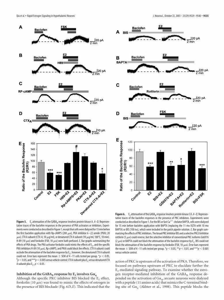
The rapid effect of E2 on the GABAB response involves proteinkinase AWe next examined the involvement of specific signaling proteinsin the E2-mediated modulation of GABAB. If activation of thePKA pathway is involved, then the effect of E2 on GABAB re-sponses should be blocked by inhibiting PKA and mimicked bystimulating PKA. To test this, we applied selective PKA activatorsand inhibitors. As shown in Figure 5, A and E, forskolin (10 �M)could mimic the actions of E2 to attenuate the GABAB response.However, the specific PKA inhibitor H-89 (10 �M) blocked theE2-induced suppression of the GABAB response (Fig. 5B,E). To
confirm the involvement of PKA in E2 modulation of GABAB
responses further, we dialyzed neurons with the specific PKA-inhibitory peptide PKI (PKA inhibitor 6 –22 amide, 20 �M) or thenonhydrolyzable cAMP analog Rp-cAMPS (200 �M) that blocksPKA activation. After �15 min of dialysis with PKI or Rp-cAMPS, the E2-induced reduction of the GABAB response wasabolished (Fig. 5C,E). CTX, which is a bacterial exotoxin secretedby vibrio cholerae, elevates intracellular cAMP levels in a variety oftissues by ADP ribosylating the G-protein Gs, thereby stimulatingadenylyl cyclase activity in an apparently irreversible manner.Intracellular dialysis with the active unit of CTX into individualcells occluded the rapid inhibition of GABAB response by estro-gen (Fig. 5D,E). These results indicate that the suppression of theGABAB response by E2 requires the activation of PKA.
Attenuation of the GABAB response involves proteinkinase C�We next examined whether activation of PKC is also critical forE2 modulation of the GABAB response using several selectivePKC inhibitors. The first, bisindolymaleimide, is a selective in-hibitor of PKC that does not distinguish among the conventional,novel, and atypical isoforms of PKC. The second, Go6976, is aselective inhibitor of the conventional PKC isoforms (Martiny-Baron et al., 1993; Way et al., 2000). Treatment of neurons withBIS nearly eliminated the effects of E2 (Fig. 6A,E). In contrast,Go6976 treatment was without effect (Fig. 6E). Indeed, whensimilar experiments were performed after replacing intracellularEGTA with 10 mM BAPTA, a calcium buffer with similar Ca 2�
affinity as EGTA but a much faster on rate, the estrogen inhibi-tion of the GABAB response was still observed (Fig. 6B,E). Be-cause conventional isoforms of PKC are unlikely to be active withthis level of calcium buffering, these results also support a role fora Ca 2�-independent, novel PKC isoform in mediating the effectsof estrogen. Finally, the selective PKC� inhibitor rottlerin (5 �M)completely blocked the ability of E2 to inhibit the GABAB re-sponse in hypothalamic neurons (Fig. 6C,E).
Table 1. Relative binding affinities of ligands to full-length ER� or ER�
Ligand
RBA
ER� ER�
17�-Estradiol 100 1004-Hydroxytamoxifen 36 43Raloxifene 34 76GW-5638 5 8STX 4.3E-6 9.0E-6
Relative binding affinities (RBA) are expressed as a percentage of the potency of 17�-estradiol. Under the experi-mental conditions described in Materials and Methods, 17�-estradiol was found to have an IC50 of 5 nM for ER� and3 nM for ER�.
Figure 4. Uteri are enlarged after estradiol treatment but not after STX or oil vehicle treat-ment. After a 48 hr treatment period, the uteri of wild-type C57BL/6 mice were collected andexamined. A, In E2-treated mice, there was a noticeable increase in uterine size after estradiolbenzoate (EB) compared with oil vehicle or STX treatment. B, Bar graph shows the uterineweights. **p � 0.01 versus oil-treated females (n � 3–5 mice per group).
9534 • J. Neurosci., October 22, 2003 • 23(29):9529 –9540 Qiu et al. • Rapid Estrogen Signaling in Hypothalamic Neurons
Inhibition of the GABAB response by E2 involves G�q
Although the specific PKC inhibitor BIS blocked the E2 effect,forskolin (10 �M) was found to mimic the effects of estrogen inthe presence of BIS blockade (Fig. 6D,E). This indicated that the
action of PKC is upstream of the activation of PKA. Therefore, wefocused on pathways upstream of PKC to elucidate further theE2-mediated signaling pathway. To examine whether the estro-gen receptor-mediated inhibition of the GABAB response de-pended on the activation of G�q, arcuate neurons were dialyzedwith a peptide (11 amino acids) that mimics the C-terminal bind-ing site of G�q (Akhter et al., 1998). This peptide blocks the
Figure 5. E2 attenuation of the GABAB response involves protein kinase A. A–D, Represen-tative traces of the baclofen responses in the presence of PKA activators or inhibitors. Experi-ments were conducted as described in Figure 1, except that cells were dialyzed for 15 min beforethe first baclofen application with Rp-cAMPS (200 �M), PKA inhibitor 6 –22 amide (PKAI; 20�M), CTX A subunit (CTX-A; 10 �g/ml), or denatured CTX A subunit (10 �g/ml, 100°C, 10 min).H-89 (10 �M) and forskolin (FSK; 10 �M) were bath perfused. E, Bar graphs summarizing theeffects of PKA drugs. The PKA activator forskolin could mimic the effects of E2 , and the specificPKA inhibitors H-89 (10 �M), Rp-cAMPS, and PKAI could block the effects. CTX A subunit couldocclude the attenuation of the baclofen response by E2 ; however, the denatured CTX A subunitcould not. Error bars represent the mean SEM of 4 –11 cells tested per group. *p � 0.05,#p � 0.05, and ***p � 0.005 versus vehicle control; CTX A subunit plus E2 versus denatured CTXA subunit plus E2 , p � 0.05.
Figure 6. E2 attenuation of the GABAB response involves protein kinase C�. A–D, Represen-tative traces of the baclofen responses in the presence of PKC inhibitors. Experiments wereconducted as described in Figure 1. For the BIS or fast Ca 2� chelator BAPTA, cells were dialyzedfor 15 min before baclofen application with BAPTA (replacing the 11 mM EGTA with 10 mM
BAPTA) or BIS (100 nM), which were included in the patch pipette solution. E, Bar graph sum-marizing the effects of PKC inhibitors. The broad PKC inhibitor BIS and a selective PKC� inhibitorrottlerin (5 �M) could reverse, but the selective inhibitor of conventional PKC isoforms Go6976(2 �M) or BAPTA could not block the attenuation of the baclofen response by E2. BIS could notblock the attenuation of the baclofen response by forskolin (FSK; 10 �M). Error bars representthe mean SEM of 4 –11 cells tested per group. *p � 0.05, **p � 0.01, and ***p � 0.005versus vehicle control.
Qiu et al. • Rapid Estrogen Signaling in Hypothalamic Neurons J. Neurosci., October 22, 2003 • 23(29):9529 –9540 • 9535

interaction between G-protein-coupled receptors and G�q pro-teins. In cells dialyzed with this peptide (200 �M), the E2-mediated reduction of the GABAB response was blocked signifi-cantly (Fig. 7A,E) compared with cells dialyzed with a control
peptide (11 amino acids) that mimics the C-terminal domain ofG�s (Fig. 7B,E).
In light of these results for a primary role for G�q in E2-mediated rapid inhibition, we tested whether the activation ofPLC, a well known G�q effector, might also play a role. To deter-mine whether the activation of PLC� is required for the estrogen-induced inhibition of GABAB response, neurons were treatedwith the broad-spectrum PLC inhibitor U73122 (10 �M).U73122 (10 �M) was perfused in the extracellular bathing media.Under these conditions, the estrogen-mediated reduction ofGABAB response was blocked (Fig. 7C,E), whereas the less activePLC inhibitor U73343 at the same concentration had no effect(Fig. 7D,E).
The attenuation of the GABAB response does not involveMAP kinaseRecent studies have shown that 17�-E2 rapidly activates the MAPkinase pathway in primary neuronal cortical cultures and in or-ganotypic cerebrocortical explant cultures (Watters et al., 1997;Singh et al., 1999, 2000). We therefore tested whether inhibitionof MAP kinase activity could prevent estrogen modulation of thebaclofen response. Treatment with MAP kinase inhibitorsPD98059 (10 �M, in the pipette) or U0126 (5 �M) did not affectE2 inhibition of baclofen responses (R2/R1 for E2, 58.6 3.4%,n � 10; vs PD98059 plus E2, 66.1 11.8%, n � 5).
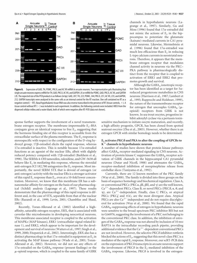
Expression of GABAB receptor PKC� and adenylate cyclaseVII transcripts in arcuate (GABA, dopamine, andPOMC) neuronsUsing single-cell RT-PCR from 75 acutely dispersed arcuate neu-rons, we found that 90% of the neurons expressed GAD65 tran-scripts, including TH-expressing and POMC-expressing neurons(data not shown). Most importantly, 92% of the neurons ex-pressed GABAB-R2 transcripts, which correlates with the 90%response rate to baclofen. Furthermore, we also determined thatdopamine and POMC neurons express PKC� and adenylyl cy-clase VII transcripts using single-cell RT-PCR. In one group ofcells (n � 22), we found that PKC� and adenylyl cyclase VIItranscripts are expressed in the majority (70%) of TH neurons(Fig. 8A), including those that coexpress GAD65. TH and GADwere colocalized in 60% of this population of neurons because ofa limited amount of cDNA from individual neurons, POMC ex-pression was determined in another group of cells (n � 29), andwe found that PKC� and adenylyl cyclase VII transcripts wereexpressed in the majority (75%) of POMC neurons, includingthose that coexpress GAD65 (Fig. 8B). POMC and GAD werecolocalized in 28% of this population of neurons. Therefore, thesingle-cell RT-PCR data support the electrophysiological find-ings that dopamine and POMC neurons express the critical tran-scripts for rapid estrogen signaling.
DiscussionA unique membrane estrogen receptor mediates the rapideffects of E2
Estrogen suppresses the action of the GABAB receptor agonistbaclofen to activate GIRK channels in GABA, POMC, and dopa-mine neurons. This E2 effect is rapid, with measurable suppres-sion occurring within minutes after addition of E2. The kinetics ofthis response support the notion that a membrane E2 receptor ismediating the response and argue against the involvement of theclassical nuclear estrogen receptors operating by transcriptionregulation.
The pharmacology we observed for this rapid estrogen re-
Figure 7. E2 attenuation of the GABAB response depends on activation of G�q , and PKCactivation is in the upstream of PKA. A–D, Representative traces of the baclofen responses in thepresence of PLC and G�q inhibitors. Experiments were conducted as described in Figure 1. Cellswere dialyzed for 15 min before baclofen application with G�q (200 �M) or G�s peptide (200�M). E, Bar graphs summarizing the effects of PLC and Gq peptide. The PLC inhibitor U73122 (10�M) and G�q peptide could inhibit, but the inactive analog of U73122, U73343 (10 �M), andG�s peptide could not block the attenuation of the baclofen response by E2. Error bars representthe mean SEM of 4 –11 cells tested per group. **p � 0.01 and ***p � 0.005 versus vehiclecontrol; U73122 plus E2 versus U73343 plus E2 , p � 0.05; Gq peptide plus E2 versus Gs peptideplus E2 , p � 0.05.
9536 • J. Neurosci., October 22, 2003 • 23(29):9529 –9540 Qiu et al. • Rapid Estrogen Signaling in Hypothalamic Neurons
sponse further supports the involvement of a novel transmem-brane estrogen receptor. The membrane-impermeable E2–BSAconjugate gives an identical response to free E2, suggesting thatthe hormone-binding site of this receptor is accessible from theextracellular surface of the plasma membrane. The E2 response isstereospecific with respect to the configuration of the D-ring hy-droxyl group; 17�-estradiol elicits the rapid response, whereas17�-estradiol is inactive. This is notable because 17�-estradiolfunctions as an agonist of the nuclear ERs, albeit with slightlyreduced potency compared with 17�-estradiol (Barkhem et al.,1998). The SERMs 4-OH tamoxifen, raloxifene, and GW-5638 allbehave like E2 in mediating this response, whereas the steroidalanti-estrogen ICI 182,780 antagonizes the E2 response. Most im-portantly, the novel SERM STX that is devoid of estrogen (oranti-estrogen) activity with the nuclear ERs is a stronger activatorof this rapid E2 response than E2, even at a 10-fold lower concen-tration. Moreover, we know that this membrane ER has a sub-nanomolar affinity for estrogen on the basis of our pharmacolog-ical (Schild) analysis (Lagrange et al., 1997). These resultsdemonstrate that the pharmacology of this rapid response is dif-ferent and, in the case of STX, separable from that of the nuclearERs (Razandi et al., 1999; Levin, 2001; Chambliss and Shaul,2002).
Recently, Toran-Allerand et al. (2002) identified a high-affinity, saturable estrogen receptor, ERX, that is associated withcaveolar-like microdomains in developing neocortical neurons.This membrane-associated receptor is coupled to the activationof MAPKs (MAP kinases), ERK1 (extracellular-signal related ki-nase 1), and ERK2, which appear to be important for the devel-opment and survival of neurons (Watters et al., 1997; Singh et al.,1999, 2000; Fitzpatrick et al., 2002). Interestingly, ERX also has adistinct pharmacology in that 17�-estradiol is equipotent as E2 inactivating the MAP kinase pathway (Wade et al., 2001; Toran-Allerand et al., 2002). However, we did not see any effects of17�-estradiol on the GABAB response (present findings) or the�-opioid response, which is coupled to the same family of GIRK
channels in hypothalamic neurons (La-grange et al., 1997). Similarly, Gu andMoss (1996) found that 17�-estradiol didnot mimic the actions of E2 in the hip-pocampus to potentiate the glutamate(kainate)-mediated currents in CA1 pyra-midal neurons. Likewise, Mermelstein etal. (1996) found that 17�-estradiol wasmuch less efficacious than E2 in reducingL-type calcium currents in neostriatal neu-rons. Therefore, it appears that the mem-brane estrogen receptor that modulateschannel activity in neurons via the PKC–PKA pathway is pharmacologically dis-tinct from the receptor that is coupled toactivation of ERK1 and ERK2 that pro-motes growth and survival.
Although the GABAA ionotropic recep-tor has been identified as a target for 5�-reduced progesterone metabolites in CNSneurons (Harrison et al., 1987; Lambert etal., 1995; Rupprecht and Holsboer, 1999),the nature of the transmembrane receptorfor estrogen that uncouples GABAB (�-opioid) receptors from GIRK is notknown. In sea trout oocytes, progestins in-hibit adenylyl cyclase via a pertussis toxin-
sensitive mechanism to initiate oocyte maturation, and recently,a high-affinity progestin, GPCR, has been cloned from spottedseatrout oocytes (Zhu et al., 2003). However, whether there is anestrogen GPCR with similar homology needs to be determined.
E2 activates PKC� and PKA to alter the coupling of GPCRs toK � channels in hypothalamic neuronsA number of studies have shown that protein kinase pathwaysaffect GABAB receptor-mediated signaling in CNS neurons. Ac-tivation of protein kinase C suppresses the GABAB receptor acti-vation of GIRK channels in the hippocampal CA1 pyramidalneurons (Dutar and Nicoll, 1988) and attenuates the GABAB
receptor-mediated inhibition of norepinephrine release fromcerebellar slices (Taniyama et al., 1992).
Currently, there are 12 known members of the PKC family(Way et al., 2000). The family is divided into three groups on thebasis of sequence homology and biochemical regulation. Class A,or conventional PKCs (PKC�, �I, �II, and �) are the well known,Ca 2�-dependent PKCs. Class B, or novel PKCs (PKC�, �, �, and), are Ca 2�-independent. Finally, class C PKCs, or atypicalPKCs (PKC and �/�), are the most divergent class. AtypicalPKCs are also Ca 2� independent and do not require diacylglyc-erol for activation (Way et al., 2000). We found that the rapidGABAB-suppressing effects of estrogen in hypothalamic neuronswere sensitive to the broad-spectrum PKC inhibitor BIS but notto Go6976, suggesting the involvement of a PKC not belonging tothe conventional PKC class. In addition, the inhibition of estro-gen of the GABAB response was not altered by inclusion of 10 mM
BAPTA in the intracellular recording patch pipette, providingadditional evidence that the Ca 2�-dependent conventional PKCsare not involved. However, the selective PKC� inhibitor rottlerinblocked the actions of E2, suggesting that this novel-class PKC is amediator of the rapid E2 response. Moreover, our scRT-PCR dataon the expression of PKC� transcripts in arcuate neurons supportthe involvement of PKC� in the E2-mediated inhibition of theGABAB response. Likewise, PKC� is involved in the estrogen-
Figure 8. Expression of GAD, TH, POMC, PKC�, and AC VII mRNA in arcuate neurons. Two representative gels illustrating thatsingle arcuate neurons express mRNA for TH, GAD, PKC�, AC VII, and GAPDH ( A) or mRNA for POMC, GAD, PKC�, AC VII, and GAPDH( B). The expected size of the PCR products is as follows (in bp): GAD, 207; TH, 223; POMC, 344; PKC�, 251; AC VII, 235; and GAPDH,212. GAPDH transcripts were analyzed in the same cells as an internal control for the RT reaction. One cell contained no RT as anegative control (�RT). Basal hypothalamic tissue RNA was also reverse transcribed in the presence of RT (tissue controls, �). Atissue control without RT (�) was included in each experiment. In addition, the following controls were included: HBSS from thedispersed cellular milieu and a water blank, both of which were negative after RT-PCR (data not shown).
Qiu et al. • Rapid Estrogen Signaling in Hypothalamic Neurons J. Neurosci., October 22, 2003 • 23(29):9529 –9540 • 9537

mediated inhibition of K� channels andfluid retention in female distal colonic ep-ithelial cells, although the upstream signal-ing pathway is not known (Doolan et al.,2000).
PKC activation is in the upstream ofPKA activationIn our study, internal perfusion of BIScould completely block the inhibition ofthe baclofen response by E2 but could notattenuate the inhibition of the baclofen re-sponse by forskolin applied via bath perfu-sion. PKC is known to activate adenylylcyclases (Jacobowitz et al., 1993; Yo-shimura and Cooper, 1993; Lin and Chen,1998); moreover, when AC is activated byPKC instead of by G�s or forskolin, it isresistant to inhibition by G�i (Pieroni etal., 1993). To date, nine AC isozymes havebeen cloned (AC types I–IX). Notably, ACVII has a potential binding site for PKC�that is not present in the sequences of theother adenylyl cyclases, which would allowPKC� to directly phosphorylate AC VII(Nelson et al., 2003). Interestingly, GABAneurons in the cortex, hippocampus, stri-atum, and cerebellum are immunoreactivefor AC VII (Mons et al., 1998), and, in thepresent study, we show that hypothalamicGABA, TH, and POMC neurons expressAC VII transcripts.
G�q mediates the inhibition of theGABAB response by E2 through PLCMost PKCs are activated by diacylglycerol,and some require the presence of Ca 2�.Thus, PKCs are downstream of the PLC–inositol triphosphate– diacylglycerol signaling cascade. Becausedifferent forms of PLC can be activated by various messengers,including G�q, G�� (PLC�), and tyrosine kinases (PLC�), thePKC family is involved in a diverse array of signaling cascades(Tanaka and Nishizuka, 1994; Battaini, 2001). Our results showthat a membrane ER is specifically coupled to G�q protein. Thisconclusion is based on experiments in which intracellular dialysiswith a peptide fragment of G�q blocked the receptor interactionwith G-protein. This G�q peptide has been used to block G�q
signaling pathways in cortical pyramidal neurons (Carr et al.,2002). In addition, the estrogen-mediated reduction of theGABAB response was significantly reduced by the phospholipaseC inhibitor U73122 compared with cells perfused with the lessactive inhibitor U73343.
Therefore, from the collective results of this study, we formu-late the signal transduction pathway for the rapid response toestrogen in hypothalamic neurons depicted in Figure 9. The se-quence of events in this model are as follows: (1) E2 binds to anovel transmembrane estrogen receptor; (2) ligand binding acti-vates G�q; (3) activated G�q in turn activates PLC; (4) activatedPLC liberates DAG; (5) free DAG stimulates PKC�; (6) PKC�activates adenylyl cyclase (VII); (7) cAMP levels are elevated; (8)cAMP stimulates PKA; and (9) PKA phosphorylates membranetargets critical for K� channel function.
Functional significance of rapid membrane effects of E2 inCNS neuronsIt was discovered previously that E2 could rapidly modulate syn-aptic efficacy via activation of PKA (Gu and Moss, 1996, 1998;Lagrange et al., 1997; Kelly et al., 1999). Presently, we delineatedthe upstream components of this signaling pathway that includesGq, phospholipase C, and PKC� activation (Fig. 9). This is a novelsignaling pathway for E2 to rapidly modulate hypothalamic neu-ronal excitability, and there is most likely a similar E2 signalingpathway in hippocampal CA1 neurons (Gu and Moss, 1996,1998). Therefore, we believe that this pathway is important forincreasing synaptic efficacy not only in hypothalamic neuronsbut also in other neurons in the CNS. In addition, we identified aspecific ligand (STX) that is selective for activating this pathway.The consequences of STX effects in hypothalamic neurons areevident in that these neurons are involved in controlling the ovu-latory cycle, lactation, stress responses, temperature, and energybalance, all of which require rapid feedback regulation by estro-gen. Furthermore, having a selective E2 agonist for rapid signalingis critical because SERMs such as tamoxifen and raloxifene in-crease the incidence of hot flashes in women, suggesting that theyact as E2 antagonists in the hypothalamus (Stearns et al., 2002;Sherwin, 2003). In addition, raloxifene treatment is no betterthan placebo treatment in maintaining cognitive performance ofpostmenopausal women (Sherwin, 2003), which suggests that
Figure 9. A cellular model of the rapid signaling of estrogen in hypothalamic neurons. Schematic overview showing the rapidversus delayed ER-mediated modulation of neurotransmitter-regulated G-protein-coupled receptors via a membrane-associatedER in hypothalamic neurons. Rapid signaling: E2 activates a membrane-associated ER that is G�q-coupled to activation of phos-pholipase C that catalyzes the hydrolysis of membrane-bound phosphatidylinositol 4,5-biphosphate (PIP2 ) to inositol 1,4,5triphosphate (IP3 ) and DAG. Calcium is released from intracellular stores (endoplasmic reticulum) by IP3 , and DAG activates PKC�.Through phosphorylation, AC VII activity is upregulated by PKC�. The generation of cAMP activates PKA, which can rapidlyuncouple GABAB and �-opioid (�) receptors from their effector system through phosphorylation of a downstream effectormolecule (e.g., the inwardly rectifying K � channel, or GIRK). ER-mediated modulation of kinase pathways either reduces thecapacity of neuromodulators such as GABA and �-endorphin (�End) to inhibit hypothalamic neuronal excitability, or augmentsthe ability of neurotransmitters such as glutamate to increase neuronal excitability (data not shown). Delayed signaling: rapidER-mediated activation of PKA can lead to phosphorylation of cAMP-responsive element-binding protein (pCREB), which can thenalter gene transcription through its interaction with the cAMP-responsive element (CRE). Moreover, other isoforms of PKC canphosphorylate raf-1 (Raf), leading to activation of the MAP kinase pathway and new gene transcription and protein (e.g., GPCRs)synthesis in an estrogen-response element-independent manner.
9538 • J. Neurosci., October 22, 2003 • 23(29):9529 –9540 Qiu et al. • Rapid Estrogen Signaling in Hypothalamic Neurons

raloxifene is not an E2 agonist in hippocampus. Most impor-tantly, raloxifene and tamoxifen bind to ER� and ER� with highaffinity (Barkhem et al., 1998). In contrast, the STX (E2) receptoris similarly coupled as the serotonin 5HT2A,C receptor (Carr et al.,2002) in CNS neurons, which may explain the ability of serotoninuptake inhibitors (SSRIs) to prevent hot flashes in postmeno-pausal women (Stearns et al., 2002). Hence, we would predictthat STX would prevent hot flashes, maintain sleep cycles, elevatemood, etc. Therefore, this rapid PLC–PKC�–PKA signaling of E2
may synergize with CNS transmitter systems to enhance synapticefficacy in brain circuits that are critical for maintaining homeo-static functions.
ReferencesAkema T, Chiba A, Kimura F (1990) On the relationship between noraden-
dritic stimulatory and GABAergic inhibitory systems in the control ofluteinizing hormone secretion in female rats. Neuroendocrinology52:566 –572.
Akhter SA, Luttrell LM, Rockman HA, Iaccarino G, Lefkowitz RJ, Koch WJ(1998) Targeting the receptor-Gq interface to inhibit in vivo pressureoverload myocardial hypertrophy. Science 280:574 –577.
Barkhem T, Carlsson B, Nilsson Y, Enmark E, Gustafsson JÅ, Nilsson S(1998) Differential response of estrogen receptor � and estrogen recep-tor � to partial estrogen agonists/antagonists. Mol Pharmacol54:105–112.
Battaini F (2001) Protein kinase C isoforms as therapeutic targets in nervoussystem disease states. Pharmacol Res 44:353–361.
Bjorklund A, Lindvall O (1984) Dopamine-containing systems in the CNS.In: Handbook of chemical neuroanatomy: classical transmitters in theCNS, Pt 1 (Bjorklund A, Hokfelt T, eds), pp 55–122. Amsterdam: Elsevier.
Carr DB, Cooper DC, Ulrich SL, Spruston N, Surmeier DJ (2002) Serotoninreceptor activation inhibits sodium current and dendritic excitability inprefrontal cortex via a protein kinase C-dependent mechanism. J Neuro-sci 22:6846 – 6855.
Chambliss KL, Shaul PW (2002) Estrogen modulation of endothelial nitricoxide synthase. Endocr Rev 23:665– 686.
Dave JR, Rubinstein N, Eskay RL (1985) Evidence that �-endorphin bindsto specific receptors in rat peripheral tissues and stimulates the adenylatecyclase-adenosine 3�, 5�-monophosphate system. Endocrinology117:1389 –1396.
Demotes-Mainard J, Arnauld E, Vincent JD (1990) Estrogens modulate theresponsiveness of in vivo recorded striatal neurons to iontophoretic ap-plication of dopamine in rats: role of D1 and D2 receptor activation.J Neuroendocrinol 2:825– 832.
Doolan CM, Condliffe SB, Harvey BJ (2000) Rapid non-genomic activationof cytosolic cyclic AMP-dependent protein kinase activity and [Ca 2�]i by17�-oestradiol in female rat distal colon. Br J Pharmacol 129:1375–1386.
Dutar P, Nicoll RA (1988) Pre- and postsynaptic GABA(B) receptors in thehippocampus have different pharmacological properties. Neuron1:585–591.
Ferin M, Van Vugt D, Wardlaw S (1984) The hypothalamic control of themenstrual cycle and the role of endogenous opioid peptides. Recent ProgHorm Res 40:441– 485.
Fitzpatrick JL, Mize AL, Wade CB, Harris JA, Shapiro RA, Dorsa DM (2002)Estrogen-mediated neuroprotection against �-amyloid toxicity requiresexpression of estrogen receptor � or � and activation of the MAPK path-way. J Neurochem 82:674 – 682.
Gu Q, Moss RL (1996) 17�-Estradiol potentiates kainate-induced currentsvia activation of the cAMP cascade. J Neurosci 16:3620 –3629.
Gu Q, Moss RL (1998) Novel mechanism for non-genomic action of 17�-estradiol on kainate-induced currents in isolated rat CA1 hippocampalneurones. J Physiol (Lond) 506:745–754.
Harrison NL, Majewska MD, Harrrington JW, Barker JL (1987) Structure-activity relationships for steroid interaction with the gamma-aminobutyric acidA receptor complex. J Pharmacol Exp Ther241:346 –353.
Herbison AE (1997) Estrogen regulation of GABA transmission in rat pre-optic area. Brain Res Bull 44:321–326.
Herbison AE, Horvath TL, Naftolin F, Leranth C (1995) Distribution ofestrogen receptor-immunoreactive cells in monkey hypothalamus: rela-
tionship to neurones containing luteinizing hormone-releasing hormoneand tyrosine hydroxylase. Neuroendocrinology 61:1–10.
Herbison AE, Skynner MJ, Sim JA (2001) Lack of detection of estrogenreceptor-� transcripts in mouse gonadotropin-releasing hormone neu-rons. Endocrinology [Erratum] 142:492– 493.
Hokfelt T, Mårtensson R, Bjorklund A, Kleinau S, Goldstein M (1984) Dis-tributional maps of tyrosine-hydroxylase-immunoreactive neurons inthe rat brain. In: Handbook of chemical neuroanatomy (Bjorklund A,Hokfelt T, eds), pp 277–379. Amsterdam: Elsevier.
Jacobowitz O, Chen J, Premont RT, Iyengar R (1993) Stimulation of specifictypes of Gs-stimulated adenylyl cyclases by phorbol ester treatment. J BiolChem 268:3829 –3832.
Jarry H, Leonhardt S, Wuttke W (1995) The inhibitory effect of�-endorphin on LH release in ovariectomized rats does not involve thepreoptic GABAergic system. Exp Clin Endocrinol Diabetes 103:317–323.
Katzenellenbogen JA, Carlson KE, Katzenellenbogen BS (1985) Facile geo-metric isomerization of phenolic non-steroidal estrogens and antiestro-gens: limitations to the interpretation of experiments characterizing theactivity of individual isomers. J Steroid Biochem 22:589 –596.
Kelly MJ, Wagner EJ (1999) Estrogen modulation of G-protein-coupled re-ceptors. Trends Endocrinol Metab 10:369 –374.
Kelly MJ, Rønnekleiv OK (1994) Electrophysiological analysis of neuroen-docrine neuronal activity in hypothalamic slices. In: Methods in neuro-sciences: pulsatility in neuroendocrine systems (Levine JE, ed), pp 47– 67.San Diego: Academic.
Kelly MJ, Rønnekleiv OK, Eskay RL (1984) Identification of estrogen-responsive LHRH neurons in the guinea pig hypothalamus. Brain ResBull 12:399 – 407.
Kelly MJ, Loose MD, Rønnekleiv OK (1990) Opioids hyperpolarize�-endorphin neurons via mu-receptor activation of a potassium conduc-tance. Neuroendocrinology 52:268 –275.
Kelly MJ, Loose MD, Rønnekleiv OK (1992) Estrogen suppresses �-opioidand GABAB-mediated hyperpolarization of hypothalamic arcuate neu-rons. J Neurosci 12:2745–2750.
Kelly MJ, Lagrange AH, Wagner EJ, Rønnekleiv OK (1999) Rapid effects ofestrogen to modulate G-protein-coupled receptors via activation of pro-tein kinase A and protein kinase C pathways. Steroids 64:64 –75.
Lagrange AH, Rønnekleiv OK, Kelly MJ (1994) The potency of �-opioidhyperpolarization of hypothalamic arcuate neurons is rapidly attenuatedby 17�-estradiol. J Neurosci 14:6196 – 6204.
Lagrange AH, Rønnekleiv OK, Kelly MJ (1995) Estradiol-17� and mu-opioid peptides rapidly hyperpolarize GnRH neurons: a cellular mecha-nism of negative feedback? Endocrinology 136:2341–2344.
Lagrange AH, Wagner EJ, Rønnekleiv OK, Kelly MJ (1996) Estrogen rapidlyattenuates a GABAB response in hypothalamic neurons. Neuroendocri-nology 64:114 –123.
Lagrange AH, Rønnekleiv OK, Kelly MJ (1997) Modulation of G-protein-coupled receptors by an estrogen receptor that activates protein kinase A.Mol Pharmacol 51:605– 612.
Lambert JJ, Belelli D, Hill-Venning C, Peters JA (1995) Neurosteroids andGABAA receptor function. Trends Pharmacol Sci 16:295–303.
Leranth C, MacLusky NJ, Brown TJ, Chen EC, Redmond Jr DE, Naftolin F(1992) Transmitter content and afferent connections of estrogen-sensitive progestin receptor-containing neurons in the primate hypothal-amus. Neuroendocrinology 55:667– 682.
Levin ER (2001) Cell localization, physiology, and nongenomic actions ofestrogen receptors. J Appl Physiol 91:1860 –1867.
Lin W-W, Chen BC (1998) Distinct PKC isoforms mediate the activation ofcPLA2 and adenylyl cyclase by phorbol ester in RAW264.7 macrophages.Br J Pharmacol 125:1601–1609.
Loose MD, Rønnekleiv OK, Kelly MJ (1990) Membrane properties and re-sponse to opioids of identified dopamine neurons in the guinea pig hy-pothalamus. J Neurosci 10:3627–3634.
Loose MD, Rønnekleiv OK, Kelly MJ (1991) Neurons in the rat arcuatenucleus are hyperpolarized by GABAB and mu-opioid receptor agonists:evidence for convergence at a ligand-gated potassium conductance. Neu-roendocrinology 54:537–544.
Martiny-Baron G, Kazanietz MG, Mischak H, Blumberg PM, Kochs G, HugH, Marme D, Schachtele C (1993) Selective inhibition of protein kinaseC isozymes by the indolocarbazole Go6976. J Biol Chem 268:9194 –9197.
McEwen BS (2001) Estrogens effects on the brain: multiple sites and molec-ular mechanisms. J Appl Physiol 91:2785–2801.
Qiu et al. • Rapid Estrogen Signaling in Hypothalamic Neurons J. Neurosci., October 22, 2003 • 23(29):9529 –9540 • 9539

Mermelstein PG, Becker JB, Surmeier DJ (1996) Estradiol reduces calciumcurrents in rat neostriatal neurons via a membrane receptor. J Neurosci16:595– 604.
Mitsushima D, Marzban F, Luchansky LL, Burich AJ, Keen KL, Durning M,Golos TG, Terasawa E (1996) Role of glutamic acid decarboxylase in theprepubertal inhibition of the luteinizing hormone releasing hormone re-lease in female rhesus monkeys. J Neurosci 16:2563–2573.
Mons N, Yoshimura M, Ikeda H, Hoffman PL, Tabakoff B (1998) Immu-nological assessment of the distribution of type VII adenylyl cyclase inbrain. Brain Res 788:251–261.
Morrell JI, McGinty JF, Pfaff DW (1985) A subset of �-endorphin ordynorphin-containing neurons in the medial basal hypothalamus accu-mulates estradiol. Neuroendocrinology 41:417– 426.
Neill JD (1980) Neuroendocrine regulation of prolactin secretion. FrontNeuroendocrinol 6:129 –155.
Nelson EJ, Hellevuo K, Yoshimura M, Tabakoff B (2003) Ethanol-inducedphosphorylation and potentiation of the activity of type 7 adenylyl cy-clase: involvement of PKC delta. J Biol Chem 278:4552– 4560.
Paradiso K, Zhang J, Steinbach JH (2001) The C terminus of the humannicotinic �4�2 receptor forms a binding site required for potentiation byan estrogenic steroid. J Neurosci 21:6561– 6568.
Pieroni JP, Jacobowitz O, Chen J, Iyengar R (1993) Signal recognition andintegration by Gs-stimulated adenylyl cyclases. Curr Opin Neurobiol3:345–351.
Razandi M, Pedram A, Greene GL, Levin ER (1999) Cell membrane andnuclear estrogen receptors (ERs) originate from a single transcript: stud-ies of ER� and ER� expressed in Chinese hamster ovary cells. Mol Endo-crinol 13:307–319.
Rupprecht R, Holsboer F (1999) Neuroactive steroids: mechanisms of ac-tion and neuropsychopharmacological perspectives. Trends Neurosci22:410 – 416.
Seltzer AM, Donoso AO (1992) Restraining action of GABA on estradiol-induced LH surge in the rat: GABA activity in brain nuclei and effects ofGABA mimetics in the medial preoptic nucleus. Neuroendocrinology55:28 –34.
Sherwin BB (2003) Estrogen and cognitive functioning in women. EndocrRev 24:133–151.
Simonian SX, Spratt DP, Herbison AE (1999) Identification and character-ization of estrogen receptor �-containing neurons projecting to the vicin-ity of the gonadotropin-releasing hormone perikarya in the rostral pre-optic area of the rat. J Comp Neurol 411:346 –358.
Singh M, Setalo Jr G, Guan X, Warren M, Toran-Allerand CD (1999)Estrogen-induced activation of mitogen-activated protein kinase in cere-bral cortical explants: convergence of estrogen and neurotrophin signal-ing pathways. J Neurosci 19:1179 –1188.
Singh M, Setalo GJ, Guan X, Frail DE, Toran-Allerand CD (2000) Estrogen-induced activation of the mitogen-activated protein kinase cascade in thecerebral cortex of estrogen receptor-� knock-out mice. J Neurosci20:1694 –1700.
Stearns V, Ullmer L, Lopez JF, Smith Y, Isaacs C, Hayes D (2002) Hotflushes. Lancet 360:1851–1861.
Sullivan KA, Witkin JW, Ferin M, Silverman AJ (1995) Gonadotropin-releasing hormone neurons in the rhesus macaque are not immunoreac-tive for the estrogen receptor. Brain Res 685:198 –200.
Takano K, Asano S, Yamashita N (1994) Activation of G-protein-coupled
K � channels by dopamine in human GH-producing cells. Am J Physiol266:E318 –E325.
Tanaka C, Nishizuka Y (1994) The protein kinase C family for neuronalsignaling. Annu Rev Neurosci 17:551–567.
Taniyama K, Niwa M, Kataoka Y, Yamashita K (1992) Activation of proteinkinase C suppresses the gamma-aminobutyric acid B receptor-mediatedinhibition of the vesicular release of noradrenaline and acetylcholine.J Neurochem 58:1239 –1245.
Toran-Allerand CD, Guan X, MacLusky NJ, Horvath TL, Diano S, Singh M,Connolly Jr ES, Nethrapalli IS, Tinnikov AA (2002) ER-X: a novel,plasma membrane-associated, putative estrogen receptor that is regulatedduring development and after ischemic brain injury. J Neurosci22:8391– 8401.
Valverde MA, Rojas P, Amigo J, Cosmelli D, Orio P, Bahamonde MI, MannGE, Vergara C, Latorre R (1999) Acute activation of Maxi-K channels(hSlo) by estradiol binding to the � subunit. Science 285:1929 –1931.
Wade CB, Robinson S, Shapiro RA, Dorsa DM (2001) Estrogen receptor(ER)alpha and ERbeta exhibit unique pharmacologic properties whencoupled to activation of the mitogen-activated protein kinase pathway.Endocrinology 142:2336 –2342.
Wagner EJ, Manzanares J, Moore KE, Lookingland KJ (1994) Neurochem-ical evidence that estrogen-induced suppression of kappa-opioid-receptor-mediated regulation of tuberoinfundibular dopaminergic neu-rons is prolactin-independent. Neuroendocrinology 59:197–201.
Wagner EJ, Bosch MA, Kelly MJ, Rønnekleiv OK (1999) A powerful GABAB
receptor-mediated inhibition of GABAergic neurons in the arcuate nu-cleus. NeuroReport 10:2681–2687.
Wagner EJ, Reyes-Vazquez C, Rønnekleiv OK, Kelly MJ (2000) The role ofintrinsic and agonist-activated conductances in determining the firingpatterns of preoptic area neurons in the guinea pig. Brain Res 879:29 – 41.
Wagner EJ, Rønnekleiv OK, Bosch MA, Kelly MJ (2001) Estrogen biphasi-cally modifies hypothalamic GABAergic function concomitantly withnegative and positive control of luteinizing hormone release. J Neurosci21:2085–2093.
Watson RE, Langub MC, Landis JW (1992) Further evidence that most lu-teinizing hormone-releasing hormone neurons are not directly estrogen-responsive: simultaneous localization of luteinizing hormone-releasinghormone and estrogen receptor immunoreactivity in the guinea-pigbrain. J Neuroendocrinol 4:311–317.
Watters JJ, Campbell JS, Cunningham MJ, Krebs EG, Dorsa DM (1997)Rapid membrane effects of steroids in neuroblastoma cells: effects ofestrogen on mitogen-activated protein kinase signaling cascade and c-fosimmediate early gene transcription. Endocrinology 138:4030 – 4033.
Way KJ, Chou E, King GL (2000) Identification of PKC-isoform-specificbiological actions using pharmacological approaches. Trends PharmacolSci 21:181–187.
Wetzel CH, Hermann B, Behl C, Pestel E, Rammes G, Zieglgansberger W,Holsboer F, Rupprecht R (1998) Functional antagonism of gonadal ste-roids at the 5-hydroxytryptamine type 3 receptor. Mol Endocrinol12:1441–1451.
Yoshimura M, Cooper DM (1993) Type-specific stimulation of adenylyl cy-clase by protein kinase C. J Biol Chem 268:4604 – 4607.
Zhu Y, Rice CD, Pang Y, Pace M, Thomas P (2003) Cloning, expression, andcharacterization of a membrane progestin receptor and evidence it is anintermediary in meiotic maturation of fish oocytes. Proc Natl Acad SciUSA 100:2231–2236.
9540 • J. Neurosci., October 22, 2003 • 23(29):9529 –9540 Qiu et al. • Rapid Estrogen Signaling in Hypothalamic Neurons