trp channel regulates egfr signaling in hair...
TRANSCRIPT
TRP Channel Regulates EGFRSignaling in Hair Morphogenesisand Skin Barrier FormationXiping Cheng,1,8 Jie Jin,2,3,8 Lily Hu,1 Dongbiao Shen,1 Xian-ping Dong,1 Mohammad A. Samie,1 Jayne Knoff,1
Brian Eisinger,1 Mei-ling Liu,1 Susan M. Huang,4 Michael J. Caterina,4 Peter Dempsey,5 Lowell Evan Michael,6
Andrzej A. Dlugosz,6 Nancy C. Andrews,2,7 David E. Clapham,3,* and Haoxing Xu1,3,*1The Department of Molecular, Cellular, and Developmental Biology, the University of Michigan, 3089 Natural Science Building (Kraus),
830 North University, Ann Arbor, MI 48109, USA2Division of Hematology and Oncology, Children’s Hospital Boston, Karp Family Building 8-125A, Boston, MA 02115, USA3The Department of Cardiology, Children’s Hospital Boston, Manton Center for Orphan Disease, Enders 1350, 320 Longwood Avenue,
Boston, MA 02115, USA4Department of Biological Chemistry and Department of Neuroscience, Johns Hopkins University School of Medicine, Baltimore,
MD 21025, USA5The Department of Pediatrics and Communicable Diseases and Department of Molecular & Integrative Physiology,
the University of Michigan, 1500 East Medical Center Drive, Room D3252, Ann Arbor, MI 48109, USA6The Department of Dermatology and Comprehensive Cancer Center, the University of Michigan, 1500 East Medical Center Drive, Ann Arbor,
MI 48109, USA7Department of Pediatrics and Department of Pharmacology and Cancer Biology, Duke University School of Medicine, DUMC 2927, Durham,
NC 27710, USA8These authors contributed equally to this work
*Correspondence: [email protected] (D.E.C.), [email protected] (H.X.)
DOI 10.1016/j.cell.2010.03.013
SUMMARY
A plethora of growth factors regulate keratinocyteproliferation and differentiation that control hairmorphogenesis and skin barrier formation. Wavyhair phenotypes in mice result from naturally occur-ring loss-of-function mutations in the genes forTGF-a and EGFR. Conversely, excessive activitiesof TGF-a/EGFR result in hairless phenotypes andskin cancers. Unexpectedly, we found that micelacking the Trpv3 gene also exhibit wavy hair coatand curly whiskers. Here we show that keratinocyteTRPV3, a member of the transient receptor potential(TRP) family of Ca2+-permeant channels, forms asignaling complex with TGF-a/EGFR. Activation ofEGFR leads to increased TRPV3 channel activity,which in turn stimulates TGF-a release. TRPV3 isalso required for the formation of the skin barrier byregulating the activities of transglutaminases, afamily of Ca2+-dependent crosslinking enzymesessential for keratinocyte cornification. Our resultsshow that a TRP channel plays a role in regulatinggrowth factor signaling by direct complex formation.
INTRODUCTION
Skin and its appendages provide a protective barrier essential
for animal survival. Hair morphogenesis and epidermal develop-
ment are orchestrated by an array of cytokines and growth
factors (Fuchs and Raghavan, 2002). Signaling by these diffus-
ible molecules provides spatially and temporally controlled
cellular programs for keratinocyte proliferation, differentiation,
migration, and finally, terminal differentiation and cornification.
TGF-a and epidermal growth factor (EGF) are related auto-
crine/paracrine growth factors that activate the EGF receptor
(EGFR; ErbB1) to regulate the balance between keratinocyte
proliferation and differentiation (Schneider et al., 2008). Defec-
tive TGF-a/EGFR signaling leads to abnormal hair morphogen-
esis, manifested by the ‘‘wavy hair’’ and ‘‘curly whiskers’’ pheno-
types of spontaneous loss-of-function mouse mutations in
TGF-a (named waved-1 or wa1) and in EGFR (named waved-2
or wa2), respectively (Ballaro et al., 2005; Luetteke et al., 1993,
1994; Mann et al., 1993; Murillas et al., 1995; Schneider et al.,
2008; Sibilia and Wagner, 1995; Threadgill et al., 1995). Exces-
sive activities of TGF-a/EGFR cause a hairless phenotype and
skin cancers (Ferby et al., 2006; Schneider et al., 2008).
The mechanisms by which TGF-a/EGFR signaling determines
cell fate (proliferation versus differentiation) of follicular and
interfollicular (epidermal) keratinocytes are not completely
understood.
Accumulated evidence suggests that both negative and
positive feedback mechanisms coexist in the TGF-a/EGFR
signaling axis. EGF binding triggers rapid degradation of the
EGFR through endocytic pathways but also leads to further
production and release/shedding of TGF-a/EGF (Coffey et al.,
1987; Peschon et al., 1998). This unique autoinduction mecha-
nism may contribute to the effects of TGF-a/EGF on keratinocyte
terminal differentiation (Peus et al., 1997; Sakai et al., 1994;
Cell 141, 331–343, April 16, 2010 ª2010 Elsevier Inc. 331
Primer B#1 #2 #2 #3#1#3
Primer A
KO Hets WTTrpv3
50
350
bp800
Animal
V3-HEK
WT
Skin Lysate
TRPV3
Tubulin
V3 KO
G
H
J
I
F
C
D E
Keratinocytes
WT
Basal 2APB + Carvacrol 4α-PDD Ionomycin
V3 KO
A
B
KO Hets WT
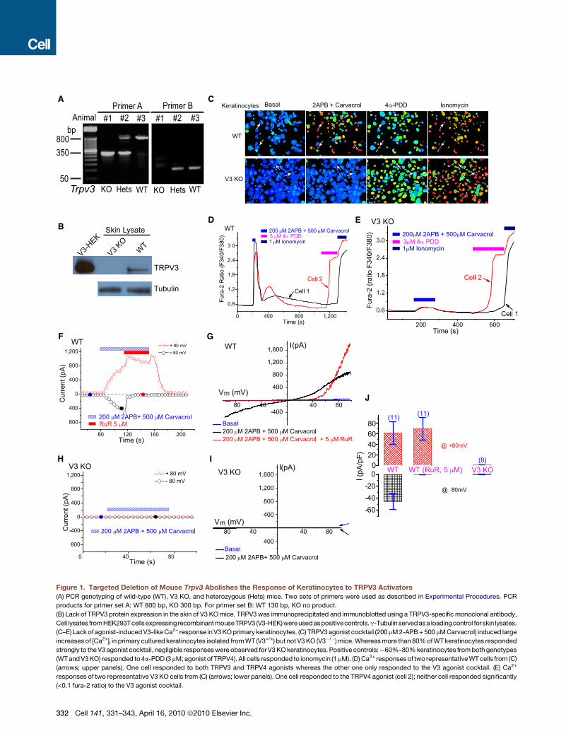
Figure 1. Targeted Deletion of Mouse Trpv3 Abolishes the Response of Keratinocytes to TRPV3 Activators
(A) PCR genotyping of wild-type (WT), V3 KO, and heterozygous (Hets) mice. Two sets of primers were used as described in Experimental Procedures. PCR
products for primer set A: WT 800 bp, KO 300 bp. For primer set B: WT 130 bp, KO no product.
(B) Lack of TRPV3 protein expression in the skin of V3 KO mice. TRPV3 was immunoprecipitated and immunoblotted using a TRPV3-specific monoclonal antibody.
Cell lysates fromHEK293Tcellsexpressing recombinant mouseTRPV3 (V3-HEK)wereusedaspositivecontrols.g-Tubulinservedasa loading control for skin lysates.
(C–E) Lack of agonist-induced V3-like Ca2+ response in V3 KO primary keratinocytes. (C) TRPV3 agonist cocktail (200 mM 2-APB + 500 mM Carvacrol) induced large
increases of [Ca2+]i in primary cultured keratinocytes isolated from WT (V3+/+) but not V3 KO (V3�/�) mice. Whereas more than 80% of WT keratinocytes responded
strongly to the V3 agonist cocktail, negligible responses were observed for V3 KO keratinocytes. Positive controls:�60%–80% keratinocytes from both genotypes
(WT and V3 KO) responded to 4a-PDD (3 mM; agonist of TRPV4). All cells responded to ionomycin (1 mM). (D) Ca2+ responses of two representative WT cells from (C)
(arrows; upper panels). One cell responded to both TRPV3 and TRPV4 agonists whereas the other one only responded to the V3 agonist cocktail. (E) Ca2+
responses of two representative V3 KO cells from (C) (arrows; lower panels). One cell responded to the TRPV4 agonist (cell 2); neither cell responded significantly
(<0.1 fura-2 ratio) to the V3 agonist cocktail.
332 Cell 141, 331–343, April 16, 2010 ª2010 Elsevier Inc.
Schneider et al., 2008). TGF-a is expressed in both basal (prolif-
erating) and suprabasal (differentiating) layers of epidermis and
in the inner root sheath of the hair follicle (Coffey et al., 1987;
Luetteke et al., 1993; Mann et al., 1993; Schneider et al.,
2008). Although most highly expressed in the basal layer, supra-
basal keratinocytes also express EGFR (Luetteke et al., 1993;
Mann et al., 1993; Schneider et al., 2008). Although induction
of differentiation dramatically increases the production of TGF-a/
EGF (Denning et al., 2000), these same growth factors promote
terminal differentiation (Wakita and Takigawa, 1999).
Previous studies suggest that TGF-a/EGF regulate keratino-
cyte terminal differentiation likely in a Ca2+-dependent manner
(Denning et al., 2000; Sakai et al., 1994). Intracellular Ca2+ regu-
lates both expression and shedding from the membrane-teth-
ered precursors of EGFR ligands (Denning et al., 2000; Horiuchi
et al., 2007). Ca2+ ionophores are sufficient to induce both
production and release of TGF-a (Horiuchi et al., 2007; Pandiella
and Massague, 1991). The Ca2+ influx pathway under physiolog-
ical conditions, however, has not been identified.
The cornified cell envelope (CE) is a protein-lipid layer that
replaces the plasma membrane of terminally differentiated kera-
tinocytes (corneocytes) and is crucial for the stratum corneum
epidermal barrier (Lorand and Graham, 2003). The CE is a com-
plex layer of lipids attached to a layer of crosslinked proteins.
The transglutaminases (TGases) primarily form 3-(g-glutamyl)
lysine isopeptide bonds between proteins, and their activi-
ties strongly depend on intracellular Ca2+ levels (Lorand and
Graham, 2003). EGF can acutely activate TGases to induce CE
formation and keratinocyte terminal differentiation (Lorand and
Graham, 2003). Cornification-promoting cellular cues may acti-
vate an unidentified Ca2+ influx channel to induce TGase activity
and subsequent CE formation.
Transient receptor potential (TRP) proteins are a large family of
Ca2+-permeable channels with diverse functions (Montell, 2005;
Nilius et al., 2007; Ramsey et al., 2006). Among these, TRPV3
and TRPV4 are functionally expressed in keratinocytes (Chung
et al., 2004; Moqrich et al., 2005). These channels detect
ambient temperature changes and are activated by various
plant-derived and synthetic compounds (Moqrich et al., 2005;
Xu et al., 2006). In this study, we find that TRPV3-deficient
mice exhibit hair phenotypes similar to wa1 and wa2. Molecular
and biochemical analyses of TRPV3-deficient mice and isolated
keratinocytes reveal defective TGF-a/EGFR signaling. We pro-
pose that TRPV3 is a Ca2+ entry pathway tightly associated
with the TGF-a/EGFR signaling complex orchestrating keratino-
cyte terminal differentiation.
(F–J) TRPV3-like currents were completely absent in V3 KO primary keratinocytes.
WT primary keratinocytes induced TRPV3-like (ITRPV3) currents. Whole-cell curren
applied every 4 s. Holding potential = 0 mV. Each symbol represents the current
Blue dashed line = zero current. (G) Representative ramp current of ITRPV3. I-V rela
rectifying and reversed near 0 mV. Ruthenium red (RuR; 5 mM) selectively blocked
No significant currents were induced by the V3 agonist cocktail in V3 KO keratinocy
or with the coapplication of RuR (5 mM). At�80 mV, inward ITRPV3 of WT keratinocy
presence of RuR, respectively. At +80 mV, outward ITRPV3 of V3+/+ keratinocytes w
of RuR, respectively. For V3 KO keratinocytes, no significant inward or outward
at +80 mV (n = 8), respectively.
Data are presented as the mean ± standard error of the mean (SEM). See also Fig
RESULTS
Whole-Animal and Keratinocyte-Specific Disruptionof Mouse Trpv3 GeneUsing a recombineering method, we inserted two loxP sites to
flank exon 13 of mouse Trpv3 (see Figure S1 available online)
and obtained mice with homozygous floxed (fl) alleles. To inves-
tigate the in vivo function(s) of TRPV3, we generated TRPV3
global knockout (KO) using Sox2-Cre transgenic mice (details
described in Experimental Procedures). To elucidate the role of
TRPV3 in the skin, we also bred the fl/fl animals to mice harboring
a K14-Cre recombinase transgene, which efficiently expresses
Cre throughout the epidermis by embryonic day 15.5 (E15.5)
(Wang et al., 1997). Mouse genotypes from both TRPV3 global
KO (Trpv3�/�; abbreviated as V3 KO) and K14-specific con-
ditional KO (V3 fl/fl: K14 Cre; abbreviated as V3 cKO) were
confirmed by PCR (Figure 1A, also see Figure S1). No TRPV3
full-length transcript was detected from V3 KO skin tissues using
RT-PCR analysis (Figure S1). No full- length TRPV3 protein was
detected by western blot in V3 KO skin lysates (Figure 1B) or
cultured primary keratinocytes (data not shown). Thus, the
mice generated completely lack TRPV3 in their skin.
Functional Characterization of TRPV3 Globaland Keratinocyte-Specific KO MiceWe performed functional studies in keratinocytes isolated from
V3 KO and control (wild-type [WT], V3+/+; heterozygous or
Hets, V3+/�) mice. Fura-2 Ca2+ imaging was employed to
study the response of keratinocytes to TRPV3 chemical agonists
(Xu et al., 2006). Application of most TRPV3 agonists alone, for
example, 2-APB (200 mM) or Carvacrol (500 mM), induced a small
increase in intracellular Ca2+ ([Ca2+]i) in a subset of cells (30% to
80% of cells; data not shown). However, coapplication of two
agonists, for example, 200 mM 2-APB + 500 mM Carvacrol
(TRPV3 agonist cocktail), reliably induced a dramatic (DF340/
F380 > 1) increase of [Ca2+]i in the majority (>80%) of keratino-
cytes isolated from WT mice (WT keratinocytes; Figures 1C
and 1D). Similar results were obtained for other combinations
of TRPV3 agonists such as 200 mM 2-APB + 5 mM Camphor;
removal of external Ca2+ abolished most of the agonist-induced
Ca2+ responses. No significant increase (DF340/F380 < 0.1) in
[Ca2+]i was seen in keratinocytes isolated from V3 KO mice (V3
KO keratinocytes; Figures 1C and 1E). In contrast, in both
WT and V3 KO cells, large [Ca2+]i increases were evoked by
4a-PDD (3 mM), an agonist of TRPV4 (Watanabe et al., 2002)
that is also expressed in keratinocytes. Similar results were
(F) Application of the V3 agonist cocktail (200 mM 2-APB + 500 mM Carvacrol) to
ts were generated in response to 400 ms voltage ramps from�100 to +100 mV,
amplitude at +80 mV (red triangles) and �80 mV (black circles), respectively.
tions were recorded at time points noted in (H) (filled circles). ITRPV3 was doubly
inward ITRPV3 with significant augmentation at very positive potentials. (H and I)
tes. (J) Average ITRPV3 current densities elicited by the V3 agonist cocktail alone
tes were�45 ± 13 pA/pF (n = 11) and�0.3 ± 0.3 pA/pF (n = 11) in the absence or
ere 62 ± 21 pA/pF (n = 11) and 70 ± 21 pA/pF (n = 11) in the absence or presence
ITRPV3 was detected: �0.3 ±± 0.3 pA/pF at �80mV (n = 8) and 0.8 ± 0.5 pA/pF
ure S1.
Cell 141, 331–343, April 16, 2010 ª2010 Elsevier Inc. 333
A
B
C
D
P1 P1
V3 cKOWT
V3 cKOWhisker
Dorsal hairP14 P14
WT
Dorsal Skin
P4 P4
V3 cKO WT
Tail Skin V3 KO
P0 P0
WT
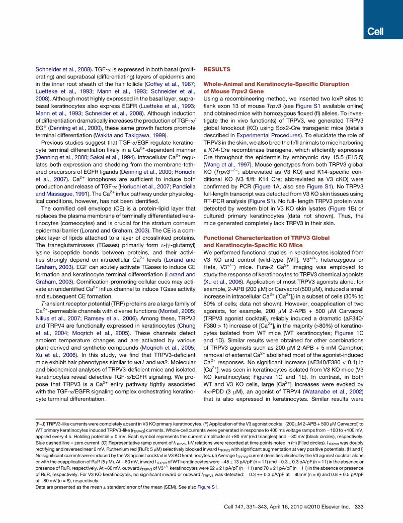
Figure 2. TRPV3-Deficient Mice Exhibit
Curly Whiskers, Wavy Hair, Misaligned Hair
Follicles, and a Thin Stratum Corneum
(A) Newborn (P1) V3 cKO (fl/fl: K14 Cre) mice: curly
whiskers; littermate WT (V3 fl/fl) animals: straight
whiskers.
(B) Whiskers in an adult WT mouse (P14) were
straight; the whiskers of littermate V3 cKO mice
were distinctively curly and hooked (upper panels).
V3 cKO mice also exhibited wavy dorsal coats
(lower panels).
(C and D) Skin and hair follicle abnormalities of
V3-deficient mice revealed by H&E staining of
dorsal (C) and tail (D) skin sections from WT and
KO or cKO mice. In the back skin of WT pups
(P4), all hair follicles lay parallel in an anterior to
posterior direction with an angle of �45� (left
panel). In contrast, hairs of littermate cKO mice
angled in different directions (right panel). Arrows
indicate two misaligned horizontally oriented hair
follicles. In addition to the hair follicle abnormality,
the stratum corneum (SC) layer (denoted by blue
rectangle bars) of the V3 KO or cKO mice was
significantly thinner but more compact than that
of the WT mice.
See also Figure S2.
obtained in keratinocytes from V3 cKO mice. These results
demonstrate that TRPV3 KO mouse keratinocytes completely
lack TRPV3-mediated Ca2+ responses.
Consistent with the Ca2+ imaging results, TRPV3 agonist
cocktail evoked a slowly developing, large TRPV3-like current
(ITRPV3) in most WT keratinocytes (Figures 1F, 1G, and 1J).
Ruthenium red (RuR, 5 mM), a nonspecific voltage-dependent
blocker of TRPV1-4 channels (Chung et al., 2004; Hu et al.,
2004; Ramsey et al., 2006; Xu et al., 2006), almost completely
(>99%) inhibited agonist-activated inward ITRPV3. Similar RuR-
sensitive ITRPV3 was also evoked by other TRPV3 agonists
(200 mM 2-APB + heat or 200 mM 2-APB + 5 mM Camphor) in
the same cells. In V3 KO keratinocytes, in contrast, no significant
current was evoked by the V3 agonist cocktail (Figures 1H, 1I,
and 1J). Similar results were obtained in keratinocytes from V3
cKO mice.
TRPV3-Deficient Mice Exhibit Curly Whiskersand Wavy HairAlthough previous animal studies identified TRPV30s function in
temperature sensation (Moqrich et al., 2005), the most obvious
phenotypic changes we observed from our V3 KO mice are
abnormalities in skin, hair, and whiskers. In contrast with litter-
mate controls (WT and heterozygotes [Hets]), most whiskers of
V3 KO mice were characteristically curled or hooked (Fig-
ure S2). The curly morphology of whiskers was apparent at birth
(Figure S2); newborn V3 KO mice could be identified based on
whisker morphology alone. Keratinocyte-specific V3 cKO mice
exhibited similar curly whiskers (Figures 2A and 2B), suggesting
that the phenotype was caused by specific V3 deficiency in ker-
atinocytes. Whisker curliness grew more obvious with age
(Figure 2B and Figure S2). Both the dorsal and ventral coat fur,
as well as the tail hair, of V3 KO and cKO mice were wavy
(Figure 2B and Figure S2) beginning 1 week after birth but was
334 Cell 141, 331–343, April 16, 2010 ª2010 Elsevier Inc.
most apparent once the hair was well formed (�3–4 weeks post-
natal), gradually reducing with age. In contrast to a previous
study reporting abnormality of ventral hairs in a subset of V3
KO mice (Moqrich et al., 2005), curly whiskers and wavy hair
were present throughout the coat at all ages with 100% pene-
trance for both V3 KO and cKO mice that we have generated, as
well as the V3 KO mice reported by Moqrich et al. (H.L., S.M.H.,
and M.J.C., unpublished data). In comparison, TRPV1 KO mice
hair shape and distribution are normal (Figure S2). Consistent
with the expression of TRPV3 in follicular keratinocytes of mouse
(Peier et al., 2002) and human (Xu et al., 2002), the wavy pheno-
type correlated with V3 deficiency in keratinocytes.
Skin contains many cell types, including sensory nerves.
However, as the K14 promoter drives the expression of Cre
recombinase specifically in all keratinocytes (both follicular and
interfollicular) in the skin (Coulombe et al., 1989), the results
obtained from V3 cKO, V3 fl/fl: K14 Cre mice suggested that
the defect in hair morphogenesis was due to the lack of TRPV3
expression in keratinocytes in a cell-autonomous manner. Both
hair and whisker phenotypes were independent of pigmentation
of the hair and genetic background, as black-coated (C57BL/6)
backcrossed (>6 generations) V3 KO mice displayed pheno-
types similar to those of mice with mixed genetic background
(BL6 and 129sv) (Figure S2). V3 KO pups were born with the
expected Mendelian ratio, and body weight was comparable
to control mice (Figure S2).
In histological examinations of skin from the mid-dorsal region
of mice of different ages, we found that a subset of hair follicles
exhibited an obvious but gentle curvature (Figure S2). In Haema-
toxylin & Eosin (H&E) stained skin sections from control mice
(P4), hair follicle shafts were parallel and posed roughly at
a 45� angle to the subcutaneous muscle layer (Figure 2C). In con-
trast, individual hair follicles of V3 cKO mice were, in many cases,
gently curved and pointed in different directions with variable

angles (Figure 2C). Several hair follicles even grew horizontally to
the subcutaneous muscle layer. Similar follicular derangement
was obvious for V3 KO mice (Figure S2). Notably, similar alter-
ations in hair follicle morphology have been reported in EGFR-
deficient mice (Threadgill et al., 1995). These results suggest
that the wavy hair phenotype of V3 KO mice was due to a defect
in follicle formation, and that mouse TRPV3 was required for
normal morphogenesis of hair and whiskers. In addition to follic-
ular abnormalities, V3 KO and cKO mice also exhibited abnor-
malities in the epidermal stratum corneum (Figures 2C and 2D;
Figure S2; details see below). The hair cycle, however, was not
significantly altered (Figure S2).
DefectiveTGF-a/EGFRSignaling inTRPV3-DeficientMiceThe hair and whisker phenotype of V3 KO mice resembled
largely those of wa1 and wa2 mice, as well as other mouse
mutations with reduced expression/release/activity in TGF-a
and/or EGFR (Ballaro et al., 2005; Du et al., 2004; Luetteke
et al., 1993, 1994; Mann et al., 1993; Miettinen et al., 1995;
Murillas et al., 1995; Peschon et al., 1998; Schneider et al.,
2008; Sibilia and Wagner, 1995; Threadgill et al., 1995) (see
Figure S3). Thus we investigated whether TGF-a and/or EGFR
signaling was altered in V3 KO mice. Real-time semiquantitative
PCR (q-PCR) analysis of the skin of V3 KO pups revealed that the
mRNA expression level of TGF-a was half that of WT (P4; Fig-
ure 3A). TGF-a mRNA levels of newborn (P0) V3 KO animals,
though significantly less than those of the P4 mice, were compa-
rable to those of the littermate controls (P0). Expression (mRNA)
levels of several other EGFR ligands and EGFR (Figure S3),
however, were not significantly altered in the skin of V3 KO
mice. These results suggest that TRPV3 affects the expression
level of TGF-a in postnatal skin in vivo.
Both expression and proteolytic shedding of the membrane-
tethered TGF-a are known to be Ca2+ dependent (Denning
et al., 2000; Horiuchi et al., 2007). We used an ELISA assay opti-
mized for human TGF-a to investigate the role of TRPV3 in TGF-a
shedding/release. In the presence of TRPV3 agonist cocktail
(100 mM 2-APB + 250 mM Carvacrol; 30 min), normal human
epidermal keratinocytes (NHEK) released more than twice the
amount of TGF-a into the culture medium. This is comparable
to the effect of PMA, a stimulus well known to induce release
and expression of TGF-a (Figure 3B). ADAM17, the principal
sheddase required for TGF-a release (Peschon et al., 1998),
was required for V3 agonist-induced TGF-a release/shedding
(Figure 3C).
As the level of TGF-a was reduced in V3 KO skin, one
would expect that its activated receptor, phosphorylated EGFR
(P-EGFR), might also be reduced. By immunoblot analysis of
P-EGFR in skin lysates of V3 KO skin, EGFR activity was only
about one-third of that of WT controls (Figures 3D and 3E). Inter-
estingly, the expression level of total EGFR was slightly but
significantly increased in V3 KO skin (1.8- ± 0.3-fold, n = 6), so
that the ratio of P-EGFR/total EGFR was about 5-fold less
(0.20 ± 0.03, n = 4) in V3 KO skin. This level of the reduction
was comparable to those of the hypofunctional EGFR mutations
causing ‘‘wavy’’ phenotypes (Du et al., 2004). Consistent with
biochemical results, EGFR staining was more prominent in V3
KO frozen skin sections (Figure S3), whereas P-EGFR immuno-
staining was weaker. These results are consistent with dramati-
cally reduced EGFR activity in V3 KO mice and suggest that the
level of total EGFR was increased as a consequence of reduced
activity, tyrosine phosphorylation-dependent endocytic degra-
dation (Schneider et al., 2008), or other compensatory mecha-
nisms. The reduction of EGFR activity in V3 KO mice was prob-
ably due to the loss of the TRPV3 channel activity, as activation
of TRPV3 in cultured keratinocytes using TRPV3 agonists re-
sulted in increases in both TGF-a release (Figures 3B and 3C)
and EGFR activity (Figure 3F and Figure S3). Notably, the
TRPV3-induced increase of EGFR activity in keratinocytes was
abolished by a neutralizing TGF-a antibody or ADAM17 shed-
dase inhibitor (Figures 3F and 3G), suggesting that activation
of TRPV3 led to an increase in TGF-a release and subsequent
EGFR activation.
Regulation of TRPV3 Channel Activityby TGF-a/EGFR SignalingSeveral in vitro studies provided evidence that TRP channels are
regulated by members of the receptor tyrosine kinase (RTK)
family (Li et al., 1999; Ramsey et al., 2006). We hypothesized
that TRPV3 channel activity could also be upregulated by EGFR
signaling. In serum-starved primary keratinocytes cultured in the
absence of TGF-a, Ca2+ responses could be induced by a high
concentration (100 mM 2-APB + 250 mM Carvacrol), but not by
a lower concentration (50 mM 2-APB + 125 mM Carvacrol), of
V3 agonist cocktail (Figures 4A and 4C). In the presence of
TGF-a (100 ng/ml for 3–5 hr), however, large [Ca2+]i increases
were recorded even with a low concentration (50 mM 2-APB +
125 mM Carvacrol) of V3 agonist cocktail; responses to a high
concentration of V3 agonists were comparable to those without
TGF-a treatment (Figures 4B and 4C). Similar results were seen
with EGF (100 ng/ml) pretreatment (Figure S4). TGF-a/EGF treat-
ment altered the sensitivity of TRPV3 in keratinocytes to V3
agonists, suggesting that the increased activity was at least
partially mediated by increased channel gating, rather than the
expression or surface expression of TRPV3 proteins. Consistent
with this interpretation, the temperature-induced response (from
22�C to 41�C) was also significantly larger in TGF-a-treated ker-
atinocytes (Figure S4). The sensitizing effect of TGF-a was most
likely mediated by EGFR, as an EGFR inhibitor (AG1478) or
shRNA knockdown of EGFR expression (Figure S4) completely
or largely eliminated its potentiation (Figures 4D and 4E).
Because inhibitors of PLC (U73122) and ERK (PD98059) com-
pletely or partially blocked potentiation (Figure 4D), these path-
ways may underlie the sensitizing effect downstream of EGFR
receptor activation. In support of this finding, shRNA knockdown
of PLC-g1 expression (Figure S4) significantly decreased the
sensitizing effect of TGF-a (Figure 4E). Consistent with our
[Ca2+]i measurements, ITRPV3 exhibited a similar dependence
on TGF-a (Figure 4F).
The results presented so far raise the possibility that TRPV3
and EGFR might be in a signaling complex. To test this hypoth-
esis, coimmunoprecipitation (co-IP) experiments were first per-
formed in a heterologous expression system. In cells transfected
with TRPV3, either alone or together with EGFR, both endoge-
nous (data not shown) and overexpressed EGFR (Figure S4)
were found to co-IP with TRPV3. Next, we confirmed this finding
Cell 141, 331–343, April 16, 2010 ª2010 Elsevier Inc. 335
A
B
D E
WT V3 KO
P-EGFR
EGFR
Tubulin
P4
F
C
DMSO
V3
-EGFR
EGFR
Tubulin
NHEK cells ( 0.5 ng/ml EGF)
Agonis
t
P
Anti- T
GF-α
BB2116
G
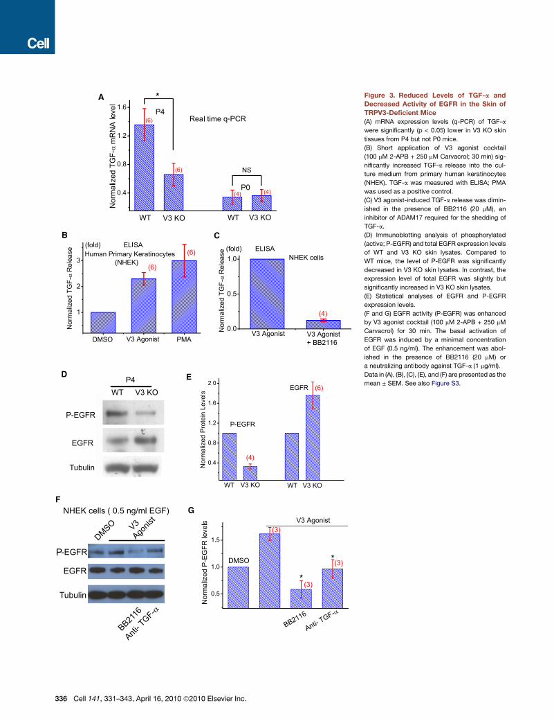
Figure 3. Reduced Levels of TGF-a and
Decreased Activity of EGFR in the Skin of
TRPV3-Deficient Mice
(A) mRNA expression levels (q-PCR) of TGF-a
were significantly (p < 0.05) lower in V3 KO skin
tissues from P4 but not P0 mice.
(B) Short application of V3 agonist cocktail
(100 mM 2-APB + 250 mM Carvacrol; 30 min) sig-
nificantly increased TGF-a release into the cul-
ture medium from primary human keratinocytes
(NHEK). TGF-a was measured with ELISA; PMA
was used as a positive control.
(C) V3 agonist-induced TGF-a release was dimin-
ished in the presence of BB2116 (20 mM), an
inhibitor of ADAM17 required for the shedding of
TGF-a.
(D) Immunoblotting analysis of phosphorylated
(active; P-EGFR) and total EGFR expression levels
of WT and V3 KO skin lysates. Compared to
WT mice, the level of P-EGFR was significantly
decreased in V3 KO skin lysates. In contrast, the
expression level of total EGFR was slightly but
significantly increased in V3 KO skin lysates.
(E) Statistical analyses of EGFR and P-EGFR
expression levels.
(F and G) EGFR activity (P-EGFR) was enhanced
by V3 agonist cocktail (100 mM 2-APB + 250 mM
Carvacrol) for 30 min. The basal activation of
EGFR was induced by a minimal concentration
of EGF (0.5 ng/ml). The enhancement was abol-
ished in the presence of BB2116 (20 mM) or
a neutralizing antibody against TGF-a (1 mg/ml).
Data in (A), (B), (C), (E), and (F) are presented as the
mean ± SEM. See also Figure S3.
336 Cell 141, 331–343, April 16, 2010 ª2010 Elsevier Inc.
A B
C D
E F
IP: Anti- GFP
Anti- GFP
Anti- EGFR
Anti- EGFR
V3-YFP
WT Skin V3-YFP TG SkinG
Anti-GFP Anti-P-Tyr
IP: Anti-EGFR
Anti-EGFR Anti-P-Tyr
IP: Anti-GFP
TGF-α
TGF-α
(-) (-)
(-) (-)
(+)(+)
(+) (+)
WB:
WB:
H
WT Keratinocytes
WB: Anti-GFP
Tubulin
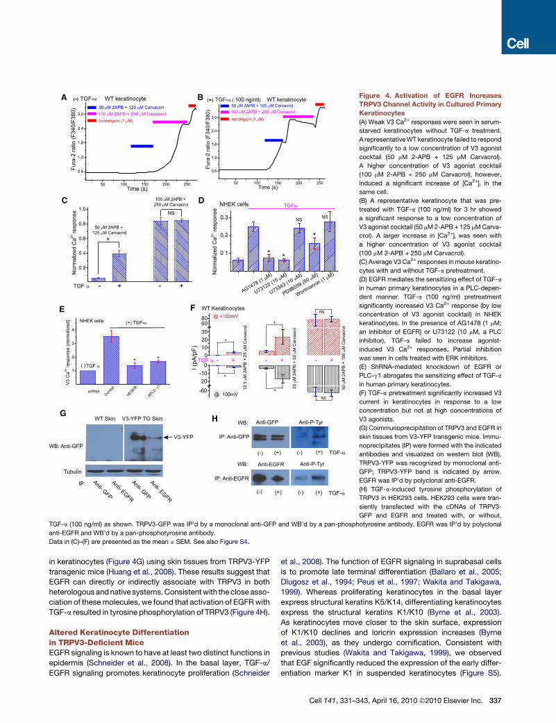
Figure 4. Activation of EGFR Increases
TRPV3 Channel Activity in Cultured Primary
Keratinocytes
(A) Weak V3 Ca2+ responses were seen in serum-
starved keratinocytes without TGF-a treatment.
A representative WT keratinocyte failed to respond
significantly to a low concentration of V3 agonist
cocktail (50 mM 2-APB + 125 mM Carvacrol).
A higher concentration of V3 agonist cocktail
(100 mM 2-APB + 250 mM Carvacrol), however,
induced a significant increase of [Ca2+]i in the
same cell.
(B) A representative keratinocyte that was pre-
treated with TGF-a (100 ng/ml) for 3 hr showed
a significant response to a low concentration of
V3 agonist cocktail (50 mM 2-APB + 125 mM Carva-
crol). A larger increase in [Ca2+]i was seen with
a higher concentration of V3 agonist cocktail
(100 mM 2-APB + 250 mM Carvacrol).
(C) Average V3 Ca2+ responses in mouse keratino-
cytes with and without TGF-a pretreatment.
(D) EGFR mediates the sensitizing effect of TGF-a
in human primary keratinocytes in a PLC-depen-
dent manner. TGF-a (100 ng/ml) pretreatment
significantly increased V3 Ca2+ response (by low
concentration of V3 agonist cocktail) in NHEK
keratinocytes. In the presence of AG1478 (1 mM;
an inhibitor of EGFR) or U73122 (10 mM, a PLC
inhibitor), TGF-a failed to increase agonist-
induced V3 Ca2+ responses. Partial inhibition
was seen in cells treated with ERK inhibitors.
(E) ShRNA-mediated knockdown of EGFR or
PLC-g1 abrogates the sensitizing effect of TGF-a
in human primary keratinocytes.
(F) TGF-a pretreatment significantly increased V3
current in keratinocytes in response to a low
concentration but not at high concentrations of
V3 agonists.
(G) Coimmunoprecipitation of TRPV3 and EGFR in
skin tissues from V3-YFP transgenic mice. Immu-
noprecipitates (IP) were formed with the indicated
antibodies and visualized on western blot (WB).
TRPV3-YFP was recognized by monoclonal anti-
GFP; TRPV3-YFP band is indicated by arrow.
EGFR was IP’d by polyclonal anti-EGFR.
(H) TGF-a-induced tyrosine phosphorylation of
TRPV3 in HEK293 cells. HEK293 cells were tran-
siently transfected with the cDNAs of TRPV3-
GFP and EGFR and treated with, or without,
TGF-a (100 ng/ml) as shown. TRPV3-GFP was IP’d by a monoclonal anti-GFP and WB’d by a pan-phosphotyrosine antibody. EGFR was IP’d by polyclonal
anti-EGFR and WB’d by a pan-phosphotyrosine antibody.
Data in (C)–(F) are presented as the mean ± SEM. See also Figure S4.
in keratinocytes (Figure 4G) using skin tissues from TRPV3-YFP
transgenic mice (Huang et al., 2008). These results suggest that
EGFR can directly or indirectly associate with TRPV3 in both
heterologous and native systems. Consistent with the close asso-
ciation of these molecules, we found that activation of EGFR with
TGF-a resulted in tyrosine phosphorylation of TRPV3 (Figure 4H).
Altered Keratinocyte Differentiationin TRPV3-Deficient MiceEGFR signaling is known to have at least two distinct functions in
epidermis (Schneider et al., 2008). In the basal layer, TGF-a/
EGFR signaling promotes keratinocyte proliferation (Schneider
et al., 2008). The function of EGFR signaling in suprabasal cells
is to promote late terminal differentiation (Ballaro et al., 2005;
Dlugosz et al., 1994; Peus et al., 1997; Wakita and Takigawa,
1999). Whereas proliferating keratinocytes in the basal layer
express structural keratins K5/K14, differentiating keratinocytes
express the structural keratins K1/K10 (Byrne et al., 2003).
As keratinocytes move closer to the skin surface, expression
of K1/K10 declines and loricrin expression increases (Byrne
et al., 2003), as they undergo cornification. Consistent with
previous studies (Wakita and Takigawa, 1999), we observed
that EGF significantly reduced the expression of the early differ-
entiation marker K1 in suspended keratinocytes (Figure S5).
Cell 141, 331–343, April 16, 2010 ª2010 Elsevier Inc. 337
A
B
C
E
D
K1/α6/DAPI K1/α6/DAPI
K14/α6/DAPI K14/α6/DAPI
Loricrin/α6/DAPI Loricrin/α6/DAPI
K1/α6/DAPI K1/α6/DAPI
K10/α6/DAPI K10/α6/DAPI
20 μ m 20 μ mV3 KO
V3 KO
V3 KO
V3 KO
P4 P4
P4 P4
P4 P4
P4 P4
P4 P4 V3 cKO
A'
B'
C'
D'
E'
WT
WT
WT
WT
WT
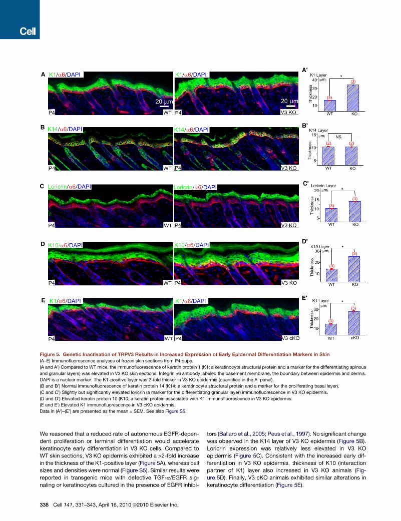
Figure 5. Genetic Inactivation of TRPV3 Results in Increased Expression of Early Epidermal Differentiation Markers in Skin
(A–E) Immunofluorescence analyses of frozen skin sections from P4 pups.
(A and A0) Compared to WT mice, the immunofluorescence of keratin protein 1 (K1; a keratinocyte structural protein and a marker for the differentiating spinous
and granular layers) was elevated in V3 KO skin sections. Integrin a6 antibody labeled the basement membrane, the boundary between epidermis and dermis.
DAPI is a nuclear marker. The K1-positive layer was 2-fold thicker in V3 KO epidermis (quantified in the A0 panel).
(B and B0) Normal immunofluorescence of keratin protein 14 (K14; a keratinocyte structural protein and a marker for the proliferating basal layer).
(C and C0) Slightly but significantly elevated loricrin (a marker for the differentiating granular layer) immunofluorescence in V3 KO epidermis.
(D and D0) Elevated keratin protein 10 (K10; a keratin protein associated with K1 immunofluorescence in V3 KO epidermis.
(E and E0) Elevated K1 immunofluorescence in V3 cKO epidermis.
Data in (A0)–(E0) are presented as the mean ± SEM. See also Figure S5.
We reasoned that a reduced rate of autonomous EGFR-depen-
dent proliferation or terminal differentiation would accelerate
keratinocyte early differentiation in V3 KO cells. Compared to
WT skin sections, V3 KO epidermis exhibited a >2-fold increase
in the thickness of the K1-positive layer (Figure 5A), whereas cell
sizes and densities were normal (Figure S5). Similar results were
reported in transgenic mice with defective TGF-a/EGFR sig-
naling or keratinocytes cultured in the presence of EGFR inhibi-
338 Cell 141, 331–343, April 16, 2010 ª2010 Elsevier Inc.
tors (Ballaro et al., 2005; Peus et al., 1997). No significant change
was observed in the K14 layer of V3 KO epidermis (Figure 5B).
Loricrin expression was relatively less elevated in V3 KO
epidermis (Figure 5C). Consistent with the increased early dif-
ferentiation in V3 KO epidermis, thickness of K10 (interaction
partner of K1) layer also increased in V3 KO animals (Fig-
ure 5D). Finally, V3 cKO animals exhibited similar alterations in
keratinocyte differentiation (Figure 5E).
A B CWT V3 KO
P0
P1 V3 cKO
V3 KO
E17
P4 P4
V3 cKOD E
WT
WT
WT
G
F
C
I
TG1/α6/DAPI
TGase1 expressionTG1/α6/DAPI
H
V3 cKO
P4
P4
DMSO V3 AgonistWT
V3 KO V3 KO
TGase Activity
WT
WT
TGase Activity
V3 cKO
P1
P1
V3 cKO
P4
P4
WT
WT
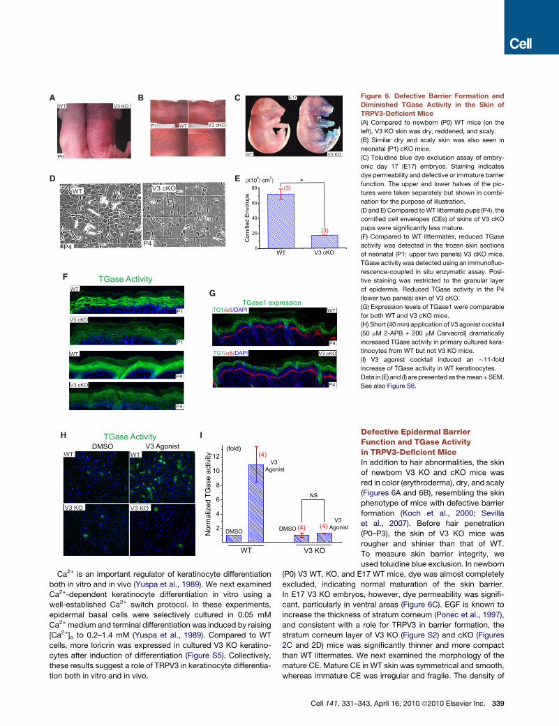
Figure 6. Defective Barrier Formation and
Diminished TGase Activity in the Skin of
TRPV3-Deficient Mice
(A) Compared to newborn (P0) WT mice (on the
left), V3 KO skin was dry, reddened, and scaly.
(B) Similar dry and scaly skin was also seen in
neonatal (P1) cKO mice.
(C) Toluidine blue dye exclusion assay of embry-
onic day 17 (E17) embryos. Staining indicates
dye permeability and defective or immature barrier
function. The upper and lower halves of the pic-
tures were taken separately but shown in combi-
nation for the purpose of illustration.
(D and E) Compared to WT littermate pups (P4), the
cornified cell envelopes (CEs) of skins of V3 cKO
pups were significantly less mature.
(F) Compared to WT littermates, reduced TGase
activity was detected in the frozen skin sections
of neonatal (P1; upper two panels) V3 cKO mice.
TGase activity was detected using an immunofluo-
rescence-coupled in situ enzymatic assay. Posi-
tive staining was restricted to the granular layer
of epidermis. Reduced TGase activity in the P4
(lower two panels) skin of V3 cKO.
(G) Expression levels of TGase1 were comparable
for both WT and V3 cKO mice.
(H) Short (40 min) application of V3 agonist cocktail
(50 mM 2-APB + 200 mM Carvacrol) dramatically
increased TGase activity in primary cultured kera-
tinocytes from WT but not V3 KO mice.
(I) V3 agonist cocktail induced an �11-fold
increase of TGase activity in WT keratinocytes.
Data in (E) and (I) are presented as the mean ± SEM.
See also Figure S6.
Ca2+ is an important regulator of keratinocyte differentiation
both in vitro and in vivo (Yuspa et al., 1989). We next examined
Ca2+-dependent keratinocyte differentiation in vitro using a
well-established Ca2+ switch protocol. In these experiments,
epidermal basal cells were selectively cultured in 0.05 mM
Ca2+ medium and terminal differentiation was induced by raising
[Ca2+]o to 0.2–1.4 mM (Yuspa et al., 1989). Compared to WT
cells, more loricrin was expressed in cultured V3 KO keratino-
cytes after induction of differentiation (Figure S5). Collectively,
these results suggest a role of TRPV3 in keratinocyte differentia-
tion both in vitro and in vivo.
Cell 141, 331–3
Defective Epidermal BarrierFunction and TGase Activityin TRPV3-Deficient MiceIn addition to hair abnormalities, the skin
of newborn V3 KO and cKO mice was
red in color (erythroderma), dry, and scaly
(Figures 6A and 6B), resembling the skin
phenotype of mice with defective barrier
formation (Koch et al., 2000; Sevilla
et al., 2007). Before hair penetration
(P0–P3), the skin of V3 KO mice was
rougher and shinier than that of WT.
To measure skin barrier integrity, we
used toluidine blue exclusion. In newborn
(P0) V3 WT, KO, and E17 WT mice, dye was almost completely
excluded, indicating normal maturation of the skin barrier.
In E17 V3 KO embryos, however, dye permeability was signifi-
cant, particularly in ventral areas (Figure 6C). EGF is known to
increase the thickness of stratum corneum (Ponec et al., 1997),
and consistent with a role for TRPV3 in barrier formation, the
stratum corneum layer of V3 KO (Figure S2) and cKO (Figures
2C and 2D) mice was significantly thinner and more compact
than WT littermates. We next examined the morphology of the
mature CE. Mature CE in WT skin was symmetrical and smooth,
whereas immature CE was irregular and fragile. The density of
43, April 16, 2010 ª2010 Elsevier Inc. 339
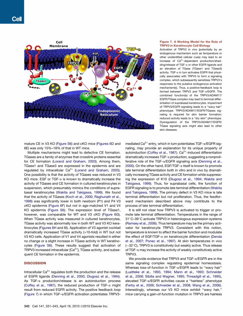
Figure 7. A Working Model for the Role of
TRPV3 in Keratinocyte Cell Biology
Activation of TRPV3 in vivo (potentially by an
endogenous mechanism such as temperature or
other unidentified cellular cues) may lead to an
increase of Ca2+-dependent production/shed-
ding/release of TGF-a or other EGFR ligands and
an elevation of TGase (TGase1 and TGase3)
activity. TGF-a in turn activates EGFR that physi-
cally associates with TRPV3 to form a signaling
complex, which subsequently sensitizes TRPV3’s
responses to the putative endogenous activation
mechanism(s). Thus, a positive-feedback loop is
formed between TRPV3 and TGF-a/EGFR. The
combined function(s) of the TRPV3/ADAM17/
EGFR/TGase complex may lead to terminal differ-
entiation of suprabasal keratinocytes. Impairment
of TRPV3/EGFR signaling leads to a ‘‘wavy hair’’
phenotype. TRPV3/ADAM17/EGFR/TGases sig-
naling is required for skin barrier formation;
reduced activity leads to a ‘‘dry skin’’ phenotype.
Dysregulation of the TRPV3/ADAM17/EGFR/
TGase signaling axis might also lead to other
skin diseases.
mature CE in V3 KO (Figure S6) and cKO mice (Figures 6D and
6E) was only 15%–18% of that in WT mice.
Multiple mechanisms might lead to defective CE formation.
TGases are a family of enzymes that crosslink proteins essential
for CE formation (Lorand and Graham, 2003). Among them,
TGase1 and TGase3 are expressed in the epidermis and are
regulated by intracellular Ca2+ (Lorand and Graham, 2003).
One possibility is that the activity of TGases was reduced in V3
KO mice. EGF or TGF-a is known to dramatically increase the
activity of TGases and CE formation in cultured keratinocytes in
suspension, which presumably mimics the conditions of supra-
basal keratinocytes (Wakita and Takigawa, 1999). We found
that the activity of TGases (Koch et al., 2000; Raghunath et al.,
1998) was significantly lower in both newborn (P1) and P4 V3
cKO epidermis (Figure 6F) but not in age-matched V1 and V4
KO epidermis (Figure S6). The expression level of TGase1,
however, was comparable for WT and V3 cKO (Figure 6G).
When TGase activity was measured in cultured keratinocytes,
TGase activity was abnormally low in both WT and V3 KO kera-
tinocytes (Figures 6H and 6I). Application of V3 agonist cocktail
dramatically increased TGase activity (>10-fold) in WT but not
V3 KO cells. Application of V1 and V4 agonists resulted in either
no change or a slight increase in TGase activity in WT keratino-
cytes (Figure S6). These results suggest that activation of
TRPV3 increased intracellular [Ca2+], TGase activity, and subse-
quent CE formation in the epidermis.
DISCUSSION
Intracellular Ca2+ regulates both the production and the release
of EGFR ligands (Denning et al., 2000; Dlugosz et al., 1994).
As TGF-a production/release is an autoinduction process
(Coffey et al., 1987), the reduced production of TGF-a might
result from reduced EGFR activity. The positive feedback loop
(Figure 7) in which TGF-a/EGFR activation potentiates TRPV3-
340 Cell 141, 331–343, April 16, 2010 ª2010 Elsevier Inc.
mediated Ca2+ entry, which in turn potentiates TGF-a/EGFR sig-
naling, may provide an explanation for its unique property of
autoinduction (Coffey et al., 1987). Ca2+-induced differentiation
dramatically increases TGF-a production, suggesting a nonproli-
ferative role of the TGF-a/EGFR signaling axis (Denning et al.,
2000). On the other hand, EGF/TGF-a itself is known to promote
late terminal differentiation both in vitro and in vivo by dramati-
cally increasing TGase activity and CE formation while suppress-
ing the expression of K10 (Dlugosz et al., 1994; Wakita and
Takigawa, 1999). Thus, for suprabasal cells, the function of
EGFR signaling is to promote late terminal differentiation (Wakita
and Takigawa, 1999). The primary defect in V3 KO mice is late
terminal differentiation but not proliferation. Thus, the feedfor-
ward mechanism described above may contribute to the
process of late terminal differentiation.
It is still not clear how TRPV3 is activated to trigger or pro-
mote late terminal differentiation. Temperatures in the range of
31�C–39�C activate TRPV3 in heterologous expression systems
(Ramsey et al., 2006). Thus temperature may be the primary acti-
vator for keratinocyte TRPV3. Consistent with this notion,
temperature is known to affect the barrier function and modulate
the effect of EGF/TGF-a on keratinocyte differentiation (Denda
et al., 2007; Ponec et al., 1997). At skin temperatures in vivo
(�32�C), TRPV3 is constitutively but weakly active. Thus release
of TGF-a may increase the activity of weakly constitutively active
TRPV3.
We provide evidence that TRPV3 and TGF-a/EGFR are in the
same signaling complex regulating epidermal homeostasis.
Whereas loss-of-function in TGF-a/EGFR leads to ‘‘wavy hair’’
(Luetteke et al., 1993, 1994; Mann et al., 1993; Schneider
et al., 2008; Sibilia and Wagner, 1995; Threadgill et al., 1995),
elevated TGF-a/EGFR activities cause a ‘‘hairless’’ phenotype
(Ferby et al., 2006; Schneider et al., 2008; Wang et al., 2006).
Interestingly, whereas our V3 KO mice exhibit ‘‘wavy hair,’’
mice carrying a gain-of-function mutation in TRPV3 are hairless

(Asakawa et al., 2006). It may prove informative to generate
transgenic mice with TRPV3 loss of function and concurrent
TGF-a/EGFR gain of function, or with TRPV3 gain of function
and concurrent TGF-a/EGFR loss of function.
EGFR is the prototype of the RTK family. EGFR signaling is
necessary for proper development and tissue homeostasis
whereas its dysregulation rapidly results in defects in cellular
proliferation and differentiation. The consequences of its mal-
function are abnormal hair follicle morphogenesis, impaired
would healing, and tumorigenesis (Schneider et al., 2008). We
have identified another key element in this important signaling
pathway, the TRPV3 channel. Our studies not only provide the
first in vivo evidence in mammals for the close interaction of
RTK and TRP channels but also suggest that TRPV3 can be
a novel target for hair growth and removal agents as well as in
the treatment of skin cancers or other dermatological diseases.
EXPERIMENTAL PROCEDURES
Conditional and Global Disruption of Trpv3 in Mice
Mouse Trpv3 was disrupted either globally or in a keratinocytes-specific man-
ner (see Extended Experimental Procedures in the Supplemental Information).
Real-Time Semiquantitative PCR
After a small piece of back skin was dissolved in TRIzol (Invitrogen, Carlsbad,
CA, USA), mRNA was purified using RNeasy columns (QIAGEN Inc., Valencia,
CA, USA). First-strand cDNA was synthesized using Superscript III RT (Invitro-
gen) and utilized for Semiquantitative PCR based on intron-spanning primers.
A Bio-Rad iQ iCycler was used to measure the expression level of transcripts.
The primer sequences are provided in the Extended Experimental Procedures.
Preparation and Culture of Mouse Keratinocytes
Mice (P0–P2) were sacrificed and soaked in 10% povidone-iodine for 5 min.
After rinsing in 70% ethanol multiple times, the skin was removed and placed
in a Petri dish containing PBS solution with 0.25% trypsin (Invitrogen) for incu-
bating at 4�C overnight. Epidermis was then separated from the subcutaneous
tissues. Vortexing dissociated cells and keratinocytes were first plated in
a high [Ca2+]o (1.4 mM) minimal essential medium (MEM; GIBCO), which
was replaced with a low [Ca2+]o (0.05 mM; differentiation-restricted) medium
after 6 hr. The keratinocytes were then cultured in MEM containing 8% Che-
lex-treated (Bio-Rad) FBS with the final [Ca2+] adjusted to 0.05 mM. Suspen-
sion cultures were on polyhydroxyethylmethacrylate (poly-HEMA)-coated
plates as described previously (Wakita and Takigawa, 1999).
NHEK Cell Culture
Normal human epidermal keratinocytes were obtained from Invitrogen and
cultured in EpiLife Medium supplemented with Human Keratinocyte Growth
Supplement (Invitrogen).
Immunoblotting and Immunoprecipitation
Back skin lysates for the immunodetection of EGFR and P-EGFR were
prepared as follows: a small piece of back skin was lysed on ice for 30 min
using 1 ml of lysis buffer (50 mM Tris [pH 7.4], 150 mM NaCl, 1 mM EDTA,
1% NP-40, 1 mM NaF, 1 mM Na3VO4, 1 mM PMSF, 0.25% sodium deoxycho-
late, and 13 protease inhibitor cocktail). Immuoprecipitation and immuoblot-
ting were then performed using skin lysates as described in the Extended
Experimental Procedures.
Histology and Immunostaining
Tissues were fixed for 3 hr with 4% paraformaldehyde (PFA) at 4�C, embedded
in OCT or paraffin. Sections (�4 mm) of paraffin-embedded tissues were
stained with hematoxylin and eosin (H&E). Immunohistochemistry was per-
formed on cryostat sections (�10 mm) using antibody dilutions described in
the Extended Experimental Procedures.
Dye Exclusion Assays
Toluidine blue staining of mouse embryos and newborn pups was performed
as described in the Extended Experimental Procedures.
In Vivo and In Vitro Transglutaminase Activity Assay
Detection of TGase activity in skin sections (in vivo) and cultured keratinocytes
(in vitro) used the amine donor substrate monodansylcadaverine (Molecular
Probes) as described in the Extended Experimental Procedures.
Analysis of Cornified Envelopes
A piece of back skin (P4) was isolated and treated as described previously
(Koch et al., 2000). Briefly, CEs were prepared by boiling skin for 30–60 min
in a buffer consisting of 20 mM Tris-HCl, pH 7.5, 5 mM EDTA, 10 mM DTT,
and 2% SDS. After centrifugation (5,000 g), CEs were washed twice at room
temperature with a buffer consisting of 20 mM Tris-HCl, pH 7.5, 5 mM
EDTA, 10 mM DTT, and 0.2% SDS. The density of CE was manually deter-
mined using a hemacytometer.
TGF-a ELISA
The medium of near confluent NHEK keratinocytes was harvested for TGF-a
measurements using an ELISA kit for human TGF-a (Calbiochem).
Electrophysiology
Whole-cell recordings were performed in primary keratinocytes. Details of
recording conditions are described in the Extended Experimental Procedures.
Ca2+ Imaging
Mouse and NHEK primary keratinocytes were loaded with 5 mM Fura-2 AM in
culture medium at 37�C for 60 min. Cells were then washed in modified
Tyrode’s solution for 10–30 min. Fluorescence at different excitation wave-
lengths was recorded on an EasyRatioPro system (Photon Technology Inter-
national, Birmingham, NJ, USA). Fura-2 ratios (F340/F380) recorded changes
in [Ca2+]i upon stimulation. Ionomycin (1 mM) was added at the conclusion of all
experiments to induce a maximal response for comparison.
Data Analysis
Data are presented as the mean ± standard error of the mean (SEM). Statistical
comparisons were made using analysis of variance (ANOVA). A p value < 0.05
was considered statistically significant.
SUPPLEMENTAL INFORMATION
Supplemental Information includes Extended Experimental Procedures and
six figures and can be found with this article online at doi:10.1016/j.cell.
2010.03.013.
ACKNOWLEDGMENTS
This work is supported by HHMI (to N.C.A and D.E.C), startup funds to H.X.
from the Department of MCDB and Biological Science Scholar Program,
and the University of Michigan and NIH RO1 grants (NS062792 to H.X. and
AR045973 to A.A.D.). We thank Dr. Leonidas Tsiokas for the EGFR construct,
Dr. Nan Hatch and Dr. Dave Ornitz for the FGFR2 constructs, and Dr. Makato
Suzuki for TRPV4 KO mice. We are grateful to Y. Fujiwara (Division of Hema-
tology Transgenic core facility), S. Hein, X. Wang (Rockefeller University), X.
Wang, E. Mills, R. Hume, J. Kuwada, M. Akaaboune, and C. Collins for assis-
tance and L. Yue and D. Ren for comments on an earlier version of the manu-
script. We appreciate the encouragement and helpful comments from other
members of the Xu and Clapham laboratories. D.E.C. is founder of Hydra,
which is currently working on TRPV3 antagonists.
Received: August 27, 2009
Revised: December 28, 2009
Accepted: March 11, 2010
Published: April 15, 2010
Cell 141, 331–343, April 16, 2010 ª2010 Elsevier Inc. 341
REFERENCES
Asakawa, M., Yoshioka, T., Matsutani, T., Hikita, I., Suzuki, M., Oshima, I.,
Tsukahara, K., Arimura, A., Horikawa, T., Hirasawa, T., et al. (2006). Associa-
tion of a mutation in TRPV3 with defective hair growth in rodents. J. Invest.
Dermatol. 126, 2664–2672.
Ballaro,C., Ceccarelli, S., Tiveron, C., Tatangelo, L.,Salvatore, A.M., Segatto,O.,
and Alema, S. (2005). Targeted expression of RALT in mouse skin inhibits
epidermal growth factor receptor signalling and generates a Waved-like
phenotype. EMBO Rep. 6, 755–761.
Byrne, C., Hardman, M., and Nield, K. (2003). Covering the limb–formation of
the integument. J. Anat. 202, 113–123.
Chung, M.K., Lee, H., Mizuno, A., Suzuki, M., and Caterina, M.J. (2004). TRPV3
and TRPV4 mediate warmth-evoked currents in primary mouse keratinocytes.
J. Biol. Chem. 279, 21569–21575.
Coffey, R.J., Jr., Derynck, R., Wilcox, J.N., Bringman, T.S., Goustin, A.S.,
Moses, H.L., and Pittelkow, M.R. (1987). Production and auto-induction of
transforming growth factor-alpha in human keratinocytes. Nature 328,
817–820.
Coulombe, P.A., Kopan, R., and Fuchs, E. (1989). Expression of keratin K14 in
the epidermis and hair follicle: insights into complex programs of differentia-
tion. J. Cell Biol. 109, 2295–2312.
Denda, M., Sokabe, T., Fukumi-Tominaga, T., and Tominaga, M. (2007).
Effects of skin surface temperature on epidermal permeability barrier homeo-
stasis. J. Invest. Dermatol. 127, 654–659.
Denning, M.F., Dlugosz, A.A., Cheng, C., Dempsey, P.J., Coffey, R.J., Jr.,
Threadgill, D.W., Magnuson, T., and Yuspa, S.H. (2000). Cross-talk between
epidermal growth factor receptor and protein kinase C during calcium-induced
differentiation of keratinocytes. Exp. Dermatol. 9, 192–199.
Dlugosz, A.A., Cheng, C., Denning, M.F., Dempsey, P.J., Coffey, R.J., Jr., and
Yuspa, S.H. (1994). Keratinocyte growth factor receptor ligands induce trans-
forming growth factor alpha expression and activate the epidermal growth
factor receptor signaling pathway in cultured epidermal keratinocytes. Cell
Growth Differ. 5, 1283–1292.
Du, X., Tabeta, K., Hoebe, K., Liu, H., Mann, N., Mudd, S., Crozat, K., Sovath, S.,
Gong, X., and Beutler, B. (2004). Velvet, a dominant Egfr mutation that causes
wavy hair and defective eyelid development in mice. Genetics 166, 331–340.
Ferby, I., Reschke, M., Kudlacek, O., Knyazev, P., Pante, G., Amann, K.,
Sommergruber, W., Kraut, N., Ullrich, A., Fassler, R., et al. (2006). Mig6 is
a negative regulator of EGF receptor-mediated skin morphogenesis and tumor
formation. Nat. Med. 12, 568–573.
Fuchs, E., and Raghavan, S. (2002). Getting under the skin of epidermal
morphogenesis. Nat. Rev. Genet. 3, 199–209.
Horiuchi, K., Le Gall, S., Schulte, M., Yamaguchi, T., Reiss, K., Murphy, G.,
Toyama, Y., Hartmann, D., Saftig, P., and Blobel, C.P. (2007). Substrate selec-
tivity of epidermal growth factor-receptor ligand sheddases and their regula-
tion by phorbol esters and calcium influx. Mol. Biol. Cell 18, 176–188.
Hu, H.Z., Gu, Q., Wang, C., Colton, C.K., Tang, J., Kinoshita-Kawada, M.,
Lee, L.Y., Wood, J.D., and Zhu, M.X. (2004). 2-aminoethoxydiphenyl borate
is a common activator of TRPV1, TRPV2, and TRPV3. J. Biol. Chem. 279,
35741–35748.
Huang, S.M., Lee, H., Chung, M.K., Park, U., Yu, Y.Y., Bradshaw, H.B.,
Coulombe, P.A., Walker, J.M., and Caterina, M.J. (2008). Overexpressed tran-
sient receptor potential vanilloid 3 ion channels in skin keratinocytes modulate
pain sensitivity via prostaglandin E2. J. Neurosci. 28, 13727–13737.
Koch, P.J., de Viragh, P.A., Scharer, E., Bundman, D., Longley, M.A., Bicken-
bach, J., Kawachi, Y., Suga, Y., Zhou, Z., Huber, M., et al. (2000). Lessons from
loricrin-deficient mice: compensatory mechanisms maintaining skin barrier
function in the absence of a major cornified envelope protein. J. Cell Biol.
151, 389–400.
Li, H.S., Xu, X.Z., and Montell, C. (1999). Activation of a TRPC3-dependent
cation current through the neurotrophin BDNF. Neuron 24, 261–273.
342 Cell 141, 331–343, April 16, 2010 ª2010 Elsevier Inc.
Lorand, L., and Graham, R.M. (2003). Transglutaminases: crosslinking
enzymes with pleiotropic functions. Nat. Rev. 4, 140–156.
Luetteke, N.C., Qiu, T.H., Peiffer, R.L., Oliver, P., Smithies, O., and Lee, D.C.
(1993). TGF alpha deficiency results in hair follicle and eye abnormalities in
targeted and waved-1 mice. Cell 73, 263–278.
Luetteke,N.C., Phillips,H.K.,Qiu,T.H., Copeland,N.G., Earp, H.S., Jenkins, N.A.,
and Lee, D.C. (1994). The mouse waved-2 phenotype results from a point
mutation in the EGF receptor tyrosine kinase. Genes Dev. 8, 399–413.
Mann, G.B., Fowler, K.J., Gabriel, A., Nice, E.C., Williams, R.L., and Dunn, A.R.
(1993). Mice with a null mutation of the TGF alpha gene have abnormal skin
architecture, wavy hair, and curly whiskers and often develop corneal inflam-
mation. Cell 73, 249–261.
Miettinen, P.J., Berger, J.E., Meneses, J., Phung, Y., Pedersen, R.A., Werb, Z.,
and Derynck, R. (1995). Epithelial immaturity and multiorgan failure in mice
lacking epidermal growth factor receptor. Nature 376, 337–341.
Montell, C. (2005). The TRP superfamily of cation channels. Sci. STKE 2005,
re3.
Moqrich, A., Hwang, S.W., Earley, T.J., Petrus, M.J., Murray, A.N., Spencer, K.S.,
Andahazy, M., Story, G.M., and Patapoutian, A. (2005). Impaired thermosensa-
tion in mice lacking TRPV3, a heat and camphor sensor in the skin. Science 307,
1468–1472.
Murillas, R., Larcher, F., Conti, C.J., Santos, M., Ullrich, A., and Jorcano, J.L.
(1995). Expression of a dominant negative mutant of epidermal growth factor
receptor in the epidermis of transgenic mice elicits striking alterations in hair
follicle development and skin structure. EMBO J. 14, 5216–5223.
Nilius, B., Owsianik, G., Voets, T., and Peters, J.A. (2007). Transient receptor
potential cation channels in disease. Physiol. Rev. 87, 165–217.
Pandiella, A., and Massague, J. (1991). Multiple signals activate cleavage of
the membrane transforming growth factor-alpha precursor. J. Biol. Chem.
266, 5769–5773.
Peier, A.M., Reeve, A.J., Andersson, D.A., Moqrich, A., Earley, T.J., Hergar-
den, A.C., Story, G.M., Colley, S., Hogenesch, J.B., McIntyre, P., et al.
(2002). A heat-sensitive TRP channel expressed in keratinocytes. Science
296, 2046–2049.
Peschon, J.J., Slack, J.L., Reddy, P., Stocking, K.L., Sunnarborg, S.W.,
Lee, D.C., Russell, W.E., Castner, B.J., Johnson, R.S., Fitzner, J.N., et al.
(1998). An essential role for ectodomain shedding in mammalian develop-
ment. Science 282, 1281–1284.
Peus, D., Hamacher, L., and Pittelkow, M.R. (1997). EGF-receptor tyrosine
kinase inhibition induces keratinocyte growth arrest and terminal differentia-
tion. J. Invest. Dermatol. 109, 751–756.
Ponec, M., Gibbs, S., Weerheim, A., Kempenaar, J., Mulder, A., and
Mommaas, A.M. (1997). Epidermal growth factor and temperature regulate
keratinocyte differentiation. Arch. Dermatol. Res. 289, 317–326.
Raghunath, M., Hennies, H.C., Velten, F., Wiebe, V., Steinert, P.M., Reis, A.,
and Traupe, H. (1998). A novel in situ method for the detection of deficient
transglutaminase activity in the skin. Arch. Dermatol. Res. 290, 621–627.
Ramsey, I.S., Delling, M., and Clapham, D.E. (2006). An introduction to TRP
channels. Annu. Rev. Physiol. 68, 619–647.
Sakai, Y., Nelson, K.G., Snedeker, S., Bossert, N.L., Walker, M.P., McLachlan,
J., and DiAugustine, R.P. (1994). Expression of epidermal growth factor in
suprabasal cells of stratified squamous epithelia: implications for a role in
differentiation. Cell Growth Differ. 5, 527–535.
Schneider, M.R., Werner, S., Paus, R., and Wolf, E. (2008). Beyond wavy hairs:
the epidermal growth factor receptor and its ligands in skin biology and
pathology. Am. J. Pathol. 173, 14–24.
Sevilla, L.M., Nachat, R., Groot, K.R., Klement, J.F., Uitto, J., Djian, P., Maatta,
A., and Watt, F.M. (2007). Mice deficient in involucrin, envoplakin, and peripla-
kin have a defective epidermal barrier. J. Cell Biol. 179, 1599–1612.
Sibilia, M., and Wagner, E.F. (1995). Strain-dependent epithelial defects in
mice lacking the EGF receptor. Science 269, 234–238.
Threadgill, D.W., Dlugosz, A.A., Hansen, L.A., Tennenbaum,T., Lichti, U., Yee,D.,
LaMantia, C., Mourton, T., Herrup, K., Harris, R.C., et al. (1995). Targeted disrup-
tion of mouse EGF receptor: effect of genetic background on mutant phenotype.
Science 269, 230–234.
Wakita, H., and Takigawa, M. (1999). Activation of epidermal growth factor
receptor promotes late terminal differentiation of cell-matrix interaction-
disrupted keratinocytes. J. Biol. Chem. 274, 37285–37291.
Wang, X., Zinkel, S., Polonsky, K., and Fuchs, E. (1997). Transgenic studies
with a keratin promoter-driven growth hormone transgene: prospects for
gene therapy. Proc. Natl. Acad. Sci. USA 94, 219–226.
Wang, X., Bolotin, D., Chu, D.H., Polak, L., Williams, T., and Fuchs, E. (2006).
AP-2alpha: a regulator of EGF receptor signaling and proliferation in skin
epidermis. J. Cell Biol. 172, 409–421.
Watanabe, H., Davis, J.B., Smart, D., Jerman, J.C., Smith, G.D., Hayes, P.,
Vriens, J., Cairns, W., Wissenbach, U., Prenen, J., et al. (2002). Activation of
TRPV4 channels (hVRL-2/mTRP12) by phorbol derivatives. J. Biol. Chem.
277, 13569–13577.
Xu, H., Ramsey, I.S., Kotecha, S.A., Moran, M.M., Chong, J.A., Lawson, D.,
Ge, P., Lilly, J., Silos-Santiago, I., Xie, Y., et al. (2002). TRPV3 is a calcium-
permeable temperature-sensitive cation channel. Nature 418, 181–186.
Xu, H., Delling, M., Jun, J.C., and Clapham, D.E. (2006). Oregano, thyme and
clove-derived flavors and skin sensitizers activate specific TRP channels. Nat.
Neurosci. 9, 628–635.
Yuspa, S.H., Kilkenny, A.E., Steinert, P.M., and Roop, D.R. (1989). Expression
of murine epidermal differentiation markers is tightly regulated by restricted
extracellular calcium concentrations in vitro. J. Cell Biol. 109, 1207–1217.
Cell 141, 331–343, April 16, 2010 ª2010 Elsevier Inc. 343
Supplemental Information
EXTENDED EXPERIMENTAL PROCEDURES
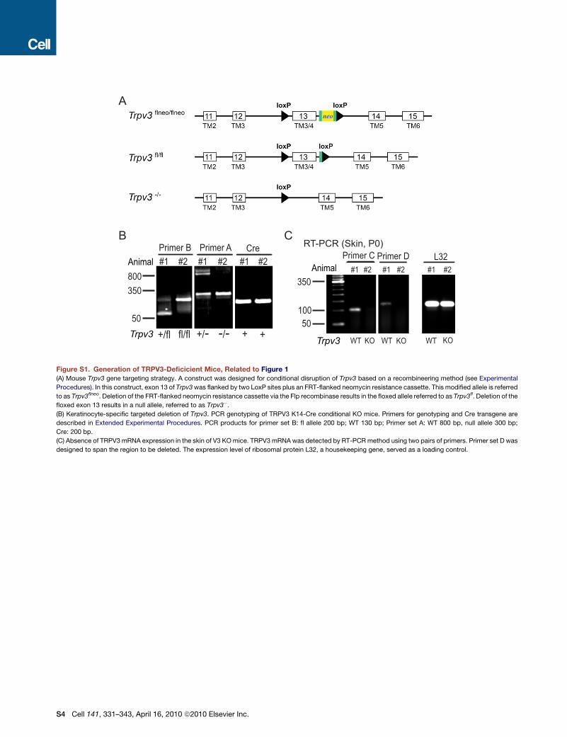
Conditional and Global Disruption of Trpv3 in MiceWe targeted exon 13 of mouse Trpv3, located on chromosome 11 B4, to disrupt its function. Deletion of exon 13 was predicted to
remove the entire 3rd transmembrane segment (TM3) and part of TM4 and shift the open reading frame thereafter (see Figure S1).
Thus, the putative pore region (TM5-TM6) would not be translated in TRPV3 KO mice regardless of whether the resulting transcript
was stable. Since global knockout mice can be easily obtained from conditional deletions via a global Cre transgenic, such as Sox2-
Cre (Hayashi et al., 2002), we made a construct for conditional disruption of Trpv3 based on a recombineering method (Liu et al.,
2003). In this construct, exon 13 of Trpv3 was flanked by two LoxP sites plus an FRT-flanked neomycin resistance cassette (see
Figure S1). This modified allele is referred to as Trpv3flneo. Deletion of the FRT-flanked neomycin resistance cassette via the recom-
binase results in the floxed allele referred to as Trpv3fl. Deletion of the floxed exon 13 results in a null allele, referred to as Trpv3�. For
ES cell targeting, the construct was electroporated into J1 embryonic stem cells and cells were selected for neomycin resistance.
Positive ES cell clones with correct homologous recombination were identified by Southern analysis. Three positive ES cell clones
with a normal karyotype were injected into C57BL/6J mouse blastocysts and transferred into the uteri of pseudopregnant females,
from which three high-percentage male chimeras were obtained. The chimeras were bred with C57BL/6J females to generate F1
offspring carrying the Trpv3flneo allele. Germline transmission of the Trpv3flneo allele was confirmed by Southern analysis using tail
DNA prepared from Agouti pups. The neomycin resistance cassette was removed from the targeted allele by breeding Trpv3flneo/+
mice with transgenic mice expressing the Flp recombinase (Farley et al., 2000) (Jackson Laboratory #003946), resulting in Trpv3fl/+
mice. The Trpv3�/+ mice were obtained by breeding Trpv3fl/+ mice with Sox-2-Cre transgenic which provides germline/embryonic
expression of Cre recombinase (Hayashi et al., 2002). Trpv3fl/fl and Trpv3�/� mice were obtained, respectively, by intercross of
heterozygotes and maintained on a mixed C57BL/6J and 129/SvEvTac background. Trpv3�/� mice were also backcrossed with
C57BL/6J females for more than 6 generations to obtain a clean genetic background. K14-Cre transgenic (Jackson laboratory #
004782) was bred with Trpv3fl/fl mice to obtain keratinocyte-specific disruption of Trpv3.
Southern Blot and PCR GenotypingFor Southern blot analysis, 10 mg of genomic DNA was digested overnight with KpnI, fractionated on a 0.7% agarose gel, and trans-
ferred to Hybond N+ membrane (Amersham). Southern analysis was performed using a standard non-radioactive labeling protocol
with DIG-labeled dTTP (Roche). The probe for identification of the Trpv3flneo allele generated by homologous recombination was
amplified with the following primers: 50- CAATGAAAAGAGTCTACAGCTTTGGA-30 and 50 CTACATGGGGCAGTTCCAAGATC-30.
Mouse genotyping was routinely done by PCR analysis. For Trpv3fl/+ mice, F8305, 50- GCTGGTTGGGCATTGGTAAGAG-30, and
R8432, 50- GTCTGTTATATGTACAGGCATGG-30 were used (Primer set B). The Trpv3fl and wt alleles yielded products of 200 bp
and 130 bp, respectively. For Trpv3-/+ mice, F7656, 50- GACATGCCATGCAAAAAACTACCA-30 and R8432 (Primer set A) were
used: the null and alleles yielded products of 300 bp and 800 bp, respectively. The primers for Cre are as follows:
forward primer (F), 50- CGTATAGCCGAAATTGCCAG-30;
reverse primer (R), 50- CAAAACAGGTAGTTATTCGG-30
Genotypes of TRPV4 KO mice and K14 promoter- driven TRPV3-YFP transgenic mice were determined by PCR as described
previously (Huang et al., 2008; Suzuki et al., 2003).
Real-Time Semiquantitative PCRThe primer sequences were as follows.
For mL32, F: 50-TGGTGAAGCCCAAGATCGTC-30; R: 50- CTTCTCCGCACCCTGTTGTC-30.
For mTGF-a, F: 50- GCGCTGGGTATCCTGTTAGC-30; R: 50-TGGGAATCTGGGCACTTGTT-30.
h EGFR F: 50-CGGGACATAGTCAGCAGTGA-30; R: 50-GGGACAGCTTGGATCACACT-30
h PLC-g1 F: 50-TGGCTCCGGAAGCAGTTTTA-30; R: 50-ATGTTGGGGACCCGGTAGTT-30
h GADPH F: 50-GAAGGTGAAGGTCGGAGTCA-30; R: 50- AATGAAGGGGTCATTGATGG-30
m EGF F: 50-GGTGGCTCCGTCCGTCTTAT-30; R: 50-CCAAATCGCCTTGCTTTTCA-30
m AR F: 50-CATCGGCATCGTTATCACAG-30; R: 50-ACAGTCCCGTTTTCTTGTCG-30
m HBEGF F: 50-ATCCACGGGGAGTGCAGATA-30; R: 50-GAGTCAGCCCATGACACCTG-30
m EGFR F: 50-CGGGACACCCAATCAGAAAA-30; R: 50-CAGCCTTCCGAGGAGCATAA-30
For each sample, the expression levels of mTGF-a, mEGF, mAR, mHBEGF, and mEGFR were normalized using that of mL32. The
expression levels of hEGFR and hPLC-g1were normalized using that of hGADPH.
Reverse Transcriptional-PCR AnalysisSingle-stranded cDNA from P0 mouse skin was prepared as described in Experimental Procedures. Primer sequences of TRPV3
were as follows.
Primer set C: forward primer, 50- CAGCGTCATGATCCAGAAGG-30; reverse primer 50- ATCAGTGAGGCCAGCGCTAC-30.
Primer set D: forward primer, 50- TGCTGAGACCCTCCGATCTT-30; reverse primer, 50- GGCAGGCGAGGTATTCTTTG-30.
Primer set D was designed based on sequences within the putative deletion region, exon 13.
Cell 141, 331–343, April 16, 2010 ª2010 Elsevier Inc. S1
Lentiviral pLKO.1-ShRNA KnockdownA series of Lentiviral pLKO.1-ShRNA constructs against human EGFR and PLC-g1 were purchased from Sigma and tested using q-
PCR in HEK293T cells. The following two ShRNA constructs were chosen to knock down EGFR and PLC-g1 in human epidermal
keratinocytes (NHEK):
EGFR: 50-CCGGGCTGCTCTGAAATCTCCTTTACTCGAGTAAAGGAGATTTCAGAGCAGCTTTTTG-30;
PLC-g1: 50-CCGGCCAGATCAGTAACCCTGAATTCTCGAGAATTCAGGGTTACTGATCTGGTTTTT-30.
The ShRNA and pLKO.1 control lentivirus stocks were generated via co-transfection of HEK293T cells with packaging plasmids
VSV-G-pMAD.G and pCMVdeltaR8.91. NHEK cells were infected with each lentivirus stock and 3 days post-puromycin (2 mg/ml)
selection, were used for Ca2+ imaging experiments.
Immunoblotting and ImmunoprecipitationFor the immunodetection of EGFR and P-EGFR, back skin lysates were incubated with 2 mg of EGFR antibody (Upstate Cell
Signaling) and rotated for 12 hr at 4�C. Protein A/G beads (30 ml; Amersham Pharmacia) were added, and after 12 hr incubation,
the beads were pulled down and washed 5–6 times with lysis buffer. Bound proteins were eluted from the beads with SDS (13)
sample buffer, vortexed, boiled for 5 min, and analyzed by immunoblotting. The total cell lysate or immunoprecipitated proteins
were separated by SDS-PAGE and transferred to nitrocellulose membranes. The membranes were blocked for 1 hr with 5% skim
milk in PBST and incubated with the anti-EGFR or P-EGFR antibody (diluted 1:1000) in PBST. Detection was carried out using Perox-
idase-conjugated anti-rabbit secondary antibody with an enhanced chemiluminescence reagent (Amersham Pharmacia Biotech).
Co-immunoprecipitation for TRPV3 and EGFR was performed in skin lysates or HEK293T cells transfected with pEGFP-C3,
TRPV3-EGFP, and EGFR plasmids. The lysis buffer contained 137 mM NaCl, 10% glycerol, 1% NP-40, 2 mM EDTA, and 20 mM
Tris-HCl (pH 8.0). The lysate was stirred on ice for 30 min and then centrifuged. The supernatant was incubated with anti-EGFR
(Upstate) or anti-GFP (Covance) at 4�C overnight. The protein complex was then visualized by western blotting using antibodies
against GFP or EGFR (Upstate).
Histology and ImmunostainingImmunohistochemistry was performed on cryostat sections (�10 mm) using antibodies for K14 (1:5000; Covance), K1 (1:2000; Co-
vance), K10 (1:1000; Sigma), Integrin a6 (1:1000; BD Lab), Integrin b4 (1:1000; BD Lab), Loricrin, (1:5000; Abcam), EGFR (1:200;
Upstate Biotechnology), and P-EGFR (anti-P-Tyr 1173 EGFR, 1:200; Upstate Biotechnology). Nuclei were counterstained with
DAPI reagents. Images were taken using an Olympus (IX 81) microscope and a Leica (TCS SP5) confocal microscope.
Dye Exclusion AssaysToluidine blue staining of mouse embryos and newborn pups was preformed as described previously (Koch et al., 2000; Sevilla et al.,
2007). The developmental stage of mouse embryos was determined based on the assumption that fertilization occurred in the middle
of the day’s dark cycle before vaginal plugs were identified. Embryos were dehydrated by incubation in 25%, 50%, and 75% meth-
anol/PBS for 1 min each followed by incubation in 100% methanol for 1 min. The embryos were then rehydrated with the same series
of methanol solution for 1 min each, washed in PBS, and stained for 10 min in 0.0125% toluidine blue O (Fisher Scientific)/PBS. The
embryos were then de-stained in PBS.
In Vivo Transglutaminase Activity AssayDetection of TGase activity in skin sections (Raghunath et al., 1998) used the amine donor substrate monodansylcadaverine (Molec-
ular Probes). A solution of 2 mg biotinylated-X-cadaverine in 0.1 N HCl (50 ml) was prepared and then mixed with 394 ml doubly
distilled H2O. This 10 mM stock solution was stored at �20�C before use. TGases substrate buffer was prepared by adding 10 ml
of the substrate stock solution plus 25 ml CaCl2 (200 mM) solution to 965 ml Tris/HCl (100 mM; pH 8.4). Cryostat sections (�10
mm) were air-dried and preincubated with 13 BSA in 0.1 M Tris/HCl (pH 8.4) for 30 min at room temperature. The sections were
then incubated for 2 hr with the substrate buffer (pH 8.4). The TGase reaction was stopped with PBS/25 mM EDTA (5 min) and washed
two times with 13 PBS, 10 min each. The sections were incubated with Streptavidin-conjugated Alexa Fluor 488 (1:1000, Invitrogen)
in PBS for 30 min, washed three times in PBS, and mounted for visualization.
In Vitro Transglutaminase Activity Assay (for Cultured Keratinocytes)TGase activity in cultured keratinocytes was detected using the amine donor substrate monodansylcadaverine. Primary cultured ker-
atinocytes were partially serum-starved overnight (1% FBS) in MEM medium containing 0. 5 mM Ca2+ medium. The cells were then
treated with 0.1% DMSO, TRPV3 agonist cocktail (50 mM 2-APB + 200 mM Carvacrol) in MEM medium containing 1.4 mM Ca2+ and
100 mM biotinylated-X-cadaverine (Invitrogen) for 40 min. Keratinocytes were then rinsed in PBS, fixed with 4% PFA, and incubated
with streptavidin-conjugated Alexa Fluor 488 (1:1000, Invitrogen) for 1 hr. Nuclei were counterstained with DAPI reagents for 10 min
before microscopic observation.
S2 Cell 141, 331–343, April 16, 2010 ª2010 Elsevier Inc.
TGF-a ELISATo measure TGF-a release, near confluent NHEK keratinocytes were pretreated with or without the metalloproteinase inhibitor BB-
2116 (20 mM; British Biotechnology, Oxford, UK) in the presence of 100 ng/ml EGF or 1 mM Tyrphostin AG 1478 (an EGFR inhibitor;
Cell Signaling), which was expected to block EGFR activation and receptor-mediated endocytosis of TGF-a. Cells were then treated
with V3 agonist cocktail (100 mM 2-APB + 250 mM Carvacrol) or 1 mM PMA (Cell Signaling) for 30 min in the presence or absence of 20
mM BB-2116. The medium was then harvested for TGF-a measurements using an ELISA kit for human TGF-a (Calbiochem).
ElectrophysiologyWhole-cell patch-clamp recordings were performed in primary keratinocytes. The pipette solution contained 147 mM Cs, 120 mM
methane-sulfonate, 4 mM NaCl, 10 mM EGTA, 2 mM Na2-ATP, 2 mM MgCl2, 20 mM HEPES (pH 7.2; free [Ca2+]i < 10 nM). Standard
extracellular bath solution (modified Tyrode’s solution) contained 153 mM NaCl, 5 mM KCl, 2 mM CaCl2, 1 mM MgCl2, 20 mM HEPES,
10 mM glucose (pH 7.4). All solutions were applied via a perfusion system to achieve a complete solution exchange within a few
seconds. Data were collected using an Axopatch 2A patch clamp amplifier, Digidata 1440, and pClamp 10.0 software (Axon Instru-
ments). Whole-cell currents were digitized at 10 kHz and low pass filtered at 2 kHz. Capacity current was reduced as much as
possible using the amplifier circuitry; series resistance compensation was 60%–85%. For heat activation experiments, the perfusate
was heated using a Warner TC-325B temperature controller and an SH-27B solution heater as described previously (Xu et al., 2006).
All other experiments were conducted at room temperature (�21�C–23�C). All recordings were analyzed with pCLAMP10 (Axon
Instruments, Union City, CA, USA) and Origin 7.5 (OriginLab, Northampton, MA, USA).
SUPPLEMENTAL REFERENCES
Farley, F.W., Soriano, P., Steffen, L.S., and Dymecki, S.M. (2000). Widespread recombinase expression using FLPeR (flipper) mice. Genesis 28, 106–110.
Hayashi, S., Lewis, P., Pevny, L., and McMahon, A.P. (2002). Efficient gene modulation in mouse epiblast using a Sox2Cre transgenic mouse strain. Mech. Dev.
119 (Suppl 1), S97–S101.
Liu, P., Jenkins, N.A., and Copeland, N.G. (2003). A highly efficient recombineering-based method for generating conditional knockout mutations. Genome Res.
13, 476–484.
Suzuki, M., Mizuno, A., Kodaira, K., and Imai, M. (2003). Impaired pressure sensation in mice lacking TRPV4. J. Biol. Chem. 278, 22664–22668.
Cell 141, 331–343, April 16, 2010 ª2010 Elsevier Inc. S3
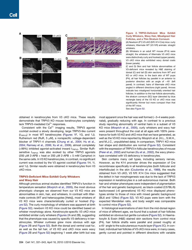
50
350
800
fl/fl -/- +/-+/fl + +
Primer B#1 #2 #2 #2 #1 #1
Primer A CreAnimal
KO WT KO WT
50
100
350
Primer C#1 #2 #2 #2 #1 #1
Primer D L32Animal
RT-PCR (Skin, P0)
Trpv3Trpv3
KO WT
A
B C
Figure S1. Generation of TRPV3-Deficicient Mice, Related to Figure 1
(A) Mouse Trpv3 gene targeting strategy. A construct was designed for conditional disruption of Trpv3 based on a recombineering method (see Experimental
Procedures). In this construct, exon 13 of Trpv3 was flanked by two LoxP sites plus an FRT-flanked neomycin resistance cassette. This modified allele is referred
to as Trpv3flneo. Deletion of the FRT-flanked neomycin resistance cassette via the Flp recombinase results in the floxed allele referred to as Trpv3fl. Deletion of the
floxed exon 13 results in a null allele, referred to as Trpv3�.
(B) Keratinocyte-specific targeted deletion of Trpv3. PCR genotyping of TRPV3 K14-Cre conditional KO mice. Primers for genotyping and Cre transgene are
described in Extended Experimental Procedures. PCR products for primer set B: fl allele 200 bp; WT 130 bp; Primer set A: WT 800 bp, null allele 300 bp;
Cre: 200 bp.
(C) Absence of TRPV3 mRNA expression in the skin of V3 KO mice. TRPV3 mRNA was detected by RT-PCR method using two pairs of primers. Primer set D was
designed to span the region to be deleted. The expression level of ribosomal protein L32, a housekeeping gene, served as a loading control.
S4 Cell 141, 331–343, April 16, 2010 ª2010 Elsevier Inc.
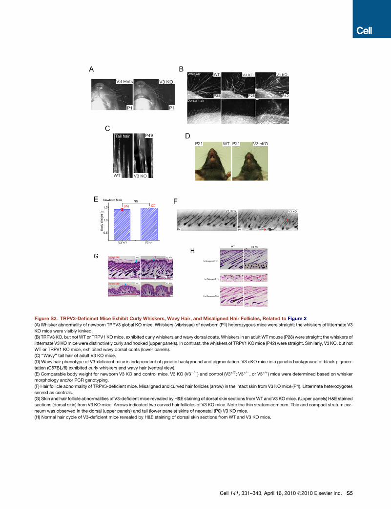
V3 KOV3 Hets
P1 P1
Whisker
Dorsal hair
V3 KO V1 KOWT
P28 P28 P42
V3 Hets V3 KO
P4 P4
Tail hair P49
V3 KOWT
P21 V3 cKOP21WT
A B
CD
E F
WT V3 KO
1st Anagen (P12)
1st Telogen (P21)
2nd Anagen (P30)
V3 KO
P4 P4
WT
Dorsal Skin
P0 P0
V3 KO WT
Dorsal SkinGH
Figure S2. TRPV3-Deficinet Mice Exhibit Curly Whiskers, Wavy Hair, and Misaligned Hair Follicles, Related to Figure 2
(A) Whisker abnormality of newborn TRPV3 global KO mice. Whiskers (vibrissae) of newborn (P1) heterozygous mice were straight; the whiskers of littermate V3
KO mice were visibly kinked.
(B) TRPV3 KO, but not WT or TRPV1 KO mice, exhibited curly whiskers and wavy dorsal coats. Whiskers in an adult WT mouse (P28) were straight; the whiskers of
littermate V3 KO mice were distinctively curly and hooked (upper panels). In contrast, the whiskers of TRPV1 KO mice (P42) were straight. Similarly, V3 KO, but not
WT or TRPV1 KO mice, exhibited wavy dorsal coats (lower panels).
(C) ‘‘Wavy’’ tail hair of adult V3 KO mice.
(D) Wavy hair phenotype of V3-deficient mice is independent of genetic background and pigmentation. V3 cKO mice in a genetic background of black pigmen-
tation (C57BL/6) exhibited curly whiskers and wavy hair (ventral view).
(E) Comparable body weight for newborn V3 KO and control mice. V3 KO (V3�/�) and control (V3+/?; V3+/�, or V3+/+) mice were determined based on whisker
morphology and/or PCR genotyping.
(F) Hair follicle abnormality of TRPV3-deficient mice. Misaligned and curved hair follicles (arrow) in the intact skin from V3 KO mice (P4). Littermate heterozygotes
served as controls.
(G) Skin and hair follicle abnormalities of V3-deficient mice revealed by H&E staining of dorsal skin sections from WT and V3 KO mice. (Upper panels) H&E stained
sections (dorsal skin) from V3 KO mice. Arrows indicated two curved hair follicles of V3 KO mice. Note the thin stratum corneum. Thin and compact stratum cor-
neum was observed in the dorsal (upper panels) and tail (lower panels) skins of neonatal (P0) V3 KO mice.
(H) Normal hair cycle of V3-deficient mice revealed by H&E staining of dorsal skin sections from WT and V3 KO mice.
Cell 141, 331–343, April 16, 2010 ª2010 Elsevier Inc. S5
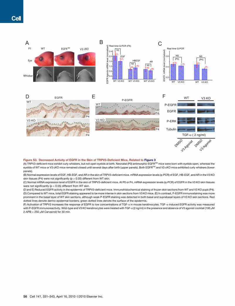
Eye
Whisker
WT EGFR Vel cKO
P-EGFRE
WT
P4
P4
D
P1
EGFR
V3 KOP4
P4
WT
V3
WT V3 KO
DMSO
V3 Ago
nist
DMSO TGF-α ( 2 ng/ml)
P-EGFR
EGFR
P-ERK
Tubulin
V3 Ago
nist
A B C
F
V3 KO
Figure S3. Decreased Activity of EGFR in the Skin of TRPV3-Deficient Mice, Related to Figure 3
(A) TRPV3-deficient mice exhibit curly whiskers, but not open eyelids at birth. Neonatal (P0) antimorphic EGFRVel mice were born with eyelids open, whereas the
eyelids of WT mice or V3 cKO mice remained closed until several days after birth (upper panels). Both EGFRVel and V3 cKO mice exhibited curly whiskers (lower
panels).
(B) Normal expression levels of EGF, HB-EGF, and AR in the skin of TRPV3-deficient mice. mRNA expression levels (q-PCR) of EGF, HB-EGF, and AR in the V3 KO
skin tissues (P4) were not significantly (p > 0.05) different from WT skin.
(C) Normal mRNA expression level of EGFR in the skin of TRPV3-deficient mice. At P0 or P4, mRNA expression levels (q-PCR) of EGFR in the V3 KO skin tissues
were not significantly (p > 0.05) different from WT skin.
(D and E) Reduced EGFR activity in the epidermis of TRPV3-deficient mice. Immunohistochemical staining of frozen skin sections from WT and V3 KO pups (P4).
(D) Compared to WT mice, total EGFR staining appeared to be more intense in skin sections from V3 KO mice. (E) In contrast, P-EGFR immunostaining was more
prominent in the basal layer of WT skin sections, although weak P-EGFR staining was detected in both basal and suprabasal layers of V3 KO skin sections. Red
dotted lines denote dermo-epidermal borders; green dotted lines denote the surface of the epidermis.
(F) Activation of TRPV3 increases the response of EGFR to low concentrations of TGF-a in mouse keratinocytes. TGF-a-induced EGFR activity was measured
with P-EGFR immunoreactivity. Wild-type and V3 KO keratinocytes were treated with TGF-a (2 ng/ml) in the presence and absence of V3 agonist cocktail (100 mM
2-APB + 250 mM Carvacrol) for 30 min.
S6 Cell 141, 331–343, April 16, 2010 ª2010 Elsevier Inc.
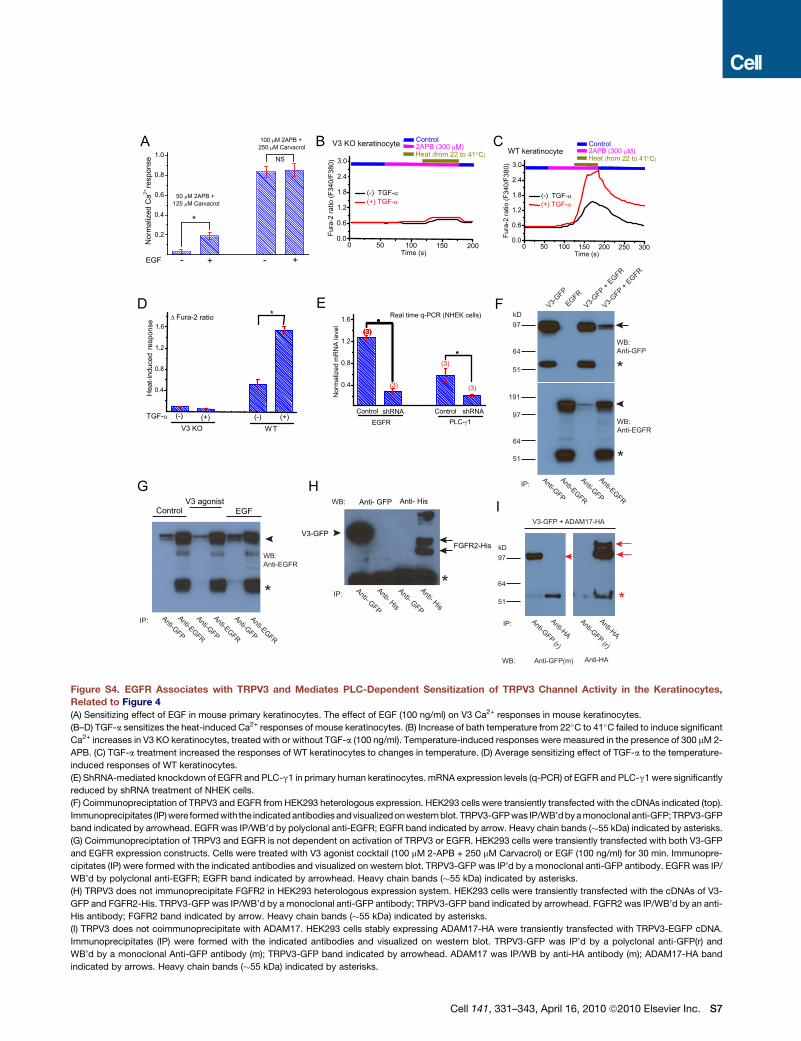
kD97
51
64
*
V3-GFP + ADAM17-HA
IP: Anti- GFP
Anti- GFP
Anti- His
Anti- His
*
WB: Anti- GFP Anti- His
V3-GFPFGFR2-His
V3-GFP
EGFRV3-G
FP + EGFR
V3-GFP +
EGFR
WB: Anti-GFP
WB: Anti-EGFR
IP: Anti-GFP
Anti-GFP
Anti-EGFR
Anti-EGFR
kD97
97
64
51
51
64
191
*
*
IP: Anti-GFP
Anti-GFP
Anti-EGFR
Anti-EGFR
*
WB: Anti-EGFR
Anti-EGFR
Anti-GFP
ControlV3 agonist
EGF
IP: Anti-GFP (r)
Anti-GFP (r)
Anti-HA
Anti-HA
Anti-GFP(m) WB: Anti-HA
A B C
D E F
G HI
Figure S4. EGFR Associates with TRPV3 and Mediates PLC-Dependent Sensitization of TRPV3 Channel Activity in the Keratinocytes,
Related to Figure 4
(A) Sensitizing effect of EGF in mouse primary keratinocytes. The effect of EGF (100 ng/ml) on V3 Ca2+ responses in mouse keratinocytes.
(B–D) TGF-a sensitizes the heat-induced Ca2+ responses of mouse keratinocytes. (B) Increase of bath temperature from 22�C to 41�C failed to induce significant
Ca2+ increases in V3 KO keratinocytes, treated with or without TGF-a (100 ng/ml). Temperature-induced responses were measured in the presence of 300 mM 2-
APB. (C) TGF-a treatment increased the responses of WT keratinocytes to changes in temperature. (D) Average sensitizing effect of TGF-a to the temperature-
induced responses of WT keratinocytes.
(E) ShRNA-mediated knockdown of EGFR and PLC-g1 in primary human keratinocytes. mRNA expression levels (q-PCR) of EGFR and PLC-g1 were significantly
reduced by shRNA treatment of NHEK cells.
(F) Coimmunopreciptation of TRPV3 and EGFR from HEK293 heterologous expression. HEK293 cells were transiently transfected with the cDNAs indicated (top).
Immunoprecipitates (IP) were formed with the indicated antibodies andvisualized on western blot. TRPV3-GFP was IP/WB’dby a monoclonal anti-GFP;TRPV3-GFP
band indicated by arrowhead. EGFR was IP/WB’d by polyclonal anti-EGFR; EGFR band indicated by arrow. Heavy chain bands (�55 kDa) indicated by asterisks.
(G) Coimmunopreciptation of TRPV3 and EGFR is not dependent on activation of TRPV3 or EGFR. HEK293 cells were transiently transfected with both V3-GFP
and EGFR expression constructs. Cells were treated with V3 agonist cocktail (100 mM 2-APB + 250 mM Carvacrol) or EGF (100 ng/ml) for 30 min. Immunopre-
cipitates (IP) were formed with the indicated antibodies and visualized on western blot. TRPV3-GFP was IP’d by a monoclonal anti-GFP antibody. EGFR was IP/
WB’d by polyclonal anti-EGFR; EGFR band indicated by arrowhead. Heavy chain bands (�55 kDa) indicated by asterisks.
(H) TRPV3 does not immunoprecipitate FGFR2 in HEK293 heterologous expression system. HEK293 cells were transiently transfected with the cDNAs of V3-
GFP and FGFR2-His. TRPV3-GFP was IP/WB’d by a monoclonal anti-GFP antibody; TRPV3-GFP band indicated by arrowhead. FGFR2 was IP/WB’d by an anti-
His antibody; FGFR2 band indicated by arrow. Heavy chain bands (�55 kDa) indicated by asterisks.
(I) TRPV3 does not coimmunoprecipitate with ADAM17. HEK293 cells stably expressing ADAM17-HA were transiently transfected with TRPV3-EGFP cDNA.
Immunoprecipitates (IP) were formed with the indicated antibodies and visualized on western blot. TRPV3-GFP was IP’d by a polyclonal anti-GFP(r) and
WB’d by a monoclonal Anti-GFP antibody (m); TRPV3-GFP band indicated by arrowhead. ADAM17 was IP/WB by anti-HA antibody (m); ADAM17-HA band
indicated by arrows. Heavy chain bands (�55 kDa) indicated by asterisks.
Cell 141, 331–343, April 16, 2010 ª2010 Elsevier Inc. S7
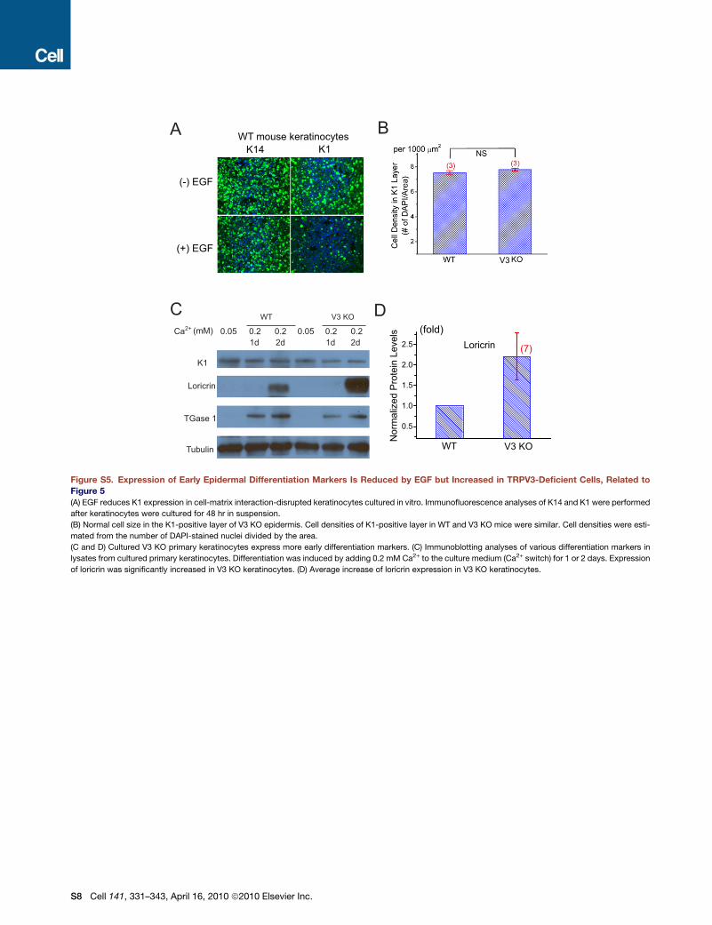
A
K1
Loricrin
TGase 1
Tubulin
Ca2+ (mM) 0.05 0.05 0.2 0.20.20.2 1d 1d 2d2d
WT V3 KO
(fold)
WT mouse keratinocytesK14 K1
(-) EGF
(+) EGFV3
B
C D
Figure S5. Expression of Early Epidermal Differentiation Markers Is Reduced by EGF but Increased in TRPV3-Deficient Cells, Related to
Figure 5
(A) EGF reduces K1 expression in cell-matrix interaction-disrupted keratinocytes cultured in vitro. Immunofluorescence analyses of K14 and K1 were performed
after keratinocytes were cultured for 48 hr in suspension.
(B) Normal cell size in the K1-positive layer of V3 KO epidermis. Cell densities of K1-positive layer in WT and V3 KO mice were similar. Cell densities were esti-
mated from the number of DAPI-stained nuclei divided by the area.
(C and D) Cultured V3 KO primary keratinocytes express more early differentiation markers. (C) Immunoblotting analyses of various differentiation markers in
lysates from cultured primary keratinocytes. Differentiation was induced by adding 0.2 mM Ca2+ to the culture medium (Ca2+ switch) for 1 or 2 days. Expression
of loricrin was significantly increased in V3 KO keratinocytes. (D) Average increase of loricrin expression in V3 KO keratinocytes.
S8 Cell 141, 331–343, April 16, 2010 ª2010 Elsevier Inc.
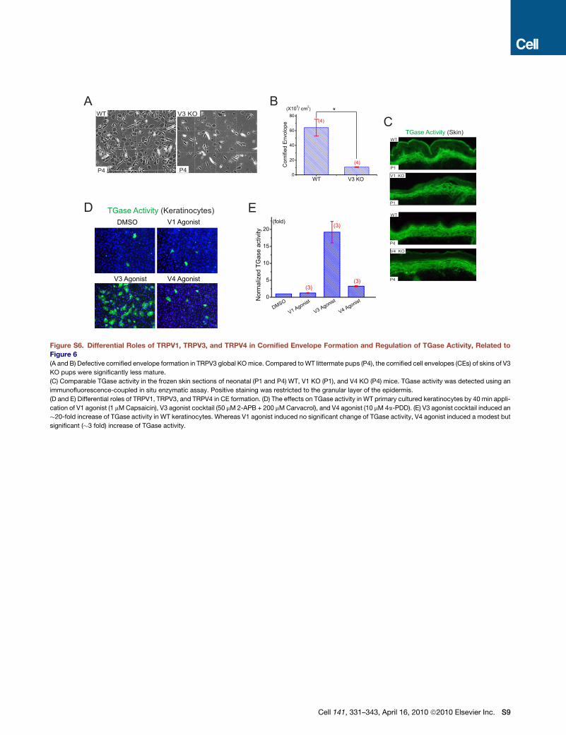
TGase Activity (Skin)WT
V1 KO
A BV3 KOWT
DMSO V1 Agonist
V3 Agonist V4 Agonist
TGase Activity (Keratinocytes)D
P4 P4 P1
P1
V4 KO
WT
P4
P4
C
E
Figure S6. Differential Roles of TRPV1, TRPV3, and TRPV4 in Cornified Envelope Formation and Regulation of TGase Activity, Related to
Figure 6
(A and B) Defective cornified envelope formation in TRPV3 global KO mice. Compared to WT littermate pups (P4), the cornified cell envelopes (CEs) of skins of V3
KO pups were significantly less mature.
(C) Comparable TGase activity in the frozen skin sections of neonatal (P1 and P4) WT, V1 KO (P1), and V4 KO (P4) mice. TGase activity was detected using an
immunofluorescence-coupled in situ enzymatic assay. Positive staining was restricted to the granular layer of the epidermis.
(D and E) Differential roles of TRPV1, TRPV3, and TRPV4 in CE formation. (D) The effects on TGase activity in WT primary cultured keratinocytes by 40 min appli-
cation of V1 agonist (1 mM Capsaicin), V3 agonist cocktail (50 mM 2-APB + 200 mM Carvacrol), and V4 agonist (10 mM 4a-PDD). (E) V3 agonist cocktail induced an
�20-fold increase of TGase activity in WT keratinocytes. Whereas V1 agonist induced no significant change of TGase activity, V4 agonist induced a modest but
significant (�3 fold) increase of TGase activity.
Cell 141, 331–343, April 16, 2010 ª2010 Elsevier Inc. S9