treatment of infectious mastitis during lactation...
TRANSCRIPT
Antibiotics vs Probiotics for Mastitis • CID 2010:50 (15 June) • 1551
M A J O R A R T I C L E
Treatment of Infectious Mastitis during Lactation:Antibiotics versus Oral Administration of LactobacilliIsolated from Breast Milk
Rebeca Arroyo, Virginia Martın, Antonio Maldonado, Esther Jimenez, Leonides Fernandez,and Juan Miguel RodrıguezDepartamento de Nutricion, Bromatologıa y Tecnologıa de los Alimentos, Universidad Complutense de Madrid, Madrid, Spain
Background. Mastitis is a common infectious disease during lactation, and the main etiological agents arestaphylococci, streptococci, and/or corynebacteria. The efficacy of oral administration of Lactobacillus fermentumCECT5716 or Lactobacillus salivarius CECT5713, two lactobacilli strains isolated from breast milk, to treat lactationalmastitis was evaluated and was compared with the efficacy of antibiotic therapy.
Methods. In this study, 352 women with infectious mastitis were randomly assigned to 3 groups. Women ingroups A ( ) and B ( ) ingested daily 9 log10 colony-forming units (CFU) of L. fermentum CECT5716n p 124 n p 127or L. salivarius CECT5713, respectively, for 3 weeks, whereas those in group C ( ) received the antibioticn p 101therapy prescribed in their respective primary care centers.
Results. On day 0, the mean bacterial counts in milk samples of the 3 groups were similar (4.35–4.47 log10
CFU/mL), and lactobacilli could not be detected. On day 21, the mean bacterial counts in the probiotic groups(2.61 and 2.33 log10 CFU/mL) were lower than that of the control group (3.28 log10 CFU/mL). L. fermentumCECT5716 and L. salivarius CECT5713 were isolated from the milk samples of women in the probiotic groups Aand B, respectively. Women assigned to the probiotic groups improved more and had lower recurrence of mastitisthan those assigned to the antibiotic group.
Conclusions. The use of L. fermentum CECT5716 or L. salivarius CECT5713 appears to be an efficient alternativeto the use of commonly prescribed antibiotics for the treatment of infectious mastitis during lactation.
ClinicalTrials.gov identifier. NCT00716183.
Mastitis is a common disease during lactation, with a
prevalence of 3%–33% of lactating mothers [1, 2]. This
inflammation of �1 lobule of the mammary gland usu-
ally has an infectious origin [3] involving staphylococci,
streptococci, and/or corynebacteria [2]. Traditionally,
Staphylococcus aureus has been considered to be the
main etiological agent of acute mastitis, although Staph-
ylococcus epidermidis is emerging as the leading cause
of chronic mastitis in both human and veterinary med-
icine [4–7]. Multidrug resistance and/or the formation
Received 30 November 2009; accepted 15 February 2010; electronicallypublished 10 May 2010.
Reprints or correspondence: Dr Juan Miguel Rodrıguez, Departamento deNutricion, Bromatologıa y Tecnologıa de los Alimentos, Universidad Complutensede Madrid, 28040 Madrid, Spain ([email protected]).
Clinical Infectious Diseases 2010; 50(12):1551–1558� 2010 by the Infectious Diseases Society of America. All rights reserved.1058-4838/2010/5012-0001$15.00DOI: 10.1086/652763
of biofilms are very common among clinical isolates of
these 2 staphylococcal species. This explains why mas-
titis is difficult to treat with antibiotics and why it con-
stitutes one of the main reasons to cease breastfeeding
[2]. In this context, the development of new strategies
based on probiotics, as alternatives or complements to
antibiotic therapy for the management of mastitis, is
particularly appealing.
In previous studies, we isolated potentially probiotic
lactobacilli strains from the milk of healthy mothers
[8–10]. Oral administration of either of 2 strains, Lac-
tobacillus salivarius CECT5713 and Lactobacillus gasseri
CECT5714, was an effective alternative for treating
staphylococcal mastitis in cases in which previous an-
tibiotic therapy had been unsuccessful [11]. The aim
of the present study was to evaluate the efficacy of oral
administration of each of 2 lactobacilli strains isolated
from breast milk, Lactobacillus fermentum CECT5716
and L. salivarius CECT5713, for treating lactational
by guest on April 1, 2014
http://cid.oxfordjournals.org/D
ownloaded from

1552 • CID 2010:50 (15 June) • Arroyo et al
Table 1. Bacterial Counts from Breast Milk and Breast Pain Score at the Beginning (Day 0) and the End (Day 21) of the Trial
Variable
Day 0 Day 21
Group A Group B Group C
P b
Group A Group B Group Ca
P bn Mean � SD n Mean � SD n Mean � SD n Mean � SD n Mean � SD n Mean � SD
Bacterial count
Total 124 4.35 � 0.57 127 4.47 � 0.53 101 4.39 � 0.41 .140 124 2.61 � 0.64 127 2.33 � 0.90 101 3.28 � 1.10 !.001
Staphylococcus epidermidis 92 4.18 � 0.70 88 4.30 � 0.59 76 4.21 � 0.52 .336 95 2.62 � 0.49 80 2.52 � 0.42 76 3.31 � 0.82 !.001
Staphylococcus aureus 67 3.83 � 0.55 55 4.06 � 0.67 30 3.95 � 0.54 .108 45 2.21 � 0.50 40 2.26 � 0.55 25 2.97 � 0.88 !.001
Streptococcus mitis 36 3.96 � 0.47 36 4.07 � 0.51 35 4.12 � 0.45 .162 32 2.35 � 0.37 28 2.29 � 0.48 31 3.14 � 0.72 !.001
Streptococcus salivarius 4 4.39 � 0.56 7 4.08 � 0.59 4 3.71 � 0.33 3 2.23 � 0.60 5 2.09 � 0.47 3 3.12 � 1.09
Rothia spp. 2 3.24 � 0.08 10 3.87 � 0.58 5 3.48 � 0.42 0 7 2.04 � 0.24 5 2.42 � 0.67
Corynebacterium spp. 5 3.65 � 0.60 2 4.64 � 0.51 6 3.86 � 0.50 5 1.94 � 0.25 2 2.27 � 0.04 5 2.39 � 0.99
Breast pain score 124 2.35 � 1.28 127 2.16 � 1.28 101 2.01 � 1.09 .185 124 8.68 � 1.06 127 8.61 � 1.25 101 5.81 � 2.50 !.001
NOTE. Data are expressed as log10 colony-forming units/mL, unless otherwise indicated. Treatment for group A was Lactobacillus fermentum CECT5716;for group B, Lactobacillus salivarius CECT5713; and for group C, antibiotic. Breast pain score ranged from extremely painful (0) to no pain (10). n, no. of womenin the group or having the listed bacterial species in their milk; SD, standard deviation.
a On day 21, group C differed significantly from group A and group B in counts for total bacteria, S. epidermidis, S. aureus, and S. mitis and in breast painscore (nonparametric multiple comparison test; ; a p 0.05).P ! .001
b Kruskal-Wallis test, .a p 0.05
mastitis in a higher number of women and to compare such
an approach with the antibiotic therapy that is usually pre-
scribed to treat this condition.
MATERIALS AND METHODS
Design of the study and collection of the milk samples.
A total of 352 women with symptoms of mastitis participated
in the study. All met the following criteria: breast inflammation,
painful breastfeeding, milk bacterial count 14 log10 colony-
forming units (CFU)/mL, and milk leukocyte count 16 log10
cells/mL. Many of the women ( ) presented fissures inn p 74
the mammary areola and/or nipple. None of them ingested
commercial probiotic foods or supplements during the study.
Women with mammary abscesses, Raynaud syndrome, or any
other mammary pathology were excluded. All volunteers gave
written informed consent to the protocol, which was approved
by the Ethical Committee of Hospital Clınico of Madrid
(Spain). The study was registered in the ClinicalTrials.gov da-
tabase (NCT00716183). The volunteers were randomly assigned
to 3 groups (2 probiotic groups and 1 antibiotic group), and
neither volunteers nor investigators knew the assignments dur-
ing the investigation.
The study lasted 21 days, and during this period, the pro-
biotic groups A ( ) and B ( ) consumed daily an p 124 n p 127
capsule with 200 mg of a freeze-dried probiotic containing ∼9
log10 CFU of L. fermentum CECT5716 [8] or L. salivarius
CECT5713 [10]. Capsules were manufactured at the probiotic
plant of Puleva Biotech (Granada, Spain) and were kept at 4�C
throughout the study. The women of the antibiotic group
(group C, ) received the antibiotic treatment prescribedn p 101
in their primary care centers. Breast milk samples were obtained
from the volunteers at the beginning (day 0) and at the end
(day 21) of the study, in accordance with a previously described
procedure [11]. The evolution of the symptoms was evaluated
at days 0 and 21 by midwives of their primary care centers. At
both times, the volunteers were asked to score their breast pain
from 0 (extremely painful) to 10 (no pain).
Count and identification of bacteria in the milk samples.
Samples were spread onto Baird-Parker, Columbia, Mac-
Conkey, and Sabouraud dextrose chloramphenicol agar plates
(BioMerieux) for selective isolation and quantification of the
main agents involved in infectious mastitis [12] and, parallel,
onto agar plates of MRS (Oxoid) supplemented with L-cysteine
(0.5 g/L) (MRS-Cys) for isolation of lactobacilli. The plates
were incubated for 48 hours at 37�C in aerobic conditions,
except for the MRS-Cys plates, which were incubated anaer-
obically (in 85% nitrogen, 10% hydrogen, and 5% carbon di-
oxide) in an anaerobic workstation (DW Scientific).
Bacteria isolated from milk were initially identified at the
species level by classic morphological and biochemical tests.
The identification of bacteria belonging to the S. epidermidis
or S. aureus species was confirmed by a multiplex polymerase
chain reaction (PCR) method based on dnaJ genes with prim-
ers J-StGen (5′-TGGCCAAAAGAGACTATTATGA-3′), J-StAur
(5′-GGATCTCTTTGTCTGCCG-3′), and J-StEpi (5′-CCACCA-
AAGCCTTGACTT-3′) in a Icycler thermocycler (Bio-Rad Lab-
oratories). The primer pair J-StGen and J-StAur results in a
337 bp S. aureus species-specific fragment, and the primer pair
J-StGen and J-StEpi results in a 249 bp S. epidermidis species-
specific fragment [11]. Identification of streptococci was per-
formed by partial amplification (488 bp) and sequencing of the
gene tuf with primers TufStrep-1 (5′-GAAGAATTGCTTGAAT-
TGGTTGAA-3′) and TufStrep-R (5′-GGACGGTAGTTGTTG-
AAGAATGG-3′) [13]. Identification of the potential Strepto-
by guest on April 1, 2014
http://cid.oxfordjournals.org/D
ownloaded from
Antibiotics vs Probiotics for Mastitis • CID 2010:50 (15 June) • 1553
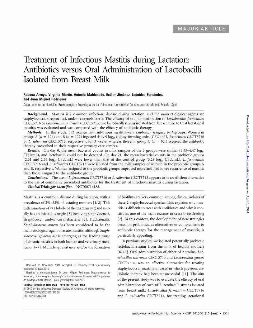
Figure 1. Box and whisker plots showing changes in bacterial count(total, Staphylococcus epidermidis, Staphylococcus aureus, and Strepto-coccus mitis) of breast milk samples and changes in breast pain scorereported by the participants after probiotic (Lactobacillus fermentumCECT5716 in group A and Lactobacillus salivarius CECT5713 in group B)or antibiotic (group C) treatment. Differences in the changes experiencedfor each group were evaluated with nonparametric multiple comparisontests and are shown with horizontal lines inside each graph (* ;P ! .01** ). The horizontal line in the middle of each box represents theP ! .001median, while the top and bottom borders of the box represent the 75%and 25% percentiles, respectively. The mean is represented as a cross,and the outliers as individual points outside the boxes. Breast pain scoreranged from 0 (extremely painful) to 10 (no pain).
coccus mitis isolates was confirmed by testing optochin sensitiv-
ity and bile solubility [14] and by testing latex agglutination with
the Slide Pneumo kit (BioMerieux).
The remaining isolates were identified by 16S rRNA se-
quencing with primers pbl16 (5′-AGAGTTTGATCCTGGCT-
CAG-3′) and mlb16 (5′-GGCTGCTGGCACGTAGTTAG-3′)
[15]. Their identity was determined on the basis of the highest
scores (�99%) among the sequences deposited in the European
Molecular Biology Laboratory database, by means of the Basic
Local Alignment Search Tool algorithm.
Identification of L. salivarius CECT5713 and L. fermentum
CECT5716 in the milk samples. A DNA-DNA colony hy-
bridization assay was developed to investigate whether oral ad-
ministration of the lactobacilli led to their presence in milk.
For this purpose, 2 species-specific probes were designed on
the basis of unique 16S rRNA sequences. In the case of L.
salivarius, a fragment (210 bp) was amplified from L. salivarius
CECT5713 genomic DNA with primers SAL91F (5′-ATTCAC-
CGTAAGAAGT-3′) and SAL285R (5′-TATCATCACCTTGG-
TAG-3′). Parallel, a fragment (192 bp) was amplified from L.
fermentum CECT5716 genomic DNA with primers Lfer-3 (5′-
ACTAACTTGACTGATCTACGA-3′) and Lfer-4 (5′-TTCACT-
GCTCAAGTAATCATC-3′) [16]. The PCR conditions were as
follows: 95�C for 2 minutes (1 cycle); 95�C for 30 seconds, 46�C
(L. salivarius) or 55�C (L. fermentum) for 30 seconds, and 72�C
for 45 seconds (40 cycles); and a final extension at 72�C for 4
minutes. Both PCR fragments were purified using the QIAquick
PCR purification kit (Qiagen) and labeled using the Amersham
ECL direct nucleic acid labelling and detection system (GE
Healthcare).
Colonies obtained on MRS-Cys plates from milk samples
(day 21) were spotted in a regular array on 2 sets of MRS-Cys
replica plates. Then, nylon Hybond-N+ discs (GE Healthcare)
were laid directly on the culture surfaces and were kept there
for 1 minute. Both hybridization and detection were performed
as previously described [11]. The identity of the isolates that
gave a positive signal after colony hybridization was confirmed
by 16S rRNA sequencing as described above.
L. salivarius and L. fermentum isolates were submitted to
pulsed-field gel electrophoresis (PFGE) genotyping as previ-
ously described [11]. Their profiles were compared with those
of L. salivarius CECT5713, L. salivarius CECT4062, L. salivarius
CECT4063, L. salivarius DSM 20492, L fermentum CECT5716,
L. fermentum CECT285, L. fermentum CECT4007, and/or L.
fermentum. The LMG 8900 Low Range PFG marker (New En-
gland BioLabs) was used as the molecular size standard.
Statistical analysis. Microbiological data, recorded as
number of CFU per mL of milk, were transformed to loga-
rithmic values before calculation of means and statistical anal-
ysis. The reported values of bacterial counts are the mean values
of duplicate or triplicate determinations. The continuous var-
iables “bacterial counts” and “breast pain score” were not nor-
mally distributed. Three bacterial species occurred in sufficient
numbers of breast milk samples to allow statistical comparison
between groups. Kruskal-Wallis tests were performed to deter-
mine statistically significant differences between the bacterial
counts (total and main bacterial species) and between the breast
pain scores at the beginning (day 0) and at the end (day 21)
of the trial. The same approach was used to determine whether
there were differences in the change of these variables among
the 3 groups. When statistically significant differences were
found, nonparametric multiple comparisons were performed
to ascertain which pair of groups was different. The association
of mastitis recurrence with the treatment was compared with
the x2 test. The relationship between total bacterial count and
breast pain score was analyzed using the Spearman rank cor-
by guest on April 1, 2014
http://cid.oxfordjournals.org/D
ownloaded from

1554 • CID 2010:50 (15 June) • Arroyo et al
Table 2. Reduction in Bacterial Counts in Breast Milk and Change in Breast Pain Score from Day 0 to Day 21, according to theAntibiotic Prescribed to Group C Women
Variable
Amoxicillin-clavulanic acid Amoxicillin Cotrimoxazole Cloxacillin Erythromycin
Pan Mean � SD n Mean � SD n Mean � SD n Mean � SD n Mean � SD
Reduction in bacterial countsb
Total 39 �1.22 � 0.84 23 �0.55 � 0.56 19 �2.50 � 1.21 18 �0.27 � 0.41 2 0 � 0.04 !.001
Staphylococcus epidermidis 32 �1.15 � 0.67 18 �0.50 � 0.59 11 �2.21 � 1.30 15 �0.17 � 0.37 1 0.03 � NA !.001
Staphylococcus aureus 10 �1.74 � 1.28 12 �0.79 � 0.59 6 �2.89 � 1.53 2 �0.05 � 0.25 0 … .006
Streptococcus mitis 15 �1.20 � 0.94 4 �1.66 � 1.67 6 �2.18 � 1.00 9 �0.85 � 1.39 1 �0.03 � NA .018
Change in breast pain scorec 39 4.67 � 1.90 23 2.61 � 2.52 19 6.05 � 1.13 18 1.50 � 2.15 2 0 � 0 !.001
NOTE. n, no. of women in the group or having the listed bacterial species in their milk; NA, not applicable.a Kruskal-Wallis test, except for erythromycin data.b Reduction in bacterial counts was calculated as D log10 colony-forming units per mL.c Breast pain score ranged from extremely painful (0) to no pain (10), and change in breast pain score used 0 for no change.
Table 3. Additional Outcomes of the Study of Treatment of Infectious Mastitis during Lactation
VariableNo. of
women
No. (%) of women
With detection oflactobacilli With recurrencea
Withvaginal candidiasisb
Withflatulence
With discontinuation ofbreastfeeding
Probiotic
Lactobacillus fermentum CECT5716 124 67 (54.0) 13 (10.5)c 0 (0) 9 (5.6) 0 (0)
Lactobacillus salivarius CECT5713 127 68 (53.5) 9 (7.1)c 0 (0) 0 (0) 0 (0)
Total 251 135 (53.8) 22 (8.8)d 0 (0) 9 (3.6) 0 (0)
Antibiotic
Amoxicillin-clavulanic acid 39 0 (0) 18 (46.1) 1 (2.56) 0 (0) 0 (0)
Amoxicillin 23 0 (0) 8 (34.8) 5 (21.7) 0 (0) 1 (4.3)
Cotrimoxazole 19 0 (0) 0 (0) 0 (0) 0 (0) 0 (0)
Cloxacillin 18 0 (0) 5 (27.8) 3 (16.7) 0 (0) 8 (44.4)
Erythromycin 2 0 (0) 0 (0) 0 (0) 0 (0) 0 (0)
Total 101 0 (0) 31 (30.7)d 9 (8.9) 0 (0) 9 (8.9)
a Recurrence was defined as a new episode of mastitis (clinical symptoms and bacterial concentration 14 log10 colony-forming units [CFU]/mL) in a follow-upperiod of 3 months after these parameters had reached physiologic values (no clinical symptoms and bacterial concentration !3 log10 CFU/mL).
b Vaginal candidiasis was defined as the presence of clinical symptoms compatible with such condition, together with a dense population of Candida albicansin culture of vaginal exudates on Sabouraud dextrose chloramphenicol agar plates (BioMerieux).
c , .2x p 0.91 P p .340d , .2x p 27.08 P ! .001
relation coefficient for nonparametric data. The significance
level was set at .05. All analyses were performed using the
software package SAS, version 9.1 (SAS Institute).
RESULTS
Bacterial counts in the milk samples. At day 0, the mean
values of total bacterial count in milk were very similar in the
3 groups and ranged 4.35–4.47 log10 CFU/mL (Table 1). S.
epidermidis (isolated from 73% of the women), S. aureus (from
43%), and S. mitis (from 30%) were the dominant species
(Table 1). Other bacterial species were identified in !5% of the
samples, and lactobacilli could not be detected in any sample.
On day 21, differences in the total bacterial counts of the 3
groups were found (Kruskal-Wallis, ) (Table 1). TheP ! .001
mean values of log10 total bacterial count in the probiotic groups
(2.61 and 2.33 log10 CFU/mL for groups A and B, respectively)
were significantly lower ( ) than the corresponding valueP ! .001
in the antibiotic group (3.28 log10 CFU/mL). Mean reductions
of 1.74 and 2.15 log10 cycles in the total bacterial count were
observed in groups A and B, respectively, whereas in the an-
tibiotic group the reduction was significantly lower (1.10 log10
cycle) (Figure 1). The distribution of the bacterial species in
the milk samples on day 21 was similar to that observed on
day 0. There were statistically significant differences in the bac-
terial counts of each dominant bacterial species (S. epidermidis,
S. aureus, and S. mitis) in the 3 groups at the end of the trial
by guest on April 1, 2014
http://cid.oxfordjournals.org/D
ownloaded from

Antibiotics vs Probiotics for Mastitis • CID 2010:50 (15 June) • 1555
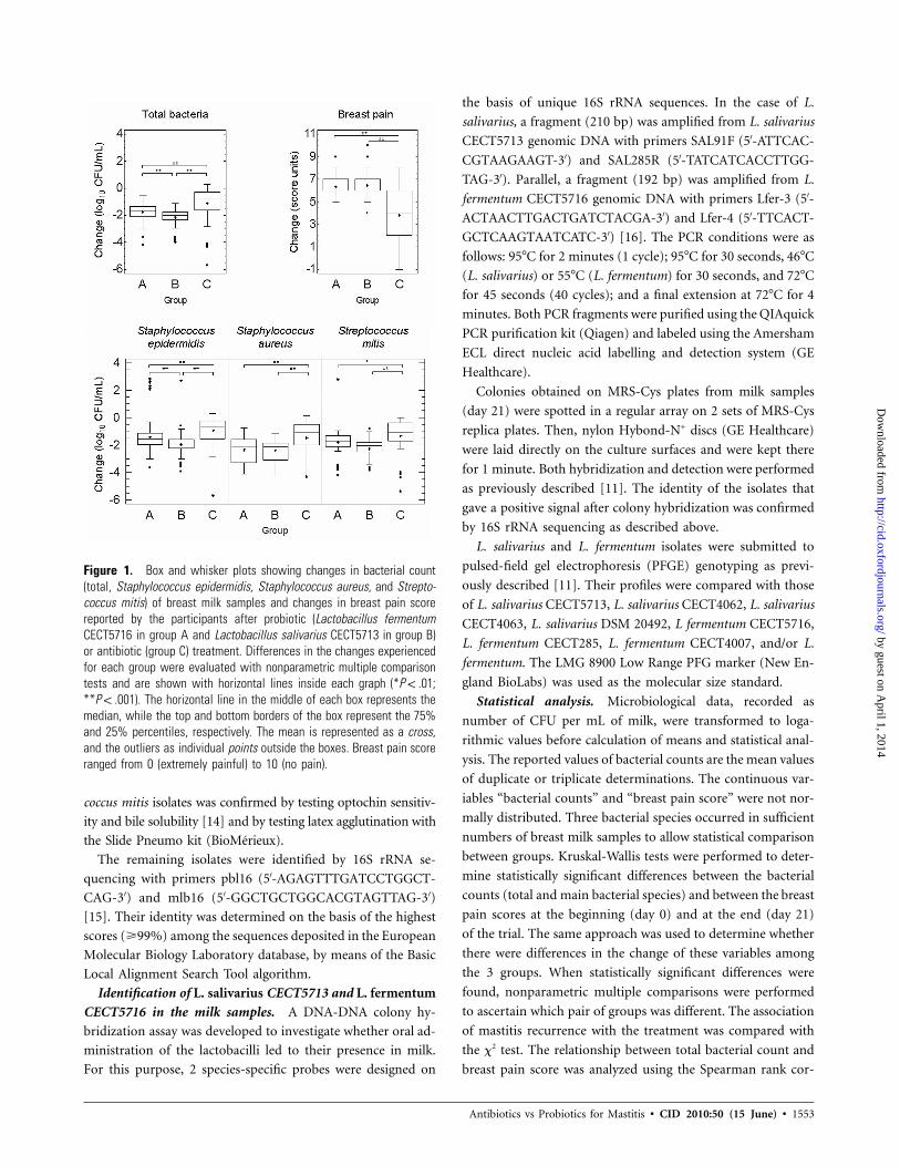
Figure 2. Distribution of breast pain scores reported by participants at the beginning (day 0) and at the end (day 21) of the trial in the probioticgroups (group A, Lactobacillus fermentum CECT5716; and group B, Lactobacillus salivarius CECT5713) and in the antibiotic group (group C). Breastpain categories were 0–4, extremely painful; 5–7, discomfort; and 8–10, no pain.
Figure 3. Banding patterns determined by pulsed-field gel electrophoresis (PFGE) of SmaI-digested genomic DNA from Lactobacillus salivariusCECT5713 (lane 1), 2 milk isolates that hybridized with the L. salivarius probe in the colony hybridization assay (lanes 2 and 3 ), L. salivarius CECT4062(lane 4 ), L. salivarius CECT4063 (lane 5 ), L. salivarius DSM 20492 (lane 6 ), Lactobacillus fermentum CECT5716 (lane 7 ), 2 milk isolates that hybridizedwith the L. fermentum probe in the hybridization assay (lanes 8 and 9 ), L. fermentum CECT285 (lane 10 ), L. fermentum CECT4007 (lane 11), and L.fermentum LMG 8900 (lane 12 ). Lane L represents the Low Range PFG standard (New England BioLabs).
(Kruskal-Wallis, ), and they were always lower (P ! .001 P !
) in the probiotic groups than in the antibiotic group (Ta-.001
ble 1).
The highest reductions in the bacterial counts were found
in group B (L. salivarius) (Figure 1). There was a statistically
significant difference ( ) in the decrease of total bacterialP ! .001
and S. epidermidis bacterial counts between the 2 probiotic
groups, although the women in both probiotic groups reported
the same change in breast pain score (Figure 1). The highest
bacterial count decrease was observed for S. aureus (2.3 and
2.4 log10 CFU/mL for groups A and B, and 1.5 log10 CFU/mL
for the antibiotic group) (Figure 1).
The antibiotics prescribed to group C women were amoxi-
cillin-clavulanic acid (38.6%), amoxicillin (22.8%), cotrimox-
azole (18.8%), cloxacillin (17.8%), and erythromycin (2%) (Ta-
ble 2). The effectiveness of these antibiotics in the reduction
of bacterial counts differed significantly (Kruskall-Wallis, P !
for total bacteria and S. epidermidis, for S. aureus,.001 P p .005 by guest on April 1, 2014
http://cid.oxfordjournals.org/D
ownloaded from

1556 • CID 2010:50 (15 June) • Arroyo et al
and for S. mitis). Cotrimoxazole lowered the meanP p .018
bacterial count by 2.5 log10 cycles and was particularly effective
against S. aureus. Amoxicillin-clavulanic acid led to a 1.22 log10
cycles reduction of the mean bacterial count, whereas the ef-
ficacy of amoxicillin and cloxacillin was lower. The counts of
the 2 women who received erythromycin did not change at the
end of the study (Table 2). On day 21, lactobacilli could not
be detected in samples from the antibiotic group, but they were
isolated from more than half of the women in the probiotic
groups (Table 3).
Evolution of the clinical symptoms. The mean score of
breast pain reported by the women was similar at day 0 in the
3 groups, ranging 2.01–2.35 (Table 1). At day 21, the breast
pain score had improved in most of the participants, but 11
women (11%) of the antibiotic group reported no change or
felt slightly worse. There were statistically significant differences
(Kruskal-Wallis, ) between the breast pain scores in theP ! .001
probiotic groups (8.68 and 8.61) and the breast pain score in
the antibiotic group (5.81) at day 21 (Table 1). The scores of
breast pain in women assigned to group C varied depending
on the antibiotic (Table 2) and were widely distributed at the
end of the trial: 27 women reported an intense pain (score 0–
4), 45 women improved but still reported discomfort for breast-
feeding (5–7), and only 29 women recovered completely (8–
10) (Figure 2). In contrast, most of the women of the probiotic
groups (88% of group A and 85% of group B) had complete
recovery at the end of the trial, whereas the rest (12% of group
A and 14% of group B) reported slight breastfeeding discom-
fort. The breast pain score was strongly related to the value of
total bacterial load in breast milk at both day 0 (Spearman
) and day 21 ( ) ( ).r p �0.750 r p �0.764 P ! .001
Clinical symptoms disappeared or notably improved among
most of the women assigned to either probiotic group (Table
1), whereas the evolution was variable among those assigned
to the antibiotic group (Table 2; Figure 2). In fact, all the women
( ) who decided to stop breastfeeding during the trialn p 9
belonged to the antibiotic group. The rate of recurrence of
mastitis in the antibiotic group (30.7%) was significantly higher
than the corresponding rate in the probiotic groups (x2 p
27.08, ), but there was no difference between the pro-P ! .001
biotic groups regarding this parameter (rate for group A, 10.5%,
and rate for group B, 7.1%; , ) (Table 3).2x p 0.91 P p .340
Some of the women who were receiving antibiotics (9 [8.9%])
developed vaginal candidiasis, whereas this effect was not re-
ported in the probiotic groups. Most of the vaginal candidiasis
cases were associated with the use of amoxicillin ( ) andn p 5
the rest with cloxacillin ( ) or amoxicillin-clavulanic acidn p 3
( ). Finally, 9 (5.6%) of the women of the group A re-n p 1
ported flatulence associated with the ingestion of the probiotic
L. fermentum, although all of them completed the trial period.
Detection of L. salivarius CECT5713 and L. fermentum
CECT5716. Lactobacilli were typified by the PFGE technique.
The profiles revealed that all the L. salivarius and L. fermentum
isolates detected by colony hybridization belonged to the strains
CECT5713 and CECT5716, respectively (Figure 3).
DISCUSSION
In previous studies, we isolated some lactobacilli strains from
human milk, including L. salivarius CECT5713 and L. fermen-
tum CECT5716 [8, 10]. These strains were particularly ap-
pealing as a probiotic alternative for the treatment of mastitis
because of their origin, safety [17], and anti-infectious [18] and
immunomodulatory [19] properties. It has already been shown
that lactic acid bacteria isolated from human milk have the
potential to prevent breast infection caused by S. aureus [20].
Recently, a pilot trial highlighted the potential of L. salivarius
CECT5713 and L. gasseri CECT5714, 2 strains isolated from
breast milk, for the treatment of staphylococcal mastitis [11].
After 30 days, probiotics reduced the mean staphylococcal
counts by ∼2 log10 cycles, compared with the value achieved
by the antibiotic group. At day 14, no clinical signs of mastitis
were observed in women who were assigned to the probiotic
group, whereas clinical signs persisted in the control group
throughout the study.
In this study, probiotic treatment led to a 1.7–2.1 log10 cycle
reduction in the bacterial count of the milk and to a rapid
improvement of the condition. The final bacterial count was
∼2.5 log10 CFU/mL, an acceptable bacterial load in the milk of
healthy women [2, 20]. After the probiotic treatment, L. sali-
varius CECT5713 and L. fermentum CECT5716 were detected
in milk, but further studies are required to elucidate the path-
ways that lactobacilli may follow to colonize the mammary
gland after oral ingestion.
The antibiotics prescribed to group C women differed sig-
nificantly in effectiveness, both in the reduction of bacterial
counts and in the improvement of the pain score. Although
hypothetical, it is probable that a change of antibiotic yielded
better results in those cases where treatment was ineffective
after the first few days. In fact, cultures of milk samples (in-
cluding antibiogram) in women with symptoms of mastitis
seem to be essential for a more rational and efficient treatment
of this condition. For example, staphylococci resistant to b-
lactams are rapidly increasing at the community level [21–24],
but such strains remain susceptible to multiple non–b-lactam
antibiotics [25]. However, widespread antibiotic therapy is
linked to the increasing rates of bacterial resistance, to molec-
ular changes that may enhance the virulence and biofilm-form-
ing ability of different microorganisms [26], and/or to a variety
of adverse effects, including antibiotic-associated diarrhea and
vaginal candidiasis [27]. Therefore, the use of probiotics con-
by guest on April 1, 2014
http://cid.oxfordjournals.org/D
ownloaded from

Antibiotics vs Probiotics for Mastitis • CID 2010:50 (15 June) • 1557
stitutes an attractive approach in the management of mastitis,
as suggested by the results of this study.
The use of lactic acid bacteria to treat bovine mastitis has
also been tested recently in 2 field trials and has been compared
with the use of conventional antibiotic therapy [28, 29]. Results
from both trials indicated that intramammary treatment with
Lactococcus lactis DPC3147 was at least as efficacious as com-
mon antibiotic treatments. Flow cytometry assays demonstrated
that live L. lactis can specifically trigger the mammary immune
response to elicit polymorphonuclear leukocyte accumulation
[29]. These results suggest that the mechanism responsible for
this probiotic treatment of mastitis is associated with stimu-
lation of the host intramammary immune system.
Staphylococci are the main etiologic agents of infectious mas-
titis during lactation. At the species level, S. aureus has been
traditionally considered to be the most common agent; how-
ever, recent studies have revealed the increasing importance of
S. epidermidis in bovine and human mastitis [4–7]. In fact,
inoculation of S. epidermidis strains isolated from human mas-
titis into the mammary glands of lactating mice leads to clinical
and histological signs of mastitis [30]. A streptococcal species
(S. mitis) was also commonly isolated from milk of women
with mastitis in this study. The S. mitis group contains 11 spe-
cies that have been traditionally considered to be prototypes
of commensals of the digestive and upper respiratory tracts,
along with one of the leading human pathogens (Streptococcus
pneumoniae). However, in recent years, it has become evident
that the pathogenic potential of S. mitis has been underrated
[14, 31].
In conclusion, the results obtained in this study suggest that
L. salivarius CECT 5713 and L. fermentum CECT5716 can be
used as an effective alternative to antibiotics for the treatment
of mastitis. Work is in progress to elucidate the mechanisms
responsible for such effects.
Acknowledgments
Financial support. Ministerio de Educacion y Ciencia, Spain (FUN-C-FOOD [Consolider-Ingenio 2010] and AGL2007–62042 projects).
Potential conflicts of interest. All authors: no conflicts.
References
1. Foxman B, D’Arcy H, Gillespie B, Bobo JK, Schwartz K. Lactationmastitis: occurrence and medical management among 946 breastfeed-ing women in the United States. Am J Epidemiol 2002; 155:103–114.
2. World Health Organization (WHO). Mastitis: causes and management.Geneva, Switzerland: WHO, 2000.
3. Lawrence RA, Lawrence RM. Breastfeeding: a guide for the medicalprofession. 6th ed. St. Louis: Elsevier Mosby, 2005.
4. Delgado S, Arroyo R, Jimenez E, et al. Staphylococcus epidermidis strainsisolated from breast milk of women suffering infectious mastitis: po-tential virulence traits and resistance to antibiotics. BMC Microbiol2009; 9:82.
5. dos Santos Nascimento J, Fagundes PC, de Paiva Brito MA, dos SantosKR, do Carmo de Freire Bastos M. Production of bacteriocins by
coagulase-negative staphylococci involved in bovine mastitis. Vet Mi-crobiol 2005; 106:61–71.
6. Thorberg BM, Kuhn I, Aarestrup FM, Brandstrom B, Jonsson P, Dan-ielsson-Tham ML.. Pheno- and genotyping of Staphylococcus epidermidisisolated from bovine milk and human skin. Vet Microbiol 2006; 115:163–172.
7. Zhang S, Maddox CW. Cytotoxic activity of coagulase-negative staph-ylococci in bovine mastitis. Infect Immun 2000; 68:1102–1108.
8. Martın R, Langa S, Reviriego C, et al. Human milk is a source of lacticacid bacteria for the infant gut. J Pediatr 2003; 143:754–758.
9. Martın R, Olivares M, Marın ML, Fernandez L, Xaus J, Rodrıguez JM.Probiotic potential of 3 lactobacilli strains isolated from breast milk.J Hum Lact 2005; 21:8–17.
10. Martın R, Jimenez E, Olivares M, et al. Lactobacillus salivarius CECT5713, a potential probiotic strain isolated from infant feces and breastmilk of a mother-child pair. Int J Food Microbiol 2006; 112:35–43.
11. Jimenez E, Fernandez L, Maldonado A, et al. Oral administration oflactobacilli strains isolated from breast milk as an alternative for thetreatment of infectious mastitis during lactation. Appl Environ Mi-crobiol 2008; 74:4650–4655.
12. Delgado S, Arroyo R, Martin R, Rodrıguez JM. PCR-DGGE assessmentof the bacterial diversity of breast milk in women with lactationalinfectious mastitis. BMC Infect Dis 2008; 8:51.
13. Collado MC, Delgado S, Maldonado A, Rodrıguez JM. Assessment ofthe bacterial diversity of breast milk of healthy women by quantitativereal-time PCR. Lett Appl Microbiol 2009; 48:523–528.
14. Whatmore AM, Efstratiou A, Pickerill AP, et al. Genetic relationshipsbetween clinical isolates of Streptococcus pneumoniae, Streptococcus or-alis, and Streptococcus mitis: characterization of “atypical” pneumococciand organisms allied to S. mitis harboring S. pneumoniae virulencefactor–encoding genes. Infect Immun 2000; 68:1374–1382.
15. Kullen MJ, Sanozky-Dawes RB, Crowell DC, Klaenhammer TR. Useof the DNA sequence of variable regions of the 16S rRNA gene forrapid and accurate identification of bacteria in the Lactobacillus aci-dophilus complex. J Appl Microbiol 2000; 89:511–516.
16. Song Y, Kato N, Liu C, Matsumiya Y, Kato H, Watanabe K. Rapididentification of 11 human intestinal Lactobacillus species by multiplexPCR assays using group- and species-specific primers derived from the16S-23S rRNA intergenic spacer region and its flanking 23S rRNA.FEMS Microbiol Lett 2000; 187:167–173.
17. Lara-Villoslada F, Sierra S, Dıaz-Ropero MP, Rodrıguez JM, Xaus J,Olivares M. Safety assessment of Lactobacillus fermentum CECT5716,a probiotic strain isolated from human milk. J Dairy Res 2009; 76:216–221.
18. Olivares M, Dıaz-Ropero MP, Martın R, Rodrıguez JM, Xaus J. An-timicrobial potential of four Lactobacillus strains isolated from breastmilk. J Appl Microbiol 2006; 101:72–79.
19. Dıaz-Ropero MP, Martın R, Sierra S, et al. Two Lactobacillus strains,isolated from breast milk, differently modulate the immune response.J Appl Microbiol 2007; 102:337–343.
20. Heikkila MP, Saris PEJ. Inhibition of Staphylococcus aureus by the com-mensal bacteria of human milk. J Appl Microbiol 2003; 95:471–478.
21. Herold BC, Immergluck LC, Maranan MC, et al. Community-acquiredmethicillin-resistant Staphylococcus aureus in children with no iden-tified predisposed risk. JAMA 1998; 279:593–598.
22. Saiman L, O’Keefe M, Graham PL III, et al. Hospital transmission ofcommunity-acquired methicillin-resistant Staphylococcus aureus amongpostpartum women. Clin Infect Dis 2003; 37:1313–1319.
23. Jones TF, Creech CB, Erwin P, Baird SG, Woron AM, Schaffner W. Familyoutbreaks of invasive community-associated methicillin-resistant Staph-ylococcus aureus infection. Clin Infect Dis 2006; 42:e76–e78.
24. Reddy P, Qi C, Zembower T, Noskin GA, Bolon M. Postpartum mastitisand community-acquired methicillin-resistant Staphylococcus aureus.Emerg Infect Dis 2007; 13:298–301.
25. Naimi TS, LeDell KH, Como-Sabetti K, et al. Comparison of com-munity- and health care–associated methicillin-resistant Staphylococcusaureus infection. JAMA 2003; 290:2976–2984.
by guest on April 1, 2014
http://cid.oxfordjournals.org/D
ownloaded from

1558 • CID 2010:50 (15 June) • Arroyo et al
26. Dancer SJ. How antibiotics can make us sick: the less obvious adverseeffects of antimicrobial chemotherapy. Lancet Infect Dis 2004; 4:611–619.
27. Pirotta MV, Garland SM. Genital Candida species detected in samplesfrom women in Melbourne, Australia, before and after treatment withantibiotics. J Clin Microbiol 2006; 44:3213–3217.
28. Klostermann K, Crispie F, Flynn J, Ross RP, Hill C, Meaney W. Intra-mammary infusion of a live culture of Lactococcus lactis for treatmentof bovine mastitis: comparison with antibiotic treatment in field trials.J Dairy Res 2008; 75:365–373.
29. Crispie F, Alonso-Gomez M, O’Loughlin C, et al. Intramammary in-fusion of a live culture for treatment of bovine mastitis: effect of livelactococci on the mammary immune response. J Dairy Res 2008; 75:374–384.
30. Thomsen AC, Mogensen SC, Love Jepsen F. Experimental mastitis inmice induced by coagulase-negative staphylococci isolated from casesof mastitis in nursing women. Acta Obstet Gynecol Scand 1985; 64:163–166.
31. Kilian M, Poulsen K, Blomqvist T, et al. Evolution of Streptococcuspneumoniae and its close commensal relatives. PLoS ONE 2008; 3:e2683.
by guest on April 1, 2014
http://cid.oxfordjournals.org/D
ownloaded from