treament of liver metastases from colon carcinoma with autologous tumor vaccine expressing...
TRANSCRIPT

Treament of Liver Metastases from ColonCarcinoma with Autologous Tumor Vaccine
Expressing Granulocyte-MacrophageColony-Stimulating Factor
KWANG WOOK SUH, MD,1 STEVEN PIANTADOSI, MD, PhD,2 HAMID A. YAZDI, MD,3
DREW M. PARDOLL, MD, PhD,4 HENRY BREM, MD,4,5* AND MICHAEL A. CHOTI, MD3
1Department of Surgery, Ajou Univeristy School of Medicine, Suwon, Korea2Department of Oncology Biostatistics, Johns Hopkins University School of Medicine,
Baltimore, Maryland3Department of Surgery, Johns Hopkins University School of Medicine,
Baltimore, Maryland4Department of Oncology, Johns Hopkins University School of Medicine,
Baltimore, Maryland5Department of Neurological Surgery, Johns Hopkins University School of Medicine,
Baltimore, Maryland
Background and Objectives: In preclinical studies, tumor cells geneti-cally altered to secrete granulocyte-macrophage colony-stimulating factor(GM-CSF) can generate systemic antitumor immunity. Clinically relevantimmunotherapeutic approaches for the treatment of colorectal cancershould address efficacy within the liver, a common site of metastaticdisease. We investigated the effect of irradiated colon cancer cells engi-neered to produce GM-CSF on protecting from and treating establishedliver metastases.Methods: Using a model of liver metastasis by intrahepatic injection ofCT-26 murine colon carcinoma cells in syngeneic BALB/c mice, GM-CSF-producing irradiated cells were given as an intradermal vaccine either14 days prior to hepatic challenge or in animals with early establishedtumor (days 5 and 10). The presence of tumor, tumor volume, and survivalwere endpoint determinants.Results: Animals receiving GM-CSF-producing vaccination demon-strated significant protection from subsequent hepatic challenge of viabletumor cells, even at the highest challenge doses. In animals with earlyestablished tumors, a significant response was seen with prolongation insurvival.Conclusions: We conclude that GM-CSF autologous tumor vaccinationwas effective for the treatment of hepatic colorectal metastases in thismurine model. These findings provide support for immunotherapeuticapproaches for metastatic liver cancer.J. Surg. Oncol. 1999;72:218–224. © 1999 Wiley-Liss, Inc.
KEY WORDS: autologous cancer vaccine; GM-CSF; liver metastases;cytokines
Grant sponsors: NIH, NCI, NCDDG U19 CA52857, the Ohrstrom Foundation, and the Norton family.*Correspondence to: Henry Brem, MD, Johns Hopkins University School of Medicine, Department of Neurosurgery, Hunterian 817, 725 N.Wolfe Street, Baltimore, MD 21205. Fax No.: (410) 614-0478. E-mail: [email protected] 16 September 1999
Journal of Surgical Oncology 1999;72:218–224
© 1999 Wiley-Liss, Inc.

INTRODUCTION
Recent efforts at developing successful tumor vaccineshave demonstrated that approaches that modify autolo-gous tumor cells to express immunostimulatory mol-ecules can result in significant antitumor effects [1–7] .When studied in a variety of tumor models, granulocyte-macrophage colony-stimulating factor (GM-CSF) hasbeen shown to be the most potent cytokine after it wastransduced into tumor cells and used as vaccine, even inpoorly immunogenic tumors [8]. This effect has beenshown to depend on the recruitment of antigen-presenting cells (APC), such as dendritic cells, to theimmunization site [9]. Vaccinated animals generate T-cell-dependent tumor-specific immunity to subsequentchallenge of viable tumor cells at a distant site.
For developing clinical applications for treatment ofgastrointestinal malignancies, demonstrating efficacy fordisease within visceral sites such as the liver is important[10]. In colorectal cancer patients, the liver is the mostcommon, and often the only site of clinical metastases.Previous reports demonstrating efficacy of immunothera-peutic strategies in animal models have examined extra-hepatic sites of tumor challenge, particularly in the sub-cutaneous tissue. These sites are easier to inoculate andevaluate for an effect, but may be immunologically dif-ferent from tumor within the liver [11]. Such “clinicallyirrelevant” challenge sites may partially explain differ-ences seen between animal models and patients withmetastatic cancer. In this study, we examined the effectof GM-CSF-transduced autologous vaccine on colorectaltumor implanted within the liver in a murine model.
MATERIALS AND METHODSMurine Tumor Lines
The murine colorectal cancer cells, CT-26 cells, weretransduced with the murine GM-CSF gene by using thereplication-defective MFG retroviral vector as previouslydescribed [8]. The transduced lines (CT-26/GM-CSF)and parental CT-26 cells from which they were derivedwere grown on Dulbecco’s modified Eagle’s medium(DMEM) containing 10% (vol/vol) fetal calf serum andpenicillin/streptomycin. All vaccine cells were tested forin vitro GM-CSF production by a standard enzyme-linked immunoabsorbent assay (ELISA) technique (En-dogen, Cambridge, MA). Transduced CT-26 cells se-creted GM-CSF levels in the range of 120–150 ng/106
cells per 24 h.
Murine Vaccination
Mice used for all vaccination studies were purchasedas 6- to 12-week-old BALB/c female mice from Harlan(Indianapolis, IN). For all experiments, 10 mice wereallocated to each group. On the day of vaccination, tumorcells were trypsinized from the culture flask, washed
once in medium containing serum, and then in Hank’sbalanced saline solution (HBSS), and counted. Cellswere resuspended in HBSS, irradiated with 3,500 cGyfrom a Csl37 source discharging 1,378 cGy/min (Gam-macell model no. 62 irradiator, Nordin International,Inc., Kanata, Ontario, Canada). Vaccination was per-formed by intradermal injection of either 105 or 106 ir-radiated CT-26/GM-CSF into the left flank by using al-cm3 tuberculin syringe with a 30-gauge needle.
Hepatic Tumor Model
Mice were anesthetized with an intraperitoneal injec-tion of 0.1 ml of a solution containing ketamine hydro-chloride (25 mg/ml), xylazine (2.5 mg/ml), and 14.25%ethyl alcohol diluted 1:3 in 0.9% sodium chloride solu-tion. Under sterile technique, laparotomy was performedthrough a midline incision and the left lobe of the liverwas exposed. With use of an operative microscope, wild-type CT-26 cells (CT-26 WT) were injected in 10mlvolume of HBSS beneath the hepatic capsule with the aidof a 30-gauge needle. Blanching of the liver parenchymaconfirmed localized injection, and gentle compressionapplied for 60 sec minimized cellular extravasation andpromoted hemostasis. The abdominal incision was closedwith 4-0 vicryl sutures. By this method, a solitary intra-parenchymal tumor can be identified by the second post-operative day (3 mm in size) without evidence of extra-hepatic diseases, such as peritoneal carcinomatoses orpulmonary metastases.
In Vivo Protection Against SubsequentHepatic Challenge
CT-26/GM-CSF vaccination was evaluated for itsability to protect animals against subsequent intrahepaticchallenge with CT-26 WT. One group of animals wasvaccinated with 105 irradiated CT-26/GM-CSF and com-pared to two other control groups: one vaccinated with105 irradiated CT-26 WT cells and one vaccinatedwith culture medium. Mice were challenged 14 days laterwith 5 × 10⁴ CT-26 WT cells intrahepatically. Twenty-one days later, animals were euthanized and the presenceor absence of tumor was determined by gross inspectionand histologic analysis. In a parallel set of experiments,animals were followed after vaccination and challenge,and survival time was determined.
To identify whether or not the antitumor effect of CT-26/GM-CSF vaccination was dependent on the challengedose, animals vaccinated with 106 irradiated CT-26/GM-CSF were allocated into three different groups (10 pergroup) in which 104, 105, and 106 CT-26 WT were in-jected intrahepatically 14 days following vaccination.The presence or absence of tumor and tumor volumewere determined 21 days following challenge, as well asparallel experiments to determine survival.
GM-CSF Vaccine for Hepatic Metastases 219

Vaccination Against Established Hepatic Tumor
In order to determine the efficacy of CT-26/GM-CSFvaccination on established tumors within the liver,groups of animals underwent tumor implantation prior tovaccination. In these series of experiments, mice werechallenged intrahepatically with 5 × 10⁴ CT-26 WT andintradermal vaccinations of CT-26/GM-CSF were given5 or 10 days later. A third group received intrahepaticchallenge on the day of vaccination (day 0). Controlgroups included those receiving tumor challenge 14 daysfollowing vaccination (day -14) and a group vaccinatedwith culture medium on day -14. Similar endpoints wereused for these experiments, including survival and as-sessment of the liver 21 days following challenge.
Tumor Assessment and Histologic Studies
Mice were euthanized by CO2 narcosis and autopsywas performed. Livers were harvested and examined inthe fresh state for the presence or absence of tumor byvisualization and palpation. In animals in which tumorwas identified, the size was measured, and tumor volumewas calculated (tumor volume4 a × b × c ×p/6, where
a, b, c are the three dimensional-diameters) and averaged.Specimens were then fixed in 10% formalin, processedfor paraffin embedment, and stained with hematoxylinand eosin. Independent pathologic review of all speci-mens was performed to confirm the presence of viabletumor.
Statistical Analysis
Differences in tumor volume were assessed for statis-tical significance using Wilcoxon’s rank score test [12].Differences in the number of animals having tumorswere assessed using Fisher’s exact test [13]. Time toevent distributions were estimated using the product-limit method and compared using the log rank test [14].All P values reported are two-sided.
RESULTSIn Vivo Protection Against Subsequent Hepatic
Tumor Challenge
Among the group vaccinated with CT-26/GM-CSF(105), no mouse had evidence of tumor within the liver.Seventy-eight percent (7 of 9) of mice vaccinated withirradiated CT-26 WT and all (10 of 10) of the nonvac-cinated mice developed tumors within the liver. In theanimals in which tumors did develop, the average tumorsize was significantly smaller in those animals vacci-nated with CT-26 WT cells compared to nonvaccinatedmice ( 67.98 ± 41.5 mm3 vs. 888.9 ± 291.1 mm3, P <0.0003, Table I ). The survival time was also signifi-cantly prolonged in animals following CT-26/GM-CSFvaccination. All of the CT-26/GM-CSF-treated mice sur-vived beyond 100 days following challenge, compared toonly 30% (3 of 10) survival in wild type cell-treatedanimals (55 days median survival). All of the nonvacci-nated animals died by 33 days after challenge (27 daysmedian survival). Differences between median survivalswere significant (P < 0.0001, Fig. 1).
Vaccination Against Established Hepatic Tumor
The efficacy of CT-26/GM-CSF vaccination was thenevaluated in animals undergoing prior tumor implanta-tion. Vaccination (106 CT-26/GM-CSF) was performed
Fig. 1. Protection against subsequent hepatic challenge. Comparisonof groups by survival. Groups of mice (10 per group) injected intra-dermally with 105 irradiated CT-26/GM-CSF, 105 irradiated CT-26WT, or media. All mice were challenged 14 days later by intrahepaticinjection of 1 × 10⁴ CT-26 WT. Differences between median survivaltimes reached statistical significance when compared to media group,P < 0.0001.
TABLE I. Protection Against Subsequent Hepatic Challenge: Comparison of Groupsby Presence of Tumors and Tumor Volume
Vaccination (day -14)No. mice
with tumor P valuea,bMean tumor
volume (mm3) P valuea,c
105 CT-26/GM-CSFd 0/10 <0.0001 0 ± 0 <0.0001105 CT-26 WTe 7/9 0.21 67.98 ± 41.5 <0.0003Control 10/10 — 888.9 ± 291.1 —
aCompared with control.bFisher’s exact test.cWilcoxon test.dGM-CSF-producing CT-26 cells.eParental CT-26.
220 Suh et al.

on day +10, day +5, and day 0 after intrahepatic chal-lenge of CT-26 WT ( Table II). By 5 and 10 days fol-lowing intrahepatic injection with 5 × 10⁴ viable tumorcells, hepatic tumors were already clearly visible, mea-suring 3 and 7 mm in diameter, respectively (data notshown). In animals vaccinated on day +5 and day 0, 6 of10 animals in each group developed visible tumors. Inanimals vaccinated 10 days following intrahepatic chal-lenge, 8 of 10 had developed tumors when euthanized 14days later. In those animals in which tumors did develop,the average tumor volumes were 13.86 ± 13.1 mm3 in theday 0 group, 15.30 ± 15.1 mm3 in the day 5 group, and148.9 ± 83.1 mm3 in the day 10 group. All of the tumorsin vaccinated animals were significantly smaller than inthe nonvaccinated group which had an average tumorvolume of 2,258.2 ± 885.9 mm3 (P < 0.0001). Survivalwas significantly prolonged in animals receiving CT-26/GM-CSF vaccination, even when administered up to 10days following challenge (Fig. 2). The median survivalof animals vaccinated at days 0, 5, and 10 were 42 days,40 days, and 35 days, respectively, compared to 32 daysin nonvaccinated animals (P < 0.0001). As in the previ-ous experiments, all animals vaccinated 14 days prior tointrahepatic challenge survived beyond 80 days.
Effect of Intrahepatic Challenge Dose Escalation onAntitumor Efficacy
The efficacy of in vivo protection from subsequentchallenge of tumor within the liver was tested with in-creasing challenge cell number. When vaccinated (106
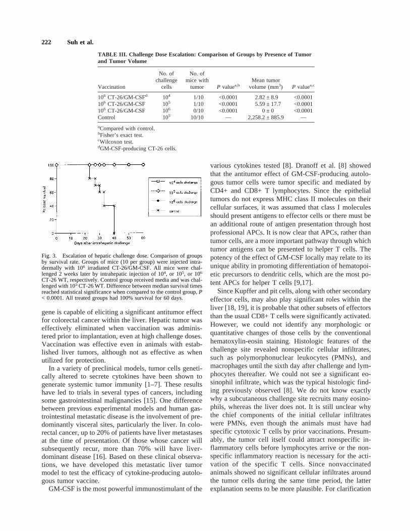
CT-26/GM-CSF) 14 days prior to intrahepatic tumor in-jection, challenge doses of 104 and 105 cells resulted inonly 1 of 10 animals developing tumors. No tumor wasseen in any animal in the group receiving the highest(106) challenge dose. Incidence of hepatic tumors waslower than that of medium treated animals and tumorvolumes were significantly smaller (Table III). Whenstudied for survival, no animals died within any of thevaccinated groups, even in those receiving 106 intrahe-patic challenge dose (Fig. 3).
Histologic Studies of the Challenge Site
Additional animals that were vaccinated with CT-26/GM-CSF (106) or control and subsequently given intra-hepatic tumor (5 × 10⁴) were sacrificed on days 2, 7, and9 following intrahepatic challenge for histologic analysisof the challenge site. In CT-26/GM-CSF-vaccinated ani-mals, a cellular infiltrate was seen, associated with a fewviable tumor cells (Fig. 4). In the control (nonvaccinated)group, viable tumors were seen but with fewer inflam-matory cells. At 2 days following challenge, the chiefcomponent of the cellular infiltrate was polymorpho-nuclear leukocytes. Eosinophils, monocytes, and lym-phocytes were also present. By 7 days following injec-tion, no viable tumor cells were seen, and the infiltratewas one of chronic inflammation composed primarily oflymphocytes. By day 9, the entire lesion was nonviableas an abscess.
DISCUSSION
We have demonstrated in this murine model that au-tologous tumor vaccine transduced with the GM-CSF
TABLE II. Established Hepatic Tumors: Comparison of Groups by Presence of Tumorand Tumor Volume
VaccinationDay from
vaccinationNo. of micewith tumor P valuea,b
Mean tumorvolume (mm3) P valuea,c
106 CT-26/GM-CSFd −14 0/10 <0.0001 0 ± 0 <0.0001106 CT-26/GM-CSF 0 6/10 0.09 13.86 ± 13.1 <0.0002106 CT-26/GM-CSF +5 6/10 0.09 15.30 ± 15.1 <0.0002106 CT-26/GM-CSF +10 8/10 0.47 148.9 ± 83.1 <0.0002Control — 10/10 — 2,258.2 ± 885.9 —
aCompared with control.bFisher’s exact test.cWilcoxon test.dGM-CSF-producing CT-26 cells.
Fig. 2. Vaccination against established tumor. Comparison of groupsby survival. Groups of mice (10 per group) were vaccinated with 106
irradiated CT-26/GM-CSF, 14 days prior to (day -14), same day (day0), or 5 days (day 5) and 10 days (day 10) following intrahepaticchallenge with 5 × l04 CT-26 WT. Control group received media 14days prior to the challenge. Differences between groups reached sta-tistical significance when compared to the media-treated group,P <0.001.
GM-CSF Vaccine for Hepatic Metastases 221
gene is capable of eliciting a significant antitumor effectfor colorectal cancer within the liver. Hepatic tumor waseffectively eliminated when vaccination was adminis-tered prior to implantation, even at high challenge doses.Vaccination was effective even in animals with estab-lished liver tumors, although not as effective as whenutilized for protection.
In a variety of preclinical models, tumor cells geneti-cally altered to secrete cytokines have been shown togenerate systemic tumor immunity [1–7]. These resultshave led to trials in several types of cancers, includingsome gastrointestinal malignancies [15]. One differencebetween previous experimental models and human gas-trointestinal metastatic disease is the involvement of pre-dominantly visceral sites, particularly the liver. In colo-rectal cancer, up to 20% of patients have liver metastasesat the time of presentation. Of those whose cancer willsubsequently recur, more than 70% will have liver-dominant disease [16]. Based on these clinical observa-tions, we have developed this metastatic liver tumormodel to test the efficacy of cytokine-producing autolo-gous tumor vaccine.
GM-CSF is the most powerful immunostimulant of the
various cytokines tested [8]. Dranoff et al. [8] showedthat the antitumor effect of GM-CSF-producing autolo-gous tumor cells were tumor specific and mediated byCD4+ and CD8+ T lymphocytes. Since the epithelialtumors do not express MHC class II molecules on theircellular surfaces, it was assumed that class I moleculesshould present antigens to effector cells or there must bean additional route of antigen presentation through hostprofessional APCs. It is now clear that APCs, rather thantumor cells, are a more important pathway through whichtumor antigens can be presented to helper T cells. Thepotency of the effect of GM-CSF locally may relate to itsunique ability in promoting differentiation of hematopoi-etic precursors to dendritic cells, which are the most po-tent APCs for helper T cells [9,17].
Since Kupffer and pit cells, along with other secondaryeffector cells, may also play significant roles within theliver [18, 19], it is probable that other subsets of effectorsthan the usual CD8+ T cells were significantly activated.However, we could not identify any morphologic orquantitative changes of those cells by the conventionalhematoxylin-eosin staining. Histologic features of thechallenge site revealed nonspecific cellular infiltrates,such as polymorphonuclear leukocytes (PMNs), andmacrophages until the sixth day after challenge and lym-phocytes thereafter. We could not see a significant eo-sinophil infiltrate, which was the typical histologic find-ing previously observed [8]. We do not know exactlywhy a subcutaneous challenge site recruits many eosino-phils, whereas the liver does not. It is still unclear whythe chief components of the initial cellular infiltrateswere PMNs, even though the animals must have hadspecific cytotoxic T cells by prior vaccinations. Presum-ably, the tumor cell itself could attract nonspecific in-flammatory cells before lymphocytes arrive or the non-specific inflammatory reaction is necessary for the acti-vation of the specific T cells. Since nonvaccinatedanimals showed no significant cellular infiltrates aroundthe tumor cells during the same time period, the latterexplanation seems to be more plausible. For clarification
TABLE III. Challenge Dose Escalation: Comparison of Groups by Presence of Tumorand Tumor Volume
Vaccination
No. ofchallenge
cells
No. ofmice with
tumor P valuea,bMean tumor
volume (mm3) P valuea,c
106 CT-26/GM-CSFd 104 1/10 <0.0001 2.82 ± 8.9 <0.0001106 CT-26/GM-CSF 105 1/10 <0.0001 5.59 ± 17.7 <0.0001106 CT-26/GM-CSF 106 0/10 <0.0001 0 ± 0 <0.0001Control 105 10/10 — 2,258.2 ± 885.9 —
aCompared with control.bFisher’s exact test.cWilcoxon test.dGM-CSF-producing CT-26 cells.
Fig. 3. Escalation of hepatic challenge dose. Comparison of groupsby survival rate. Groups of mice (10 per group) were injected intra-dermally with 106 irradiated CT-26/GM-CSF. All mice were chal-lenged 2 weeks later by intrahepatic injection of 104, or 105, or 106
CT-26 WT, respectively. Control group received media and was chal-lenged with 105 CT-26 WT. Difference between median survival timesreached statistical significance when compared to the control group,P< 0.0001. All treated groups had 100% survival for 60 days.
222 Suh et al.

of these findings, studies on animals depleted of variableeffector subsets should be conducted.
We found that the vaccinated host effectively scav-enged the large number (106) of tumor cells that wouldbe an overwhelming tumor burden to the mouse immunesystem. There have been few preclinical studies in whichmice were challenged with as large a cell number. The
effectors of immune system seem to be able to clear thechallenged tumor cells independent of their numbersonce they are stimulated.
To determine whether this tumor vaccine strategy willbe effective, it needs to be evaluated in relevant animalmodels under conditions that resemble the clinical con-ditions prevailing in cancer patients [10]. For this reason,
Fig. 4. Photomicrograph of intrahepatic challenge site. Sections were prepared 2 days(A,B), 7 days(C,D), and 9 days(E,F) followingintraheptaic challenge. Animals vaccinated with CT26/GM-CSF (A,C,E); control animals (B,D,F). A: A few scattered, but viable tumor cells(arrow) are surrounded by PMNs. B, D: No definite inflammatory cell infiltrates with increasing tumor size. C: No viable tumor cells are seen.Chief components of cellular infiltrates are lymphocytes. E: Challenge sites shrunk as microabscesses. F: A well-established tumor. Hematoxylinand eosin stains, ×64.
GM-CSF Vaccine for Hepatic Metastases 223

we conducted the second set of experiments in whichanimals were challenged first and then vaccinated. Therewere no long-term survivors in the animals vaccinatedsubsequently. However, the mean tumor volumes and themedian survival times of the animals with subsequentvaccinations were significantly smaller and longer re-spectively than those of the control group. Prolongationof median survival was observed even when the vaccinewas administered up to 10 days following tumor chal-lenge. Five to 10 days may be necessary for the fullstimulation of tumor-specific T cells following treatmentof GM-CSF-producing cells. Probably, GM-CSF-producing autologous tumor vaccine can eradicate theestablished tumor if smaller numbers of tumor cells arechallenged.
We conclude that GM-CSF-producing autologous tu-mor cells effectively enhance the immunogenicity oftheir parental forms of the tumor so that the immunizedhost becomes able to reject the further challenge of pa-rental tumor cells into the liver. The efficacy of the GM-CSF gene-transduced vaccine to treat established tumorswarrants further investigation. This study provides sup-port for cell-based immunotherapeutic strategies for thetreatment of colorectal cancer. The demonstration thatthis approach is effective within the liver should form thebasis of clinical trials to assess active immunotherapy forliver cancer.
CONCLUSIONS
GM-CSF autologous tumor vaccination was effectivefor the treatment of hepatic colorectal metastases in thismurine model. These findings provide support for immu-notherapeutic approaches for the treatment of metastaticliver cancer.
ACKNOWLEDGMENTS
We thank Dr. John Boitnott for review of the histo-logic findings. We also thank Betty Tyler for her excel-lent technical support.
REFERENCES1. Zoller M, Douvdevani A, Segal S, et al.: Interleukin-l produced by
tumorigenic fibroblasts influences tumor rejection. Int J Cancer1992;50:443–449.
2. Porgador A, Gansbacher B, Bannerji R, et al.: Antimetastatic vac-cination of tumor-bearing mice with IL-2-gene-inserted tumorcells. Int J Cancer 1993;53:471–477.
3. Golumbek, P, Lazenby A, Levitsky H, et al.: Treatment of estab-lished renal cancer by tumor cells engineered to secrete interleu-kin-4. Science 1991;254:713–716.
4. Porgador A, Tzehoval E, Katz A, et al.: Interleukin 6 gene trans-fection into Lewis lung carcinoma tumor cells suppresses the ma-lignant phenotype and confers immunotherapeutic competenceagainst parental metastatic cells. Cancer Res 1992;52:3679–3686.
5. Hock H, Dorsch M, Diamantstein T, et al.: Interleukin 7 inducesCD4+ T cell-dependent tumor rejection. J Exp Med 1991;174:1291–1298.
6. Porgador A, Bannerji R, Watanabe Y, et al.: Antimetastatic vac-cination of tumor-bearing mice with two types of IFN-gammagene-inserted tumor cells. J Immunol 1993; 150:1458–1470.
7. Thompson R, Pardoll DM, Jaffee EM, et al.: Systemic and localparacrine cytokine therapies utilizing transduced tumor cells aresynergistic in treating brain tumors. J Immunol 1996;19:405–413.
8. Dranoff G, Jaffee E, Lazenby A, et al.: Vaccination with irradiatedtumor cells engineered to secrete murine granulocyte macro-phage-colony stimulating factor stimulate potent, specific, andlong-lasting antitumor immunity. Proc Natl Acad Sci USA 1993;90:3539–3543.
9. Steinman R: The dendritic system and its role in immunogenicity.Annu Rev Immunol 1991;9:271–281.
10. Gilboa E: Murine models for cancer immunotherapy using cyto-kine gene modified tumor vaccines. In Forni G, Foa R, Santoni A,et al. (eds): “Cytokine-Induced Tumor Immunogenicity.” London:Academic Press, 1994:130–143.
11. Shiraki K, Tsuji N, Shioda T, et al.:Expression of Fas ligand inliver metastases of human colonic adenocarcinomas. Proc NatlAcad Sci USA 1997;94:6420–6425.
12. Wilcoxon F: Individual comparisons by ranking methods. Bio-metrics 1945;1:80–83.
13. Agresti A: “Categorical Data Analysis.” New York: John Wileyand Sons, 1990.
14. Zar JH (ed): “Biostatistical analysis.” Englewood-Cliffs, NJ: Pren-tice-Hall, 1984.
15. Roth JA, Cristiano RJ: Gene therapy for cancer: What have wedone and where are we going? J Natl Cancer Inst 1997;89:21–39.
16. Boring CC, Squires TS, Tong T, et al.: Cancer statistics. CACancer J Clin 1994; 44:7–26.
17. Pardoll DM: Genetically engineered tumor vaccines. Ann N YAcad Sci 1993; 690:301–310.
18. Bayon LG, Izquierdo MA, Sirovich I, et al.: Role of Kupffer cellsin arresting circulating tumor cells and controlling metastaticgrowth in the liver. Hepatology 1996;23:1224–1231.
19. Griffini P, Smorenburg SM, Vogels IM, et al.: Kupffer cells andpit cells are not effective in the defense against experimentallyinduced colon carcinoma metastases in rat liver. Clin Exp Metas-tasis 1996;14:367–380.
224 Suh et al.