translational discoveries lead to clinical advancements
TRANSCRIPT
W I LL I A M R . P R I TC H A R D V E T E R I N A RY M E D I C A L T E AC H I N G H O S P I TA L • U N I V E R S I T Y O F C A LI F O R N I A , DAV I S
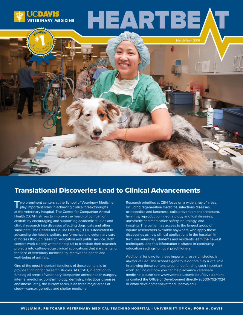
Two prominent centers at the School of Veterinary Medicine play important roles in achieving clinical breakthroughs
at the veterinary hospital. The Center for Companion Animal Health (CCAH) strives to improve the health of companion animals by encouraging and supporting academic studies and clinical research into diseases affecting dogs, cats and other small pets. The Center for Equine Health (CEH) is dedicated to advancing the health, welfare, performance and veterinary care of horses through research, education and public service. Both centers work closely with the hospital to translate their research projects into cutting-edge clinical applications that are changing the face of veterinary medicine to improve the health and well-being of animals.
One of the most important functions of these centers is to provide funding for research studies. At CCAH, in addition to funding all areas of veterinary companion animal health (surgery, internal medicine, ophthalmology, dentistry, infectious diseases, anesthesia, etc.), the current focus is on three major areas of study—cancer, genetics and shelter medicine.
March/April 2016
Translational Discoveries Lead to Clinical Advancements
RANKED
U.S. News and World
Rep
ort
QS World Unive
rsity
Research priorities at CEH focus on a wide array of areas, including regenerative medicine, infectious diseases, orthopedics and lameness, colic prevention and treatment, laminitis, reproduction, neonatology and foal diseases, anesthetic and medication safety, neurology, and imaging. The center has access to the largest group of equine researchers available anywhere who apply these discoveries as new clinical applications in the hospital. In turn, our veterinary students and residents learn the newest techniques, and this information is shared in continuing education settings for local practitioners.
Additional funding for these important research studies is always valued. The school’s generous donors play a vital role in allowing these centers to continue funding such important work. To find out how you can help advance veterinary medicine, please see www.vetmed.ucdavis.edu/development or contact the Office of Development directly at 530-752-7024 or email [email protected].

2 | W I LL I A M R . P R I TC H A R D V E T E R I N A RY M E D I C A L T E AC H I N G H O S P I TA L • U C DAV I S
Latest Clinical Trials at UC Davis
Since its inception in 2013, the UC Davis Veterinary
Center for Clinical Trials (VCCT) has completed dozens of studies, and more than 100 new trials are currently being conducted. VCCT works closely with the veterinary hospital and other campus institutions including the School of Medicine, the Clinical and Translational Science Center, the Center for Companion Animal Health, the Center for Equine Health, and the Veterinary Institute for Regenerative Cures. Clinical investigators have active trials aimed at advancing medical care for veterinary patients in a variety of disciplines, including oncology, neurology/neurosurgery, ophthalmology, dermatology and cardiology.
While the hospital provides the highest standard of care through conventional methods, clinical trials allow UC Davis veterinarians to evaluate new scientific breakthroughs that have the potential to improve the diagnosis and treatment of diseases. Veterinary clinical trials through the VCCT assess promising new treatments, drugs or procedures, but only after preliminary studies have established that the new methods are safe and have the potential to work better than existing protocols.
For more information about the VCCT and the current clinical trials at UC Davis, visit www.vetmed.ucdavis.edu/clinicaltrials or email [email protected].
A few current trials at UC Davis include:
Bladder Stones (Struvite) in DogsDr. Jodi Westropp is recruiting for struvite in dogs to provide another option to bladder stone dissolution. The purpose of this trial is to evaluate the efficacy of a therapeutic diet to help determine another dietary method for non-invasive dissolution.
Chronic Gingivostomatitis in CatsDr. Boaz Arzi is having success with a new stem cell approach to treating feline chronic gingivostomatitis (FCGS). None of the treatments for FCGS that are currently available are ideal, predictable and without possible complications. The purpose of this study is to treat cats for which all current treatment
modalities have failed and have a poor quality of life.
Osteoarthritis in DogsDr. Duane Robinson is recruiting for osteoarthritis in dogs. The primary purpose of this study is to test two investigational medications to determine if either one, or both, work to potentially decrease signs of pain of osteoarthritis in dogs.
Bilateral Corneal Stromal Loss in Friesian HorsesDr. Mary Lassaline
is recruiting for a new clinical trial for Friesian horses diagnosed with bilateral corneal stromal loss (BCSL). The trial focuses on determining the incidence of BCSL in the breed and the mode of inheritance if a single gene is involved, and identifying candidate genes for further investigation. Owners are encouraged to enroll Friesian horses with and without the diagnosis of BCSL.
Fungal Infections (Aspergillus spp) in German Shepherds, Rhodesian Ridgebacks, and Hungarian VizslasDr. Jonathan Dear is recruiting patients for his study on aspergillosis, a rare infection in animals with competent immune systems; however, certain dog breeds (namely the German shepherd, Rhodesian ridgeback and Hungarian vizsla) are reported to have a higher risk of this uncommon disease. A genome-wide association analysis will be used to evaluate the differences in the genetic material of affected dogs.
Leopard Complex Spotting in Appaloosa HorsesDr. Rebecca Bellone is recruiting for leopard complex spotting in Appaloosa horses, which is characterized by the progressive loss of pigment and has been associated with uveitis and night blindness in several breeds of horses. Two genes have been previously implicated in the loss of pigment in Appaloosas. This trial will investigate the morphology of the pigment producing cells (melanocytes) and determine if any ultrastructural differences exist among varying Appaloosa genotypes.
Dr. Boaz Arzi and Megan Badgley, RVT, examine Morris, a patient in the feline chronic gingivostomatitis trial.

W W W.V E T M E D.U C DAV I S . E D U/ V M T H | 32 | W I LL I A M R . P R I TC H A R D V E T E R I N A RY M E D I C A L T E AC H I N G H O S P I TA L • U C DAV I S
The School of Veterinary Medicine continues to receive “thanks and praise” from people all over the world who
have tried Dr. John Madigan’s dummy foal squeeze technique. Dubbed the Madigan Foal Squeeze Procedure, it is utilized to reduce the symptoms of neonatal maladjustment syndrome, or dummy foal syndrome, a disorder that occurs in only three to five percent of live horse births.
The technique uses a simple rope harness to gently squeeze the foal and mimic the pressure normally experienced in the birth canal. To recreate that pressure, Dr. Madigan and his team developed a method for wrapping a foal’s upper torso with several loops of a soft rope, creating a temporary harness. When pressure is applied with the rope, creating a gentle squeeze, the foal lies down and appears to be asleep.
After 20 minutes—about the same time a foal would spend in the birth canal—the rope is loosened and the squeeze pressure released. In initial cases, the foals have responded well to the procedure and recovered, some rising to their feet within minutes, bounding over to join the mare and nurse from her.
Worldwide Kudos of Foal Maladjustment Treatment Many equine clinicians, trainers and owners throughout the world have picked up on this technique—mostly via videos on social media or magazine articles—and successfully attempted the procedure – some after only watching the video once.
Here is a sample of what equine enthusiasts are saying about this astonishing procedure:
“I just wanted to pass on a huge thank you, as we had a dummy foal born which we squeezed and reverted to normal behavior (utilizing Dr. Madigan’s technique). He was showing mild classic signs…we squeezed for 25 minutes…the foal has never looked back.”
– Dr. Emma A., South Africa
“After reading an article about this procedure, we had a dummy foal born. We struggled with bottle feeding him for several hours and then remembered Dr. Madigan’s procedure. We tried it, and when we let him up, he was so ‘normal.’ It was quite amazing! Thank goodness we had seen this and were able to implement it.”
– Suzanna J., Saint Albans, West Virginia
“I wanted to write a message of a huge thank you to Dr. Madigan. I had a client’s mare give birth to a dummy foal. He never once tried to follow the mare, who was screaming for him. While researching orphan foals, I stumbled across UC Davis’ video on newborn horse syndrome. We attempted to duplicate your method. (Just after administering) he got up and trotted over to his mom, and went under her belly.”
– Kayleigh H., Toano, Virginia
“After successfully utilizing Dr. Madigan’s dummy foal squeeze technique, I wrote about it in Miniature Horse World, and posted it to Facebook. My original posts (from August 2015) have been shared thousands of times all around the world. Even now, I get several notifications a day that the post is still being shared. In my opinion, Dr. Madigan is a genius to have put all the pieces of the puzzle together and come up with this technique. Thank you for saving so many foals.”
– Dr. Lynn G., Alexandria, Virginia
Dr. John Madigan examines a maladjusted foal shortly after birth.
Dr. John Madigan administers the Madigan Foal Squeeze Procedure on a newborn foal.
The foal’s upper torso is wrapped with several loops of soft rope to administer the procedure.

4 | W I LL I A M R . P R I TC H A R D V E T E R I N A RY M E D I C A L T E AC H I N G H O S P I TA L • U C DAV I S
New Clinicians
Two new veterinarians have joined the faculty recently, and will have clinical duties in the hospital with their
respective specialty services. These additions help the hospital continue to provide the highest quality patient care. With more than 120 faculty veterinarians and more than 100 resident veterinarians, the UC Davis veterinary hospital has more clinicians than any hospital in the nation.
Dr. Amandeep Chohan – Anesthesia/Critical Patient Care ServiceDr. Chohan earned his veterinary degree and Master’s degree in Clinical Veterinary Medicine from the Punjab Agricultural University in Ludhiana, India in 2003 and 2005 respectively.
Subsequently, he joined the Department of Veterinary Clinical Sciences at Washington State University, where he completed a residency in anesthesiology (2006-2009) and obtained a second Master’s degree in Science. After finishing his residency, he joined the section of anesthesiology at Washington State University as a clinical instructor, where he served in that capacity until 2015. He is board certified (Diplomate) in the American College of Veterinary Anesthesia and Analgesia. Dr. Chohan
Dr. Amandeep Chohan, BVSc, AH, MVSc, MS, DACVAA
Through research funding assistance from the Center for Companion Animal Health (CCAH), hospital
clinicians are continually looking for ways to enhance their understanding of and treatments for oncologic diseases. Many of these research projects materialize as clinical trials to bring about new procedures that may revolutionize the standard of care for many common forms of cancer.
Dr. Michele Steffey of the Soft Tissue Surgery Service is leading current studies on thermal ablation. The process involves placing a patient under general anesthesia and ablation probes are placed in a minimally invasive way into
the pet’s mass using imaging guidance, such as computed tomography or ultrasound. Ablation, or killing of the tumor cells, is then performed using thermal, chemical or electrical methods. With thermal ablation, the procedure directly changes the temperature of the lesion with either heating (microwave ablation) or freezing (cryoablation) for the purpose of causing cells to die.
Oncology specialists Drs. Jenna Burton, Michael Kent and Rob Rebhun are all currently conducting separate clinical trials and research projects on dogs diagnosed with osteosarcoma. The team has focused on osteosarcoma in dogs as both a way to understand this cancer in canines and as an animal model for humans. With osteosarcoma in dogs, the primary tumor can usually be addressed effectively through removal or radiation. The more worrisome problem is that most of these cancers metastasize and spread to the lungs. The prognosis for these patients remains poor. Identifying and targeting therapy-resistant tumor cells is a major step to improving the standard of care. With Dr. Kent’s trial, the focus is on finding a way to slow or stop the spread of the tumor to the lungs.
Dr. Kent, who serves as CCAH director, is also recruiting for dogs diagnosed with either soft tissue sarcoma or melanoma. That trial focuses on treating dogs with combined immunotherapy and radiation therapy at the primary tumor site to see if the immune system is induced to attack the tumor and prevent metastatis.
Currently, 20 clinical trials are focused on oncology.
New Therapies to Treat Cancer Being Explored
Lindsay Stevens, RVT, comforts a patient receiving chemotherapy treatment.

W W W.V E T M E D.U C DAV I S . E D U/ V M T H | 54 | W I LL I A M R . P R I TC H A R D V E T E R I N A RY M E D I C A L T E AC H I N G H O S P I TA L • U C DAV I S
Livestock Services Enhance Student Training
Whether veterinary
students plan to enter a career in livestock medicine or not, fourth-year clinical rotations through the livestock services can prove to be a valuable resource. For those actively pursuing a livestock career path, the rotations are obviously absolute necessities, but there are many aspects of the rotations that will enhance the futures of students who may never encounter large animals again.
Basic technical skills and knowledge of livestock are impressed upon students in their third year for them to later utilize in fourth-year clinical rotations. Many of these skills—basic understanding of diseases, pathophysiology, efficient physical examinations, basic surgical skills—are transferable to any species. For those geared toward livestock paths, the skills are expanded in the clinics.
Rotations through the Livestock Medicine Service allow students to participate in surgeries such as castrations, common abdominal surgeries, C-sections and leg fracture repairs. Many small animal track students want to rotate
Dr. Munashe Chigerwe trains DVM students to care for the hospital’s blood donor goats during their fourth-year clinical rounds.
provides teaching, clinical service and clinical research in the veterinary anesthesiology discipline. He also provides leadership in directing research projects of residents and graduate students.
Dr. Po-Yen Chou – Orthopedic Surgery ServiceDr. Chou recently joined the Orthopedic Surgery Service, and will help increase offerings of this bustling service. He received his veterinary degree from National Taiwan University in 2003, where he stayed and completed a combined Master’s degree and small animal surgery residency from 2004 to 2008. Dr. Chou then completed a small animal
surgery fellowship at Michigan State University and his rotating internship and small animal surgical residency at Atlantic Veterinary College, University of Prince Edward Island in Canada. He is board certified (Diplomate) in the American College of Veterinary Surgeons. Dr. Chou’s areas of interest are minimally invasive orthopedic surgery, canine elbow disease and small animal trauma surgery.
Po-Yen Chou, BVM, MVM, DACVS (Small Animal)
through livestock medicine to gain additional hands-on surgery experiences, giving them more practice suturing and more practice with tissue handling and manipulation—skills they will utilize when performing surgery on cats and dogs. This rotation also offers the students additional experience with CTs, MRIs, radiology, laparoscopy and endoscopy.
In the Livestock Herd Health and Reproduction Service, it’s an opportunity for future livestock clinicians to hone their skills in some of the newest techniques being offered. Students become skilled in artificial insemination, embryo transfers and other advanced reproductive technologies, as well as gaining better understandings of basic herd health concepts like preventive medicine, vaccinations, disease outbreak investigations and interventions during a crisis, improving their critical thinking skills.
The school strives to give every student the best-rounded veterinary education possible. Livestock medicine plays a vital role in that process, always increasing and enhancing learning opportunities for both large and small animal-focused students.

6 | W I LL I A M R . P R I TC H A R D V E T E R I N A RY M E D I C A L T E AC H I N G H O S P I TA L • U C DAV I S
UC Davis Veterinary Surgeons Utilize Artificial Ureter in Kitten
Googgie, a 17-week-old spayed female Sphynx, was brought to the UC Davis veterinary hospital following
a three-day bout of inappetence, lethargy and vomiting. Originally seen by the Emergency and Critical Care Service, she was noted to have an enlarged, painful left kidney.
With ultrasound, it was discovered that an obstruction of the left ureter was preventing flow of urine to the bladder. It could not be determined through imaging what was causing the blockage, and surgery was needed to discover, then alleviate, the obstruction.
Specialists from the Soft Tissue Surgery Service were brought in to review Googgie’s case and work with the Anesthesia Service to prepare her for surgery. After opening Googgie’s abdomen, surgeons were able to see the extent of the damage to her ureter. A traumatic stricture (scarred down area) was
present around the ureter, preventing it from being open. There was a solution, however. Dr. Bill Culp and his team had success previously in dogs and cats with a relatively new procedure creating an artificial ureter using catheters, and they were hopeful it would succeed in a kitten as well.
Bypassing the natural ureter, Dr. Culp was able to create two distinct ends of a new ureter with tubing—one end coming from the kidney, one end leading into the bladder. The two pieces were connected with a titanium shunting port that was placed just under the skin for easy access for collecting urine samples from the system. Known as a subcutaneous ureteral bypass (SUB), the system has primarily been utilized in older animals. The longevity of the system for longer installation into young animals is unknown, and Googgie’s device will need to be monitored long term to ensure functionality.
Tests following the successful surgery showed normal kidney function. Had Googgie’s signs of an illness been delayed longer, she could have lost complete function of the affected kidney, potentially leading to medical issues later in life.
Googgie recovered from the procedure without complications. She appeared bright, and was eating and drinking several hours after surgery. The following morning, she continued to recover and maintained an excellent appetite. For the next few days, she remained under the watchful eyes of technicians and veterinarians who supported her with IV fluid therapy and pain medications while she recovered. A follow-up ultrasound revealed complete resolution of the swollen kidney, and showed the SUB system remained in an appropriate position.
After nearly a week, Googgie was discharged from the hospital to continue her recovery at home. Three-week and three-month recheck examinations continued to be positive. Her weight had doubled since her initial visit to the emergency room, and she is growing like a normal cat.
The placement of the SUB in Googgie is unique due to Googgie’s age. As conventional methods for correcting ureteral obstructions are often less than optimal, it is hopeful that implantation of this SUB system will hold promise as a viable alternative. Initial data regarding use of SUB systems indicate fewer long-term complications compared with traditional ureteral surgery.
While the surgeons did need to open Googgie’s abdominal cavity to perform the surgery, they were able to place the implant minimally invasively, and utilize interventional radiology to help guide the procedure. Minimally invasive procedures at UC Davis are dramatically changing the face of veterinary surgery with equipment and technology allowing for new procedures to be performed that were not available less than a generation ago.
MARCH CASE OF THE MONTH – GOOGGIE
Googgie now has an artificial ureter after one of her natural ureters became permanently obstructed.

W W W.V E T M E D.U C DAV I S . E D U/ V M T H | 76 | W I LL I A M R . P R I TC H A R D V E T E R I N A RY M E D I C A L T E AC H I N G H O S P I TA L • U C DAV I S
UC Davis Veterinary Radiologists Help Save Dog with Foxtail Near Aorta
Joey, a 4-year-old male German shepherd mix, loves to play frisbee. While playing nine months ago, he ran into a
tree stump. Initially, he appeared to be alright following the event, however, swelling developed over his left hip the next day. Joey’s veterinarian aspirated the wound and drained fluid from it that was consistent with a hematoma.
About a month later, the swelling returned as an abscess. Cultures of the wound revealed a bacterial infection. It was drained again, and Joey was placed on antibiotics which resolved the swelling. A few days following completion of the medications, though, the swelling returned.
After months of unsuccessful treatment, Joey was brought to the UC Davis veterinary hospital, where specialists discussed potential causes of non-healing wounds, including self trauma, infection, or a foreign body in the area.
Dr. Anthony DeRouen—a fourth-year resident with the Diagnostic Imaging Service—and faculty member Dr. Allison Zwingenberger, reviewed imaging options with the Soft Tissue Surgery Service to identify the best way to investigate the cause of the wound. The hospital’s imaging team has more board-certified radiologists than any veterinary hospital in California. Their facilities and expertise include radiographs (x-rays), ultrasound, CT, MRI, and nuclear medicine. Each of these different imaging tests can provide different and useful information about the source of infection. In Joey’s case, their expertise proved invaluable in saving his life.
They recommended starting with an ultrasound examination with an option to do a CT scan if more information was needed. Dr. DeRouen performed an ultrasound scan on Joey that revealed a tract that could be traced from the initial wound area on his hip toward his abdomen. Near the end of that tract was a large foreign body. The tract indicated that the foreign body had burrowed its way into the muscles near the spine, and was likely a foxtail. The ultrasound also located the foreign body to be beside the lumbar vertebrae and about an eighth of an inch from the aorta. The foreign body was clearly visible with ultrasound, and the CT scan was not needed for more information before surgery. X-rays of his back were performed to ensure the infection had not affected his spine.
Based on these findings, the radiologists recommended that surgical removal of the suspected foxtail was the best course of action. Not only was it important to remove it to eliminate the source of infection of Joey’s hip wound, but to prevent the foxtail from migrating toward the aorta, which if penetrated, could result in life-threatening internal bleeding.
The Anesthesia/Critical Patient Care Service placed Joey under general anesthesia, and he was taken to surgery. Surgeons were assisted by Dr. DeRouen, who used ultrasound during the surgery to guide them to the foxtail, and more importantly, avoid the aorta. Following extraction of the foxtail, as much of the infected tissue as could safely be removed was debrided.
Joey remained stable under anesthesia throughout his surgical procedure and recovered well. He was hospitalized after surgery, where he received supportive care from the hospital’s technical staff as they kept him comfortable and monitored him closely for any signs of post-operative or post-anesthetic complications. After two days, Joey was discharged and made a full recovery at home.
Diagnostic imaging saved Joey by discovering a foxtail dangerously close to his aorta.
APRIL CASE OF THE MONTH – JOEY

LE A D I N G V E T E R I N A RY M E D I C I N E , A D D R E S S I N G S O C I E TA L N E E D S
Veterinary Continuing Education
(530) 752-3905, Fax: (530) 752-6728, [email protected]
Upcoming Veterinary Continuing Education Events:
n May 21-22 Beginning Practical Ultrasound, UC Davis
n June 4-5 Biomedicine Symposium, UC Davisn July 23-24 Back to School Seminar, UC Davisn September 24-25 Fall Festival, UC Davisn October 23-27 Explorer Series, Milan, Italyn November 6 Feline Forum, UC Davis
For more information on these and other upcoming CE events, please visit www.vetmed.ucdavis.edu/ce.
Featured Clinical TrialDr. Lance Visser is recruiting for a new clinical trial for dogs diagnosed with subaortic or pulmonic stenosis. The trial aims to study the effects of two commonly utilized medications—butorphanol and atenolol—on heart function and assessment of disease severity. Owners are encouraged to enroll any dog diagnosed with pulmonic stenosis or subaortic stenosis with a pressure gradient over 50 mmHg. For more information about this and other groundbreaking trials, visit www.vetmed.ucdavis.edu/clinicaltrials or email [email protected].
For Appointments Call:Small Animal Clinic: (530) 752-1393Large Animal Clinic: (530) 752-0290
www.vetmed.ucdavis.edu/vmth
To subscribe to VMTH Heartbeat, email [email protected]
Please consider supporting UC Davis Veterinary Medicinewww.vetmed.ucdavis.edu/development
Like us on Facebook https://www.facebook.com/ucdavisvetmed
Watch us on YouTube http://www.youtube.com/ucdvetmed
Follow us on Twitter http://twitter.com/ucdavisvetmed
Follow us on Instagram https://www.instagram.com/ucdavisvetmed
Did You Know?… that fourth-year ophthalmology resident Dr. Lionel Sebbag
won 2016 American Association of Veterinary Clinicians Outstanding Resident Award? “I feel very honored and flattered to be selected for the award,” said Dr. Sebbag. “Being a resident at UC Davis has exceeded my expectations by far, and I am grateful every day for the chance of being here.”
… that SVM Information Technology Services recently completed their WiFi Project in the VMTH? This important step in providing uniform and reliable Internet service throughout the hospital is greatly appreciated.
… that third-year surgery resident Dr. Ingrid Balsa was recently presented a Spot Award? SVM Dean Michael Lairmore, VMTH Interim Director Dr. Jane Sykes and faculty surgeon Dr. Bill Culp surprised Dr. Balsa with the award for her outstanding service to the hospital over the past three years.
… that Lisa Winn is the new referral coordinator for the Neurology/Neurosurgery Service? Congratulations and welcome, Lisa!
Dean Michael Lairmore, along with Drs. Bill Culp and Jane Sykes, presents Dr. Ingrid Balsa with a Spot Award.