transforming growth factor-p stimulates trophoblast ... 78 … · 0.1 ml thrombin (thromboquik,...
TRANSCRIPT

0021-972x/94/7805-1241$03.00/0 Journal of Clinical Endocrinology and Metabolism Copyright 0 1994 by The Endocrine Society
Vol. ‘IS, No. 5 Printed in U.S.A.
Transforming Growth Factor-P Stimulates Trophoblast Oncofetal Fibronectin Synthesis in Vitro: Implications for Trophoblast Implantation in Viuo*
RONALD F. FEINBERG, HARVEY J. KLIMAN, AND CAI-LIANG WANG
Department of Obstetrics and Gynecology, Division of Reproductive Biology, University of Pennsylvania Medical Center (R.F.F., C.-L. W.), Philadelphia, Pennsylvania 19104-4283; and the Departments of Pathology and Obstetrics and Gynecology, Yale University School of Medicine (H.J.K.), New Haven, Connecticut 06510
ABSTRACT In pregnancy tissues, oncofetal fibronectin (onfFN) has been local-
ized specifically to the extracellular matrix (ECM) surrounding extra- villous anchoring trophoblasts of the placental-uterine junction and chorion. When isolated from first or third trimester placentas, human cytotrophoblasts in culture secrete and deposit onfFN in the ECM. In addition, onfFN synthesis is significantly up-regulated in response to serum stimulatory factor(s). The goal of this study was to examine the role of transforming growth factor-p (TGFP), a cytokine present in uterine decidua, as a stimulator of trophoblast onfFN production. Our initial insight into the significance of TGFp resulted from the seren- dipitous use of cord serum from a neonate with severe alloimmune thrombocytopenia. Trophoblasts cultured in medium containing this serum underwent normal morphological differentiation, but produced markedly less onfFN. In an analogous fashion, trophoblasts cultured in normal serum preincubated with anti-TGFP neutralizing antibodies also produced significantly less onfFN. Exogenously added TGFPl
restored the ability of trophoblasts to produce onfFN by a factor of 4- to 5-fold in medium containing thrombocytopenic serum. In platelet- poor serum derived from human or bovine plasma, TGFPl also induced onfFN synthesis, as assayed both in the conditioned medium and by immunocytochemical localization of onfFN in cell-associated ECM fibrils. Dose-response analysis demonstrated that the onfFN stimula- tory response is sensitive to TGFP, with an ED,, of 0.1-0.2 rig/ml. In a reciprocal fashion, TGFp inhibited @hCG secretion 3- to 4-fold. Our results demonstrate that TGFp is a significant stimulator of tropho- blast onfFN production. Furthermore, TGFp appears to modulate trophoblast differentiation by up-regulating the expression of an an- choring trophoblast marker (onfFN) and down-regulating a phenotypic marker of villous syncytiotrophoblast (hCGp). We speculate that troph- oblast responsiveness to TGFp in the implantation milieu contributes to trophoblast adhesion by stimulating the production of a trophoblast- derived implantation site fibronectin. (J Clin Endocrinol Metab 78: 1241-1248,1994)
0 NCOFETAL fibronectin (onfFN) is closely associated with extravillous trophoblasts in the placental-uterine
junction throughout pregnancy (l-3). In normal human im- plantation sites, onfFN is localized to a highly specific region, the extracellular matrix (ECM), connecting extravillous troph- oblasts and trophoblastic cell columns to the uterine decidua. In extrauterine pregnancies, onfFN is absent from the uterus, but it is found at ectopic trophoblastic implantation sites, further suggesting that the protein is trophoblast derived. Trophoblast-associated onfFN has been identified as early as 20 days postconception, and at this gestation it is specifically localized to the early cytotrophoblastic shell. Based on these immunolocalization studies, it has been hypothesized that onfFN functions in the placental-uterine junction as a tro- pho-uteronectin or trophoblast-uterine connecting protein (l-3). Similar, if not identical, oncofetal fibronectins are also found within the ECM of the chorion-decidual junction (2, 4), and these chorion-derived forms may be the source of
Received June 18, 1993. Accepted January 5, 1994. Address all correspondence and requests for reprints to: Ronald F.
Feinberg, M.D., Ph.D., Department of Obstetrics and Gynecology, Uni- versity of Pennsylvania Medical Center, 106 Dulles Building, 3400 Spruce Street, Philadelphia, Pennsylvania 19104.4283.
* This work was supported by grants from the March of Dimes (to R.F.F.), the University of Pennsylvania Research Foundation (to R.F.F. and H.J.K.), and Ares-Serono (to R.F.F. and H.J.K.).
cervico-vaginal “fetal” fibronectin detected clinically in pa- tients at risk for preterm labor and delivery (4, 5). The novel feature of these fibronectins, as originally described by Mat- suura and Hakomori (6-g), is the presence of a specific O- glycosylated hexapeptide epitope within the type III con- necting segment (IIICS) region, reactive with monoclonal antibody FDC-6.
Human cytotrophoblasts isolated from first trimester or term placentas synthesize and secrete abundant quantities of FDC-6-reactive fibronectin, and we have found that this fibronectin is specifically deposited at sites of trophoblast- ECM attachment in vitro (1, 2). As such, we have hypothe- sized that trophoblasts in vitro are recapitulating an impor- tant in vivo trophoblast function by producing and depositing an endogenous fibronectin within newly formed ECM. More recently, we have found that onfFN production in vitro is regulated by a specific stimulatory factor(s) in serum. Troph- oblasts cultured in medium containing increasing percentages of serum produce increasing quantities of onfFN (2). Inter- estingly, human newborn serum derived from cord blood is 5-10 times more stimulatory for onfFN production than equivalent amounts of fetal calf serum (2).
We have further hypothesized that identification of an onfFN stimulatory factor(s) in serum might give us insight into potential uterine-derived promoters of trophoblast im- plantation. While working with numerous cord sera, we
1241
at Yale Med Libr on March 26, 2010 jcem.endojournals.orgDownloaded from
1242 FEINBERG. KLIMAN. AND WANG JCE & M .1994 Vol78.No5
observed that trophoblasts cultured in one serum derived from a neonate with severe alloimmune thrombocytopenia exhibited normal morphology, yet produced significantly less onfFN. As transforming growth factor-p (TGFP), a potent growth factor released into serum by activated platelets, has also been localized to the placental-uterine junction (9), we initiated studies to determine whether TGFP affects tropho- blast synthesis of onfFN. Using cultured cytotrophoblasts, we assayed the production of two phenotypic markers of trophoblast differentiation, onfFN and hCG@, and found a reciprocal response to TGFP.
Materials and Methods
Cytotrophoblast preparation and culture
Human cytotrophoblasts were purified from the placentas of uncom- plicated term pregnancies immediately after delivery by serial trypsin- DNase digestions followed by Percoll gradient centrifugation, as previ- ously described by Kliman ei al. (10). Yields of viable &ophoblasi cells ranged from 60-100 X lo6 cells/30 e starting ulacental tissue. The pugfied cytotrophoblasts were culturedvin Dulbic’co’s Modified Eagle’s Medium (DMEM) containing 25 mmol/L glucose and 25 mmol/L HEPES supplemented with 4 mmol/L glutamine and 50 pg/mL gentamicin. The DMEM contained different sera, depending on the experimental condi- tions Trophoblasts were typically cultured for 48 or 72 h, and the media were collected at these time points for immunoblot and/or enzyme- linked immunoassay (ELISA) analyses.
Human cord blood samples (SO-100 mL) were collected from the umbilical vein immediately after normal vaginal deliveries of healthy neonates, but before delivery of the placenta. For normal serum collec- tion, the blood was transferred to red-top Vacutainer tubes (Becton Dickinson, Rutherford, NJ) containing no additives and allowed to clot, The clotted blood was centrifuged for 20 min in a clinical centrifuge, and the resultant serum supernatant was collected and aliquoted. Hu- man and bovine plasma were either purchased commercially (Sigma Chemical Co., St. Louis, MO) or prepared from whole blood by collection in blue-top Vacutainer tubes containing buffered sodium citrate as an anticoagulant. These tubes were centrifuged to remove all cells and platelets, and the resultant plasma supematant was collected. Platelet- poor serum was then prepared from plasma by generating a clot with 0.1 mL thrombin (Thromboquik, Organon Teknika, Durham, NC)/l.O mL plasma, which was removed from the serum.
TGFP immunoneutralization and stimulation
For immunoneutralization of endogenous TGFP activity in DMEM- containing normal serum, turkey (Collaborative-Becton Dickinson, Bed- ford, MA) or chicken (R & D Systems, Minneapolis, MN) antihuman TGFP panneutralizing antibodies were employed. The neutralization results we obtained were identical for each antibody. DMEM containing either 2% normal newborn human serum or 5% fetal calf serum were incubated at antibody concentrations of lo-100 pg/mL for 6 h before and throughout the duration of the cell culture.
To examine the effects of exogenous TGFP on onfFN production, purified human TGF/31 was obtained from two different commercial sources (Collaborative-Becton Dickinson and R & D Systems). As rec- ommended by the manufacturers, the lyophilized samples were recon- stituted with sterile 4 mmol/L hydrogen chloride containing 1 mg/mL BSA to a final concentration of 1 ng/mL TGFPl. Appropriate dilutions were made from frozen aliquots of TGFPl stock solutions, as needed for stimulation experiments,
Quantitative onfFN immunoassays
For Western immunoblots, trophoblast-conditioned medium samples were electrophoresed in 6% sodium dodecyl sulfate-polyacrylamide gels under reducing conditions. Known standards of 50-1000 ng amniotic
fluid fibronectin, previously purified by gelatin-Sepharose 48 chroma- tography (Pharmacia, Piscataway, NJ) (ll), were electrophoresed in parallel. The gels were then electrotransferred to nitrocellulose (Schleicher and Schuell, Keene, NH) overnight. To detect onfFN, the nitrocellulose blots were incubated with a 1:50 dilution of hybridoma supematant containing murine monoclonal antibody FDC-6 (American Type Culture Collection, Rockville, MD). Immunodetection of FDC-6 was performed with a biotinylated antimouse secondary antibody, avi- din, and a biotinylated horseradish peroxidase, according to the manu- facturer’s instructions (ABC Vectastain, Vector Laboratories, Burlin- game, CA). The chromagen reaction was carried out with 3,3’-diami- nobenzidine and hydrogen peroxide (Sigma). For more exact quantitation of medium samples, an ELISA was used. Known purified standards (O- 400 ng) of amniotic fluid fibronectin were plated on 96-well plates (Coming, Corning, NY) in parallel with medium samples diluted 1:20 to 1:lOO. Immunodetection was identical to that described for the Western blots (ABC Vectastain), except that o-phenylenediamine was used as the soluble chromagen. Samples were read at 492 nm. A detailed standard curve for each ELISA, based on quadruplicate samples, was created with CricketGraph 1.3.2 for the Macintosh (Computer Associates, Garden City, NY), with r2 values typically greater than 0.98.
Trophoblast immunocytochemistry
Cytotrophoblasts were cultured on sterile glass coverslips (Coming) for 48 or 72 h in defined DMEM containing 2% platelet-poor human or bovine serum, with or without 2.0 ng/mL TGFPl. For fixation, coverslips were washed with phosphate-buffered saline, fixed for 15 min with formalin, and stored in 0.1% sodium azide until use. For immunostain- ing, FDC-6 supernatant was diluted 1:50 and used as the primary antibody. Immunodetection was performed as described above for im- munoblots with ABC Vectastain. Coverslips were counterstained with hematoxylin. Undiluted P3X63Ag8 mouse myeloma cell line supematant (American Type Culture Collection) was used as a control for negative staining. The specificity of the FDC-6 antibody has previously been demonstrated by immunoabsorption (1, 4) and the inability of the antibody to bind to 0-deglycosylated oncofetal fibronectin (7, 8).
Results
Trophoblasts cultured in thrombocytopenic serum produce significantly less onfFN than trophoblasts cultured in normal serum
We reported previously that isolated human cytotropho- blasts demonstrate normal morphological differentiation (i.e. attachment and spreading, formation of aggregates, and syncytia) when cultured in medium containing as little as 1% human cord blood serum (2). Although the cord sera we collected postdelivery were generally derived from normal pregnancies, we obtained one cord serum sample from a neonate who was later found to have an extremely low platelet count of 11,000/mm3 (normal range, 150-400 X 103/ mm”). This neonate was subsequently diagnosed with severe alloimmune thrombocytopenia and did well with platelet transfusions. As with other cord sera we had collected, this particular sample was unremarkable in its clotting and serum separation characteristics.
When trophoblasts were cultured in medium containing the thrombocytopenic cord serum, they appeared indistin- guishable morphologically from cells cultured with normal cord sera. Figure 1 demonstrates the results of an immunoblot assay comparing the quantity of onfFN secreted into the media when trophoblasts from the same placental isolation prep were cultured in medium containing 10% cord serum from a normal neonate thrombocytopenic cord serum (lanes
at Yale Med Libr on March 26, 2010 jcem.endojournals.orgDownloaded from
TGFp STIMULATES ONCOFETAL FIBRONECTIN SYNTHESIS 1243
TGFB Neutralization Experiments
2% NHS 5% FCS
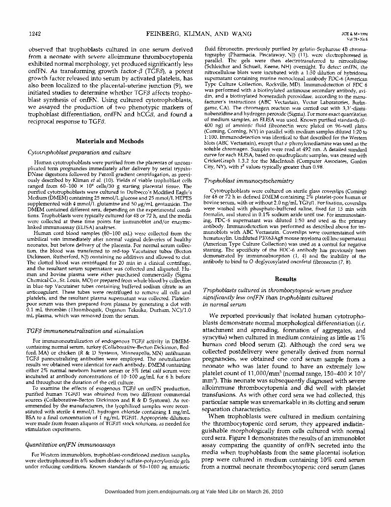
abed FIG. 1. Normal us. thrombocytopenic serum as a stimulator of onfFN synthesis. Immunoblot comparison of secreted onfFN in parallel troph- oblast cultures containing 10% cord serum from a normal neonate (lanes a and b) us. 10% cord serum from a thrombocytopenic neonate (lanes c and d) after 48 h (lanes a and c) and 72 h (lanes b and d). Unconcentrated conditioned medium (50 rL) was loaded in each lane.
a and b) VS. 10% thrombocytopenic cord serum from a normal neonate (lanes c and d). After culture times of 48 h (lanes a and c) and 72 h (lanes b and d), onfFN levels in the medium were 4- to 5-fold lower in the trophoblast cultures containing the thrombocytopenic serum. We further confirmed this ob- servation by comparing the onfFN-stimulating capacity of the thrombocytopenic serum with other normal cord sera (not shown). Depending on the serum sample used, medium containing normal cord sera induced 4- to IO-fold higher levels of onfFN secretion than the thrombocytopenic serum, based on quantitative onfFN immunoblot and ELISA assays. These findings suggested that a platelet-derived soluble serum factor, such as TGFP, might be required for trophoblast production of onfFN in vitro.
Neutralization of endogenous TGF@ in medium containing normal serum
Although a lack of various platelet-derived factors could have been responsible for the diminished onfFN production observed in the thrombocytopenic serum, we focused specif- ically on TGFP for the following reasons: 1) TGF/3 up- regulates fibronectin production in other cell systems (12- 15); 2) TGF/3 has been reported to modulate trophoblast differentiation in vitro (9, 16, 17); and 3) TGFP has recently been immunolocalized in viva to the placental-uterine junc- tion in human pregnancies (9). To assess the significance of TGF/3 in stimulating trophoblast onfFN production, we used a strategy of incubating trophoblast medium before and during the culture with anti-TGF/3 neutralizing antibodies. As an assay for onfFN production by the cultured tropho- blasts, conditioned media were analyzed by immunoblot. Figure 2 demonstrates the results of two different TGF/3 neutralization experiments, in which 100 pg/mL added an- tibody (lanes a and e) were effective in reducing onfFN
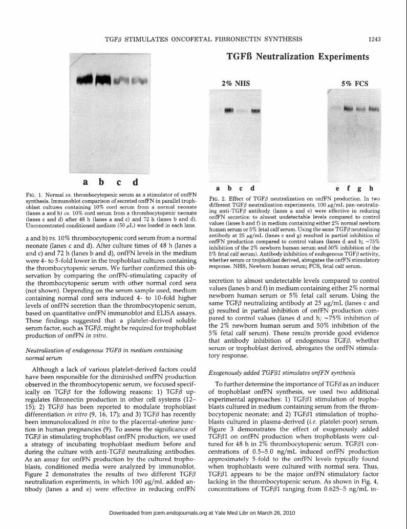
a b c d e f g b FIG. 2. Effe.ct of TGF@ neutralization on onfFN production. In two different TGFp neutralization experiments, 100 pg/mL pan-neutraliz- ing anti-TGFP antibody (lanes a and e) were effective in reducing onfFN secretion to almost undetectable levels compared to control values (lanes b and f) in medium containing either 2% normal newborn human serum or 5% fetal calf serum. Using the same TGF@ neutralizing antibody at 25 rg/mL (lanes c and g) resulted in partial inhibition of onfFN production compared to control values (lanes d and h; -75% inhibition of the 2% newborn human serum and 50% inhibition of the 5% fetal calf serum). Antibody inhibition of endogenous TGFp activity, whether serum or trophoblast derived, abrogates the onfFN stimulatory response. NHS, Newborn human serum; FCS, fetal calf serum.
secretion to almost undetectable levels compared to control values (lanes b and f) in medium containing either 2% normal newborn human serum or 5% fetal calf serum. Using the same TGFP neutralizing antibody at 25 pg/mL (lanes c and g) resulted in partial inhibition of onfFN production com- pared to control values (lanes d and h; -75% inhibition of the 2% newborn human serum and 50% inhibition of the 5% fetal calf serum). These results provide good evidence that antibody inhibition of endogenous TGFP, whether serum or trophoblast derived, abrogates the onfFN stimula- tory response.
Exogenously added TGF@l stimulates onfFN synthesis
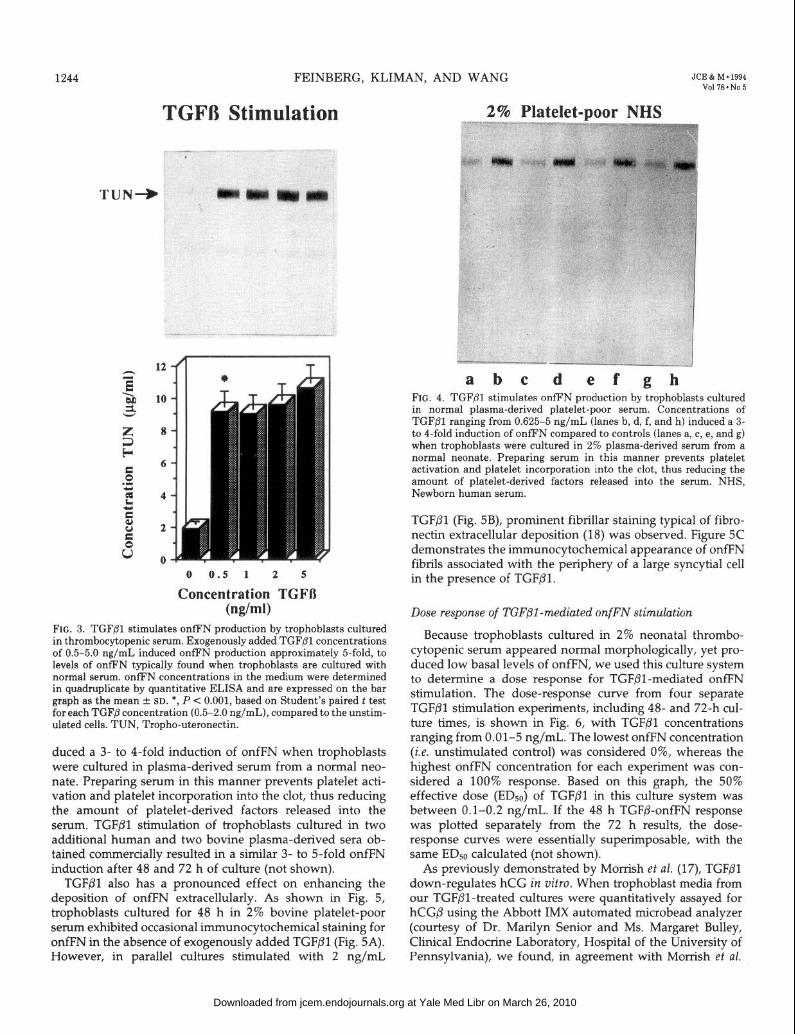
To further determine the importance of TGFP as an inducer of trophoblast onfFN synthesis, we used two additional experimental approaches: 1) TGF/31 stimulation of tropho- blasts cultured in medium containing serum from the throm- bocytopenic neonate; and 2) TGFPl stimulation of tropho- blasts cultured in plasma-derived (i.e. platelet-poor) serum. Figure 3 demonstrates the effect of exogenously added TGF/31 on onfFN production when trophoblasts were cul- tured for 48 h in 2% thrombocytopenic serum. TGFPl con- centrations of 0.5-5.0 ng/mL induced onfFN production approximately 5-fold to the onfFN levels typically found when trophoblasts were cultured with normal sera. Thus, TGFPl appears to be the major onfFN stimulatory factor lacking in the thrombocytopenic serum. As shown in Fig. 4, concentrations of TGFPl ranging from 0.625-5 ng/mL in-
at Yale Med Libr on March 26, 2010 jcem.endojournals.orgDownloaded from
1244 FEINBERG, KLIMAN, AND WANG JCE & M .1994 Vol78.No5
TGFB Stimulation
0 0.5 1 2 5
Concentration TGFD (w/ml)
FIG. 3. TGF@l stimulates onfFN production by trophoblasts cultured in thrombocytopenic serum. Exogenously added TGF@l concentrations of 0.5-5.0 ng/mL induced onfFN production approximately &fold, to levels of onfFN typically found when trophoblasts are cultured with normal serum. onfFN concentrations in the medium were determined in quadruplicate by quantitative ELISA and are expressed on the bar graph as the mean + SD. *, P < 0.001, based on Student’s paired t test for each TGFj3 concentration (0.5-2.0 ng/mL), compared to the unstim- ulated cells. TUN, Tropho-uteronectin.
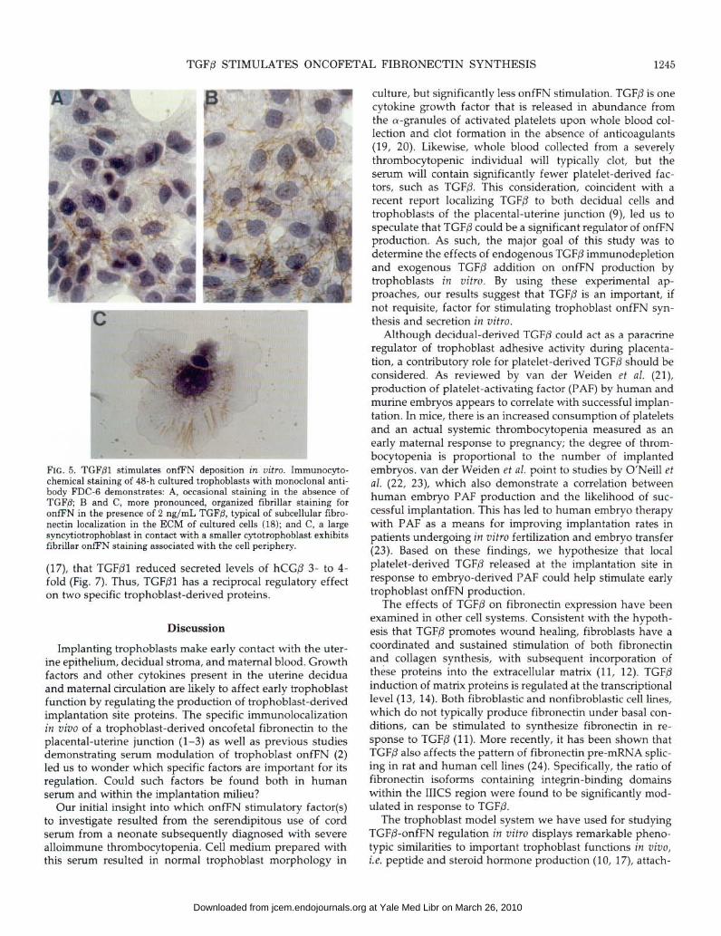
duced a 3- to 4-fold induction of onfFN when trophoblasts were cultured in plasma-derived serum from a normal neo- nate. Preparing serum in this manner prevents platelet acti- vation and platelet incorporation into the clot, thus reducing the amount of platelet-derived factors released into the serum. TGFPl stimulation of trophoblasts cultured in two additional human and two bovine plasma-derived sera ob- tained commercially resulted in a similar 3- to 5-fold onfFN induction after 48 and 72 h of culture (not shown).
TGFPl also has a pronounced effect on enhancing the deposition of onfFN extracellularly. As shown in Fig. 5, trophoblasts cultured for 48 h in 2% bovine platelet-poor serum exhibited occasional immunocytochemical staining for or&N in the absence of exogenously added TGFPl (Fig. 5A). However, in parallel cultures stimulated with 2 ng/mL
2% Platelet-poor NHS *1”“*NF--
se,.
a b c d ef gh FIG. 4. TGFfll stimulates onfFN production by trophoblasts cultured in normal plasma-derived platelet-poor serum. Concentrations of TGFPl ranging from 0.625-5 ng/mL (lanes b, d, f, and h) induced a 3- to 4-fold induction of onfFN compared to controls (lanes a, c, e, and g) when trophoblasts were cultured in 2% plasma-derived serum from a normal neonate. Preparing serum in this manner prevents platelet activation and platelet incorporation into the clot, thus reducing the amount of platelet-derived factors released into the serum. NHS, Newborn human serum.
TGFPl (Fig. 5B), prominent fibrillar staining typical of fibro- nectin extracellular deposition (18) was observed. Figure SC demonstrates the immunocytochemical appearance of onfFN fibrils associated with the periphery of a large syncytial cell in the presence of TGFPl.
Dose response of TGF/31 -mediated onfFN stimulation
Because trophoblasts cultured in 2% neonatal thrombo- cytopenic serum appeared normal morphologically, yet pro- duced low basal levels of onfFN, we used this culture system to determine a dose response for TGF/31-mediated onfFN stimulation. The dose-response curve from four separate TGFPl stimulation experiments, including 48- and 72-h cul- ture times, is shown in Fig. 6, with TGFPl concentrations ranging from 0.01-5 ng/mL. The lowest onfFN concentration (i.e. unstimulated control) was considered 0%, whereas the highest onfFN concentration for each experiment was con- sidered a 100% response. Based on this graph, the 50% effective dose (EDso) of TGFPl in this culture system was between 0.1-0.2 ng/mL. If the 48 h TGFP-o&N response was plotted separately from the 72 h results, the dose- response curves were essentially superimposable, with the same EDs0 calculated (not shown).
As previously demonstrated by Morrish et al. (17), TGFPl down-regulates hCG in vitro. When trophoblast media from our TGF@l-treated cultures were quantitatively assayed for hCG/3 using the Abbott IMX automated microbead analyzer (courtesy of Dr. Marilyn Senior and Ms. Margaret Bulley, Clinical Endocrine Laboratory, Hospital of the University of Pennsylvania), we found, in agreement with Morrish et al.
at Yale Med Libr on March 26, 2010 jcem.endojournals.orgDownloaded from
TGFP STIMULATES ONCOFETAL FIBRONECTIN SYNTHESIS 1245
FIG. 5. TGFPl stimulates onfEN deposition in uitro. Immunocyto- chemical staining of 48-h cultured trophoblasts with monoclonal anti- body FDC-6 demonstrates: A, occasional staining in the absence of TGFP; B and C, more pronounced, organized fibrillar staining for onfFN in the presence of 2 ng/mL TGFB, typical of subcellular fibro- nectin localization in the ECM of cultured cells (18); and C, a large syncytiotrophoblast in contact with a smaller cytotrophoblast exhibits tibrillar onfFN staining associated with the cell periphery.
(17), that TGFPl reduced secreted levels of hCG/l 3- to 4- fold (Fig. 7). Thus, TGFPl has a reciprocal regulatory effect on two specific trophoblast-derived proteins.
Discussion
Implanting trophoblasts make early contact with the uter- ine epithelium, decidual stroma, and maternal blood. Growth factors and other cytokines present in the uterine decidua and maternal circulation are likely to affect early trophoblast function by regulating the production of trophoblast-derived implantation site proteins. The specific immunolocalization in viva of a trophoblast-derived oncofetal fibronectin to the placental-uterine junction (l-3) as well as previous studies demonstrating serum modulation of trophoblast onfFN (2) led us to wonder which specific factors are important for its regulation. Could such factors be found both in human serum and within the implantation milieu?
Our initial insight into which onfFN stimulatory factor(s) to investigate resulted from the serendipitous use of cord serum from a neonate subsequently diagnosed with severe alloimmune thrombocytopenia. Cell medium prepared with this serum resulted in normal trophoblast morphology in
culture, but significantly less onfFN stimulation. TGF/3 is one cytokine growth factor that is released in abundance from the a-granules of activated platelets upon whole blood col- lection and clot formation in the absence of anticoagulants (19, 20). Likewise, whole blood collected from a severely thrombocytopenic individual will typically clot, but the serum will contain significantly fewer platelet-derived fac- tors, such as TGFP. This consideration, coincident with a recent report localizing TGFP to both decidual cells and trophoblasts of the placental-uterine junction (9), led us to speculate that TGFP could be a significant regulator of onfFN production. As such, the major goal of this study was to determine the effects of endogenous TGFP immunodepletion and exogenous TGFP addition on onfFN production by trophoblasts in vitro. By using these experimental ap- proaches, our results suggest that TGFP is an important, if not requisite, factor for stimulating trophoblast onfFN syn- thesis and secretion in vitro.
Although decidual-derived TGFP could act as a paracrine regulator of trophoblast adhesive activity during placenta- tion, a contributory role for platelet-derived TGF/3 should be considered. As reviewed by van der Weiden et al. (21), production of platelet-activating factor (PAF) by human and murine embryos appears to correlate with successful implan- tation. In mice, there is an increased consumption of platelets and an actual systemic thrombocytopenia measured as an early maternal response to pregnancy; the degree of throm- bocytopenia is proportional to the number of implanted embryos. van der Weiden et al. point to studies by O’Neill et al. (22, 23), which also demonstrate a correlation between human embryo PAF production and the likelihood of suc- cessful implantation. This has led to human embryo therapy with PAF as a means for improving implantation rates in patients undergoing in vitro fertilization and embryo transfer (23). Based on these findings, we hypothesize that local platelet-derived TGF/3 released at the implantation site in response to embryo-derived PAF could help stimulate early trophoblast onfFN production.
The effects of TGFP on fibronectin expression have been examined in other cell systems. Consistent with the hypoth- esis that TGFP promotes wound healing, fibroblasts have a coordinated and sustained stimulation of both fibronectin and collagen synthesis, with subsequent incorporation of these proteins into the extracellular matrix (11, 12). TGF/3 induction of matrix proteins is regulated at the transcriptional level (13, 14). Both fibroblastic and nonfibroblastic cell lines, which do not typically produce fibronectin under basal con- ditions, can be stimulated to synthesize fibronectin in re- sponse to TGFP (11). More recently, it has been shown that TGFP also affects the pattern of fibronectin pre-mRNA splic- ing in rat and human cell lines (24). Specifically, the ratio of fibronectin isoforms containing integrin-binding domains within the IIICS region were found to be significantly mod- ulated in response to TGFP.
The trophoblast model system we have used for studying TGFP-onfFN regulation in vitro displays remarkable pheno- typic similarities to important trophoblast functions in viva, i.e. peptide and steroid hormone production (10, 17), attach-
at Yale Med Libr on March 26, 2010 jcem.endojournals.orgDownloaded from
1246 FEINBERG, KLIMAN, AND WANG JCE & M. 1994 Vol7S.No5
TGFfil Dose Response of TUN Stimulation
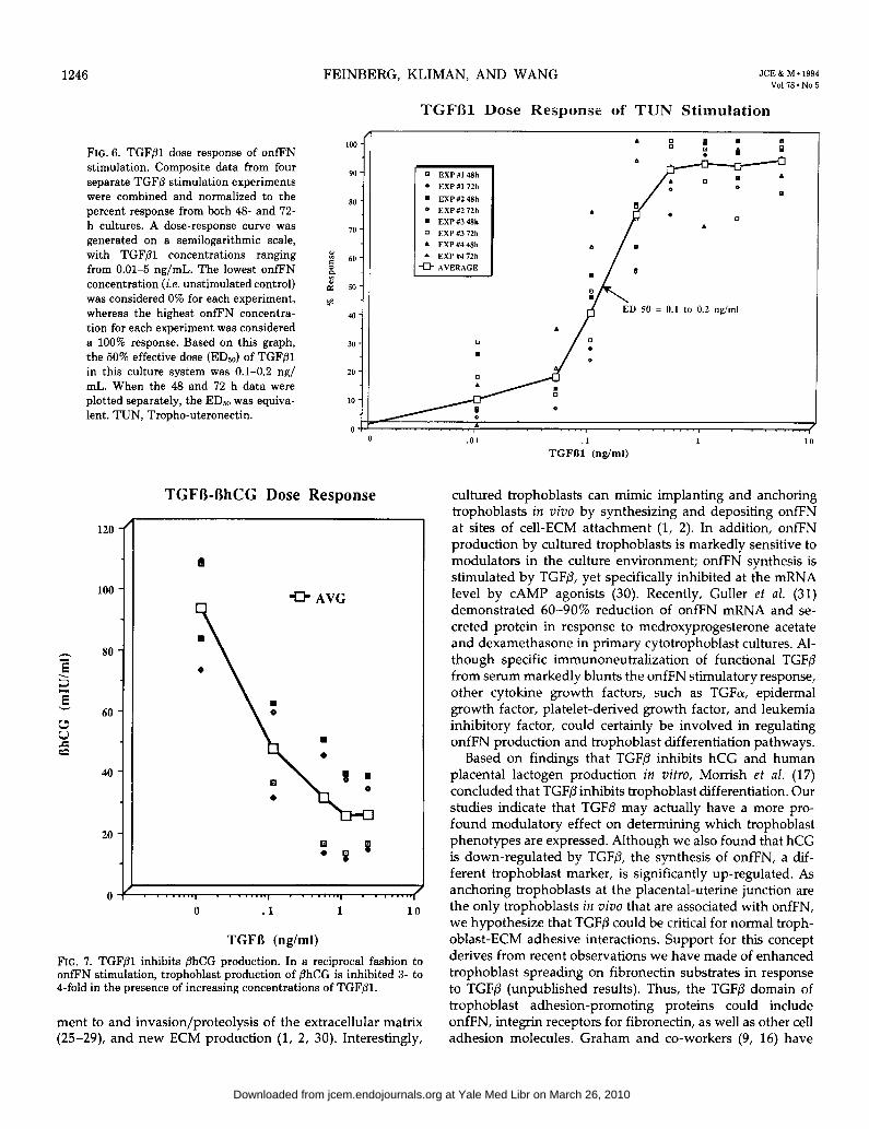
FIG. 6. TGFPl dose response of onfFN stimulation. Composite data from four separate TGFp stimulation experiments were combined and normalized to the percent response from both 46 and 72- h cultures. A dose-response curve was generated on a semilogarithmic scale, with TGFpl concentrations ranging from 0.01-5 ng/mL. The lowest onfFN concentration (i.e. unstimulated control) was considered 0% for each experiment, whereas the highest onfFN concentra- tion for each experiment was considered a 100% response. Based on this graph, the 50% effective dose (ED& of TGFPl in this culture system was 0.1-0.2 ng/ mL. When the 48 and 12 h data were plotted separately, the ED,, was equiva- lent. TUN, Tropho-uteronectin.
100 -
90 -
80 -
70 -
x 5 60.
:: a” SO-
*
40 -
30 -
20 -
to -
Ol- 0
TGFB-OhCG Dose Response
100 -
= 80 -
E 3
E 60 -
t: zz
40-
20 -
04
0 EXPlfl48h l EXPIf172h
q EXPU248h
0 EXPb272h
I n EXP#348h
q EXP#372h
A EXPM48h
A EXP11472h
0 AVERAGE
-D AVG
: p 0
ED 50 = 0.1 to 0.2 q/ml
.
d I
0 .l 1 10
TGFD (rig/ml)
FIG. 7. TGF@l inhibits phCG production. In a reciprocal fashion to onfEN stimulation, trophoblast production of PhCG is inhibited 3- to I-fold in the presence of increasing concentrations of TGFPl.
ment to and invasion/proteolysis of the extracellular matrix (25-29), and new ECM production (1, 2, 30). Interestingly,
J .Ol .t 1 10
TGFBl (&ml)
cultured trophoblasts can mimic implanting and anchoring trophoblasts in viva by synthesizing and depositing onfFN at sites of cell-ECM attachment (1, 2). In addition, onfFN production by cultured trophoblasts is markedly sensitive to modulators in the culture environment; onfFN synthesis is stimulated by TGFP, yet specifically inhibited at the mRNA level by CAMP agonists (30). Recently, Guller et al. (31) demonstrated 60-90s reduction of onfFN mRNA and se- creted protein in response to medroxyprogesterone acetate and dexamethasone in primary cytotrophoblast cultures. Al- though specific immunoneutralization of functional TGFP from serum markedly blunts the onfFN stimulatory response, other cytokine growth factors, such as TGFo, epidermal growth factor, platelet-derived growth factor, and leukemia inhibitory factor, could certainly be involved in regulating onfFN production and trophoblast differentiation pathways.
Based on findings that TGFP inhibits hCG and human placental lactogen production in vitro, Morrish et al. (17) concluded that TGFP inhibits trophoblast differentiation. Our studies indicate that TGF/3 may actually have a more pro- found modulatory effect on determining which trophoblast phenotypes are expressed. Although we also found that hCG is down-regulated by TGFP, the synthesis of onfFN, a dif- ferent trophoblast marker, is significantly up-regulated. As anchoring trophoblasts at the placental-uterine junction are the only trophoblasts in vim that are associated with onfFN, we hypothesize that TGFP could be critical for normal troph- oblast-ECM adhesive interactions. Support for this concept derives from recent observations we have made of enhanced trophoblast spreading on fibronectin substrates in response to TGF/3 (unpublished results). Thus, the TGFP domain of trophoblast adhesion-promoting proteins could include onfFN, integrin receptors for fibronectin, as well as other cell adhesion molecules. Graham and co-workers (9, 16) have
at Yale Med Libr on March 26, 2010 jcem.endojournals.orgDownloaded from

TGFB STIMULATES ONCOFETAL FIBRONECTIN SYNTHESIS 1247
provided considerable evidence that TGFP modulates troph- oblast differentiation. In addition to their in vim localization of TGFP to decidual and trophoblast cells at the human placental-uterine interface (9), these investigators found that TGFP promotes trophoblast syncytial formation and limits invasiveness in vitro (9, 16).
Although our work has generally focused on the adhesive aspects of trophoblast-uterine interaction, onfFN production and deposition within the placental-uterine junction could have additional roles involved with promoting and maintain- ing pregnancy. Fibronectin-lymphocyte and fibronectin- macrophage interactions have been well described, and fi- bronectin appears to have an important function in modu- lating leukocyte adhesiveness, chemotaxis, proliferation, and differentiation. Typically, these leukocyte-matrix interactions are mediated via cell surface integrins, such as the fibronectin receptors VLA-4 and VLA-5 (32-38). One particularly inter- esting feature of the VLA-4 integrin is its binding specificity for the CS-1 region of fibronectin IIICS, specifically the tripeptide Leu-Asp-Val (39). This VLA-4-binding site is sep- arated from the FDC-6 epitope by only nine amino acids (18), raising the possibility that 0-glycosylation at this IIICS site could have an effect on CSl conformation and binding affinity. However, the precise functional consequences of the FDC-6 epitope on cellular VLA-4-fibronectin interactions are not known. TGFP has also been found to have significant immunomodulatory properties, such as diminishing the pro- duction of inflammatory cytokines implicated in septic shock (40). In a model relevant to implantation, TGF/3 can function as a potent immunosuppressant in vim during organ trans- plant, and it has been suggested that one immunosuppressive effect of drugs such as cyclosporin-A may be mediated through induction of endogenous TGFP (40). Thus, it is interesting to speculate that both TGF/3 and onfFN, localized to the trophoblast-decidual junction, could act as immuno- modulators to protect the implanting pregnancy from im- munological rejection.
In summary, we have proposed one important role for TGFP in normal human implantation and trophoblast devel- opment: the up-regulation of onfFN, an extravillous troph- oblast-derived adhesive glycoprotein. Future studies will fo- cus on other potential decidual and serum-derived modula- tors of trophoblast function as well as the implications of aberrant onfFN expression and regulation in the pathophys- iology of implantation failure and pregnancy loss.
Acknowledgments
The authors thank the nurses, residents, and secretaries on the Labor and Delivery Suite of the Hospital of the University of Pennsylvania for their help with collecting the normal placentae required for this work. We also thank Dr. Marilyn Senior and Ms. Margaret Bulley for their help with the hCGP assays, and Dr. Jerry Strauss for his suggestions and thoughtful reading of the manuscript.
References
1. Feinberg RF, Kliman HJ, Lockwood CJ. 1991 Is oncofetal fibro- nectin a trophoblast glue for human implantation? Am J Pathol. 138:537-543.
2.
3.
4.
5.
6.
7.
8
9
10
11
12
13.
14.
15.
16.
17.
18. 19.
20.
21.
22.
23.
Feinberg RF, Kliman HJ. 1993 Tropho-uteronectin (TUN): a unique oncofetal fibronectin deposited in the extracellular matrix of the tropho-uterine junction and regulated in vitro by cultured human trophoblast cells. Troph Res. 7:167-179. Cunningham FG, MacDonald PC, Gant NF, Leveno KJ, Gilstrap LC. 1993 Williams’ obstetrics, 19th ed. Norwalk: Appleton, Lange; 122. Lockwood CJ, Senyei AE, Dische MR, et al. 1991 Fetal fibronectin in cervical and vaginal secretions as a predictor of preterm delivery. N Engl J Med. 325:669-674. Feinberg RF, Kliman HJ. 1992 ‘Fetal’ fibronectin and preterm labor [Letter]. N Engl J Med. 326:708. Matsuura H, Hakomori S. 1985 The oncofetal domain of fibronectin defined by monoclonal antibody FDC-6: its presence in fibronectins from fetal and tumor tissues and its absence in those from normal adult tissues and plasma. Proc Nat1 Acad Sci USA. 82:6517-6521. Matsuura H, Takio K, Titani K, et al. 1988 The oncofetal structure of human fibronectin defined by monoclonal antibody FDC-6: unique structural requirement for the antigenic specificity-provided bv a elvcosvlhexaueutide. I Biol Chem. 263:3314-3322. Gat&ra h, Gre&e T, fiakomori S. 1989 An alpha-l\i-acetylga- lactosaminylation at the threonine residue of a defined peptide sequence creates the oncofetal peptide epitope in human fibronectin. J Biol Chem. 264:10472-10476. Graham CH, Lysiak JJ, McRae KR, Lala PK. 1992 Localization of transforming growth factor-p at the human fetal-maternal interface: role in trophoblast growth and differentiation. Biol Reprod. 46:561- 572. Kliman HJ, Nestler JE, Sermasi E, Sanger JM, Strauss III JF. 1986 Purification, characterization and in vitro differentiation of cyto- trophoblasts from human term placentae. Endocrinology. 118:1567- 1582. Engvall E, Ruoslahti E. 1977 Binding of a soluble form of fibroblast surface protein, fibronectin, to collagen. Int J Cancer. 20:1-5. Ignotz RA, Massague J. 1986 Transforming growth factor-0 stim- ulates the expression of fibronectin and collagen and their incorpo- ration into the extracellular matrix. J Biol Chem. 261:4337-4345. Ignotz RA, Endo T, Massague J. 1987 Regulation of fibronectin and type I collagen mRNA levels by transforming growth factor-p. J Biol Chem. 262:6443-6446. Keski-Oja J, Raghow R, Sawdey M, et al. 1988 Regulation of mRNAs for type-l plasminogen activator inhibitor, fibronectin, and type I procollagen by transforming growth factor-p. J Biol Chem. 263:3111-3115. Blatti SP, Foster DN, Ranganathan G, Moses HL, Getz M. 1988 Induction of fibronectin gene transcription and mRNA is a primary response to growth-factor stimulation of AKR-2B cells. Proc Nat1 Acad Sci USA. 85:1119-1123. Graham CH, Lala PK. 1991 Mechanism of control of trophoblast invasion in situ. J Cell Physiol. 148:228-234. Morrish DW, Bhardwaj D, Paras MT. 1991 Transforming growth factor 01 inhibits placental differentiation and human chorionic gonadotropin and human placental lactogen secretion. Endocrinol- ogy. 129:22-26. Hynes RO. 1990 Fibronectins. New York: Springer-Verlag. Childs CB, Proper JA, Tucker RF, Moses HL. 1982 Serum contains a platelet derived transforming growth factor. Proc Nat1 Acad Sci USA. 79:5312-5316. Assoian RK, Komoriya A, Meyers CA, Miller DM, Sporn MB. 1983 Transforming growth factor-beta in human platelets. J Biol Chem. 258:7155-7160. van der Weiden RMF, Helmerhorst FM, Keirse MJNC. 1991 Influence of prostaglandins and platelet activating factor on implan- tation. Hum Reprod 6:436-442. O’Neill C, Gidley-Baird AA, Pike IL, Porter RN, Sinosich MJ, Saunders DM. 1985 Maternal blood platelet physiology and luteal phase endocrinology as a means of monitoring pre- and postim- plantation embryo viability following in vitro fertilization. J In Vitro kertil Embryo Tiansfer. 2:67-93. - O’Neill C, Collier M, Ammit AI, Rvan IP, Saunders DM, Pike IL. 1989 Supplementation of in-v;tro’fertiii;ation culture m&urn with platelet activating factor. Lancet. 2:769-772.
at Yale Med Libr on March 26, 2010 jcem.endojournals.orgDownloaded from

24.
25.
26.
27.
28.
29.
30.
31.
FEINBERG, KLIMAN, AND WANG
Magnuson VL, Young M, Schattenberg DG, et al. 1991 The alternative splicing of fibronectin pre-mRNA is altered during aging and in response to growth factors. J Biol Chem. 266:14654-14662. Kao LC, Caltabiano S, Wu S, Strauss JF3, Kliman HJ. 1988 The human villous cytotrophoblast: interactions with extracellular matrix proteins, endocrine function, and cytoplasmic differentiation in the absence of syncytium formation, Dev Biol. 130:693-702. Queenan Jr JT, Kao L-C, Arboleda CE, et al. 1987 Regulation of urokinase-type plasminogen activator production by cultured hu- man cytotrophoblasts. J Biol Chem. 262:10903-10906, Feinberg RF, Kao LC, Haimowitz JE, et al. 1989 Plasminogen activator inhibitor types 1 and 2 in human trophoblasts. PA1-1 is an immunocytochemical marker of invading trophoblasts. Lab Invest. 61:20-26. Kliman HJ, Feinberg RF. 1990 Human trophoblast-extracellular matrix (ECM) interactions in vitro: ECM thickness modulates mor- phology and proteolytic activity. Proc Nat1 Acad Sci USA. 87:3057- 3061. Kliman HJ, Feinberg RF, Haimowitz JE. 1990 Human trophoblast- endometrial interactions in an in vitro suspension culture system. Placenta. 11:349-367. Ulloa-Aguirre A, August AM, Golos TG, et al. 1987 8-Bromo- 3’,5’-adenosine monophosphate regulates expression of chorionic gonadotropin and fibronectin in human cytotrophoblasts. J Clin Endocrinol Metab. 64:1002-1009. Guller S, LaCroix NC, Kirkun G, et al. 1993 Steroid regulation of oncofetal fibronectin expression in human cytotrophoblasts. J Ste-
32.
33.
34.
35.
36.
37.
38.
39.
40.
JCE & M -1994 Vol%.No5
roid Biochem Mol Biol. 46:1-10. Klingemann HG, Kohn FR. 1991 Involvement of fibronectin and its receptor in human lymphocyte proliferation. J Leuk Biol. 50:464- 470. Blue ML, Conrad P, Davis G, Kelley KA. 1991 Enhancement of CD2-mediated T cell activation by the interaction of VLA-4 with fibronectin. Cell Immunol. 138:238-244. Freedman AS, Rhynhart K, Nojima Y, et al. 1993 Stimulation of protein tyrosine phosphorylation in human B cells after ligation of the beta 1 integrin VLA-4. J Immunol. 150:1645-1652. Ferguson TA, Kupper TS. 1993 Antigen-independent processes in antigen-specific immunity. A role for alpha 4 integrin. J Immunol. 150:1172-1182. Charm ZL, Beezhold DH, Personius CD, Shen ZL. 1993 Fibronec- tin cefi-binding domain triggered transmembrane signal transduc- tion in human monocvtes. I Leuk Biol. 53:79-85. Hershkoviz R, Alonk, Gilat D, Lider 0. 1992 Activated T lym- phocytes and macrophages secrete fibronectin which strongly sup- ports cell adhesion. Cell Immunol. 141:352-361. Ferguson TA, Mizutani H, Kupper TS. 1991 Two integrin-binding peptides abrogate T cell-mediated immune responses in vim. Proc Nat1 Acad Sci USA. 88:8072-8076. Wayner EA, Kovach NL. 1992 Activation-dependent recognition by hematopoietic cells of the LDV sequence in the V region of fibronectin. J Cell Biol. 116:489-497. Palladino MA, Morris RE, Starnes HF, Levinson AD. 1991 The transforming growth factor-betas: a new family of immunoregula- tory molecules, Ann NY Acad Sci. 593:181-187.
at Yale Med Libr on March 26, 2010 jcem.endojournals.orgDownloaded from