aquaporin 3 promotes human extravillous trophoblast
TRANSCRIPT

RESEARCH Open Access
Aquaporin 3 promotes human extravilloustrophoblast migration and invasionYingqi Nong1,2†, Shifen Li3†, Wenjuan Liu2, Xiqian Zhang2, Lin Fan2, Ye Chen2, Qianwen Huang2,Qianyu Zhang2 and Fenghua Liu2*
Abstract
Problem: Does aquaporin 3 (AQP3) affect the migration and invasion of human extravillous trophoblast (HTR8/Svneo) cells?
Method of study: A lentivirus infection system was used to construct stable cell lines with either AQP3 knockdownor overexpression. RT-PCR and western blotting were used to verify the efficiencies of AQP3 knockdown oroverexpression in HTR8/Svneo cells at mRNA and protein levels, respectively. Cell Counting Kit-8 and flowcytometry assays were used to detect the influence of AQP3 knockdown or overexpression on proliferation andapoptosis of HTR8/Svneo cells. In addition, wound healing and Transwell invasion assays were used to detect theeffects of AQP3 knockdown or overexpression on migration and invasion capabilities of HTR8/Svneo cells. AnAgilent gene chip was used to screen for significant differentially expressed genes after AQP3 knockdown. Finally,mechanisms by which AQP3 influences the migration and invasion of HTR8/Svneo cells were explored usingbioinformatic analysis.
Results: Compared with controls, migration and invasion capabilities of HTR8/Svneo cells were significantlyreduced after AQP3 knockdown, and significantly increased after AQP3 overexpression. Subsequent bioinformaticanalysis of gene chip expression profiles indicated downregulation of genes related to adhesion such as PDGF-B, aswell as signaling pathways (such as PIK3/AKT, NF-κB, and TNF) after AQP3 knockdown.
Conclusions: AQP3 could significantly promote migration and invasion capabilities of human extravilloustrophoblasts, it may mediate embryo invasion and adhesion to endometrium by regulating PDGF-B, PIK3/AKTsignaling pathways, although this requires further verification.
Keywords: Aquaporin 3 (AQP3), Embryo implantation, Human extravillous trophoblast, Migration, Invasion
IntroductionRecurrent implantation failure (RIF) is one of the bottle-necks of in vitro fertilization-embryo transfer and its de-rivative techniques. Currently, there is no uniformdefinition on RIF. However, it is widely accepted as adiagnosis criteria which include an age less than 40 yearsand failure to achieve a clinical pregnancy after transfer
of at least four good-quality embryos in a minimum ofthree fresh or frozen cycles [1]. Incidence of RIF is up to10–15% [2], but its pathogenesis is still unclear. Manystudies have reported that two-thirds of RIF cases can beattributed to endometrial receptivity, while the otherthird is caused by inherent factors within embryos [3].Embryo implantation is an important step of mamma-
lian reproduction, and is critical for determining preg-nancy. Two factors that determine embryo implantationare the embryo’s implantation capability and the receiv-ing status of the endometrium. The process of embryo
© The Author(s). 2021 Open Access This article is licensed under a Creative Commons Attribution 4.0 International License,which permits use, sharing, adaptation, distribution and reproduction in any medium or format, as long as you giveappropriate credit to the original author(s) and the source, provide a link to the Creative Commons licence, and indicate ifchanges were made. The images or other third party material in this article are included in the article's Creative Commonslicence, unless indicated otherwise in a credit line to the material. If material is not included in the article's Creative Commonslicence and your intended use is not permitted by statutory regulation or exceeds the permitted use, you will need to obtainpermission directly from the copyright holder. To view a copy of this licence, visit http://creativecommons.org/licenses/by/4.0/.The Creative Commons Public Domain Dedication waiver (http://creativecommons.org/publicdomain/zero/1.0/) applies to thedata made available in this article, unless otherwise stated in a credit line to the data.
* Correspondence: [email protected]†Yingqi Nong and Shifen Li contributed equally to this work.2Department of Reproductive Health and Infertility, Guangdong Women andChildren Hospital, Guangzhou, Guangdong, ChinaFull list of author information is available at the end of the article
Nong et al. Reproductive Biology and Endocrinology (2021) 19:49 https://doi.org/10.1186/s12958-021-00726-z

implantation includes localization, adhesion, and inva-sion of the maternal endometrium until embedment intothe matrix. This behavior of the embryo invading theendometrium at a specific time and space is the primi-tive motive for embryo implantation. The invasionprocess is completed when EVT differentiate from cyto-trophoblasts [4]. Decidualization happens in the endo-metrium during the implantation window afterblastocyst adhesion. Endometrial interstitial cells grad-ually transform into decidual stromal cells, which receivethe invasion of EVT [5]. It has been shown that invasionof trophoblasts into the endometrium is similar to themetastasis of malignant tumors [6], as both involve cellinvasion. Interestingly, an in vitro co-culture study re-ported that trophoblasts in mouse embryos exhibitedstronger invasion than malignant tumor cells [7, 8]. Dir-ectional migration of trophoblasts, which involves aseries of cell signaling events, is a central step of inva-sion behavior. At present, widely accepted cell migrationmechanisms [9] include: (1) actin depolymerization,transport of ions into cells, and increased osmotic pres-sure in the front of cells; (2) penetration of waterthrough the cell membrane to increase local hydrostaticpressure, whereby the cell membrane forms local crown-ing including ruga, pseudopod, and vesicles; and (3)actin re-polymerization. Thus, migration speed can becontrolled by osmotic pressure in extracellular medium.This means that high osmotic pressure accelerates mi-gration, while low pressure slows down migration. Dur-ing this process, AQPs play a critical role for compliancewith the acceleration of intracellular and extracellularosmotic pressure changes, as well as rapid changes ofcellular morphology [10, 11].AQPs, a type of channel protein, can regulate levels of
water and small molecular substances (such as glycerin,urea, and nitrogen), which is very important for themaintenance of body fluid equilibrium [12]. MultipleAQPs have been detected in embryos before implant-ation [12–14]. Expression of AQP3 lasts from the zygoteto the blastula stage [15], and is the most abundantaquaporin expressed in villi during early pregnancy [16].This suggests that AQP3 may play a role in early growthand implantation of embryos.Our previous study found high expression of AQP3 in
the cell membrane of trophoblasts in blastocysts of Kun-ming mice [17]. AQP3 could significantly promote bothadhesion and expansion capabilities of blastocysts [18],suggesting that AQP3 participates in the process of tro-phoblasts invading the endometrium. However, it is un-known if AQP3 is expressed in human extravilloustrophoblasts (EVT), or whether it participates in humanembryo implantation. If so, the mechanism by whichAQP3 regulates embryo implantation is unknown. All ofthese issues were addressed in the current study.
Methods and materialsMethodsCell cultureThe human trophoblast cell line HTR8/SVneo has beenwidely applied as an early invasion and migration modelof extravillous cytotrophoblasts [19]. HTR8/SVneo,which was provided by American Type Culture Collec-tion (USA), was cultured with Dulbecco’s Modified Ea-gle’s Medium (DMEM) containing 10% fetal bovineserum (FBS; Gibco, USA) at 37 °C and 5% CO2.
Construction of stable cell linesPlasmids containing AQP3 knockdown, AQP3 overex-pression, or their respective negative control plasmidswere purchased from Weijiang Biotechnology (China).Lentiviral packaging was performed in 293 T cells ac-cording to the manufacturer’s instructions (Suzhou Gen-ePharma, China). Lentivirus in the supernatant wascollected to transfect cells. Controls were transfectedwith empty vector. Cells were divided into four groups:interfering AQP3 (AQP3-shRNA) group, interferingempty vector (CON-shRNA) group, overexpressingAQP3 (AQP3-OE) group and overexpressing empty vec-tor (CON-OE) group. Cells were infected with viruses atthe following multiplicities of infection (MOI): AQP3-shRNA (MOI =100); CON-shRNA (MOI =100); AQP3-OE (MOI =150); CON-OE (MOI =100). Strict pheno-type selection was performed on stably infected HTR8/SVneo cells with 0–10 μg/mL puromycin (MPbio, USA)to use resistance as a screening index. Furthermore, cellswere stably cloned in 0.5 μg/mL puromycin.
RNA isolation, cDNA synthesis and qRT-PCR analysisTotal RNA was extracted from HTR8/Svneo cells ac-cording to the instructions of a TRIzol kit (Taraka Bio-technology, China), and measured with aspectrophotometer (Nanodrop 2000; Thermo Scientific,USA).
cDNA synthesis The annealing mixture contained1 μg of RNA, 1 μl of 0.5 μg/ul Oligo (dT) 18, 1 μl ofdNTPs Mix (2.5 mM), and RNase-free water to a totalvolume of 10 μl. Then, it was incubated in a thermalcycler at 65 °C for 5 min and placed on ice for 1 minat least. The contents of the tube were collected bybrief centrifugation before the following were addedto the tube: Random primer, 1 μl, 5 × Buffer, 4 μl,RNase Inhibitor, 0.5 μl, and RNase Free H2O 4.5 μl.The pipette was gently sucked and tapped severaltimes to obtain a better mixture. Incubation was doneas follows: 10 min at 30 °C, 60 min at 42 °C and then15 min at 70 °C. The cDNA synthesis reaction wasstored at − 20 °C, or PCR was performed immediately.
Nong et al. Reproductive Biology and Endocrinology (2021) 19:49 Page 2 of 11

RT-qPCR reactions were conducted with a QuantStu-dio 5 real-time PCR system (Thermo Fisher Scientific,Waltham, MA USA). The primers were designed byPrimer-BLAST (NCBI). The primer sequences and PCRproduct sizes were detailed in Table 1. The real timePCR mixture contained SYBR Green Premix, 10 μl, For-ward Primer (10 μM), 0.4 μl, Reverse Primer (10 μM),0.4 μl, Template,1.2 μl, ROX Reference Dye II (50×),0.4 μl, DNA template 2.0 μl, and RNase Free H2O to atotal volume of 20 μl. After the solution was mixed andcentrifuged at 5000 RPM for a short time, the reactionmixtures (8 μl) were added into a 384-well PCR plate,and the cDNA samples (2 μl) were added. The plate wassealed and placed on ice. The PCR was initiated by heat-ing the mixture to 95 °C for 5 min, followed by 40 cyclesof 15 s at 95 °C and 60s at 60 °C and then 15 s at 95 °C.To establish the melting curve, the mixture was heatedto 95 °C for 10 s, 60 °C for 60 s, and 95 °C for 15 s se-quentially after the amplification reaction was over. The2-ΔΔCT method was used to quantify relative expressionof AQP3 mRNA. Each real-time PCR included a no-template control. The experiments were repeated 3times with triplicates of each sample.
Western blottingCells were collected and lysed with RIPA lysis buffer andphenylmethylsulfonyl fluoride (Beyotime Biotechnology,China) on ice for 30 min, quantified by bicinchoninicacid assay. Equal amounts of protein (~ 10 μg) were sep-arated by 10% sodium dodecyl sulfate polyacrylamide gelelectrophoresis (SDS-PAGE) and loaded for SDS-PAGEelectrophoresis. After transfer to a polyvinylidene fluor-ide membrane, the membrane was blocked in 5% skimmilk, sealed for 1 h at room temperature with shaking,and incubated with a primary antibody over-night at4 °C. The primary antibodies used were rabbit polyclonalanti-AQP3 antibody (1:1000, Abcam, UK, ab125219),rabbit polyclonal anti-PDGFB antibody (1:1000, Cohe-sion, UK, CPA1865). IgG from rabbit serum (1:1000,Sigma, USA, I5006) was used for negative control.
Finally, the membrane was incubated with horseradishperoxidase-labeled secondary antibody (120,000, BosterBiological Technology, China, BA1054) at roomtemperature for 1 h, and developed by enhanced chemi-luminescence. The experiments were repeated 3 times.Image Pro-Plus 6.0 software (Media Cybernetics, USA)was used to analyze gray values.
Cell proliferation/CCK-8 assayHTR8/Svneo cells (100 μL; 1 × 10⁵ cells/mL) in vectorcontrol, knockdown, and overexpression groups wereadded into 96-well plates in triplicate, and cultured at37 °C overnight. CCK-8 kit reagent (10 μL; Dojindo,Japan) was added into each well and incubated for 2 h, 3h, and 4 h. Optical density at 450 nm of each well wasdetected each time point with a multifunctional micro-plate reader.
Flow cytometry assayAnnexin V-APC/7-AAD double staining was performedon HTR8/Svneo cells according to the instructions of anAnnexin V-APC/7AAD Apoptosis Detection Kit [Multi-sciences (Lianke) Biotech, China]. Live, early apoptotic,and late apoptotic or necrotic cells were classified usingflow cytometry (AccuriC6, Becton Dickinson, USA) andFlow Jo 7.6.1 software (FlowJo, USA).
Wound healing assayHTR8/Svneo cells (1 × 10⁶ cells/mL) were seeded in asix-well plate and routinely cultured in an incubator.When cells grew into a monolayer, they were treatedwith mitomycin for 1 h to inhibit cell division. Next, asterile 10 μl-pipette tip was used to scrape cell cultureplates. Scraped cells were washed twice with phosphate-buffered saline, cultured in serum-free medium, incu-bated in an incubator, and photographed at 0, 6, 24 and48 h after scratching. Image Pro-Plus 6.0 was used tomeasure scratch depth at any five sites at the same timepoint to calculate migration rates, thus reflecting cellmobility and migratory capabilities.
Transwell invasion assayA Transwell invasion system (8-μm, 24-well; Corning,UK) coated with Matrigel (40 μL; Becton Dickinson) wasused. Briefly, 1 × 10⁵ cells were suspended in DMEMwithout serum, and seeded in the upper chamber.DMEM containing 10% FBS was then added to the lowerchamber and the plate was incubated at 37 °C and 5%CO2. After 24 h, cells were fixed with methanol andstained with 0.1% crystal violet. The quantity of coloredcells in five random visual fields was counted using aninverted microscope (Nikon, Japan).
Table 1 The Primer sequences and the product size of targetand control genes
Primer Primer sequence (5′-3′) size (bp)
AQP3 Forward primer: ACCATCAACCTGGCCTTTGGReverse primer: GGGGACGGGGTTGTTGTAG
390
PDGFB Forward primer: ACTGATGGGGTCGCTCTTTGReverse primer: CAGGGATCAGGCAGGCTATG
126
FOS Forward primer: GTGCCAACTTCATTCCCACGReverse primer: GGCCTCCTGTCATGGTCTTC
186
SNAIL1 Forward primer: CCTGTCTGCGTGGGTTTTTGReverse primer: ACCTGGGGGTGGATTATTGC
198
GAPDH Forward primer: GAAGCTCATTTCCTGGTATGACAReverse primer: GGGAGATTCAGTGTGGTGGG
189
Nong et al. Reproductive Biology and Endocrinology (2021) 19:49 Page 3 of 11

Whole genome expression profileThe Gene expression profiles assay was performed ac-cording to a previously described method [20]. Gene ex-pression profiles of AQP3-shRNA and CON-shRNAwere analyzed by two-color gene expression microarray(Agilent Technologies, USA) according to the instruc-tions of a Low Input Quick Amp Labeling Kit Two-Color (Agilent). Total RNA obtained in the extractionphase was used as a template, and the first strand ofcDNA was reverse transcribed using T7 RNA polymer-ase. The second strand of cDNA was used as the synthe-sis template to perform in vitro transcription andpromote generation of cRNA. An Agilent cRNA labeling
Fig. 1 Verification of AQP3 knockdown and overexpression efficiency. (a) Relative expression levels of AQP3 mRNA in AQP3 knockdowned andoverexpressed HTR8/SVneo. (b) Relative expression levels of AQP3 protein in AQP3 knockdowned and overexpressed HTR8/SVneo. Suggestingeffective AQP3 knockdown or overexpression in HTR8/SVneo. **P < 0.01; ****P < 0.0001
Table 2 Apoptosis rates of HTR8/SVneo cells. The apoptoticcells include late apoptotic (Q2:APC+/7AAD) and early apoptoticcells (Q3:APC+/7AAD–)
Group Apoptosis rates (Mean ± SEM) p-value
AQP3-shRNA 7.157% ± 4.391% < 0.05
CON-shRNA 5.36% ± 2.594%
AQP3-OE 4.727% ± 1.984% > 0.05
CON-OE 5.28% ± 0.1353%
Nong et al. Reproductive Biology and Endocrinology (2021) 19:49 Page 4 of 11

Fig. 2 Flow cytometry and CCK-8 assays. (a) Apoptosis rates of HTR8/SVneo cells as analyzed by flow cytometry. (b) Proliferation rates as detectedby CCK-8 assay. AQP3 knockdown expression increased the apoptosis of HTR 8/SVneo, but had not effect on cell proliferation. Overexpression ofAQP3 had no effect either on the cell apoptosis or proliferation rates. * P < 0.05, NS:P > 0.05
Nong et al. Reproductive Biology and Endocrinology (2021) 19:49 Page 5 of 11

kit was used to incorporate cRNA with Cy-3, whichallowed purification and qualification of cRNA (Nano-drop 2000). After hybridization, washing, and chip scan-ning, data were extracted to perform bioinformaticanalysis using Agilent Feature Extraction Software.Doing q-PCR verfication for FDGF-B, FOS and Snail1,which showed significantly decrease in the results of thegene expression profile experiment.
Statistical analysisData were analyzed by SPSS 23.0 software (IBM, USA).Each experiment was performed in triplicate, and datawere expressed as mean ± standard deviation (SD). Mi-gration rate and invasion index were analyzed using atwo independent-samples t-test. Proliferation and apop-tosis rates were analyzed by analysis of variance.P < 0.05 was considered statistically significant.
ResultsVerification of AQP3 knockdown and overexpressionefficiencyAfter construction of stable cell lines, AQP3 knockdownand overexpression efficiencies at mRNA and proteinlevels in HTR8/Svneo cells were detected using RT-PCRand western blotting, respectively. The results indicatedthat compared with the CON-shRNA group, AQP3
knockdown resulted in downregulation of AQP3 mRNAexpression by 50.8% (P < 0.0001), and downregulation ofAQP3 protein levels by 40.3% (P < 0.0001). Comparedwith the CON-OE group, AQP3 overexpression resultedin upregulation of AQP3 mRNA expression by 3579-fold(P < 0.0001), and upregulation of AQP3 protein levels by2-fold (P < 0.0001) (Fig. 1).
AQP3 knockdown expression increased the apoptosis ofHTR 8/SVneo cells but had no significant effect on theircell proliferationApoptosis rates of HTR8/SVneo cells were analyzed byflow cytometry while proliferation rates were detectedby CCK-8 assay. Q2 (APC+/7AAD+) indicates late apop-totic/necrotic cells, Q3 (APC+/7AAD–) indicates earlyapoptotic cells, Q4 (APC−/7AAD–) indicates living cells.The apoptotic cells include late apoptotic and early apop-totic cells (Q2 +Q3). Compared with the control, apop-tosis in the AQP3-shRNA group was significantlyupregulated by 33.5% (P < 0.05). However, the rate ofapoptosis in HTR8/Svneo cells in the AQP3-OE groupwas downregulated by 11.8% (P > 0.05) (one-wayANOVA) (Table 2, Fig. 2a). Proliferation rates in AQP3-shRNA, CON-shRNA, AQP3-OE, and CON-OE groupsat 2 h,3 h,4 h after CCK-8 addition were not significantlydifferent (P > 0.05). (two-way ANOVA). (Table 3, Fig. 2b).
Migratory rate and invasive ability of HTR8/Svneo cellswere increased after overexpressing AQP3 while bereduced after knockdown of AQP3Wound healing assay show that at 6 h after scratch, cellmigration rate in the AQP3-shRNA group was downreg-ulated by 40.50% compared with the CON-shRNA group(P < 0.0001), while AQP3-OE group was upregulated by15.44% compared with the CON-OE group (P < 0.05). At24 h after scratch, migration rate of AQP3-shRNA wasdownregulated by 25.09% compared with CON-shRNA(P < 0.0001), while AQP3-OE group was upregulated by32.56% compared with the CON-OE group (P < 0.0001).At 48 h after scratch, migration rate of AQP3-shRNA
Table 3 Proliferation rates of HTR8/SVneo cells
Time (after CCK-8 addition) Proliferation rates (Mean ± SEM) p-value
AQP3-shRNA CON-shRNA
2 h 79.3% ± 3.5%, 79.3% ± 2.3% > 0.05
3 h 94.3% ± 3.5% 95.0% ± 2.6% > 0.05
4 h 100.3 ± 3.0% 100.4% ± 1.7% > 0.05
Time (after CCK-8 addition) Proliferation rates (Mean ± SEM) p-value
AQP3-OE CON-OE
2 h 83.7% ± 3.5% 79.7% ± 3.1% > 0.05
3 h 98.3% ± 3.1% 95.0% ± 1.7% > 0.05
4 h 100.70% ± 2.0% 100.3 ± 2.1% > 0.05
Table 4 Migration rates of HTR8/Svneo cells
Time (after scratch) Migration rates (Mean ± SEM) p-value
AQP3-shRNA CON-shRNA
6 h 20.96% ± 0.4032% 35.22% ± 1.099% < 0.0001
24 h 43.29% ± 0.5446% 57.79% ± 1.531% < 0.0001
48 h 57.32% ± 0.5719% 86.57% ± 0.8777% < 0.0001
Time (after scratch) Migration rates (Mean ± SEM) p-value
AQP3-OE CON-OE
6 h 40.35% ± 2.074% 34.95% ± 0.7426%, < 0.05
24 h 80.31% ± 1.425% 60.59% ± 1.425% < 0.0001
48 h 99.61% ± 0.2702% 87.34% ± 1.09% < 0.0001
Nong et al. Reproductive Biology and Endocrinology (2021) 19:49 Page 6 of 11

was downregulated by 33.79% compared with CON-shRNA (P < 0.0001), while AQP3-OE group was upregu-lated by 14.05% compared with the CON-OE group (P <0.0001) (Table 4, Fig. 3a). After AQP3 knockdown, num-bers in the AQP3-shRNA group were downregulated by50.25% compared with the CON-shRNA group (P <0.0001). After AQP3 overexpression, numbers in theAQP3-OE group were upregulated by 34.38% comparedwith the CON-OE group (P < 0.0001) (t-test), (×10 amp-lification) (Table 5, Fig. 3b).
Whole genome expression profileTo further study signaling pathways regulated by AQP3during embryo implantation, an Agilent gene expressionmicroarray was used to examine AQP3-shRNA and
CON-shRNA groups. Genes with differential expressionfold-changes≥2 and q < 0.05 as screened with Signifi-cance Analysis of Microarrays software were taken assignificant differentially expressed genes. The results in-dicated that after AQP3 gene downregulation, therewere 311 significant differentially expressed genes (150upregulated and 161 downregulated) (Fig. 4). The resultof gene ontology (GO) analysis of genes with differentialexpression (fold-change> 2) between AQP3 knockdownand its control group indicated these genes involved inangiogenesis, cell migration, inflammatory response, celladhesion, and extracellular matrix recombination.Among them, 11 differentially expressed genes were re-lated to cell migration (GO: 0030335) were notablydownregulated (P = 0.000119), resulting in some criticalfactors (e.g. PDFGF-B, FOS and SNAIL1). Fourteen dif-ferentially expressed genes related to cell adhesion (GO:0007155), such as ICAM-1, COL18A, and JUP, were sig-nificantly downregulated (P = 0.00345) (Fig. 5a, b; Fig. 6).Screening of differentially expressed genes by KyotoEncyclopedia of Genes and Genomes (KEGG) enrich-ment analysis revealed participation primarily in tumorcell metastasis, adhesion, and apoptosis, as well asMAPK, PIK3-AKT, cell adhesion-related, tumor necrosis
Fig. 3 Wound healing assay and transwell assay. (a) Wound healing assay show the average migration rates in AQP3 knockdowned andoverexpressed HTR8/SVneo (4×). (b) Transwell assay show the numbers of invading cells of AQP3knockdowned and overexpressed in HTR8/SVneo(10×). After AQP3 knockdown, the migratory and invasion capabilities of cells were significantly reduced, while overexpression of AQP3significantly promoted the migration and invasion of cells
Table 5 Numbers of invading cells in HTR8/Svneo cells
group invading cells (Mean ± SEM) p-value
AQP3-shRNA 59.4 ± 1.29 < 0.0001
CON-shRNA 119.4 ± 2.952
AQP3-OE 159.7 ± 3.046 < 0.0001
CON-OE 118.9 ± 2.914
Nong et al. Reproductive Biology and Endocrinology (2021) 19:49 Page 7 of 11

factor (TNF), and NF-κB signaling pathways. Amongthem, the majority of genes involved in cell migration,adhesion, PIK3 and NF-κB signaling pathway weredown-regulated, and AQP3-shRNA was significantlydown-regulated compared with CON-shRNA, with sta-tistically significant differences (Fig. 5c, SupplementaryTable). Of all these pathways, changes in cell migrationand adhesion-related signaling were the most significant.
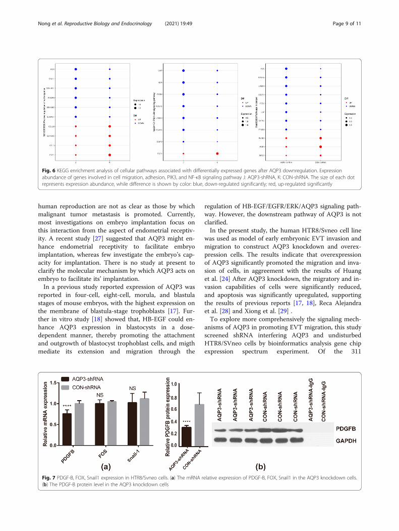
To verify the expression of some differentially expressedgenes selected from the results of whole genomeexpression profileThe results of q-PCR verfication for FDGF-B,FOS andSnail1 showed that, the mRNA expression level ofFDGF-B in AQP3-shRNA group was significantly lowerthan that in CON-shRNA group(P < 0.0001), while FOS,
Snail1 mRNA expression were lower than that of CON-shRNA group, but there were no significant difference(P values were 0.068 and 0.168, respectively) (Fig. 7a).The protein level of FDGF-B was further verified, West-ern blot showed that the FDGF-B protein level inAQP3-shRNA group was significantly lower than that inCON-shRNA group (P < 0.0001) (Fig. 7b).
DiscussionIt is becoming increasingly evident that AQP3 isexpressed in multiple malignant tumor cells [21] andparticipates in both tissue oncogenicity and tumor cellmigration [22–26]. The invasion process of the tropho-blast into the maternal endometrium and metastasis ofmalignant tumors both rely on cell invasion behaviors.However, mechanisms by which AQP3 influences
Fig. 4 Clustering gene expression patterns. J: AQP3-shRNA, K: CON-shRNA. Red represents gene upregulation, while green representsgene downregulation
Fig. 5 GO enrichment analysis of differentially expressed genes after AQP3 downregulation. (a), (b): Red represents result of biologicalenrichment, green represents result of cell component enrichment and blue represents result of molecular function enrichment. (c) Some entriesof the Pathway on KEGG enrichment analysis of differentially expressed genes after down-regulation of AQP3 genes
Nong et al. Reproductive Biology and Endocrinology (2021) 19:49 Page 8 of 11
human reproduction are not as clear as those by whichmalignant tumor metastasis is promoted. Currently,most investigations on embryo implantation focus onthis interaction from the aspect of endometrial receptiv-ity. A recent study [27] suggested that AQP3 might en-hance endometrial receptivity to facilitate embryoimplantation, whereas few investigate the embryo’s cap-acity for implantation. There is no study at present toclarify the molecular mechanism by which AQP3 acts onembryo to facilitate its’ implantation.In a previous study reported expression of AQP3 was
reported in four-cell, eight-cell, morula, and blastulastages of mouse embryos, with the highest expression onthe membrane of blastula-stage trophoblasts [17]. Fur-ther in vitro study [18] showed that, HB-EGF could en-hance AQP3 expression in blastocysts in a dose-dependent manner, thereby promoting the attachmentand outgrowth of blastocyst trophoblast cells, and migthmediate its extension and migration through the
regulation of HB-EGF/EGFR/ERK/AQP3 signaling path-way. However, the downstream pathway of AQP3 is notclarified.In the present study, the human HTR8/Svneo cell line
was used as model of early embryonic EVT invasion andmigration to construct AQP3 knockdown and overex-pression cells. The results indicate that overexpressionof AQP3 significantly promoted the migration and inva-sion of cells, in aggreement with the results of Huanget al. [24] After AQP3 knockdown, the migratory and in-vasion capabilities of cells were significantly reduced,and apoptosis was significantly upregulated, supportingthe results of previous reports [17, 18], Reca Alejandraet al. [28] and Xiong et al. [29] .To explore more comprehensively the signaling mech-
anisms of AQP3 in promoting EVT migration, this studyscreened shRNA interfering AQP3 and undisturbedHTR8/SVneo cells by bioinformatics analysis gene chipexpression spectrum experiment. Of the 311
Fig. 6 KEGG enrichment analysis of cellular pathways associated with differentially expressed genes after AQP3 downregulation. Expressionabundance of genes involved in cell migration, adhesion, PIK3, and NF-κB signaling pathway J: AQP3-shRNA, K: CON-shRNA. The size of each dotrepresents expression abundance, while difference is shown by color: blue, down-regulated significantly; red, up-regulated significantly
Fig. 7 PDGF-B, FOX, Snail1 expression in HTR8/Svneo cells. (a) The mRNA relative expression of PDGF-B, FOX, Snail1 in the AQP3 knockdown cells.(b) The PDGF-B protein level in the AQP3 knockdown cells
Nong et al. Reproductive Biology and Endocrinology (2021) 19:49 Page 9 of 11

differentially expressed genes screened, 150 were upreg-ulated and 161 were downregulated. GO analysis andGO enrichment analysis found that these genes aremainly involved in angiogenesis, cell migration, inflam-matory response, cell adhesion, extracellular matrix re-combination. Bioinformatics analysis of gene chipexpression profiling data reveals that key genes relatedto migration, such as platelet-derived growth factor-B(PDGF-B), Snai1, and FOS, were notably down-regulatedafter AQP3 knockdown expression.Further q-PCR validation of these two key genes re-
vealed that PDGF-B mRNA expression was prominentlydownregulated after knockdown expression, while Snai1,FOS mRNA expression was non-significantly downregu-lated. PDGFB protein levels were also observablydownregulated.PDGF is a dimeric molecule existing as homodimers
or heterodimers of related polypeptide chains (A and B).PDGF-B is a member of the PDGF family, which has theability to link with cystine and play a crucial role in de-velopment, cell proliferation, cell survival, and angiogen-esi s[30]. PI3K/AKT, JNK, and PLCγ pathways wereinvolved in the process of PDGF-B binding to PDGFRβand inducing receptor dimerizatio n[31]. Schwenke et.al[32]. found that PDGF-B trigger undirected motility inendometrial stromal cells, while pathway inhibitor-basedstudies have shown that ERK1/2, PI3K/Akt and p38 sig-naling are associated with chemotactic motility, whereaschemokines (PDGF-B) are mainly dependent on PI3 kin-ase/Akt activation. Jing et al .[33] found that PDGFBand PI3K/AKT signaling pathways have co-expressionnetworks together with the false detection rate is verylow, and PDGFB promote the metastasis of oral squa-mous cell carcinoma through the PI3K/AKT signalingpathway. In this study, the differentially expressed genesscreened KEGG pathway analysis are mainly involved intumor-related pathways, MAPK pathways, PI3K-AKTpathways, and NF-B signaling pathways. PI3K-AKTpathways have also been identified as closely related totumor cell migration. Therefore, we speculate thatAQP3 may play a role by acting on PDGF-B gene andPI3K/AKT signaling pathways that mediate the migra-tion and invasion of extravillous trophoblastic cells,thereby mediating embryo implantation. However, fur-ther investigation is needed to confirm this conjecture.
ConclusionsCollectively, these results reveal that AQP3 is an import-ant positive regulatory factor for fetal-maternal crosstalkduring the first trimester of pregnancy, whereby it mayact on PDGF-B gene to promote migration and invasionability of trophoblast cells, the underlying mechanismstill requires a further investigation.
AbbreviationsAQP3: Aquaporin3; RIF: Recurrent implantation failure; EVT: Extravilloustrophoblasts; EMT: Epithelial-mesenchymal transition; HB-EGF: Heparin-binding epidermal growth factor-like growth factor
Supplementary InformationThe online version contains supplementary material available at https://doi.org/10.1186/s12958-021-00726-z.
Additional file 1. Supplementary table. Functional categories ofselected genes differentially expressed in the HTR8/Svneo cells AQP3-shRNA and CON-shRNA.
AcknowledgementsWe are particularly grateful to the Medical Transformation Center ofGuangdong Women and Children Hospital for technical assistance.
Authors’ contributionsNong YQ designed the study and contributed to article revision. Li SFperformed the research and contributed to writing the article. They sharefirst authorship. Liu WJ, Fan L, Chen Y and Huang QW contributed toexperiment design and figures performed. Zhang XQ and Zhang QY wereresponsible for analysis of data. Liu FH designed the work, providedtechnical guidance and final approved of manuscript. All authors read andapproved the final manuscript.
FundingThis study was supported by Natural Science Foundation of GuangdongProvince, China (No. 2016A030313817), Science and Technology Program ofGuangzhou, China (201704020217), and the In-Hospital Training Project ofthe National Natural Science Foundation of Guangdong Women and Chil-dren Hospital, China (No. YN2017G10).
Availability of data and materialsNot applicable.
Declaration
Ethics approval and consent to participateThis study was approved by the Ethics Committee of Guangdong Womenand Children Hospital and has been performed in accordance with theprinciples of Declaration of Helsinki.
Consent for publicationNot applicable.
Competing interestsThe authors have no conflict of interest to declare.
Author details1The First Affiliated Hospital of Jinan University, Guangzhou, China.2Department of Reproductive Health and Infertility, Guangdong Women andChildren Hospital, Guangzhou, Guangdong, China. 3Reproductive MedicineCenter, Affiliated Shenzhen City Maternity and Child Healthcare Hospital ofSouthern Medical University, Shenzhen, China.
Received: 4 September 2020 Accepted: 24 February 2021
References1. Coughlan C. What to do when good-quality embryos repeatedly fail to
implant. Best Pract Res Clin Obstet Gynaecol. 2018;53:48–59.2. Shohayeb A, El-Khayat W. Does a single endometrial biopsy regimen (S-EBR)
improve ICSI outcome in patients with repeated implantation failure? Arandomised controlled trial. Eur J Obstet Gynecol Reprod Bio. 2012;164:176–9.
3. Achache H, Revel A. Endometrial receptivity markers, the journey tosuccessful embryo implantation. Hum Reprod Update. 2006;12:731–46.
4. Burrows TD, King A, Loke YW. Trophoblast migration during humanplacental implantation. Hum Reprod Update. 1996;2:307–21.
Nong et al. Reproductive Biology and Endocrinology (2021) 19:49 Page 10 of 11

5. Cross JC, Werb Z, Fisher SJ. Implantation and the placenta: key pieces of thedevelopment puzzle. Science. 1994;266:1508–18.
6. Murray MJ, Lessey BA. Embryo implantation and tumor metastasis: commonpathways of invasion and angiogenesis. Semin Reprod Endocrinol. 1999;17:275–90.
7. Verkman AS, Mitra AK. Structure and function of aquaporin water channels.Am J Physiol Renal Physiol. 2000;278:F13–28.
8. Maggio M, et al. Association of hormonal dysregulation with metabolicsyndrome in older women: data from the InCHIANTI study. Am J PhysiolEndocrinol Metab. 2007;292:E353–8.
9. Dong C, Aznavoorian S, Liotta LA. Two phases of pseudopod protrusion intumor cells revealed by a micropipette. Microvasc Res. 1994;47:55–67.
10. Verkman AS. More than just water channels: unexpected cellular roles ofaquaporins. J Cell Sci. 2005;118:3225–32.
11. Steffens S, et al. Low dose oral cannabinoid therapy reduces progression ofatherosclerosis in mice. Nature. 2005;434:782–6.
12. Xiong Y, et al. Expression of aquaporins in human embryos and potentialrole of AQP3 and AQP7 in preimplantation mouse embryo development.Cell Physiol Biochem. 2013;31:649–58.
13. King LS, Kozono D, Agre P. From structure to disease: the evolving tale ofaquaporin biology. Nat Rev Mol Cell Biol. 2004;5:687–98.
14. Ducza E, Csanyi A, Gaspar R. Aquaporins during pregnancy: their functionand significance. Int J Mol Sci. 2017;18.
15. Martinez N, Damiano AE. Aquaporins in fetal development. Adv Exp MedBiol. 2017;969:199–212.
16. Escobar J, et al. Expression of aquaporins early in human pregnancy. EarlyHum Dev. 2012;88:589–94.
17. Nong YQ, et al. The expression and distribution of aquaporin 3 in mouseembryos before and after vitrification. J Assist Reprod Genet. 2013;30:601–6.
18. Fang CX, et al. Heparin-binding epidermal growth factor-like growth factorenhances aquaporin 3 expression and function during mouse embryoimplantation. Reprod Sci. 2017;24:463–70.
19. Zhang Q, et al. Expression of CD82 in human trophoblast and its role introphoblast invasion. PLoS One. 2012;7:e38487.
20. Alekseev OM, et al. Analysis of gene expression profiles in HeLa cells inresponse to overexpression or siRNA-mediated depletion of NASP. ReprodBiol Endocrinol. 2009;7:45.
21. Marlar, S., et al., Aquaporin-3 in Cancer. Int J Mol Sci. 2017;18(10).22. Hara-Chikuma M, Verkman AS. Prevention of skin tumorigenesis and
impairment of epidermal cell proliferation by targeted aquaporin-3 genedisruption. Mol Cell Biol. 2008;28:326–32.
23. Hara-Chikuma M, Verkman AS. Aquaporin-3 facilitates epidermal cellmigration and proliferation during wound healing. J Mol Med (Berl). 2008;86:221–31.
24. Huang YT, et al. Identification of estrogen response element in Aquaporin-3gene that mediates estrogen-induced cell migration and invasion inestrogen receptor-positive breast Cancer. Sci Rep. 2015;5:12484.
25. Chen J, et al. Aquaporin 3 promotes prostate cancer cell motility andinvasion via extracellular signal-regulated kinase 1/2-mediated matrixmetalloproteinase-3 secretion. Mol Med Rep. 2015;11:2882–8.
26. Hou SY, et al. Aquaporin-3 inhibition reduces the growth of NSCLC cellsinduced by hypoxia. Cell Physiol Biochem. 2016;38:129–40.
27. Cui D, et al. Aquaporin-3 mediates ovarian steroid hormone-inducedmotility of endometrial epithelial cells. Hum Reprod. 2018;33:2060–73.
28. Alejandra R, Natalia S, Alicia ED. The blocking of aquaporin-3 (AQP3) impairs extravilloustrophoblast cell migration. Biochem Biophys Res Commun. 2018;499(2):227–32.
29. Xiong G, et al. RNA interference influenced the proliferation and invasion ofXWLC-05 lung cancer cells through inhibiting aquaporin 3. BiochemBiophys Res Commun. 2017;485(3):627–34.
30. Andrae J, Gallini R, Betsholtz C. Role of platelet-derived growth factors inphysiology and medicine. Genes Dev. 2008;22:1276–312.
31. Honda M, et al. Mesothelioma cell proliferation through autocrine activationof PDGF-betabeta receptor. Cell Physiol Biochem. 2012;29:667–74.
32. Schwenke M, et al. Control of human endometrial stromal cell motility byPDGF-BB, HB-EGF and trophoblast-secreted factors. PLoS One. 2013;8:e54336.
33. Jing Y, et al. SPARC promotes the proliferation and metastasis of oral squamous cellcarcinoma by PI3K/AKT/PDGFB/PDGFRbeta axis. J Cell Physiol. 2019.
Publisher’s NoteSpringer Nature remains neutral with regard to jurisdictional claims inpublished maps and institutional affiliations.
Nong et al. Reproductive Biology and Endocrinology (2021) 19:49 Page 11 of 11