toward understanding respiratory sinus arrhythmia: relations
TRANSCRIPT

www.elsevier.com/locate/biopsycho
Biological Psychology 74 (2007) 263–285
Toward understanding respiratory sinus arrhythmia: Relations
to cardiac vagal tone, evolution and biobehavioral functions
Paul Grossman a,*, Edwin W. Taylor b
a Department of Psychosomatic and Internal Medicine, Psychophysiology Research Laboratory,
University of Basel Hospital, Hebelstrasse 2, CH-4031 Basel, Switzerlandb School of Biosciences, University of Birmingham, Birmingham, United Kingdom
Accepted 16 November 2005
Available online 1 November 2006
Abstract
Respiratory sinus arrhythmia (RSA, or high-frequency heart-rate variability) is frequently employed as an index of cardiac vagal tone or even
believed to be a direct measure of vagal tone. However, there are many significant caveats regarding vagal tone interpretation:
1. R
*
e.w
030
doi
espiratory parameters can confound relations between RSA and cardiac vagal tone.
2. A
lthough intraindividual relations between RSA and cardiac vagal control are often strong, interindividual associations may be modest.3. R
SA measurement is profoundly influenced by concurrent levels of momentary physical activity, which can bias estimation of individualdifferences in vagal tone.
4. R
SA magnitude is affected by beta-adrenergic tone.5. R
SA and cardiac vagal tone can dissociate under certain circumstances.6. T
he polyvagal theory contains evolution-based speculations that relate RSA, vagal tone and behavioral phenomena. We present evidence thatthe polyvagal theory does not accurately depict evolution of vagal control of heart-rate variability, and that it ignores the phenomenon of cardiac
aliasing and disregards the evolution of a functional role for vagal control of the heart, from cardiorespiratory synchrony in fish to RSA in
mammals.
Unawareness of these issues can lead to misinterpretation of cardiovascular autonomic mechanisms. On the other hand, RSA has been shown to
often provide a reasonable reflection of cardiac vagal tone when the above-mentioned complexities are considered. Finally, a recent hypothesis is
expanded upon, in which RSA plays a primary role in regulation of energy exchange by means of synchronizing respiratory and cardiovascular
processes during metabolic and behavioral change.
# 2006 Elsevier B.V. All rights reserved.
Keywords: Cardiac vagal control; Cardiac vagal tone; Respiratory sinus arrhythmia; Heart-rate variability; Parasympathetic; Vagus; Autonomic; Cardiovascular;
Evolution; Respiration; Polyvagal theory
1. Introduction
Respiratory sinus arrhythmia (RSA) is a cardiorespiratory
phenomenon characterized in mammals by heart rate (HR) or
R-R interval (RRI) fluctuations that are in phase with inhalation
and exhalation. Typically, HR accelerates during inspiration
and slows down during expiration, but the exact phase
relationship between respiratory and HR oscillations is
Corresponding author.
E-mail addresses: [email protected] (P. Grossman),
[email protected] (E.W. Taylor).
1-0511/$ – see front matter # 2006 Elsevier B.V. All rights reserved.
:10.1016/j.biopsycho.2005.11.014
dependent upon the prevailing respiration rate (Eckberg,
1983). Furthermore, even when autonomic tone remains stable,
the amplitude of these rhythmic HR fluctuations (i.e. the
magnitude of RSA) is greatly dependent upon both respiratory
frequency and depth of ventilation (i.e. tidal volume; Hirsch
and Bishop, 1981). The central, neural, humoral and mechan-
ical feedback mechanisms that together generate RSA are a
complex of integrated respiratory and cardiovascular responses
(Grossman, 1983; Jordan and Spyer, 1987; Spyer, 1990).
Therefore, RSA must be conceptualized as a phenomenon that
directly results from the interaction between the cardiovascular
and respiratory systems.

P. Grossman, E.W. Taylor / Biological Psychology 74 (2007) 263–285264
RSA has been shown to importantly reflect rhythmic waxing
and waning of cardiac vagal efferent effects upon the sinoatrial
node and, therefore, HR (Eckberg, 2003; Hedman et al.,
1995b). The relationship between RSA and vagal control of HR
has generated great interest among scientists who wish to
explore and exploit noninvasive estimates of cardiovascular
autonomic control. Along a related line, evidence that RSA
magnitude in humans is sometimes predictive of both
physiologic and psychological morbidity has also engendered
research focused upon using RSA more as a marker of risk than
as an index of discrete parasympathetic cardiac control (e.g.
Bigger et al., 1992; Hayano et al., 1990; Janszky et al., 2004;
Kluge et al., 1988; Nishimura et al., 2004).
Whatever the basic motivation for investigation, a firm
understanding of what RSA is – and what RSA is not – would
seem essential. All too commonly, a thorough grasp of RSA
appears missing in the literature, and this has led to
contradiction, confusion, misinterpretation and misattribution
with respect to research findings and appropriate methods of
measurement and analysis. The major purposes of this article
are (1) to clarify the nature of RSA, (2) to elucidate certain
misconceptions regarding RSA, and (3) to elaborate upon a new
theoretical model that integrates RSA and biobehavioral
functioning. In this model, RSA plays a significant role in
coordinating physical energy requirements and continuously
changing behavioral activities.
RSA – in addition to being simply employed as a cardiac
vagal index – has become embedded in a theoretical framework
of evolutionary, biological and psychobiological adaptation.
Therefore, we will address both proximal concerns about its
accuracy as a parasympathetic measure and broader aspects of
its functional roles and its evolutionary origins in non-
mammalian vertebrates. Specifically, we will discuss the
biological function that RSA is likely to serve in coordinating
and maintaining interplay between the respiratory and
cardiovascular systems, which are together responsible for
meeting metabolic demands over a range of highly variable
internal and external conditions. Both respiratory and
cardiovascular processes are responsive not only to gross
metabolic demands but also to levels of alertness and, in
humans at least, different types of emotion, mental activity and
arousal (and the latter may only minimally or not at all change
metabolism). Coupling of respiratory and cardiovascular
systems, consequently, are likely to be pertinent to psycholo-
gical and behavioral variations, as well as physiological state.
Our arguments fall under two themes: (1) clarification of the
relationships between RSA and vagal tone in mammals and
particularly humans, and (2) the evolution of central, vagal
control of cardiorespiratory interactions in vertebrates. With
respect to evolutionary issues, we will also critique the
polyvagal theory (Porges, 1995), a currently popular view
based upon assumptions about the evolution of the autonomic
nervous system. The polyvagal theory attempts to introduce an
evolutionary perspective into relations between parasympa-
thetic activity and behavior and to explain situations in which
changes in RSA clearly do not correspond to alterations in
vagally mediated HR (i.e. cardiac vagal tone; Porges, 1995,
2001, 2003b). The theory maintains that RSA is generated in
functionally distinct vagal systems that first evolved in the
brainstem of mammals (Porges, 1995, 2003b). In recent years,
this theory has been expanded to encompass a wide range of
postulates regarding physical, psychophysiological and even
social functioning in humans (e.g. Porges, 2003b; Sahar et al.,
2001).
The following six points will guide the structure of our
presentation:
1. R
espiratory parameters of rate and volume can confoundrelations between RSA and cardiac vagal tone.
2. A
lthough within-subject relations between RSA and cardiacvagal control are often strong (when properly measured),
between-subject associations may be relatively weak.
3. R
SA measurement is strongly influenced by concurrentlevels of momentary physical activity, which can bias
estimation of individual differences in vagal tone.
4. R
SA amplitude is affected by beta-adrenergic tone and maynot be a pure vagal measure.
5. R
SA and cardiac vagal tone may dissociate under certaincircumstances.
6. B
asic assumptions of the polyvagal theory regarding RSAare at odds with current knowledge of the neuroanatomical
and functional evolution of cardiac vagal control.
RSA can be quantified in a number of different ways, most
commonly including spectral analysis, time-domain peak-
valley analysis or application of a band-pass filter. Units of
measurement can also consequently vary. For time-domain
measures, RSA is typically estimated in ms (e.g. the
inspiratory–expiratory difference in RRI). With spectral
analysis and other frequency-domain approaches, the variation
of RRI occurring within the range of the respiratory frequency
is estimated; thus ms2 is frequently employed, consistent with
usual statistical units of variance. Often RSA measures are
logarithmically transformed to normalize distribution, but this
is not always the case. Because different methods are almost
perfectly correlated with each other when properly employed
(Grossman et al., 1990b), we will not detail quantification
methods when reviewing the literature, except when it might be
pertinent to a specific topic. A fuller treatment of measurement
issues is beyond the scope of this article.
2. Respiratory confounds in RSA estimation of cardiac
vagal tone
Numerous studies have documented the effects upon RSA of
voluntary and spontaneous changes in respiration rate and tidal
volume under steady-state conditions and during mental tasks.
Steady state, in this context, connotes conditions during which
metabolic activity and autonomic tone remain largely constant.
A sample of these studies include the following: Ahmed et al.
(1982, 1986), Althaus et al. (1998), Angelone and Coulter
(1964), Badra et al. (2001), Ben Lamine et al. (2004), Brown
et al. (1993), Clynes (1960), Cooke et al. (1998), Eckberg
(1983, 2003), Eckberg et al. (1984), Grossman (1992),

P. Grossman, E.W. Taylor / Biological Psychology 74 (2007) 263–285 265
Grossman et al. (1991, 2004), Grossman and Kollai (1993),
Hayano et al. (1994), Hedman et al. (1995a), Hirsch and Bishop
(1981), Patwardhan et al. (1995), Penttila et al. (2001),
Poyhonen et al. (2004), Ritz et al. (2001), Saul et al. (1989),
Schipke et al. (1999), Scott et al. (2004), Selman et al. (1982),
Stark et al. (2000), Strauss-Blasche et al. (2000), Taylor et al.
(2001b), and Wilhelm et al. (2004).
Results across studies are highly consistent and very similar,
no matter how RSA is quantified (e.g. including the most
common methods of spectral analysis, peak-valley analysis or
digitally filtered time-domain procedures). RSA magnitude
under steady-state conditions is inversely related to respiration
rate and directly related to tidal volume. Hence, rapid shallow
breathing will greatly attenuate RSA, and slow deep breathing
will produce maximal RSA levels. Respiration rate and tidal
volume produce independent and interactive effects (e.g.
Hirsch and Bishop, 1981). For example, increases of tidal
volume at slower respiration rates will cause larger RSA
elevations than the same tidal volume increases at more rapid
respiration rates, although natural changes in respiration and
tidal volume are usually reciprocal at stable levels of metabolic
activity. Furthermore, these respiratory influences are sizeable
within the normal physiological range of respiration rates and
tidal volumes that characterize states of rest and mental or
emotional activation (e.g. Althaus et al., 1998; Bernardi et al.,
2000; Grossman et al., 1991). Therefore, an increase of only 3–
4 breaths/min from rest to mental task may attenuate RSA
magnitude substantially. Consequently, RSA magnitude under
conditions of mental activation or emotion can be significantly
altered solely as a function of those respiratory changes.
Measurements of RSA magnitude are commonly taken to
indicate the level of cardiac vagal tone. This, as defined and
understood in cardiovascular physiology, is the mean vagal
Fig. 1. Mean respiratory sinus arrhythmia (RSA; a) and respiration period (b) across
(base), four paced-breathing phases (pb; 7, 11 and 17 cpm; max = 7 cpm at large tida
pressor (cold), bicycle ergometry at 0, 25, 50, 75, 100 and 125 W (erg), and recovery (
of tasks, suggesting the very close within-individual relationship that was found.
efferent effect upon HR or RRI (Grossman and Kollai, 1993;
Hayano et al., 1991; Kollai et al., 1994). Cardiac vagal tone is
not constant for an individual but changes greatly as a
consequence of age, posture, metabolic activity and other
factors (e.g. Craft and Schwartz, 1995; Robinson et al., 1966).
A number of investigations have shown that pharmacologically
induced changes in cardiac vagal tone can be tracked by RSA
amplitude: thus, stepwise decreases in cardiac vagal tone
produced by the cumulative action of increasing doses of the
muscarinic cholinergic receptor antagonist atropine, or other
vagolytic (i.e. vagally blocking) agents, are accompanied by a
dose-related reduction in RSA magnitude (Dellinger et al.,
1987; Medigue et al., 2001; Pyetan et al., 2003; Raczkowska
et al., 1983; Scheinin et al., 1999). Atropine does not produce a
systematic effect upon respiratory parameters; therefore,
alterations in cardiac vagal tone and RSA during atropine
administration are not accompanied by changes in respiration
rate or tidal volume (Elstad et al., 2001; Grossman and Kollai,
1993; Rauniar et al., 1998).
Respiratory influences upon RSA amplitude become a
problem for assessment of cardiac vagal tone (1) whenever
respiratory rate and/or tidal volume substantially differ between
groups or conditions, and (2) whenever RSA, respiratory
parameters and cardiac vagal tone all do not systematically
covary with each other. We have examined this issue
(Grossman et al., 1991) by adapting the same kind of
pharmacological autonomic blockade paradigm that has long
been employed in physiological studies to characterize
autonomic control during specific challenges such as dynamic
or isometric exercise. To evaluate the degree of covariation of
RSA, respiration and vagal tone, we expanded the task domain
to include mental tasks (a cognitive reaction-time task) and
ventilatory tests (e.g. voluntary hyperventilation and CO2
different behavioral task; data from Grossman et al. (1991): five baseline phases
l volume), hyperventilation (hyp), two cognitive reaction-time phases (rt), cold
recov). Note the similar patterning of respiratory period and RSA over the range

P. Grossman, E.W. Taylor / Biological Psychology 74 (2007) 263–285266
rebreathing): healthy subjects were intravenously administered
a dosage of propranolol sufficient to effectively block
sympathetic, beta-adrenergic influences upon HR. Because
variations in normal HR almost exclusively derive from
sympathetic beta-adrenergic and vagal effects, this meant that
HR changes under pharmacological beta-blockade could be
ascribed solely to alterations in vagal tone. We were able,
therefore, to answer the question of whether changes in RSA
necessarily reflect alterations in cardiac vagal tone when
respiratory rate and tidal volume vary.
Our findings clearly indicated that behaviorally induced
fluctuations in cardiac vagal tone were not discernibly tracked
by changes in RSA magnitude under variable respiration. RSA
magnitude was much more closely related to changes in
respiratory parameters – particularly respiration rate – than to
any changes in cardiac vagal tone (Fig. 1a and b). It was only
when respiratory variables were statistically controlled (using a
within-individual regression approach) that there was a clearly
improved but still imperfect association between RSA and
vagal tone (Fig. 2a–c).
A second autonomic blockade study (Grossman and Kollai,
1993) that manipulated respiratory rate and volume under
individual and dual beta-adrenergic and vagal blockade
provided additional evidence that respiratory variation con-
founds RSA indices of within-subject changes in cardiac vagal
tone. Alterations in respiration rate and tidal volume had
profound effects upon RSA magnitude that were unrelated to
directly determined levels of cardiac vagal tone.
We recently addressed the question of whether, under
relatively constant levels of cardiac autonomic tone, normal
respiratory variations to psychological tasks would signifi-
cantly alter RSA independently of cardiac vagal tone (Gross-
man and Stemmler, in preparation). Within-subject relations
between RSA and respiratory parameters were examined
during performance of a variety of mental tasks during which
Fig. 2. Cardiac vagal tone (a), respiratory sinus arrhythmia adjusted within individua
(Grossman et al., 1991). Cardiac vagal tone (beta-blocked HR) and unadjusted RSA
parameters, shows a clear covariation with changes in cardiac vagal tone.
autonomic tone appeared to be relatively constant. Tasks
included completion of written psychological inventories, two
rest phases, guided meditation, nonstressful speaking, quiet
reading, attention to auditory stimuli (five levels), a memory-
comparison reaction-time task (three levels), a computerized
multiple-choice math task, and a post-task recovery period.
Forty healthy adults (24–40 years) were tested. In order to
create the least stressful environment possible, tasks were
administered in a comfortable, nonclinical setting by an female
assistant chosen and instructed to be very friendly and relaxed
with subjects. We wanted to examine the physiological effects
of discrete mental tasks, themselves, upon cardiorespiratory
variables, relatively unconfounded by emotional stress.
No task-related HR changes were found. Self-reported
negative affect (anxiety and anger/irritation complaints) also
barely changed on a 10-point scale (from a mean low of 0.4
during meditation to a high of 0.9 during stress tasks).
Therefore, results indicated that cardiac autonomic tone and
mood were not significantly altered as a function of task.
Respiratory parameters, however, did reliably vary across
tasks. Both respiration rate and tidal volume showed large
condition effects, as did peak-valley RSA (see Fig. 3a and b).
Using multiple regression with RSA as criterion measure and
respiration rate and tidal volume as predictors, the average
within-subject R was .76, indicating that almost 60% of the
RSAvariance could be accounted for by respiratory parameters.
In the complete absence of parallel HR changes (i.e. no
suggestion of cardiac autonomic change), these findings
underscore the powerful confounding effects of respiratory
parameters on RSA under conditions of psychological
activation.
A recent paper has suggested that respiratory rate and tidal
volume need not be considered in RSA studies of cardiac vagal
control during non-exercise behavioral variations (Houtveen
et al., 2002). However, the single mental stress task employed in
l for respiratory variables (b) and raw unadjusted RSA (c) across conditions from
are obviously not strongly associated. However, RSA, corrected for respiratory
P. Grossman, E.W. Taylor / Biological Psychology 74 (2007) 263–285 267
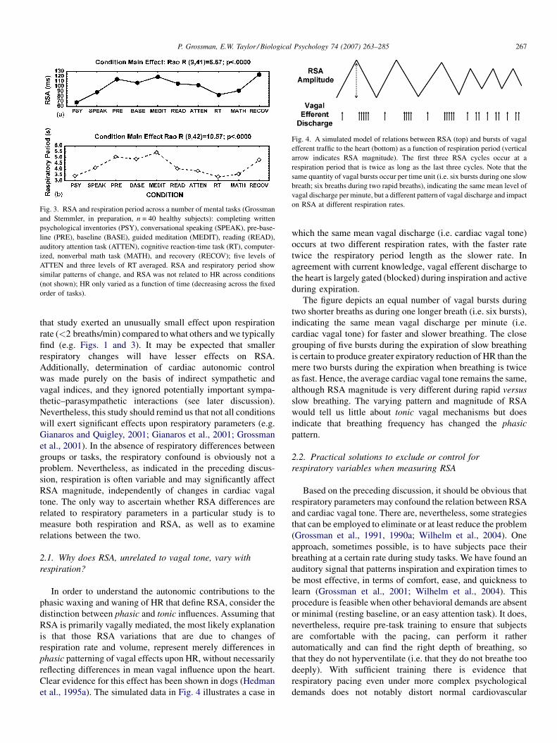
Fig. 3. RSA and respiration period across a number of mental tasks (Grossman
and Stemmler, in preparation, n = 40 healthy subjects): completing written
psychological inventories (PSY), conversational speaking (SPEAK), pre-base-
line (PRE), baseline (BASE), guided meditation (MEDIT), reading (READ),
auditory attention task (ATTEN), cognitive reaction-time task (RT), computer-
ized, nonverbal math task (MATH), and recovery (RECOV); five levels of
ATTEN and three levels of RT averaged. RSA and respiratory period show
similar patterns of change, and RSA was not related to HR across conditions
(not shown); HR only varied as a function of time (decreasing across the fixed
order of tasks).
Fig. 4. A simulated model of relations between RSA (top) and bursts of vagal
efferent traffic to the heart (bottom) as a function of respiration period (vertical
arrow indicates RSA magnitude). The first three RSA cycles occur at a
respiration period that is twice as long as the last three cycles. Note that the
same quantity of vagal bursts occur per time unit (i.e. six bursts during one slow
breath; six breaths during two rapid breaths), indicating the same mean level of
vagal discharge per minute, but a different pattern of vagal discharge and impact
on RSA at different respiration rates.
that study exerted an unusually small effect upon respiration
rate (<2 breaths/min) compared to what others and we typically
find (e.g. Figs. 1 and 3). It may be expected that smaller
respiratory changes will have lesser effects on RSA.
Additionally, determination of cardiac autonomic control
was made purely on the basis of indirect sympathetic and
vagal indices, and they ignored potentially important sympa-
thetic–parasympathetic interactions (see later discussion).
Nevertheless, this study should remind us that not all conditions
will exert significant effects upon respiratory parameters (e.g.
Gianaros and Quigley, 2001; Gianaros et al., 2001; Grossman
et al., 2001). In the absence of respiratory differences between
groups or tasks, the respiratory confound is obviously not a
problem. Nevertheless, as indicated in the preceding discus-
sion, respiration is often variable and may significantly affect
RSA magnitude, independently of changes in cardiac vagal
tone. The only way to ascertain whether RSA differences are
related to respiratory parameters in a particular study is to
measure both respiration and RSA, as well as to examine
relations between the two.
2.1. Why does RSA, unrelated to vagal tone, vary with
respiration?
In order to understand the autonomic contributions to the
phasic waxing and waning of HR that define RSA, consider the
distinction between phasic and tonic influences. Assuming that
RSA is primarily vagally mediated, the most likely explanation
is that those RSA variations that are due to changes of
respiration rate and volume, represent merely differences in
phasic patterning of vagal effects upon HR, without necessarily
reflecting differences in mean vagal influence upon the heart.
Clear evidence for this effect has been shown in dogs (Hedman
et al., 1995a). The simulated data in Fig. 4 illustrates a case in
which the same mean vagal discharge (i.e. cardiac vagal tone)
occurs at two different respiration rates, with the faster rate
twice the respiratory period length as the slower rate. In
agreement with current knowledge, vagal efferent discharge to
the heart is largely gated (blocked) during inspiration and active
during expiration.
The figure depicts an equal number of vagal bursts during
two shorter breaths as during one longer breath (i.e. six bursts),
indicating the same mean vagal discharge per minute (i.e.
cardiac vagal tone) for faster and slower breathing. The close
grouping of five bursts during the expiration of slow breathing
is certain to produce greater expiratory reduction of HR than the
mere two bursts during the expiration when breathing is twice
as fast. Hence, the average cardiac vagal tone remains the same,
although RSA magnitude is very different during rapid versus
slow breathing. The varying pattern and magnitude of RSA
would tell us little about tonic vagal mechanisms but does
indicate that breathing frequency has changed the phasic
pattern.
2.2. Practical solutions to exclude or control for
respiratory variables when measuring RSA
Based on the preceding discussion, it should be obvious that
respiratory parameters may confound the relation between RSA
and cardiac vagal tone. There are, nevertheless, some strategies
that can be employed to eliminate or at least reduce the problem
(Grossman et al., 1991, 1990a; Wilhelm et al., 2004). One
approach, sometimes possible, is to have subjects pace their
breathing at a certain rate during study tasks. We have found an
auditory signal that patterns inspiration and expiration times to
be most effective, in terms of comfort, ease, and quickness to
learn (Grossman et al., 2001; Wilhelm et al., 2004). This
procedure is feasible when other behavioral demands are absent
or minimal (resting baseline, or an easy attention task). It does,
nevertheless, require pre-task training to ensure that subjects
are comfortable with the pacing, can perform it rather
automatically and can find the right depth of breathing, so
that they do not hyperventilate (i.e. that they do not breathe too
deeply). With sufficient training there is evidence that
respiratory pacing even under more complex psychological
demands does not notably distort normal cardiovascular

P. Grossman, E.W. Taylor / Biological Psychology 74 (2007) 263–285268
responses (Grossman et al., 1990a,b), although this approach
may not be easy to achieve, especially with mentally more
demanding tasks.
Objections that paced breathing affects RSA differently than
spontaneous breathing are largely unsupported: the vast
majority of carefully performed investigations support the
idea that relations between RSA and respiratory measures
remain the same under spontaneous and voluntarily paced
respiratory conditions (e.g. Ben Lamine et al., 2004; Bloom-
field et al., 2001; Eckberg et al., 1976; Grossman et al., 1991;
Hirsch and Bishop, 1981; Patwardhan et al., 2001, 1995). This
makes sense, because respiration during awake states is
importantly under the regulation of higher brain centers that
control voluntary behavior (Longobardo et al., 2002). There-
fore, covariation between RSA magnitude and respiratory
parameters during alert states inherently reflects the interaction
of cardiovascular control mechanisms and higher central
nervous system (CNS) behavioral control of breathing.
Another strategy is statistical control for respiratory
parameters, using covariance procedures. This is a frequently
employed approach (e.g. Burleson et al., 2003; Hughes and
Stoney, 2000), but is problematic when a simple between-group
analysis of covariance is performed, with respiratory measures
as covariates: relations between RSA and respiratory measures
are systemic, that is to say that these relationships are typically
very strong within individuals but not between individuals (e.g.
Ben Lamine et al., 2004). This is because (a) the magnitude of
RSA is highly variable between subjects, and (b) the slope of
the regression between RSA and respiratory variables can also
vary greatly from subject to subject, independent of individual
differences in RSA magnitude, even at very similar levels of
within-individual correlation. A normal analysis of covariance
approach pools between- and within-subject variance and can
lead to erroneous conclusions about the contribution of
respiratory parameters to RSA fluctuation, due to violations
of assumptions concerning homogeneous variances/covar-
iances per cell (Browne and Shapiro, 1991). Simulated data
in Fig. 5a–d illustrate this point, by displaying that even with a
large difference between groups in respiratory period response
from baseline to task, analysis of covariance effects for RSA
controlled for respiratory period are still significant; each of the
simulated subjects showed a covariation between RSA and
respiratory period, but the slopes of the within-subject relations
were highly variable within and between cells.
A more appropriate procedure may be a within-subject
approach in which RSA is residualized against respiratory
variables (Grossman et al., 1991). This is performed when
respiration rate or tidal volume changes as a consequence of
experimental conditions or time. Several condition or measure-
ment epochs are needed to compute individual regressions.
Condition levels of RSA are regressed against respiration
variables using a multiple regression analysis, and the residuals
are used as an index of vagal tone (RSA variation dispropor-
tional to respiratory change; positive residuals index increase in
cardiac vagal tone, and negative values reflect vagal with-
drawal; see Fig. 2b). However, this procedure may yield a
somewhat conservative estimate of cardiac vagal control under
circumstances in which respiratory parameters partially covary
with vagal control.
Yet another approach is to pace subjects across the normal
physiological range of respiration rates during a baseline
period. Once again, care must be taken in training subjects to
ensure that subjects remain eucapnic (i.e. do not hyperventilate)
and are not so uncomfortable or distressed by the pacing that
autonomic tone changes. We have found that with clear
instruction, most subjects can ordinarily learn within 10 min to
comfortably pace their breathing at a range of rates, at the same
time maintaining baseline HRs and avoiding hyperventilation.
Also stable estimates of RSA can be acquired with pacings of
about 1–1.5 min per respiratory rate. Therefore, even pacing
across five frequencies can be accomplished without exces-
sively prolonging a protocol. The regression line of RSA on
respiratory parameters can then be used to estimate task-related
RSA changes that systematically exceed or are lower than
expected values (i.e. reflecting cardiac vagal augmentation or
decline, respectively).
Tidal volume is often considered a less important respiratory
variable to control in RSA research (Berntson et al., 1997).
Nevertheless, tidal volume can have marked independent
effects upon RSA magnitude (Hirsch and Bishop, 1981), and
attention to tidal volume may be warranted whenever it changes
substantially and these changes are not tightly reciprocally
related to changes in respiration rate (e.g. Grossman et al.,
1991, 2004; Ritz et al., 2001). Tidal volume can be
nonintrusively assessed rather easily and inexpensively using
air bellows strain gauges (see Morel et al., 1983).
A common approach that adjusts for the influence of tidal
volume upon RSA is to calculate the transfer function (Berger
et al., 1989; Grossman et al., 1991, 2004; Wilhelm et al., 2004).
This procedure derives the gain of RSA related to tidal volume
(ms RSA per liter tidal volume). The measure is simply RSA
divided by tidal volume when time domain-measures are used,
or the transfer function from cross-spectral analysis of the RRI
and respiratory time series when spectral analysis is employed.
In both cases, it characterizes the amount of RSA amplitude
change per liter tidal volume. Due to the reciprocal relation
between rate and tidal volume, this adjustment may dampen or
even eliminate the RSA dependency upon respiration rate: in an
ambulatory study of alert, active subjects (Grossman et al.,
2004), the transfer function was more closely associated with
cardiac vagal tone than raw, unadjusted RSA; additionally
statistical control for respiration rate did not improve the degree
of correlation between the adjusted measure and cardiac vagal
tone. On the other hand, the transfer function measure appears
to remain importantly related to respiration rate under
laboratory conditions (where physical activity does not exert
a large impact on cardiac vagal tone; Berger et al., 1989;
Grossman et al., 1991; Wilhelm et al., 2004). Therefore,
transfer function analysis can be used as an adjustment to
variations in tidal volume, and, perhaps, requires no additional
control for respiration rate when the major focus is upon
metabolically associated changes in cardiac vagal tone (as in
most ambulatory studies). However, this parameter alone does
not appear adequate for evaluating tonic vagal changes under

P. Grossman, E.W. Taylor / Biological Psychology 74 (2007) 263–285 269
Fig. 5. Simulated respiration period and RSA data of two groups (experimental vs. control) during rest and task. RSA and respiration period decreased from rest to
task; the control group showed larger changes (mean changes in a and b). (c) and (d), both individual data, indicate that the control group consistently manifested
larger decreases in respiration period, although the main effect for RSA change was still significant after a group analysis of covariance (ANCOVA) adjustment (see
b). In all cases, RSA decrease was accompanied by respiration period reduction, although, corresponding to real findings, within-individual slopes of the relation were
highly variable.
the typically sedentary circumstances of most psychophysio-
logical or clinical investigations.
The above-described approaches all have methodological
and practical limitations. Furthermore, there is no systematic
evidence about which method is superior for estimation of
cardiac vagal tone. Hopefully enhanced awareness of the
problem of respiratory confounding of vagal tone estimation
will lead to additional research regarding this matter.
3. Individual differences in vagal tone may not always
be reflected by variations of RSA
Studies validating RSA as a within-individual index of
cardiac vagal tone have shown RSA to decrease proportionately
to levels of atropine-induced cardiac vagal withdrawal. Along a
different line, individual differences in cardiac vagal tone
among humans are defined as the decrease in mean RRI (ms),
produced by complete vagal blockade using atropine or some
other vagolytic drug under basal conditions. In other words,
cardiac vagal tone is operationalized as the difference between
the average RRI before atropine infusion and the mean RRI
after maximal dose of atropine (at which HR change is
maximal). Several studies have examined the correlation
between RSA and this pharmacological measure of cardiac
vagal tone, considered the gold standard of estimation in human
cardiovascular physiology (Cacioppo et al., 1994; Fouad et al.,
1984; Grossman and Kollai, 1993; Hayano et al., 1991; Kollai
and Mizsei, 1990; Maciel et al., 1985). All but two studies
(Cacioppo et al., 1994; Maciel et al., 1985) found a significant
correlation between cardiac vagal tone and RSA. However, the
extent of the association varied widely in those investigations
that did find a relationship, with r’s ranging from 0.5 to 0.9. In
the two studies with the highest level of association (both
r = 0.9), methodological issues cast doubt over the actual
strength of the relation between RSA and vagal tone in a
homogeneous population: in one study (Fouad et al., 1984),
hypertensives and normotensives were combined in the data
set. It is well known that hypertension reduces RSA and vagal
tone, and this would serve to exaggerate both the range of
normal variation of measures and the correlation coefficient. A
similar effect could be expected from the other investigation
(Hayano et al., 1991) that pooled half sedentary and half
physically active subjects in the analyzed sample (physically
active individuals often show higher levels of RSA, e.g. Dixon
et al., 1992; Shin et al., 1997). The other larger and homoge-
neous sample of young healthy subjects (Grossman and Kollai,
1993; Kollai and Mizsei, 1990) reported much more modest
correlations between 0.5 and 0.6, indicating that only 1/4 to 1/3
of the individual variation in cardiac vagal tone could be
explained by RSA alone, even when respiratory parameters
were controlled. However, the inclusion of HR as an additional
predictor increased explained variance to 76% (r = 0.9).
Taken together, these studies indicate that caution is
warranted when employing RSA as an index of individual
differences in cardiac vagal tone. There is apparently a
relationship, but it has not been demonstrated to be close
enough to assume RSA to be more than roughly associated with
individual variations in cardiac vagal tone. In fact, in one set of
studies, resting HR, in comparison to RSA, was much more
highly correlated to cardiac vagal tone (Grossman and Kollai,
1993; Kollai and Mizsei, 1990); a later report of a small sample
of female students yielded similar results (Cacioppo et al.,
1994). These findings suggest the utility of including both
variables whenever predicting individual differences in
cardiac vagal tone. They also suggest that baseline HR may
be as good or better an index of individual differences in

P. Grossman, E.W. Taylor / Biological Psychology 74 (2007) 263–285270
cardiac vagal tone than RSA. This evidence has long been
neglected in the literature and may have implications for our
understanding of RSA and autonomic control. Exactly what
individual differences in RSA represent, thus, remains an
intriguing question.
4. Concurrent physical activity alters cardiac vagaltone and RSA
Recent evidence (Bernardi et al., 1996; Grossman et al.,
2004) shows that accurate estimation of RSA is seriously biased
by variations in concurrent physical activity. Heart rate during
mild-to-moderate change in physical activity, characteristic of
normal variations of daily activity, is predominantly under
parasympathetic control (Boushel et al., 2001; Ekblom et al.,
1972; Epstein et al., 1965; Grossman et al., 1991; Hopkins
et al., 2003; Janicki et al., 1996; Maciel et al., 1986; O’Leary
and Seamans, 1993; Robinson et al., 1966, 1953; Rowell and
O’Leary, 1990; Vatner and Pagani, 1976). Thus, disease-
specific, temperamental or other psychological effects upon
range, frequency and duration of daily activity may interact
with and confound the assessment of individual differences in
autonomic regulation and RSA during ambulatory monitoring.
Markers of cardiac vagal activity may reflect not only
individual differences in constitutional parasympathetic control
but also variations in daily activity pattern. Fig. 6 illustrates the
extent of effects of concurrent activity upon RSA magnitude
among young, healthy adults (Grossman et al., 2004), displayed
as a function of quintiles of physical activity during awake
hours. There is a systematic decrease in RSA as activity
increases.
Although it seems clear that certain clinical groups may
differ in activity from healthy individuals, it is less obvious that
even small differences of activity in the laboratory or field may
also have significant effects upon RSA magnitude: we found in
Fig. 6. RSA magnitude (ln spectral power) as a function of quintile of physical
activity (based on minute ventilation, Vmin) during awake ambulatory recording
over 12 h in 40 healthy subjects (from Grossman et al., 2004). Quintiles were
determined by evenly distributing activity levels over time (i.e. the highest
quintile being equivalent to mean RSA for that 20% of the day during which
Vmin was highest; this analysis was made for each subject, then averaged over all
subjects). RSA varied lawfully across all levels of activity, as indicated by the p
values.
the same study that the distinction in activity level in the lowest
two quintiles was extremely subtle (see Fig. 7). Nevertheless,
RSA differed markedly and very reliably between these lowest
quintiles of activity (see Fig. 6). Therefore, it is plausible that
even small laboratory differences in movement during baseline
measurement (i.e. frequent postural shifts, tapping or even
subtle limb or body motion) may produce effects upon RSA that
could be wrongly inferred as evidence of constitutional
differences in autonomic control. Simultaneous monitoring
of activity in field, experimental and clinical settings may,
therefore, be required when examining group differences in
RSA, especially when group effects, as so often the case, are
significant but relatively small in magnitude.
5. RSA is affected by sympathetic tone and may not be
a ‘pure’ vagal index
As previously mentioned, within-individual validation
studies of RSA are based upon evidence of changes in RSA
during progressive pharmacological blockade of cardiac
parasympathetic control (Ali-Melkkila et al., 1991; Coker
et al., 1984; Dellinger et al., 1987; Hayano et al., 1991; Julu and
Hondo, 1992; Medigue et al., 2001; Pyetan et al., 2003;
Raczkowska et al., 1983; Scheinin et al., 1999). In almost all
studies, an exponential reduction of RSA (quantified in
different ways) is found as HR rises. In other words, HR
increases as a function of progressive attenuation of vagal tone,
and RSA tracks that change (exponentially) and is, conse-
quently, assumed to be a noninvasive marker of the changing
mean level of cardiac vagal tone.
HR fluctuations, as previously noted, are under the joint
control of sympathetic beta-adrenergic and parasympathetic
branches of the autonomic nervous system. In some of these
validation investigations, pretreatment with a beta-adrenergic
blocking drug was performed to prevent cardiac sympathetic
influences from affecting the findings (e.g. Coker et al., 1984;
Hayano et al., 1991; Medigue et al., 2001). Sympathetic
blockade made little difference, in terms, of the relative
relations found between RRI and RSA during progressive vagal
Fig. 7. Physical activity levels (Vmin) across the quintiles (Grossman et al.,
2004), but not reported there); quintile calculation described in Fig. 6. Note that
physical activity is relatively sedentary during normal awake functioning,
especially at the lowest two quintiles.

P. Grossman, E.W. Taylor / Biological Psychology 74 (2007) 263–285 271
withdrawal, attesting to the parasympathetic effects of the
vagolytic drugs (e.g. Grossman and Kollai, 1993). However, an
important finding in a large number of dual cardiac blockade
investigations (vagal and sympathetic) was that beta-adrenergic
blockade augmented RSA under baseline conditions (e.g.
Eckberg et al., 1976; Coker et al., 1984; Grossman and Kollai,
1993; Pitzalis et al., 1998; Taylor et al., 2001b), unrelated to
respiratory alteration. This effect was often relatively large
(RSA increases typically �30%) and was independent of
whether the beta-blocker was cardiac-specific (i.e. affected
only the periphery) or had central effects because the drug
crossed the blood–brain barrier (Pitzalis et al., 1998). Even in
small-sample studies in which this effect was not significant,
mean RSA tended to increase under beta-blockade, e.g.
Cacioppo et al. (1994). For example, beta-blockade during
baseline sitting in the latter investigation induced a mean RSA
increase of 65% when reported natural log units were
transformed to ms2 spectral power (28% if converted to time
domain measures).
Beta-adrenergic effects upon RSA magnitude are relevant for
physiological and psychophysiological studies, because the
change in cardiac sympathetic tone (often about 150 ms RRI or
10 bpm HR; e.g. Cacioppo et al., 1994) is within the range of
what can be expected in laboratory situations or among different
clinical samples. The finding of sympathetic effects upon RSA
magnitude points to a serious problem of intra-individual
validation studies. Namely, throughout the validation literature,
the covarying RSA and RRI decline during vagal withdrawal has
always been examined within the context of stable levels of
cardiac sympathetic tone (either during or without beta-
blockade). These investigations have provided necessary but
insufficient evidence for the general utility of RSA as a cardiac
vagal index: they showed that RSAwas sensitive to variations in
cardiac vagal tone when cardiac sympathetic tone was absent, or
was relatively low and stable. However, they failed to confirm the
parasympathetic specificity of RSA, i.e. that no matter how
cardiac sympathetic activity changes, RSA always specifically
reflects cardiac vagal control. Beta-adrenergic effects upon RSA
challenge the vagal specificity of RSA.
There are several possible interpretations of these findings:
(a) cardiac vagal tone is directly altered by varying levels of
sympathetic effects upon HR; (b) RSA, but not cardiac vagal
tone, is influenced by changes in cardiac sympathetic tone; or
(c) vagal-sympathetic interactions can occur that alter the
relation between RSA and cardiac vagal tone in predictable or
unpredictable ways. Whichever in the end proves correct, any
of these possibilities, at the very least, clearly indicates that
concurrent levels of cardiac sympathetic activity can alter
respiratory modulation of HR (i.e. RSA).
One invasive dog study (Hedman et al., 1995b), indeed, may
confirm that RSA, but not mean level of cardiac vagal efferent
activity (vagal outflow from the brain to the heart), is altered by
beta-adrenergic blockade: sympathetic and vagal nerves to the
heart were electrically stimulated after their pathways from the
brain were severed. Vagal nerve impulses were rhythmically
stimulated so as to simulate RSA patterns at a frequency of
12 cpm, and this patterning of impulses was presented both in
the presence and in the absence of mild-to-moderate
sympathetic stimulation. In comparison to a background of
no sympathetic activity, even mild sympathetic stimulation
drastically attenuated RSA, despite the fact that mean levels of
efferent vagal traffic to the heart remained constant.
What seems clear from this and other investigations is that
even mild levels of sympathetic activity can substantially
depress RSA, as a function of either sympathetic–vagal
interactions or a direct suppressive effect upon RSA magnitude.
In human studies, there are indications that the influence of
beta-blockade on RSA can be removed or greatly attenuated by
a simple procedure that normalizes RSA for mean RRI (Hayano
et al., 1990):
RSAnorm
¼
ffiffiffiffiffiffiffiffiffiffiffiffiffiffiffiffiffiffiffiffiffiffiffiffiffiffiffiffiffiffiffiffiffiffiffiffiffiffiffiffiffiffiffiffiffiffiffiffiffiffiffiffiffiffiffiffiffiffiffiffiffiffiffiffiffiffiffiffiffiffiffiffiffiffiffiffiffiffiffiffiffiffiffiffiffiffiffiffiffiffiffiffiffiffiffiffiffiffiffiffiffiffiffiffiffiðspectral power of high-frequency RRI power ðms2ÞÞ
mean RRI ðmsÞ
s
In the case of time-domain peak-valley amplitude measures
(which are linearly related to the square root of the spectral
power; Laude et al., 1995), the peak-valley measure is divided
by RRI.
This normalized index has been found to be more sensitive
to incremental vagal blockade than more commonly used
indices (e.g. the spectral power uncorrected) in one sophisti-
cated and well-documented pharmacological investigation
(Scheinin et al., 1999). Therefore, we explored whether this
RSA measure, which corrects for changes in mean RRI, would
be less sensitive to beta-adrenergic effects than uncorrected
measures of RSA.
We found one study that reported beta-blockade to have no
effect upon this measure (Schachinger et al., 2001). Addition-
ally, we calculated this normalized index from mean group data
in three studies that had reported clear effects of beta-blockade
upon RSA (Grossman and Kollai, 1993; Pitzalis et al., 1998;
Taylor et al., 2001b). Beta-blocker effects were almost
completely removed (all differences between beta-blockade
and no blockade <10%). This was also confirmed in data
recalculated from our own study (Grossman and Kollai, 1993),
which found a 30% increase in peak-valley RSA with beta-
blockade before normalizing, and a 5% difference after
normalizing for mean RRI.
These findings suggest that RRI correction of RSA can
reduce or eliminate the influence of basal levels of cardiac
sympathetic tone (resting HR with versus without the normally
mild level of sympathetic activity). In the autonomic
stimulation study described above (Hedman et al., 1995b),
normalized RSA also substantially lowered the impact of
sympathetic stimulation: with a constant level of vagal efferent
discharge, normalized RSA was reduced 50% during sympa-
thetic stimulation, compared to no sympathetic stimulation;
without correction, RSA was reduced by a factor of 12.
5.1. RSA and vagal–sympathetic interactions
In light of preceding conclusions, it may be useful to
consider more precisely the meaning of the term cardiac vagal

P. Grossman, E.W. Taylor / Biological Psychology 74 (2007) 263–285272
tone. Many scientists assume that cardiac vagal tone reflects the
extent of central vagal discharge to the heart, but the term is
actually operationalized as the mean level of cardiac vagal
effects upon HR over a defined interval of time. This definition
focuses upon the final vagal effects upon HR. Vagally mediated
HR changes depend, in turn, upon a number of prior processes
and events.
Limiting discussion to the efferent side of vagal neural
transmission, the vagus nerve must first transmit efferent
activity in the direction of the sinoatrial node, located in the
posterior wall of the heart’s right atrium. In healthy individuals,
the sinoatrial node initiates each beat of the heart, and vagal
effects on HR are contingent upon the release of acetylcholine
from the parasympathetic nerve endings at junctures to the
sinoatrial node. Any process that interferes with acetylcholine
transmission can alter the relationship between central vagal
efferent traffic and its effect on HR. For example, as dosage
rises, atropine appears to progressively increase average central
vagal efferent traffic to the heart, while, at the same time, it
blocks the action of acetylcholine at the periphery, i.e. at the
sinoatrial node (Katona et al., 1977). The net result at higher
doses of atropine is complete vagal blockade – elimination of
cardiac vagal tone – despite elevated, mean levels of central
vagal efferent activity.
Findings from the previously described experimental study
(Hedman et al., 1995b) showed that mild-to-moderate
stimulation of cardiac sympathetic activity greatly attenuated
beat-to-beat modulation of RRI (simulated RSA) in dogs,
although average level of vagal efferent traffic to the heart
remained constant during both the absence and presence of
sympathetic stimulation. Perhaps related to the effects of the
latter study, numerous investigations have demonstrated that
neuropeptide Y, an important neurotransmitter, inhibits the
release of acetylcholine at parasympathetic effector junctions of
the heart, itself (including the sinoatrial node), and thereby
reduces effectiveness of vagal action on the heart (e.g. Moriarty
et al., 1993a,b; Serone and Angus, 1999; Smith-White et al.,
1999, 2002; Warner and Levy, 1989).
In mammals, it has long been recognized that the cardiac
response to neural activity in one autonomic division depends
on levels of activity in the other division (Levy, 1984). Complex
peripheral interactions between sympathetic and parasympa-
thetic nerve supplies to the heart are of importance in
modulating control of cardiac function. Many of the terminal
fibers of the two subdivisions of the autonomic nervous system
lie close to each other on the mammalian heart (Jacobowitz,
1967). Consequently, transmitters released from the nerve
endings of one division can readily diffuse to the nerve
terminals of the other, as well as to the cells of the cardiac
ganglion and the myocardium (Revington and McCloskey,
1990).
These and many others studies illustrate that what we
measure with any HR index of vagal tone are only the final
functional vagal effects on cardiac activity. Attenuation of
cardiac vagal tone may be caused by reduced central vagal
efferent activity, blocking of preganglionic or postganglionic
neuronal actions of acetylcholine, impaired vagus nerve
conduction, or combined mechanisms. A pronounced decrease
in RSA magnitude may therefore signify (1) true reduction of
vagal outflow from brain to heart, (2) a primary increase in
sympathetic tone that leads to an interaction with vagal activity
in the periphery, or (3) both. Such considerations underline the
difficulties in inferring CNS mechanisms whenever there is a
reduction in RSA, even when respiratory parameters are
controlled and when RSA does reflect some aspect of
parasympathetic modulation.
These interactions, nevertheless, may not always be
apparent in pharmacological blockade studies (e.g. Berntson
et al., 1994; Cacioppo et al., 1994). However, autonomic
blockade studies, such as the latter, in which sympathetic and
vagal influences are eliminated one at a time or jointly, may not
be informative about interactions between the two autonomic
branches when both are active to varying degrees. Interactions
also depend upon the extent of activation of both autonomic
branches (Levy, 1984). For example, in the previously
described canine study (Hedman et al., 1995b), substantial
RSA attenuation was found at mild to moderate levels of
cardiac sympathetic stimulation. On the other hand, under
conditions during which sympathetic stimulation is consis-
tently very modest (e.g. estimated sympathetic effects upon
RRI � 30 ms, in Berntson et al., 1994), autonomic interactions
may play an insignificant role.
To further complicate the matter, there are numerous ways in
which autonomic blocking drugs may potentially influence
estimations of cardiac autonomic tone: drugs like atropine and
different beta-blockers may sometimes have central effects, as
well as induce indirect, secondary effects due to distressing
symptoms. However, the use of double blockade studies would
seem to eliminate such sources of bias when only two
influences can be responsible (e.g. vagal and sympathetic
effects upon HR). Moreover, sequential studies of dual vagal
and beta-sympathetic blockade do not indicate that central or
indirect concomitants of autonomic blockade significantly bias
estimates of cardiac autonomic tone (e.g. Kollai et al., 1994).
Nevertheless, further research may still be useful to examine
such possible influences upon pharmacological determination
of cardiac parasympathetic and sympathetic tone.
Whatever the explanation for occasionally disparate find-
ings, the repeated evidence of beta-sympathetic influence upon
RSA and peripheral sympathetic–parasympathetic interactions
should be of concern for both physiological and psychophy-
siological investigations of RSA and cardiac vagal control. This
does not mean we should cease employing RSA as a marker of
final vagal effects upon the heart. However, it does imply that
variations in RSA magnitude currently provide an unreliable
index of central vagal outflow or tone. The implications for
interpretation of past and present RSA research are far-
reaching.
6. RSA and cardiac vagal tone can dissociate under
certain circumstances
There is evidence that RSA magnitude can sometimes
dissociate from cardiac vagal tone, even under conditions of no

P. Grossman, E.W. Taylor / Biological Psychology 74 (2007) 263–285 273
apparent sympathetic–vagal interaction and when respiration is
controlled. One example replicated in several studies is when
extreme levels of cardiac vagal tone are provoked by
pharmacological enhancement of the cardiac baroreflex
(Goldberger et al., 1994, 1996, 2001). The baroreflex is a
cardiovascular feedback system with a primary function to
stabilize blood pressure within a certain range. An important
component of the baroreflex is a vagally mediated slowing of
HR when blood pressure rises above a set point (slowing the
heart will reduce levels of arterial blood flow and consequently
blood pressure). Therefore, phenylephrine, a drug that raises
blood pressure produces increased cardiac vagal tone in a dose–
response manner, i.e. HR progressively slows as a result of
enhanced vagal tone. The relation between RSA and vagal tone
(vagal HR change) is relatively proportional and linear until HR
slows down to very low levels. As HR further decreases, so does
magnitude of RSA (e.g. Goldberger et al., 2001). The relation
between RSA and vagal tone is quadratic across the entire range
of vagally mediated HR change (see Fig. 8). Why this happens
is unclear but may have something to do with saturation of the
vagal effects across the respiratory cycle, loss of phasic
respiratory changes in vagal nerve discharge, or a simple floor
effect in which minimal HR has no room left to oscillate.
Another instance of dissociation between cardiac vagal tone
and RSA appears to occur during stimulation of the carbon
dioxide chemoreceptors (involved in the control of respiration;
Sasano et al., 2002; Yasuma and Hayano, 2001; Yasuma et al.,
2001). When high levels of inspired CO2 stimulate these
receptors, RSA increases disproportionately to changes in
cardiac vagal tone (or respiratory variables; also see the CO2
rebreathing condition of Fig. 2a and b; Grossman et al., 1991). On
the basis of this and other evidence, it has been suggested that the
primary adaptive function of this RSA augmentation during
elevated levels of inspired and arterial CO2 is to expel excess
levels of CO2 from pulmonary circulation, thus enhancing
pulmonary gas exchange (Hayano and Yasuma, 2003).
Fig. 8. Dissociation between RSA and cardiac vagal tone during baroreflex
stimulation (from Goldberger et al., 2001). RR-interval alteration reflects
pharmacologically induced (phenylephrine) change in cardiac vagal tone as
a consequence of baroreflex-mediated parasympathetic responses; RSA (y axis)
is the natural logarithm of high-frequency HR variability (ln HF) in bpm2. There
is a quadratic relation between RSA and cardiac vagal tone that flattens
somewhat with increasing age (to be reprinted with permission).
These and other investigations (e.g. infant RSA during
attentional responses; Richards and Casey, 1991) point to
specific conditions during which RSA is not an accurate index
of cardiac vagal tone. It should be noted, however, that these
instances often seem to be at the outer range of normal
physiological functioning (see Fig. 8). In the baroreflex studies
described above, the association between RSA and vagally
induced HR change was directly proportional until HR fell to
approximately 40 bpm, clearly at the bottom range of HR seen
during normal physiological states. Likewise, the elevated
levels of CO2 in the second set of experiments could only be
expected during respiratory distress approaching asphyxiation.
Therefore, these studies, by themselves, do not severely
compromise RSA as an index of cardiac vagal tone within a
wide spectrum of normal physiological functioning. However,
the findings once again underline the fact that RSA is not
synonymous with vagal tone and point to the risks in assuming
that RSA variations necessarily reflect cardiac vagal tone in
every circumstance.
The preceding discussion highlighted a number of issues that
should be considered when employing RSA as an index of
cardiac vagal tone. A substantial body of literature documents
the value of RSA, although it is often replete with contrasting
findings. Our preceding review should not be taken to negate
the utility of RSA as a marker of vagal control. To the contrary,
we hope it many be used as a guide to improve understanding
and measurement of RSA and cardiac vagal activity. With these
aims in mind, Table 1 provides a list of possible solutions to the
various concerns and caveats we have here raised.
7. A critique and an alternative to the polyvagal theory
As stated earlier, the polyvagal theory was first proposed in
1995 (Porges, 1995, 2001, 2003b) as an attempt to (a) introduce
an evolutionary perspective into relations between parasympa-
thetic activity and behavior (possibly on the basis of a
phylogenetic overview of vagal control of the heart in
vertebrates reviewed by Taylor, 1994, as suggested by Medigue
et al., 2001), and (b) to explain situations in which changes in
RSA clearly do not correspond to alterations in vagally
mediated HR (i.e. cardiac vagal tone). In recent years, this
theory has been expanded to encompass a wide range of
postulates regarding physical, psychophysiological and even
social functioning, including speculations regarding various
disorders including asthma, autism, and post-traumatic stress
disorder (e.g. Porges, 2003b; Sahar et al., 2001).
At the core of the polyvagal theory lie several assumptions
regarding two separate populations of vagal nuclei that reside in
the brainstem – the ventrally located nucleus ambiguus (nA)
and the dorsal motor nucleus (DMN) – and their role in
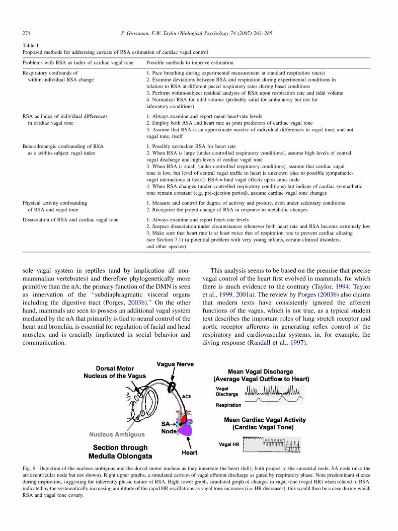
vertebrate evolution. Fig. 9 provides a cartoon of the two
brainstem nuclei, their cardiac projections, and the functional
respiratory gating of central vagal outflow that characterizes
RSA. The polyvagal theory maintains that these areas not only
represent functionally distinct vagal systems in the mammal but
also that they can be clearly characterized in terms of their
evolution (Porges, 1995, 2003b): the DMN is proposed as the
P. Grossman, E.W. Taylor / Biological Psychology 74 (2007) 263–285274
Table 1
Proposed methods for addressing caveats of RSA estimation of cardiac vagal control
Problems with RSA as index of cardiac vagal tone Possible methods to improve estimation
Respiratory confounds of
within-individual RSA change
1. Pace breathing during experimental measurement at standard respiration rate(s)
2. Examine deviations between RSA and respiration during experimental conditions in
relation to RSA at different paced respiratory rates during basal conditions
3. Perform within-subject residual analysis of RSA upon respiration rate and tidal volume
4. Normalize RSA for tidal volume (probably valid for ambulatory but not for
laboratory conditions)
RSA as index of individual differences
in cardiac vagal tone
1. Always examine and report mean heart-rate levels
2. Employ both RSA and heart rate as joint predictors of cardiac vagal tone
3. Assume that RSA is an approximate marker of individual differences in vagal tone, and not
vagal tone, itself
Beta-adrenergic confounding of RSA
as a within-subject vagal index
1. Possibly normalize RSA for heart rate
2. When RSA is large (under controlled respiratory conditions), assume high levels of central
vagal discharge and high levels of cardiac vagal tone
3. When RSA is small (under controlled respiratory conditions), assume that cardiac vagal
tone is low, but level of central vagal traffic to heart is unknown (due to possible sympathetic-
vagal interactions at heart): RSA = final vagal effects upon sinus node
4. When RSA changes (under controlled respiratory conditions) but indices of cardiac sympathetic
tone remain constant (e.g. pre-ejection period), assume cardiac vagal tone changes
Physical activity confounding
of RSA and vagal tone
1. Measure and control for degree of activity and posture, even under sedentary conditions
2. Recognize the potent change of RSA in response to metabolic changes
Dissociation of RSA and cardiac vagal tone 1. Always examine and report heart-rate levels
2. Suspect dissociation under circumstances whenever both heart rate and RSA become extremely low
3. Make sure that heart rate is at least twice that of respiration rate to prevent cardiac aliasing
(see Section 7.1) (a potential problem with very young infants, certain clinical disorders,
and other species)
sole vagal system in reptiles (and by implication all non-
mammalian vertebrates) and therefore phylogenetically more
primitive than the nA; the primary function of the DMN is seen
as innervation of the ‘‘subdiaphragmatic visceral organs
including the digestive tract (Porges, 2003b).’’ On the other
hand, mammals are seen to possess an additional vagal system
mediated by the nA that primarily is tied to neural control of the
heart and bronchia, is essential for regulation of facial and head
muscles, and is crucially implicated in social behavior and
communication.
Fig. 9. Depiction of the nucleus ambiguus and the dorsal motor nucleus as they in
atrioventricular node but not shown). Right upper graphs, a simulated cartoon of va
during inspiration, suggesting the inherently phasic nature of RSA. Right lower gra
indicated by the systematically increasing amplitude of the rapid HR oscillations as
RSA and vagal tone covary.
This analysis seems to be based on the premise that precise
vagal control of the heart first evolved in mammals, for which
there is much evidence to the contrary (Taylor, 1994; Taylor
et al., 1999, 2001a). The review by Porges (2003b) also claims
that modern texts have consistently ignored the afferent
functions of the vagus, which is not true, as a typical student
text describes the important roles of lung stretch receptor and
aortic receptor afferents in generating reflex control of the
respiratory and cardiovascular systems, in, for example, the
diving response (Randall et al., 1997).
nervate the heart (left); both project to the sinoatrial node, SA node (also the
gal efferent discharge as gated by respiratory phase. Note predominant silence
ph, simulated graph of changes in vagal tone (vagal HR) when related to RSA,
vagal tone increases (i.e. HR decreases); this would then be a case during which

P. Grossman, E.W. Taylor / Biological Psychology 74 (2007) 263–285 275
Fig. 10. The proportion of VPN located in ventro-lateral locations outside the
DMN in a range of vertebrates. In mammals this location is chiefly the nAwith a
few cells scattered in the reticular formation. In ‘‘lower’’ vertebrates a discrete
area similar to the nA exists in birds and some amphibians and reptiles while in
fish and other reptiles the ventro-lateral cells have a more scattered distribution
across the medulla. The neotenous axolotl has no cells outside the DMN but
15% following induced metamorphosis (see text). The location of CVPN is
known for a small number of species. The proportion of CVPN located outside
the DMN is similar to or much larger than the proportion of VPN in this
location. This implies that two locations for CVPN in the brainstem have early
origins in vertebrate phylogeny, linked to a significant functional role that we
postulate is the generation of cardio-respiratory coupling.
An additional and very essential implicit premise of the
polyvagal theory is that RSA always reflects tonic vagal
efferent discharge originating in the nA, and that dissociation
between vagally mediated HR changes and RSA can be
explained by divergent activities of the nA and the DMN. The
theory assumes that nA activity is at all times associated with
beat-to-beat changes in HR, i.e. RSA, whereas the DMN
produces flat changes in HR that show no respiratory
modulation or any other type of beat-to-beat variation.
Therefore, according to this theory, vagally mediated HR
reductions that are not accompanied by RSA increase are
supposed to originate from the DMN; changes in RSA
magnitude, on the other hand, from the nA. Hence the accuracy
of RSA as a measure of mean levels of nA-generated vagal
efferent discharge is critical to the theory, because RSA is the
vagal measure used in studies to evaluate premises and
postulates of the theory.
The polyvagal theory has been very broadly stated in terms
of premises, extrapolations and speculations. Yet the above-
mentioned major assumptions provide a basis by which we may
evaluate the very fundament of the theory. There are a number
of lines of evidence that challenge this foundation of the
polyvagal theory. Several of these arguments relate to points
already discussed. Others concern questionable inferences
made in the polyvagal theory regarding the evolution of the
vertebrate parasympathetic nervous system. Each of these
points is addressed below.
7.1. Evolution of vagal control of cardiorespiratory
interactions in vertebrates
7.1.1. Evolution of the nA and two sites for cardiac vagal
preganglionic neurons
The polyvagal theory first proposed that the nA regulation of
cardiac vagal tone is only to be found in mammals (Porges,
1995): ‘‘Reptiles, unlike mammals, have only the older vagal
system.’’ Later this was qualified (Porges, 2003b): ‘‘In general,
phylogenetic development results in increased neural control of
the heart via the myelinated mammalian vagal system [i.e.
nucleus ambiguus].’’ This assumption, in either form, appears
to be unfounded.
It seems that a dual location for vagal preganglionic neurons
(VPN) has important functional correlates in all vertebrates
(Taylor, 1994; Taylor et al., 1999, 2001a). This may be
particularly the case with the central vagal control exerted over
the heart by cardiac vagal preganglionic neurons (CVPN). About
30% of VPN but up to 70% CVPN are in the nA of mammals
where inhibitory inputs from neighboring inspiratory neurons are
the primary central mechanism generating RSA (Jordan and
Spyer, 1987). There is a similar proportional representation of
VPN between the major vagal nuclei in amphibians and turtles.
When stimulated to metamorphose by injection of thyroxine, the
neotenous axolotl shows a doubling of numbers of VPN and
relocation of 15% into a ventrolateral group outside the DMN
that may constitute a primitive nA (Taylor et al., 2001a,b) This
change is accompanied by an increased HRV (Taylor and
Choudhury, unpublished observations). In fish and crocodilians
the proportion of VPN in the nA is closer to 10% and in some
lizards and birds it is 2–5%. However, the CVPN are distributed
unequally between these nuclei so that 45% of CVPN are located
in the nA of the dogfish; and about 30% in Xenopus and the duck
(Taylor et al., 2001a; see Fig. 10). This topographical separation
of CVPN seems to be of importance in the central control of the
heart. Cells in one location may show respiration-related activity
(e.g. those in the DMN of dogfish and in the nA of mammals,
where they are in close proximity to respiratory neurons), while
cells in the other location are sporadically active. Their different
activities and separate functions will be determined by their
different afferent inputs from the periphery or elsewhere in the
CNS, which in turn will relate to their central topography.
What emerges from this brief overview of the vertebrates is
that separation of VPN and in particular CVPN into two major
nuclei seems virtually ubiquitous and has a long evolutionary
history (Taylor et al., 2001a). Its functional significance is the
subject of debate, with elasmobranch fish providing a viable
model for studying basic mechanisms of cardiac vagal control
(Taylor, 1989).
7.1.2. Relationships between ventilatory and heart rates
The polyvagal theory has suggested that the beat-to-beat
control of HR that generates RSA is restricted to mammals,
which have evolved myelinated vagal pathways that originate
in the nA (Porges, 1995, 2003a,b). This idea is not supported by
existing research.
In fish there is a close matching of the rates of respiratory
water flow and cardiac output, according to their relative
P. Grossman, E.W. Taylor / Biological Psychology 74 (2007) 263–285276
capacities for oxygen (the ventilation/perfusion ratio), that is
thought to optimize respiratory gas exchange over the
functional counter-current at the gills (Hughes and Shelton,
1962; Piiper and Scheid, 1977; Taylor, 1992). As both water
and blood flow have been shown to be pulsatile over the gills
(e.g. Jones et al., 1974), close beat-to-beat temporal relation-
ships between heart beat and ventilation, or cardiorespiratory
synchrony (CRS), has long been hypothesized for fish (Satchell,
1960). CRS has been reported in both resting dogfish (Taylor,
1992) and hypoxic trout (Randall and Smith, 1967). Cardiac
vagotomy or injection of atropine abolished CRS in the dogfish
(Taylor, 1992) and HR variability in the unanaesthetized trout
(Le Mevel et al., 2002). In the sculpin, Myoxocephalus
scorpius, injection of atropine raised mean HR in normoxia and
abolished a hypoxic bradycardia (Taylor et al., 2006), and
cardiac vagotomy abolished HR variability (Campbell et al.,
2004). These observations confirm the dependence of beat-to-
beat variability of HR on tonic vagal control in these non-
mammalian vertebrate species.
Peripheral, phasic electrical stimulation of a cardiac vagus in
the dogfish (an elasmobranch) can recruit the heart, so that it
beats at a frequency determined by the rate of the phasic bursts
of stimuli (Taylor et al., 2006). This frequency can be either
higher or lower than the intrinsic HR (Young et al., 1993).
Current work on pacu (a Brazilian teleost) has shown that the
heart can be paced by peripheral stimulation of the cardiac
vagus and by central stimulation of respiratory branches of the
vagus (Taylor et al., unpublished observations). Thus, in both of
these two major groups of fish, the generation of CRS is likely
to depend on a combination of central, feed-forward and reflex,
receptor-based control (Taylor et al., 1999).
Recordings from the central cut ends of cardiac nerves in
decerebrate and paralyzed dogfish showed two different forms
of activity, comprising smaller units that fired sporadically or
continuously but without any clear pattern and larger units
showing respiration related, bursting activity (Barrett and
Taylor, 1985b,c; Taylor, 1992; Taylor and Butler, 1982). These
different units were shown to originate from CVPN having
separate locations in the medulla (Barrett and Taylor, 1985a,c).
Central recordings revealed that the sporadically active units
arose from a clearly distinguishable lateral group of VPN that
contribute axons solely to the cardiac branch of the vagus
(Barrett and Taylor, 1985c). These CVPN comprise 8% of the
total population of VPN but supply 45% of vagal preganglionic
output to the heart and cardiac ganglion in the dogfish. About
9% of VPN are in a scattered ventrolateral location in the ray,
Raja clavata, and they are also solely CVPN, innervating the
heart via a single pair of cardiac vagi (J.J. Levings and E.W.
Taylor, unpublished).
Thus in elasmobranch fishes, a very ancient group (dating
back some 400 million years), CVPN are in two locations; in
the DMN, where they are contiguous with and possibly directly
influenced by respiratory vagal motor-neurons (RVM), gen-
erating respiration-related activity, and in a ventrolateral
location, outside the DMN, possibly homologous to and even
the evolutionary antecedent of the mammalian nA, where they
are less likely to be influenced by the respiration-related
activity in RVM. Stimuli that caused a marked bradycardia,
such as hypoxia or capsaicin (the latter an airway irritant, which
stimulates J-receptors innervated by pulmonary C-fibers in
mammals), induced increased activity in the sporadically active
units, thus increasing cardiac vagal tone (Taylor, 1992). When
vagal tone is relatively low, in settled normoxic or hyperoxic
fish (Taylor, 1992), firing rates in the sporadically active units
were reduced while the respiration related activity in the larger
units continued. This bursting activity may then recruit HR,
generating CRS. This implies that reflex control of HR in
response to environmental change is influenced by the
sporadically active units while cardiorespiratory synchrony
can be generated in the dogfish by central, feed-forward control
from units showing respiration-related activity, arising from the
central respiratory pattern generator (CRPG). The dogfish heart
lacks sympathetic innervation; however, vagal tone on the heart
is influenced by levels of circulating catecholamines, with
normal resting levels augmenting vagal tone and elevated levels
associated with hypoxia or disturbance antagonizing vagal tone
(Agnisola et al., 2003). Thus, similar complex interactions
between cholinergic (i.e. parasympathetic) and adrenergic (i.e.
sympathetic) control of the heart as have been reported in
mammals already exist in this primitive group of vertebrates.
In trout, CRS developed during progressive hypoxia, as HR
slowed to match ventilation, and could be generated by
pulsatile forced ventilation, independently of central respira-
tory rhythmicity, presumably due to reflex control, arising from
stimulation of branchial mechanoreceptors (Randall and Smith,
1967). Thus, fundamentally different mechanisms may underlie
the generation of CRS in dogfish and trout (Taylor et al., 1999).
However, both species have CVPN located both in the dorsal
motor nucleus of the vagus (DMN) and in a ventrolateral
location outside the DMN that may constitute a primitive nA
(Taylor, 1992).
7.1.3. Myelinated versus unmyelinated vagal efferent fibers
In contra-distinction to mammals, the interaction between
respiratory neurons and CVPN occurs in the DMN of the
dogfish rather than in the equivalent of the nA of the mammal.
This difference probably reflects the migration of respiratory
neurons into dorsal and ventral groups during the evolution of
the lung and aspiratory air-breathing. In mammals, CVPN in
the DMN have unmyelinated fibers while those in the nA have
myelinated fibers. It is these latter neurons that are thought to
generate RSA, possibly dependent on their faster conduction
velocities. This is the root of the polyvagal theory, which
postulates that the nA and the associated myelinated fibers are a
late innovation, coming with evolution of the mammals.
However, all fibers in the branchial cardiac nerves of the
dogfish are myelinated (Short et al., 1977) with conduction
velocities varying between 7 and 35 m/s�1 (Barrett and Taylor,
1985c). These velocities fall within the range of mammalian B
fibers (e.g. Katona et al., 1970) and definitely above the range
for unmyelinated C fibers, reported by Bennett et al. (1985).
The faster conduction velocity of mammalian fibers may
relate in part to the high body temperature and to faster HRs of
these animals. However, beat-by-beat modulation of HR may

P. Grossman, E.W. Taylor / Biological Psychology 74 (2007) 263–285 277
Fig. 11. Simulated examples of RSA (a) and cardiac aliasing (b). Solid line,
respiration; dotted line, HR; CVE, simulated cardiac vagal efferent discharge.
Note the clear phase relation in (a) between respiratory phase and HR; this
example represents the minimal condition in which there are just two beats per
respiratory cycle. In (b), aliasing occurs because HR approximates respiration
rate; there is little more than a single beat during each respiratory cycle making
it impossible for RSA to occur. Also note the changing (and drifting) phase
relation between signals in (b). This can happen even when vagal efferent
discharge is phasically related to respiration (see CVE).
be possible in fish, despite the somewhat slower conduction
velocities of the vagal fibers innervating the heart, because of
their relatively slower HRs. Additionally, there is also evidence,
even in mammals, that the DMN is capable of generating beat-
to-beat modulation of HR: dorsal vagal neurons in the rabbit
apparently manifest conduction properties that are indistin-
guishable from nA vagal neurons, including expiratory-related
firing patterns (Jordan et al., 1982).
7.1.4. Mistaken inferences when heart rate is not at least
double the respiration rate—aliasing and the Nyquist
frequency
One-to-one CRS is not the only possible temporal
interaction between HR and ventilation rate. For many years
researchers seeking piscine CRS reported complex and often
drifting phase relationships between HR and ventilatory
frequency (Satchell, 1960; Taylor and Butler, 1971). These
relationships were often subject to relatively complex analysis
(e.g. Hughes, 1972), but the techniques of power spectral
analysis were not available to fish physiologists of that
generation. Interest in HR variability (HRV) research increased
once it became clear that in both the time and frequency
domains, oscillations in cardiac interval generated by
sympathetic, parasympathetic and circadian inputs could be
detected. Power spectral statistics has, of course, become well
established for research and clinical application in mammals
and especially in humans.
The few power spectral studies undertaken on non-
mammalian vertebrates have shown interspecific differences,
with lizards and some fish having dual spectral peaks
(Campbell et al., 2004; De Vera and Priede, 1991; Gonzalez
Gonzalez and De Vera Porcell, 1988; Le Mevel et al., 2002)
whilst other fish have a single main component (Altimiras et al.,
1995, 1996; Armstrong and Priede, 1988). These components
have been characterized as of a relatively low frequency, well
below that of the ventilation rate. For example, spectral analysis
of instantaneous ECG in the sculpin produced dual spectral
peaks at 0.018 and 0.05 Hz in the frequency domain, equivalent
to periodicities of 55 and 20 s, respectively, in the time domain.
As ventilation occurred every 3.3 � 0.2 s (0.30 Hz), it was
about six times faster than the highest-frequency component
(0.05 Hz). It was concluded that this component probably did
not represent centrally generated respiration-related activity,
analogous to the RSA described in recordings of HRV in
mammals, and consequently that the sculpin did not show CRS
(Campbell et al., 2004).
However, one very important point that has gone overlooked
in the vertebrate literature is that RSA is only possible when HR
is at least twice as fast as the ventilation (i.e. respiration rate see
Fig. 11). Two cardiac intervals (RRIs) are required for each
breath in order to have a characteristic waxing and waning of
HR with each respiratory cycle. In other words, at the very
minimum, there must be a longer RRI associated with
expiratory phase and a shorter one associated with inspiratory
phase. This minimum two-to-one relationship is known as the
Nyquist frequency. Therefore, if ventilation rate is greater than
one-half the HR, another phenomenon occurs that is termed
aliasing: it is no longer possible to meaningfully calculate RSA
because there are less than two beats per breath, and any
possible synchrony between respiration period and RRI must be
evaluated over a longer duration than the single breath. If one
knows exactly what the cardiac frequency and the ventilation
frequency are for an animal, then the exact aliasing frequency
can be determined as the difference between the sampled signal
(HR) and the sampling frequency (respiration rate; Mintchev
et al., 2000).
Application of the Nyquist criterion and an anti-aliasing
digital filter to the data from Campbell et al. (2004) revealed
that HRV in the sculpin included a component that appeared to
be synchronous with the respiratory rhythm (Taylor et al.,
2006), so that, contrary to previous analysis of these data, a
respiratory coupling may indeed contribute to HRV in fish and
perhaps other lower vertebrates (see Fig. 12). Campbell et al.
(2004) noted that as rates of oxygen uptake (MO2) in the
sculpin fell during recovery from surgery, a time-domain index
of HRV (standard deviation of successive RRIs) mirrored this
change more closely than the progressive reduction in HR.
Reexamination of their data, using anti-aliasing techniques has
led them to conclude that HRV was an important component of
the mechanisms optimizing respiratory gas exchange (Taylor
et al., 2006).
This problem of cardiac aliasing has also been thoroughly
documented and discussed in the human infant HRV literature
for over a decade (Rother et al., 1989; Witte et al., 1988;
Zwiener et al., 1994, 1995, 1990) but has been completely
ignored in the discussion of the polyvagal theory. As we shall
see, aliasing may lead to important misinterpretations regarding
the polyvagal theory.
In a study comparing two types of lizards (Porges et al.,
2003), the authors concluded on the basis of RSA findings that
vagal tone deriving from the nA is tied to the evolution of

P. Grossman, E.W. Taylor / Biological Psychology 74 (2007) 263–285278
Fig. 12. Power spectra calculated from IBIs of a resting sculpin Myocephalus
scorpious; only the amplitude and frequency of major peaks displayed. Peaks of
HR variability are apparent in the calculated spectra at 0.018 and 0.05 Hz. The
Nyquist criterion (N) shows limit of spectral window set by the mean HR
frequency ( fh). The actual frequency component set by ventilation rate ( fv)
would be folded back as the aliased component (A). In M. scorpious (Campbell
et al., 2004), the recorded mean RRI was 2.85s thus producing an actual
sampling frequency ( fa) of 0.35 Hz, and consequently a Nyquist limit (N) of
0.175 Hz. The fish was observed to ventilate its gills every 3.33 � 0.2 s
( fs = 0.3 Hz). Therefore, any spectral component at the frequency of ventilation
would appear above the Nyquist criterion and outside the limits of the spectral
bandwidth. The consequence of this is a true component (0.018 Hz) close to the
aliased, folded-back component (0.05 Hz). When an anti-aliasing filter (a low
pass bandwidth filter at the limit of Nyquist) was applied to the spectra
calculated for M. scorpious this peak at 0.05 Hz was abolished confirming
that it was aliased, but the low frequency peak at 0.018 Hz was unaffected,
confirming that it is a true component in the spectra and perhaps corresponds to
adrenergic effects as described by De Vera and Priede (1991).
certain aspects of social behavior among reptiles. However,
data presented in the tables and figures of the article clearly
indicate that aliasing occurred during measurement. Conse-
quently, RSA could not have possibly been identified in the
presumably ‘less vagally and socially evolved’ species of lizard
because the respiration rate approximated the HR (greater than
0.5 the cardiac frequency). Nevertheless, examination of the
spectral analysis data suggests that beat-to-beat cardiorespira-
tory synchrony did, in fact, occur. Thus, due to the aliasing,
there is no basis to believe that the mere numerical relationship
between respiration rate and HR should be meaningful in terms
of the evolution of cardiac vagal control.
Both reptiles and amphibians regularly show marked
changes in HR associated with periodic breathing. Modulation
of HR by ventilation can be clearly seen in species that show
prolonged periods of regular breathing, particularly when this is
at a markedly slower rate than HR (e.g. Wang et al., 2001). In a
recent study on the rattlesnake, Crotalus durissus, long-term
measurements of HR, using data-loggers in undisturbed and
resting snakes, clearly documented an oscillatory component in
the HRV signal at the frequency of ventilation (Campbell et al.,
2006). These oscillations resemble the rise in HR during
inspiration in mammals, and are very likely an example of
reptilian RSA. RSA in rattlesnakes was most pronounced at low
HRs and was diminished when HR was increased due to
spontaneous activity, handling stress or recovery from
instrumentation. Cholinergic tone is highest at low HRs in
reptiles (Wang et al., 2001), and as in mammals, the incidence
of RSA seems to require a relatively high vagal tonus on the
heart. This interpretation is also consistent with our observation
that the cholinergic antagonist atropine abolished RSA in the
rattlesnakes.
Thus evidence of cardiorespiratory coupling and sometimes
apparent RSA is found throughout the vertebrate literature,
among not only fish but amphibians (Shelton, 1985; Wang
et al., 1999a,b, 2004; West and Burggren, 1983); reptiles
(Birchard and Reiber, 1996; Burggren, 1975; Wang and Hicks,
1996; Campbell et al., 2006), and birds (Butler, 1982; Butler
and Jones, 1997; Butler and Taylor, 1983), as well as, of course,
mammals. These findings do not provide support for the
polyvagal theory. To the contrary, they suggest a clear primacy
of cardiorespiratory coupling in the evolution of cardiac vagal
control. It may well be neglect of the respiratory system within
the polyvagal theory that has led to a series of unsupported
assumptions.
7.1.5. Mammalian evidence of DMN contribution to
cardiac vagal tone
We were unable to discover any publications that document
DMN effects upon mammalian vagally mediated HR during
any conditions other than severe experimental irritation of the
airways, extreme hypoxia, or lung congestion (Jones et al.,
1998; Jordan et al., 1982; Wang et al., 2000). DMN generation
of bradycardia has only been suggested as a response to
stimulation of pulmonary C fibers (also called J receptors),
which do not have a clear role in normal breathing. Existing
research, therefore, suggests that tonic DMN HR alterations are
restricted to physiological circumstances in which ventilation
may be critically compromised by toxic airborne agents, lung
edema or asphyxia (Paintal, 1983). These conditions in no way
correspond to any hypotheses in the polyvagal theory
concerning when DMN influence upon HR may be active,
e.g. during behavioral immobilization. Hence, there appears to
be little or no evidence of DMN effects upon vagal HR under
circumstances that reflect behavioral variation and not
physiological, respiratory emergency.
7.1.6. Accuracy of RSA as a measure of mean level of nA-
generated vagal efferent tone
A clear and fundamental assumption of the polyvagal theory
is that RSA is a precise measure of vagal efferent discharge to
the sinoatrial node emanating from the nA. Several lines of
evidence already reviewed appear to contradict this assump-
tion: (a) Some fish studies mentioned above indicate that a
phenomenon similar or identical to RSA can be produced via
vagal efferent traffic originating in the DMN. (b) Changes in
respiratory pattern can confound any relationship between RSA
magnitude and cardiac vagal tone. (c) RSA magnitude can be
influenced by concurrent changes in sympathetic activity at
constant rates of vagal efferent discharge. (d) Peripheral
blocking of vagal activity at the sinoatrial node can mask

P. Grossman, E.W. Taylor / Biological Psychology 74 (2007) 263–285 279
Fig. 13. Respiratory responses from resting baseline to varying mental physical
tasks (from Grossman and Wientjes, 2001). Arrow up, increase; arrow down
decrease; =, unchanged from baseline; length of arrow proportional to amount
of change. Note that each condition is associated with a distinctive pattern of
respiratory adjustment. Also note changes both respiration rate and tidal volume
even during moderate mental demand.
increased central vagal activity, indicating that, at best, RSA
reflects the final vagal effects upon HR and may not necessarily
be correlated with the amount of central vagal efferent traffic.
(e) RSA may even sometimes dissociate from mean vagal
effects upon HR, in a manner that cannot be explained by the
polyvagal theory (e.g. a quadratic relation over the entire range
of normal vagal heart variation). (f) Beat-to-beat vagal
cardiorespiratory coupling can occur in the absence of RSA
when respiration rate is above the Nyquist frequency. There-
fore, RSA cannot be assumed to be a direct measure of vagal
tone or nA-generated vagal efferent discharge as suggested by
the polyvagal theory, nor is it always a relatively accurate index
of central efferent vagal traffic, or vagal tone, in general, or
cardiac vagal tone, specifically.
The extent to which cardiac vagal tone and RSA are
differentially affected by sympathetic mechanisms remains
unclear, because it unknown whether sympathetic effects upon
RSA reflect true autonomic interactions or only affect the
amplitude of RSA. RSA, however, does not provide a measure
of nA vagal activity. For all these reasons, the pivotal role of
RSA in the polyvagal theory is seriously challenged. Without
an adequate metric to accurately measure some aspect of tonic
vagal control, or nA efferent discharge specifically, the
polyvagal theory would appear to lack basic supporting
evidence for several of its fundamental assumptions.
7.2. RSA and energy regulation during behavioral and
metabolic demands
The evolutionary literature on cardiac vagal control
emphasizes the biological advantage of tight coupling between
ventilation and cardiovascular function in order to achieve
optimal gas exchange and to meet changing metabolic and
behavioral requirements (Taylor et al., 1999; Taylor, 1994;
Barrett and Taylor, 1985b). It is plainly the case that the
respiratory and cardiovascular systems are intimately related
structurally, neurally and functionally, with a common goal of
coordinating and modulating energy expenditure and uptake in
all vertebrates. Within this theoretical framework, it makes sense
to consider that RSA has evolved as a mechanism that contributes
to functional cardiorespiratory interactions (Hayano and
Yasuma, 2003; Hayano et al., 1996; Yasuma and Hayano,
2004). Because the heart is largely under vagal control during the
normal range of metabolic activity, it is also logical to assume
that a covariation of cardiorespiratory coupling, e.g. RSA, CRS
and vagal tone, may often occur. This has, indeed, been found to
be the case even among ‘‘lower’’ non-mammalian vertebrates,
such as the dogfish and the sculpin (Campbell et al., 2004; Taylor
and Barrett, 1985; Taylor et al., 2006). The precise mechanisms
underlying the covariation of cardiorespiratory synchrony and
vagal control may vary from species to species of vertebrates
(Taylor et al., 2001a). At present we can only speculate on how
the dramatic ventilatory adaptations involved in the switch from
aquatic to terrestrial breathing may have influenced the evolution
of mechanisms concerned with cardiorespiratory coupling, but it
seems likely that these ventilatory modifications played a
significant role.
A number of experimental studies have recently begun to
provide support for the hypothesis that RSA serves an active
biological function, in that it enhances the efficiency of
pulmonary gas exchange by matching blood perfusion to air
flow in the lung throughout each breathing cycle, particularly
during rest (Hayano and Yasuma, 2003; Hayano et al., 1996;
Sasano et al., 2002; Yasuma and Hayano, 2001, 2004; Yasuma
et al., 2001). It is proposed that in basal states, cardiovascular
and respiratory systems save energy by reducing unnecessary
heartbeats and, therefore, pulmonary blood flow during
expiration when alveolar gas volume is reduced. The extent
of resting RSA is seen as reflecting a functional energy reserve
capacity from which the organism can draw during more active
states. Evidence for this theory comes primarily from invasive
dog investigations in which ventilation and cardiac activity
were experimentally manipulated. However, human studies,
employing paced breathing, also provide preliminary support
(Giardino et al., 2003, 2004). Hence, the theory appears to be
gathering direct and indirect confirmation and is a plausible
alternative to the polyvagal theory.
This gas exchange hypothesis of RSA alludes to but does not
elaborate upon behavioral and psychophysiological mechan-
isms that may be involved (Hayano and Yasuma, 2003). Nor
does it consider the diversity of respiratory patterns that modify
RSA amplitude and that mediate both behavioral and metabolic
demands during changing levels of activity. The human
breathing pattern varies systematically and often precisely to
degree of alertness, behavioral task, emotional state and
motivation factors (Boiten, 1998; Boiten et al., 1994; Grossman
and Wientjes, 2001; Longobardo et al., 2002; Wientjes and
Grossman, 1994; Wientjes et al., 1998; e.g. see Fig. 13).
Variations of breathing are obviously essential during
behavioral acts such as speaking, expression of emotions like
laughing and crying, eating, drinking and defecation (Boiten,
1998; Overeem et al., 2004; Phillipson et al., 1978; Reilly and
Moore, 2003). Breathing also changes more covertly during
quiet performance of different mental tasks (e.g. see Section 2;
and Grossman and Wientjes, 2001) and may subserve clear

P. Grossman, E.W. Taylor / Biological Psychology 74 (2007) 263–285280
social communication functions, e.g. modulation of utterances
and facial expressions that occur with anger, disgust, sadness,
excitement or joy. Emotional experience and expression occur
within a context of physical activity, whether this means
changes in muscle tone or in animation of head, trunk and/or
limbs. Such alterations are inherently tied to metabolic change,
to which ventilation must also respond. Thus, breathing
necessarily adapts to both behavioral and metabolic require-
ments.
Vagal control of HR often dominates the rapid metabolic
cardiac adaptations required by a range of behavioral activities
in humans (e.g. Grossman et al., 2004). Furthermore, according
to arguments above, concomitant variations in breathing must
be integrated into an efficient pattern of cardiorespiratory
synchrony in order to achieve optimal gas exchange for a given
level of physical or mental activity. Therefore, it is unsurprising
that RSA, vagal mechanisms and behavioral function have
evolved in a manner in which coordination, and covariation,
often occur. The cranial nerves (including the Xth, the vagus)
are implicated in ventilation and in head, neck and upper torso
muscle activity in mammals. In fact, this has been the case all
along the evolutionary path of vertebrates with respect to
corresponding anatomical regions (Taylor et al., 1999): in fish,
cranial nerves V, VII, IX and X innervate the respiratory
muscles, as well as other musculature in the head, and in the
case of X (i.e. the vagus), the heart and other viscera are
innervated. Fish recruit the feeding muscles, innervated by the
hypobranchial nerves, into the respiratory rhythm when the
level of respiratory drive is high (Taylor et al., 2006). It is these
same muscles that are responsible for buccal (approximately,
the floor of the mouth) ventilation in amphibians and reptiles.
The hypobranchial nerve of fish is replaced in four-limbed
vertebrates by its equivalent the hypoglossal, which innervates
the tongue. It is interesting that the muscles of the tongue and
jaws are used in suckling by infant mammals, or even during an
expressive sigh, in humans.
The very rapid beat-to-beat modulation of HR that occurs
with RSA can only be realized by means of parasympathetic
influences upon HR: vagal neural effects upon HR are almost
instantaneous (i.e. within milliseconds), whereas sympathetic
effects are delayed for a few seconds and then take several more
seconds to achieve a maximum response (e.g. Berger et al.,
1989). Therefore, phasic vagal modulation, but not necessarily
tonic vagal activity, must be implicated in cardiac changes
occurring within a single breath.
RSA magnitude, cardiac vagal tone and behavioral
adjustment, in fact, need not always covary, if the primary
biological role of RSA is to enhance cardiopulmonary
efficiency. The rapid, relatively shallow breathing characteristic
of continuous mental processing (e.g. a cognitive reaction-time
task) may require a different kind of coupling between
ventilation and perfusion (reflected by reduced RSA) than that
occurring during quiet relaxation, when breathing is slower and
deeper (Boiten, 1998; Boiten et al., 1994; Grossman and
Wientjes, 2001; Wientjes and Grossman, 1994; Wientjes et al.,
1998). Cardiac vagal tone may, at times, be hardly altered
between these two conditions (Grossman and Stemmler, in
preparation; see Section 2), but the pattern of cardiorespiratory
synchrony will be different, solely as a function of the
respiratory change.
On the other hand, RSA amplitude locked to variations in
vagal tone would mean that vagal tone was invariably bound to
respiration rate and depth (i.e. every respiratory adaptation
would be accompanied by a HR change). Not only has this idea
been disproved (see Section 2), but it would also imply that
behaviorally mediated changes in breathing pattern would
necessarily trigger changes in cardiac vagal tone, even in the
absence of metabolic alterations. Such interdependency
between respiration and cardiac autonomic control is not
likely to confer any biologically adaptive advantage: behavioral
control of respiration, which prevails during alert states, can
greatly alter time and volume parameters of ventilation
independently of metabolic activity (Boiten, 1998; Boiten
et al., 1994; Grossman and Wientjes, 2001; Wientjes and
Grossman, 1994; Wientjes et al., 1998; also Section 2).
Within a context of behavioral and metabolic demands, this
hypothesis linking RSA, energy efficiency and behavior is
compatible with the concept of allostatic regulation, which
posits that in a world of ever-occurring exogenous and
endogenous perturbations, the organism maintains stability
by means of active change (Sterling and Eyer, 1988). The
notion of allostatic regulation, indeed, originated to describe
how the cardiovascular system adjusts from resting to active
states by means of autonomic and other mechanisms. We
propose that RSA may help match the behavioral and metabolic
requirements of respiratory and circulatory systems by
adjusting the relationship of ventilation and pulmonary
perfusion to immediate demands. Thus, the almost instanta-
neous changes in RSA accommodate the diversity of often
rapid and interactive metabolic, behavioral and psychological
alterations that characterize our waking (and even much of our
sleeping) hours (e.g. scratching an itch, simple posture changes,
experience and expression of emotion, or verbal and nonverbal
communication). The range of RSA from rest to maximal
exertion would then indicate the reserve energy capacity
available to an individual under physical or mental load.
In conclusion, we have presented evidence that clearly
indicates that RSA is not synonymous with cardiac vagal tone
but may sometimes reflect it. Our theoretical formulation,
derived from evolutionary biology, cardiovascular physiology
and respiratory psychophysiology, posits that vagally modu-
lated RSA may facilitate enhanced gas exchange and closely
interact with behavioral, respiratory and both phasic and tonic
cardiac parasympathetic mechanisms. Thus it seems likely that
coordination of breathing and blood flow through pulmonary
capillaries (in land-dwelling vertebrates) would contribute to
the efficiency of oxygen uptake and carbon dioxide release in
response to a variety of tasks and settings.
Consistent with the concept of allostasis, this hypothesis
suggests that dynamic changes in RSA may reflect broad
plasticity of function across a wide variety of physiological,
behavioral and psychological conditions. As such, it supports
the concept, not so much of autonomic flexibility (Friedman
and Thayer, 1998), as of the extent of capacity to integrate

P. Grossman, E.W. Taylor / Biological Psychology 74 (2007) 263–285 281
behavioral and metabolic demands by means of efficient energy
exchange. Aberrant RSA levels may signal breakdown of a
range of processes, including impaired ventilatory, cardiovas-
cular or autonomic function, psychological or behavioral
disorder, or disturbed integration of behavioral and metabolic
function. We propose this model in the hope it may offer a new,
testable and more inclusive perspective upon RSA.
Acknowledgements
Preparation of this article was partially supported by a grant
from HRCA Research and Training Institute, Boston MA,
U.S.A. We would like to thank Dr. Frank H. Wilhelm for
discussion and comments, as well as all reviewers for their
remarks.
References
Agnisola, C., Randall, D.J., Taylor, E.W., 2003. The modulatory effects of
noradrenaline on vagal control of heart rate in the dogfish, Squalus
acanthias. Physiological and Biochemical Zoology 76 (3), 310–320.
Ahmed, A.K., Fakhouri, S.Y., Harness, J.B., Mearns, A.J., 1986. Modelling of
the control of heart rate by breathing using a kernel method. Journal of
Theoretical Biology 119 (1), 67–79.
Ahmed, A.K., Harness, J.B., Mearns, A.J., 1982. Respiratory control of heart
rate. European Journal of Applied Physiology 50, 95–104.
Ali-Melkkila, T., Kaila, T., Antila, K., Halkola, L., Iisalo, E., 1991. Effects of
glycopyrrolate and atropine on heart rate variability. Acta Anaesthesiolo-
gica Scandinavica 35 (5), 436–441.
Althaus, M., Mulder, L.J., Mulder, G., Van Roon, A.M., Minderaa, R.B., 1998.
Influence of respiratory activity on the cardiac response pattern to mental
effort. Psychophysiology 35 (4), 420–430.
Altimiras, J., Aissaoui, A., Tort, L., 1995. Is the short-term modulation of heart
rate in teleost fish physiologically significant? Assessment by spectral
analysis techniques. Brazilian Journal of Medical and Biological Research
28 (11–12), 1197–1206.
Altimiras, J.A., Johnstone, D.F., Lucas, M.C., Priede, I.G., 1996. Sex differ-
ences in the heart rate variability spectrum of free-swimming Atlantic
salmon (Salmo salar L.) during spawning season. Physiological Zoology
69, 770–784.
Angelone, A., Coulter Jr., N.A., 1964. Respiratory sinus arrhythemia: a frequency
dependent phenomenon. Journal of Applied Physiology 19, 479–482.
Armstrong, J.D.L.D., Priede, I.G., 1988. Short-term oscillations in heart rate of
teleost fishes: Esox lucius L. and Salmo trutta L. Journal of Physiology 409,
41.
Badra, L.J., Cooke, W.H., Hoag, J.B., Crossman, A.A., Kuusela, T.A., Tahva-
nainen, K.U., et al., 2001. Respiratory modulation of human autonomic
rhythms. American Journal of Physiology-Heart and Circulatory Physiol-
ogy 280 (6), H2674–H2688.
Barrett, D.J., Taylor, E.W., 1985a. The location of cardiac vagal preganglionic
neurones in the brain stem of the dogfish Scyliorhinus canicula. Journal of
Experimental Biology 117, 449–458.
Barrett, D.J., Taylor, E.W., 1985b. Spontaneous efferent activity in branches of
the vagus nerve controlling heart rate and ventilation in the dogfish. Journal
of Experimental Biology 117, 433–448.
Barrett, D.J., Taylor, E.W., 1985c. The characteristics of cardiac vagal preg-
anglionic motoneurones in the dogfish. Journal of Experimental Biology
117, 459–470.
Ben Lamine, S., Calabrese, P., Perrault, H., Dinh, T.P., Eberhard, A., Benchetrit,
G., 2004. Individual differences in respiratory sinus arrhythmia. American
Journal of Physiology-Heart and Circulatory Physiology 286 (6), 29.
Bennett, J.A., Goodchild, C.S., Kidd, C., McWilliam, P.N., 1985. Neurones in
the brain stem of the cat excited by vagal afferent fibres from the heart and
lungs. Journal of Physiology 369, 1–15.
Berger, R.D., Saul, J.P., Cohen, R.J., 1989. Transfer function analysis of
autonomic regulation. I. Canine atrial rate response. American Journal of
Physiology 256 (1 Pt 2), H142–H152.
Bernardi, L., Valle, F., Coco, M., Calciati, A., Sleight, P., 1996. Physical activity
influences heart rate variability and very- components in Holter electro-
cardiograms. Cardiovascular Research 32 (2), 234–237.
Bernardi, L., Wdowczyk-Szulc, J., Valenti, C., Castoldi, S., Passino, C.,
Spadacini, G., et al., 2000. Effects of controlled breathing, mental activity
and mental stress with or without verbalization on heart rate variability.
Journal of the American College of Cardiology 35 (6), 1462–1469.
Berntson, G.G., Bigger Jr., J.T., Eckberg, D.L., Grossman, P., Kaufmann, P.G.,
Malik, M., Nagaraja, H.N., Porges, S.W., Saul, J.P., Stone, P.H., van der
Molen, M.W., 1997. Heart rate variability: origins, methods, and inter-
pretive caveats. Psychophysiology 34 (6), 623–648.
Berntson, G.G., Cacioppo, J.T., Binkley, P.F., Uchino, B.N., Quigley, K.S.,
Fieldstone, A., 1994. Autonomic cardiac control. III. Psychological stress
and cardiac response in autonomic space as revealed by pharmacological
blockades. Psychophysiology 31 (6), 599–608.
Bigger Jr., J.T., Fleiss, J.L., Steinman, R.C., Rolnitzky, L.M., Kleiger, R.E.,
Rottman, J.N., 1992. Frequency domain measures of heart period variability
and mortality after myocardial infarction. Circulation 85 (1), 164–171.
Birchard, G.F., Reiber, C.L., 1996. Heart rate during development in the turtle
embryo: effect of temperature. Journal of Comparative Physiology B-
Biochemical Systemic and Environmental Physiology 166 (8), 461–466.
Bloomfield, D.M., Magnano, A., Bigger Jr., J.T., Rivadeneira, H., Parides, M.,
Steinman, R.C., 2001. Comparison of spontaneous vs. metronome-guided
breathing on assessment of vagal modulation using RR variability. Amer-
ican Journal of Physiology-Heart and Circulatory Physiology 280 (3),
H1145–H1150.
Boiten, F.A., 1998. The effects of emotional behaviour on components of the
respiratory cycle. Biological Psychology 49 (1–2), 29–51.
Boiten, F.A., Frijda, N.H., Wientjes, C.J., 1994. Emotions and respiratory
patterns: review and critical analysis. International Journal of Psychology
17 (2), 103–128.
Boushel, R., Calbet, J.A., Radegran, G., Sondergaard, H., Wagner, P.D., Saltin,
B., 2001. Parasympathetic neural activity accounts for the lowering of heart
rate at high altitude. Circulation 104 (15), 1785–1791.
Brown, T.E., Beightol, L.A., Koh, J., Eckberg, D.L., 1993. Important influence
of respiration on human R-R interval power spectra is largely ignored.
Journal of Applied Physiology 75 (5), 2310–2317.
Browne, M.W., Shapiro, A., 1991. Invariance of covariance structures under
groups of transformations. Metrika 38, 335–345.
Burggren, W.W., 1975. A quantitative analysis of ventilation tachycardia and its
control in two chelonians, Pseudemys scripta and Testudo graeca. Journal of
Experimental Biology 63 (2), 367–380.
Burleson, M.H., Poehlmann, K.M., Hawkley, L.C., Ernst, J.M., Berntson, G.G.,
Malarkey, W.B., Kiecolt-Glaser, J.K., Glaser, R., Cacioppo, J.T., 2003.
Neuroendocrine and cardiovascular reactivity to stress in mid-aged and
older women: long-term temporal consistency of individual differences.
Psychophysiology 40 (3), 358–369.
Butler, P.J., 1982. Respiratory and cardiovascular control during diving in birds
and mammals. Journal of Experimental Biology 100, 195–221.
Butler, P.J., Jones, D.R., 1997. Physiology of diving of birds and mammals.
Physiological Reviews 77 (3), 837–899.
Butler, P.J., Taylor, E.W., 1983. Factors affecting the respiratory and cardio-
vascular responses to hypercapnic hypoxia, in mallard ducks. Respiration
Physiology 53 (1), 109–127.
Cacioppo, J.T., Berntson, G.G., Binkley, P.F., Quigley, K.S., Uchino, B.N.,
Fieldstone, A., 1994. Autonomic cardiac control. II. Noninvasive indices
and basal response as revealed by autonomic blockades. Psychophysiology
31 (6), 586–598.
Campbell, H.A., Leite, C.A.C., Wang, T., Skals, M., Abe, A.S., Egginton, S.,
Rantin, F.T., Bishop, C.M., Taylor, E.W., 2006. Evidence for a respiratory
component, similar to mammalian respiratory sinus arrhythmia, in the heart
rate variability signal from the rattlesnake, Crotalus durissus terrificus.
Journal of Experimental Biology 209, 2628–2636.
Campbell, H.A., Taylor, E.W., Egginton, S., 2004. The use of power spectral
analysis to determine cardiorespiratory control in the short-horned sculpin

P. Grossman, E.W. Taylor / Biological Psychology 74 (2007) 263–285282
Myoxocephalus scorpius. Journal of Experimental Biology 207 (Pt 11),
1969–1976.
Clynes, M., 1960. Respiratory sinus arrhythmia: laws derived from computer
simulation. Journal of Applied Physiology 15, 863–874.
Coker, R., Koziell, A., Oliver, C., Smith, S.E., 1984. Does the sympathetic
nervous system influence sinus arrhythmia in man? Evidence from com-
bined autonomic blockade. Journal of Physiology 356, 459–464.
Cooke, W.H., Cox, J.F., Diedrich, A.M., Taylor, J.A., Beightol, L.A., Ames,
J.E.t., et al., 1998. Controlled breathing protocols probe human autonomic
cardiovascular rhythms. American Journal of Physiology 274 (2 Pt 2),
H709–H718.
Craft, N., Schwartz, J.B., 1995. Effects of age on intrinsic heart rate, heart rate
variability, and AV conduction in healthy humans. American Journal of
Physiology 268 (4 Pt 2), H1441–H1452.
De Vera, L., Priede, I.G., 1991. The heart rate variability signal in rainbow
trout (Oncorhynchus mykiss). Journal of Experimental Biology 156, 611–
617.
Dellinger, J.A., Taylor, H.L., Porges, S.W., 1987. Atropine sulfate effects on
aviator performance and on respiratory-heart period interactions. Aviation
Space and Environmental Medicine 58 (4), 333–338.
Dixon, E.M., Kamath, M.V., McCartney, N., Fallen, E.L., 1992. Neural
regulation of heart rate variability in endurance athletes and sedentary
controls. Cardiovascular Research 26 (7), 713–719.
Eckberg, D.L., 1983. Human sinus arrhythmia as an index of vagal cardiac
outflow. Journal of Applied Physiology 54 (4), 961–966.
Eckberg, D.L., 2003. The human respiratory gate. Journal of Physiology 548 (Pt
2), 339–352.
Eckberg, D.L., Abboud, F.M., Mark, A.L., 1976. Modulation of carotid
baroreflex responsiveness in man: effects of posture and propranolol.
Journal of Applied Physiology 41 (3), 383–387.
Eckberg, D.L., Mohanty, S.K., Raczkowska, M., 1984. Trigeminal-baroreceptor
reflex interactions modulate human cardiac vagal efferent activity. Journal
of Physiology 347, 75–83.
Ekblom, B., Goldbarg, A.N., Kilbom, A., Astrand, P.O., 1972. Effects of
atropine and propranolol on the oxygen transport exercise in man.
Scandinavian Journal of Clinical & Laboratory Investigation 30 (1),
35–42.
Elstad, M., Toska, K., Chon, K.H., Raeder, E.A., Cohen, R.J., 2001. Respiratory
sinus arrhythmia: opposite effects on systolic and mean arterial pressure in
supine humans. Journal of Physiology 536 (Pt 1), 251–259.
Epstein, S., Robinson, B.F., Kahler, R.L., Braunwald, E., 1965. Effects of beta-
adrenergic blockade on the cardiac response to submaximal exercise in man.
Journal of Clinical Investigation 44 (11), 1745–1753.
Fouad, F.M., Tarazi, R.C., Ferrario, C.M., Fighaly, S., Alicandri, C., 1984.
Assessment of parasympathetic control of heart rate by a noninvasive
method. American Journal of Physiology 246 (6 Pt 2), H838–H842.
Friedman, B.H., Thayer, J.F., 1998. Anxiety and autonomic flexibility: a
cardiovascular approach. Biological Psychology 49 (3), 303–323.
Gianaros, P.J., Quigley, K.S., 2001. Autonomic origins of a nonsignal stimulus-
elicited bradycardia and its habituation in humans. Psychophysiology 38
(3), 540–547.
Gianaros, P.J., Quigley, K.S., Mordkoff, J.T., Stern, R.M., 2001. Gastric
myoelectrical and autonomic cardiac reactivity to laboratory stressors.
Psychophysiology 38 (4), 642–652.
Giardino, N.D., Chan, L., Borson, S., 2004. Combined heart rate variability and
pulse oximetry biofeedback for chronic obstructive pulmonary disease:
preliminary findings. Applied Psychophysiology and Biofeedback 29 (2),
121–133.
Giardino, N.D., Glenny, R.W., Borson, S., Chan, L., 2003. Respiratory sinus
arrhythmia is associated with efficiency of pulmonary gas exchange in
healthy humans. American Journal of Physiology-Heart and Circulatory
Physiology 284 (5), 23.
Goldberger, J.J., Ahmed, M.W., Parker, M.A., Kadish, A.H., 1994. Dissociation
of heart rate variability from parasympathetic tone. American Journal of
Physiology 266 (5 Pt 2), H2152–H2157.
Goldberger, J.J., Challapalli, S., Tung, R., Parker, M.A., Kadish, A.H., 2001.
Relationship of heart rate variability to parasympathetic effect. Circulation
103 (15), 1977–1983.
Goldberger, J.J., Kim, Y.H., Ahmed, M.W., Kadish, A.H., 1996. Effect of
graded increases in parasympathetic tone on heart rate variability. Journal of
Cardiovascular Electrophysiology 7 (7), 594–602.
Gonzalez Gonzalez, J., De Vera Porcell, L., 1988. Spectral analysis of heart rate
variability of lizard, Gallotia galloti. American Journal of Physiology 254 (2
Pt 2), R242–R248.
Grossman, P., 1983. Respiration, stress, and cardiovascular function. Psycho-
physiology 20 (3), 284–300.
Grossman, P., 1992. Respiratory and cardiac rhythms as windows to central and
autonomic biobehavioral regulation: selection of window frames, keeping
the panes clean and viewing the neural topography. Biological Psychology
34 (2–3), 131–161.
Grossman, P., Karemaker, J., Wieling, W., 1991. Prediction of tonic parasym-
pathetic cardiac control using respiratory sinus arrhythmia: the need for
respiratory control. Psychophysiology 28 (2), 201–216.
Grossman, P., Kollai, M., 1993. Respiratory sinus arrhythmia, cardiac vagal
tone, and respiration: within- and between-individual relations. Psycho-
physiology 30 (5), 486–495.
Grossman, P., Stemmler, G., Meinhardt, E., 1990a. Paced respiratory sinus
arrhythmia as an index of cardiac parasympathetic tone during varying
behavioral tasks. Psychophysiology 27 (4), 404–416.
Grossman, P., Wientjes, C.J., 2001. How breathing adjusts to mental and
physical demands. In: Haruki, Y., Homma, I., Umezawa, A., Masaoka,
Y. (Eds.), Respiration and Emotion. Springer, New York, pp. 43–55.
Grossman, P., Wilhelm, F.H., Kawachi, I., Sparrow, D., 2001. Gender differ-
ences in psychophysiological responses to speech stress among older social
phobics: congruence and incongruence between self-evaluative and cardi-
ovascular reactions. Psychosomatic Medicine 63 (5), 765–777.
Grossman, P., Wilhelm, F.H., Spoerle, M., 2004. Respiratory sinus arrhythmia,
cardiac vagal control, and daily activity. American Journal of Physiology-
Heart and Circulatory Physiology 287 (2), 29.
Grossman, P., van Beek, J., Wientjes, C., 1990b. A comparison of three
quantification methods for estimation of respiratory sinus arrhythmia.
Psychophysiology 27 (6), 702–714.
Hayano, J., Mukai, S., Sakakibara, M., Okada, A., Takata, K., Fujinami, T.,
1994. Effects of respiratory interval on vagal modulation of heart rate.
American Journal of Physiology 267 (1 Pt 2), H33–H40.
Hayano, J., Sakakibara, Y., Yamada, M., Ohte, N., Fujinami, T., Yokoyama, K.,
et al., 1990. Decreased magnitude of heart rate spectral components in
coronary artery disease. Its relation to angiographic severity. Circulation 81
(4), 1217–1224.
Hayano, J., Sakakibara, Y., Yamada, A., Yamada, M., Mukai, S., Fujinami, T.,
et al., 1991. Accuracy of assessment of cardiac vagal tone by heart rate
variability in normal subjects. American Journal of Cardiology 67 (2), 199–
204.
Hayano, J., Yasuma, F., 2003. Hypothesis: respiratory sinus arrhythmia is an
intrinsic resting function of cardiopulmonary system. Cardiovascular
Research 58 (1), 1–9.
Hayano, J., Yasuma, F., Okada, A., Mukai, S., Fujinami, T., 1996. Respiratory
sinus arrhythmia. A phenomenon improving pulmonary gas exchange and
circulatory efficiency. Circulation 94 (4), 842–847.
Hedman, A.E., Hartikainen, J.E., Tahvanainen, K.U., Hakumaki, M.O., 1995a.
The high frequency component of heart rate variability reflects cardiac
parasympathetic modulation rather than parasympathetic ‘tone’. Acta Phy-
siologica Scandinavica 155 (3), 267–273.
Hedman, A.E., Tahvanainen, K.U., Hartikainen, J.E., Hakumaki, M.O., 1995b.
Effect of sympathetic modulation and sympatho-vagal interaction on heart
rate variability in anaesthetized dogs. Acta Physiologica Scandinavica 155
(2), 205–214.
Hirsch, J.A., Bishop, B., 1981. Respiratory sinus arrhythmia in humans: how
breathing pattern modulates heart rate. American Journal of Physiology 241
(4), H620–H629.
Hopkins, S.R., Bogaard, H.J., Niizeki, K., Yamaya, Y., Ziegler, M.G., Wagner,
P.D., 2003. {beta}-Adrenergic or parasympathetic inhibition, heart rate and
cardiac output during normoxic and acute hypoxic exercise in humans.
Journal of Physiology 550, 605–616.
Houtveen, J.H., Rietveld, S., de Geus, E.J., 2002. Contribution of tonic vagal
modulation of heart rate, central respiratory drive, respiratory depth, and

P. Grossman, E.W. Taylor / Biological Psychology 74 (2007) 263–285 283
respiratory frequency to respiratory sinus arrhythmia during mental stress
and physical exercise. Psychophysiology 39 (4), 427–436.
Hughes, G.M., 1972. The relationship between cardiac and respiratory rhythms
in the dogfish, Scyliorhinus canicula L. Journal of Experimental Biology 57,
415–434.
Hughes, G.M., Shelton, G., 1962. Respiratory mechanisms and their nervous
control in fish. Advances in Comparative Physiology and Biochemistry 1,
275–364.
Hughes, J.W., Stoney, C.M., 2000. Depressed mood is related to high-frequency
heart rate variability during stressors. Psychosomatic Medicine 62 (6), 796–
803.
Jacobowitz, D., 1967. Histochemical studies of the relationship of chromaffin
cells and adrenergic nerve fibers to the cardiac ganglia of several species.
Journal of Pharmacology and Experimental Therapeutics 158 (2), 227–240.
Janicki, J.S., Sheriff, D.D., Robotham, J.L., Wise, R.A., 1996. Cardiac output
during exercise:contribution of the cardiac, circulatory, and respiratory
systems. In: Rowell, L.B., S, J.T. (Eds.), Handbook of Physiology, Section
12, Exercise: Regulation and Integration of Multiple Systems. Oxford
University Press, New York, pp. 649–704.
Janszky, I., Ericson, M., Mittleman, M.A., Wamala, S., Al-Khalili, F., Schenck-
Gustafsson, K., et al., 2004. Heart rate variability in long-term risk assessment
in middle-aged women with coronary heart disease: the Stockholm Female
Coronary Risk Study. Journal of Internal Medicine 255 (1), 13–21.
Jones, D.R., Langille, B.L., Randall, D.J., Shelton, G., 1974. Blood flow in
dorsal and ventral aortas of the cod, Gadus morhua. American Journal of
Physiology 226 (1), 90–95.
Jones, J.F., Wang, Y., Jordan, D., 1998. Activity of C fibre cardiac vagal
efferents in anaesthetized cats and rats. Journal of Physiology 507 (Pt 3),
869–880.
Jordan, D., Khalid, M.E., Schneiderman, N., Spyer, K.M., 1982. The location
and properties of preganglionic vagal cardiomotor neurones in the rabbit.
Pflugers Archiv 395 (3), 244–250.
Jordan, D., Spyer, K.M., 1987. Central neural mechanisms mediating respira-
tory-cardiovascular interactions. In: Taylor, E.W. (Ed.), Neurobiology of
the Cardiorespiratory System. Manchester University Press, Manchester,
pp. 322–341.
Julu, P.O., Hondo, R.G., 1992. Effects of atropine on autonomic indices based
on electrocardiographic R-R intervals in healthy volunteers. Journal of
Neurology Neurosurgery and Psychiatry 55 (1), 31–35.
Katona, P.G., Lipson, D., Dauchot, P.J., 1977. Opposing central and peripheral
effects of atropine on parasympathetic cardiac control. American Journal of
Physiology 232 (2), H146–H151.
Katona, P.G., Poitras, J.W., Barnett, G.O., Terry, B.S., 1970. Cardiac vagal
efferent activity and heart period in the carotid sinus reflex. American
Journal of Physiology 218 (4), 1030–1037.
Kluge, K.A., Harper, R.M., Schechtman, V.L., Wilson, A.J., Hoffman, H.J.,
Southall, D.P., 1988. Spectral analysis assessment of respiratory sinus
arrhythmia in normal infants and infants who subsequently died of sudden
infant death syndrome. Pediatric Research 24 (6), 677–682.
Kollai, M., Jokkel, G., Bonyhay, I., Tomcsanyi, J., Naszlady, A., 1994. Relation
between baroreflex sensitivity and cardiac vagal tone in humans. American
Journal of Physiology 266 (1 Pt 2), H21–H27.
Kollai, M., Mizsei, G., 1990. Respiratory sinus arrhythmia is a limited measure
of cardiac. Journal of Physiology (London) 424, 329–342.
Laude, D., Weise, F., Girard, A., Elghozi, J.L., 1995. Spectral analysis of
systolic blood pressure and heart rate oscillations related to respiration.
Clinical and Experimental Pharmacology and Physiology 22 (5), 352–357.
Le Mevel, J.C., Mimassi, N., Lancien, F., Mabin, D., Boucher, J.M., Blanc, J.J.,
2002. Heart rate variability, a target for the effects of angiotensin II in the
brain of the trout Oncorhynchus mykiss. Brain Research 947 (1), 34–40.
Levy, M.N., 1984. Cardiac sympathetic–parasympathetic interactions. Federa-
tion Proceedings 43 (11), 2598–2602.
Longobardo, G., Evangelisti, C.J., Cherniack, N.S., 2002. Effects of neural
drives on breathing in the awake state in humans. Respiration Physiology
129 (3), 317–333.
Maciel, B.C., Gallo Junior, L., Marin Neto, J.A., Lima Filho, E.C., Terra Filho,
J., Manco, J.C., 1985. Parasympathetic contribution to bradycardia induced
by endurance training in man. Cardiovascular Research 19 (10), 642–648.
Maciel, B.C., Gallo Jr., L., Marin Neto, J.A., Lima Filho, E.C., Martins, L.E.,
1986. Autonomic nervous control of the heart rate during dynamic normal
man. Clinical Science (London) 71 (4), 457–460.
Medigue, C., Girard, A., Laude, D., Monti, A., Wargon, M., Elghozi, J.L., 2001.
Relationship between pulse interval and respiratory sinus arrhythmia: a
time- and frequency-domain analysis of the effects of atropine. Pflugers
Archiv 441 (5), 650–655.
Mintchev, M.P., Rashev, P.Z., Bowes, K.L., 2000. Misinterpretation of human
electrogastrograms related to inappropriate data conditioning and acquisi-
tion using digital computers. Digestive Diseases and Sciences 45 (11),
2137–2144.
Morel, D.R., Forster, A., Suter, P.M., 1983. Noninvasive ventilatory monitoring
with bellows pneumographs in supine subjects. Journal of Applied Physiol-
ogy 55 (2), 598–606.
Moriarty, M., Potter, E.K., McCloskey, D.I., 1993a. Desensitization by neuropep-
tide Y of effects of sympathetic stimulation on cardiac vagal action in
anaesthetised dogs. Journal of the Autonomic Nervous System 45 (1), 21–28.
Moriarty, M., Potter, E.K., McCloskey, D.I., 1993b. Pharmacological separation
of cardio-accelerator and vagal inhibitory capacities of sympathetic nerves.
Journal of the Autonomic Nervous System 43 (1), 7–16.
Nishimura, M., Hashimoto, T., Kobayashi, H., Fukuda, T., Okino, K., Yama-
moto, N., et al., 2004. Association between cardiovascular autonomic
neuropathy and left ventricular hypertrophy in diabetic haemodialysis
patients. Nephrology Dialysis Transplantation 19 (10), 2532–2538.
O’Leary, D.S., Seamans, D.P., 1993. Effect of exercise on autonomic mechan-
isms of baroreflex control of heart rate. Journal of Applied Physiology 75
(5), 2251–2257.
Overeem, S., Taal, W., Ocal Gezici, E., Lammers, G.J., Van Dijk, J.G., 2004. Is
motor inhibition during laughter due to emotional or respiratory influences?
Psychophysiology 41 (2), 254–258.
Paintal, A.S., 1983. Lung and airway receptors. In: Pallot, D.J. (Ed.), Control of
Respiration. Croom Helm, London, pp. 78–107.
Patwardhan, A., Evans, J., Bruce, E., Knapp, C., 2001. Heart rate variability
during sympatho-excitatory challenges: comparison between spontaneous
and metronomic breathing. Integrative Physiological and Behavioral
Science 36 (2), 109–120.
Patwardhan, A.R., Evans, J.M., Bruce, E.N., Eckberg, D.L., Knapp, C.F., 1995.
Voluntary control of breathing does not alter vagal modulation of heart rate.
Journal of Applied Physiology 78 (6), 2087–2094.
Penttila, J., Helminen, A., Jartti, T., Kuusela, T., Huikuri, H.V., Tulppo, M.P.,
et al., 2001. Time domain, geometrical and frequency domain analysis of
cardiac vagal outflow: effects of various respiratory patterns. Clinical
Physiology 21 (3), 365–376.
Phillipson, E.A., McClean, P.A., Sullivan, C.E., Zamel, N., 1978. Interaction of
metabolic and behavioral respiratory control during hypercapnia and
speech. American Review of Respiratory Disease 117 (5), 903–909.
Piiper, J., Scheid, P., 1977. Comparative physiology of respiration: functional
analysis of gas exchange organs in vertebrates.In: Respiratory Physiology
II. In: Widdicombe, J.G. (Ed.), International Review of Physiology, vol.
14. University Park Press, Baltimore, pp. 219–253.
Pitzalis, M.V., Mastropasqua, F., Massari, F., Passantino, A., Totaro, P., Forleo,
C., et al., 1998. Beta-blocker effects on respiratory sinus arrhythmia and
baroreflex gain in normal subjects. Chest 114 (1), 185–191.
Porges, S.W., 1995. Orienting in a defensive world: mammalian modifications
of our evolutionary heritage. A Polyvagal Theory. Psychophysiology 32 (4),
301–318.
Porges, S.W., 2001. The polyvagal theory: phylogenetic substrates of a social
nervous system. International Journal of Psychophysiology 42, 123–146.
Porges, S.W., 2003a. Social engagement and attachment: a phylogenetic
perspective. Annals of the New York Academy of Sciences 31–47.
Porges, S.W., 2003b. The Polyvagal Theory: phylogenetic contributions to
social behavior. Physiology & Behavior 79 (3), 503–513.
Porges, S.W., Riniolo, T.C., McBride, T., Campbell, B., 2003. Heart rate and
respiration in reptiles: contrasts between a sit-and-wait predator and an
intensive forager. Brain Cognition 52 (1), 88–96.
Poyhonen, M., Syvaoja, S., Hartikainen, J., Ruokonen, E., Takala, J., 2004. The
effect of carbon dioxide, respiratory rate and tidal volume on human heart
rate variability. Acta Anaesthesiologica Scandinavica 48 (1), 93–101.

P. Grossman, E.W. Taylor / Biological Psychology 74 (2007) 263–285284
Pyetan, E., Toledo, E., Zoran, O., Akselrod, S., 2003. Parametric description of
cardiac vagal control. Autonomous Neuroscience 109 (1–2), 42–52.
Raczkowska, M., Eckberg, D.L., Ebert, T.J., 1983. Muscarinic cholinergic
receptors modulate vagal cardiac responses in man. Journal of the Auto-
nomic Nervous System 7 (3–4), 271–278.
Randall, D.J., Burggren, W., French, K., 1997. Animal Physiology: Mechan-
isms and Adaptations, 4th ed. W.H. Freeman, New York.
Randall, D.J., Smith, J.C., 1967. The regulation of cardiac activity in fish in a
hypoxia environment. Physiological Zoology 40, 104–113.
Rauniar, G.P., Gitanjali, B., Shashindran, C., 1998. Comparative effects of
hyoscine butylbromide and atropine sulphate on sleep architecture in
healthy human volunteers. Indian Journal of Physiology and Pharmacology
42 (3), 395–400.
Reilly, K.J., Moore, C.A., 2003. Respiratory sinus arrhythmia during speech
production. Journal of Speech Language and Hearing Research 46 (1), 164–
177.
Revington, M.L., McCloskey, D.I., 1990. Sympathetic-parasympathetic inter-
actions at the heart, possibly involving neuropeptide Y, in anaesthetized
dogs. Journal of Physiology 428, 359–370.
Richards, J.E., Casey, B.J., 1991. Heart rate variability during attention phases
in young infants. Psychophysiology 28, 43–53.
Ritz, T., Thons, M., Dahme, B., 2001. Modulation of respiratory sinus arrhyth-
mia by respiration rate and volume: stability across posture and volume
variations. Psychophysiology 38 (5), 858–862.
Robinson, B.F., Epstein, S.E., Beiser, G.D., Braunwald, E., 1966. Control of
heart rate by the autonomic nervous system. Studies in man the interrelation
between baroreceptor mechanisms and exercise. Circulation Research 19
(2), 400–411.
Robinson, S., Pearcy, M., Brueckman, F.R., Nicholas, J.R., Miller, D.I., 1953.
Effects of atropine on heart rate and oxygen intake in working. Journal of
Applied Physiology 5 (9), 508–512.
Rother, M., Witte, H., Zwiener, U., Eiselt, M., Fischer, P., 1989. Cardiac
aliasing—a possible cause for the misinterpretation of cardiorespirographic
data in neonates. Early Human Development 20 (1), 1–12.
Rowell, L.B., O’Leary, D.S., 1990. Reflex control of the circulation during
exercise: chemoreflexes and mechanoreflexes. Journal of Applied Physiol-
ogy 69 (2), 407–418.
Sahar, T., Shalev, A.Y., Porges, S.W., 2001. Vagal modulation of responses to
mental challenge in posttraumatic stress disorder. Biological Psychiatry 49
(7), 637–643.
Sasano, N., Vesely, A.E., Hayano, J., Sasano, H., Somogyi, R., Preiss, D., et al.,
2002. Direct effect of Pa(CO2) on respiratory sinus arrhythmia in conscious
humans. American Journal of Physiology-Heart and Circulatory Physiology
282 (3), H973–H976.
Satchell, G.H., 1960. The reflex co-ordination of the heart beat with respiration
in dogfish. Journal of Experimental Biology 37, 719–731.
Saul, J.P., Berger, R.D., Chen, M.H., Cohen, R.J., 1989. Transfer function
analysis of autonomic regulation. II. Respiratory sinus arrhythmia. Amer-
ican Journal of Physiology 256 (1 Pt 2), H153–H161.
Schachinger, H., Weinbacher, M., Kiss, A., Ritz, R., Langewitz, W., 2001.
Cardiovascular indices of peripheral and central sympathetic activation.
Psychosomatic Medicine 63 (5), 788–796.
Scheinin, H., Helminen, A., Huhtala, S., Gronroos, P., Bosch, J.A., Kuusela, T.,
et al., 1999. Spectral analysis of heart rate variability as a quantitative
measure of parasympatholytic effect—integrated pharmacokinetics and
pharmacodynamics of three anticholinergic drugs. Therapeutic Drug Mon-
itoring 21 (2), 141–151.
Schipke, J.D., Pelzer, M., Arnold, G., 1999. Effect of respiration rate on short-
term heart rate variability. Journal of Clinical Basic Cardiology 2, 92–94.
Scott, A.S., Eberhard, A., Ofir, D., Benchetrit, G., Dinh, T.P., Calabrese, P.,
et al., 2004. Enhanced cardiac vagal efferent activity does not explain
training-induced bradycardia. Autonomous Neuroscience 112 (1–2), 60–68.
Selman, A., McDonald, A., Kitney, R., Linkens, D., 1982. The interaction
between heart rate and respiration. Part I. Experimental studies in man.
Automedica 4, 131–139.
Serone, A.P., Angus, J.A., 1999. Neuropeptide Y is a prejunctional inhibitor of
vagal but not sympathetic inotropic responses in guinea-pig isolated left
atria. British Journal of Pharmacology 127 (2), 383–390.
Shelton, G., 1985. Functional and evolutionary significance of cardiovascular
shunts in the Amphibia. In: K. Johansen, W. Burggren (Eds.), Cardiovas-
cular shunts: Phylogenetic, ontogenetic and clinical aspects (Vol. Munks-
gaard). Copenhagen.
Shin, K., Minamitani, H., Onishi, S., Yamazaki, H., Lee, M., 1997. Autonomic
differences between athletes and nonathletes: spectral analysis approach.
Medicine and Science in Sports and Exercise 29 (11), 1482–1490.
Short, S., Butler, P.J., Taylor, E.W., 1977. The relative importance of nervous,
humoral and intrinsic mechanisms in the regulation of heart and stroke
volume in the dogfish Scyliorhinus canicula. Journal of Experimental
Biology 70, 77–92.
Smith-White, M.A., Herzog, H., Potter, E.K., 2002. Role of neuropeptide Y
Y(2) receptors in modulation of cardiac parasympathetic neurotransmis-
sion. Regulatory Peptides 103 (2–3), 105–111.
Smith-White, M.A., Wallace, D., Potter, E.K., 1999. Sympathetic–parasympa-
thetic interactions at the heart in the anaesthetised rat. Journal of the
Autonomic Nervous System 75 (2–3), 171–175.
Spyer, K.M., 1990. The central nervous organisation of reflex circulatory
control. In: Loewy, A.D., Spyer, K.M. (Eds.), Central Regulation of
Autonomic Functions. Oxford University Press, New York, pp. 168–188.
Stark, R., Schienle, A., Walter, B., Vaitl, D., 2000. Effects of paced respiration on
heart period and heart period variability. Psychophysiology 37 (3), 302–309.
Sterling, P., Eyer, J., 1988. Allostasis: a new paradigm to explain arousal
pathology. In: Fisher, S., R, J. (Eds.), Handbook of Life Stress, Cogintion
and Health. John Wiley & Sons, New York, pp. 629–649.
Strauss-Blasche, G., Moser, M., Voica, M., McLeod, D.R., Klammer, N.,
Marktl, W., 2000. Relative timing of inspiration and expiration affects
respiratory sinus arrhythmia. Clinical and Experimental Pharmacology and
Physiology 27 (8), 601–606.
Taylor, E.W., 1989. Nervous control of ventilation and heart rate in elasmo-
branch fish, a model for the study of the central neural mechanisms
mediating cardio-respiratory interactions in mammals. In: Woodhead,
A.D. (Ed.), Non-mammalian Animal Models for Bio-medical Research.
CRC Press, Boca Raton, FL.
Taylor, E.W., 1992. Nervous control of the heart and cardiorespiratory inter-
actions. In: Hoar, W.S., Randall, D.J., Farrell, A.P. (Eds.), Fish Physiology,
vol. 12B. Academic, New York, pp. 343–387.
Taylor, E.W., 1994. The evolution of efferent vagal control of the heart in
vertebrates. Cardioscience 5 (3), 173–182.
Taylor, E.W., Al-Ghamdi, M.S., Ihmied, I.H., Wang, T., Abe, A.S., 2001a. The
neuranatomical basis of central control of cardiorespiratory interactions in
vertebrates. Experimental Physiology 86 (6), 771–776.
Taylor, E.W., Barrett, D.J., 1985. Evidence of a respiratory role for the hypoxic
bradycardia in the dogfish, Scyliorhinus canicula L. Comparative Biochem-
istry and Physiology 80A, 99–102.
Taylor, E.W., Butler, P.J., 1971. Some observations on the relationship between
heart beat and respiratory movement in the dogfish (Scyliorhinus canicula
L.). Comparative Biochemistry and Physiology 39A, 297–305.
Taylor, E.W., Butler, P.J., 1982. Nervous control of heart rate: activity in the
cardiac vagus of the dogfish. Journal of Applied Physiology 53 (6), 1330–
1335.
Taylor, E.W., Campbell, H.A., Levings, J.J., Young, M.J., Butler, P.J., Egginton,
S., 2006. Coupling of the respiratory rhythm in fish with activity in
hypobranchial nerves and with instantaneous heart rate. Physiological
and Biochemical Zoology 79 (6), 1000–1009 (Epub 2006 Oct 2).
Taylor, E.W., Jordan, D., Coote, J.H., 1999. Central control of the cardiovas-
cular and respiratory systems and their interactions in vertebrates. Physio-
logical Review 79 (3), 855–916.
Taylor, J.A., Myers, C.W., Halliwill, J.R., Seidel, H., Eckberg, D.L., 2001b.
Sympathetic restraint of respiratory sinus arrhythmia: implications for
vagal-cardiac tone assessment in humans. American Journal of Physiol-
ogy-Heart and Circulatory Physiology 280 (6), H2804–H2814.
Vatner, S.F., Pagani, M., 1976. Cardiovascular adjustments to exercise: hemody-
namics and mechanisms. Progress in Cardiovascular Diseases 19 (2), 91–108.
Wang, T., Hedrick, M.S., Ihmied, Y.M., Taylor, E.W., 1999a. Control and
interaction of the cardiovascular and respiratory systems in anuran amphi-
bians. Comparative Biochemistry and Physiology A-Molecular and
Integrative Physiology 124 (4), 393–406.

P. Grossman, E.W. Taylor / Biological Psychology 74 (2007) 263–285 285
Wang, T., Hicks, J.W., 1996. Cardiorespiratory synchrony in turtles. Journal of
Experimental Biology 199 (Pt 8), 1791–1800.
Wang, T., Taylor, E.W., Reid, S.G., Milsom, W.K., 1999b. Lung deflation
stimulates fictive ventilation in decerebrated and unidirectionally ventilated
toads. Respiration Physiology 118, 181–191.
Wang, T., Taylor, E.W., Reid, S.G., Milsom, W.K., 2004. Interactive effects of
mechano- and chemo-receptor inputs on cardiorespiratory outputs in the
toad. Respiratory Physiology & Neurobiology 140, 63–76.
Wang, T., Warburton, S., Abe, A., Taylor, T., 2001. Vagal control of heart rate
and cardiac shunts in reptiles: relation to metabolic state. Experimental
Physiology 86, 777–784.
Wang, Y., Jones, J.F., Jeggo, R.D., de Burgh Daly, M., Jordan, D., Ramage,
A.G., 2000. Effect of pulmonary C-fibre afferent stimulation on cardiac
vagal neurones in the nucleus ambiguus in anaesthetized cats. Journal of
Physiology 1, 157–165.
Warner, M.R., Levy, M.N., 1989. Inhibition of cardiac vagal effects by neurally
released and exogenous neuropeptide Y. Circulation Research 65 (6), 1536–
1546.
West, N.H., Burggren, W.W., 1983. Reflex interactions between aerial and
aquatic gas exchange organs in larval bullfrogs. American Journal of
Physiology 244 (6), R770–R777.
Wientjes, C.J., Grossman, P., 1994. Overreactivity of the psyche or the soma?
Interindividual associations between psychosomatic symptoms, anxiety,
heart rate, and end-tidal partial carbon dioxide pressure. Psychosomatic
Medicine 56 (6), 533–540.
Wientjes, C.J., Grossman, P., Gaillard, A.W., 1998. Influence of drive and
timing mechanisms on breathing pattern and ventilation during mental task
performance. Biological Psychology 49 (1–2), 53–70.
Wilhelm, F.H., Grossman, P., Coyle, M.A., 2004. Improving estimation of
cardiac vagal tone during spontaneous breathing using a paced breathing
calibration. Biomedical Science Instruments 40, 317–324.
Witte, H., Zwiener, U., Rother, M., Glaser, S., 1988. Evidence of a pre-
viously undescribed form of respiratory sinus arrhythmia (RSA)—the
physiological manifestation of ‘‘cardiac aliasing’’. Pflugers Archiv 412
(4), 442–444.
Yasuma, F., Hayano, J., 2001. Augmentation of respiratory sinus arrhythmia in
response to progressive hypercapnia in conscious dogs. American Journal of
Physiology-Heart and Circulatory Physiology 280 (5), H2336–H2341.
Yasuma, F., Hayano, J., 2004. Respiratory sinus arrhythmia: why does the
heartbeat synchronize with respiratory rhythm? Chest 125 (2), 683–690.
Yasuma, F., Hirai, M., Hayano, J.I., 2001. Differential effects of hypoxia and
hypercapnia on respiratory sinus arrhythmia in conscious dogs. Japanese
Circulation Journal 65 (8), 738–742.
Young, M.J., Taylor, E.W., Butler, P.J., 1993. Peripheral electrical stimulation of
the cardiac nerves in the decerebrate dogfish (Scyliorhinus). Journal of
Physiology 473, 259.
Zwiener, U., Luthke, B., Bauer, R., Hoyer, D., Richter, A., Wagner, H., 1995.
Heart rate fluctuations of lower frequencies than the respiratoryrhythm but
caused by it. Pflugers Archiv 429 (4), 455–461.
Zwiener, U., Luthke, B., Bauer, R., Richter, A., Hoyer, D., Wagner, H., 1994.
Forms of physiological aliasing within the heart rate fluctuations by higher
frequent respiratory movements. Journal of Physiology and Pharmacology
45 (4), 563–572.
Zwiener, U., Richter, A., Schumann, N.P., Glaser, S., Witte, H., 1990. Heart rate
fluctuations in rabbits during different behavioural states. Biomedica Bio-
chimica Acta 49 (1), 59–68.