tough day at the office: lupus, sarcoidosis, and morphea tips … · disclosure slide •...
TRANSCRIPT

Tough Day at the Office: Lupus, Sarcoidosis, and Morphea - Tips for Effective
Management
Adam Friedman, MD, FAADAssistant Professor of Dermatology
Assistant Professor of Physiology and BiophysicsDirector of Dermatologic Research

Disclosure Slide
• Consulting/Ad board: Sanova works, Oakstoneinstitute, Loreal, La Roche Posay, Galderma, Amgen, Aveeno, Valeant, Microcures, Nano Bio-Med, Biogen, Pfizer, Nerium, G&W Laboratories, Novartis, Occulus, Intraderm, Encore, Ferndale
• Speaker: Amgen, Valeant
• Grants: Valeant

Agenda
• Lupus Erythematosus
–Cutaneous
• Chronic
• Sarcoidosis
• Morphea
Oh my!

Question 1
I order ANAs all patients with cutaneous
lupus
• Agree
• Disagree

Autoantibodies
• Circulating immunoglobulins detected in
autoimmune diseases
• Profile contributes to disease phenotype
• Etiology / inciting event not completely
understood

Awesome Autoantibodies
• ANA
• SSA (Ro)
• SSB (La)
• dsDNA
• ssDNA
• Sm
• U1RNP
• U2RNP
• Th/To RNP
• Cardiolipin
• B2-glycoprotein
I
Histone
RF
Ku
Mi-2
Jo-1
Se
PCNA
A-fodrin
PL-7
PL-12
OJ/EJ
PM-Scl
Centromere
Scl-70
Calpastatin
HMG
Fer
Mas
KJ
SRP
C1q
U3RNP (fibrillarin)

Antinuclear Antibody (ANA)
• Screening tool
– Good sensitivity (assay-dependent)
– Low disease specificity
– False positives

Antinuclear Antibody (ANA)
• Immunofluorescence staining pattern
Bolognia et al. Dermatology. 2007
A: HomogeneousB: PeripheralC: SpeckledD: NucleolarE: Centromeric

Antinuclear Antibody (ANA)
Mutasim, et al. JAAD Feb 2000

Antinuclear Antibody (ANA)
False Positives
Mutasim, et al. JAAD Feb 2000
ANA + and loving it

Antinuclear Antibody (ANA)Lupus Erythematosus (LE)
“Normal” ANA cut-off < 1:160
Mutasim, et al. JAAD Feb 2000

Bockle et al. JAAD 2013; 68(3): 385-393
• Internal organ involvement was most
commonly observed in:
– Ro/SS-A antibody + patients with LE
presenting with LE-nonspecific cutaneous
manifestations
– Ro/SS-A antibody + patients presenting with
acute cutaneous LE and mucosal LE high risk
• Highest frequency of lupus nephritis and serositis
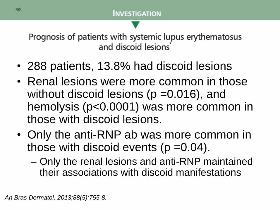
• 288 patients, 13.8% had discoid lesions
• Renal lesions were more common in those without discoid lesions (p =0.016), and hemolysis (p<0.0001) was more common in those with discoid lesions.
• Only the anti-RNP ab was more common in those with discoid events (p =0.04).
– Only the renal lesions and anti-RNP maintained their associations with discoid manifestations
An Bras Dermatol. 2013;88(5):755-8.

• Cross-sectional study of patients with DLE (small N)
• Correlation between anti-RNP antibodies and skin disease
activity, both in patients with SLE and in those with DLE.
– ANA, anti-RNP, and anti-dsDNA correlated with SLE activity
index scores
• These and anti-ssDNA IgG correlated with C3 and C4
levels, the number of SLE criteria
– Need for oral medications!
• Measuring autoantibodies to predict skin disease activity and
associated systemic findings
3/24/2016Kim, A et al.JAMA Dermatol 2014 Apr 30;[EPub Ahead of Print],

• Approximately 50% of patients with subacutecutaneous lupus erythematosus
• Patients with SCLE/SLE vs SCLE – Oral ulcers
– +anti-dsDNA antibodies
– + ANA
– Low complement

Cutaneous Lupus Erythematosus (LE)
• Chronic Cutaneous LE
• Subacute Cutaneous LE
• Acute (systemic) LE

The Lupus Spectrum: God Help Us
Increased prevalence/severity in black/hispanicpeds
Greater risk of renal
disease then adults!
X

Chronic Cutaneous LE
• CCLE
– Classic Discoid LE
– Tumid LE
– Neonatal LE (sort of)
– Lupus Panniculitis
– Chilblain LE

Case 1
• CC: 49 yo M with rash for 2 years.
• HPI: First lesion appeared on the trunk and subsequently spread to the face and scalp.
– Lesions are generally asymptomatic, but are occasionally itchy.
– Past tx: topical steroids and hydroquinone 4% cream without improvement
• PMH: DM, HTN
• Meds:Glucophage, Lisinopril, Metoprolol
• Allergies: NKDA
• Social Hx: No tobacco, alcohol, or drug use
• Family Hx: Father hx laryngeal ca


Clinical picture


| 23Science at the heart of medicine
3/24/2016

| 24 Science at the heart of medicine
3/24/2016

Discoid lupus erythematosus (DLE)
• Can be seen w/o serologic or
systemic manifestations of
SLE
– May be early sign of SLE
– Up to 28% progress!!*
• Follicular plugging*
• Chronic lesions may show
hyper/hypopigmentation w/
atrophy
• ? exacerbated by UV light
• m/c head and face
– Also scalp, extremities,
back, chest, abdomen
Chong B et al Br J Dermatol. 2012 Jan;166(1):29-35.

DLE

DLE

What about peds?


DLE Management
• Spontaneous involution with scarring is common
• Rarely BCCs or SCCs may occur in scars
– Lower lips with hypopigmented scars
• Treatment of localized disease: topical steroids and/or intralesional steroids.
• Photoprotection + Vit D supplementation
• Generalized DLE requires systemic therapy with hydroxychloroquine (Plaquenil) dosed up to 6.5 mg/kg/day.
– Low dose (50-100 mg /d) seems to work better for acral
• If Plaquenil is not effective, add quinacrine (~ 100 mg qd vsbid)
• MTX, retinoids, dapsone, cellcept also options
Dermatology Online Journal 7(1): 2

Plaquenil Clinical Pearls
• High affinity for melanin-containing tissue, with a tendency to accumulate in ocular tissues such as the choroids and ciliary body
• < 1% risk of eye damage
• Risk factors:
– Daily dosage of HCQ exceeding 6.5mg/kg
– Obesity
– Duration of use longer than 5 years
• After year 5, MUST have yearly optho exams
– Renal or hepatic functional impairment.
– > 60 yrs
• Chloroquine + hydroxychloroquine NO NO
• G6P-deficiency
• ? Pregnancy
Lupus. 2001;10(6):401-4.

A tough one
ANA went from 1:40 to 1:360over 6 month period

What now?

No Butts About It: CLE and Smoking Do Not Mix
• *Current smokers had higher median CLASI scores (9.5) than did never (7.0) and past (6.0) smokers (P=0.02)
– Current smokers more likely than nonsmokers to receive combination antimalarial therapies and significantly less likely to improve than never or past smokers
• **4 risk increase was seen for buccal cancer, lymphomas, respiratory cancer and nonmelanoma skin cancer
Piette E et al Arch Dermatol. 2012;148(3):317-322Gronhagen et al. British Journal of Dermatology 2012; 166: 1053-1059.

Frances C et al., Arch Dermatol 2012 Apr; 148:479
Plaquenil not working? There’s an app for that
• 300 patients (DLE, SCLE, LP)
– 38 of these patients had 2 or more associated cutaneous forms
• Median blood hydroxychloroquine significantly higher in patients with complete remission compared with partial remission and treatment failure (P = .007)
– Thirty patients (10.0%) had very low blood hydroxychloroquine concentrations and considered nonadherent to the treatment regimen
• Prove poor adherence

On the horizon: Alitretinoin for CLE
• Alitretinoin (9-cis retinoic acid)
– Approved in Europe as an oral formulation for use in recalcitrant hand eczema,
– Topical version is approved for Kaposi sarcoma.
• 3 patients with varying types of CLE tx oral alitretinoin at 30 mg/day.
– 1 severe DLE with SLE
– 1 hypertrophic LE
– 1 SCLE.
• Total clearance in 2 patients and nearly complete resolution in the third.
• No serious AE
• 2 patients tapered to 10 mg/day
Kuhn A et al., J Am Acad Dermatol 2012 Sep; 67:e123.

De Souza A et al., J Drugs Dermatol 2012 Oct; 11:1224
On the horizon: Apremilast for CLE
• Apremilast
– PDE4 enzyme inhibitor capable of blocking leukocyte production of IL-12, IL-23, TNF-a, INF-γwith subsequent suppression of Th1 and Th17-mediated immune responses
• 1st open label study
• CLASI scores significantly (P<0.05) decrease after 85 days of treatment with apremilast 20 mg twice daily in 8 patients with active discoid lupus.

Case 2
• 74 yo M presented with 8 week history progressively
enlarging “rash” L eye and mid chest
• Mildly painful/burning
• No past episodes
• No family history of similar lesions
• PMH: HTN, High Chol, OA
• Meds: HCTZ, Amlodipine, Simvistatin,
Metoprolol
• Otherwise well

| 40


What to Know: Diagnostic Criteria
Kuhn et al Arch Dermatol. 2000;136(8):1044-9.

But of course…
PMLE
PMLE
Pseudolymphoma
Jessner’s

| 47 Science at the heart of medicine 3/24/2016

What to Know about Treatment
• Antimalarials– up to 90% effective (with
rapid resolution of lesions in 1 study)
• Topical steroids/ILK
• Dapsone
• Sun protection
Arch Dermatol 2000;136:1033-1041

Case 3
• Newborn baby boy presents with a red macular facial rash
• PMH: born at 37 wks. Mother had a URI x 2 wks prior to delivery, but otherwise the pregnancy had been going well.
• All: NKDA
• Medications: none
• FH: non-contributory
• SH: has 2 siblings (11 yr old brother, 5 yr old sister)


Path

Lab studies
12/17-28/09:
TORCH panel:
HS Ab (type 1/type 2), Rubella Ab IgM, Toxo Ab IgM/IgG, CMV IgM/IgG:
negative, Urine CMV cx negative
Rubella Ab IgG + (?)
Syphilis screen NR
CSF HSV-1/2 DNA negative
CSF VDRL NR
CSF pink WBC 21, RBC 15684, glucose 40, protein 217 (10-40)
CSF culture no growth
Eye cx, viral eye cx, nasal viral cx, rectal viral cx, HS cx: no growth
Anti-SSA (Ro) > 8.0 (< 1.0)
Anti-SSB (La) < 0.2 (< 1.0) EKG: WNL

Lab studies - Mother
6/1/09:
VZ Ab IgG +
Rubella Ab IgG -
Syphilis screen NR
10/30/09:
HIV-1/2 Ab -

NICU course
• Pt was given ampicillin and gentimicin after a full
sepsis work-up, and abx stopped once blood and
CSF cultures were negative x 48 hrs.
• Pt was given acyclovir until CSF HSV PCR
returned negative
• Pt was discharged on DOL 4 with f/u with PMD,
rheumatology, and dermatology

Neonatal Lupus Erythematosus (NLE)

Neonatal Lupus Erythematosus (NLE)
• Caused by maternally transmitted autoantibodies (anti-Ro/SS-A in 95% of cases)
– Anti-La/SS-B (60%-80%)—mean level higher in mothers of infants with congenital heart block
– Anti-U1RNP (small subset
• The major manifestations of NLE involve which two organ systems, and are the effects transient or permanent in the newborn?– 1) Dermatologic disease:
• transient effects
• 50% of pts with NLE
– 2) Cardiac disease: • permanent effects
• responsible for morbidity & mortality of NLE (congenital heart block)
• 10% of pts with NLE

NLE: Cutaneous Findings
• Develop at a few weeks of age
• ~ SCLE
– Papulosquamous variant (most common)
• Erythematous, nonindurated scaly plaques
• No scarring
– Annular variant
• Annular, more inflammatory plaques
• Occurs almost exclusively in Japanese
• MC face & scalp, especially periorbital and malar areas (“raccoon eyes” appearance)
• NLE lesions may occur in sun-protected areas
• Transient hypopigmentation & epidermal atrophy may result
• Telangiectasia may be a permanent sequela

Neonatal Lupus Erythematosus (NLE)
• Heart block
– seen in up to 50%
– Permanent defect developing in utero
• Inflammation due to antibody-antigen reaction on
conduction system causes scarring
• Require pacemaker
– 10% die with cardiac related disease
– 90% of pts have HLA-DR2, DR3
– 100% have lupus anticoagulants

NLE
Treatment & Prognosis• Workup:
– complete physical exam, EKG, CBC , LFTs
• Skin disease treatment:
– sun protection, topical steroids, PDL for residual telangiectasias
• Congenital heart block treatment:
– 50% of newborns with complete congenital heart block require pacemaker in neonatal period
– 15% mortality in neonatal period from complete congenital heart block
• Risks for mothers with anti-Ro or anti-La antibodies:
– 1%-20% risk of developing infant with NLE
– 25% risk of recurrence of congenital heart block in subsequent pregnancies

Poll 2
I order ACE levels on all patients with
cutaneous sarcoidosis
• Agree
• Disagree

Case 3
• CC: Arm rash and Dry skin
• HPI: 72 Year old woman with a 2 year history of a solitary pruritic plaque on the right upper arm.
– No previous episodes
• PMH: Bronchiectasis, Gastritis
• PSH: Not Contributory Meds: Flovent All: NKA FH: HTN
• SH:
– Never smoked, no hx of passive exposure
– No work health hazards; worked as nurses aide for many years
– One child, grown; No pets; Lives alone


| 63 Science at the heart of medicine
3/24/2016| 63Science at the heart of medicine 3/24/2016

Pathology

Clinical Data
• PMH: Pulmonary Sarcoidosis, Granulomatous gastritis
• Labs:
• ANA: negative; RF: 20 (nrml) ACE: 87 (↑) in 2007
• Multiple negative PPDs
• Current: Ca2+ 9.7
• High Ca2+ ; low PTH 2010
• Imaging:
• CXR 7/11: Parenchymal findings of sarcoidosis with apparent improvement from 2010
• CT Thorax 8/10: Mild splenomegaly, calcified granuloma in the right lower lobe of lung, enlarged bilateral hilar and mediastinallymph nodes.

Sarcoidosis
• Female preponderance 1.3:1 among all ethnic and racial groups
• US: increased incidence among black Americans, ranging from 35.5-64.4 per 100,000
• Peak incidence in black Americans occurs in the fourth decade.
• Sarcoidosis is also more likely to be chronic and fatal in black Americans

Clinical
• Key Clinical Features
– Cutaneous disease in 30%• Early manifestation
– Red-brown to purple dermal papules/plaques
– Atypical presentations include alopecia, atrophic forms, erythrodermic sarcoidosis, hypopigmented, ichthyosiform, and lichenoid forms.
• Erythema nodosum is predictive of an subacute, transient form of sarcoid


The Many Faces of Sarcoid

And don’t forget…

| 71 Science at the heart of medicine 3/24/2016
Indian Dermatol Online J. 2014 Jan;5(1):77-9. doi: 10.4103/2229-5178.126041.

Sarcoid as a complication of neurotoxins?

So what’s going on here?

Sarcoidosis Syndromes
• Lofgren’s syndrome: • Erythema nodosum, bilateral hilar/right
paratracheal lymphadenopathy, fever, polyarthralgia
• Heerfordt's syndrome:
• Parotid enlargement, uveitis, fever, and facial nerve palsy.
• Darier-Roussy Disease:
• Subcutaneous sarcoidosis• Keep in your ddx with RA nodule and Subq GA

Diagnosis
• Diagnosis of exclusion
– Chest radiograph evidence, accompanied by non-necrotizing granuloma on biopsy and supportive clinical history
• Radiologic findings: paratracheal or hilaradenopathy +/- infiltration
– PFTs: restrictive ventilatory dysfunction
• Opthalmologic: Uveitis
• CBC, Ca2+, LFTs; ↑ANA in 30%; ↑ serum ACE in 60%, and check for ↑ESR, anemia, eosinophilia, lymphopenia, and hypercalcemia

Treatment of Cutaneous Sarcoidosis
• Corticosteroids• Topical, intralesional, and systemic*
• Tetracycline Abx (minocycline 100 mg BID)
• Antimalarials• Hydroxychloroquine (200-400 mg daily) for 12
weeks
• Methotrexate (10-25 mg weekly)
• TNF-α antagonists• Inflixumab
• Thalidomide

• A randomized, placebo-controlled, single-masked trial on 30 patients
– 22 patients biopsy- proven cutaneous disease; 8 had biopsy-proven
pulmonary disease with dermatologic lesions
• 8- week regimen consisted of:
– levofloxacin,750 mg on day 1, followed by 500 mg/d
– ethambutol, 25 mg/kg/d up to a maximum dosage of 1200 mg/d
– azithromycin, 500 mg on day 1, followed by 250 mg/d
– rifampin, 10 mg/kg/dup to a maximum dosage of 300 mg/ d
• The placebo regimen consisted of riboflavin (in place of rifampin)
and lactose (in place of the other 3 drugs).
• 6 patients withdrew due to AE
• Observed improvement with the 8-week CLEAR regimen was still present
180 days after baseline

Caveats

The Future:

Poll 3
Imaging studies can be prudent in the
setting of morphea
• Agree
• Disagree

Case 4
• 9 yo AA girl presented with asx, progressively enlarging rash on RLE x 1 year
– Began distally progressed proximally
– No past episodes
– No associated symptoms
• Pmhx
– PPD+ s/p tx with INH
– Asthma
• NKA
• Meds
– Albuterol
– INH
• Family hx
– Unremarkable


Path
Square BX

Serologic W/U
• ANA: 1:80 speckled
• ANA-ENA: -
• Anti-centromere: -
• ESR: 6
• Lyme titer: .2 (wnl)
• CBC: wnl

Morphea
• 0.4 - 2.7 per 100,000 people
• Female predominance of 2.4 to 4.2:1 has been reported
• Etiology:
– Autoantibodies? Environmental (L-tryptophan, bleomycin), infection (viral)?, vaccination (bcg)?
– Fibroblasts produce increased collagen
– Abnormal collagen production due to instruction from surrounding T cells
– Is morphea a TH1 or TH2 response pattern?
• TH2 - specifically IL-4 and TGF-beta enhance collagen I, II, III production

Morphea - Linear
• Most common subtype in children, affecting 41.8% to
67%
• Tends to involve the underlying fascia, muscle and
tendons
• Primary type of morphea to cause disability
• Variants?
– “en coup de sabre’
– Parry - Romberg
• + anti-ssDNA abs
• Melorheostosis
• ? Assoc with spina bifida


Morphea - Linear
• Initially harmless
looking but…
– Muscle weakness
– Shortens muscles
– Immobilizes joint
– Growth retardation
in kids

Parry-Romberg
• Progressive
hemifacial atrophy
• May affect what
Trigeminal nerve
distribution

En Coup de Sabre
• Unilateral
• Forehead to scalp
• Paramedian m/c
• Can involve
underlying muscle
and bone and rarely
meningies and brain
causing seizures

Treatments: Level of Evidence
Indian J Dermatol Venereol Leprol 2012;78:135-41
•NB-UVB equivalent to low-dose (800 J/cm2, ), but not medium(2000 J/cm2,) UVA
J Am Acad Dermatol. 2011;64(2):231-42


Treatment with pirfenidone was three times daily for 6 months.

• Prednisolone (1 mg/kg/day)and methotrexate (MTX; 9.5 mg/m2 once a week) were started.
– So severe - treatment with imatinib• Imatinib was started at a dosage of 235 mg/ m2/day
• Imatinib was continued at the same dosage for 1 year, and the prednisolone dosage was gradually decreased until it was stopped after 3 months
• MTX was continued at the same dose regimen for 4 years as maintenance therapy.


Thank you for your attention