tnf-alpha expression, evaluation of collagen, and tunel of ... · 2- universidade federal do...
TRANSCRIPT
J Appl Oral Sci. 278
ABSTRACT
www.scielo.br/jaoshttp://dx.doi.org/10.1590/1678-775720150481
TNF-alpha expression, evaluation of collagen, and TUNEL of Matricaria recutita L. extract and triamcinolone on oral ulcer in diabetic ratsBruna Vasconcelos OLIVEIRA1, Paulo Goberlânio de BARROS SILVA1 2, Luiz André Cavalcante BRIZENO3 4, Fabrício Bitú SOUSA1, Mário Rogério Lima MOTA1, Ana Paula Negreiros Nunes ALVES1
1- Universidade Federal do Ceará, Setor de Patologia Oral, Departamento de Odontologia Clínica, Fortaleza, CE, Brasil.2- Universidade Federal do Ceará, Setor de Cariologia e Odontologia Restauradora, Departamento de Odontologia Restauradora, Fortaleza, CE, Brasil.3- Universidade Estadual do Ceará, Setor de Fisiologia e Farmacologia, Departamento de Ciências Biomédicas, Fortaleza, CE, Brasil.4- Universidade Federal do Ceará, Departamento de Fisiologia e Farmacologia, Fortaleza, CE, Brasil.
Corresponding address: Paulo Goberlânio de Barros Silva - Departamento de Odontologia Clínica - Setor de Patologia Oral - Faculdade de Odontologia -
2: +55 85 8705 7151 - e-mail: [email protected]
Diabetes mellitus (DM) is a disease associated with delayed wound healing of oral ulcers
) and apoptosis in rats with DM treated with chamomile extract or triamcinolone. Material and Methods: Wistar male
diabetes; positive control group (PCG) with DM (alloxan, 45 mg/kg); and groups treated with chamomile extract (normoglycemic= NCG group and diabetic= DCG group) and with triamcinolone (TG). Traumatic ulcers were performed on all animals that received topical
and ten the animals were euthanized and the ulcers were analyzed by light microscopy, TUNEL assay, and immunohistochemically (TNF- ). The NCG (p=0.0062), PCG (p=0.0285), NCG (p=0.0041), and DCG (p<0.0001) groups were completely healed on the 10th day, however, there was no healing on the TG (p=0.5127) group. The TNF- expression
th to the 10th day in NCG (p=0.0266) and DCG
number of positive cells in NCG (p=0.0273) and CNG (p=0.0469) and in the epithelium only in CDG (p=0.0320). Conclusions: Chamomile extract can optimize the healing of traumatic oral ulcers in diabetic rats through the reduction of apoptosis in the epithelium and TNF- expression.
Keywords: Oral ulcer. Diabetes mellitus. Chamomile. Matricaria. Triamcinolone.
INTRODUCTION
Diabetes Mellitus (DM) is a chronic metabolic disease characterized by deficiency in insulin production or resistance to its action, resulting in hyperglycemia and metabolic alterations4. The incidence of DM is increasing in the world, and it is considered the biggest health problem in XXI century18. It is estimated that in 2025 there will be twice as many diabetic patients compared with the year 2000, totaling approximately 300 million
affected individuals worldwide20.Chronic hyperglycemia causes numerous events
that promote structural changes in tissue. It is associated with delayed wound healing, increased susceptibility to infection, alterations in neutrophil activity, and reduction of chemotaxis, adhesion, phagocytosis and angiogenesis4,7. Clinically, the wound healing disorder manifests itself as hypertrophic scars or chronic unhealed wounds (ulcers), being ulcers the most prevalent problem in healing18.
2016;24(3):278-90
J Appl Oral Sci. 279
In the oral cavity, traumatic ulcers are caused by mechanical trauma due to maladjusted dentures, orthodontic brackets, accidental bites, or iatrogenic factors. Typically, when the causal agent is removed, healing occurs spontaneously from one to two weeks; however, in a few cases, the ulcer can persist for longer periods of time. It can be extremely painful and interfere with eating and speaking6.
Corticosteroids are commonly prescribed for the treatment of painful symptoms of traumatic oral ulcers6; however, conflicting results have been reported in literature regarding the effects of this therapeutic modality on the healing process. Glucocorticoids have potent anti-inflammatory and immunosuppressive effects1. Triamcinolone is commonly used in clinical dentistry because of its analgesic effects on oral ulcers. It has a potent anti-
scores of mucositis and pain in patients undertaking radiotherapy12. But corticoids used in treatment
wound healing1,12.In contrast, the use of natural products in the
treatment of ulcerated oral lesions has increased over the past several decades23. The plant chamomile, also known as Chamomilla recutita L. or Matricaria recutita L., which is a member of the Asteraceae family, is one of the most widely used medicinal plants10
apigenin), terpenes and acetylated derivatives,
antifungal, antioxidant, hypocholesterolemic, and sedative properties9,10,14,19,21,26,27.
These drugs have been indiscriminately used in dental clinic for the treatment of persistent ulcerative lesions, but mechanisms of the diseases can be different in diabetic patients. Hyperglycemia
wound closure and healing and interfere in collagen deposition3,25. Drugs used in the treatment of oral
modifying these parameters. Thus, the aim of this
Factor alpha (TNF- ) and apoptosis in rats with DM treated with chamomile extract or triamcinolone.
MATERIAL AND METHODS
AnimalsThis study was approved by the Ethics Committee
for Animal Research (protocol no. 11/11), and was performed in accordance with the Ethical Principles in Animal Experimentation adopted by the Brazilian College of Animal Experimentation (COBEA).
We used male Wistar rats (Rattus norvegicus albinus, Rodentia mammalia) weighing 210.0±4.2 g (Mean±SD) that were provided by the Central Animal Facility of the Federal University of Ceará (UFC). The rats were kept in the Animal Laboratory of Experimental Oncology (LOE), given an initial examination for systemic health conditions and stored in boxes with sawdust. All animals received industrial feed (Bio-base®, Águas Frias, SC, Brazil) accordingly and water ad libitum and were kept at room temperature with controlled humidity for a photoperiod of 12 hours.
Diabetes inductionThe induction of diabetes was performed by
injection of alloxan (45 mg/kg) in diluted sterile saline (0.9%) intravenously after mild sedation with ether. Two milliliters of blood were collected 48 h after the induction of diabetes from the retro-orbital plexus for determination of blood glucose. The animals were considered diabetic when the blood glucose was equal to or greater than 200 mg/dL24.
blood was collected again for glucose measurement
Animals with a blood glucose level of less than 200 mg/dL were excluded.
Experimental protocol to induce the ulcersFor the induction of ulcers, the animals were
anesthetized with intraperitoneal ketamine (80 mg/kg) and xilazin (10 mg/kg). Antisepsis was performed with an oral solution of 0.12% chlorhexidine gluconate in cotton pellets. The ulceration in the left buccal mucosa was performed by abrasion with a number 15 scalpel blade, and a marker with an 8 mm diameter was used for standardization of the lesion area. The surgical technique was standardized for all animals and performed by the same operator9.
Groups and treatment
groups by lot:- Groups with Saline Treatment: Negative
Control Group (normoglycemic rats) and a Positive Control Group (diabetic rats);
- Groups with Chamomile Treatment: Chamomile Normoglycemic Group (normoglycemic rats treated with chamomile) and Chamomile Diabetic Group (diabetic rats treated with chamomile);
- Groups with Triamcinolone Treatment: Triamcinolone Group (diabetic rats treated with Triamcinolone).
topical application of sterile saline solution in the Negative Control Group and Positive Control Group,
TNF-alpha expression, evaluation of collagen, and TUNEL of Matricaria recutita L. extract and triamcinolone on oral ulcer in diabetic rats
2016;24(3):278-90
J Appl Oral Sci. 280
Omcilon-A, orabase®, 1 mg/g (B-MS, São Paulo, SP, Brazil) in the Triamcinolone Group, or Ad-Muc® 10% ointment (BIOLAB, São Paulo, SP, Brazil) in the Chamomile Group (diabetic and normoglycemic). The application of the drugs was performed with an individual sterile and disposable microbrush (KG Sorensen®, Cotia, SP, Brazil).
and ten days after ulcer induction.
Clinical evaluationThe animals were weighed and had the blood
a 0.5 mm digital pachymeter (L=larger diameter; m=minor diameter) to calculate the area of the
All measurements were performed by the same operator8. Weight, blood glucose, and ulcer diameter were evaluated in the 5th and 10th days.
Histological analysis
the specimens were macroscopically analyzed, subjected to dehydration in crescent alcoholic series,
melted at 60º C. Then, the fragments were placed
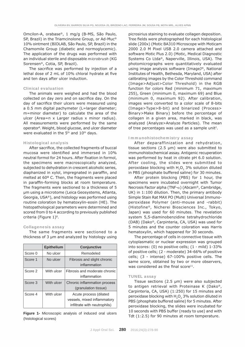
The fragments were sectioned to a thickness of 5 μm using a microtome (Leica Geosystems, Atlanta, Georgia, USA®), and histology was performed using routine coloration by hematoxylin-eosin (HE). The histopathological parameters were determined and scored from 0 to 4 according to previously published criteria (Figure 1)8.
Collagenesis assayThe same fragments were sectioned to a
thickness of 3 μm and analyzed by histology using
picrosirius staining to evaluate collagen deposition.
slide (200x) (Motic BA310 Microscope with Moticam 2000 2.0 M Pixel USB 2.0 camera attached and software Motic Plus 2.0) (Motic, Medical Diagnostic Systems Co Ltda®, Naperville, Illinois, USA). The photomicrographs were quantitatively evaluated using image analysis software (ImageJ®, National Institutes of Health, Bethesda, Maryland, USA) after calibrating images by the Color Threshold command (Image>Adjust>Color Threshold) in the RGB function for colors Red (minimum 71, maximum 255), Green (minimum 0, maximum 69) and Blue (minimum 0, maximum 92). After calibration, images were converted to a color scale of 8-bits (Image>Type>8-bit) and binarized (Process> Binary>Make Binary) before the percentage of collagen in a given area, marked in black, was analyzed (Analyze>Analyze Particles). The mean of tree percentages was used as a sample unit2.
Immunohistochemistry assayAfter deparaffinization and rehydration,
immunohistochemical assay. Antigenic recuperation was performed by heat in citrate pH 6.0 solution. After cooling, the slides were submitted to peroxidase blocking with H2O2 3% solution diluted in PBS (phosphate buffered saline) for 30 minutes.
After protein blocking (PBS) for 1 hour, the specimens were incubated overnight with Tumor Necrosis Factor alpha (TNF- ) (Abcam®, Cambridge, UK) in 1:100 dilution. Then, the primary antibody Simple Stain Rat MAX PO (Multi) Universal Immuno-peroxidase Polymer (anti-mouse and -rabbit) (Histofine®, Nicherei Biosciences Inc., Tokyo, Japan) was used for 60 minutes. The revelation system 5,5-diaminobenzidine tetrahydrochloride (DAB) (Dako®, Carpinteria, CA, USA) was used for 5 minutes and the counter coloration was Harris hematoxylin, which happened for 30 seconds.
The percentage of cells in connective tissue with cytoplasmatic or nuclear expression was grouped into scores: (0) no positive cells; (1 - mild) 1-33% of positive cells; (2 - moderate) 34-66% of positive cells; (3 - intense) 67-100% positive cells. The same score, obtained by two or more observers,
11.
TUNEL assay
to antigen retrieval with Proteinase K (Dako®, Carpinteria, CA, USA) (1:250) for 15 minutes and peroxidase blocking with H2O2 3% solution diluted in PBS (phosphate buffered saline) for 5 minutes. After peroxidase blocking, the slides were incubated for 10 seconds with PBS buffer (ready to use) and with Tdt (1:2.5) for 90 minutes at room temperature.
Figure 1- Microscopic analysis of induced oral ulcers (histological scores)
Epithelium ConjunctiveScore 0 No ulcer Remodeled
Score 1 No ulcer Fibrosis and slight chronic
Score 2 With ulcer Fibrosis and moderate chronic
Score 3 With ulcer(granulation tissue)
Score 4 With ulcer Acute process (dilated
OLIVEIRA BV, BARROS SILVA PG, NOJOSA JS, BRIZENO LAC, FERREIRA JM, SOUSA FB, MOTA MRL, ALVES APNN
2016;24(3):278-90
J Appl Oral Sci. 281
The tissue slides were rinsed with the wash buffer (1:15) for 10 minutes and after this the anti-digoxigenin conjugate (ready to use) was added for 30 minutes. The revelation system was done with DAB (Dako®, Carpinteria, CA, USA). The tissue slides were stained with metil green for 10 minutes followed by dehydration, diaphanization, and mounting with coverslips.
The percentage of positive cells in connective tissue and epithelium was grouped into scores: (0) no positive cells; (1 - mild) 1-33% of positive cells; (2 - moderate) 34-66% of positive cells; (3 - intense) 67-100% positive cells. The same score, obtained by two or more observers, was considered
11.
Statistical analysisThe area of ulceration, weight loss, blood glucose
change, and percentage of collagen in an area were analyzed by the t-test and ANOVA/Tukey test. The Mann-Whitney test and Kruskall-Wallis/Dunn test were used to histological scores. The relationship between the area of ulceration, weight loss and blood glucose variation in the four experimental groups was analyzed by Pearson correlation. The relationship between these parameters and the histological scores were analyzed by Spearman correlation. All analyses were performed with the software GraphPad Prism 5.0® (GraphPad Software Inc., San Diego, California, USA), with a p<0.05
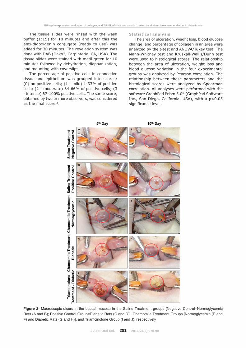
Figure 2-
F) and Diabetic Rats (G and H)], and Triamcinolone Group (I and J), respectively
TNF-alpha expression, evaluation of collagen, and TUNEL of Matricaria recutita L. extract and triamcinolone on oral ulcer in diabetic rats
2016;24(3):278-90
J Appl Oral Sci. 282
RESULTS
Clinical evaluation
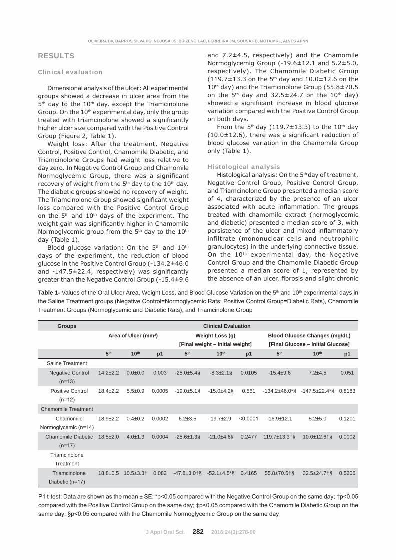
Dimensional analysis of the ulcer: All experimental groups showed a decrease in ulcer area from the 5th day to the 10th day, except the Triamcinolone Group. On the 10th experimental day, only the group
higher ulcer size compared with the Positive Control Group (Figure 2, Table 1).
Weight loss: After the treatment, Negative Control, Positive Control, Chamomile Diabetic, and Triamcinolone Groups had weight loss relative to day zero. In Negative Control Group and Chamomile
recovery of weight from the 5th day to the 10th day. The diabetic groups showed no recovery of weight.
loss compared with the Positive Control Group on the 5th and 10th days of the experiment. The
Normoglycemic group from the 5th day to the 10th day (Table 1).
Blood glucose variation: On the 5th and 10th days of the experiment, the reduction of blood glucose in the Positive Control Group (-134.2±46.0
greater than the Negative Control Group (-15.4±9.6
and 7.2±4.5, respectively) and the Chamomile Normoglycemig Group (-19.6±12.1 and 5.2±5.0, respectively). The Chamomile Diabetic Group (119.7±13.3 on the 5th day and 10.0±12.6 on the 10th day) and the Triamcinolone Group (55.8±70.5 on the 5th day and 32.5±24.7 on the 10th day)
variation compared with the Positive Control Group on both days.
From the 5th day (119.7±13.3) to the 10th day
blood glucose variation in the Chamomile Group only (Table 1).
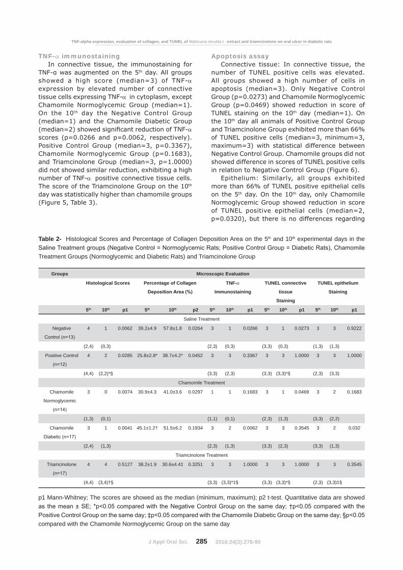
Histological analysisHistological analysis: On the 5th day of treatment,
Negative Control Group, Positive Control Group, and Triamcinolone Group presented a median score of 4, characterized by the presence of an ulcer
treated with chamomile extract (normoglycemic and diabetic) presented a median score of 3, with
infiltrate (mononuclear cells and neutrophilic granulocytes) in the underlying connective tissue. On the 10th experimental day, the Negative Control Group and the Chamomile Diabetic Group presented a median score of 1, represented by
Table 1- Values of the Oral Ulcer Area, Weight Loss, and Blood Glucose Variation on the 5th and 10th experimental days in
Treatment Groups (Normoglycemic and Diabetic Rats), and Triamcinolone Group
Groups Clinical Evaluation
Area of Ulcer (mm²) Weight Loss (g)[Final weight – Initial weight]
Blood Glucose Changes (mg/dL)[Final Glucose – Initial Glucose]
5th 10th p1 5th 10th p1 5th 10th p1
Saline Treatment
Negative Control 14.2±2.2 0.0±0.0 0.003 -25.0±5.4§ -8.3±2.1§ 0.0105 -15.4±9.6 7.2±4.5 0.051
Positive Control 18.4±2.2 5.5±0.9 0.0005 -19.0±5.1§ -15.0±4.2§ 0.561 -134.2±46.0*§ -147.5±22.4*§ 0.8183
Chamomile Treatment
Chamomile 18.9±2.2 0.4±0.2 0.0002 6.2±3.5 19.7±2.9 -16.9±12.1 5.2±5.0 0.1201
Chamomile Diabetic 18.5±2.0 4.0±1.3 0.0004 -25.6±1.3§ -21.0±4.6§ 0.2477 119.7±13.3†§ 10.0±12.6†§ 0.0002
Triamcinolone Treatment
Triamcinolone 18.8±0.5 10.5±3.3† 0.082 -47.8±3.0†§ -52.1±4.5*§ 0.4165 55.8±70.5†§ 32.5±24.7†§ 0.5206
OLIVEIRA BV, BARROS SILVA PG, NOJOSA JS, BRIZENO LAC, FERREIRA JM, SOUSA FB, MOTA MRL, ALVES APNN
2016;24(3):278-90
J Appl Oral Sci. 283
assigned a median score of 2 because there was a
inflammation. The Triamcinolone Group had a median score of 4, characterized by the presence
Only Chamomile Normoglycemic Group shows
in connective tissue (score of 0).
reduction of the histological scores of the ulcers
from the 5th day to the 10th day in all experimental groups, except in the Triamcinolone Group. On the 5th
groups (Figure 3, Table 1). On the 10th day, the Positive Control Group showed histological scores
Control Group (median=1) and Chamomile Normoglycemic group (median=0). The Positive Control Group (median=2) and the Chamomile
however, the Triamcinolone Group (median=4)
Figure 3- Photomicrograph of ulceration in the buccal mucosa in the Saline Treatment groups [Negative
[Normoglycemic (E and F) and Diabetic Rats (G and H)], and Triamcinolone Group (I and J), respectively. (Hematoxylin &
TNF-alpha expression, evaluation of collagen, and TUNEL of Matricaria recutita L. extract and triamcinolone on oral ulcer in diabetic rats
2016;24(3):278-90

J Appl Oral Sci. 284
higher than the Positive Control Group (Table 2).
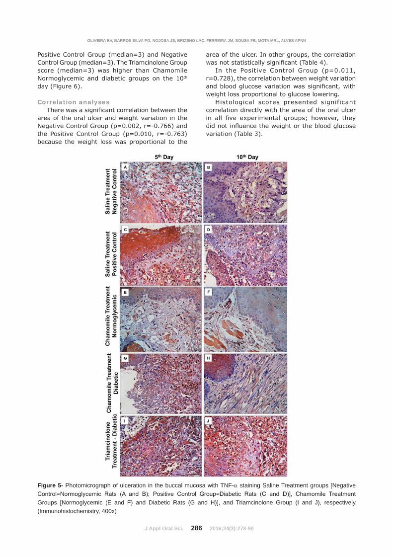
CollagenesisOn the 5th experimental day, the collagen
Control Group (25.8±2.8) compared with the Negative Control Group (39.2±4.9). The Chamomile
higher collagen deposition than the Positive Control Group. On the 10th day, the Positive Control Group (38.7±4.2) also had collagen deposition
(57.8±1.8). Positive Control Group (38.7±4.2), Chamomile Diabetic Group (51.5±6.2), and Chamomile Normoglycemic Group (41.0±3.6) did not differ statistically, but the Chamomile Diabetic
higher than the Triamcinolone Group (30.6±4.4). Only Negative Control Group, Positive Control Group, and Chamomile Normoglycemic Group showed augment in collagenesis from the 5th to the 10th day (Figure 4, Table 2).
Figure 4- Photomicrograph of ulceration in the buccal mucosa with picrosirius staining in Saline Treatment groups [Negative
[Normoglycemic (E and F) and Diabetic Rats (G and H)], and Triamcinolone Group (I and J), respectively (Picrosirius, 200x)
OLIVEIRA BV, BARROS SILVA PG, NOJOSA JS, BRIZENO LAC, FERREIRA JM, SOUSA FB, MOTA MRL, ALVES APNN
2016;24(3):278-90
J Appl Oral Sci. 285
TNF- immunostainingIn connective tissue, the immunostaining for
th day. All groups showed a high score (median=3) of TNF-expression by elevated number of connective tissue cells expressing TNF- in cytoplasm, except Chamomile Normoglycemic Group (median=1). On the 10th day the Negative Control Group (median=1) and the Chamomile Diabetic Group
scores (p=0.0266 and p=0.0062, respectively). Positive Control Group (median=3, p=0.3367), Chamomile Normoglycemic Group (p=0.1683), and Triamcinolone Group (median=3, p=1.0000) did not showed similar reduction, exhibiting a high number of TNF- positive connective tissue cells. The score of the Triamcinolone Group on the 10th day was statistically higher than chamomile groups (Figure 5, Table 3).
Apoptosis assayConnective tissue: In connective tissue, the
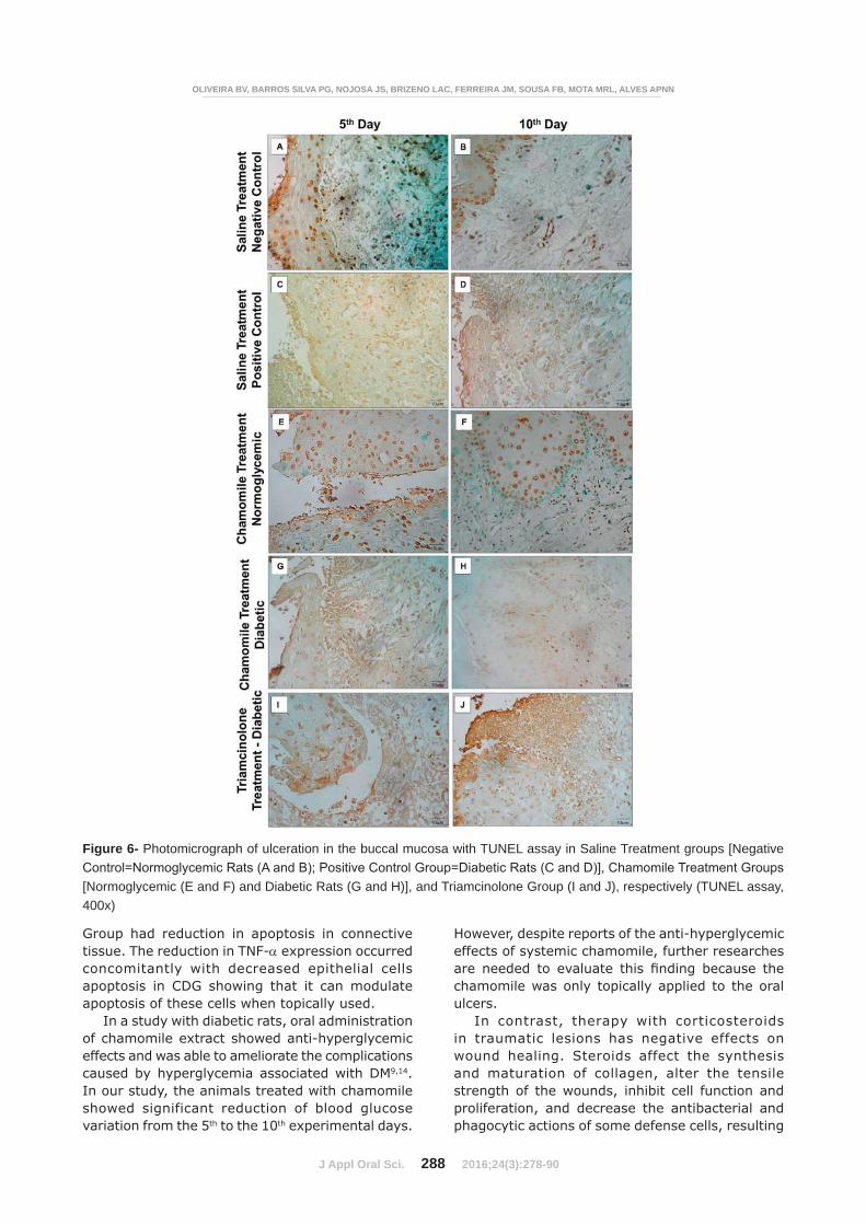
number of TUNEL positive cells was elevated. All groups showed a high number of cells in apoptosis (median=3). Only Negative Control Group (p=0.0273) and Chamomile Normoglycemic Group (p=0.0469) showed reduction in score of TUNEL staining on the 10th day (median=1). On the 10th day all animals of Positive Control Group and Triamcinolone Group exhibited more than 66% of TUNEL positive cells (median=3, minimum=3, maximum=3) with statistical difference between Negative Control Group. Chamomile groups did not showed difference in scores of TUNEL positive cells in relation to Negative Control Group (Figure 6).
Epithelium: Similarly, all groups exhibited more than 66% of TUNEL positive epithelial cells on the 5th day. On the 10th day, only Chamomile Normoglycemic Group showed reduction in score of TUNEL positive epithelial cells (median=2, p=0.0320), but there is no differences regarding
Table 2- Histological Scores and Percentage of Collagen Deposition Area on the 5th and 10th experimental days in the
Treatment Groups (Normoglycemic and Diabetic Rats) and Triamcinolone Group
Groups Microscopic Evaluation
Histological Scores Percentage of Collagen TNF-
Immunostaining
TUNEL connective
tissue
Staining
TUNEL epithelium
Staining
5th 10th p1 5th 10th p2 5th 10th p1 5th 10th p1 5th 10th p1
Saline Treatment
Negative 4 1 0.0062 39.2±4.9 57.8±1.8 0.0264 3 1 0.0266 3 1 0.0273 3 3 0.9222
(2,4) (0,3) (2,3) (0,3) (3,3) (0,3) (1,3) (1,3)
Positive Control 4 2 0.0285 25.8±2.8* 38.7±4.2* 0.0452 3 3 0.3367 3 3 1.0000 3 3 1.0000
(4,4) (2,2)*§ (3,3) (2,3) (3,3) (3,3)*§ (2,3) (3,3)
Chamomile Treatment
Chamomile
Normoglycemic
3 0 0.0074 30.9±4.3 41.0±3.6 0.0297 1 1 0.1683 3 1 0.0469 3 2 0.1683
(1,3) (0,1) (1,1) (0,1) (2,3) (1,3) (3,3) (2,2)
Chamomile 3 1 0.0041 45.1±1.2† 51.5±6.2 0.1934 3 2 0.0062 3 3 0.3545 3 2 0.032
(2,4) (1,3) (2,3) (1,3) (3,3) (2,3) (3,3) (1,3)
Triamcinolone Treatment
Triamcinolone 4 4 0.5127 38.2±1.9 30.6±4.4‡ 0.3251 3 3 1.0000 3 3 1.0000 3 3 0.3545
(4,4) (3,4)†§ (3,3) (3,3)*‡§ (3,3) (3,3)*§ (2,3) (3,3)‡§
p1 Mann-Whitney; The scores are showed as the median (minimum, maximum); p2 t-test. Quantitative data are showed
compared with the Chamomile Normoglycemic Group on the same day
TNF-alpha expression, evaluation of collagen, and TUNEL of Matricaria recutita L. extract and triamcinolone on oral ulcer in diabetic rats
2016;24(3):278-90
J Appl Oral Sci. 286
Positive Control Group (median=3) and Negative Control Group (median=3). The Triamcinolone Group score (median=3) was higher than Chamomile Normoglycemic and diabetic groups on the 10th day (Figure 6).
Correlation analyses
area of the oral ulcer and weight variation in the Negative Control Group (p=0.002, r=-0.766) and the Positive Control Group (p=0.010, r=-0.763) because the weight loss was proportional to the
area of the ulcer. In other groups, the correlation
In the Positive Control Group (p=0.011, r=0.728), the correlation between weight variation
weight loss proportional to glucose lowering.Histological scores presented significant
correlation directly with the area of the oral ulcer
variation (Table 3).
Figure 5- Photomicrograph of ulceration in the buccal mucosa with TNF- staining Saline Treatment groups [Negative
Groups [Normoglycemic (E and F) and Diabetic Rats (G and H)], and Triamcinolone Group (I and J), respectively (Immunohistochemistry, 400x)
OLIVEIRA BV, BARROS SILVA PG, NOJOSA JS, BRIZENO LAC, FERREIRA JM, SOUSA FB, MOTA MRL, ALVES APNN
2016;24(3):278-90

J Appl Oral Sci. 287
DISCUSSION
High levels of blood glucose affect oral wound healing negatively. Wound healing in an uncontrolled diabetic patient occurs more slowly compared with normoglycemic or controlled diabetic patients13. This delay in oral wound healing can cause chronicity of oral lesions in patients with DM18.
In this study, a wound healing deficit was observed in diabetic rats. On the 10th experimental day, we observed characteristics compatible with tissue healing in the normoglycemic group (score=1), but the diabetic group showed
of the underlying connective tissue (score=2).
in collagen deposition in the diabetic rats when compared with the normoglycemic rats at both time points. Reinforcing the histopathology analysis, the macroscopic evaluation showed that on the 10th experimental day the oral ulcers were completely healed in the normoglycemic rats, but not in the diabetic animals.
Animals treated with chamomile extract showed
the 5th experimental day, unlike Negative Control, Positive Control, and Triamcinolone Groups. Additionally, on the 10th
for the Chamomile Group were similar to those for the Negative Control Group (normoglycemic), showing total epithelial healing and mild chronic inflammation. In the fibrogenesis evaluation, collagen deposition in the Chamomile Group was
Group (diabetic) and did not differ from the Negative Control Group (normoglycemic).
been linked to compounds present in its extract
derivatives21,26,27. The mechanism of action of
direct inhibition of ciclooxigenase-2 and synthesis
E228. In this study, the chamomile group (DCG) was
expression in diabetic rats that are likely to augment
actions lead to a reduction in vascular and cellular
alpha-bisabolol has been associated with promoter activity in the formation of granulation tissue during the process of wound repair4.
Additionally, the reduction of apoptosis scores only in connective tissue of NCG may be associated
that is proportional to TNF- activation29. NCG showed high reduction in TNF scores in connective tissue and have a potent antioxidant activity, leading to reduction of apoptosis only in connective tissue.
The increase in TNF- and oxidative stress due to elevated levels of glucose decreases cell viability and survival of various cell types,
17,30. However, there was no
cells in any treatment. Only Negative Control
TNF-alpha expression, evaluation of collagen, and TUNEL of Matricaria recutita L. extract and triamcinolone on oral ulcer in diabetic rats
Table 3- Correlation between clinical and histological parameters
Correlation Negative Control (n=13)
Positive Control(n=12)
Chamomile(n=17)
Triamcinolone(n=17)
Pearson Correlation
Area of Ulcer x Weight Loss
Area of Ulcer x Blood Glucose
Changes
Blood Glucose Changes x Weight
Loss
Spearman Correlation
Histologic Scores x Area of Ulcer
Histologic Scores x Weight Loss
Histologic Scores x Blood Glucose
Changes
2016;24(3):278-90
J Appl Oral Sci. 288
Group had reduction in apoptosis in connective tissue. The reduction in TNF- expression occurred concomitantly with decreased epithelial cells apoptosis in CDG showing that it can modulate apoptosis of these cells when topically used.
In a study with diabetic rats, oral administration of chamomile extract showed anti-hyperglycemic effects and was able to ameliorate the complications caused by hyperglycemia associated with DM9,14. In our study, the animals treated with chamomile showed significant reduction of blood glucose variation from the 5th to the 10th experimental days.
However, despite reports of the anti-hyperglycemic effects of systemic chamomile, further researches
chamomile was only topically applied to the oral ulcers.
In contrast, therapy with corticosteroids in traumatic lesions has negative effects on wound healing. Steroids affect the synthesis and maturation of collagen, alter the tensile strength of the wounds, inhibit cell function and proliferation, and decrease the antibacterial and phagocytic actions of some defense cells, resulting
Figure 6- Photomicrograph of ulceration in the buccal mucosa with TUNEL assay in Saline Treatment groups [Negative
[Normoglycemic (E and F) and Diabetic Rats (G and H)], and Triamcinolone Group (I and J), respectively (TUNEL assay, 400x)
OLIVEIRA BV, BARROS SILVA PG, NOJOSA JS, BRIZENO LAC, FERREIRA JM, SOUSA FB, MOTA MRL, ALVES APNN
2016;24(3):278-90
J Appl Oral Sci. 289
in a delay in wound healing1,22. In this study, the Triamcinolone Group exhibited an oral ulcer area
Negative Control Group and the NCG on the 10th experimental day. The triamcinolone administered topically delayed the repair process, which caused
the 5th and 10th experimental days. This effect was not observed in other groups. The Triamcinolone Group exhibited less collagen deposition and higher weight loss than the Positive Control Group (diabetic rats), suggesting that this drug inhibits additional wound healing in the oral cavity in diabetic rats.
of blood glucose variation on both the 5th and 10th experimental days in animals treated with chamomile and triamcinolone when compared with the untreated diabetic rats. These data can be associated with the antinociceptive effects of both drugs10. Chamomile and triamcinolone can reduce nociception, leading to increased food intake and a consequent augment in blood glucose. The size of the ulcerative process in the oral cavity is proportional to the painful symptomatology, and topical drugs with anesthetic or analgesic effects diminish this relationship16. Thus, this model is supported by the analysis of the correlation between the area of the oral ulcer and weight loss, since the weight loss was proportional to the area of the ulcer only in the untreated animals (Positive Control Group and Negative Control Group).
The weight loss occurs due to diabetic anorexia, which is coupled with nociception and consequent dysphagia resulting from oral ulceration3. This combination may interfere with feeding and be responsible for the variation in blood glucose and
diabetic animals15. The results of the present study showed weight loss in all of the experimental diabetic groups; however, the Triamcinolone Group had
Control Group. Corticosteroids promote a complex metabolic process that results in malnutrition and catabolism. Consequently, this poor health condition of the animals may contribute to the delayed healing and the increase in weight loss1.
The corticosteroids, even administered topically,
wound healing. The extract of Matricaria recutita L. (chamomile) may be an important tool to optimize the healing process of oral traumatic ulcers in DM
CONCLUSION
Thus, chamomile extract can optimize the healing of traumatic oral ulcers in diabetic rats
through the reduction of apoptosis in the epithelium and TNF- expression. On the other hand, steroid therapy had negative effects on the healing process of oral ulcers.
ACKNOWLEDGMENTS
The authors would like to thank CNPq (National
and FUNCAP (Ceará Foundation for the Support
Oncology (LOE, Federal University of Ceará) for part of the physical structure for the experiments.
CONFLICTS OF INTEREST AND SOURCE OF FUNDING
The authors declare no sources of funding and
REFERENCES
systemic corticosteroids on skin wound healing resistance. Acta Cir Bras. 2012;27(4):295-9.2- Andrade TA, Iyer A, Das PK, Foss NT, Garcia SB, Coutinho-
biomembrane improves healing in rats. Braz J Med Biol Res. 2011;44(10):1036-47.3- Argyropoulos AJ, Robichaud P, Balimunkwe RM, Fisher GJ, Hammerberg C, Yan Y, Quan T. Alterations of dermal connective tissue collagen in diabetes: molecular basis of aged-appearing skin. PLoS One. 2016;11(4):e0153806.4- Bastos AS, Leite AR, Spin-Neto R, Nassar PO, Massucato ES, Orrico SR. Diabetes mellitus and oral mucosa alterations: prevalence and risk factors. Diabetes Res Clin Pract. 2011;92(1):100-5.5- Bedi MK, Shenefelt PD. Herbal therapy in dermatology. Arch Dermatol. 2002;138(2):232-42.6- Campisi G, Compilato D, Cirillo N, Ciavarella D, Panzarella V, Amato S, et al. Oral ulcers: three questions on their physiopathology. Minerva Stomatol. 2007;56(5):293-302.
nm) on the healing of skin wounds in diabetic rats. Acta Cir Bras. 2010;25(1):71-9.8- Cavalcante GM, Paula RJ, Souza LP, Sousa FB, Mota MR, Alves AP. Experimental model of traumatic ulcer in the cheek mucosa of rats. Acta Cir Bras. 2011;26(3):227-34.
M. Antihyperglycemic and antioxidative potential of Matricaria chamomilla L. in streptozotocin-induced diabetic rats. J Nat Med. 2008;62(3):284-93.10- Duarte CM, Quirino MR, Patrocínio MC, Anbinder AL. Effects of Chamomilla recutita (L.) on oral wound healing in rats. Med Oral Patol Oral Cir Bucal. 2011;16(6):716-21.11- Etemad-Moghadam S, Khalili M, Tirgary F, Alaeddini, M.
squamous cell carcinoma. J Oral Pathol Med. 2009;38(8):639-43.12- Ghalayani P, Emami H, Pakravan F, Nasr Isfahani M.
a randomized double-blinded clinical trial. Asia Pac J Clin Oncol. 2014. doi: 10.1111/ajco.12295.
TNF-alpha expression, evaluation of collagen, and TUNEL of Matricaria recutita L. extract and triamcinolone on oral ulcer in diabetic rats
2016;24(3):278-90
J Appl Oral Sci. 290
13- Guggenheimer J, Moore PA, Rossie MD, Myers D, Mongelluzzo MB, Block HM, et al. Insulin-dependent diabetes mellitus and oral soft tissue pathologies: I. Prevalence and characteristics of non-candidal lesions. Oral Surg Oral Med Oral Pathol. 2000;89(5):563-9.14- Kato A, Minoshima Y, Yamamoto J, Adachi I, Watson AA, Nash RJ. Protective effects of dietary chamomile tea on diabetic complications. J Agric Food Chem. 2008;56(17):8206-11.15- Keogh JB, Clifton PM. Meal replacements for weight loss in type 2 diabetes in a community setting. J Nutr Metab. 2012;2012:918571.16- Khandwala A, Van Inwegen RG, Alfano MC. 5% Amlexanox oral paste, a new treatment for recurrent minor aphtous ulcers: I. Clinical demonstration of acceleration of healing and resolution of pain. Oral Surg Oral Med Oral Pathol Oral Radio Endod. 1997:83(2):222-30.17- Liu R, Bal HS, Desta T, Behl Y, Graves DT. Tumor necrosis
production cells and impairs diabetic healing. Am J Pathol. 2006;168(3):757-64.18- Liu ZJ, Vlazques OC. Hyperoxia, endothelial progenitor cell mobilization, and diabetic wound healing. Antioxid Redox Signal. 2008;10(11):69-82.19- Malheiros FB, Garcia AC, Souza LM. Cicatrizant effect of the
Chamomile recutita (L.) Rauschert in magistral semisolid formulations on cutaneous lesions in rats. Conscientia Saúde. 2011;10(3):425-32.20- Malucelli DA, Malucelli FJ, Fonseca VR, Zeigeboim B, Ribas A, Trotta F, et al. Hearing loss prevalence in patients with diabetes mellitus type 1. Braz J Otorhinolaryngol. 2012;78(3):105-15.21- Martins MD, Marques MM, Bussadori SK, Martins MA, Pavesi VC, Mesquita-Ferrari RA, et al. Comparative analysis between Chamomilla recutita and corticosteroids on wound healing. An in vitro and in vivo study. Phytother Res. 2009;23(2):274-8.
22- Mathiesen O, Wetterslev J, Kontinen VK, Pommergaard HC, Nikolajsen L, Rosenberg J, et al. Adverse effects of perioperative paracetamol, NSAIDs, glucocorticoids, gabapentinoids and their combinations: a topical review. Acta Anaesthesiol Scand. 2014;58(10):1182-98.23- Mendonça FA, Passarini Junior JR, Esquisatto MA, Mendonça JS, Franchini CC, Santos GM. Effects of the application of Aloe vera (L.) and microcurrent on the healing of wounds surgically induced in Wistar rats. Acta Cir Bras. 2009;24(2):150-5.24- Seidel AC, Fagundes DJ, Novo NF, Juliano Y, Meister H, Bazotte RB. Response of the lung after pneumonectomy in alloxan diabetic rats. Braz Arch Biol Technol. 2003;46(2):211-4.25- Shkumat MS, Klymenko PP, Leonov YI, Pishel IN, Glukhovskiy,
phase of wound healing in the diabetic K14/mIGF1 transgenic mice. J Med Biol Sci. 2013;6(1):1-12.26- Singh MV, Dias LO, Baldini NL, Silveira D, Zago R. Pharmaceutical development and gel stability evaluation containing chamomile aqueous extract for buccal use. Rev Bras Farm. 2008;89(2):134-8.27- Srivastava JK, Gupta S. Antiproliferative and apoptotic effects of chamomile extract in various human cancer cells. J Agric Food Chem. 2007;55(23):9470-8.28- Srivastava JK, Mitali P, Gupta S. Chamomile, a novel and
Sciences. 2009;85(19-20):663-9.
induced by oxidative stress. Exp Lung Res. 2002;28(4):275-84.30- Yu P, Wang Z, Sun X, Chen X, Zeng S, Chen L, et al. Hydrogen-
or mannitol induced oxidative damage. Biochem Biophys Res Commun. 2011;409(2):350-5.
OLIVEIRA BV, BARROS SILVA PG, NOJOSA JS, BRIZENO LAC, FERREIRA JM, SOUSA FB, MOTA MRL, ALVES APNN
2016;24(3):278-90